Role of the Laboratory in TB Diagnosis and Management Michael Pentella, Ph.D., D(ABMM), CIC Associate Director University Hygienic Lab Clinical Associate Professor, College of Public Health, University of Iowa
Transcript
Role of the Laboratory in TB Diagnosis and Management
Michael Pentella, Ph.D., D(ABMM), CIC Associate Director University Hygienic Lab
Clinical Associate Professor, College of Public Health, University of Iowa
Objectives
• At the completion of this TB webinar, participants will: – Be familiar with the tests to diagnose latent
tuberculosis and active tuberculosis – Recognize the tests available to detect
Mycobacterium tuberculosis in clinical specimens
– Understand the value of molecular tests to detect TB
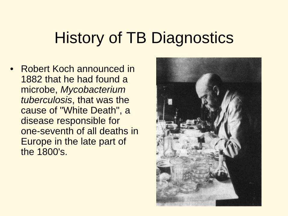
History of TB Diagnostics
• Robert Koch announced in 1882 that he had found a microbe, Mycobacterium tuberculosis, that was the cause of "White Death", a disease responsible for one-seventh of all deaths in Europe in the late part of the 1800's.
Timeline of TB Infection
Exposure
4-6 wks Adaptive T cell
response
Latent TB
(LTBI)*
Active TB
Yrs-decades Lifelong Containment
*Prevention efforts focus on detecting LTBI, most LTBI do not advance to active disease but those patients are at high risk particularly if they become immunocompromised.
TB Infection vs. TB Disease TB in the body TB in the body Chest X-ray normal Chest X-ray abnormal Sputum not done Sputum smear and
culture positive No symptoms Symptoms: cough,
fever, weight loss Not infectious Infectious Not a case of TB Case of TB
TB Algorithm
• Collect sputum specimens at 3 different times and 8 hours apart (at least one must be a first morning specimen) for AFB smear and mycobacterial culture.
• Perform MTD or NAAT test on the first smear positive sputum specimen
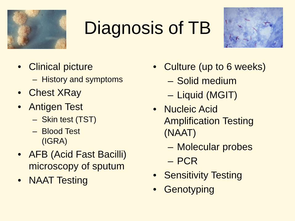
Diagnosis of TB
• Clinical picture – History and symptoms
• Chest XRay • Antigen Test
– Skin test (TST) – Blood Test
(IGRA)
• AFB (Acid Fast Bacilli) microscopy of sputum
• NAAT Testing
• Culture (up to 6 weeks) – Solid medium – Liquid (MGIT)
• Gastric Lavage (children) • Tissue and Body fluids (CSF, pleural,
blood) • Wounds, skin lesions (exudates)
Specimen Collection and Processing: special considerations
• Biohazard – Aerosol transmission
• Prevent contamination of specimen – Slow growth rate of TB
• Evaluate at least 3 specimens per patient
Sputum collection considerations
• Instruct patients that nasopharyngeal discharge and saliva are not sputum
• Sputum = thick, yellowish (sometimes blood-tinged) exudative material brought up from the lungs after a deep, productive cough
• First rinse mouth with mouthwash to decrease bacterial contamination
• Collect specimen into appropriate container
Sputum cont…
• About 10 ml of sputum is sufficient • If patient cannot provide an adequate
specimen then sputum induction is acceptable – Warm, aerosolized hypertonic salt solution – Be certain to label the specimen as “induced
sputum”
Specimen Transport
• From the time of collection until the specimen is processed, the other bacteria present will over grow (contaminate) the slower growing Mycobacteria sp. – Speed is important
• Courier • Ship cold when possible • Shipping cold slows bacterial growth
Specimen Processing
• If collected from a non-sterile site (sputum), then digest and decontaminate before culture – Kill off other microbes – Liquefy mucin – Remove organic debris – Homogenize tissue
• Weakness – 50-60% of TB patients are smear negative
• Need at least 10,000 CFU/ml sputum for positive result
– Cannot differentiate Mycobacteria species
Importance of acid-fast bacilli smear microscopy as a primary diagnostic tool • Initial diagnosis • Monitoring treatment • Determination of time to release from
isolation
How sensitive is the smear? • Peterson et. al. JCM 1999 vol. 37:3564-68.
Number of specimens
Direct AFB smear sensitivity
Concentrated AFB smear sensitivity
Comment
353 culture positive for Mycobacteria
34% 58% Direct smear cannot be relied on
208 cultures positive for M. tuberculosis
42% 74% Concentrated smear most reliable
Analysis of 3 specimens per patient
81% 91% Concentrated smear is still the most reliable
Direct detection of TB in the specimen
• MTD test – Genprobe – transcription mediated amplification
• In house developed Nucleic Acid Amplification test (NAAT)
• GeneXpert Cepheid NAAT
Interpretation of NAAT Result Smear NAAT Interpretation
+ + Presumed Positive TB, No Additional Testing
+
-- If first sputum specimen: smear positive and NAAT-negative, repeat on one additional specimens, if negative then presume negative for TB.
-- + Additional specimens (limit 2). Presumptive positive for TB if the subsequent specimen positive
-- -- Presumptive negative for TB. Two specimens recommended.
TB NAAT Algorithm Respiratory Specimen
Smear Negative Smear Positive
NAAT NAAT
Positive: Presumed TB, pending culture
results
Negative
Use clinical judgment to determine whether to begin therapy while
awaiting culture results and determine if
additional diagnostic testing is needed.
If a second specimen is smear positive, NAAT negative, the patient is presumed to have an infection with non-tuberculous mycobacteria, pending culture results.
Consider testing another specimen (not to exceed a total of two).
NAAT Positive: A patient can be presumed to have tuberculosis, pending culture results, if two specimens are NAA positive.
Positive Negative
Consider testing another specimen (not to exceed a total of two).
Inhibitors Detected: Test result is of no
diagnostic help. Consider testing second specimen (not to exceed
a total of two).
Use clinical judgment to determine whether to begin therapy while
awaiting culture results and determine if
additional diagnostic testing is needed.
Use clinical judgment to determine whether to begin therapy while
awaiting results of culture and other diagnostic tests.
Currently available NAA tests are not
sufficiently sensitive to exclude the
diagnosis of TB in AFB smear negative patients suspected
of having TB.
Diagnosis of TB: Culture • Solid Media Culutre
– Agar Middlebrook 7H10/7H11 – Egg based Lowenstein-Jensen
• Liquid – Broth Culture – 7H9 – Commercial broth and monitoring systems
• High performance liquid chromatography (HPLC) of cell wall mycolic acids
• Molecular probes – Culture confirmation – Direct from growth in broth or on slant
• DNA sequence analysis – 16S rRNA gene
Molecular Probes for Mycobacteria identification
• MYCOBACTERIUM TUBERCULOSIS Complex Culture Identification Test – For identification of M. tuberculosis, M. bovis, M. bovis (BCG), M. africanum, M. canetti, M. microti etc. isolated from culture.
• MYCOBACTERIUM AVIUM Culture Identification Test - For the identification of Mycobacterium avium isolated from culture.
• MYCOBACTERIUM INTRACELLULARE Culture Identification Test - For the identification of Mycobacterium intracellulare isolated from culture.
• MYCOBACTERIUM AVIUM Complex Culture Identification Test - For the identification of Mycobacterium avium complex (M. avium, M. intracellulare, and other members) isolated from culture.
• MYCOBACTERIUM GORDONAE Culture Identification Test - For the identification of Mycobacterium gordonae isolated from culture.
• MYCOBACTERIUM KANSASII Culture Identification Test - For the identification of Mycobacterium kansasii isolated from culture.
QuantiFERON TB- Gold In-Tube •Blood test that measures and compares amount of interferon-gamma (IFN-γ) released by blood cells in response to antigens •FDA approved in May 2005 –Cellestis, Carnegie, Australia
TB
QFT Procedure – Clinic and Lab
• Procedures in Clinic – Blood Collection – Shaking of Tubes – Blood Incubation – Plasma Separation
• Procedures in Lab
– ELISA – Data Analysis
Data Analysis and Results • Results are reported as
– Positive – Negative – Indeterminate
• Indeterminate – Low mitogen
• CMI response
– High Nil • live vaccines • secondary infection
Technology Comparison T-SPOT QFTB In Tube TST
Antigens ESAT-6 & CFP10 ESAT-6 , CFP10, TB7.7
PPD
Boosting effect with repeat tests
No No Yes
TAT 16-20 h 16-24 h 48-72 h Readout units IFN-Gamma spot
forming cells International units of IFN-Gamma
Millimeters of induration
Technology ELISpot ELISA NA Readout system Count of spots Measurement of
optical density values using an automated reader
Palpable induration
Subjective Reading Yes No Yes
CDC advises that IGRA’s can be used in all circumstances in which the TST
is used, including…
• Contact investigations • Evaluation of recent immigrants who
have had BCG vaccination • TB screening of health care workers
and other individuals in high risk settings
• IGRA is in place of (not an addition to) TST
General Benefits of IGRA over TST
• Requires only one patient visit • Assesses responsiveness to M.
tuberculosis antigens • Does not boost previous responses • Interpretation less subjective than for TST
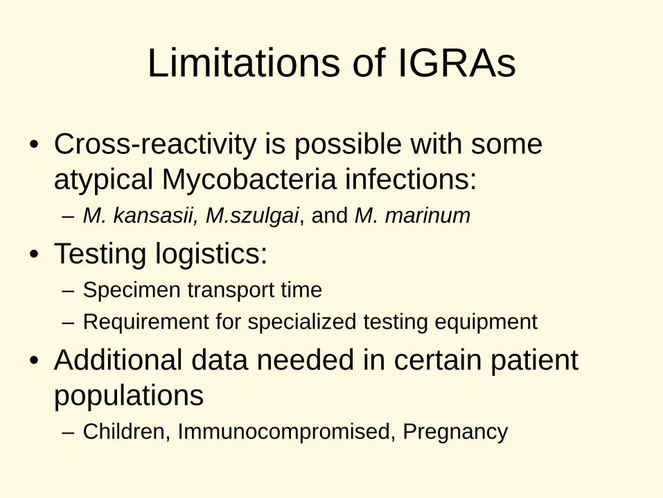
Limitations of IGRAs
• Cross-reactivity is possible with some atypical Mycobacteria infections: – M. kansasii, M.szulgai, and M. marinum
• Testing logistics: – Specimen transport time – Requirement for specialized testing equipment
• Additional data needed in certain patient populations – Children, Immunocompromised, Pregnancy
BCG Vaccinated Patients
• IGRA benefit the BCG vaccinated patient • Many false positive TST due to
vaccination status • Treatment is costly, carries risk of
significant side effects • Treatment is not always needed since
most do not have LTBI
Performance of IGRAs and the TST:
An up-to-date TB Test Meta-Analysis
R Diel, R Loddenkemper and A Nienhaus Evidence based comparison of commercial interferon-gamma release assays for detecting active tuberculosis – a meta-analysis. Chest, 2009, Published on Dec 18, 2009 in electronic format;
Chest April 2010 137:952-968; doi:10.1378/chest.09-2350
Contact Investigations • For persons with recent TB exposure, negative IGRA
results should be confirmed with a repeat test 8-10 weeks after exposure (end of window period) per CDC. This is the same as for a negative TST.
• Yoshiyama, et al. Timing of Quantiferon TB-G test for the contact examination of tuberculosis. Kekkaku. 2007 Aug;82(8):655-8. – “3 months interval from the diagnosis of the index
case will be enough for the final decision of the infection of contacts.”
• N=25, 8 positive QFTB-G
For high risk contacts… • When “window prophylaxis” has been started
for high-risk contacts exposed to an infectious TB patient, a negative IGRA result at the end of the window period should be interpreted in light of all other clinical and epi data – A full course of LTBI TX should be considered
even with a negative result when the rate of TB transmission to other contacts is high or when a false-negative is suspected because of immune status.
Use of IGRA Baseline and Serial Testing
• Baseline testing with IGRA – Establish baseline with single negative IGRA – HCWs with positive IGRA result should be
referred for diagnostic evaluation • Serial testing for infection control
– A conversion is a change from negative to positive
Cost Barrier? – Cost-effective alternative to TST
• Reduction in false positive test results • No second visit needed to complete
testing • Two-step testing not needed • Reduction in rates of CXR (due to higher
specificity for M. tuberculosis)
Are IGRAs cost effective?
• DePerio et al: Arch Intern Med. 2009 – Use of IGRA “leads to superior clinical
outcomes and lower costs than the TST and should be considered in screening non-BCG-vaccinated and BCG vaccinated new HCWs for LTBI.”
• Marra et al: Int J Tuberc Lung Dis. 2008 – “Selected use of QFT-G appears to be cost
effective if used in targeted fashion.”
IGRA Summary
• IGRAs are more specific than TST and are not confounded by previous BCG vaccination – Less unnecessary preventive treatment
• IGRA are more sensitive than TST
TB antibody tests
• Tests that detect IgG antibody to TB • Highly variable results for sensitivity and
specificity • Do not have a roll in the diagnosis of TB • Not FDA approved • Recently confused with IGRA in the news.
Take Home Message
• Culture of TB remains the gold standard • AFB smears are the most cost effective • NAAT are sensitive and rapid but cannot
differentiate between dead and viable TB • IGRA do not differentiate between active
and latent TB • There are new tests on the horizon
Let’s not forget…
Bibliography • Requirements to get a high quality specimen to the laboratory
– Salfinger, M, and AJ Morris. 1994. The role of the microbiology laboratory in diagnosing mycobacterial diseases. American Journal of Clinical Pathology. 101: S6-13.
• • Standard diagnostic techniques for the detection and identification of Mycobacteria
– Chegou, NN, et.al. 2011. Tuberculosis assays: past, present and future. Expert Reviews in Anti Infective Therapy. 9(4):457-69. • • Importance of acid-fast bacilli smear microscopy as a primary diagnostic tool
– Peterson, EM, et.al. 1999. Comparison of direct and concentrated acid-fast smears to identify specimens culture positive for Mycobacterium species. Journal of Clinical Microbiology. 37(11): 3564-8.
• • Drug susceptibility testing of M. tuberculosis
– O’Grady, J. et.al. 2011. New and improved diagnostics for detection of drug-resistant pulmonary tuberculosis. Current Opinion in Pulmonary Medicine. 17(3):134-41.
• • Rapid methods for the identification and drug susceptibility testing of M. tuberculosis as they compare to the traditional methods
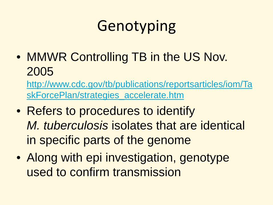
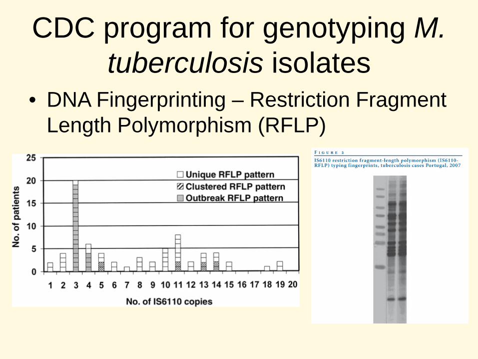
– Lalvani A. 2007. Diagnosing tuberculosis infection in the 21st century: new tools to tackle an old enemy. Chest 131:1898-1906. • • CDC program for genotyping M. tuberculosis isolates
– Kato-Maeda, M, JZ Metcalfe, and L Flores. 2011. Genotyping of Mycobacterium tuberculosis: application in epidemiologic studies. Future Microbiology. 2011. 6(2): 203-16.
• • Interferon Gamma Assays (IGRA) – Pro’s and Con’s
– Diel, R. et.al. 2010. Evidence-based comparison of commercial interferon-gamma release assays for detecting active TB: a metaanalysis. Chest. 137:952-968.