Page 1
This is a repository copy of Role of the small intestine, colon and microbiota in determiningthe metabolic fate of polyphenols.
White Rose Research Online URL for this paper:http://eprints.whiterose.ac.uk/113962/
Version: Accepted Version
Article:
Williamson, G orcid.org/0000-0002-5624-6267 and Clifford, MN (2017) Role of the small intestine, colon and microbiota in determining the metabolic fate of polyphenols. Biochemical Pharmacology, 139. pp. 24-39. ISSN 0006-2952
https://doi.org/10.1016/j.bcp.2017.03.012
© 2017 Elsevier Inc. This manuscript version is made available under the CC-BY-NC-ND 4.0 license http://creativecommons.org/licenses/by-nc-nd/4.0/
[email protected] ://eprints.whiterose.ac.uk/
Reuse
Unless indicated otherwise, fulltext items are protected by copyright with all rights reserved. The copyright exception in section 29 of the Copyright, Designs and Patents Act 1988 allows the making of a single copy solely for the purpose of non-commercial research or private study within the limits of fair dealing. The publisher or other rights-holder may allow further reproduction and re-use of this version - refer to the White Rose Research Online record for this item. Where records identify the publisher as the copyright holder, users can verify any specific terms of use on the publisher’s website.
Takedown
If you consider content in White Rose Research Online to be in breach of UK law, please notify us by emailing [email protected] including the URL of the record and the reason for the withdrawal request.
Page 2
Accepted Manuscript
Review
Role of the small intestine, colon and microbiota in determining the metabolicfate of polyphenols
Gary Williamson, Michael N. Clifford
PII: S0006-2952(17)30141-7DOI: http://dx.doi.org/10.1016/j.bcp.2017.03.012Reference: BCP 12766
To appear in: Biochemical Pharmacology
Received Date: 26 January 2017Accepted Date: 14 March 2017
Please cite this article as: G. Williamson, M.N. Clifford, Role of the small intestine, colon and microbiota indetermining the metabolic fate of polyphenols, Biochemical Pharmacology (2017), doi: http://dx.doi.org/10.1016/j.bcp.2017.03.012
This is a PDF file of an unedited manuscript that has been accepted for publication. As a service to our customerswe are providing this early version of the manuscript. The manuscript will undergo copyediting, typesetting, andreview of the resulting proof before it is published in its final form. Please note that during the production processerrors may be discovered which could affect the content, and all legal disclaimers that apply to the journal pertain.
Page 3
1
Role of the small intestine, colon and microbiota in determining the metabolic fate of
polyphenols
Gary Williamson1 and Michael N. Clifford2
1School of Food Science and Nutrition
University of Leeds
Leeds
LS2 9JT
UK
2 School of Bioscience and Medicine,
Faculty of Health and Medical Sciences,
University of Surrey,
Guildford, GU2 7XH,
UK
Page 4
2
Abstract
(Poly)phenols are a large group of compounds, found in food, beverages, dietary supplements
and herbal medicines. Owing to their biological activities, absorption and metabolism of the
most abundant compounds in humans are well understood. Both the chemical structure of the
phenolic moiety and any attached chemical groups define whether the polyphenol is absorbed
in the small intestine, or reaches the colon and is subject to extensive catabolism by colonic
microbiota. Untransformed substrates may be absorbed, appearing in plasma primarily as
methylated, sulfated and glucuronidated derivatives, with in some cases the unchanged
substrate. Many of the catabolites are well absorbed from the colon and appear in the plasma
either similarly conjugated, or as glycine conjugates, or in some cases unchanged.
Although many (poly)phenol catabolites have been identified in human plasma and / or urine,
the pathways from substrate to final catabolite, and the species of bacteria and enzymes
involved, are still scarcely reported. While it is clear that the composition of the human gut
microbiota can be modulated in vivo by supplementation with some (poly)phenol-rich
commodities, such modulation is definitely not an inevitable consequence of
supplementation, it depends on the treatment, length of time and on the individual
metabotype, and it is not clear whether the modulation is sustained when supplementation
ceases. Some catabolites have been recorded in plasma of volunteers at concentrations similar
to those shown to be effective in in vitro studies suggesting that some benefit may be
achieved in vivo by diets yielding such catabolites.
Page 5
3
1. (Poly)phenols covered and main classes.
(Poly)phenols are a large group of compounds synthesised by plants for a variety of
functions, such as protection against UV radiation, mechanical damage, and microbial
infection [1, 2]. A wide variety of polyphenols are consumed as part of the normal diet [3],
and are also present at high levels in supplements [4], and form essential components of
many Chinese medicines [5]. A study within the European Prospective Investigation into
Cancer and Nutrition (EPIC) using the Phenol Explorer database reported that the cohort
studied consumed 427 different polyphenols including 94 that were consumed at a rate of >1
g/day. The estimated total polyphenol consumption ranged from 584 mg/day by Greek
women to 1786 mg/day for men in Aarhus-Denmark. The corresponding data for Greek men
and for Aarhus women were 744 mg/day and 1626 mg/ day, respectively, with all data
adjusted for age and weighted by season and weekday of dietary recall [6]. This study defined
the major contributors to be the phenolic acids (predominantly the caffeoylquinic acids (27–
53%), also called chlorogenic acids) and flavonoids (predominantly flavanols (16–29%),
proanthocyanidins (5–9%) and theaflavins (14–25%)) [6]. Hydroxybenzoic acids (3.3–7.4%),
alkyl-phenols (1.6–4.)%), tyrosols (0.4–3.6%) and the glycosides of flavanones (2.6–4.0%),
flavonols (2.8–5.1%) and flavones (0.7–1.5%) were also recorded along with smaller
contributions from stilbenes (0.1–0.5%), lignans (0.1–0.7%) and trace amounts from several
other subgroups. It is feasible that individuals with heavy coffee consumption might have
greater total polyphenol intakes. Recent analyses of coffee beverage as sold in retail outlets
can supply as much as 423 mg chlorogenic acids per cup [7]. Heavy consumers of black tea
might also have greater total polyphenol intakes because the EPIC study did not consider
thearubigins. Black tea theaflavins account for some 3–5% of the beverage solids compared
Page 6
4
with some 17–20% for the extremely complex thearubigins which can equate to ≈100 mg/cup
[8-10]. It has not been possible to trace consumption data for the transformed anthocyanins
characteristic of matured red wines, but it is clear that an extensive range is present [11, 12],
and regular consumers of red wine might have a significant intake. It has been demonstrated
that foods also contain a significant amount of non-extractable (poly)phenols which,
nevertheless, are gut microbiota substrates, and these will further raise the (poly)phenol
intake [13]. The phenol-explorer database is a useful compilation of good quality
compositional data [3], but lacks data for the transformed anthocyanins, thearubigins and
unextractable (poly)phenols [14]. Since phenolic acids are mostly monomeric phenols,
flavonoids contain 2 phenolic rings, and polymerised flavonoids contain multiple phenolic
rings, we designate the term “(poly)phenols” to cover this group [15].
Flavonols, flavanones and anthocyanins exist in planta as glycosides, where the predominant
attached sugars are glucose and rhamnose. Flavanols are present in planta mostly in their free
forms, but can be galloylated (as in green tea) or polymerised to form proanthocyanidins,
common components of many foods such as cocoa. Phenolic acids are usually attached to an
organic acid, most commonly quinic acid, found at very high levels in coffee and at moderate
levels in most fruits. Here, we will focus on the main compounds present in a normal diet,
and only include those which have been well studied and understood. The route of absorption
can be either through the stomach, small intestine or, if not absorbed at those sites, by the
colon, after chemical modification by the colonic microbiota. During this process, the
(poly)phenols become modified by various catabolic and conjugation reactions, appear in the
blood, and are then excreted either in the urine or through the bile. Some unabsorbed
substrate and catabolites are voided in the faeces. Recently, advances have been made
Page 7
5
particularly in defining the effects of the microbiota on (poly)phenols, and how, in parallel,
(poly)phenol-rich foods can affect the composition and activity of the microbiota.
2. Absorption into the bloodstream
Chlorogenic acid is a general term for the esters of a phenolic acid (e.g. ferulic, caffeic or
dimethoxycinnamic acids) with quinic acid [16], and these classes are found at particularly
high levels in coffee [17]. After reaching the small intestine, some hydrolysis of
caffeoylquinic acid and of dimethoxycinnamoyl quinic acid occurs owing to the action of
mammalian esterases, but the hydrolysis is relatively slow and only a proportion of the
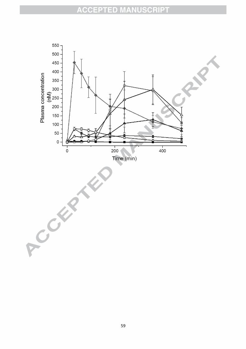
chlorogenic acid is hydrolysed [18]. The resulting free caffeic acid is absorbed through the
small intestinal epithelium [19], and is rapidly sulfated, to form caffeic acid-3ガ-O-sulfate, or
sulfated and methylated, resulting in ferulic acid-4ガ-O-sulfate, both with a Tmax of ~30 min
[20]. Dimethoxycinnamic acid is rapidly and efficiently absorbed after hydrolytic removal of
its quinic acid moiety, and circulates in plasma as the unmodified dimethoxycinnamic acid,
again with a Tmax ~30 min [21, 22] (Fig. 1). Although dimethoxycinnamic acid is only a
minor component of the coffee chlorogenic acids, it exhibits the highest concentration in
plasma of all of the coffee-derived phenolic acids that are absorbed in the upper
gastrointestinal tract. Feruloylquinic acids are not substrates for gut esterases, but a very
small amount is absorbed intact, although the level only reaches low nM concentrations and
this is not considered a major route of absorption and metabolism [23]. Most of the
chlorogenic acids arrive in the colon intact. Here, the microbiota have abundant esterases for
hydrolysing the phenolic-quinic acid linkage [24]. Released phenolic acids are readily
converted by the microbiota to the dihydro forms, such as dihydroferulic acid and
Page 8
6
dihydrocaffeic acid, and then absorbed through the colonic epithelium. Dihydroferulic,
dihydroferulic acid-4ガ-O-sulfate and dihydrocaffeic acid-3ガ-O-sulfate circulate at relatively
high concentrations [20] (Fig. 1 and 2). Some of the conjugated phenolic acids appear in the
urine together with glycine conjugates such as feruloyl glycine. Having breakfast with coffee
somewhat affected the timing of absorption, but not the overall amount absorbed [25], and
non-dairy creamer, but not milk, has the same effect [26] and so the overall effect of food or
beverages on the absorption and metabolism of chlorogenic acids appears to be minimal
despite some reports to the contrary [27].
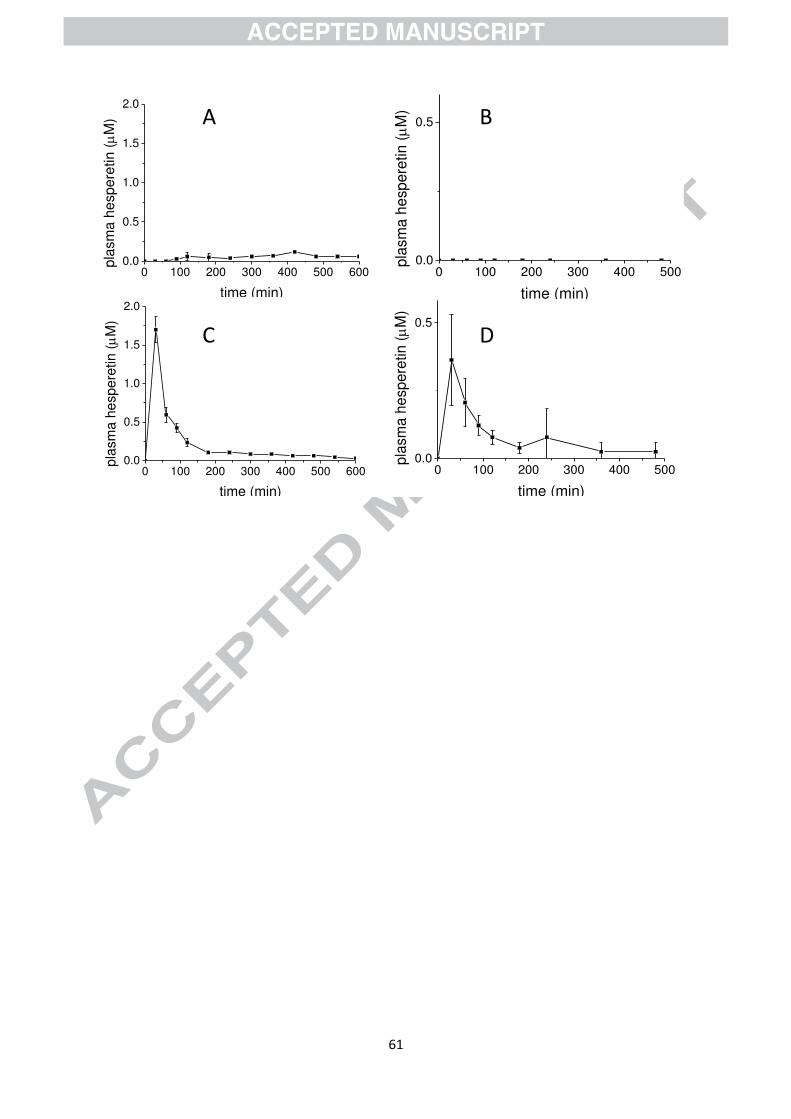
Hesperidin is found at high levels in citrus fruits and is the rutinoside of hesperetin
(hesperetin-7-O-rhamnoglucoside) [28]. Intact hesperidin is not absorbed across the small
intestine epithelium as shown by direct jejunal perfusion in humans [29] (Fig. 3). If the
terminal rhamnose moiety is hydrolysed before consumption, the product, hesperetin-7-O-
glucoside, becomes a substrate for the brush border enzyme lactase phloridzin hydrolase.
After hydrolysis of the attached glucose at the surface of the intestine, the resulting hesperetin
is efficiently absorbed with a Tmax of ~30 min after direct perfusion of the pure compound
into the jejunum [29] (Fig. 3). When hesperidin is consumed orally, hesperetin (conjugates of
sulfate and glucuronide) appear in the plasma with a Tmax of 4-6 h. When hesperetin-7-O-
glucoside is consumed orally, the same conjugates appear in the plasma but with a much
higher Cmax and a much shorter Tmax of ~ 30 min indicating absorption in the small intestine
[30]. The microbiota, and not mammalian cells [31], can produce -rhamnosidases which
cleave the rhamnose from hesperidin [32], and this is a critical step in its absorption. Intact
hesperidin is not absorbed passively, but hesperetin, after removal of the two sugars, is
readily absorbed [33]. This is a clear cut example of where the attached sugars make an
enormous and critical difference to both the site and extent of absorption. Consumption of
Page 9
7
hesperidin with other foods can affect the shape of the pharmacokinetic curve [34]. The
absorption, metabolism and excretion of hesperidin shows substantial inter-individual
variation, but is only slightly affected by age [35]. Quercetin is found in nature attached to
sugar moieties, such as the 3-O-glucoside and 3,4ガ-O-diglucoside forms found in onions [36],
the 3-O-rutinoside as found in tea [37] and various glycosides in apple [38]. The same
mechanism as described above for hesperidin applies to these quercetin derivatives, where
the glucoside is absorbed rapidly and efficiently in the small intestine, but the rutinoside is
absorbed only after hydrolysis of the sugars by the gut microbiota in the colon, leading to less
efficient absorption with a longer Tmax [39, 40]. The numerous conjugates of quercetin have
been described [41, 42] and kinetic modelling has indicated that rats are not an adequate
model to study quercetin pharmacokinetics since the pattern of conjugation is very different
[43].
picatechin (( 札 )-epicatechin is the most abundant form) is found in most foods as the free
form [44], is absorbed in the small intestine, and does not need prior action of the microbiota
for absorption [45]. It undergoes sulfation, methylation and glucuronidation with typical Tmax
for conjugates of ~2 h [46, 47]. After both oral ingestion and direct jejunal perfusion, the EC-
3ガ-O-glucuronide conjugate is the most abundant metabolite found in plasma [48] (Fig. 4).
Despite reported differences in analytical procedures between laboratories [47], the
consensus is that (−)-epicatechin-3ガ-O-glucuronide, (−)-epicatechin-3ガ-O-sulfate, and a 3ガ-O-
methyl-(−)-epicatechin-5/7-sulfate are the predominant epicatechin metabolites in humans
after oral consumption [47, 49]. The apparent half-life of most epicatechin conjugates is 2-4 h
[49]. The proportion of epicatechin which is not absorbed in the small intestine will reach the
colonic microbiota and will be converted to lower molecular weight compounds (see below)
with no evidence for absorption of intact epicatechin in the colon.
Page 10
8
Anthocyanins and proanthocyanidins are very poorly absorbed in the small intestine in their
intact forms [50, 51]. These classes of compound are subject to extensive catabolism by the
gut microbiota, and the products absorbed through the colon (see below). Anthocyanins are
relatively unstable in the gastrointestinal tract, even in the conditions of the small intestine
[52], and phenolic products from anthocyanins could arise from chemical degradation. Any
anthocyanins which survived the small intestine would be rapidly transformed into lower
molecular weight compounds by the colonic microbiota (see below [53, 54]).
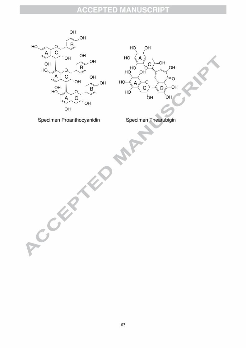
Proanthocyanidins are oligomeric flavanols, and there are no mammalian enzymes reported
which have the ability to degrade the high molecular weight molecules into forms which are
small enough to be easily absorbed in the small intestine. Only the dimer of epicatechin
(procyanidin B2) is absorbed intact, but to a very limited extent [51]. The majority of the
procyanidin B2, and other oligomers with a degree of polymerisation ≥2 reach the colon and
are extensively metabolised by colonic microbiota. In rats, 80% of the microbiota-catalysed
catabolites of radiolabelled procyandin B2 were absorbed [55].
3. Conversion of polyphenols by the gut microbiota
In the last decade there have been major advances in defining the composition of the gut
microbiota with greatly increased use of methods based on the detection and sequencing of
16S rDNA and MALDI-TOF MS replacing conventional culture methods, but these diverse
approaches remain complementary with more traditional methods required to determine
microbiota functionality [56, 57]. The human gastro-intestinal tract hosts up to 100 trillion
microbes [58], which have been assigned to well over 1000 species. The three most dominant
bacterial phyla in the colon are the Firmicutes, Bacteroidetes, and Actinobacteria, with
Page 11
9
Proteobacteria and Verrucomicrobia generally less abundant [56]. However, a detailed
discussion of microbiota composition, reported variations therein with age [59, 60] or disease
[61, 62], and the techniques used to determine it [56], are outside the scope of this review.
With regard to microbiota-catabolism, it is convenient to divide the substrates into three
categories. There remain some for which no data are available, the transformed anthocyanins
(pyranoanthocyanins) of red wines, plus some minor dietary components such as algal
phlorotannins, alkyl resorcinols, urushiols, cardanols and anacardic acids, naphthoquinones
and anthraquinones, coumarins, isocoumarins and furanocoumarins [63]. The second group
are those substrates associated with unique catabolites, and the third group consists of some
structurally diverse substrates that nevertheless yield essentially the same set of catabolites,
this latter group predominating (see Figs. 2 and 5) [64]. The unique catabolites include 4-
hydroxy-mandelic acid from p-sympatol [65], tyrosols from oleuropein and related
compounds [66], S-equol, 5-hydroxy-equol and dihydrocinnamic acids with the aryl residue
at C2 formed from isoflavones [67, 68], urolithins and nasutins from ellagitannins [69-71],
diarylbutanes from lignans [32;35;36], and dihydroresveratrol from piceid and resveratrol
[72]. The avenanthramides of oats, N-cinnamoyl conjugates of anthranilic acids, yield some
unique catabolites, e.g. the corresponding dihydrocinnamoyl conjugates and 5-hydroxy-
anthranilic acid (2-amino-5-hydroxybenzoic acid) in addition to the much commoner
cinnamic and dihydrocinnamic acids [73]. Many of these unique catabolites have received
considerable attention but their precursors are minor components of the diet, albeit with some
considerable variation between subgroups [6], depending upon whether they consume
significant amounts of oranges [65], olive oil [74], soya beans [75, 76], nuts, pomegranates,
strawberries and raspberries [71, 77, 78], whole grains [76], wine and peanuts [79, 80] or oats
[81], respectively.
Page 12
10
The predominant catabolites generated by the microbiota from (poly)phenols are the aromatic
and phenolic acids with zero to three aromatic hydroxyls, or their mono- or di-methoxy
analogues, possessing also a sidechain of one to five carbons which might bear an aliphatic
hydroxyl [64]. These are formed from the diet predominantly from chlorogenic acids,
flavanols, proanthocyanidins, theaflavins [6] and thearubigins [82-85], but also from many
other minor components of the diet (Figs. 2 and 5). Human studies in vivo and gut
microbiota-fermentation studies have demonstrated that at least some of these C6–C1 and
possibly C6–C2 phenolic acids can be decarboxylated yielding the corresponding phenols (C6)
or methyl-phenols (C6–C1) [84, 86-90], respectively. Phloroglucinol can also be formed from
chalcones and dihydrochalcones [91, 92], and at least certain rumen microorganisms can
reduce this to non-aromatic dihydrophloroglucinol [93, 94]. There are several catabolites, for
example some mandelic and phenylhydracrylic acids, for which the origin remains uncertain.
Although the microbiota-mediated transformation of dietary (poly)phenols is generally
considered to occur in the colon, studies in vitro using ileostomy fluid establish that some
transformation can be expected to occur in the small intestine [95]. Studies with ileostomists
where plasma and / or urine have been analysed support this observation, but studies in which
ileostomists are compared with volunteers having an intact colon indicate that microbiota
catabolism in the small intestine is very much less than in the colon [23].
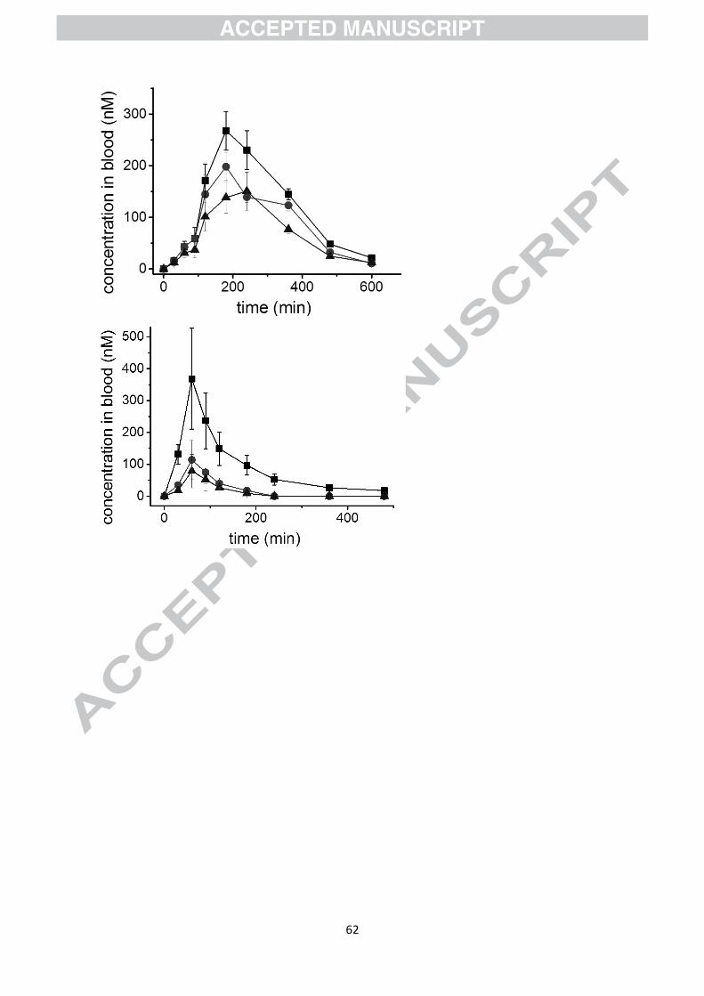
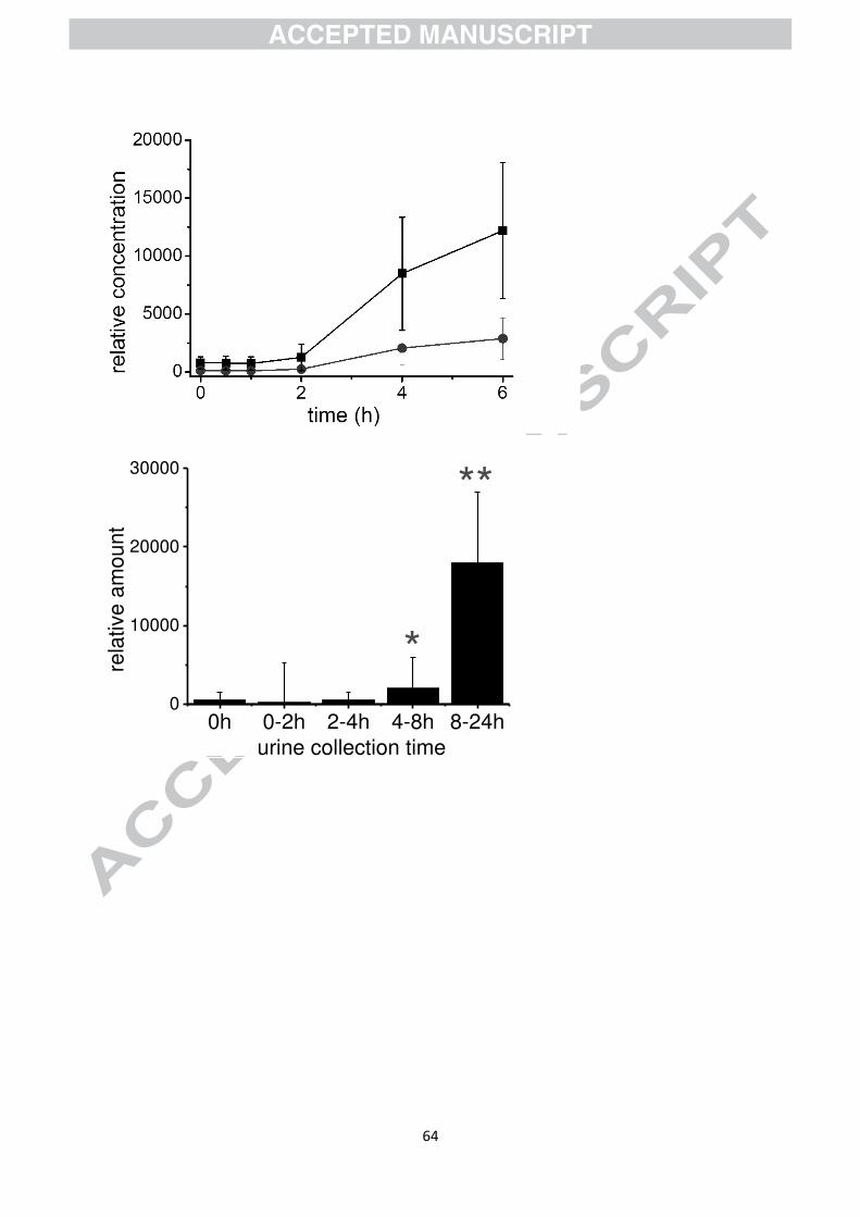
As a result of the catabolic reactions indicated above, the products appear in the blood and
circulate either as free or conjugated forms. The concentration of microbiota catabolites are
often much higher than the parent compounds. As an example, Fig. 6 shows the appearance
of low molecular weight phenolics in the blood after consumption of a mixture of fruits.
Page 13
11
Catechol and 1-methyl pyrogallol are not found in foods, but are products of catabolism by
the gut microbiota. These are characterised by a high Tmax and a late appearance in the urine.
As for the parent compounds, the catabolic products are also sulfated, glucuronidated and
methylated, and although this does not necessarily account for 100% of the catabolite
absorbed, catechol-O-sulfate is highly abundant in plasma after consumption of polyphenol-
rich foods.
4. Pharmacokinetic consequences of multiple doses
(Poly)phenols associated with coffee, green tea and black tea are generally consumed
frequently during the waking day, with the result that the gut microbiota have access to
several bolus doses for a prolonged period of time. This would predispose to a larger and
more uniform concentration of the associated catabolites in the gastrointestinal tract and in
plasma after absorption, but little attention has been given to how this would affect the levels
of (poly)phenols in circulation in human intervention studies. Polyphenols undergo several
reactions before this stage, such as deglycosylation, conjugation, ring cleavage, etc., and so
are not amenable to classical pharmacokinetic analysis and modelling to assess the
implications of frequent consumption. There are very few data on the human
pharmacokinetics of intravenous doses, particularly for the gut flora catabolites, which are
needed for such modelling. Volunteer studies of Chinese herbal preparations which contain
preformed protocatechuic acid given intravenously have revealed a clearance half-life of ca
10–20 minutes [96, 97].
Such rapid clearance is potentially-misleading when the catabolite is not produced until the
colon, and is then produced throughout the transit of the large bowel, resulting in absorption
Page 14
12
over the same substantial time period and creating a plasma profile more closely resembling a
prolonged intravenous infusion. There are no intravenous dosing data for the C6–C3, C6–C2 or
C6–C5 catabolites. A study in which volunteers consumed a bolus dose of strawberry purée
containing ellagitannins has demonstrated that the unique urolithin catabolites were still
detected in urine 90 h later suggesting that the ellagitannin substrate may have bound to the
mucosa and not moved with the unbound digesta [98]. It is likely that a similar phenomenon
will occur with proanthocyanidins which also bind strongly to proteins, and possibly
thearubigins, but because their catabolites are not unique it would be more difficult to
demonstrate than with the ellagitannins (see Figs 2 and 5). Such binding would extend the
period during which the microbiota could attack such substrates, potentially increasing the
yield of catabolites and the area under the concentration–time curve for these catabolites in
plasma.
Volunteer studies of typical real world repeated consumption would, if available, circumvent
the lack of data for intravenous dosing, but in their absence we performed a simple additive
modelling using real data from a single dose study in order to obtain an indication of the
potential concentrations that might be attained. This approach assumes that the rate of
clearance from plasma does not change with dose, and would only be true if clearance was
saturated, and thus the estimate obtained is on the high side.
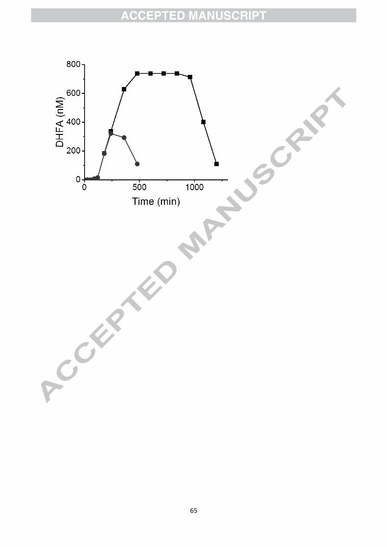
Figure 7 shows the data obtained for dihydroferulic acid in plasma after consumption of a
single cup of coffee from the study of Stalmach et al [20]. We then used these data for
estimating the consequences of human subjects consuming seven servings at two-hour
intervals, by adding the values expected at different times using the single serving data. The
Cmax is ~300 nM after a single serving, but reaches a steady state of ~750 nM after multiple
Page 15
13
doses spaced 2 h apart, an increase of ~2.5-fold, and the total area under the curve is also
increased 7-fold with a much longer time of exposure. It would be interesting to determine
how multiple doses of polyphenols affect the plasma concentrations throughout the day using
healthy volunteers, since, for example, many free-living people consume multiple cups of
coffee at regular intervals each day. In contrast, pre-existing (poly)phenols that are absorbed
in the stomach or duodenum (e.g. caffeoylquinic acids) will for any bolus dose be absorbed
only during the comparatively brief residence period, and if cleared rapidly from the plasma
will not accumulate to the same extent even if consumed at 2-hour intervals.
5. Prebiotic effects
There have been many studies of the potential of (poly)phenols substrates and catabolites to
modulate the composition of the gut microbiota. These include studies using animals, in vitro
incubations with faecal flora, and volunteer studies. Animal studies permit strategies not
otherwise available, for example introducing a particular organism into a germ-free animal
with suitable controls to assess its impact on the health of the gnotobiotic animal [68, 99,
100]. Studies in which conventional animals are given an atypical, human-type diet (e.g. high
fat, high sugar, obesogenic) supplemented with a substantial dose of a specific (poly)phenols-
rich commodity (e.g. table grapes [101] or cocoa [102]) or test substance (e.g. trans-
resveratrol [103]) have been used extensively. Often, such studies demonstrate that the test
substance or (poly)phenol-rich commodity modulate the composition of the gut microbiota
and / or ameliorate the harmful effects of the test animal’s less than ideal diet [101-108].
However, as noted in an earlier review, the metabolic competence of the normal gut
microbiota may vary markedly between species [64], and it is uncertain whether or not a
similar effect would be achieved in humans following a realistically supplemented diet. For
Page 16
14
example, after supplementation for three months in 58 volunteers with nine capsules of
flavanol-rich green tea extract per day, equivalent to several 100 ml cups daily, there was no
change in the gut microbiota [109]. The oral microbiota is also stable to red wine
consumption [110]. Similarly, a study in which 25 volunteers consumed a (poly)phenol-rich
boysenberry beverage, an apple fibre-rich beverage, or both in combination (2 × 175 ml/day),
failed to establish any change in the gut microbiota or short chain fatty acid production [111].
The consumption of seven dates per day by 22 volunteers for 21 days also did not alter the
composition of the faecal microbiota, but stool ammonia was significantly lowered [112].
Similarly, the consumption of 200 g per day of raspberry purée for 4 days by 10 free-living
volunteers failed to produce a detectable change in the composition of the gut microbiota
[53]. A 5 × 4 dietary crossover study in which 23 volunteers consumed whole apples (550
g/day), apple pomace (22 g/day), clear or cloudy apple juice (500 ml/day) as supplements to a
restricted diet also did not elicit any change in the gut microbiota [113].
Daily consumption of 200 ml pomegranate juice for four weeks by 12 healthy adults did not
produce any statistically significant change in the faecal microbiota possibly because of
considerable inter-individual variation in profile at all time points, and such inter-individual
variation is commonplace. However, the faecal concentration of catechol was positively
correlated with Oscillospira spp. and negatively correlated with Paraprevotella spp., whereas
the concentration of 3-phenylpropionic acid was positively correlated with Odoribacter spp,
and these catabolites increased significantly over the four week study [114], but such
correlations do not establish a simple cause and effect relationship.
In contrast, 20 volunteers consumed 1 g/day of pomegranate extract for four weeks resulting
in significant changes in microbiota composition (increases in Bacteroidetes and
Proteobacteria, decreases in Firmicutes and Actinobacteria) in those volunteers at zero time
Page 17
15
excreting urolithins, and those who were excreting urolithins at the end of the treatment
period. Over the test period, there were significant increases in Veillonella spp., Serratia spp.,
Prevotella spp., Lactobacillus spp., Escherichia spp., Enterobacter spp. and Butyrivibrio spp.
and a decline in Collinsella spp. However, six volunteers were still not excreting urolithins
after four weeks pomegranate supplementation, but a reduction in Actinobacteria was still
detected and there was a marginally significant decline in Bifidobacteria spp., Actinobacillus
spp. and Thermovenabulum spp. [115]. It was noted that urolithin producers hosted at zero
time a 33-fold greater population of Akkermansia muciniphila (phylum Actinobacteria) than
the non-producers, and that after supplementation this differential was 47-fold [115], whereas
Romo-Vaquero et al. were able to associate urolithin A production with Gordonibacter [116],
an organism not detected by Li et al. These studies clearly demonstrate that there are two
metabotypes with regard to ellagitannin catabolism — one for whom urolithin production is
easily inducible and the other for which induction by dietary supplementation is more
difficult or impossible, and that certain transformations can be achieved by more than one
genus.
Moreno-Indias et al. reported that red wine (poly)phenols, given either as red wine (272
ml/day) or dealcoholized red wine (272 ml/day) for one month, reduced the incidence of
undesirable species (Escherichia coli and Enterobacter cloacae) and increased the incidence
of desirable species (bifidobacteria, Lactobacillus spp., Faecalibacterium prausnitzii and
Roseburia spp.) in 10 patients suffering from metabolic syndrome. There were some similar,
but more modest, improvements in the faecal flora of the healthy controls. There were
significant improvements also in systolic and diastolic blood pressure and blood glucose
levels in the metabolic syndrome group [117]. It was noted that more 4ガ-hydroxyphenylacetic
acid was excreted by the healthy metabotype than the obese-diabetic metabotype after red
Page 18
16
wine consumption. Although 4ガ-hydroxyphenylacetic acid is associated with tyrosine and
phenylalanine catabolism, it is also a gut microbiota catabolite of some dietary (poly)phenols
and the greater excretion was ascribed provisionally to a greater proportion of
Faecalibacterium prausnitzii, Bifidobacterium spp. and Lactobacillus spp. in the gut
microbiota of the healthy metabotype both before and after red wine consumption [118]. This
result confirmed earlier studies by this research group which showed increases in
bifidobacteria and excretion of anthocyanin catabolites (syringic, p-coumaric, gallic acids and
pyrogallol) and flavanol catabolites (hydroxyphenylvalerolactones) [119, 120]. The effect of
consuming anthocyanin-rich blueberry-beverages on the faecal concentration of
bifidobacteria has also been investigated, with consumption reported to produce significant
increases in Bifidobacterium longum subsp. infantis averaged across 20 volunteers but with
considerable inter-individual variation such that this organism sometimes declined [121].
In an 8-week placebo controlled study of 80 healthy overweight / obese volunteers with low
intake of fruit and vegetables and a sedentary lifestyle, a subgroup of 40 where refined wheat
products were replaced with isocaloric whole grain products, there were modest increases in
Prevotella spp. and modest reductions in Dialister spp., Bifidobacterium spp., Blautia spp.
and Collinsella spp., with reduction in markers of inflammation associated with whole grain
consumption [122]. In 38 adult volunteers assessed by using an annual food frequency
questionnaire followed by plasma and stool analysis, the regular consumers of red wine (100
ml/day) had lower levels of Bifidobacterium spp., B. coccoides, C. leptum, and Lactobacillus
spp., albeit with lower serum malondialdehyde than those who did not consume red wine
[123], in contrast to the results discussed above. The markedly lower wine consumption of
the regular drinkers is likely one factor contributing to this difference, but other diet and life-
Page 19
17
style factors, and differences in the particular species, strains and metabolic competence of
bifidobacteria might also be important.
Studies on volunteers must be viewed as the gold standard but they are susceptible to several
confounding factors, such as faulty recall when food frequency questionnaires are used, or
non-compliance when supplements are provided, and to substantial inter-individual variation.
It has been suggested that analysis of gut microbiota catabolites in urine collected over 24-
hours, expressed relative to creatinine, can be used as a compliance monitor [124], but unless
the supplement generates a unique catabolite there is a possibility that the consumption of for
example coffee or tea, whether approved or not, can invalidate the compliance assessment by
yielding the same catabolites. Even with a unique catabolite variation in yield with volunteer
metabotype may also limit its power, especially as the yield of S-equol following soy-milk
consumption is significantly increased when consumed with coffee [125], and Crozier et al.
have demonstrated a more general modulating effect of cream consumed with strawberries
and yogurt consumed with orange juice [126, 127]. Similarly consumption of coffee with
either a high-fat or high-carbohydrate meal also delayed absorption of the gut microbiota
catabolites [25], as did non-dairy creamer, but not milk [26]. Coupled with the noted inter-
individual variability, these factors limit the power of such studies, especially those with few
volunteers.
These limitations notwithstanding, collectively these studies demonstrate that the
composition of the human gut microbiota can be modulated in vivo by supplementation with
some (poly)phenol-rich commodities, but that modulation is not an inevitable consequence,
depending at least in part on the individual metabotype. Because these supplements are
chemically complex it is not possible to identify the substrate(s) and / or catabolite(s)
Page 20
18
responsible, nor define the mechanisms responsible for any consequential changes such as
reduction in blood pressure. It is also not possible at present to estimate the minimum
effective dose necessary to achieve a beneficial effect in vivo, the percentage of the
population to which it would apply, nor how long-lasting is the change in microbiota and any
consequential benefit.
That differences in customary diet can markedly influence the composition of the gut
microbiota is strikingly illustrated by Cavalieri et al’s study of children in Europe and
children in Burkina Faso. Compared with European children, those in Burkina Faso had far
fewer Firmicutes (12% vs 51%) and far more Bacteroidetes (73% vs 27%), with a unique and
significant content of Prevotella spp. and Xylanibacter spp. capable of degrading cellulose
and xylans in dietary fibre. The Enterobacteriaceae (Shigella spp. and Escherichia spp) were
significantly lower in the Burkina Faso children [128]. A comparison of their faecal and
urinary (poly)phenols catabolites supported by in vitro faecal fermentation studies would
make a fascinating study, as would determination of the age at which babies / young children
acquired the (poly)phenol catabolising microbiota. In contrast to the results of this study, a
comparison of the microbiota of urban-dwelling vegans and omnivores living in the same US
environment revealed only modest differences, mainly in the Firmicutes (but not Prevotella
spp.), Actinobacter and Proteobacter. There were much larger differences in the metabolome:
The vegans had much higher plasma concentrations of 3-hydroxyhippuric acid, hippuric acid
and catechol-O-sulfate. It was not possible to explain the small difference in microbiota
composition associated with such substantial differences in diet, but it was suggested that if
differences in microbiota observed in globally distinct human populations are attributable to
differences in diet then possibly these differences take several generations to develop, or
Page 21
19
possibly the young must be exposed to the different diet at an early age. Another possibility
suggested was an environmental factor other than diet [129].
Although it has long been accepted that the gut microbiota can catabolise dietary
(poly)phenols, remarkably little attention has been paid to exactly how this is achieved.
Controls used in model fermentations have clearly demonstrated that the catabolites produced
by live microbiota are not observed with microbiota inactivated by chloroform suggesting
that enzymes released from dead cells were not responsible [130, 131], and implying that
uptake mechanisms for the test substance were available to at least some of the organisms
present. Escherichia coli and Bifidobacterium bifidum can, under anaerobic conditions, take
up quercetin much more efficiently than either rutin or quercetin-3-O-glucoside, and B.
bifidum took up twice as much as E. coli. Both organisms could hydrolyse the glucoside.
Under anaerobic conditions, E. coli mutants lacking either porins 0mpF and 0mpC or the
efflux transporter multi-drug efflux pump AcrAB, did not differ in behaviour relative to the
wild type [132], and so the precise uptake mechanisms remain unclear.
Some dietary (poly)phenols are unstable under the conditions employed for in vitro
fermentations, and it is important to use an uninoculated control to detect purely chemical
transformations. In vitro studies have demonstrated that not all species of Lactobacillus and
Bifidobacterium found in the human microbiota are resistant to (poly)phenols and / or able to
metabolise them. For example, L. fermentum, L. acidophilus and L. vaginalis were very
intolerant even of (+)-catechin and (–)-epicatechin, whereas L. plantarum, L. casei and L.
bulgaricus grew best in the presence of oligomeric procyanidins. L. plantarum was able also
to produce non-aromatic catabolites from flavanol monomers [133]. Exposure of L.
acidophilus to rutin initiated stress response mechanisms that protected the integrity of the
Page 22
20
cell boundary, modulated protein and carbohydrate metabolism, and triggered enzymes
involved in oxidation–reduction mechanisms, indicating that adaptation is possible [134].
Similar studies have demonstrated that individual flavonoid aglycone substrates such as (+)-
catechin, quercetin and puerarin have significantly different effects on the microbiota
composition. Catechin and quercetin induced significant increases in Bifidobacterium
whereas puerarin was ineffective, and while quercetin and puerarin produced significant
reductions in Bacteroides, catechin was much more potent [135]. Intact cell walls might
significantly limit access of (poly)phenols-catabolising bacteria to their substrates [136], and
Mosele et al. demonstrated that a simulated duodenal digestion that degraded pectin
increased microbial access to many substrates [137]. It has also been demonstrated in vitro
that the exposure to (poly)phenols modulates the ability of the microbiota to metabolise
fructo-oligosaccharides and to generate short chain fatty acids [138, 139]. It must be
anticipated that interactions absent from in vitro fermentations are likely in the
gastrointestinal tract in vivo.
In vitro fermentation studies of flavanol diastereoisomers have demonstrated how subtle
differences in structure and culture media can influence the catabolism. For example,
Adlercreutzia equolifaciens JCM 14793 removed the 4胡-OH from (–)-epigallocatechin but did
not produce diaryl-propanols when stimulated by hydrogen, whereas with (–)-gallocatechin,
diaryl propanol production dominated, with the 4胡-dehydroxylation being a secondary
pathway. However, with Ad. equolifaciens MT4s-5, the diaryl-propanol pathway
predominated with both substrates [140]. Ad. equolifaciens JCM 14793 was able to ring open
(+)-catechin and (–)-epicatechin more efficiently than (–)-catechin and (+)-epicatechin.
Access to hydrogen dramatically increased ring opening with (–)-catechin, more modestly
increased ring opening with (+)-catechin and (–)-epicatechin, but had little effect with (+)-
Page 23
21
epicatechin [141]. This research group has obtained similar data for several other organisms,
some of which are also stimulated by formate [142].
One must accept that human faecal samples do not truly represent the microbiota composition
or the metabolic competence of the gastrointestinal tract from which it was voided. Some
species are strongly bound to the gut surface and may not be voided. Some are very sensitive
to oxygen and may not survive transfer to the culture medium, and the medium may be less
than ideal for some species. Examples of tightly bound microorganisms are Lactobacillus
gasseri, L. mucosae [143] and a strain of the facultative anaerobe Bacillus subtilis isolated
from the human gastrointestinal tract [144]. When faecal samples from 10 healthy individuals
were applied to the in vitro catabolism of black tea or a mixture of red wine and grape juice
considerable variation was observed, each individual’s microbiota producing a distinct
catabolite profile [89]. Interestingly, all 10 microbiota were more efficient at producing 3-(3胡-
hydroxyphenyl) propionic acid from red wine than from black tea, whereas the yield of 3胡 -
hydroxyphenylacetic acid was much greater from black tea than red wine. While vanillic acid
(a known peonidin catabolite) was easily produced from red wine, three microflora were able
to produce it from black tea, which typically does not contain peonidin [89], and the
immediate precursor is not known. In contrast, pyrogallol, a known catabolite of delphinidin
and gallate, both of which are typically found in red wine, and 2,6-dihydroxybenzoic acid,
were produced only from black tea. Polyphenols with a 2,6-substitution are comparatively
rare (minor components of olives, olive oil and beer) and the immediate precursor of this
catabolite is uncertain. Thearubigins formed either by the route described in the oxidative
cascade hypothesis [145-148], or by peroxidase [149], are possibilities.
Page 24
22
Following the development of powerful LC–MS procedures, increasingly complex mixtures
of catabolites are reported in both in vitro fermentation and volunteer studies, and it is routine
to propose one or more pathways to account for the multiple transformations that have been
detected. While plausible, in the absence of detailed studies using labelled substrates and
putative intermediates, these pathways are speculative. In view of the very large number of
discrete species, and the recognised potential for subtle variations in metabolic competence of
even closely related species, it is almost certain that there is more than one organism capable
of effecting any particular transformation, and probably more than one route connecting
substrate to any catabolite. It is routine to assume that the conversion of a C6–C5
phenylvaleric acid proceeds via the C6–C3 phenylpropionic and C6–C2 phenylacetic
intermediates en route to the C6–C1 benzoic acid and C6 phenol products. However, the
absence of the C6–C4 intermediate argues against repeated g-oxidations and might suggest
that the C6–C2 intermediate arises from a flavonoid ring scission different from that providing
the C6–C5 and / or C6–C3 catabolites. This inference is supported by the failure to detect the
C6–C2 intermediate during in vitro fermentation of chlorogenic acids [150], or after dosing
rats with dihydrocaffeic acid [151], although it is possible that its turnover is too rapid for it
to be detected. The construction of these complex pathways is further complicated because
some transformations can be made after absorption, such as hydrogenation and く-oxidation. It
is also clear from numerous studies on volunteers that both C6–C5 and C6–C3 catabolites can
be absorbed but it is not possible to define where the く-oxidation occurs.
The chemistry of many transformations has been reviewed [152], but some transformations
are still poorly understood. Several mandelic acids have sometimes been considered as gut
microbiota catabolites, but it is now recognised that 4-hydroxymandelic acid is a metabolite
of p-sympatol (p-synephrine) and / or p-octopamine, which in dietary terms are known only
Page 25
23
from citrus fruit [65, 153]. However, the rapid appearance of 4胡-hydroxymandelic acid in
plasma (Cmax≈ 1 h) indicates that the gut microbiota are not necessarily involved [65, 154].
The origin of 3胡-methoxy-4胡-hydroxymandelic acid and 4胡-hydroxymandelic acid after the
consumption of raspberries [71], or red wine and grape juice [155], remains uncertain but
might be a consequence of dietary (poly)phenols or associated gut microbiota catabolites
modulating endogenous mammalian catecholamine metabolism. Catecholamines are
endogenous precursors of mandelic acids, and the potential for such interactions are receiving
increasing attention [156]. The precise origin of phenylhydracrylic acids also is unclear.
Despite being strongly associated with citrus flavanone consumption, phenylhydracrylic acids
do not seem to be produced during in vitro flavanone fermentations [15, 157], suggesting that
they are either purely endogenous metabolites, or at least a microbial catabolite which
requires further metabolism after absorption. Volunteer studies have established that oral
neohesperidin dihydrochalcone [92] and hesperetin [29] yield 3胡-hydroxy-4胡-
methoxyphenylhydracrylic acid, i.e. the B-ring hydroxylation pattern is retained. This
catabolite and 3胡-hydroxyphenylhydracrylic acid have both been reported in urine samples
from volunteers after orange juice consumption [65, 158]. 3胡-Hydroxyphenylhydracrylic acid
excretion increased also after consumption of a red wine-grape juice mixture [159], and
raspberries [71], both of which lack flavanones, and there are older studies on volunteers
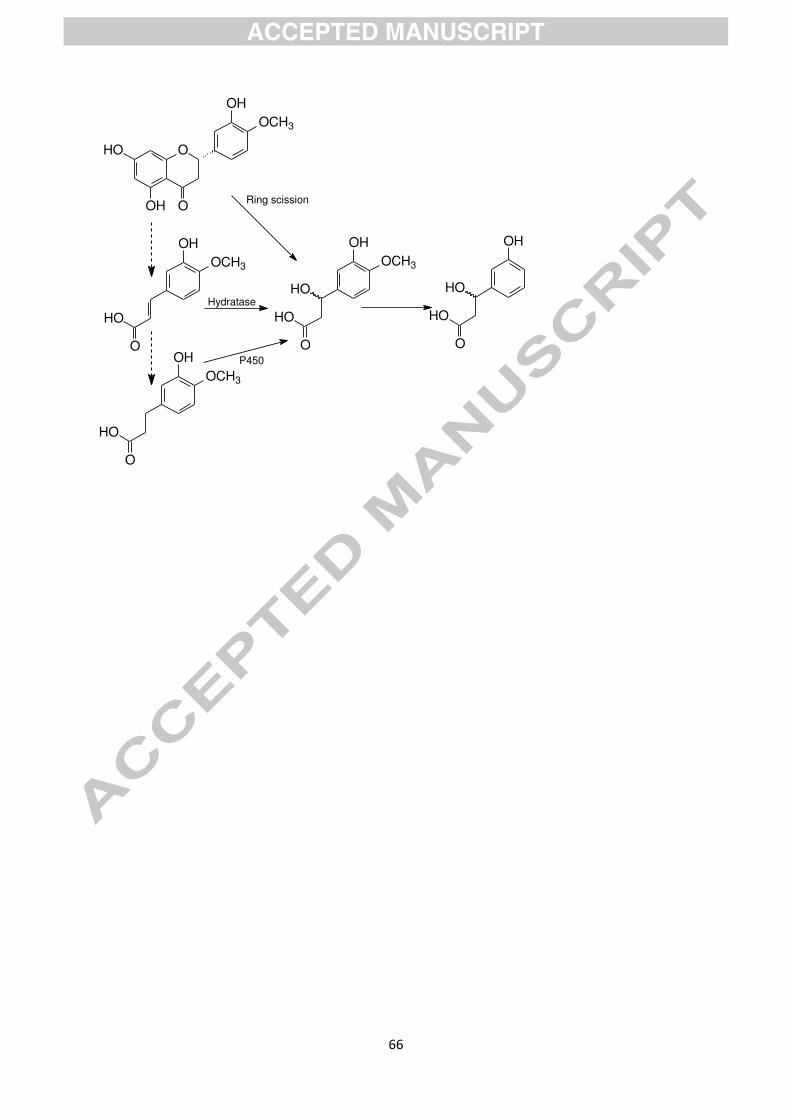
which report its production from rutin [160], and after consumption of coffee [161]. Plausible
immediate precursors are the appropriate cinnamic acid plus hydratase enzyme or
dihydrocinnamic (3-(phenyl)propionic) acid plus P450 mono-oxygenase enzyme, but such
precursors arise from so many substrates that many more reports of the analogous
phenylhydracrylic acids would be expected. It has been suggested that an alternative route
might be the formation of a phenylhydracrylic acid from a flavonoid by an unusual C-ring
fission in which the pyran oxygen is retained on the side chain of the B-ring fragment rather
Page 26
24
than on the A-ring fragment (see Fig. 8) [157]. 3胡-Hydroxy-phenylhydracrylic acid has been
identified as a phenylalanine catabolite of incompletely characterised metronidazole-sensitive
bacteria [162], and proposed as a urinary biomarker for a range of neurological,
gastrointestinal, and psychiatric disorders [163], and 4胡-hydroxyphenylhydracrylic acid has
been observed in patients with gastro-intestinal disease [164]. The definitive answer to the
origin and significance of phenylhydracrylic acids is still lacking, but it has been shown that
some Bacillus spp. can produce 3胡-methoxy-4胡-hydroxyphenylhydracrylic acid from ferulic
acid [165], and it is now recognised that some distinctive strains of Bacillus are found in the
human gut, including some B. subtilis that bind strongly to the mucosa possibly precluding
their elimination in faeces and availability in in vitro fermentations [144, 166].
6. Effects of catabolites
In vitro cell culture studies are probably the most effective way to investigate the effects of
gut microflora catabolites because when effects are observed there is the potential also to
investigate the underlying mechanism(s). Choice of tissue, choice of catabolite, concentration
and time of exposure, and choice of biomarker require careful consideration.
The chosen biomarker(s) must be biologically relevant, but more importantly, those selected
should be measurable with precision. Many biomarkers can only be quantified with a
replicate precision in excess of 10%, or even 20%, and such imprecision makes it very
difficult to detect small treatment-related changes in the biomarker. This constraint can be
circumvented by increasing the catabolite concentration, but when the catabolite is used at
even 10-fold the likely sustained plasma concentration, the relevance to real diets is
Page 27
25
questionable. Certainly extrapolation to lower concentrations is unreliable as effects are likely
to reach a threshold.
Ideally, the range of catabolite concentration should centre on plasma / tissue concentrations
associated with real-world diets perceived as being beneficial to health. Such data for tissues
are conspicuous by their absence but plasma Cmax values for metabolites absorbed in the
proximal gastrointestinal tract rarely exceed a transient 50 nM for a single bolus dose (unless
supplemented) [25, 167-169] although 3ガ,4ガ-dimethoxycinnamic acid (ca 0.6 µM after ca two
cups of coffee) [170] and 4-hydroxybenzoic acid (2.5 µM after 300 g fresh strawberries) [22]
are notable exceptions. In contrast, as discussed above, for those catabolites derived from
multiple substrates, and especially those associated with commodities consumed repeatedly at
short intervals during the day, a higher Cmax is to be expected, in some cases exceeding 1 µM.
More importantly, a concentration of at least 0.5 Cmax will be maintained for a considerable
period, possibly overnight. Concentrations in human faecal water can be much higher [171],
exposing the gut microbiota and the colonic mucosa to individual catabolites at
concentrations in excess of 10 µM. Because multiple catabolites are present simultaneously,
the total exposure in the plasma might realistically be some 2–3-fold higher and in the colon
in excess of 1 mM. The relatively long-term exposure, possibly every day, give these
catabolites a real potential to exert biological effects in vivo.
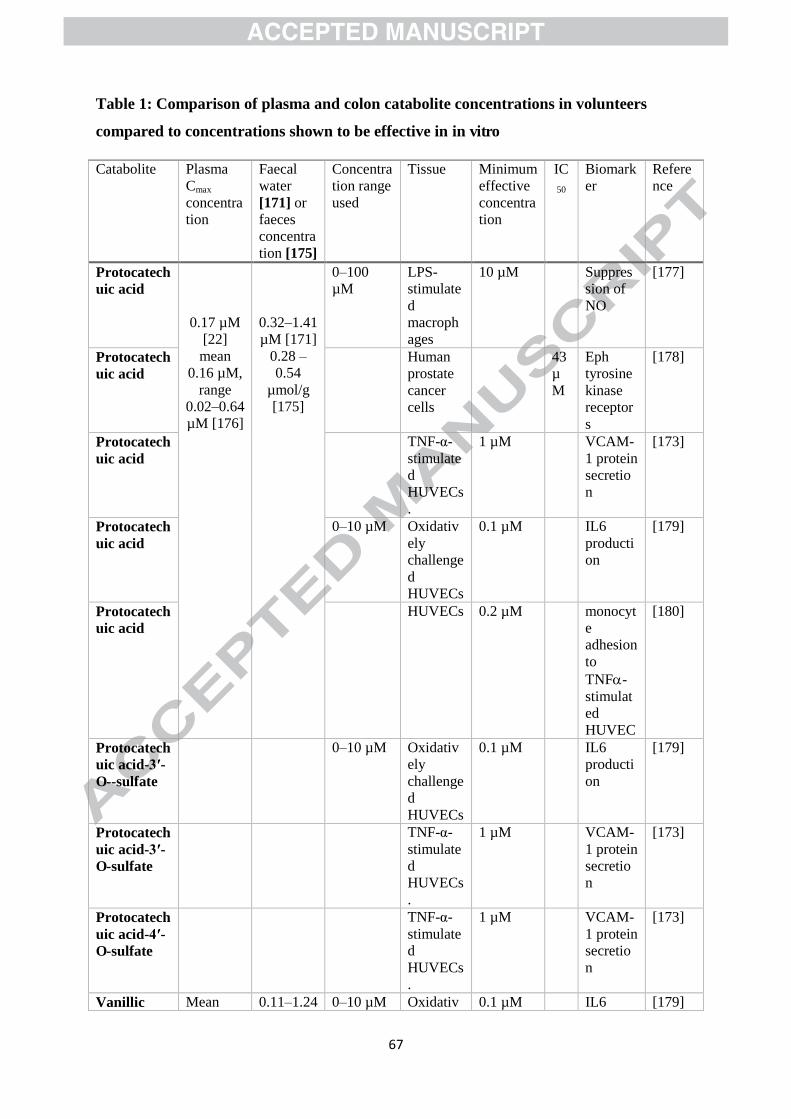
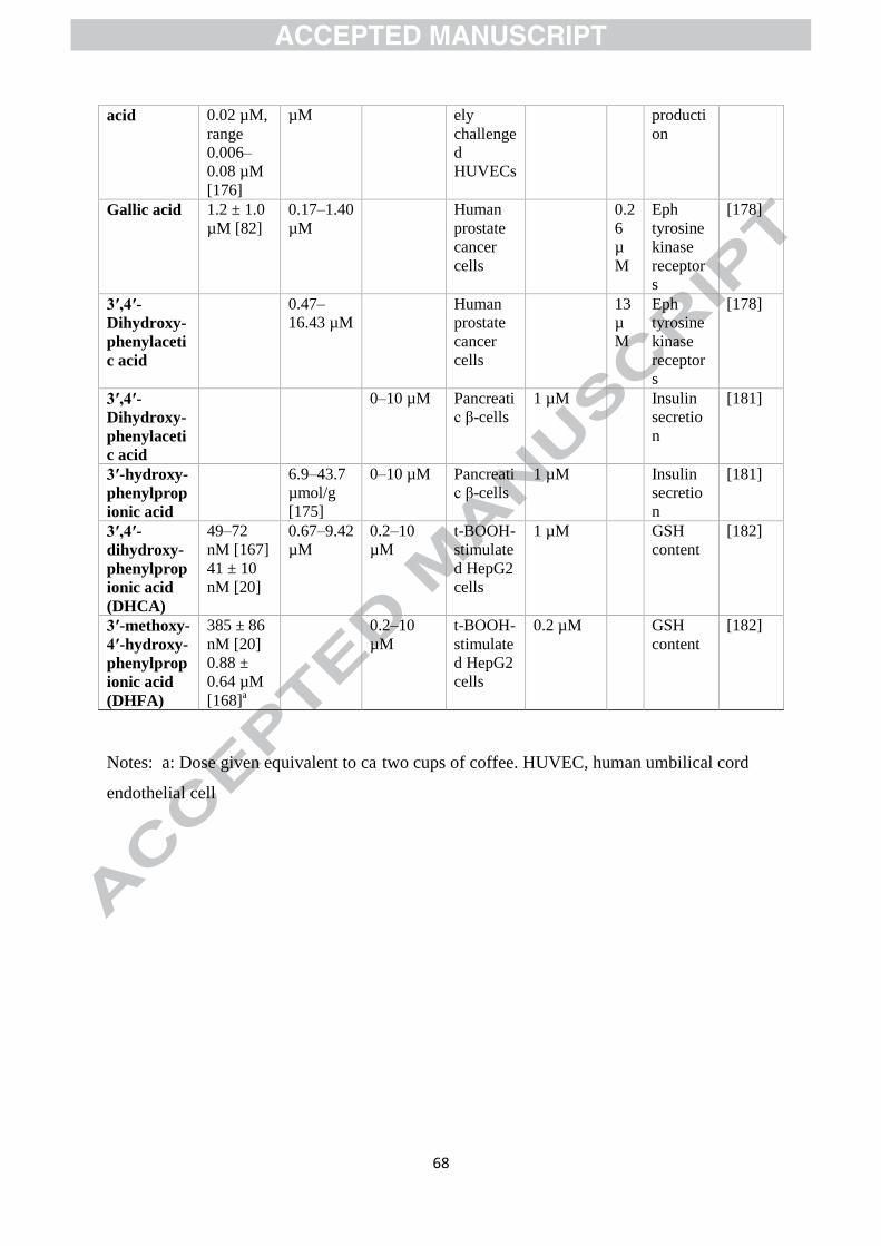
Data for a range of C6–C1, C6–C2, and C6–C3 phenolic acids are presented in Table 1, so far as
possible comparing concentrations achieved in plasma and the colon following
unsupplemented diets with data for biological effects observed in vitro. None of these studies
observed any effect below 0.1 µM — indeed none even investigated a lower concentration. A
simple comparison of plasma and colon concentrations with concentrations shown to be
Page 28
26
effective in vitro suggests that there is potential for protocatechuic acid, vanillic acid, gallic
acid and 3ガ,4ガ-dihydroxyphenylacetic acid to exert modest effects in vivo in at least some
volunteers on real-world diets. 3胡-Hydroxyphenylpropionic acid, 3胡,4胡-
dihydroxyphenylpropionic and 3胡-methoxy-4胡-hydroxyphenylpropionic might also achieve
sufficient concentrations following repeated consumption of coffee or tea. Note, however,
that at least some of these catabolites, including protocatechuic and vanillic acids, bind to
human serum albumin [172], and this might reduce their potency in vivo.
There has been limited investigation of mixtures of (poly)phenolic catabolites. In one study
there was no evidence of any additive effect [173], but in another study a mixture of 4-
hydroxybenzoic acid, protocatechuic acid and vanillic acid (albeit each at 0.33 µM) was more
effective than any component alone at the same concentration [174], suggesting that there is
some synergy rather than merely an additive effect. There is clearly a potential for some
carefully-targeted studies seeking synergy, and these should not be restricted to (poly)phenol
mixtures but embrace other classes of phytonutrients, and a need for further data on plasma
and tissue catabolite concentrations in free-living volunteers, preferably in association with
their dietary history. The development of biomarker assays, and the requisite reagents, to
facilitate the generation of more precise replicate data would be a distinct advantage.
Page 29
27
7. Summary, conclusions and recommendations for future research
The colonic microbiota transform a very complex range of (poly)phenol substrates with
coffee chlorogenic acids, black tea theaflavins and thearubigins often dominating, with
substantial contributions from proanthocyanidins and flavanols, flavonols, flavanones and
anthocyanin glycosides, and unextractable (poly)phenols, from many fruit and vegetables.
These transformations can be extensive, and while some microbial catabolites are substrate-
specific (e.g. equol, urolithins and nasutins, mammalian lignans, hydroxyanthranilic acid),
certain catabolites are common to many of the major substrates, implying that the spectrum
of catabolites produced is less complex and qualitatively less variable than the spectrum of
substrates consumed. The catabolites most likely to dominate are the C6 phenols, C6–C1, C6–
C2 and C6–C3 dihydro acids derived from chlorogenic acids/cinnamates, and most flavonoids
including black tea thearubigins and theaflavins (see Figs. 2 and 5).
There is growing evidence from in vitro and intervention studies that some of these
catabolites are biologically active, and that, along with untransformed substrates, may
function as prebiotics capable of modulating the human gut microbiota composition. In
addition, some catabolites after absorption may have further beneficial effects provided that
sufficient concentrations are achieved for a sufficient time in the relevant tissue(s).
Consumption of multiple doses, such as regular coffee and tea drinking throughout the day,
have the potential to increase the plasma concentration of catabolites, especially for those
derived from microbial action.
Page 30
28
Future investigations must address the minimum effective dose of potentially prebiotic
substrates, determine what percentage of the population are susceptible, whether
susceptibility can be induced, and how long any associated benefits persist, especially if the
supplementation is subsequently curtailed. There is also a pressing need for investigation of
free-living volunteers who regularly consume (poly)phenol-rich beverages at frequent
intervals to better define the circulating catabolite profiles after consumption of multiple
doses. The answers to these are crucial if we are to understand and fully exploit the effect of
(poly)phenol consumption on human health.
Acknowledgements: GW thanks the European Research Council for an advanced grant
(project number 322467, POLYTRUE?).
Page 31
29
References
[1] E. Haslam, Plant polyphenols (syn. vegetable tannins) and chemical defense-A
reappraisal, J. Chem. Ecol. 14 (10) (1988) 1789-1805.
[2] A. Scalbert, Antimicrobial properties of tannins, Phytochemistry 30 (1991) 3875-3883.
[3] V. Neveu, J. Perez-Jimenez, F. Vos, V. Crespy, C.L. Du, L. Mennen, C. Knox, R. Eisner,
J. Cruz, D. Wishart, A. Scalbert, Phenol-Explorer: an online comprehensive database on
polyphenol contents in foods, Database (Oxford) (2010) bap024.
[4] A. Garcia-Alvarez, B. Egan, K.S. de, L. Dima, F.M. Maggi, M. Isoniemi, L. Ribas-Barba,
M.M. Raats, E.M. Meissner, M. Badea, F. Bruno, M. Salmenhaara, R. Mila-Villarroel, V.
Knaze, C. Hodgkins, A. Marculescu, L. Uusitalo, P. Restani, L. Serra-Majem, Usage of plant
food supplements across six European countries: findings from the PlantLIBRA consumer
survey, PLoS. ONE 9 (3) (2014) e92265.
[5] J. Liu, P.G. Xiao, Recent advances in the study of antioxidative effects of chinese
medicinal plants - review, Phytother. Res 8 (1994) 445-451.
[6] R. Zamora-Ros, V. Knaze, J.A. Rothwell, B. Hemon, A. Moskal, K. Overvad, A.
Tjonneland, C. Kyro, G. Fagherazzi, M.C. Boutron-Ruault, M. Touillaud, V. Katzke, T.
Kuhn, H. Boeing, J. Forster, A. Trichopoulou, E. Valanou, E. Peppa, D. Palli, C. Agnoli, F.
Ricceri, R. Tumino, M.S. de Magistris, P.H.M. Peeters, H.B. Bueno-de-Mesquita, D.
Engeset, G. Skeie, A. Hjartaker, V. Menendez, A. Agudo, E. Molina-Montes, J.M. Huerta, A.
Barricarte, P. Amiano, E. Sonestedt, L.M. Nilsson, R. Landberg, T.J. Key, K.T. Khaw, N.J.
Wareham, Y.X. Lu, N. Slimani, I. Romieu, E. Riboli, A. Scalbert, Dietary polyphenol intake
Page 32
30
in Europe: the European Prospective Investigation into Cancer and Nutrition (EPIC) study,
Eur. J. Nutr. 55 (4) (2016) 1359-1375.
[7] T.W. Crozier, A. Stalmach, M.E. Lean, A. Crozier, Espresso coffees, caffeine and
chlorogenic acid intake: potential health implications, Food Funct. 3 (1) (2012) 30-33.
[8] M.N. Clifford, J.J. van der Hooft, A. Crozier, Human studies on the absorption,
distribution, metabolism, and excretion of tea polyphenols, Am. J Clin. Nutr 98 (2013)
1619S-1630S.
[9] D.A. Balentine, S.A. Wiseman, C.M. Bouwens, The chemistry of tea flavonoids, CRC
Crit. Rev. Food Sci. Nutr. 37 (1997) 693-704.
[10] M.E. Harbowy, D. Balentine, Tea chemistry, CRC Crit. Rev. Plant Sci. 16 (1997) 415-
480.
[11] V. Cheynier, Phenolic compounds: from plants to foods, Phytochem. Rev. 11 (2012)
153-177.
[12] M. Lambert, E. Meudec, A. Verbaere, G. Mazerolles, J. Wirth, G. Masson, V. Cheynier,
N. Sommerer, A high-throughput UHPLC-QqQ-MS method for polyphenol profiling in rosé
wines, Molecules 20 (5) (2015) 7890-7914.
[13] F. Saura-Calixto, J. Perez-Jimenez, S. Tourino, J. Serrano, E. Fuguet, J.L. Torres, I.
Goni, Proanthocyanidin metabolites associated with dietary fibre from in vitro colonic
fermentation and proanthocyanidin metabolites in human plasma, Mol. Nutr. Food Res. 54
(7) (2010) 939-946.
[14] P. Pinto, C.N. Santos, Worldwide (poly)phenol intake: assessment methods and
identified gaps, Eur. J. Nutr. (2017) DOI 10.1007/s00394-016-1354-2.
Page 33
31
[15] G. Pereira-Caro, B. Fernandez-Quiros, I.A. Ludwig, I. Pradas, A. Crozier, J.M. Moreno-
Rojas, Catabolism of citrus flavanones by the probiotics Bifidobacterium longum and
Lactobacillus rhamnosus, Eur. J. Nutr. DOI: 10.1007/s00394-016-1312-z (2016) 1-12.
[16] M.N. Clifford, Chlorogenic acids and other cinnamates — nature, occurrence and dietary
burden, J. Sci. Food Agric. 79 (1999) 362-372.
[17] M.N. Clifford, Chlorogenic acids and other cinnamates - nature, occurrence, dietary
burden, absorption and metabolism, J. Sci. Food Agric. 80 (7) (2000) 1033-1043.
[18] J.A. da Encarnacao, T.L. Farrell, A. Ryder, N.U. Kraut, G. Williamson, In vitro enzymic
hydrolysis of chlorogenic acids in coffee, Mol. Nutr. Food Res. 59 (2) (2015) 231-239.
[19] M.R. Olthof, P.C. Hollman, M.B. Katan, Chlorogenic acid and caffeic acid are absorbed
in humans, J. Nutr. 131 (1) (2001) 66-71.
[20] A. Stalmach, W. Mullen, D. Barron, K. Uchida, T. Yokota, C. Cavin, H. Steiling, G.
Williamson, A. Crozier, Metabolite profiling of hydroxycinnamate derivatives in plasma and
urine after the ingestion of coffee by humans: identification of biomarkers of coffee
consumption, Drug Metab. Dispos. 37 (8) (2009) 1749-1758.
[21] K. Nagy, K. Redeuil, G. Williamson, S. Rezzi, F. Dionisi, K. Longet, F. Destaillats, M.
Renouf, First identification of dimethoxycinnamic acids in human plasma after coffee intake
by liquid chromatography-mass spectrometry, J. Chromatogr. A 1218 (3) (2011) 491-497.
[22] T.L. Farrell, M. Gomez-Juaristi, L. Poquet, K. Redeuil, K. Nagy, M. Renouf, G.
Williamson, Absorption of dimethoxycinnamic acid derivatives in vitro and pharmacokinetic
profile in human plasma following coffee consumption, Mol. Nutr. Food Res. 56 (9) (2012)
1413-1423.
Page 34
32
[23] A. Stalmach, H. Steiling, G. Williamson, A. Crozier, Bioavailability of chlorogenic acids
following acute ingestion of coffee by humans with an ileostomy, Arch. Biochem. Biophys.
501 (1) (2010) 98-105.
[24] G.W. Plumb, M.T. Garcia Conesa, P.A. Kroon, M. Rhodes, S. Ridley, G. Williamson,
Metabolism of chlorogenic acid by human plasma, liver, intestine and gut microflora, J. Sci.
Food Agr. 79 (1999) 390-392.
[25] D. Scherbl, M. Renouf, C. Marmet, L. Poquet, I. Cristiani, S. Dahbane, S. Emady-Azar,
J. Sauser, J. Galan, F. Dionisi, E. Richling, Breakfast consumption induces retarded release of
chlorogenic acid metabolites in humans, Eur. Food Res. Technol. (2016)
doi:10.1007/s00217-016-2793-y.
[26] M. Renouf, C. Marmet, P. Guy, A.L. Fraering, K. Longet, J. Moulin, M. Enslen, D.
Barron, C. Cavin, F. Dionisi, S. Rezzi, S. Kochhar, H. Steiling, G. Williamson, Nondairy
creamer, but not milk, delays the appearance of coffee phenolic acid equivalents in human
plasma, J. Nutr. 140 (2) (2010) 259-263.
[27] G.S. Duarte, A. Farah, Effect of simultaneous consumption of milk and coffee on
chlorogenic acids' bioavailability in humans, J. Agric. Food Chem. 59 (14) (2011) 7925-
7931.
[28] F. Vallejo, M. Larrosa, E. Escudero, M.P. Zafrilla, B. Cerda, J. Boza, M.T. Garcia-
Conesa, J.C. Espin, F.A. Tomas-Barberan, Concentration and solubility of flavanones in
orange beverages affect their bioavailability in humans, J. Agric. Food Chem. 58 (10) (2010)
6516-6524.
[29] L. Actis-Goretta, T.P. Dew, A. Leveques, G. Pereira-Caro, M. Rein, A. Teml, C.
Schafer, U. Hofmann, M. Schwab, M. Eichelbaum, A. Crozier, G. Williamson,
Page 35
33
Gastrointestinal absorption and metabolism of hesperetin-7-O-rutinoside and hesperetin-7-O-
glucoside in healthy humans, Mol. Nutr. Food Res. 59 (9) (2015) 1651-1662.
[30] I.L. Nielsen, W.S. Chee, L. Poulsen, E. Offord-Cavin, S.E. Rasmussen, H. Frederiksen,
M. Enslen, D. Barron, M.N. Horcajada, G. Williamson, Bioavailability is improved by
enzymatic modification of the citrus flavonoid hesperidin in humans: a randomized, double-
blind, crossover trial, J. Nutr. 136 (2) (2006) 404-408.
[31] D. Monti, A. Pisvejcova, V. Kren, M. Lama, S. Riva, Generation of an alpha-L-
rhamnosidase library and its application for the selective derhamnosylation of natural
products, Biotechnol. Bioeng. 87 (6) (2004) 763-771.
[32] L. Bredsdorff, I.L. Nielsen, S.E. Rasmussen, C. Cornett, D. Barron, F. Bouisset, E.
Offord, G. Williamson, Absorption, conjugation and excretion of the flavanones, naringenin
and hesperetin from alpha-rhamnosidase-treated orange juice in human subjects, Br. J. Nutr.
103 (11) (2010) 1602-1609.
[33] H. Takumi, R. Mukai, S. Ishiduka, T. Kometani, J. Terao, Tissue distribution of
hesperetin in rats after a dietary intake, Biosci. Biotechnol. Biochem. 75 (8) (2011) 1608-
1610.
[34] W. Mullen, M.A. Archeveque, C.A. Edwards, H. Matsumoto, A. Crozier, Bioavailability
and metabolism of orange juice flavanones in humans: impact of a full-fat yogurt, J. Agric.
Food Chem. 56 (23) (2008) 11157-11164.
[35] G.M. Brett, W. Hollands, P.W. Needs, B. Teucher, J.R. Dainty, B.D. Davis, J.S.
Brodbelt, P.A. Kroon, Absorption, metabolism and excretion of flavanones from single
portions of orange fruit and juice and effects of anthropometric variables and contraceptive
pill use on flavanone excretion, Br. J. Nutr. 101 (5) (2009) 664-675.
Page 36
34
[36] M.J.C. Rhodes, K.R. Price, Analytical problems in the study of flavonoid compounds in
onions, Food Chem. 56 (1996) 1-5.
[37] K.R. Price, M.J.C. Rhodes, K.A. Barnes, Flavonol glycoside content and composition of
tea infusions made from commercially available teas and tea products, J. Agric. Food Chem.
46 (1998) 2517-2522.
[38] A. Lommen, M. Godejohann, D.P. Venema, P.C. Hollman, M. Spraul, Application of
directly coupled HPLC-NMR-MS to the identification and confirmation of quercetin
glycosides and phloretin glycosides in apple peel, Analyt. Chem. 72 (8) (2000) 1793-1797.
[39] P.C. Hollman, J.M. van Trijp, M.N. Buysman, M.S. van der Gaag, M.J. Mengelers, J.H.
de Vries, M.B. Katan, Relative bioavailability of the antioxidant flavonoid quercetin from
various foods in man, FEBS Lett. 418 (1-2) (1997) 152-156.
[40] P.C. Hollman, M.N. Bijsman, Y. van Gameren, E.P. Cnossen, J.H. de Vries, M.B.
Katan, The sugar moiety is a major determinant of the absorption of dietary flavonoid
glycosides in man, Free Radic. Res. 31 (6) (1999) 569-573.
[41] W. Mullen, C.A. Edwards, A. Crozier, Absorption, excretion and metabolite profiling of
methyl-, glucuronyl-, glucosyl- and sulpho-conjugates of quercetin in human plasma and
urine after ingestion of onions, Br. J. Nutr. 96 (1) (2006) 107-116.
[42] Y.J. Hong, A.E. Mitchell, Identification of glutathione-related quercetin metabolites in
humans, Chem. Res. Toxicol. 19 (11) (2006) 1525-1532.
[43] R. Boonpawa, N. Moradi, A. Spenkelink, I.M. Rietjens, A. Punt, Use of physiologically
based kinetic (PBK) modeling to study interindividual human variation and species
Page 37
35
differences in plasma concentrations of quercetin and its metabolites, Biochem. Pharmacol.
98 (4) (2015) 690-702.
[44] S. Pascual-Teresa, C. Santos-Buelga, J.C. Rivas-Gonzalo, Quantitative analysis of
flavan-3-ols in Spanish foodstuffs and beverages, J. Agric. Food Chem. 48 (11) (2000) 5331-
5337.
[45] G. Kuhnle, J.P. Spencer, H. Schroeter, B. Shenoy, E.S. Debnam, S.K. Srai, C. Rice-
Evans, U. Hahn, Epicatechin and catechin are O-methylated and glucuronidated in the small
intestine, Biochem Biophys. Res. Commun. 277 (2) (2000) 507-512.
[46] S. Saha, W. Hollands, P.W. Needs, L.M. Ostertag, R.B. de, G.G. Duthie, P.A. Kroon,
Human O-sulfated metabolites of (-)-epicatechin and methyl-(-)-epicatechin are poor
substrates for commercial aryl-sulfatases: implications for studies concerned with quantifying
epicatechin bioavailability, Pharmacol. Res. 65 (6) (2012) 592-602.
[47] J.I. Ottaviani, T.Y. Momma, G.K. Kuhnle, C.L. Keen, H. Schroeter, Structurally related
(-)-epicatechin metabolites in humans: assessment using de novo chemically synthesized
authentic standards, Free Radic. Biol. Med. 52 (8) (2012) 1403-1412.
[48] L. Actis-Goretta, A. Leveques, M. Rein, A. Teml, C. Schafer, U. Hofmann, H. Li, M.
Schwab, M. Eichelbaum, G. Williamson, Intestinal absorption, metabolism, and excretion of
(-)-epicatechin in healthy humans assessed by using an intestinal perfusion technique, Am. J.
Clin. Nutr. 98 (2013) 924-933.
[49] L. Actis-Goretta, A. Leveques, F. Giuffrida, F. Romanov-Michailidis, F. Viton, D.
Barron, M. Duenas-Paton, S. Gonzalez-Manzano, C. Santos-Buelga, G. Williamson, F.
Dionisi, Elucidation of (-)-epicatechin metabolites after ingestion of chocolate by healthy
humans, Free Radic. Biol. Med. 53 (4) (2012) 787-795.
Page 38
36
[50] R. Gonzalez-Barrio, G. Borges, W. Mullen, A. Crozier, Bioavailability of anthocyanins
and ellagitannins following consumption of raspberries by healthy humans and subjects with
an ileostomy, J. Agric. Food Chem. 58 (7) (2010) 3933-3939.
[51] R.R. Holt, S.A. Lazarus, M.C. Sullards, Q.Y. Zhu, D.D. Schramm, J.F. Hammerstone,
C.G. Fraga, H.H. Schmitz, C.L. Keen, Procyanidin dimer B2 [epicatechin-(4 beta-8)-
epicatechin] in human plasma after the consumption of a flavanol-rich cocoa, Am. J. Clin.
Nutr. 76 (4) (2002) 798-804.
[52] G.M. Woodward, P.W. Needs, C.D. Kay, Anthocyanin-derived phenolic acids form
glucuronides following simulated gastrointestinal digestion and microsomal glucuronidation,
Mol. Nutr. Food Res. 55 (3) (2011) 378-386.
[53] C.I.R. Gill, G.J. McDougall, S. Glidewell, D. Stewart, Q. Shen, K. Tuohy, A. Dobbin, A.
Boyd, E. Brown, S. Haldar, I.R. Rowland, Profiling of phenols in human fecal water after
raspberry supplementation, J. Agric. Food Chem. 58 (19) (2010) 10389-10395.
[54] A.M. Aura, P. Martin-Lopez, K.A. O'Leary, G. Williamson, K.M. Oksman-Caldentey,
K. Poutanen, C. Santos-Buelga, In vitro metabolism of anthocyanins by human gut
microflora, Eur. J. Nutr. 44 (3) (2005) 133-142.
[55] S. Stoupi, G. Williamson, F. Viton, D. Barron, L.J. King, J.E. Brown, M.N. Clifford, In
vivo bioavailability, absorption, excretion, and pharmacokinetics of [14C]procyanidin B2 in
male rats, Drug Metab. Dispos. 38 (2) (2010) 287-291.
[56] A.W. Walker, S.H. Duncan, P. Louis, H.J. Flint, Phylogeny, culturing, and
metagenomics of the human gut microbiota, Trends Microbiol. 22 (5) (2014) 267-274.
Page 39
37
[57] P.E. Fournier, J.C. Lagier, G. Dubourg, D. Raoult, From culturomics to
taxonomogenomics: A need to change the taxonomy of prokaryotes in clinical microbiology,
Anaerobe 36 (2015) 73-78.
[58] F. Bäckhed, R.E. Ley, J.L. Sonnenburg, D.A. Peterson, J.I. Gordon, Host-bacterial
mutualism in the human intestine, Science 307 (5717) (2005) 1915-1920.
[59] S.H. Park, K.A. Kim, Y.T. Ahn, J.J. Jeong, C.S. Huh, D.H. Kim, Comparative analysis
of gut microbiota in elderly people of urbanized towns and longevity villages, BMC
Microbiology 15 (2015) 15: 49 doi: 10.1186/s12866-015-0386-8.
[60] M.J. Claesson, I.B. Jeffery, S. Conde, S.E. Power, E.M. O'Connor, S. Cusack, H.M.B.
Harris, M. Coakley, B. Lakshminarayanan, O. O'Sullivan, G.F. Fitzgerald, J. Deane, M.
O'Connor, N. Harnedy, K. O'Connor, D. O'Mahony, D. van Sinderen, M. Wallace, L.
Brennan, C. Stanton, J.R. Marchesi, A.P. Fitzgerald, F. Shanahan, C. Hill, R.P. Ross, P.W.
O'Toole, Gut microbiota composition correlates with diet and health in the elderly, Nature
488 (7410) (2012) 178.
[61] J.J. Goedert, G. Jones, X. Hua, X. Xu, G.Q. Yu, R. Flores, R.T. Falk, M.H. Gail, J.X.
Shi, J. Ravel, H.S. Feigelson, Investigation of the Association Between the Fecal Microbiota
and Breast Cancer in Postmenopausal Women: a Population-Based Case-Control Pilot Study,
J. Nat. Cancer Inst. 107 (8) (2015) doi: 10.1093/jnci/djv147.
[62] E. Vogtmann, X. Hua, G. Zeller, S. Sunagawa, A.Y. Voigt, R. Hercog, J.J. Goedert, J.X.
Shi, P. Bork, R. Sinha, Colorectal Cancer and the Human Gut Microbiome: Reproducibility
with Whole-Genome Shotgun Sequencing, PLoS One 11(5) (2016)
http://dx.doi.org/10.1371/journal.pone.0155362
Page 40
38
[63] M.N. Clifford, Miscellaneous phenols in foods and beverages - nature, occurrence and
dietary burden, J. Sci. Food Agric. 80 (7) (2000) 1126-1137.
[64] G. Williamson, M.N. Clifford, Colonic metabolites of berry polyphenols: the missing
link to biological activity?, Br. J. Nutr. 104 (2010) S48-S66.
[65] G. Pereira-Caro, I.A. Ludwig, T. Polyviou, D. Malkova, A. Garcia, J.M. Moreno-Rojas,
A. Crozier, Identification of plasma and urinary metabolites and catabolites derived from
orange juice (poly)phenols: analysis by high-performance liquid chromatographyhigh-
resolution mass spectrometry, J. Agric. Food Chem. 64(28) (2016) 5724-5735.
[66] J.I. Mosele, S. Martin-Pelaez, A. Macia, M. Farras, R.M. Valls, U. Catalan, M.J.
Motilva, Faecal microbial metabolism of olive oil phenolic compounds: In vitro and in vivo
approaches, Mol. Nutr. Food Res. 58 (9) (2014) 1809-1819.
[67] M. Blaut, L. Schoefer, A. Braune, Transformation of flavonoids by intestinal
microorganisms, Int. J. Vitam. Nutr. Res. 73 (2) (2003) 79-87.
[68] A. Matthies, G. Loh, M. Blaut, A. Braune, Daidzein and genistein are converted to equol
and 5-hydroxy-equol by human intestinal Slackia isoflavoniconvertens in gnotobiotic rats, J.
Nutr. 142 (1) (2012) 40-46.
[69] P. Gaya, M. Medina, A. Sanchez-Jimenez, J.M. Landete, Phytoestrogen metabolism by
adult human gut microbiota, Molecules 21 (8) (2016) doi: 10.3390/molecules21081034.
[70] M.V. Selma, F.A. Tomas-Barberan, D. Beltran, R. Garcia-Villalba, J.C. Espin,
Gordonibacter urolithinfaciens sp nov., a urolithin-producing bacterium isolated from the
human gut, Int. J. Systematic Evolutionary Microbiol. 64 (2014) 2346-2352.
Page 41
39
[71] R. Gonzalez-Barrio, C.A. Edwards, A. Crozier, Colonic catabolism of ellagitannins,
ellagic acid, and raspberry anthocyanins: in vivo and in vitro studies, Drug Metab. Dispos. 39
(9) (2011) 1680-1688.
[72] M.E. Juan, I. Alfaras, J.M. Planas, Determination of dihydroresveratrol in rat plasma by
HPLC, J. Agric. Food Chem. 58 (12) (2010) 7472-7475.
[73] P. Wang, H. Chen, Y. Zhu, J. McBride, J. Fu, S. Sang, Oat avenanthramide-C (2c) is
biotransformed by mice and the human microbiota into bioactive metabolites, J. Nutr. 145 (2)
(2015) 239-245.
[74] A.F. Vinha, F. Ferreres, B.M. Silva, P. Valentao, A. Goncalves, J.A. Pereira, M.B.
Oliveira, R.M. Seabra, P.B. Andrade, Phenolic profiles of Portuguese olive fruits (Olea
europaea L.): Influences of cultivar and geographical origin, Food Chem. 89 (4) (2005) 561-
568.
[75] R. Zamora-Ros, V. Knaze, L. Lujan-Barroso, G.G. Kuhnle, A.A. Mulligan, M.
Touillaud, N. Slimani, I. Romieu, N. Powell, R. Tumino, P.H. Peeters, M.S. de Magistris, F.
Ricceri, E. Sonestedt, I. Drake, A. Hjartaker, G. Skie, T. Mouw, P.A. Wark, D. Romaguera,
H.B. Bueno-de-Mesquita, M. Ros, E. Molina, S. Sieri, J.R. Quiros, J.M. Huerta, A.
Tjonneland, J. Halkjaer, G. Masala, B. Teucher, R. Kaas, R.C. Travis, V. Dilis, V. Benetou,
A. Trichopoulou, P. Amiano, E. Ardanaz, H. Boeing, J. Forster, F. Clavel-Chapelon, G.
Fagherazzi, F. Perquier, G. Johansson, I. Johansson, A. Cassidy, K. Overvad, C.A. Gonzalez,
Dietary intakes and food sources of phytoestrogens in the European Prospective Investigation
into Cancer and Nutrition (EPIC) 24-hour dietary recall cohort, Eur. J. Clin. Nutr. 66 (8)
(2012) 932-941.
Page 42
40
[76] G.G. Kuhnle, C. Dell'aquila, S.M. Aspinall, S.A. Runswick, A.A. Mulligan, S.A.
Bingham, Phytoestrogen content of cereals and cereal-based foods consumed in the UK,
Nutr. Cancer 61 (3) (2009) 302-309.
[77] S.G. Kasimsetty, D. Bialonska, M.K. Reddy, G. Ma, S.I. Khan, D. Ferreira, Colon
cancer chemopreventive activities of pomegranate ellagitannins and urolithins, J. Agric. Food
Chem. 58 (4) (2010) 2180-2187.
[78] J. Regueiro, C. Sanchez-Gonzalez, A. Vallverdu-Queralt, J. Simal-Gandara, R. Lamuela-
Raventos, M. Izquierdo-Pulido, Comprehensive identification of walnut polyphenols by
liquid chromatography coupled to linear ion trap-Orbitrap mass spectrometry, Food Chem.
152 (2014) 340-348.
[79] Y. Ma, A. Kosinska-Cagnazzo, W.L. Kerr, R. Amarowicz, R.B. Swanson, R.B. Pegg,
Separation and characterization of phenolic compounds from dry-blanched peanut skins by
liquid chromatography-electrospray ionization mass spectrometry, J. Chromatogr. A 1356
(2014) 64–81.
[80] R. Zamora-Ros, C. ndres-Lacueva, R.M. Lamuela-Raventos, T. Berenguer, P. Jakszyn,
C. Martinez, M.J. Sanchez, C. Navarro, M.D. Chirlaque, M.J. Tormo, J.R. Quiros, P.
Amiano, M. Dorronsoro, N. Larranaga, A. Barricarte, E. Ardanaz, C.A. Gonzalez,
Concentrations of resveratrol and derivatives in foods and estimation of dietary intake in a
Spanish population: European Prospective Investigation into Cancer and Nutrition (EPIC)-
Spain cohort, Br. J. Nutr. 100 (1) (2008) 188-196.
[81] F.W. Collins, Oat phenolics: avenanthramides, novel substituted N-
cinnamoylanthranilate alkaloids from oat groats and hulls, J. Agric. Food Chem. 37 (1989)
60-66.
Page 43
41
[82] P.C. van der Pijl, M. Foltz, N.D. Glube, S. Peters, G.S.M.J. Duchateau,
Pharmacokinetics of black tea-derived phenolic acids in plasma, J. Functional Foods 17
(2015) 667-675.
[83] H.D. Chen, S.M. Sang, Biotransformation of tea polyphenols by gut microbiota, J.
Functional Foods 7 (2014) 26-42.
[84] J. van Duynhoven, J.J. van der Hooft, F.A. van Dorsten, S. Peters, M. Foltz, V. Gomez-
Roldan, J. Vervoort, R.C. De Vos, D.M. Jacobs, Rapid and sustained systemic circulation of
conjugated gut microbial catabolites after single-dose black tea extract consumption, J.
Proteome Res. 13 (5) (2014) 2668-2678.
[85] M.N. Clifford, E.L. Copeland, J.P. Bloxsidge, L.A. Mitchell, Hippuric acid as a major
excretion product associated with black tea consumption, Xenobiotica 30 (3) (2000) 317-326.
[86] R.C. Pimpao, M.R. Ventura, R.B. Ferreira, G. Williamson, C.N. Santos, Phenolic
sulfates as new and highly abundant metabolites in human plasma after ingestion of a mixed
berry fruit puree, Br. J. Nutr. 113 (3) (2015) 454-463.
[87] R.C. Pimpao, T. Dew, M.E. Figueira, G.J. McDougall, D. Stewart, R.B. Ferreira, C.N.
Santos, G. Williamson, Urinary metabolite profiling identifies novel colonic metabolites and
conjugates of phenolics in healthy volunteers, Mol. Nutr. Food Res. 58 (7) (2014) 1414-1425.
[88] D.M. Jacobs, J.C. Fuhrmann, F.A. van Dorsten, D. Rein, S. Peters, E.J. van Velzen, B.
Hollebrands, R. Draijer, D.J. van, U. Garczarek, Impact of short-term intake of red wine and
grape polyphenol extract on the human metabolome, J Agric. Food Chem. 60 (12) (2012)
3078-3085.
Page 44
42
[89] G. Gross, D.M. Jacobs, S. Peters, S. Possemiers, D.J. van, E.E. Vaughan, W.T. Van de,
In vitro bioconversion of polyphenols from black tea and red wine/grape juice by human
intestinal microbiota displays strong interindividual variability, J Agric. Food Chem. 58 (18)
(2010) 10236-10246.
[90] M. Dall'asta, L. Calani, M. Tedeschi, L. Jechiu, F. Brighenti, R.D. Del, Identification of
microbial metabolites derived from in vitro fecal fermentation of different polyphenolic food
sources, Nutrition 28 (2) (2012) 197-203.
[91] L.-Q. Zhang, X.-W. Yang, Y.-B. Zhang, Y.-Y. Zhai, W. Xu, B. Zhao, D.-L. Liu, H.-J.
Yu, Biotransformation of phlorizin by human intestinal flora and inhibition of
biotransformation products on tyrosinase activity, Food Chem. 132 (2012) 936-942.
[92] A. Braune, W. Engst, M. Blaut, Degradation of neohesperidin dihydrochalcone by
human intestinal bacteria, J Agric. Food Chem. 53 (5) (2005) 1782-1790.
[93] J.D. Haddock, J.G. Ferry, Purification and properties of phloroglucinol reductase from
Eubacterium oxidoreducens G-41, J. Biol. Chem. 264 (1989) 4423-4427.
[94] L.R. Krumholz, M.P. Bryant, Eubacterium oxidoreducens sp. nov. requiring H2 or
formate to degrade gallate, pyrogallol, phloroglucinol and quercetin, Arch. Microbiol. 144
(1986) 8-14.
[95] M. Schantz, T. Erk, E. Richling, Metabolism of green tea catechins by the human small
intestine, Biotechnol. J. 5 (10) (2010) 1050-1059.
[96] M. Li, F. Wang, Y. Huang, F. Du, C. Zhong, O.E. Olaleye, W. Jia, Y. Li, F. Xu, J. Dong,
J. Li, J.B. Lim, B. Zhao, L. Jia, L. Li, C. Li, Systemic exposure to and disposition of
Page 45
43
catechols derived from Salvia miltiorrhiza roots (Danshen) after intravenous dosing DanHong
injection in human subjects, rats, and dogs, Drug Metab. Dispos. 43 (5) (2015) 679-690.
[97] X. Li, C. Cheng, F. Wang, Y. Huang, W. Jia, O.E. Olaleye, M. Li, Y. Li, C. Li,
Pharmacokinetics of catechols in human subjects intravenously receiving XueBiJing
injection, an emerging antiseptic herbal medicine, Drug Metab. Pharmacokinet. 31 (1) (2016)
95-98.
[98] P. Truchado, M. Larrosa, M.T. Garcia-Conesa, B. Cerda, M.L. Vidal-Guevara, F.A.
Tomas-Barberan, J.C. Espin, Strawberry processing does not affect the production and
urinary excretion of urolithins, ellagic acid metabolites, in humans, J. Agric. Food Chem. 60
(23) (2012) 5749-5754.
[99] A. Woting, N. Pfeiffer, L. Hanske, G. Loh, S. Klaus, M. Blaut, Alleviation of high fat
diet-induced obesity by oligofructose in gnotobiotic mice is independent of presence of
Bifidobacterium longum, Mol. Nutr. Food Res. 59 (11) (2015) 2267-2278.
[100] A. Woting, N. Pfeiffer, G. Loh, S. Klaus, M. Blaut, Clostridium ramosum promotes
high-fat diet-Induced obesity in gnotobiotic mouse models, Mbio 5 (5) (2014) 5. e01530-14.
[101] J. Baldwin, B. Collins, P.G. Wolf, K. Martinez, W. Shen, C.C. Chuang, W. Zhong, P.
Cooney, C. Cockrell, E. Chang, H.R. Gaskins, M.K. McIntosh, Table grape consumption
reduces adiposity and markers of hepatic lipogenesis and alters gut microbiota in butter fat-
fed mice, J. Nutr. Biochem. 27 (2016) 123-135.
[102] Y.Y. Gu, S. Yu, J.Y. Park, K. Harvatine, J.D. Lambert, Dietary cocoa reduces
metabolic endotoxemia and adipose tissue inflammation in high-fat fed mice, J. Nutr.
Biochem. 25 (4) (2014) 439-445.
Page 46
44
[103] U. Etxeberria, N. Arias, N. Boque, M.T. Macarulla, M.P. Portillo, J.A. Martinez, F.I.
Milagro, Reshaping faecal gut microbiota composition by the intake of trans-resveratrol and
quercetin in high-fat sucrose diet-fed rats, J. Nutr. Biochem. 26 (6) (2015) 651-660.
[104] D.E. Roopchand, R.N. Carmody, P. Kuhn, K. Moskal, P. Rojas-Silva, P.J. Turnbaugh,
I. Raskin, Dietary polyphenols promote growth of the gut bacterium Akkermansia
muciniphila and attenuate high-fat diet-induced metabolic syndrome, Diabetes 64(8) (2015)
2847-2858.
[105] D.B. Seo, H.W. Jeong, D. Cho, B.J. Lee, J.H. Lee, J.Y. Choi, I.H. Bae, S.J. Lee,
Fermented green tea extract alleviates obesity and related complications and alters gut
microbiota composition in diet-induced obese mice, J. Medicinal Food 18 (5) (2015) 549-
556.
[106] T. Taira, S. Yamaguchi, A. Takahashi, Y. Okazaki, A. Yamaguchi, H. Sakaguchi, H.
Chiji, Dietary polyphenols increase fecal mucin and immunoglobulin A and ameliorate the
disturbance in gut microbiota caused by a high fat diet, J. Clin. Biochem. Nutr. 57 (3) (2015)
212-216.
[107] T. Unno, M. Sakuma, S. Mitsuhashi, Effect of dietary supplementation of ()-
epigallocatechin gallate on gut microbiota and biomarkers of colonic fermentation in rats, J.
Nutr. Sci. Vitaminol. 60 (3) (2014) 213-219.
[108] Q.Y. Wang, Z.Y. Du, H. Zhang, L. Zhao, J. Sun, X.N. Zheng, F.Z. Ren, Modulation of
gut microbiota by polyphenols from adlay (Coix lacryma-jobi L. var. ma-yuen Stapf.) in rats
fed a high-cholesterol diet, Int. J. Food Sci. Nutr. 66 (7) (2015) 783-789.
Page 47
45
[109] P.L.H.R. Janssens, J. Penders, R. Hursel, A.E. Budding, P.H.M. Savelkoul, M.S.
Westerterp-Plantenga, Long-term green tea supplementation does not change the human gut
microbiota, PLoS One 11 (4) (2016) doi.org/10.1371/journal.pone.0153134.
[110] E. Barroso, V. Martin, M.C. Martinez-Cuesta, C. Pelaez, T. Requena, Stability of saliva
microbiota during moderate consumption of red wine, Arch. Oral Biol. 60 (12) (2015) 1763-
1768.
[111] A.J. Wallace, S.L. Eady, D.C. Hunter, M.A. Skinner, L. Huffman, J. Ansell, P.
Blatchford, M. Wohlers, T.D. Herath, D. Hedderley, D. Rosendale, H. Stoklosinski, T.
McGhie, D. Sun-Waterhouse, C. Redman, No difference in fecal levels of bacteria or short
chain fatty acids in humans, when consuming fruit juice beverages containing fruit fiber, fruit
polyphenols, and their combination, Nutr. Res. 35 (1) (2015) 23-34.
[112] N. Eid, H. Osmanova, C. Natchez, G. Walton, A. Costabile, G. Gibson, I. Rowland,
J.P. Spencer, Impact of palm date consumption on microbiota growth and large intestinal
health: a randomised, controlled, cross-over, human intervention study, Br. J. Nutr. 114 (8)
(2015) 1226-1236.
[113] G. Ravn-Haren, L.O. Dragsted, T. Buch-Andersen, E.N. Jensen, R.I. Jensen, M.
Nemeth-Balogh, B. Paulovicsova, A. Bergstrom, A. Wilcks, T.R. Licht, J. Markowski, S.
Bugel, Intake of whole apples or clear apple juice has contrasting effects on plasma lipids in
healthy volunteers, Eur. J. Nutr. 52 (8) (2013) 1875-1889.
[114] J.I. Mosele, M.J. Gosalbes, A. Macia, L. Rubio, J.F. Vazquez-Castellanos, H.N.
Jimenez, A. Moya, A. Latorre, M.J. Motilva, Effect of daily intake of pomegranate juice on
fecal microbiota and feces metabolites from healthy volunteers, Mol. Nutr Food Res 59
(2015) 1942–1953.
Page 48
46
[115] Z.P. Li, S.M. Henning, R.P. Lee, Q.Y. Lu, P.H. Summanen, G. Thames, K. Corbett, J.
Downes, C.H. Tseng, S.M. Finegold, D. Heber, Pomegranate extract induces ellagitannin
metabolite formation and changes stool microbiota in healthy volunteers, Food Funct. 6 (8)
(2015) 2487-2495.
[116] M. Romo-Vaquero, R. Garcia-Vi llalba, A. Gonzalez-Sarrias, D. Beltran, F.A. Tomas-
Barberan, J.C. Espin, M.V. Selma, Interindividual variability in the human metabolism of
ellagic acid: Contribution of Gordonibacter to urolithin production, J Functional Foods 17
(2015) 785-791.
[117] I. Moreno-Indias, L. Sanchez-Alcoholado, P. Perez-Martinez, C. Andres-Lacueva, F.
Cardona, F. Tinahones, M.I. Queipo-Ortuno, Red wine polyphenols modulate fecal
microbiota and reduce markers of the metabolic syndrome in obese patients, Food Funct. 7
(4) (2016) 1775-1787.
[118] R. Vazquez-Fresno, R. Llorach, A. Perera, R. Mandal, M. Feliz, F.J. Tinahones, D.S.
Wishart, C. Andres-Lacueva, Clinical phenotype clustering in cardiovascular risk patients for
the identification of responsive metabotypes after red wine polyphenol intake, J. Nutr.
Biochem. 28 (2016) 114-120.
[119] M. Boto-Ordonez, M. Urpi-Sarda, M.I. Queipo-Ortuno, S. Tulipani, F.J. Tinahones, C.
Andres-Lacuevea, High levels of Bifidobacteria are associated with increased levels of
anthocyanin microbial metabolites: a randomized clinical trial, Food Funct 5 (8) (2014) 1932-
1938.
[120] M. Boto-Ordonez, M. Urpi-Sarda, M.I. Queipo-Ortuno, D. Corella, F.J. Tinahones, R.
Estruch, C. Andres-Lacueva, Microbial metabolomic fingerprinting in urine after regular
Page 49
47
dealcoholized red wine consumption in humans, J. Agric. Food Chem. 61 (38) (2013) 9166-
9175.
[121] S. Guglielmetti, D. Fracassetti, V. Taverniti, C. Del Bo', S. Vendrame, D. Klimis-
Zacas, S. Arioli, P. Riso, M. Porrini, Differential modulation of human intestinal
Bifidobacterium populations after consumption of a wild blueberry (Vaccinium
angustifolium) drink, J. Agric. Food Chem. 61 (34) (2013) 8134-8140.
[122] P. Vitaglione, I. Mennella, R. Ferracane, A.A. Rivellese, R. Giacco, D. Ercolini, S.M.
Gibbons, A. La Storia, J.A. Gilbert, S. Jonnalagadda, F. Thielecke, M.A. Gallo, L. Scalfi, V.
Fogliano, Whole-grain wheat consumption reduces inflammation in a randomized controlled
trial on overweight and obese subjects with unhealthy dietary and lifestyle behaviors: role of
polyphenols bound to cereal dietary fiber, Am. J. Clin. Nutr. 101 (2) (2015) 251-261.
[123] A. Cuervo, C.G. Reyes-Gavilan, P. Ruas-Madiedo, P. Lopez, A. Suarez, M.
Gueimonde, S. Gonzalez, Red wine consumption is associated with fecal microbiota and
malondialdehyde in a human population, J Am. Coll. Nutr. 34 (2) (2015) 135-141.
[124] C. Vetrani, A.A. Rivellese, G. Annuzzi, I. Mattila, E. Meudec, T. Hyotylainen, M.
Oresic, A.M. Aura, Phenolic metabolites as compliance biomarker for polyphenol intake in a
randomized controlled human intervention, Food Res. Int. 63 (2014) 233-238.
[125] I. Felberg, A. Farah, M.C. Monteiro, R.L. Godoy, S. Pacheco, V. Calado, C.M.
Donangelo, Effect of simultaneous consumption of soymilk and coffee on the urinary
excretion of isoflavones, chlorogenic acids and metabolites in healthy adults, J. Functional
Foods 19 (2015) 688-699.
Page 50
48
[126] S. Roowi, W. Mullen, C.A. Edwards, A. Crozier, Yoghurt impacts on the excretion of
phenolic acids derived from colonic breakdown of orange juice flavanones in humans, Mol.
Nutr. Food Res. 53 Suppl 1 (2009) S68-S75.
[127] W. Mullen, C.A. Edwards, M. Serafini, A. Crozier, Bioavailability of pelargonidin-3-
O-glucoside and its metabolites in humans following the ingestion of strawberries with and
without cream, J. Agric. Food Chem. 56 (3) (2008) 713-719.
[128] F.C. de, D. Cavalieri, P.M. Di, M. Ramazzotti, J.B. Poullet, S. Massart, S. Collini, G.
Pieraccini, P. Lionetti, Impact of diet in shaping gut microbiota revealed by a comparative
study in children from Europe and rural Africa, Proc. Natl. Acad. Sci. 107 (33) (2010) 14691-
14696.
[129] G.D. Wu, C. Compher, E.Z. Chen, S.A. Smith, R.D. Shah, K. Bittinger, C. Chehoud,
L.G. Albenberg, L. Nessel, E. Gilroy, J. Star, A.M. Weljie, H.J. Flint, D.C. Metz, M.J.
Bennett, H.Z. Li, F.D. Bushman, J.D. Lewis, Comparative metabolomics in vegans and
omnivores reveal constraints on diet-dependent gut microbiota metabolite production, Gut 65
(1) (2016) 63-72.
[130] S. Stoupi, G. Williamson, J.W. Drynan, D. Barron, M.N. Clifford, A comparison of the
in vitro biotransformation of ()-epicatechin and procyanidin B2 by human faecal microbiota,
Mol. Nutr. Food Res. 54 (2010) 747-759.
[131] S. Stoupi, G. Williamson, J.W. Drynan, D. Barron, M.N. Clifford, Procyanidin B2
catabolism by human fecal microflora: Partial characterization of 'dimeric' intermediates,
Arch. Biochem. Biophys. 501 (2010) 73-78.
Page 51
49
[132] I.H. Said, R.L. Shah, M.S. Ullrich, N. Kuhnert, Quantification of microbial uptake of
quercetin and its derivatives using an UHPLC-ESI-QTOF mass spectrometry assay, Food
Funct 7 (9) (2016) 4082-4091.
[133] R. Tabasco, F. Sanchez-Patan, M. Monagas, B. Bartolome, M. Victoria Moreno-
Arribas, C. Pelaez, T. Requena, Effect of grape polyphenols on lactic acid bacteria and
bifidobacteria growth: resistance and metabolism, Food Microbiol. 28 (7) (2011) 1345-1352.
[134] M.F. Mazzeo, R. Lippolis, A. Sorrentino, S. Liberti, F. Fragnito, R.A. Siciliano,
Lactobacillus acidophilus-rutin interplay investigated by proteomics, PLoS One 10 (11)
(2015) e0142376.
[135] J.C. Huang, L. Chen, B. Xue, Q.Y. Liu, S.Y. Ou, Y. Wang, X.C. Peng, Different
flavonoids can shape unique gut microbiota profile in vitro, J. Food Sci. 81 (9) (2016)
H2273-H2279.
[136] D.Y. Low, M.P. Hodson, B.A. Williams, B.R. D'Arcy, M.J. Gidley, Microbial
biotransformation of polyphenols during in vitro colonic fermentation of masticated mango
and banana, Food Chem. 207 (2016) 214-222.
[137] J.I. Mosele, A. Macia, M.P. Romero, M.J. Motilva, Stability and metabolism of
Arbutus unedo bioactive compounds (phenolics and antioxidants) under in vitro digestion and
colonic fermentation, Food Chem. 201 (2016) 120-130.
[138] B. Xue, J.L. Xie, J.C. Huang, L. Chen, L.J. Gao, S.Y. Ou, Y. Wang, X.C. Peng, Plant
polyphenols alter a pathway of energy metabolism by inhibiting fecal Bacteroidetes and
Firmicutes in vitro, Food Funct 7 (3) (2016) 1501-1507.
Page 52
50
[139] L. Zhou, W. Wang, J. Huang, Y. Ding, Z.Q. Pan, Y. Zhao, R.K. Zhang, B. Hu, X.X.
Zeng, In vitro extraction and fermentation of polyphenols from grape seeds (Vitis vinifera) by
human intestinal microbiota, Food Funct 7 (4) (2016) 1959-1967.
[140] A. Takagaki, F. Nanjo, Biotransformation of ()-epigallocatechin and ()-gallocatechin
by intestinal bacteria involved in isoflavone metabolism, Biol. Pharm. Bull. 38 (2) (2015)
325-330.
[141] A. Takagaki, F. Nanjo, Biotransformation of ()-epicatechin, (+)-epicatechin, ()-
catechin, and (+)-catechin by intestinal bacteria involved in isoflavone metabolism, Biosci.
Biotechnol Biochem. (2015) DOI: 10.1080/09168451.2015.1079480.
[142] A. Takagaki, F. Nanjo, Bioconversion of ()-epicatechin, (+)-epicatechin, ()-catechin,
and (+)-catechin by ()-epigallocatechin-metabolizing bacteria, Biol. Pharm. Bull. 38 (5)
(2015) 789-794.
[143] S. Fakhry, N. Manzo, E. D'Apuzzo, L. Pietrini, I. Sorrentini, E. Ricca, F.M. De, L.
Baccigalupi, Characterization of intestinal bacteria tightly bound to the human ileal
epithelium, Res. Microbiol. 160 (10) (2009) 817-823.
[144] S. Fakhry, I. Sorrentini, E. Ricca, F.M. De, L. Baccigalupi, Characterization of spore
forming Bacilli isolated from the human gastrointestinal tract, J. Appl. Microbiol. 105 (6)
(2008) 2178-2186.
[145] N. Kuhnert, J.W. Drynan, J. Obuchowicz, M.N. Clifford, M. Witt, Mass spectrometric
characterization of black tea thearubigins leading to an oxidative cascade hypothesis for
thearubigin formation, Rapid Commun. Mass Spectrom. 24 (23) (2010) 3387-3404.
Page 53
51
[146] N. Kuhnert, M.N. Clifford, A. Muller, Oxidative cascade reactions yielding
polyhydroxy-theaflavins and theacitrins in the formation of black tea thearubigins: Evidence
by tandem LCMS, Food Funct 1 (2) (2010) 180-199.
[147] G.H. Yassin, J.H. Koek, S. Jayaraman, N. Kuhnert, Identification of novel homologous
series of polyhydroxylated theasinensins and theanaphthoquinones in the SII fraction of black
tea thearubigins using ESI /HPLC tandem mass spectrometry, J. Agric. Food Chem. 62
(2014) 9848–9859.
[148] G.H. Yassin, J.H. Koek, N. Kuhnert, Identification of trimeric and tetrameric flavan-3-
ol derivatives in the SII black tea thearubigin fraction of black tea using ESI-tandem and
MALDI-TOF mass spectrometry, Food Res. Int. 63, Part C (2014) 317-327.
[149] A.J.W. Verloop, J.P. Vincken, H. Gruppen, Peroxidase can perform the hydroxylation
step in the oxidative cascade during oxidation of tea catechins, J. Agric. Food Chem. (2016)
DOI: 10.1021/acs.jafc.6b03029.
[150] I.A. Ludwig, P.M. Paz de, C. Concepcion, A. Crozier, Catabolism of coffee
chlorogenic acids by human colonic microbiota, Biofactors 39 (2013) 623–632.
[151] L. Poquet, M.N. Clifford, G. Williamson, Investigation of the metabolic fate of
dihydrocaffeic acid, Biochem. Pharmacol. 75 (2008) 1218-1229.
[152] J.F. Stevens, C.S. Maier, The chemistry of gut microbial metabolism of polyphenols,
Phytochem. Rev. 15 (3) (2016) 425-444.
[153] J.F. da Silva-Pereira, G.A. Bubna, G.d.A. Goncalves, F. Bracht, R.M. Peralta, A.
Bracht, Fast hepatic biotransformation of p-synephrine and p-octopamine and implications
for their oral intake, Food Funct. 7 (3) (2016) 1483-1491.
Page 54
52
[154] J.H. Hengstmann, H. Aulepp, Pharmacokinetics and metabolism of synephrine-H-3,
Arzneimittel-Forschung/Drug Research 28 (12) (1978) 2326-2331.
[155] F.A. van Dorsten, C.H. Grun, E.J. van Velzen, D.M. Jacobs, R. Draijer, J.P. van
Duynhoven, The metabolic fate of red wine and grape juice polyphenols in humans assessed
by metabolomics, Mol. Nutr Food Res. 54 (7) (2010) 897-908.
[156] D.R. Brown, Catecholamine-directed epithelial cell interactions with bacteria in the
intestinal mucosa, Adv. Exp. Med. Biol. (2016) 874:79-99.
[157] G. Pereira-Caro, G. Borges, I. Ky, A. Ribas, L. Calani, D. Del Rio, M.N. Clifford, S.A.
Roberts, A. Crozier, In vitro colonic catabolism of orange juice (poly)phenols, Mol. Nutr.
Food Res. 59 (2015) 465-475.
[158] G. Pereira-Caro, G. Borges, J. van der Hooft, M.N. Clifford, D. Del Rio, M.E.J. Lean,
S.A. Roberts, M.B. Kellerhals, A. Crozier, Orange juice (poly)phenols are highly
bioavailable in humans, Am. J. Clin. Nutr. 100 (2014) 1378-1384.
[159] D.M. Jacobs, L. Spiesser, M. Garnier, N. de Roo, F. van Dorsten, B. Hollebrands, E.
van Velzen, R. Draijer, J. van Duynhoven, SPE-NMR metabolite sub-profiling of urine,
Anal. Bioanal. Chem. 404 (8) (2012) 2349-2361.
[160] S. Baba, T. Furuta, M. Horie, H. Nakagawa, Studies on drug metabolism by use of
isotopes XXVI: Determination of urinary metabolites of rutin in humans, J Pharm. Sci. 70
(1981) 780-782.
[161] K.N. Shaw, J. Trevarthen, Exogenous sources of urinary phenol and indole acids,
Nature 182 (4638) (1958) 797-798.
Page 55
53
[162] W. Shaw, Increased urinary excretion of a 3-(3-hydroxyphenyl)-3-hydroxypropionic
acid (HPHPA), an abnormal phenylalanine metabolite of Clostridia spp. in the
gastrointestinal tract, in urine samples from patients with autism and schizophrenia, Nutr.
Neurosci. 13 (3) (2010) 135-143.
[163] W. Shaw, Usefulness of HPHPA marker in a wide range of neurological,
gastrointestinal, and psychiatric disorders, 2013.
https://www.greatplainslaboratory.com/articles-1/2015/11/13
[164] S.K. Wadman, H.C. van der, D. Ketting, J.P. Kamerling, J.F. Vliegenthart, -p-
Hydroxyphenylhydracrylic acid as a urinary constituent in a patient with gastrointestinal
disease, Clin. Chim. Acta 47 (2) (1973) 307-314.
[165] X. Peng, N. Misawa, S. Harayama, Isolation and characterization of thermophilic
bacilli degrading cinnamic, 4-coumaric, and ferulic acids, Appl. Environ. Microbiol. 69 (3)
(2003) 1417-1427.
[166] L. Hoyles, H. Honda, N.A. Logan, G. Halket, R.M. La Ragione, A.L. McCartney,
Recognition of greater diversity of Bacillus species and related bacteria in human faeces, Res.
Microbiol. 163 (1) (2012) 3-13.
[167] A. Stalmach, G. Williamson, A. Crozier, Impact of dose on the bioavailability of coffee
chlorogenic acids in humans, Food Funct. 5 (2014) 1727-1737.
[168] R. Lang, N. Dieminger, A. Beusch, Y.M. Lee, A. Dunkel, B. Suess, T. Skurk, A. Wahl,
H. Hauner, T. Hofmann, Bioappearance and pharmacokinetics of bioactives upon coffee
consumption, Anal. Bioanal. Chem. 405 (2013) 8487-8503.
Page 56
54
[169] E. Azzini, R. Bugianesi, F. Romano, D. di Venere, S. Miccadei, A. Durazzo, M.S.
Foddai, G. Catasta, V. Linsalata, G. Maiani, Absorption and metabolism of bioactive
molecules after oral consumption of cooked edible heads of Cynara scolymus L. (cultivar
Violetto di Provenza) in human subjects: a pilot study, Br. J. Nutr. 97 (5) (2007) 963-969.
[170] T.L. Farrell, M. Gomez-Juaristi, L. Poquet, K. Redeuil, K. Nagy, M. Renouf, G.
Williamson, Absorption of dimethoxycinnamic acid derivatives in vitro and pharmacokinetic
profile in human plasma following coffee consumption, Mol. Nutr. Food Res. 56 (2012)
1413-1423.
[171] A.M. Jenner, J. Rafter, B. Halliwell, Human fecal water content of phenolics: the extent
of colonic exposure to aromatic compounds, Free Radic. Biol. Med. 38 (6) (2005) 763-772.
[172] A. Nozaki, T. Kimura, H. Ito, T. Hatano, Interaction of polyphenolic metabolites with
human serum albumin: a circular dichroism study, Chem. Pharm. Bull. (Tokyo) 57 (9) (2009)
1019-1023.
[173] E.F. Warner, Q.Z. Zhang, K.S. Raheem, D. O'Hagan, M.A. O'Connell, C.D. Kay,
Common phenolic metabolites of flavonoids, but not their unmetabolized precursors, reduce
the secretion of vascular cellular adhesion molecules by human endothelial cells, J. Nutr. 146
(3) (2016) 465-473.
[174] J.L. di Gesso, J.S. Kerr, Q.Z. Zhang, S. Raheem, S.K. Yalamanchili, D. O'Hagan, C.D.
Kay, M.A. O'Connell, Flavonoid metabolites reduce tumor necrosis factor- secretion to a
greater extent than their precursor compounds in human THP-1 monocytes, Mol. Nutr. Food
Res. 59 (6) (2015) 1143-1154.
Page 57
55
[175] F. Sanchez-Patan, M. Monagas, M. Victoria Moreno-Arribas, B. Bartolome,
Determination of microbial phenolic acids in human faeces by UPLC-ESI-TQ MS, J. Agric.
Food Chem. 59 (6) (2011) 2241-2247.
[176] K.M. Keane, P.G. Bell, J.K. Lodge, C.L. Constantinou, S.E. Jenkinson, R. Bass, G.
Howatson, Phytochemical uptake following human consumption of Montmorency tart cherry
(L. Prunus cerasus) and influence of phenolic acids on vascular smooth muscle cells in vitro,
Eur. J. Nutr. 55 (4) (2016) 1695-1705.
[177] M. Hidalgo, S. Martin-Santamaria, I. Recio, C. Sanchez-Moreno, B. de Pascual-Teresa,
G. Rimbach, S. de Pascual-Teresa, Potential anti-inflammatory, anti-adhesive,
anti/estrogenic, and angiotensin-converting enzyme inhibitory activities of anthocyanins and
their gut metabolites, Genes Nutr. 7 (2) (2012) 295-306.
[178] M. Tognolini, C. Giorgio, M. Hassan, I, E. Barocelli, L. Calani, E. Reynaud, O.
Dangles, G. Borges, A. Crozier, F. Brighenti, R.D. Del, Perturbation of the EphA2-EphrinA1
system in human prostate cancer cells by colonic (poly)phenol catabolites, J. Agric. Food
Chem. 60 (2012) 8877-8884.
[179] H.P. Amin, C. Czank, S. Raheem, Q.Z. Zhang, N.P. Botting, A. Cassidy, C.D. Kay,
Anthocyanins and their physiologically relevant metabolites alter the expression of IL-6 and
VCAM-1 in CD40L and oxidized LDL challenged vascular endothelial cells, Mol. Nutr.
Food Res. 59 (6) (2015) 1095-1106.
[180] I. Krga, L.E. Monfoulet, A. Konic-Ristic, S. Mercier, M. Glibetic, C. Morand, D.
Milenkovic, Anthocyanins and their gut metabolites reduce the adhesion of monocyte to
TNFalpha-activated endothelial cells at physiologically relevant concentrations, Arch.
Biochem. Biophys. 599 (2016) 51-59.
Page 58
56
[181] E. Fernandez-Millan, S. Ramos, C. Alvarez, L. Bravo, L. Goya, M.A. Martin,
Microbial phenolic metabolites improve glucose-stimulated insulin secretion and protect
pancreatic beta cells against tert-butyl hydroperoxide-induced toxicity via ERKs and PKC
pathways, Food Chem. Toxicol. 66 (2014) 245-253.
[182] G. Baeza, B. Sarri, R. Mateos, L. Bravo, Dihydrocaffeic acid, a major microbial
metabolite of chlorogenic acids, shows similar protective effect than a yerba mate phenolic
extract against oxidative stress in HepG2 cells, Food Res. Int. 87 (2016) 25-33.
Page 59
57
Figure legends
Figure 1. Appearance of phenolic acids in plasma after consumption of chlorogenic acids
from coffee. Healthy volunteers (n=11) drank 3.4 g instant coffee a 200 ml cup of coffee, or
4.0 g of instant coffee in 400 ml (n=8) for the dimethoxycinnamic acid estimation.
Dimethoxycinnamic acid (ヰ), dihydroferulic acid 4ガ-O-sulfate (メ), dihydroferulic acid (∆),
ferulic acid 4ガ-O-sulfate (ズ), 5-feruloylquinic acid (ミ), dihydrocaffeic acid-3ガ-O-sulfate (ゴ),
caffeic acid-3ガ-O-sulfate (ヨ). Redrawn using data from [20] and [22].
Figure 2: Diagrammatic illustration of the conversion of three classes of dominant dietary
(poly)phenols to a series of common catabolites
Figure 3. Appearance of hesperetin in plasma after consumption of hesperetin 7-O-glucoside
or 7-O-rhamnoside. Oral consumption (Panel A) or direct jejunal perfusion (Panel B) of
hesperidin (hesperetin-7-O-rhamnoside), and oral consumption (Panel C) or direct jejunal
perfusion (Panel D) of hesperetin-7-O-glucoside. Hesperetin after absorption is found as
glucuronidated and sulfated conjugates, but here enzymic deconjugation was used to convert
them back to hesperetin aglycone. Redrawn using data from [30] and [29].
Figure 4. Appearance of epicatechin conjugates in plasma. Upper panel: after oral
administration of chocolate containing 79 mg of epicatechin; n=5. Lower panel: after direct
jejunal perfusion of 50 mg pure epicatechin; n=6; EC-3ガ-O-glcA (ミ) C-3ガ-O-sulfate (ズ) Me-
EC-5-O-sulfate (メ). Redrawn from [48, 49]. Note that the y axes are different scales.
Page 60
58
Figure 5: Specimen structures for two classes of dominant dietary (poly)phenol substrates
that are not absorbed unchanged. The initial catabolic steps are not precisely known but lead
to products similar to those shown in Fig. 2 for (–)-epicatechin.
Figure 6. Appearance of microbial metabolites in the plasma and urine from volunteers
(n=22) after consumption of mixed berry fruits. Top panel: Plasma catechol-O-sulfate (ミ) and
1-methyl-pyrogallol-O-sulfate (ズ). Lower panel: Urinary catechol-O-sulfate. Redrawn from
[87] and [86]. *, p < 0.05; **, p < 0.01 relative to time zero. Relative quantification is based
on peak area corrected for the internal standard.
Figure 7. Measured concentration of 3胡-methoxy-4胡-hydroxy-phenylpropionic acid (DHFA) in
plasma after consumption of one cup of coffee (ズ), or calculated after seven cups, one every
120 min (ミ). Data on single dose taken from [20] and is the DHFA plasma concentration
after consumption of a cup of coffee (3.4 g of instant coffee in 200 ml water) containing 412
mg total chlorogenic acid. Effect of seven doses consumed every 120 min was calculated
using simple addition of each time point from the single dose data, without taking into
account any change in plasma clearance as a function of plasma concentration.
Figure 8: Possible catabolic routes from hesperetin to hydroxyphenylhydracrylic acids.
Page 62
60
HOOC
OHOH
OOHO
OH
HO
OHO
OH
OH
OH
OH
A C
B O
OCH3
OH
OOH
ORutinose
A C
B
5-Caffeoylquinic acid ()-Epicatechin Hesperidin (Hesperetin-7-O-rutinoside
Representatives of three classes of major dietary (poly)phenol substrates
Microbiota catabolism
Ester hydrolysis Opening of Ring C and partial fragmentation of Ring A
Removal of rutinose, opening of Ring C and partial fragmentationof Ring A
HO
O
OH
OH
O
OH
OH
OH
OH
B
OCH3
OH
O
HO
B
Mammalian metabolism and / or microbiota catabolism
The catabolites above might be absorbed and subject to mammalian metabolism (glucuronidation,
sulfation, methylation, hydrogenation, dehydrogenation, -oxidation) and / or further microbiota catabolism
(hydrogenation, -oxidation, de-methoxylation, de-hydroxylation)
HO
O
OH
OH
HO
O
OH
HO
O
OH
OH
HO
O
OH
OH
OH
HO
O
OH
HO
O
A selection of catabolites that are common to these substrates, found in plasma and / or urine as drawn or as conjugates (glucuronidation, sulfation, methylation or glycination)
Page 63
61
0 100 200 300 400 5000.0
0.5
pla
sm
a h
esp
ere
tin
(M
)
time (min)
0 100 200 300 400 5000.0
0.5
pla
sm
a h
espere
tin (M
)
time (min)
0 100 200 300 400 500 6000.0
0.5
1.0
1.5
2.0
pla
sm
a h
espere
tin (M
)
time (min)
0 100 200 300 400 500 6000.0
0.5
1.0
1.5
2.0pla
sm
a h
espere
tin (M
)
time (min)
A B
C D
Page 65
63
O
O
OH
HO
OHHO
HO
OH
OHHO
HO
HO
O
OH
OH
OH
OHO
OH
OH
OH
OH
OHO
OH
OH
OH
OH
OHO
OH
OH
OH
OH
Specimen Proanthocyanidin Specimen Thearubigin
A C
B
A
AC
C
B
A
A
C
C
B
B
Page 66
64
0h 0-2h 2-4h 4-8h 8-24h0
10000
20000
30000
rela
tive
am
ou
nt
urine collection time
**
*
Page 68
66
O
OCH3
OH
OOH
HO
HO
OCH3
OH
O
HO
HO
OH
O
HO
OCH3
OH
O
HO
OCH3
OH
O
HO
Ring scission
Hydratase
P450
Page 69
67
Table 1: Comparison of plasma and colon catabolite concentrations in volunteers
compared to concentrations shown to be effective in in vitro
Catabolite Plasma Cmax
concentration
Faecal water [171] or faeces concentration [175]
Concentration range used
Tissue Minimum effective concentration
IC50
Biomarker
Reference
Protocatechuic acid
0.17 µM [22] mean
0.16 µM, range
0.02–0.64 µM [176]
0.32–1.41 µM [171]
0.28 – 0.54
µmol/g [175]
0–100 µM
LPS-stimulated macrophages
10 µM Suppression of NO
[177]
Protocatechuic acid
Human prostate cancer cells
43 µM
Eph tyrosine kinase receptors
[178]
Protocatechuic acid
TNF-g-stimulated HUVECs.
1 µM VCAM-1 protein secretion
[173]
Protocatechuic acid
0–10 µM Oxidatively challenged HUVECs
0.1 µM IL6 production
[179]
Protocatechuic acid
HUVECs 0.2 µM monocyte adhesion to TNF-stimulated HUVEC
[180]
Protocatechuic acid-3ガ-O--sulfate
0–10 µM Oxidatively challenged HUVECs
0.1 µM IL6 production
[179]
Protocatechuic acid-3ガ-O-sulfate
TNF-g-stimulated HUVECs.
1 µM VCAM-1 protein secretion
[173]
Protocatechuic acid-4ガ-O-sulfate
TNF-g-stimulated HUVECs.
1 µM VCAM-1 protein secretion
[173]
Vanillic Mean 0.11–1.24 0–10 µM Oxidativ 0.1 µM IL6 [179]
Page 70
68
acid 0.02 µM, range 0.006–0.08 µM [176]
µM ely challenged HUVECs
production
Gallic acid 1.2 ± 1.0 µM [82]
0.17–1.40 µM
Human prostate cancer cells
0.26 µM
Eph tyrosine kinase receptors
[178]
3ガ,4ガ-Dihydroxy-phenylacetic acid
0.47–16.43 µM
Human prostate cancer cells
13 µM
Eph tyrosine kinase receptors
[178]
3ガ,4ガ-Dihydroxy-phenylacetic acid
0–10 µM Pancreatic く-cells
1 µM Insulin secretion
[181]
3胡-hydroxy-phenylpropionic acid
6.9–43.7 µmol/g [175]
0–10 µM Pancreatic く-cells
1 µM Insulin secretion
[181]
3胡,4胡-dihydroxy-phenylpropionic acid (DHCA)
49–72 nM [167] 41 ± 10 nM [20]
0.67–9.42 µM
0.2–10 µM
t-BOOH-stimulated HepG2 cells
1 µM GSH content
[182]
3胡-methoxy-4胡-hydroxy-phenylpropionic acid (DHFA)
385 ± 86 nM [20] 0.88 ± 0.64 µM [168]a
0.2–10 µM
t-BOOH-stimulated HepG2 cells
0.2 µM GSH content
[182]
Notes: a: Dose given equivalent to ca two cups of coffee. HUVEC, human umbilical cord
endothelial cell