120
ROLE OF THE TRANSCRIPTION FACTOR DRG11 IN THE EMBRYONIC DEVELOPMENT OF THE NOCICEPTIVE SYSTEM Sandra Paula da Costa Pinto da Silva Rebelo e Sousa Porto, 2010
ROLE OF THE TRANSCRIPTION FACTOR DRG11 IN THE EMBRYONIC DEVELOPMENT OF THE NOCICEPTIVE SYSTEM
Sandra Paula da Costa Pinto da Silva Rebelo e Sousa
Porto, 2010


Sandra Paula da Costa Pinto da Silva Rebelo e Sousa
DISSERTAçãO DE CANDIDATuRA AO GRAu DE DOuTOR APRESENTADA à FACuLDADE DE MEDICINA DA uNIVERSIDADE DO PORTO
Orientação do Professora Doutora Deolinda Maria Alves de Lima TeixeiraCo-orientação do Professor Doutor David Anderson
ROLE OF THE TRANSCRIPTION FACTOR DRG11 IN THE EMBRYONIC DEVELOPMENT OF THE NOCICEPTIVE SYSTEM
Artigo 48o, § 3o“A Faculdade não responde pelas doutrinas expendidas na dissertação”
(Regulamento da Faculdade de Medicina do Porto, Decreto-Lei nº 19 337 de 29 de Janeiro de 1931)
Professores Efectivos
Alberto Manuel Barros da SilvaAltamiro Manuel Rodrigues Costa PereiraÁlvaro Jerónimo Leal Machado de AguiarAntónio Carlos Freitas Ribeiro SaraivaDaniel Filipe Lima MouraDeolinda Maria Valente Alves Lima TeixeiraFrancisco Fernando Rocha GonçalvesIsabel Maria Amorim Pereira RamosJoão Francisco Montenegro Andrade Lima BernardesJorge Manuel Mergulhão Castro TavaresJosé Agostinho Marques LopesJosé Carlos Neves da Cunha AreiasJosé Eduardo Torres Eckenroth GuimarãesJosé Henrique Dias Pinto de BarrosJosé Manuel Lopes Teixeira AmaranteJosé Manuel Pereira Dias de Castro LopesManuel Alberto Coimbra Sobrinho SimõesManuel António Caldeira Pais ClementeManuel Jesus Falcão Pestana VasconcelosMaria Amélia Duarte FerreiraMaria Dulce Cordeiro MadeiraMaria Fátima Machado Henriques CarneiroMaria Leonor Martins Soares DavidPatrício Manuel Vieira Araújo Soares SilvaRui Manuel Almeida Mota CardosoRui Manuel Lopes Nunes
Professores Jubilados ou Aposentados
Abel José Sampaio da Costa TavaresAbel Vitorino Trigo CabralAlexandre Alberto Guerra Sousa PintoAmândio Gomes Sampaio TavaresAntónio Augusto Lopes VazAntonio Carvalho Almeida CoimbraAntónio Fernandes da FonsecaAntónio Fernandes Oliveira Barbosa Ribeiro BragaAntónio Germano Pina Silva LealAntónio José Pacheco PalhaAntónio Luís Tomé da Rocha RibeiroAntónio Manuel Sampaio de Araújo TeixeiraBelmiro dos Santos PatrícioCândido Alves Hipólito ReisCarlos Rodrigo Magalhães RamalhãoCassiano Pena de Abreu e LimaDaniel Santos Pinto SerrãoEduardo Jorge Cunha Rodrigues PereiraFernando de Carvalho Cerqueira Magro FerreiraFernando Tavarela VelosoFrancisco de Sousa LéHenrique José Ferreira Gonçalves Lecour de MenezesJoaquim Germano Pinto Machado Correia da SilvaJosé Augusto Fleming TorrinhaJosé Carvalho de OliveiraJosé Fernando Barros Castro CorreiaJosé Luís Medina VieiraJosé Manuel Costa Mesquita GuimarãesLevi Eugénio Ribeiro GuerraLuís Alberto Martins Gomes de AlmeidaManuel Augusto Cardoso de OliveiraManuel Machado Rodrigues GomesManuel Maria Paula BarbosaMaria da Conceição Fernandes Marques MagalhãesMaria Isabel Amorim de AzevedoMário José Cerqueira Gomes BragaSerafim Correia Pinto GuimarãesValdemar Miguel Botelho dos Santos CardosoWalter Friedrich Alfred Osswald
CORPO CATEDRÁTICO DA FACuLDADE DE MEDICINA DO PORTO

Presidente:
Reitor da Universidade do Porto
Vogais:
Doutora Deolinda Maria Valente Alves Lima Teixeira, professora catedrática da Faculdade de Medicina da Universidade do Porto, e orientadora da tese;
Doutor Quifu Ma, professor associado of Harvard Medical School;
Doutora Alexandra Matias Pereira da Cunha Coelho de Macedo, professora associada convidada da Faculdade de Medicina da Uni-versidade do Porto;
Doutor Vasco Miguel Clara Lopes Galhardo, professor auxiliar da Faculdade de Medicina da Universidade do Porto;
Doutora Isabel Maria Mestre Marques Palmeirim de Alfarra Este-ves, professora auxiliar da Universidade do Algarve;
Doutor Paulo Jorge Sousa Nunes Pereira, investigador do Instituto de Biologia Molecular e Celular do Porto.
JÚRI NOMEADOPARA A PROVA DE DOuTORAMENTO


À memória do Professor Doutor Manuel Miranda Guimarães
À Professora Doutora Deolinda Maria Alves de Lima Teixeira


Aos meus Pais e Irmão
Ao Miguel, Inês e Joana


PREFÁCIO
Finalmente! Não posso esconder o alívio que sinto ao escrever estas linhas pois fazem parte daquele que é o último texto que introduzo a esta dissertação an-tes de a dar como concluída. Passaram-se muitos anos desde o dia em que fui aceite como aluna de doutoramento da Faculdade de Medicina do Porto. Mais do que aqueles que gostaria que tivessem passado, mas foi o preço que tive de pagar por ter escolhido doutorar-me numa área, a do Desenvolvimento Em-brionário, que até então nunca tinha sido explorada pela equipa de investigação dedicada ao estudo da dor chefiada pelo Professor Doutor Antonio Coimbra e mais tarde pela Professora Doutora Deolinda Lima. O desenvolvimento em-brionário é uma área fascinante e desde os tempos de liceu que fiquei seduzida com as questões científicas e filosóficas subjacentes a esta área do conhecimen-to. Confesso, no entanto, que no remoto Junho de 1994, enquanto aluna do 5º ano de Ciências Farmacêuticas, estava muito longe de imaginar que um dia esta seria a minha área de especialização. Estava a preparar-me para fazer o último exame do curso (e aquele que julgava ser o último da minha vida … mal ima-ginava nos tantos que me esperavam em Medicina) quando recebi um convite surpreendente, por parte de uma Assistente que tinha sido minha professora de Farmacologia no 3º ano do curso, a Drª Sílvia Cunha, para participar num projecto de investigação que estava a desenvolver sob orientação da Professora Doutora Deolinda Lima. Foi de facto pela sua mão que entrei no mundo da investigação e pela porta do Instituto de Histologia e Embriologia da Faculdade de Medicina da Universidade do Porto. Agradeço-lhe o convite endereçado, o muito que me ensinou, a amizade que fomos desenvolvendo nos anos que juntas trabalhamos e a oportunidade de me apresentar a Professora Doutora Deolinda Lima. O entusiasmo e a paixão pela investigação transmitidas pela Professora Doutora Deolinda Lima, aquando do nosso primeiro encontro, juntamente com o clima acolhedor em que fui recebida, foram de tal modo contagiantes que não tive dúvidas que era consigo que queria trabalhar. Esta tese representa o esforço, dedicação, entusiasmo, rigor, empenho, dinamismo, determinação que são típicos da sua personalidade e que sabe transmitir aos seus discípulos. Estou-lhe profundamente agradecida e sensibilizada pela for-ma como sempre me apoiou e guiou. Não foi apenas uma orientadora, acima de tudo uma grande amiga. Ao Professor Doutor David Anderson, agradeço-lhe o privilégio que me concedeu, ao receber-me no seu laboratório, na Division of Biology, CalTech, EUA. Dotado de uma invulgar inteligência e rigor científi-co, acolheu-me calorosamente no seu grupo e deu-me a oportunidade de cres-cer cientificamente. Ao Professor Doutor Zhoufeng Chen, agradeço o facto de me ter concedido a honra de consigo partilhar a análise fenótipica do ratinho Drg11 knockout e que culminou no percurso escolhido para desenvolver esta dissertação. O seu incentivo e sentido de humor foram preciosos. Ao Professor Carlos Reguenga, desejo exprimir o meu mais sincero agradecimento por todo o dinamismo e conhecimento que soube imprimir quando se integrou nosso
pequeno grupo do Desenvolvimento. A presente dissertação beneficiou imen-so com todo o seu contributo. À Doutora Liliana Osório, Dr. Carlos Pereira e Dra. Claúdia Lopes estou particularmente reconhecida por toda a colaboração e amizade dedicada enquanto bolseiros dos projectos aprovados pela FCT que financiaram os estudos conducentes à presente dissertação. Às Dras Mariana Matos e Isabel Regadas pelo seu contributo e alegria. À Professora Doutora Dulce Madeira e Professor Doutor Manuel Paula Barbosa o meu sincero re-conhecimento por toda a ajuda prestada sobre métodos estereológicos que foi inestimável para um dos artigos aqui apresentados. Ao Professor Doutor Filipe Monteiro gostaria de agradecer a sua disponibilidade e precioso espírito crítico. Ao Professor Doutor António Coimbra, o meu sincero agradecimento por ter contribuído para a minha ida para os EUA, fiquei muito sensibilizada por o ver tão entusiasmado com a escolha do desenvolvimento embrionário como área do meu doutoramento. Aos Professores Doutores Claudio Sunkel e Elsa Bronze-da-Rocha, agradeço o estágio em técnicas de biologia molecular que me ofereceram antes da minha ida para os EUA. À Professora Doutora Maria da Conceição Magalhães expresso a minha gratidão, agradecendo todas as pa-lavras de ânimo que me soube dar e ainda dá. Ao Professor Doutor José Castro Lopes, agradeço toda a amizade e apoio com que sempre me brindou, assim como a disponibilidade e espírito crítico que demonstrou quando solicitado. Aos Professores Doutores Duarte Pignatelli e Francisco Cruz, companheiros de antigo gabinete, quero expressar a minha admiração pela vossa boa disposi-ção. Às Professoras Doutoras Fani Neto, Delminda Neves, Ana Charrua e Célia Cruz, o meu especial obrigada por toda a ajuda e recomendações/sugestões de quem já teve que passar pelo processo de escrita de dissertação. À Professora Doutora Isaura Tavares, gostava de reconhecer a honra que me concedeu ao co-orientar informalmente a tese de mestrado integrado em medicina do Nuno Gonçalves. Ao Professor Doutor Henrique Almeida gostaria de agradecer os bons momentos que juntos passamos a trabalhar nos Mestrados de Medicina e Oncologia Molecular, serviram para aliviar o stress e foram retemperadores para o cérebro. Às Dras Joana Gomes e Clara Monteiro, amigas especiais, obri-gada pelo vosso ombro amigo e excelentes momentos de “destilação”. Um espe-cial obrigado aos anos de excelente convívio e discussões científicas partilhadas com a Professora Doutora Ana Rita Castro. A todos os restantes elementos dos corpos docentes e de investigação do Serviço de Biologia Celular e Molecular e do Instituto de Histologia e Embriologia, agradeço o convívio enriquecedor e agradável com que sempre contei. Um agradecimento muito sentido a todos os meus alunos que contribuí-ram de uma forma muito particular para a minha sanidade mental. Obrigada por todos os momentos joviais que juntos partilhamos. Foi um prazer poder participar de alguma forma na vossa formação enquanto excelentes futuros e presentes médicos.
Na Division of Biology, CalTech, gostaria de agradecer a todos os que contribuí-ram para que a minha estadia fosse tão agradável e aos preciosos ensinamentos, muito em particular aos Doutores Sherry Perez, Sean Morrison, Quifu Ma, Ma-riela Zirlinger, Sebastian Gerety, Emma Dormond, Amy Greenwood and Liching Lo. À Gabriele Mosconi, a Lab Manager mais eficiente do mundo, agradeço toda a amizade e preocupação durante a minha estadia no Anderson Lab. Reconheço também o grande valor de todo o corpo técnico do Instituto de Histologia e Embriologia gostaria de agradecer a boa vontade, colaboração e preciosos ensinamentos com que sempre contei, muito em particular à Dª Alice Neves, Dª Maria Amélia Ferreira e Dª Elisa Nova no apoio laboratorial, ao Sr. Fernando Pinto no apoio do biotério e das Srs.ª D. Maria Teresa Laranjeira, Eli-sabete Ferreira, Raquel Madanços, no trabalho de secretariado. À Ana Tavares, quero realçar a amizade que sempre me dedicou. A todas as pessoas responsáveis pela manutenção da colónia de ratinhos Drg11 knockout do Biotério do IBMC agradeço toda a dedicação e esforço em manter os animais nas melhores condições possíveis e gestão da colónia, muito em particular à Drª Luísa Guardão, Drª Isabel Carvalho, Isabel Duarte e Maria de Fátima Martins. A presente tese de dissertação foi possível devido à concessão de bolsas de apoio à minha estadia no CalTech por parte da Fundação Calouste Gulbenkian e Fundação Luso-Americana (FLAD), assim com à concessão de uma Bolsa de Doutoramento pela Fundação para a Ciência e Tecnologia (PRAXIS XXI/ BD/11519/97). Ao Professor Doutor Vasco Galhardo gostaria agradecer de forma perso-nalizada. A tua inteligência, amizade e incondicional apoio são uma bênção e motivo de orgulho. Sou tão sortuda de te ter como amigo. Tens o dom de tornar simples aquilo que por vezes é complicado, obrigada por tudo! Ao Pedro Augusto, Isabel Reimão, Marta Drumond e Joana Queiróz Ma-chado obrigada por terem sempre uma palavra de incentivo e de confiança. É com imensa saudade que gostaria de reconhecer toda a amizade, confian-ça e reconhecimento prestados pelo Professor Doutor Manuel Miranda Maga-lhães, que um dia se lembrou de mim para engrossar o corpo docente da Bio-logia Celular e Molecular. Relembro comovida da vontade que manifestou em ver-me doutorada em vida. Não fui capaz de lhe realizar o desejo mas gostava dedicar à sua memória esta tese. Aos meus pais, quero deixar expresso o meu reconhecimento pelo apoio in-condicional, paciência, confiança, coragem, boa disposição e amor que sempre transmitiram. Imagino a preocupação que sentiram durante a minha ausência nos EUA mas souberam heroicamente guardá-la e aliviar-me as saudades. Não tenho palavras para vos agradecer e exprimir o quanto são importantes na mi-nha vida, tudo o que sou a vocês devo. Obrigada por estarem sempre presentes e serem quem são.
Ao meu querido irmão, agradeço todo o incentivo, amizade e cumplicidade incondicionais, as palavras sempre apropriadas e revigorantes para os momen-tos de desânimo que senti ao longo deste processo. É um privilégio ter-te como irmão. À Inês e Joana, quero agradecer-lhes a oportunidade que me ofereceram de vivenciar o desenvolvimento embrionário de outra perspectiva. Souberam ser uns embriões fortes e deixaram-me trabalhar com o mesmo vigor até ao último dia de gestação. Foi um prazer ter a vossa companhia na escrita da discussão de dois dos artigos aqui apresentados. Por fim, quero expressar o meu mais sentido reconhecimento à pessoa que mais ”sofreu” com toda esta tese. O Miguel teve que sobreviver às minhas au-sências, ao desgaste e sacrifício implícito pela minha escolha profissional, que levou a que tivéssemos de adiar muitos projectos. Apesar de tudo, mantiveste-te sempre a meu lado, fiel companheiro dos bons e maus momentos. Quero que saibas que te admiro imenso e que me sinto muito abençoada por te ter como marido e pai das nossas meninas.

Em obediência ao disposto no Decreto-Lei nº 388/70, Artigo 8o, pará-grafo 2, declaro que efectuei o planeamento e execução das experiências, observação e análise de resultados e participei activamente na redacção de todas as publicações que fazem parte integrante desta dissertação:
. I Rebelo S, Reguenga C, Osório L, Pereira C, Lopes C, Lima D (2007) DRG11 immunohistochemical expression during embryonic develop-ment in the mouse. Dev. Dyn. 236: 2653-2660.
. II Chen ZF, Rebelo S, White F, Malmberg AB, Baba H, Lima D, Woolf CJ, Basbaum AI, Anderson DJ (2001) The paired homeodomain protein DRG11 is required for the projection of cutaneous sensory afferent fibers to the dorsal spinal cord. Neuron 31: 59-73.
. III Rebelo S, Chen ZF, Anderson DJ, Lima D (2006) Involvement of DRG11 in the development of the primary afferent nociceptive system. Mol. Cell Neurosci. 33: 236-246.
. IV Rebelo S, Reguenga C, Lopes C, Lima D (2010) Prrxl1 is required for the generation of a subset of nociceptive glutamatergic superficial spi-nal dorsal horn neurons. Dev. Dyn. 239: 1684-1694.
. V Rebelo S, Lopes C, Lima D, Reguenga C (2009) Expression of a Prrxl1 alternative splice variant during the development of the mouse nocicep-tive system. Int. J. Dev. Biol. 53: 1089-1095.
A reprodução destas publicações foi feita com autorização das respectivas editoras.

ÍNDICE
I. INTRODuCTION The nociceptive systemDevelopment of the nociceptive systemNeural tube formation and regionalizationSpecification of the primary sensory pathway Development of peripheral and central primary afferent connectionsSpecification of sensory spinal neuronsSpinal circuitry establishment and maturationObjectives and Study OutlineReferences
II. PuBLICATIONS Publication I Publication II Publication III Publication IVPublication V
III. DISCuSSION Drg11 is involved in the development of the nociceptive systemDrg11 appears not to be involved in the differentiation of nociceptive primary afferent neu-rons but is required for their postnatal survivalDrg11 commands the differentiation of nociceptive spinal neuronsThe relative concentration of Drg11 and its splice variant along development may contribute to its differential role in the DRG and spinal cordThe role of Drg11 in the development of the primary afferent - spinal nociceptive circuitReferences
IV. SuMMARY AND CONCLuSIONS
V. RESuMO E CONCLuSõES
21
23
25
25
26
27
28
29
30
32
39
42
52
68
80
92
101
103
104
105
106
107
109
111
115


IINTRODuCTION
Pain is described by the International Association for the Study of Pain as “an unpleasant sensory and emotional experience associated with actual or potential tissue damage, or described in terms of such damage”. Various stimuli, classically grouped as high threshold mechanical, thermal or chemical may cause pain perception. Pain can be categorized in many different forms depending on the origin and nature of the triggering input and its intensity and duration. Of particular relevance is the division into acute (or physiological) and chronic pain (for review see Portenoy and Kanner, 1996). The latter can be subdivided into inflammatory and neuropathic pain (for review see Cervero and Laird, 1996), de-pending on its origin either on nociceptive stimulation of peripheral tissues or on lesionning of the nervous system, respectively.
THE NOCICEPTIVE SYSTEM
Pain is normally triggered when noxious stimuli of particular qualities are encoded by nociceptors (for review see Sherrington, 1906). Nociceptors consist on a specialized class of primary afferent sensory neurons located in the cranial and dorsal root ganglia that respond with a sole discharge to high threshold stimulation (Burgess and Perl, 1967). They are commonly divided in two classes: Aδ-fiber nociceptors and C-fiber nociceptors. Aδ-fiber nociceptors have fast-conducting, lightly myelinated axons and a broad cell body size spectrum (Lawson, 1992). They mediate sharp, prick-ing quality pain and are activated more efficiently by strong mechanical pressure and extreme heat. C-fiber nociceptors have slow-conducting, unmyelinated axons and small-diameter cell bod-ies (McCarthy and Lawson 1990; Lawson et al., 1996). They mediate burning quality pain and are activated by a variety of high-intensity mechanical, thermal, and chemical stimuli, therefore being commonly called polymodal (McCarthy and Lawson 1990; Lawson et al., 1996). Nociceptors differ in neurotransmitter content and receptor and ion channel expression. They are commonly divided in two classes: peptidergic neurons, which contain substance P (SP) or cal-citonin gene-related peptide (CGRP), express TrkA receptors and are NGF-responsive (Averill et al., 1995; Michael et al., 1997), and non-peptidergic neurons, which exhibit fluoride-resistant acid phosphatase (FRAP) and thiamine monophosphatase (TMP) activity (Silvermann and Kruger, 1990), bind to the lectin Griffonia simplicifolia (IB4) (Nagy and Hunt, 1982; Streit et al., 1986; Alva-rez et al., 1991) and are GDNF-responsive (Moliver et al., 1997b; Bennett et al., 1998). Nociceptors express several transient receptor potential cation channels (TRPs) (for review see Vriens et al., 2009) such as TRPV1 (Caterina et al., 1997; Tominaga et al., 1998), TRPV2 (Caterina et al., 1999), TRPV3 (Peier et al., 2002b; Smith et al., 2002; Xu et al., 2002), TRPV4 (Schumacher et al., 2000; Guler et al., 2002), TRPA1 (Jaquemar et al., 1999; Story et al., 2003), TRPM8 (McKemy et al., 2002; Peier et al., 2002a), the purinergic receptor P2X3 (Chen et al., 1995; Lewis et al., 1995; Cook et al., 1997; for review see Wirkner et al., 2007) and Mas-related G-protein-coupled receptors (Mrgprs) (Dong et al., 2001; Lembo et al., 2002). From the large variety of Na+ channel subunits (Nav) pres-ent in sensory fibers, Nav1.7, Nav1.8 and Nav1.9 are expressed preferentially in small DRG neurons, suggesting a possible role in nociception (Akopian et al., 1999; Dib-Hajj et al., 1998; Fang et al. 2002; Djouhri et al., 2003). The dorsal root acid sensing ion channel (DRASIC/ASIC3) (Waldmann et al., 1997) was shown to be present in peptidergic neurons (Price et al., 2001). Nociceptors project to the dorsal horn of the spinal cord or to cranial sensory nuclei, where they impinge upon postsynaptic second-order neurons. They enter the spinal cord through the lateral di-vision of the dorsal root to form the Lissauer tract, where they give rise to ascending and descending branches that extend for one to three segments (for review see Fyffe, 1992). Both Aδ and C primary afferents terminate in the most superficial laminae of the spinal cord dorsal horn. Peptidergic neu-rons project to lamina I and outer lamina II, while non-peptidergic neurons project to inner lamina II. Aδ primary afferents also project more deeply to terminate in lamina V (for review see Fyffe, 1992).
23
Information processed at the spinal level is conveyed to supraspinal centers via nociceptive-specific and wide dynamic range projection neurons. Projection neurons are located in laminae I and IV-V. Around 80% of lamina I projection neurons express the neurokinin 1 (NK1) receptor (Manthy et al., 1997; Doyle and Hunt, 1999; Todd, 2002). Spinal projection neurons are feedback modulated, either directly or through local circuit neu-rons, upon activation of inhibitory and facilitatory descending pathways originated in multiple brain areas (Manthy et al., 1997; Stone et al., 1998; Stewart and Maxwell, 2000; Suzuki et al., 2002; Olave and Maxwell, 2003; for review see Lima and Almeida, 2002; Gebhart, 2004; Ossipov et al., 2010). Their final output is dependent on the interaction of various supraspinal and spinal neu-rotransmitter systems that are subjected to adjustment and plasticity, particularly under pathologi-cal conditions. Local systems include the fast inhibitory neurotransmitters γ-aminobutyric acid (GABA), which acts on ionotropic GABAA or G-protein-coupled metabotropic GABAB receptors, and glycine, which acts as a cotransmitter on ionotropic glycine receptors (Todd and McKenzie, 1989). Local inhibition is also mediated by endogenous opioids, such as met- and leu-enkephalin, β-endorphin, and dynorphin (Fields et al. 2006). Supraspinal descending modulatory systems use monoamines, such as noradrenalin, serotonin and dopamine (for review see Millan, 2002). De-scending inhibition largely involves the spinal release of noradrenalin from brainstem nuclei such as the locus coeruleus and nucleus subcoerulus, which acts predominantly at the α2-adrenoceptor subclass to inhibit transmitter release from primary afferent terminals and suppress firing of pro-jection neurons in the dorsal horn (for review see Millan, 2002). Neurotransmission of descending facilitation is much less studied. Serotoninergic pathways arising from the rostral ventromedial medulla (RVM) were initially shown to play a role in descending inhibition (Basbaum and Fields, 1984), but later revealed to exert bidirectional effects upon spinal nociception (Zhuo and Gebhart, 1991; Kovelowski et al., 2000; Buhler et al., 2005). Recent studies, using regional shRNA interfer-ence of neuronal tryptophan hydroxylase-2, showed that serotonin from spinal projecting RVM neurons is an important contributor to pain facilitation during the development of persistent pain (Wei et al., 2010). A large variety of discrete brain areas are involved in pain perception (Apkarian et al., 2005), revealing the complexity of nociceptive processing in the central nervous system (for review see Tracey and Manthy, 2007). These areas are mainly located in the thalamus, hypothalamus, limbic system and cortex. The development of chronic pain of either inflammatory or neuropathic nature is accompanied by dramatic changes at the various components of the nociceptive system. Such changes are based on multiple molecular alterations and result in the increase of receptive field size and in peripheral and central sensitization, with recruitment of unresponsive synapses and increased spontaneous and evoked firing (for review see Melzack and Wall, 1965; Treede et al., 1992; Cervero and Laird, 1996; Alvares and Fitzgerald, 1999; Hunt and Manthy, 2001; Julius and Basbaum, 2001). Spontaneous pain, which results from intermittent axonal depolarization and is characteristic of neuropathic pain, is ac-counted for by an increase in sodium channel expression (for review see Lai e at. 2004) and a decrease in potassium channel expression (Devor, 1983) in the DRG of the injured nerve. Hyperexcitability also develops in dorsal horn neurons, making both peripheral and spinal elements contributors to neuropathic pain (for review see Dubner and Ruda, 1992; Woolf and Salter, 2000). Inflammatory pain leads to altered activity of ion channels within affected sensory fibers, namely the purinergic P2X3 receptors and ASIC channels, (for review see Linley et al., 2010). Inflammatory mediators, which include bradykinin, SP, ATP, prostaglandins, growth factors, proteases, protons, nitric oxide (NO), cytokines and chemokines, among others (for review see McMahon et al., 2006), are capable of either sensitizing or directly exciting the peripheral terminals of nociceptive neurons (Shubayev and Myers, 2002; Schafers et al., 2003; Leinninger et al., 2004; for review see Anand, 2004). Both aberrant neuronal activity and inflammatory mediators trigger several signaling pathways in primary afferent and dorsal horn sensory neurons, such as those involving protein kinases A and C,
24

calcium/calmodulin-dependent protein kinase and mitogen-activated protein kinases (MAPKs) (for review see Ji and Strichartzg, 2004). Moreover, activation of MAPKs in nonneuronal cells in the spinal cord, such as microglia and/or astrocytes, plays an important role in regulating excit-ability through the control of extracellular glutamate levels, and leads to the production of inflam-matory mediators and sensitization of dorsal horn neurons (for review see Watkins et al., 2001a, b).
DEVELOPMENT OF THE NOCICEPTIVE SYSTEM
In order for the brain to accurately perceive noxious events, this complex nociceptive neuronal circuitry must be assembled with precision during embryonic development (for review see Gil-lespie and Walker, 2001; Julius and Basbaum, 2001). A comprehensive appraisal of the underlying mechanisms is essential for understanding how the nociceptive system functions and reacts to the establishment of chronic pain, and opens new frontiers for the development of more effective and specific pain therapies. In this respect, the neuronal circuitry linking the periphery with the central nervous system is of particular importance as a privileged site for therapeutical manipulation.
NEuRAL TuBE FORMATION AND REGIONALIzATION
The anatomical outline of the mature central nervous system (CNS) is shaped first in the neuroepi-thelium and later in the early neural plate, as molecularly distinct progenitor regions are formed through the expression of unique combinations of specific transcription factors (for review see Lumsden and Krumlauf, 1996; Pituello, 1997; Rubenstein et al., 1998; Lee and Jessell, 1999; Shira-saki and Pfaff, 2002). In the mouse, neural folds begin to close at embryonic day 8 (E8) to form the neural tube (for review see Copp, 1990; Copp et al., 2003a, b; Greene and Copp, 2009). Between E8.5 and E10 (Serbedzija et al., 1990), a migratory cell population delaminates from the dorsal neural tube to form the neural crest cells (NCCs) (for review see LeDourin, 1980; LaBonne and Bronner-Fraser, 1999). Their migration occurs in chain-like structures to form the dorsal root gan-glia (DRG) in a ventral to dorsal order, following a strict spatio-temporal signalling mechanism (Teillet et al., 1987; Lallier and Bronner-Fraser, 1988; Kasemeire-Kulesa, 2005). During migration and shortly after coalescing into a ganglion, NCCs are exposed to signals from the adjacent somites and neural tube (Liem et al., 1997; Martinsen and Bronner-Fraser, 1998; Garcia-Castro et al., 2002; for review see LaBonne and Bronner-Fraser, 1999) to become committed to a sensory neuronal fate. Then, they diversify into nociceptive, mechanoreceptive and proprioceptive sensory neurons. The neural tube is patterned along its rostro-caudal and dorsal-ventral axes early in development (Jacobson and Gordon, 1976; Colas and Schoenwolf, 2001; for review see Schoenwolf and Smith, 1990; Diez del Corral and Storey, 2004). A series of constrictions appear in its wall, subdividing its anterior end into expanded vesicles, the forebrain, the midbrain and the hindbrain. The forebrain is later subdivided into telencephalon and diencephalon, and the hindbrain into the metencephalon and myelencephalon (for review see Gilbert, 2000). Initially, neural tube patterning is controlled by secreted extracellular signalling molecules that spread over variable distances, forming gradients across the neural tissue. These signals are spatio-temporally induced and define the specific tran-scriptional code that needs to be activated in distinct regions of the CNS for them to acquire their final structure (for review see Pituello, 1997; Harland, 2000; Tabata and Takei, 2004; Wilson and Houart, 2004). Morphologically distinct subsets of cells can be recognized at predictable times and at precise positions in the neural tube (for review see Tanabe and Jessell, 1996). In the midline there is a narrow strip of non-neuronal cells forming dorsally the roof plate and ventrally the floor plate. Between these regions is the ventricular zone, which is formed by a pseudostratified epithelium of
25
proliferating neural progenitors (for review see Tanabe and Jessell, 1996). The position of progeni-tor cells along rostro-caudal and dorso-ventral axes is thought to influence their fate, but this is ultimately defined by the identity and concentration of exposing inductive signals. The acquisition of dorsal and ventral fates is dependent on short-range signals from non-neural ectoderm and notochord, respectively (for review see Tanabe and Jessell, 1996). Several TGF-β family members, including bone morphogenetic protein (BMP), are expressed in the roof plate and prospective neu-roectoderm, and are critical in the specification of dorsal cell types (Basler et al., 1993; Liem et al., 1995; Liem et al., 1997; Lee et al., 1998; Wilson and Edlund, 2001; Timmer et al., 2002; Chesnutt et al., 2004; Win-Lee et al., 2004; for review see Lee and Jessell, 1999; Stern, 2001; Munoz-Sanjuan and Brivanlou, 2002; Chizhikov and Millen, 2005). Sonic hedgehog (Shh) signals from the notochord and first induces the formation of the floor plate to then promote the specification of ventral cell types (Marti et al., 1995; Roelink et al., 1995; Briscoe et al., 2001; Gritli-Linde et al., 2001 ; for review see Jessell and Dodd, 1990; Placzek, 1995;). Ventral neural tube patterning is also influenced by BMP signalling (Dale et al., 1999; McMahon et al., 1998; Liem et al, 2000). At a later time, ventral cell fate determination is dependent on Wnt ligands in conjunction with Shh signalling (Ulloa and Briscoe, 2007; Alvarez-Medina et al., 2008). Fibroblast growth factors (FGFs), produced by caudal mesoderm, are down-regulated before neural differentiation (for review see Wilson and Maden, 2005). In response to FGF down-regulation, retinoic acid (RA) is produced by the paraxial meso-derm and induces neural differentiation (Pierani et al., 1999; Wichterle et al., 2002; Diez del Corral et al., 2003; Novitch et al., 2003). Fate mapping and molecular analyses of the spinal neural tube have depicted 11 neural progenitor domains, which produce distinct subpopulations of neurons in the dorsal (D1-D6) and ventral (po-p3, pMN) horns (for review see Caspary and Anderson, 2003; Helms and Johnson, 2003; Wilson and Maden, 2005). Progenitor domains (p0-p3, pMN) express differential combinatory codes of Class I and Class II homeodomain transcription factors and differentiate into distinct motor neuron subtypes (V0-V3, MN) in the ventral horn (for review see Wilson and Maden, 2005). Deep dorsal horn neurons are born after motor neurons from progenitor domains D1-D3, and superficial dorsal horn neurons, the last to mature, from progenitor domains D4-D6 (Altman and Bayer, 1984; for review see Wilson and Maden, 2005). While the mechanisms underlying the differentiation of spinal motor neurons are well understood, our knowledge on the molecular determinants of dorsal neuronal diversity is still limited.
SPECIFICATION OF THE PRIMARY SENSORY PATHwAY
DRG cells are born in successive waves (Frank and Sanes, 1991; Ma et al., 1999) that largely deter-minate their fate, connectivity, trophic factor dependence and function. In the mouse, cells from the first wave of neurogenesis are born between E9.5 and E11.5 and produce large-diameter-fiber TrkB and TrkC neurons, which mediate proprioceptive and mechanoceptive information, respec-tively (Lawson and Biscoe, 1979; Ma et al., 1999; for review see Marmigère and Ernfors, 2007). Cells from the second wave of neurogenesis are born between E10.5 and E13.5 and produce the major-ity of small-diameter-fiber TrkA-positive neurons, which mediate pain (Carr and Simpson, 1978; Lawson and Biscoe, 1979; Altman and Bayer, 1984; Kitao et al. 1996; Rifkin et al., 2000; Montelius et al., 2007; for review see Fariñas et al., 2002; Marmigère and Ernfors, 2007). Between E11.5-13.5, the boundary cap cells, a neural crest derivative, migrate along the central axonal projections of the already formed DRG neurons to colonize the DRG, thus feeding a secondary wave of peripheral neurogenesis (Maro et al., 2004). In the rat, at E15.5-16.5, a subpopulation of small-diameter-fiber neurons, probably the one that expresses CGRP, is produced (Kitao et al., 1996). All these neurons require the bHLH transcription factors neurogenin 1 (Ngn1) and neurogenin 2 (Ngn2) early in specification (Perez et al., 1997; Fode et al., 1998, Ma et al., 1998; Ma, et al., 1999; Lo et al., 2002; for review see Anderson, 1999). Ngn2 is primarily needed for the generation of
26
TrkC+ and TrkB+ neurons, and Ngn1 for the generation of TrkA+ neurons (Ma et al., 1999). Com-petitive interactions between these precursors may control the final proportions of different neuro-nal subtypes (for review see Fitzgerald, 2005). Runx1 and Runx3, from the Runt-related (Runx) family of transcription factors (Levanon et al., 2001, 2002; Inoue et al., 2002; Marmigere et al., 2006; Chen et al., 2006a,b; Kramer et al., 2006; Nakamura et al., 2008) are required for further differentiation of sensory neurons. Runx3 differ-entiates the TrkC-positive, proprioceptor population from Ngn2-dependent neurons (Kramer et al., 2006; Marmigere et al., 2006) and regulates the spinal cord proprioceptor projection (Chen et al., 2006a). Runx1 differentiates subtypes of nociceptive neurons from the TrkA-positive popula-tion and regulates their projection to the dorsal horn (Yoshikawa et al., 2007; Chen et al., 2006b). Runx1 also acts postnatally on Ngn1-dependent neurons to suppress CGRP and TrkA expression, and thus differentiate a non-peptidergic subpopulation of DRG neurons that begins to express Ret and IB4 (Kramer et al., 2006, Molliver et al., 1997a, b). In contrast to proprioceptors and nociceptors, little is known about the molecular mechanisms controlling the diversification of TrkB mechanosensitive neurons into distinct subtypes of low-threshold mechanoreceptors. Recently, it was shown that their differentiation depends on selective expression of the transcription factor MafA in combination with the Ret tyrosine kinase receptor and its coreceptor GFRα2 (Luo et al., 2007; 2009; Bourane et al., 2009). The final numbers of DRG cells are determined by the balance between cell birth and pro-grammed cell death, their survival being regulated by neurotrophic factors (for review see Kirst-ein and Fariñas, 2002). Peptidergic TrkA-positive neurons depend on nerve growth factor (NGF) (Silos-Santiago et al., 1995; Molliver et al., 1997a), while non-peptidergic TrkA-negative neurons (IB4-positive) depend on glial-derived neurotrophic factor (GDNF) (Molliver et al., 1997b; Bennett et al., 1996, 1998, 2000; Orozco et al., 2001; Zwick et al., 2002).
DEVELOPMENT OF PERIPHERAL AND CENTRAL PRIMARY AFFERENT CONNECTIONS
In the mouse, outgrowth of axons from the DRG to peripheral and central targets takes place at E10.5 (Ozaki and Snider, 1997). Innervation of the skin occurs in an organized manner, indepen-dently of motor innervation. The cutaneous nerve plexus is first build up by large-diameter A-fibers and immediately after by small-diameter C-fibers (Jackman and Fitzgerald, 2000) in a process that is regulated by neurotrophins (Kirsten and Farinas, 2002). DRG axons arise at the dorsal root entry zone (DREZ) by day E10.5, but it takes 48 hours for them to extend collateral branches into the spinal gray matter (Ozaki and Snider, 1997). The physi-ological meaning of this waiting period, although documented in different species such as the rat, frog, cat and mice (Smith, 1983; Lee et al., 1988; Smith and Frank, 1988; Davis et al., 1989; Fitzger-ald et al., 1991; Mirnics and Koerber, 1995), is not yet understood. At E13.5, a few primary affer-ent axons have entered the dorsal gray matter and course along the midline toward to the ventral spinal cord (Ozaki and Snider, 1997). By E15.5, axon projections to both the superficial and deep dorsal horn have developed (Ozaki and Snider, 1997). Each class of sensory axons projects directly to its target lamina, never branching into inappropriate laminae en route (Ozaki and Snider, 1997). Although the laminar architecture of the spinal cord is already established at E15.5 (for review see Jessell, 2000), the onset of terminal branching occurs later, at E18-19, after morphological and biochemical differentiation of distinct spinal cell groups is achieved (Fitzgerald, 1987; Mirnics and Koerber, 1995; Ozaki and Snider, 1997; Jackman and Fitzgerald, 2000). As to the molecular mechanisms that guide DRG axons to their targets in the spinal cord, the role of chemorepulsive signals from the surrounding “nontarget” tissues, such as the dermamyo-tome, the notochord and the ventral spinal cord, is well established (Keynes et al., 1997; Nakamoto
27
and Shiga, 1998). Growing DRG axons express axonin-1, a GPI-anchored cell adhesion molecule of the immunoglobulin superfamily (Zuellig et al., 1992) that mediates notochord-derived chemore-pulsion (Masuda et al., 2000, 2003). They also exhibit neuropilin-1 receptor, which is required for semaphorin 3A (Sema3A) signaling (Takagi et al., 1995; Kawakami et al., 1996; He and Tessier-Lavigne, 1997; Kitsukawa et al.,1997; Kolodkin et al., 1997; White and Behar, 2000; for review see Kolodkin and Ginty, 1997). Sema3A is a diffusible chemorepulsive from the ventral spinal cord that is involved in the regulation of the timing of DRG axonal entry into the spinal cord dorsal horn (Fu et al., 2000; Puschel et al., 1996; Shepherd et al., 1997; for review see Fujisawa and Kitsukawa, 1998), as well as in lamina-specific projection of NGF-dependent DRG axons (Messersmith et al., 1995). Synaptic connections with primary afferent central targets in the spinal cord are established around birth (for review see Fitzgerald, 2005). Although data on the molecular mechanisms that guide proper connectivity are largely missing, it appears to depend on the same gene programs that direct subtype specification. In vertebrates, subtypes of primary sensory neurons have unique patterns of axon outgrowth and receptor expression immediately before target innervation (Guan et al., 2003). At birth, the primary afferent-spinal nociceptive pathway is established, but robust action potentials can not be evoked until the second postnatal week due to the low frequency of neurotransmitter release and immature state of the synapses (Fitzgerald and Jennings, 1999; Baccei et al., 2003).
SPECIFICATION OF SENSORY SPINAL NEuRONS
The spinal dorsal horn hosts a large variety of sensory neurons specifically lodged in its differ-ent laminae (for review see Gillespie and Walker, 2001; Hunt and Mantyh, 2001; Julius and Bas-baum, 2001). Several transcription factors have been uncovered as important to drive specification mechanisms and instruct neurons to fulfil their differentiation program (Muller et al., 2002; Qian et al., 2002; Zhou and Anderson, 2002; Cheng et al., 2004; Ding et al., 2004). Early born spinal dorsal horn neurons are generated at E10 from six progenitor domains (dp1-6), which express the proneural genes encoding the bHLH transcription factors Math1, Ngn1, Ngn2, Mash1 and Dbx2 (Gowan et al., 2001; for review see Caspary and Anderson, 2003; Helms and Johnson, 2003; Wilson and Maden, 2005; Lupo et al., 2006). Between E10-11.5, these progenitors give rise to six early-born dorsal neuronal populations (dI1-6), which will lodge in the deep dorsal horn (Gowan et al., 2001; Gross et al., 2002; Muller et al., 2002; Helms et al. 2005; for review see Lee and Jessell, 1999; Jessell, 2000; Chizhikov and Millen, 2005). Math 1-expressing progenitors give rise to dI1 interneurons (Helms and Johnson, 1998), Ngn1 and Ngn2 progenitors to dI2 interneurons (Gowan et al., 2001), Mash1 progenitors to dI3-5 interneurons (Qian et al., 2002) and Dbx2 progenitors to dI6 inter-neurons (Helms and Johnson, 2003). Early-born neurons can be subdivided into class A (dI1-3) and class B (dI4-6) neurons. Class A neurons arise from the dorsal alar plate, depend on roof plate signals and are Lbx1-independent (Liem et al., 1997; Lee et al., 1998, 2000; Wine-Lee et al., 2004); class B neurons arise from the ventral alar plate, are not dependent on roof plate signals and are Lbx1-dependent (Pierani et al., 2001; Gross et al., 2002; Muller et al., 2002; Cheng et al., 2004 ; for review see Matise et al., 2002). dI1-3 neurons are thought to be involved in proprioceptive process-ing (Bermingham et al., 2001; Gowan et al., 2001) and dI4-6 in nociceptive processing (Muller et al., 2002; for review see Goulding et al., 2002). Olig3 drives a marked increase in the number of dI3 cells in the presence of Mash1, and is therefore thought to impose, together with Mash1, the dI3 fate (Muller et al., 2005). Pax7, Dbx2 and Mash1 have been proposed as possible candidates for dI6 class-specific neuronal markers (Helms and Johnson, 2003; Muller et al, 2002). At E12-14.5, a second neurogenic wave, derived from Mash1 expressing progenitors, produces two late-born neuronal populations, dILA and dILB. They arise in a salt-and-pepper pattern and migrate dorsally to form the superficial laminae of the dorsal horn (Gross et al., 2002; Muller et al.,
28
2002). dILA neurons differentiate into inhibitory neurons, which use GABA or glycine as fast trans-mitters. They require Ptf1a and Lbx1 for development and express the transcription factors Pax2 and Lhx1/5, as well as Gad1 (Glasgow et al., 2005; Cheng et al., 2004, 2005; Pillai et al., 2007). The expression of Lbx1, another homeobox gene, specifies default inhibitory GABAergic differentia-tion (Cheng et al., 2005). Gbx1 is also specifically expressed in dILA neurons, which, as develop-ment proceeds, differentiate into a subpopulation of GABAergic neurons (John et al., 2005). dILB neurons differentiate into excitatory neurons and use glutamate as neurotransmitter. They require Gsx1/2 for development, and express the transcription factors Tlx1/3 and Lmx1b, as well as vGlut2 (Gross et al., 2002; Muller et al., 2002, Cheng et al., 2004, 2005; Glasgow et al., 2005; Brohl et al., 2008; Xu et al., 2008). Tlx-class homeobox genes are determinant for the establishment of an excit-atory glutamatergic nature (Cheng et al., 2004). Between E18-18.5 peptidergic dorsal horn neurons are already differentiated in the various sub-populations. dlLA derived inhibitory neurons express category A neuropetides, which include NPY, nociceptin, dynorphin and enkephalin (Marti et al., 1987; Todd and Spike, 1993; Polgar et al., 2006). dILB derived excitatory neurons express category B neuropetides, such as CCK, TAC1, GRP and PACAP (Brohl et al., 2008; Xu et al., 2008).
SPINAL CIRCuITRY ESTABLISHMENT AND MATuRATION
Maturation and tuning of spinal nociceptive circuits critically depends on the development of ex-citatory and inhibitory neurotransmitter/receptor functioning in the neonatal dorsal horn (for re-view see Fitzgerald, 2005). This depends as much on primary afferents and spinal neurons as on neurons sending descending projections from multiple brainstem nuclei. Spontaneous activity, appearing early during spinal development, is regulated by the expres-sion pattern of ion channels in individual neurons (for review see Fitzgerald, 2005). It is thought to be crucial for expression of distinct neuronal phenotypes, axonal growth, initial set of synap-tic connections and signalling processes (for review see Moody, 1998; Moody and Bosma, 2005; Spitzer, 2006). While emerging excitability of embryonic motoneurons has been widely investi-gated (for review see Barbeau, 1999; Bate, 1999) little is known about that of spinal dorsal horn neurons. Spinal networking strongly depends on the activity of glycinergic/GABAergic neurons, whose action is excitatory until shortly before birth (for review see Sibilla and Ballerini, 2009). The inter-play between the glycinergic and GABAergic components in the spinal cord is subjected to dynam-ic changes throughout development, where the “predominance” of one transmitter system over the other depends on the stage of spinal maturation. In the mouse spinal cord, glycine levels are higher than GABA levels, indicating that at this early age glycinergic interneurons are already abundant (Miranda-Contreras et al. 2002). A progressive additional increment in glycine contents takes place between E17 and postnatal day 3 due to the appearance of numerous glycinergic neurons (Mi-randa-Contreras et al. 2002). As to GABA contents, there is also a gradual increase between E14 and P3 (Miranda-Contreras et al. 2002). These results are in line with previous data indicating an increased of the GABAergic component in the embryonic rat spinal cord activity up to E20 (Wu et al., 1992). However, immediately before birth GABA-mediated excitation is replaced by synaptic inhibition. The large majority of GABAergic neurons are located in the dorsal horn. Functional elimination of synaptic inputs plays an important role in shaping adult connectivity in many parts of the nervous system (Shatz, 1983; Katz and Shatz, 1996; Katz and Crowley, 2002; Kim and Kandler, 2003; for review see Kano and Hashimoto, 2009), but its role on determining synaptic connectivity in the spinal dorsal horn is unclear. In the mouse, during the first postnatal week, a massive loss of glycinergic synapses occurs, together with a similar, but less pronounced loss in GABAergic synapses (Miranda-Contreras et al. 2002).
29
Brainstem nuclei differentiate between E11 and E16 in the rat and present their final anatomical features by E18 (Altman and Bayer, 1984). Axons descend from the brainstem to the spinal cord long before birth (Cabana and Martin, 1984), but they do not extend collateral branches into the dorsal horn for some time (Gilbert and Stelzner, 1979; Fitzgerald and Koltzenburg, 1986). This late development, which appears to depend on afferent C-fiber activity, is thought to explain the delayed postnatal onset of functional descending inhibition (Cervero and Plenderleith, 1985). Elec-trical activation of the PAG does not produce analgesia until P21 (van Praag and Frenk, 1991) and stimulation of the dorsolateral funiculus cannot inhibit firing of dorsal horn neurons until P10 (Boucher et al., 1998; Fitzgeral and Koltzenburg, 1986). Descending fibers transection before P15 has less impact on spinal sensory circuits than it does later in life (Weber and Stelzner, 1977).
OBJECTIVES AND STuDY OuTLINE
Experimental data concerning the molecular mechanisms of development of the nervous system were scarce in the late nineties of the past century. At that time, however, mouse genetics had reached sufficient sophistication to allow the combination of molecular, embryological, biochemi-cal and genetic approaches, which proved to be capable of revealing the principles that control the diversification and patterning of the vertebrate nervous system (Tanabe and Jessell, 1996). From then on, seminal studies have uncovered the basic mechanisms that govern neuronal differentia-tion at the ventral and dorsal spinal cord (reviewed above). The acknowledgment that transcription factors coordinate several key biological processes in nervous system development points to a new way of thinking the development and plasticity of neuronal circuits. A set of transcription factors involved in the development of sensory neurons and their differentiation into excitatory and inhibitory populations was identified, but very scarce data were obtained on the molecular mechanisms that govern the development of the nociceptive system. Only one study by the group of David Anderson (Saito and collaborators, 1995) approached this issue by revealing a novel paired-like homeodomain transcription factor, Drg11 (recently re-named as Prrxl1), which is specifically expressed in small size DRG neurons and in the superficial spinal cord dorsal horn. Based on its early expression and particular location, Drg11 was regarded as possibly playing a role as a master regulator of differentiation of the spinal nociceptive circuit. Following an old venture of unravelling the molecular processes that underlie the specification of the various categories of superficial dorsal horn neurons, a collaboration was set up with David Anderson aimed at functionally characterizing Drg11 as a putative determinant of the differentia-tion of the nociceptive system through the study of a Drg11 knockout mouse model. The resulting studies, which make up the bulk of the present thesis, were guided by the following objectives: 1) To determine whether Drg11 may extend its role to the cranial level 2) To determine whether Drg11 is involved in the development of the nociceptive system 3) To determine the specific role of Drg11 in the differentiation of nociceptive primary afferent and spinal neurons 4) To evaluate whether the differential involvement of Drg11 in DRG and spinal cord develop-ment is explained by the occurrence of Drg11 splice variants The data collected during this study were published in the following five original papers. In the first publication (Developmental Dynamics, vol. 236), systematic spatio-temporal immu-nohistochemical analysis of Drg11 expression in the entire peripheral and central mouse nervous system was carried out along embryonic development and postnatally. To accomplish this purpose, a polyclonal anti-Drg11 antibody was raised in rabbit against the C-terminal region. The second publication (Neuron, vol. 31) analysed the phenotypic profile resulting from the deletion of the Drg11 gene (the two exons that correspond to the putative DNA binding region) in mice using homologous recombination in embryonic stem cells. Early developmental phenotypic
30
abnormalities in the DRG and spinal cord dorsal horn of Drg11-/- embryos were searched using in situ hibridization and Nissl staining. Persistent anatomical and molecular deficiencies in the adult DRG and spinal cord were also looked for. Nociceptive function was experimentally assessed by performing a battery of behavioural tests in adult mice. In the third publication (Molecular Cell Neuroscience, vol. 33), the involvement of Drg11 in the development of primary afferent nociceptive neurons was addressed. The various subclasses of DRG neurons were quantified in wild-type and Drg11-/- embryos and adult mice by the use of stereological methods, and the extent to which the innervation of various peripheral tissues was affected by the deletion mutant was investigated. The fourth publication (Developmental Dynamics, vol. 239) addressed the immunohistochemi-cal expression of spinal Drg11 along development together with two other functionally related transcription factors, Tlx3 and Lmx1b, as a way of defining various subpopulations of spinal cord dorsal horn Drg11-dependent neurons. By the use of the Golgi-Rio Hortega silver impregnation method, the extent to which the loss of Drg11-dependent neurons in the Drg11 knockout mice af-fected the anatomy of the spinal cord dorsal horn was evaluated. The way in which noxious-evoked neuronal activation at the spinal level was affected was also investigated by immunohistochemical detection of c-fos induction. In the fifth publication (International Journal of Developmental Biology, vol. 53), a Drg11 alter-native splice variant was reported and its expression along development at the DRG and spinal lev-els characterized. Mouse Drg11 isoform mRNA sequences were obtained by Rapid Amplification cDNA Ends (RACE) analysis and the distribution of the splice isoform at different developmental ages was analysed by in situ hibridization and quantitative real-time PCR. All experiments were carried out in accordance with the European Community Council Direc-tive (86/609/EEC) and the ethical guidelines for pain investigation in animals (Zimmerman, 1983).
31

REFERENCES
Akopian AN, Souslova V, England S, Okuse K, Ogata N, Ure J, Smith A, Kerr BJ, McMahon SB, Boyce S, Hill R, Stanfa LC, Dickenson AH, Wood JN (1999). The tetrodotoxin-resistant sodium channel SNS has a specialized function in pain pathways. Nat. Neurosci. 2: 541-548.Altman J, Bayer SA (1984). The development of rat spinal cord. Adv. Anat. Embryol. Cell Biol. 85: 1-168.Alvarez FJ, Morris H, Priestley JV (1991). Sub-populations of smaller diameter trigeminal primary afferent neurons defined by expression of calcitonin gene-related peptide and the cell surface oligosaccharide recognized by monoclonal antibody LA4. J. Neurocytol. 20: 716-731.Alvarez-Medina R, Cayuso J, Okubo T, Takada S, Martí E (2008). Wnt canonical pathway restricts graded Shh/Gli patterning activity through the regulation of Gli3 expression. Development135: 237-247.Alvares D, Fitzgerald M (1999) Building blocks of pain: the regulation of key molecules in spinal sensory neurones during development and following peripheral axotomy. Pain Suppl 6: S71-85.Anand P (2004).Neurotrophic factors and their receptors in human sensory neuropathies. Prog. Brain Res 146: 477–492.Anderson DJ (1999). Lineages and transcription factors in the specification of vertebrate primary sensory neurons. Curr. Opin Neuro-biol. 9: 517-524.Apkarian AV, Bushnell MC, Treede RD, Zubieta JK (2005). Human brain mechanisms of pain perception and regulation in health and disease. Eur. J. Pain 9: 463-484.Averill S, McMahon SB, Clary DO, Reichardt LF, Priestley JV (1995). Immunocytochemical localization of trkA receptors in chemically identified subgroups of adult rat sensory neurons. Eur. J. Neurosci. 7: 1484-1494.Baccei ML, Bardoni R, Fitzgerald M (2003). Development of nociceptive synaptic inputs to the neonatal rat dorsal horn: glutamate release by capsaicin and menthol. J. Physiol. 549: 231-242.Basbaum AI, Fields HL (1984). Endogenous pain control systems: brainstem spinal pathways and endorphin circuitry. Annu. Rev. Neurosci. 7: 309-338.Basler K, Edlund T, Jessell TM, Yamada T (1993). Control of cell pattern in the neural tube: regulation of cell differentiation by dorsa-lin-1, a novel TGF beta family member. Cell 73: 687-702.Barbeau H, McCrea DA, O’Donovan MJ, Rossignol S, Grill WM, Lemay MA (1999). Tapping into spinal circuits to restore motor func-tion. Brain Res. Rev. 30: 27–51.Bate M (1999). Development of motor behaviour. Curr. Opin. Neurobiol. 9: 670–675. Bennett DLH, Averill S, Clary DO, Priestley JV, McMahon SB (1996). Postnatal changes in the expression of the trkA high-affinity NGF receptor in primary sensory neurons. Eur. J. Neurosci. 8: 2204-2208.Bennett DLH, Boucher TJ, Armanini MP, Poulsen KT, Michael GJ, Priestley JV, Philips HS, McMahon SB, Shelton DL (2000). The glial cell line-derived neurotrophic factor family receptor components are differentially regulated within sensory neurons after nerve injury. J. Neurosci. 20: 427-437.Bennett DLH, Michael GJ, Ramachandran N, Munson JB, Averill S, Yan Q, McMahon SB, Priestley JV (1998). A distinct subgroup of small DRG cels express GDNF receptor components and GDNF is protective for these neurons after nerve injury. J. Neurosci. 18: 3059-3072.Bermingham NA, Hassan BA, Wang VY, Fernandez M, Banfi S, Bellen HJ, Fritzsch B, Zoghbi HY (2001). Proprioceptor pathway deve-lopment is dependent on Math1. Neuron30: 411-422.Boucher T, Jennings E, Fitzgerald M (1998). The onset of diffuse noxious inhibitory controls in postnatal rat pups: a C-Fos study. Neu-rosci. Lett. 257: 9-12.Bourane S, Garces A, Venteo S, Pattyn A, Hubert T, Fichard A, Puech S, Boukhaddaoui H, Baudet C, Takahashi S, Valmier J, Carroll P (2009). Low-threshold mechanoreceptor subtypes selectively express MafA and are specified by Ret signaling. Neuron 64: 857-870.Burgess PR, Perl ER (1967). Myelinated afferent fibers responding specifically to noxious stimulation of the skin. J. Physiol. 190: 541-562.Briscoe J, Chen Y, Jessell TM, Struhl G (2001). A hedgehog-insensitive form of patched provides evidence for direct long-range morpho-gen activity of sonic hedgehog in the neural tube. Mol. Cell. 7: 1279-1291.Bröhl D, Strehle M, Wende H, Hori K, Bormuth I, Nave KA, Müller T, Birchmeier C (2008). A transcriptional network coordinately determines transmitter and peptidergic fate in the dorsal spinal cord. Dev. Biol. 322: 381-393.Buhler AV, Choi J, Proudfit HK, Gebhart GF (2005). Neurotensin activation of the NTR1 on spinally-projecting serotonergic neurons in the rostral ventromedial medulla is antinociceptive. Pain 114: 285-294.Cabana T, Martin GF (1984). Developmental sequence in the origin of descending spinal pathways. Studies using retrograde transport techniques in the North American opossum (Didelphis virginiana). Brain Res. 317: 247-263.Carr VM, Simpson SB Jr (1978). Proliferative and degenerative events in the early development of chick dorsal root ganglia. I. Normal development. J. Comp. Neurol. 182: 727-739.Caspary T, Anderson KV (2003). Patterning cell types in the dorsal spinal cord: what the mouse mutants say. Nat. Rev. Neurosci. 4: 289-297.Caterina MJ, Schumacher MA, Tominaga M, Rosen TA, Levine JD, Julius D (1997). The capsaicin receptor: a heat-activated ion channel in the pain pathway. Nature 389: 816-824.Caterina MJ, Rosen TA, Tominaga M, Brake AJ, Julius D (1999). A capsaicin-receptor homologue with a high threshold for noxious heat. Nature 398: 436-441.Cervero F, Laird JM (1996). From acute to chronic pain: mechanisms and hypoyheses. Prog. Brain Res. 110: 3-15.Cervero F, Plenderleith MB (1985). C-fibre excitation and tonic descending inhibition of dorsal horn neurones in adult rats treated at birth with capsaicin. J. Physiol. 365: 223-237.Chen AI, Broom DC, Liu Y, de Nooij JC , Li Z, Cen C, Samad OA, Jessell TM, Woolf CJ, Ma Q (2006b). Runx1 determines nociceptive sensory neuron phenotype and is required for thermal and neuropathic pain. Neuron 49: 365-377.Chen AI, de Nooij JC, Jessell TM (2006a). Graded activity of transcription factor Runx3 specifies the laminar termination pattern of sensory axons in the developing spinal cord. Neuron 49: 395-408.Chen CC, Akopian AN, Sivilotti L, Colquhoun D, Burnstock G, Wood JN (1995). A P2X purinoceptor expressed by a subset of sensory neurons. Nature 377: 428-431.Cheng L, Arata A, Mizuguchi R, Qian Y, Karunaratne A, Gray PA, Arata S, Shirasawa S, Bouchard M, Luo P, Chen C-L, Busslinger M, Goulding M, Onimaru H, Ma Q (2004). Tlx3 and Tlx1 are post-mitotic selector genes determining glutamatergic over GABAergic cell fates. Nat. Neurosci. 7:510-517.Cheng L, Samad OA, Xu Y, Mizuguchi R, Luo P, Shirasawa S, Goulding M, Ma Q (2005). Lbx1 and Tlx3 are opposing switches in deter-mining GABAergic versus glutamatergic transmitter phenotypes. Nat. Neurosci. 8: 1510-1515.
32
Chesnutt C, Burrus LW, Brown AM, Niswander L (2004). Coordinate regulation of neural tube patterning and proliferation by TGFβ and WNT activity. Dev. Biol. 274: 334-347.Chizhikov, VV, Millen, KJ (2005). Roof plate-dependent patterning of the vertebrate dorsal central nervous system. Dev. Biol. 277: 287-295.Colas JF, Schoenwolf GC (2001). Towards a cellular and molecular understanding of neurulation. Dev. Dyn. 221: 117-145.Cook SP, Vulchanova L, Hargreaves KM, Elde R, McCleskey EW (1997). Distinct ATP receptors on pain-sensing and stretch-sensing neurons. Nature 387: 505-508. Copp AJ, Brook FA, Estibeiro JP, Shum AS, Cockroft DL (1990). The embryonic development of mammalian neural tube defects. Prog. Neurobiol. 35: 363-403. Copp AJ, Greene ND, Murdoch JN (2003a). Disheyelled: linking convergent extension with neural tube closure. Trends Neurosci. 26: 453-455. Copp AJ, Greene ND, Murdoch JN (2003b). The genetic basis of mammalian neurolation. Nat. Rev. Genet. 4: 784-793. Dale K, Sattar N, Heemskerk J, Clarke JD, Placzek M, Dodd J (1999). Differential patterning of ventral midline cells by axial mesoderm is regulated by BMP7 and chordin. Development 126: 397-408.Davis BM, Frank E, Johnson FA, Scott SA (1989). Development of central projections of lumbosacral sensory neurons in the chick. J. Comp. Neurol. 279: 556-566.Devor M (1983). Potassium channels moderate ectopic excitability of nerve-end neuromas in rats. Neurosci. Lett. 40: 181–186.Dib-Hajj SD, Tyrrell L, Black JA, Waxman SG (1998). NaN, a novel voltage-gated Na channel, is expressed preferentially in peripheral sensory neurons and down-regulated after axotomy. Proc. Natl. Acad. Sci. U S A 95: 8963-8968.Diez del Corral R, Olivera-Martinez I, Goriely A, Gale E, Maden M, Storey KG (2003). Opposing FGF and retinoid pathways control ventral neural pattern, neuronal differentiation, and segmentation during body axis extension. Neuron 40: 65-79 Diez del Corral R, Storey KG (2004). Opposing FGF and retinoid pathways: a signalling switch that controls differentiation and patter-ning onset in the extending vertebrate body axis. BioEssays 26: 857-869Ding YQ, Yin J, Kania A, Zhao ZQ, Johnson RL, Chen ZF (2004). Lmx1b controls the differentiation and migration of the superficial dorsal horn neurons of the spinal cord. Development 131: 3693-3703. Djouhri L, Fang X, Okuse K, Wood JN, Berry CM, Lawson SN (2003). The TTX-resistant sodium channel Nav1.8 (SNS/PN3): expression and correlation with membrane properties in rat nociceptive primary afferent neurons. J. Physiol. 550: 739-752. Dong X, Han S, Zylka MJ, Simon MI, Anderson DJ (2001). A diverse family of GPCRs expressed in specific subsets of nociceptive sen-sory neurons. Cell 106: 619-632.Doyle CA, Hunt SP (1999). Substance P receptor (neurokinin-1)-expressing neurons in lamina I of the spinal cord encode for the inten-sity of noxious stimulation: a c-Fos study in rat. Neuroscience 89: 17-28.Dubner R, Ruda MA (1992). Activity-dependent neuronal plasticity following tissue injury and inflammation. Trends Neurosci. 15: 96-103. Fang X, Djouhri L, Black JA, Dib-Hajj SD, Waxman SG, Lawson SN (2002). The presence and role of the tetrodotoxin-resistant sodium channel Na(v)1.9 (NaN) in nociceptive primary afferent neurons. J. Neurosci 22: 7425-7433. Fariñas I, Cano-Jaimez M, Bellmunt E, Soriano M (2002). Regulation of neurogenesis by neurotrophins in developing spinal sensory ganglia. Brain Res. Bull. 57: 809-816.Fitzgerald M (2005). The development of nociceptive circuits. Nat. Rev. Neurosci. 6: 507-520.Fitzgerald M (1987). The prenatal growth of fine diameter afferents into rat spinal cord - a transganglionic tracer study. J. Comp. Neurol. 261: 98-104.Fitzgerald M, Jennings E (1999). The postnatal development of spinal sensory processing. Proc. Natl. Acad. Sci. U.S.A. 96: 7719-7722.Fitzgerald M, Koltzenburg M (1986). The functional development of descending inhibitory pathways in the dorsolateral funiculus of the newborn rat spinal cord. Brain Res. 389: 261-270.Fitzgerald M, Reynolds ML, Benowitz LI (1991). GAP-43 expression in the developing rat lumbar spinal cord. Neuroscience 41: 187-199.Fode C, Gradwohl G, Morin X, Dierich A, LeMeur M, Goridis C, Guillemot F (1998). The bHLH protein NEUROGENIN 2 is a deter-mination factor for epibranchial placode-derived sensory neurons. Neuron 20: 483-494.Frank E, Sanes JR (1991). Lineage of neurons and glia in chick dorsal root ganglia: analysis in vivo with a recombinant retrovirus. De-velopment 111: 895-908.Fu SY, Sharma K, Luo Y, Raper JA, Frank E (2000). SEMA3A regulates developing sensory projections in the chicken spinal cord. J. Neurobiol. 45: 227-236.Fujisawa H, Kitsukawa T (1998). Receptors for collapsin/semaphorins. Curr. Opin. Neurobiol. 8: 587-592. Fyffe REW. Laminar Organization of Primary afferent terminations in the mammalian spinal cord. Sensory neurons. Diversity, deve-lopment and plasticity. Edited by S.A.Scott. OUP, New York. García-Castro MI, Marcelle C, Bronner-Fraser M (2002). Ectodermal Wnt function as a neural crest inducer. Science 297: 848-851.Gebhart GF (2004). Descending modulation of pain. Neurosci. Biobehav. Rev. 27: 729-737. Gilbert M, Stelzner DJ (1979). The development of descending and dorsal root connections in the lumbosacral spinal cord of the post-natal rat. J. Comp. Neurol. 184: 821-838. Gilbert SF (2000). The central nervous system and the epidermis. In: Sinauer Associatess (Ed), Development Biology, 6Th Ed., Sunder-land, Massachusetts, pp. 379-410.Gillespie PG, Walker RG ( 2001). Molecular basis of mechanosensory transduction. Nature 413: 194-202.Glasgow SM, Henke RM, MacDonald RJ, Wright CVE, Jonhson JE (2005). Ptf1 determines GABAergic over glutamatergic neuronal cell fate in the spinal cord dorsal horn. Development 132: 5461-5469.Gowan K, Helms AW, Hunsaker TL, Collisson T, Ebert PJ, Odom R, Johnson JE (2001). Crossinhibitory activities of Ngn1 and Math1 allow specification of distinct dorsal interneurons. Neuron 31: 219-232.Goulding M, Lanuza G, Sapir T, Narayan S (2002). The formation of sensorimotor circuits. Curr. Opin. Neurobiol. 12: 508-515.Greene ND, Copp AJ (2009). Development of the vertebrate central nervous system: formation of the neural tube. Prenat. Diagn. 29: 303-311. Gritli-Linde A, Lewis P, McMahon AP, Linde A (2001). The whereabouts of a morphogen: direct evidence for short- and graded long-range activity of hedgehog signaling peptides. Dev. Biol. 236: 364-86.Gross MK, Dottori M, Goulding M (2002). Lbx1 specifies somatosensory association interneurons in the dorsal spinal cord. Neuron 34: 535-549.Guan W, Puthenveedu MA, Condic ML (2003). Sensory neuron subtypes have unique substratum preference and receptor expression before target innervation. J. Neurosci. 23: 1781-1791.Güler AD, Lee H, Iida T, Shimizu I, Tominaga M, Caterina M (2002). Heat-evoked activation of the ion channel, TRPV4. J. Neurosci. 22: 6408-6414.
33

34
Harland R (2000). Neural induction. Curr. Opin. Genet. Dev. 10: 357-362. He Z, Tessier-Lavigne M (1997). Neuropilin is a receptor for the axonal chemorepellent Semaphorin III. Cell 90: 739-751.Helms AW, Johnson JE (2003). Specification of dorsal spinal cord interneurons. Curr. Opin. Neurobiol. 13: 42-49.Helms AW, Johnson JE (1998). Progenitors of dorsal commissural interneurons are defined by MATH1 expression. Development 125: 919-928. Helms AW, Battiste J, Henke RM, Nakada Y, Simplicio N, Guillemot F, Johnson JE (2005). Sequential roles for Mash1 and Ngn2 in the generation of dorsal spinal cord interneurons. Development 132: 2709-2 719. Hunt SP, Mantyh PW (2001). The molecular dynamics of pain control. Nat. Rev. Neurosci. 2: 83-91.Inoue K, Ozaki S, Shiga T, Ito K, Masuda T, Okado N, Iseda T, Kawaguchi S, Ogawa M, Bae SC, Yamashita N, Itohara S, Kudo N, Ito Y (2002). Runx3 controls the axonal projection of proprioceptive dorsal root ganglion neurons. Nat. Neurosci. 5: 946-954.Jackman A, Fitzgerald M (2000). Development of peripheral hindlimb and central spinal cord innervation by subpopulations of dorsal root ganglion cells in the embryonic rat. J. Comp. Neurol. 418: 281-298.Jacobson AG, Gordon R (1976). Changes in the shape of the developing vertebrate nervous system analyzed experimentally, mathema-tically and by computer simulation. J. Exp. Zool. 197: 191-246.Jaquemar D, Schenker T, Trueb B (1999). An ankyrin-like protein with transmembrane domains is specifically lost after oncogenic transformation of human fibroblasts. J. Biol. Chem. 274: 7325-7333.Jessell TM, Dodd J (1990). Floor plate-derived signals and the control of neural cell pattern in vertebrates. Harvey Lect. 86: 87-128.Jessell TM (2000). Neuronal specification in the spinal cord: inductive signals and transcriptional codes. Nat. Rev. Genet. 1: 20-29. Ji RR, Gereau RW 4th, Malcangio M, Strichartz GR (2009). MAP kinase and pain. Brain Res. Rev. 60: 135-48. John A, Wildner H, Britsch S (2005). The homeodomain transcription factor Gbx1 identifies a subpopulation of late-born GABAergic interneurons in the developing dorsal spinal cord. Dev. Dyn. 234: 767-771.Julius D, Basbaum AI (2001). Molecular mechanisms of nociception. Nature 413: 203-210.Kano M, Hashimoto K (2009). Synapse elimination in the central nervous system. Curr. Opin. Neurobiol. 19: 154-161. Kasemeier-Kulesa JC, Kulesa PM, Lefcort F (2005). Imaging neural crest cell dynamics during formation of dorsal root ganglia and sympathetic ganglia. Development. 132: 235-245. Katz LC, Crowley JC (2002). Development of cortical circuits: lessons from ocular dominance columns. Nat. Rev. Neurosci. 3: 34-42. Katz LC, Shatz CJ (1996). Synaptic activity and the construction of cortical circuits. Science 274: 1133-1138.Kawakami A, Kitsukawa T, Takagi S, Fujisawa H (1996). Developmentally regulated expression of a cell surface protein, neuropilin, in the mouse nervous system. J. Neurobiol. 29: 1-17.Keynes R, Tannahill D, Morgenstern DA, Johnson AR, Cook GM, Pini A (1997). Surround repulsion of spinal sensory axons in higher vertebrate embryos. Neuron 18: 889-897.Kim G, Kandler K (2003). Elimination and strengthening of glycinergic/GABAergic connections during tonotopic map formation. Nat. Neurosci. 6: 282-290.Kirstein M, Fariñas I (2002). Sensing life: regulation of sensory neuron survival by neurotrophins. Cell. Mol. Life Sci. 59: 1787-1802.Kitao Y, Robertson B, Kudo M, Grant G (1996). Neurogenesis of subpopulations of rat lumbar dorsal root ganglion neurons including neurons projecting to the dorsal column nuclei. J. Comp. Neurol. 371: 249-257.Kitsukawa T, Shimizu M, Sanbo M, Hirata T, Taniguchi M, Bekku Y, Yagi T, Fujisawa H (1997). Neuropilin-semaphorin III/D-media-ted chemorepulsive signals play a crucial role in periph eral nerve projection in mice. Neuron 19: 995-1005.Kolodkin AL, Ginty DD (1997). Steering clear of semaphorins: neuropilins sound the retreat. Neuron 19: 1159-1162.Kolodkin AL, Levengood DV, Rowe EG, Tai YT, Giger RJ, Ginty DD (1997). Neuropilin is a semaphorin III receptor. Cell 90: 753-762.Kovelowski CJ, Ossipov MH, Sun H, Lai J, Malan TP, Porreca F. Supraspinal cholecystokinin may drive tonic descending facilitation mechanisms to maintain neuropathic pain in the rat. Pain 87: 265-273.Kramer I, Sigrist M, de Nooij JC, Taniuchi I, Jessell TM, Arber S (2006). A role for Runx transcription factor signalling in dorsal root ganglion sensory neuron diversification. Neuron 49: 379-393.LaBonne C, Bronner-Fraser M (1999). Molecular mechanisms of neural crest formation. Annu. Rev. Cell Dev. Biol. 15: 81-112. Lai J, Porreca F, Hunter JC, Gold MS (2004). Voltage-gated sodium channels and hyperalgesia. Annu. Rev. Pharmacol.Toxicol. 44: 371–397.Lallier TE, Bronner-Fraser M (1988). A spatial and temporal analysis of dorsal root and sympathetic ganglion formation in the avian embryo. Dev. Biol. 127: 99-112.Lawson SN (1992). Morphological and biochemical cell types of sensory neurons. In Sensory neurons. Diversity, development and plasticity. Edited by SA Scott. OUP, New York. pp.27-59. Lawson SN, Biscoe TJ (1979). Development of mouse dorsal root ganglia: an autoradiographic and quantitative study. J. Neurocytol. 8: 265-274.Lawson SN, Crepps B, Buck H, Perl ER (1996). Correlation of CGRP-like immunoreactivity (CGRP-LI) with sensory receptor properties in dorsal root ganglion (DRG) neurons in guinea pigs. J. Physiol. 493P:P45.Le Douarin N (1980). Migration and differentiation of neural crest cells. Curr. Top. Dev. Biol. 16: 31-85.Levanon D, Bettoun D, Harris-Cerruti C, Woolf E, Negreanu V, Eilam R, Bernstein Y, Goldenberg D, Xiao C, Fliegauf M, Kremer E, Otto F, Brenner O, Lev-Tov A, Groner Y (2002). The Runx3 transcription factor regulates development and survival of TrkC dorsal root ganglia neurons. EMBO J. 21: 3454-3463.Levanon D, Brenner O, Negreanu V, Bettoun D, Woolf E, Eilam R, Lotem J, Gat U, Otto F, Speck N, Groner Y (2001). Spatial and tem-poral expression pattern of Runx3 (Aml2) and Runx1 (Aml1) indicates non-redundant functions during mouse embryogenesis. Mech. Dev. 109: 413-417.Lewis C, Neidhart S, Holy C, North RA, Buell G, Surprenant A (1995). Coexpression of P2X2 and P2X3 receptor subunits can account for ATP-gated currents in sensory neurons. Nature 377: 432-435. Lee KJ, Dietrich P, Jessell TM (2000). Genetic ablation reveals that the roof plate is essential for dorsal interneuron specification. Nature 403: 734-740.Lee KJ, Jessell TM (1999). The specification of dorsal cell fates in the vertebrate central nervous system. Annu. Rev. Neurosci. 22: 261-294.Lee KJ, Mendelsohn M, Jessell TM (1998). Neuronal patterning by BMPs: a requirement for GDF7 in the generation of a discrete class of commissural interneurons in the mouse spinal cord. Genes Dev. 12: 3394-3407.Leinninger GM, Vincent AM, Feldman EL (2004). The role of growth factors in diabetic peripheral neuropathy, J. Peripher. Nerv. Syst. 9: 26–53.Lembo PM, Grazzini E, Groblewski T, O’Donnell D, Roy MO, Zhang J, Hoffert C, Cao J, Schmidt R, Pelletier M, Labarre M, Gosselin M, Fortin Y, Banville D, Shen SH, Ström P, Payza K, Dray A, Walker P, Ahmad S (2002). Proenkephalin A gene products activate a new family of sensory neuron--specific GPCRs. Nat. Neurosci. 5: 201-209.
Liem KF Jr, Jessell TM, Briscoe J (2000). Regulation of the neural patterning activity of sonic hedgehog by secreted BMP inhibitors expressed by notochord and somites. Development 127: 4855-4866.Liem KF Jr, Tremmi G, Jessell TM (1997). A role for the roof plate and its resident TGFbeta-related proteins in neuronal patterning in the dorsal spinal cord. Cell 91: 127-138.Liem KF Jr, Tremmi G, Roelink H, Jessell TM (1995). Dorsal differentiation of neural plate cells induced by BMP-mediated signals from epidermal ectoderm. Cell 82: 969-979. Lima D, Almeida A (2002). The medullary dorsal reticular nucleus as a pronociceptive centre of the pain control system. Prog. Neuro-biol. 66: 81–108.Linley JE, Rose K, Ooi L, Gamper N (2010). Understanding inflammatory pain: ion channels contributing to acute and chronic noci-ception. Pflugers Arch. 459: 657-669. Lo L, Dormand E, Greenwood A, Anderson DJ (2002). Comparison of the generic neuronal differentiation and neuron subtype speci-fication functions of mammalian achaete-scute and atonal homologs in cultured neural progenitor cells. Development 129: 1553-1567.Lumsden A, Krumlauf R (1996). Patterning the vertebrate neuraxis. Science 274: 1109-1115.Luo W, Enomoto H, Rice FL, Milbrandt J, Ginty DD (2009). Molecular identification of rapidly adapting mechanoreceptors and their developmental dependence on ret signaling. Neuron 64: 841-856.Luo W, Wickramasinghe SR, Savitt JM, Griffin JW, Dawson TM, Ginty DD (2007). A hierarchical NGF signaling cascade controls Ret-dependent and Ret-independent events during development of nonpeptidergic DRG neurons. Neuron 54: 739-754.Lupo G, Harris WA, Lewis KE (2006). Mechanisms of ventral patterning in the vertebrate nervous system. Nat. Rev. Neurosci. 7: 103-114. Ma Q, Chen Z, del Barco Barrantes I, de la Pompa JL, Anderson DJ (1998). neurogenin1 is essential for the determination of neuronal precursors for proximal cranial sensory ganglia. Neuron 20: 469-482.Ma Q, Fode C, Guillemot F, Anderson DJ (1999). Neurogenin1 and neurogenin2 control two distinct waves of neurogenesis in develo-ping dorsal root ganglia. Genes Dev. 13: 1717-1728.Mantyh PW, Rogers SD, Honore P, Allen BJ, Ghilardi JR, Li J, Daughters RS, Lappi DA, Wiley RG, Simone DA (1997). Inhibition of hyperalgesia by ablation of lamina I spinal neurons expressing the substance P receptor. Science 278: 275-279.Marmigère F, Ernfors P (2007). Specification and connectivity of neuronal subtypes in the sensory lineage. Nat. Rev. Neurosci. 8 : 114-127.Marmigère F, Montelius A, Wegner M, Groner Y, Reichardt LF, Ernfors P (2006). The Runx1/AML1 transcription factor selectively regulates development and survival of TrkA nociceptive sensory neurons. Nat. Neurosci. 9: 180-187.Maro GS, Vermeren M, Voiculescu O, Melton L, Cohen J, Charnay P, Topilko P (2004). Neural crest boundary cap cells constitute a source of neuronal and glial cells of the PNS. Nat. Neurosci. 7: 930-938.Marti E, Bumcrot DA, Takada R, McMahon AP (1995). Requirement of 19K form of Sonic hedgehog for induction of distinct ventral cell types in CNS explants. Nature 375: 322-325.Marti E, Gibson SJ, Polak JM, Facer P, Springall DR, Van Aswegen G, Aitchison M, Koltzenburg M (1987). Ontogeny of peptide- and amine-containing neurones in motor, sensory, and autonomic regions of rat and human spinal cord, dorsal root ganglia, and rat skin. J. Comp. Neurol. 266: 332-359.Martinsen BJ, Bronner-Fraser M (1998). Neural crest specification regulated by the helix-loop-helix repressor Id2. Science 281: 988-991.Masuda T, Okado N, Shiga T (2000). The involvement of axonin-1/SC2 in mediating notochord-derived chemorepulsive activities for dorsal root ganglion neurites. Dev. Biol. 224: 112-121.Masuda T, Tsuji H, Taniguchi M, Yagi T, Tessier-Lavigne M, Fujisawa H, Okado N, Shiga T (2003). Differential non-target-derived re-pulsive signals play a critical role in shaping initial axonal growth of dorsal root ganglion neurons. Dev. Biol. 254: 289-302. Mattise M (2002). A dorsal elaboration in the spinal cord. Neuron 34: 491-493.McCarthy PW, Lawson SN (1990). Cell type and conduction velocity of rat primary sensory neurons with calcitonin gene related peptide-like immunoreactivity. Neuroscience 34: 623-632.McKemy DD, Neuhausser WM, Julius D (2002). Identification of a cold receptor reveals a general role for TRP channels in thermosen-sation. Nature 416: 52-58.McMahon SB, Bennet DLH, Bevan S (2006). Inflammatory mediators and modulators. In: Koltzenburg M, McMahon SB (eds) Wall and Melzack’s Textbook of Pain. Elsevier, Edinburgh, pp 49–72.McMahon JA, Takada S, Zimmerman LB, Fan CM, Harland RM, McMahon AP (1998). Noggin-mediated antagonism of BMP signa-ling is required for growth and patterning of the neural tube and somite. Genes Dev. 12: 1438-1452.Melzack R, Wall PD (1965). Pain mechanisms: a new theory. Science 150: 971-9. Messersmith EK, Leonardo ED, Shatz CJ, Tessier-Lavigne M, Goodman CS, Kolodkin AL (1995). Semaphorin III can function as a selective chemorepellent to pattern sensory projections in the spinal cord. Neuron 14: 949-959.Michael G, Averill S, Nitkunan A, Rattray M, Bennett DLH, Yan Q, Priestley JV (1997). Nerve growth factor treatment increases brain-derived neurotrophic factor selectively in trkA-expressing dorsal root ganglion cells and their central terminations within the spinal cord. J. Neurosci. 17: 8476-8490.Millan MJ (2002). Descending control of pain. Prog. Neurobiol. 66: 355-474.Miranda-Contreras L, Benítez-Díaz P, Peña-Contreras Z, Mendoza-Briceño RV, Palacios-Prü E (2002). Levels of amino acid neuro-transmitters during neurogenesis and in histotypic cultures of mouse spinal cord. Dev Neurosci. 24: 59-70.Mirnics K, Koerber HR (1995). Prenatal development of rat primary afferent fibers: II. Central projections. J. Comp. Neurol. 355: 601-614.Molliver DC, Snider WD (1997a). Nerve growth factor receptor TrkA is down-regulated during postnatal development by a subset of dorsal root ganglion neurons. J. Comp. Neurol. 381: 428-438.Molliver DC, Wright DE, Leitner ML, Parsadanian AS, Doster K, Wen D, Yan Q, Snider WD (1997b). IB4-binding DRG neurons switch from NGF to GDNF dependence in early postnatal life. Neuron 19: 849-861.Montelius A, Marmigère F, Baudet C, Aquino JB, Enerbäck S, Ernfors P (2007). Emergence of the sensory nervous system as defined by Foxs1 expression. Differentiation 75: 404-417. Moody WJ (1998). Control of spontaneous activity during development. J. Neurobiol. 37: 97-109.Moody WJ, Bosma MM (2005). Ion channel development, spontaneous activity, and activity-dependent development in nerve and muscle cells. Physiol. Rev. 85: 883-941.Müller T, Anlag K, Wildner H, Britsch S, Treier M, Birchmeier C (2005). The bHLH factor Olig3 coordinates the specification of dorsal neurons in the spinal cord. Genes Dev. 19: 733-743.Müller T, Brohmann H, Pierani A, Heppenstall PA, Lewis GR, Jessell TM, Birchmeier C (2002). The homedomain factor Lbx1 distin-guishes two major programs of neuronal differentiation in the dorsal spinal cord. Neuron 34: 551-562.
35
36
Munoz-Sanjuan, I and Brivanlou, AH (2002). Neural induction, the default model and embryonic stem cells. Nat. Rev. Neurosci. 3: 271-280.NagyJI, Hunt SP (1982). Fluoride-resistant acid phosphatase-containing neurones in dorsal root ganglia are separate from those contai-ning substance P or somatostatin. Neuroscience 7: 89-97.Nakamoto K, Shiga T (1998). Tissues exhibiting inhibitory [correction of inhibiory] and repulsive activities during the initial stages of neurite outgrowth from the dorsal root ganglion in the chick embryo. Dev. Biol. 202: 304-314.Nakamura S, Senzaki K, Yoshikawa M, Nishimura M, Inoue K, Ito Y, Ozaki S, Shiga T (2008). Dynamic regulation of the expression of neurotrophin receptors by Runx3. Development 135: 1703-1711.Novitch BG, Wichterle H, Jessell TM, Sockanathan S (2003). A requirement for retinoic acid-mediated transcription activation in ven-tral neural patterning and motor neuron specification. Neuron 40: 81-95.Olave MJ, Maxwell DJ (2003). Neurokinin-1 projection cells in the rat dorsal horn receive synaptic contacts from axons that possess alpha2C-adrenergic receptors. J. Neurosci. 23: 6837-6846.Orozco OE, Walus L, Sah DWY, Pepinsky RB, Sanicola M (2001). GFRalpha3 is expressed predominantly in nociceptive sensory neu-rons. Eur. J. Neurosci. 13: 2177-2182.Ossipov MH, Dussor GO, Porreca F (2010). Central modulation of pain. J. Clin. Invest. 120: 3779-3787. Ozaki S, Snider WD (1997). Initial trajectories of sensory axons toward laminar targets in the developing mouse spinal cord. J. Comp. Neurol. 380: 215-229. Peier AM, Moqrich A, Hergarden AC, Reeve AJ, Andersson DA, Story GM, Earley TJ, Dragoni I, McIntyre P, Bevan S, Patapoutian A (2002a). A TRP channel that senses cold stimuli and menthol. Cell 108: 705-715.Peier AM, Reeve AJ, Andersson DA, Moqrich A, Earley TJ, Hergarden AC, Story GM, Colley S, Hogenesch JB, McIntyre P, Bevan S, Patapoutian A (2002b). A heat-sensitive TRP channel expressed in keratinocytes. Science 296: 2046-2049.Perez SE, Rebelo S, Anderson DJ (1999). Early specification of sensory neuron fate revealed by expression and function of neurogenins in the chick embryo. Development 126: 1715-1728.Pierani A, Brenner-Morton S, Chiang C, Jessell TM (1999). A sonic hedgehog-independent, retinoid-activated pathway of neurogenesis in the ventral spinal cord. Cell 97: 903-915.Pierani A, Moran-Rivard L, Sunshine MJ, Littman DR, Goulding M, Jessell TM (2001). Control of interneuron fate in the developing spinal cord by the progenitor homeodomain protein Dbx1. Neuron 29: 367-384.Pillai A, Mansouri A, Behringer R, Westphal H, Goulding M (2007). Lhx1 and Lhx5 maintain the inhibitory-neurotransmitter status of interneurons in the dorsal spinal cord. Development 134: 357-366. Pituello F (1997). Neuronal specification: generating diversity in the spinal cord. Curr. Biol. 7: R701-704.Placzek M (1995). The role of the notochord and floor plate in inductive interactions. Curr. Opin. Genet. Dev. 5: 499-506.Polgár E, Furuta T, Kaneko T, Todd A (2006). Characterization of neurons that Express preprotachykinin B in the dorsal horn of the rat spinal cord. Neuroscience 139: 687-697.Porreca F, Ossipov MH, Gebhart GF (2002). Chronic pain and medullary descending facilitation. Trends Neurosci. 25: 319-325.Portenoy RK, Kanner RM (1996). Definition and assessment of pain. In: Pain management: theory and practice. Edited by Portenoy RK and Kanner RM, Philadelphia, FA Davis Company, pp. 3-18.Price MP, McIlwrath SL, Xie J, Cheng C, Qiao J, Tarr DE, Sluka KA, Brennan TJ, Lewin GR, Welsh MJ (2001). The DRASIC cation channel contributes to the detection of cutaneous touch and acid stimuli in mice. Neuron 32: 1071-1083. Püschel AW, Adams RH, Betz H (1996). The sensory innervation of the mouse spinal cord may be patterned by differential expression of and differential responsiveness to semaphorins. Mol. Cell Neurosci. 7: 419-431.Qian Y, Shirasawa S, Chen CL, Cheng L, Ma Q (2002). Proper development of relay somatic sensory neurons and D2/D4 interneurons requires homeobox genes Rnx/Tlx-3 and Tlx-1. Genes Dev. 16: 1220-1233.Rifkin JT, Todd VJ, Anderson LW, Lefcort F (2000). Dynamic expression of neurotrophin receptors during sensory neuron genesis and differentiation. Dev. Biol. 227: 465-480. Roelink H, Porter JA, Chiang C, Tanabe Y, Chang DT, Beachy PA, Jessell, TM (1995). Floor plate and motor neuron induction by diffe-rent concentrations of the amino-terminal cleavage product of sonic hedgehog autoproteolysis. Cell 81: 445-455. Rubenstein JL, Shimamura K, Martinez S, Puelles L (1998). Regionalization of the prosencephalic neural plate. Annu. Rev. Neurosci. 21: 445-477.Saito T, Greenwood A, Sun Q, Anderson DJ (1995). Identification by differential RT-PCR of a novel paired homeodomain protein spe-cifically expressed in sensory neurons and a subset of their CNS targets. Mol. Cell. Neurosci. 6: 280–292.Schafers M, Lee DH, Brors D, Yaksh TL, Sorkin LS (2003). Increased sensitivity of injured and adjacent uninjured rat primary sensory neurons to exogenous tumor necrosis factor-alpha after spinal nerve ligation, J. Neurosci. 23: 3028–3038.Schoenwolf GC, Smith JL (1990). Mechanisms of neurulation: traditional viewpoint and recent advances. Development 109: 243-270.Schumacher MA, Jong BE, Frey SL, Sudanagunta SP, Capra NF, Levine JD (2000). The stretch-inactivated channel, a vanilloid receptor variant, is expressed in small-diameter sensory neurons in the rat. Neurosci. Lett. 287: 215-218.Serbedzija GN, Fraser SE, Bronner-Fraser M (1990). Pathways of trunk neural crest cell migration in the mouse embryo as revealed by vital dye labelling. Development 108: 605-612.Sherrington DS (1906). The integrative action of the nervous system, 2ª Ed, Yale Univ. Press, New Haven. Sibilla S, Ballerini L (2009). GABAergic and glycinergic interneuron expression during spinal cord development: dynamic interplay between inhibition and excitation in the control of ventral network outputs. Prog. Neurobiol. 89: 46-60.Silos-Santiago I, Molliver DC, Ozaki S, Smeyne RJ, Fagan AM, Barbacid M, Snider WD (1995). Non-TrkA-expressing small DRG neu-rons are lost in TrkA deficient mice. J. Neurosci. 15: 5929-5942.Silvermann JD, Kruger L (1990). Selective neuronal glycoconjugate expression in sensory and autonomic ganglia: relation of lectin reactivity to peptide and enzyme markers. J. Neurocytol. 19: 789-801.Shatz CJ (1983). The prenatal development of the cat’s retinogeniculate pathway. J. Neurosci. 3: 482-499.Shepherd IT, Luo Y, Lefcort F, Reichardt LF, Raper JA (1997). A sensory axon repellent secreted from ventral spinal cord explants is neutralized by antibodies raised against collapsing-1. Development 124: 1377-1385. Shirasaki R, Pfaff SL (2002). Transcriptional codes and the control of neuronal identity. Annu. Rev. Neurosci. 25: 251-281.Shubayev VI, Myers RR (2002). Endoneurial remodeling by TNFalph- and TNFalpha-releasing proteases. A spatial and temporal co-localization study in painful neuropathy, J. Peripher. Nerv. Syst. 7: 28–36.Smith CL (1983). The development and postnatal organization of primary afferent projections to the rat thoracic spinal cord. J. Comp. Neurol. 220: 29-43.Smith CL, Frank E (1988). Specificity of sensory projections to the spinal cord during development in bullfrogs. J. Comp. Neurol. 269: 96-108.Smith GD, Gunthorpe MJ, Kelsell RE, Hayes PD, Reilly P, Facer P, Wright JE, Jerman JC, Walhin JP, Ooi L, Egerton J, Charles KJ, Smart
D, Randall AD, Anand P, Davis JB (2002). TRPV3 is a temperature-sensitive vanilloid receptor-like protein. Nature 418: 186-190. Spitzer NC (2006). Electrical activity in early neuronal development. Nature 444:707-712. Stern, CD (2001). Initial patterning of the central nervous system: how many organizers? Nat. Rev. Neurosci. 2: 92-98.Stewart W, Maxwell DJ (2000). Morphological evidence for selective modulation by serotonin of a subpopulation of dorsal horn cells which possess the neurokinin-1 receptor. Eur. J. Neurosci. 12(12): 4583-4588.Stone LS, Broberger C, Vulchanova L, Wilcox GL, Hökfelt T, Riedl MS, Elde R (1998).Differential distribution of alpha2A and alpha2C adrenergic receptor immunoreactivity in the rat spinal cord. J. Neurosci. 18: 5928-5937.Story GM, Peier AM, Reeve AJ, Eid SR, Mosbacher J, Hricik TR, Earley TJ, Hergarden AC, Andersson DA, Hwang SW, McIntyre P, Jegla T, Bevan S, Patapoutian A (2003). ANKTM1, a TRP-like channel expressed in nociceptive neurons, is activated by cold temperatures. Cell 112: 819-829.Streit WJ, Schulte BA, Balentine JD, Spicer SS (1986). Evidence for glycoconjugate in nociceptive primary sensory neurons and its origin from the Golgi Complex. Brain Res. 377: 1-17.Suzuki R, Morcuende S, Webber M, Hunt SP, Dickenson AH (2002). Superficial NK1-expressing neurons control spinal excitability through activation of descending pathways. Nat. Neurosci. 5: 1319-1326.Tabata T, Takei Y (2004). Morphogens, their identification and regulation. Development 131: 703-712.Takagi S, Kasuya Y, Shimizu M, Matsuura T, Tsuboi M, Kawakami A, Fujisawa H (1995). Expression of a cell adhesion molecule, neu-ropilin, in the developing chick nervous system. Dev. Biol. 170: 207-222.Tanabe Y, Jessell TM (1996). Diversity and pattern in the developing spinal cord. Science 274: 1115-1123. Teillet MA, Kalcheim C, Le Douarin NM (1987). Formation of the dorsal root ganglia in the avian embryo: segmental origin and mi-gratory behavior of neural crest progenitor cells. Dev. Biol. 120: 329-347.Timmer, JR, Wang, C and Niswander, L (2002). BMP signalling patterns the dorsal and intermediate neural tube via regulation of ho-meobox and helix-loop-helix transcription factors. Development 129: 2459-2472.Tracey I, Manthy PW (2007). The cerebral signature for pain perception and its modulation Neuron 55: 377-391.Treede RD, Meyer RA, Raja SN, Campbell JN (1992). Peripheral and central mechanisms of cutaneous hyperalgesia. Prog Neurobiol. 38: 397-421.Todd AJ. McKenzie J (1989). GABA-imunoreactive neurons in the dorsal horn of the rat spinal cord. Neuroscience 31: 799-806.Todd AJ, Puskar Z, Spike RC, Hughes C, Watt C, Forrest L (2002). Projection neurons in lamina I of rat spinal cord with the neurokinin 1 receptor are selectively innervated by substance p-containing afferents and respond to noxious stimulation. J. Neurosci. 22: 4103-4113.Todd AJ, Spike RC (1993). The localization of classical transmitters and neuropeptides within neurons in laminae I-III of the mamma-lian spinal dorsal horn. Prog. Neurobiol. 41: 609-645. Tominaga M, Caterina MJ, Malmberg AB, Rosen TA, Gilbert H, Skinner K, Raumann BE, Basbaum AI, Julius D (1998). The cloned capsaicin receptor integrates multiple pain-producing stimuli. Neuron 21: 531-543.Ulloa F, Brisco J (2007). Morphogens and the control of cell proliferation and patterning in the spinal cord. Cell Cycle 6: 2640-2649.van Praag H, Frenk H (1991). The development of stimulation-produced analgesia (SPA) in the rat. Brain Res. Dev. Brain Res. 64: 71-76.Vriens J, Appendino G, Nilius B (2009). Pharmacology of vanilloid transient receptor potential cation channels. Mol. Pharmacol. 75: 1262-1279. Waldmann R, Bassilana F, de Weille J, Champigny G, Heurteaux C, Lazdunski M (1997). Molecular cloning of a non-inactivating proton-gated Na+ channel specific for sensory neurons. J. Biol. Chem. 272: 20975-20978.Watkins LR, Milligan ED, Maier SF (2001). Glial activation: a driving force for pathological pain. Trends Neurosci. 24: 450–455.Watkins LR, Milligan ED, Maier SF (2001). Spinal cord glia: new players in pain. Pain 93: 201–205.Weber ED, Stelzner DJ (1977). Behavioral effects of spinal cord transection in the developing rat. Brain Res 125: 241-255.Wei F, Dubner R, Zou S, Ren K, Bai G, Wei D, Guo W (2010). Molecular depletion of descending serotonin unmasks its novel facilitatory role in the development of persisten pain. J. Neurosci. 10: 8624-8636.White FA, Behar O (2000). The development and subsequent elimination of aberrant peripheral axon projections in Semaphorin3A null mutant mice. Dev. Biol. 225: 79-86.Wichterle H, Lieberam I, Porter JA, Jessell TM (2002). Directed differentiation of embryonic stem cells into motor neurons. Cell 110: 385-397.Wilson SI, Edlund T (2001). Neural induction: towards a unifying mechanism. Nat. Neurosci. Suppl. 4: 1161-1168. Wilson SW, Houart C (2004). Early steps in the development of the forebrain. Dev. Cell 6: 167-181.Wilson L, Maden M (2005). The mechanisms of dorsalventral patterning in the vertebrate neural tube. Dev. Biol. 282: 1-13. Wine-Lee L, Ahn KJ, Richardson RD, Mishina Y, Lyons KM, Crenshaw III EB (2004). Signaling through BMP type 1 receptors is requi-red for development of interneuron cell types in the dorsal spinal cord. Development 131: 5393-5403.Wirkner K, Sperlagh B, Illes P (2007). P2X3 receptor involvement in pain states.Mol. Neurobiol. 36: 165-183. Woolf CJ, Salter MW (2000). Neuronal plasticity: increasing the gain in pain. Science 88: 1765–1768. Wu WL, Ziskind-Conhaim L, Sweet MA (1992). Early development of glycine- and GABA-mediated synapses in rat spinal cord. J Neurosci. 12: 3935-3945.Xu H, Ramsey IS, Kotecha SA, Moran MM, Chong JA, Lawson D, Ge P, Lilly J, Silos-Santiago I, Xie Y, DiStefano PS, Curtis R, Clapham DE (2002). TRPV3 is a calcium-permeable temperature-sensitive cation channel. Nature 418: 181-186.Xu Y, Lopes C, Qian Y, Liu Y, Cheng L, Goulding M, Turner EE, Lima D, Ma Q (2008). Tlx1 and Tlx3 coordinate specification of dorsal horn pain-modulatory peptidergic neurons. J Neurosci. 28: 4037-4046.Yoshikawa M, Senzaki K, Yokomizo T, Takahashi S, Ozaki S, Shiga T (2007). Runx1 selectively regulates cell fate specification and axonal projections of dorsal root ganglion neurons. Dev. Biol. 303:663-674.Zhou Q, Anderson DJ (2002). The bHLH transcription factors OLIG2 and OLIG1 couple neuronal and glial subtype specification. Cell 109:61-73.Zhuo M, Gebhart GF (1991). Spinal serotonin receptors mediate descending facilitation of a nociceptive reflex from the nuclei reticularis gigantocellularis and gigantocellularis pars alpha in the rat. Brain Res. 550: 35-48.Zimmermann M (1983). Ethical guidelines for investigations of experimental pain in conscious animals. Pain 16: 109-110.Zuellig RA, Rader C, Schroeder A, Kalousek MB, Von Bohlen und Halbach F, Osterwalder T, Inan C, Stoeckli ET, Affolter HU, Fritz A, Hafen E, Sonderegger P (1992). The axonally secreted cell adhesion molecule, axonin-1. Primary structure, immunoglobulin-like and fibronectin-type-III-like domains and glycosyl-phosphatidylinositol anchorage. Eur. J. Biochem. 204: 452-463. Zwick M, Davis BM, Woodbury J, Burkett JN, Koerber HR, Simpson JF, Albers KM (2002). Glial cell line-derived neurotrophic factor is a survival factor for isolectin B4-positive, but not vanilloid receptor 1-positive, neurons in the mouse. J. Neurosci. 22: 4057-4065.
37


IIPuBLICATIONS

42
I
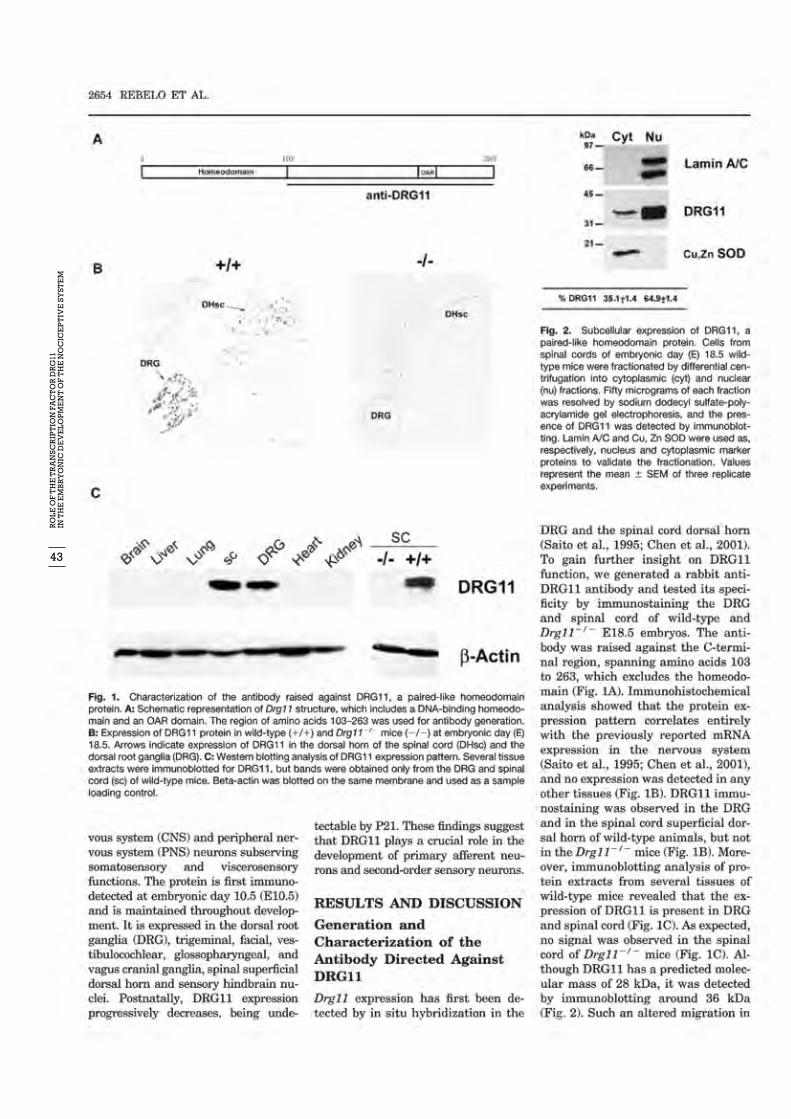
43
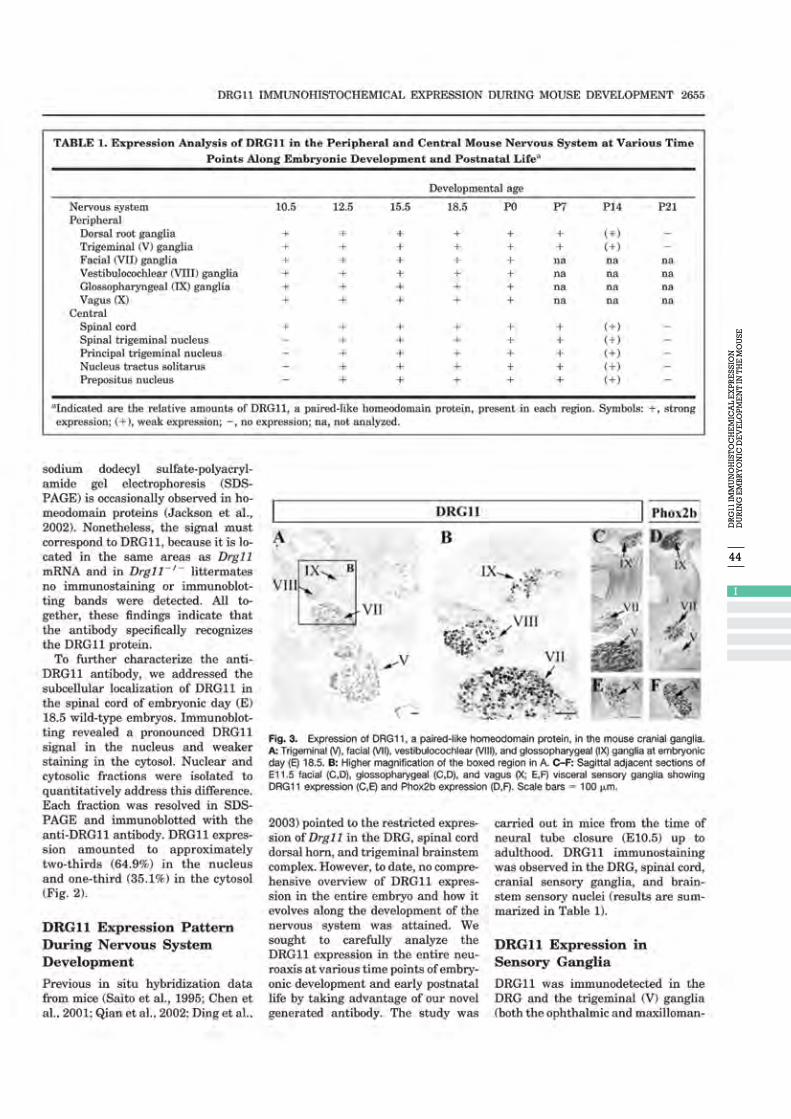
44
I
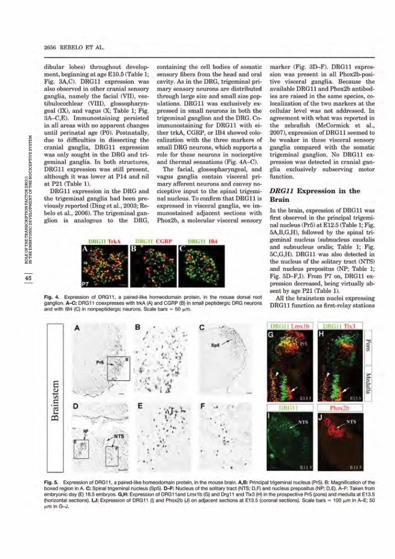
45
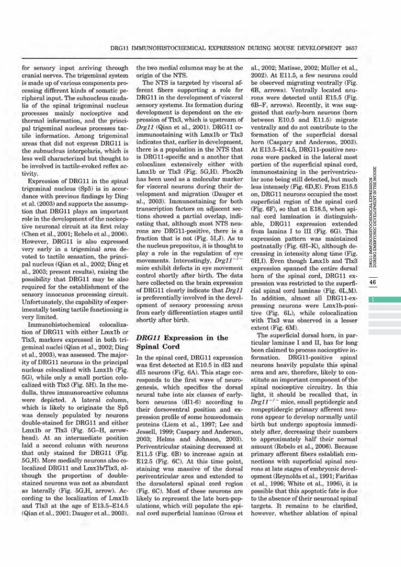
46
I

47
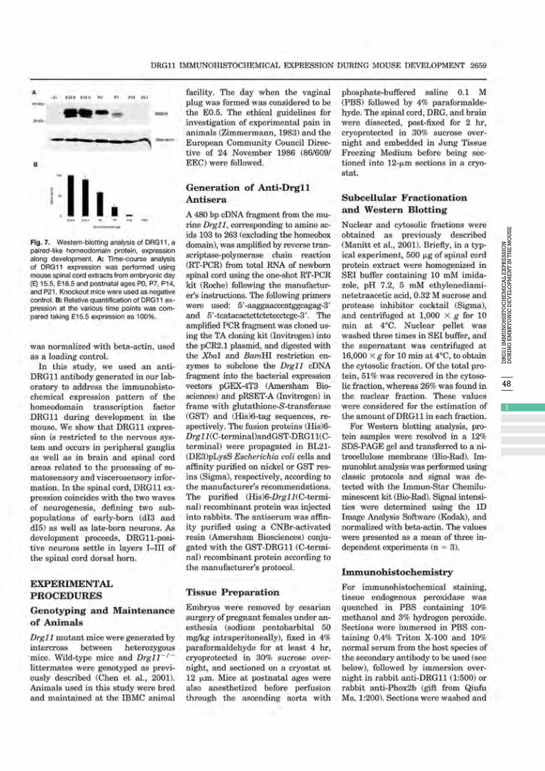
48
I
49


52
II
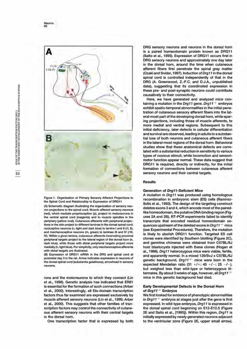
53
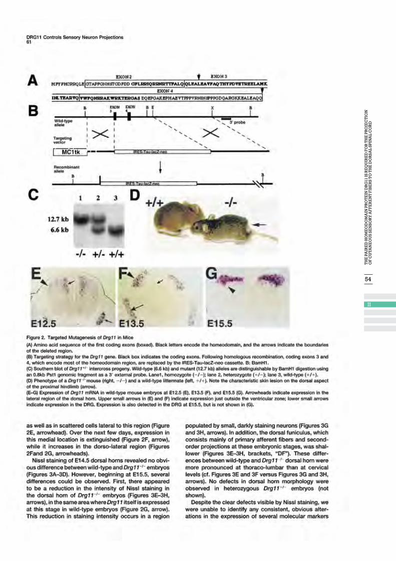
54
II
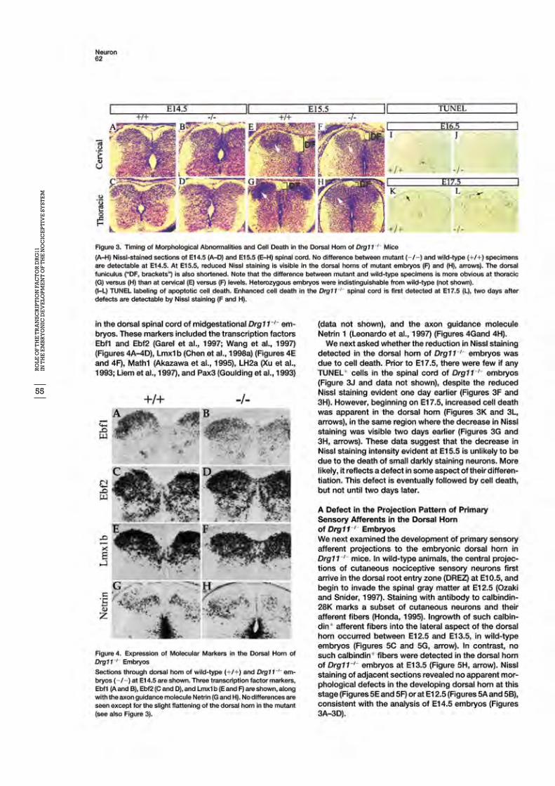
55
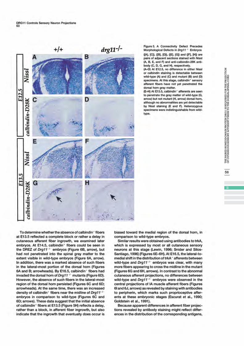
56
II
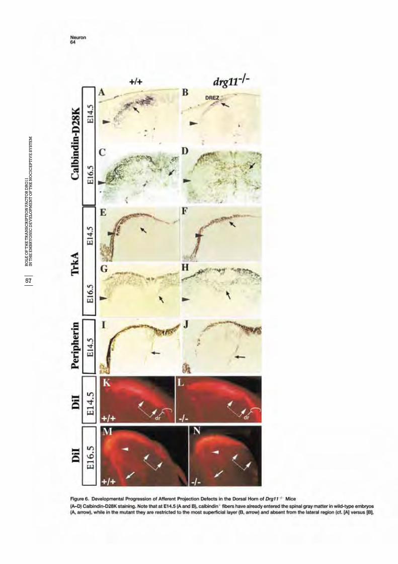
57
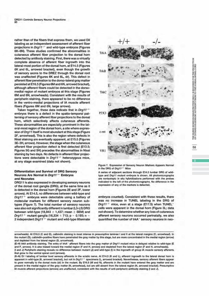
58
II

59
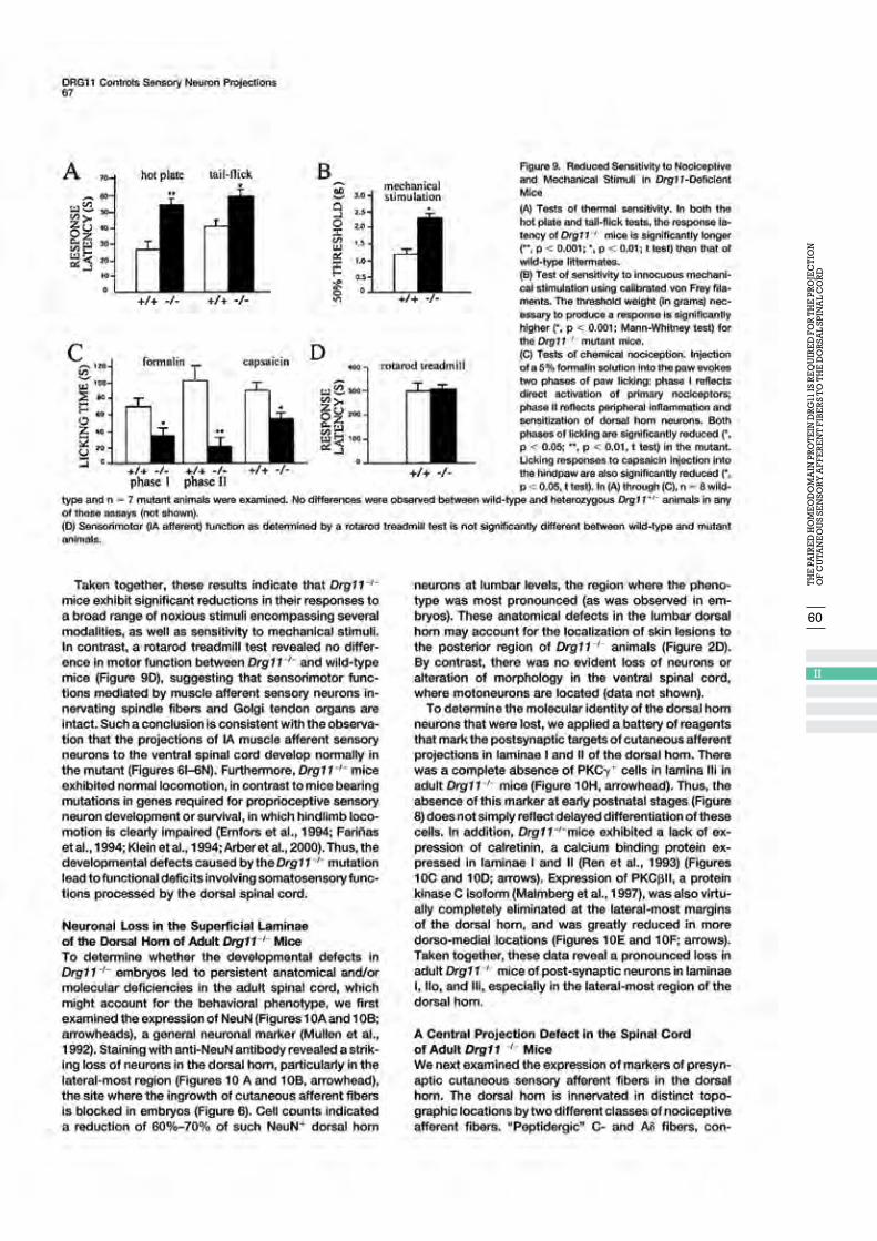
60
II
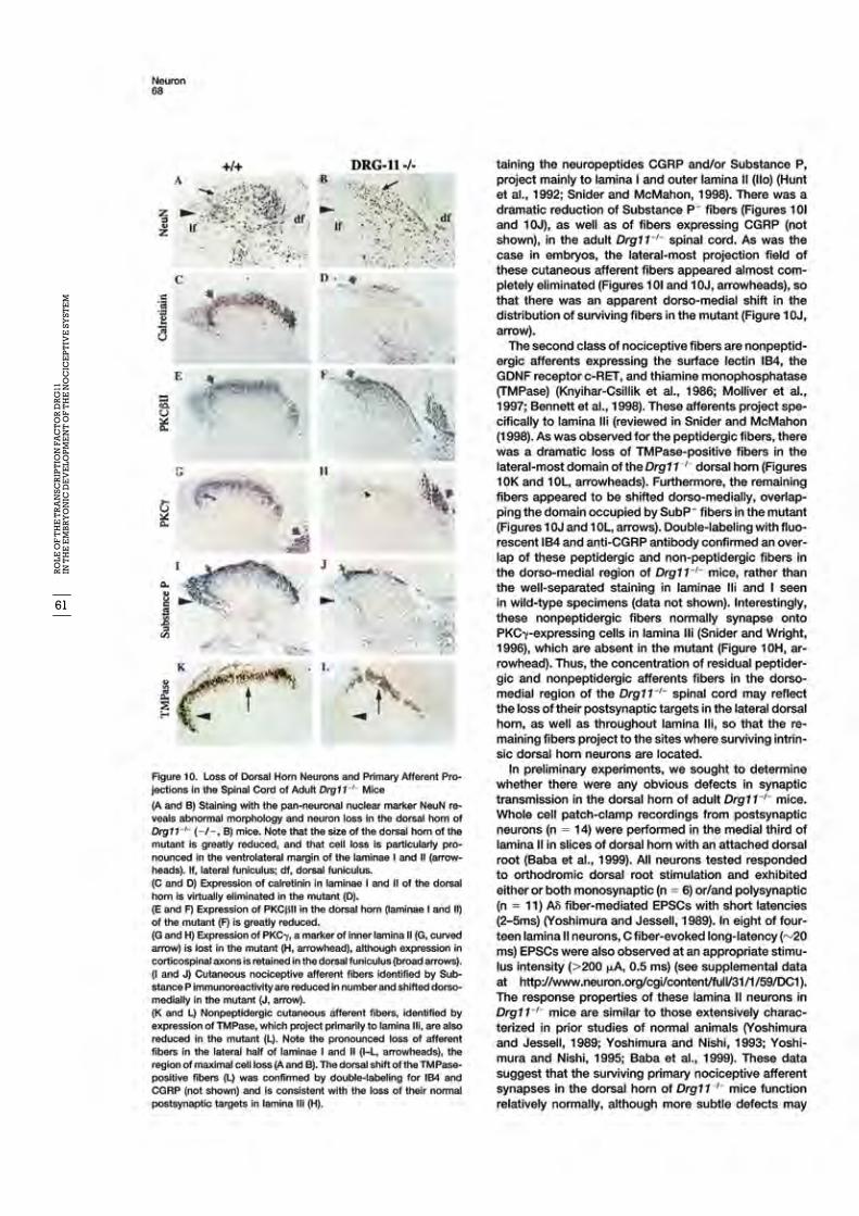
61
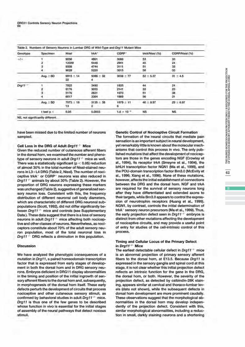
62
II
63

64
II
65
66
II

68
III
69
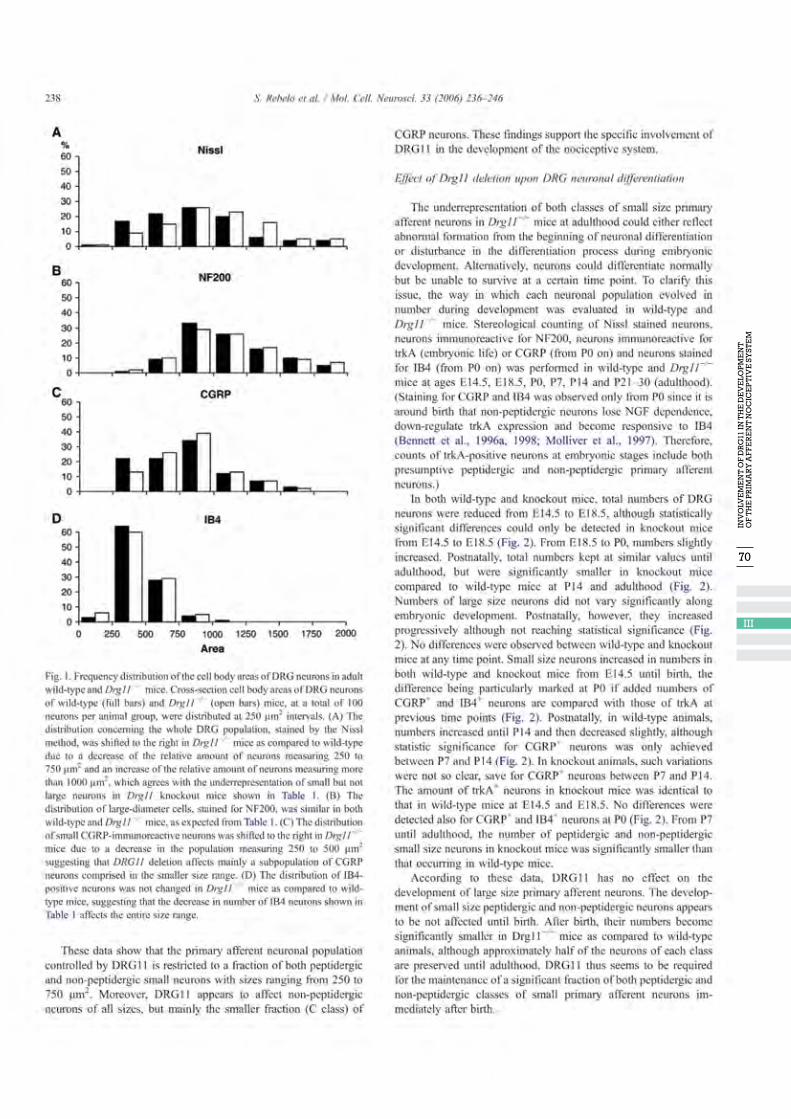
70
III
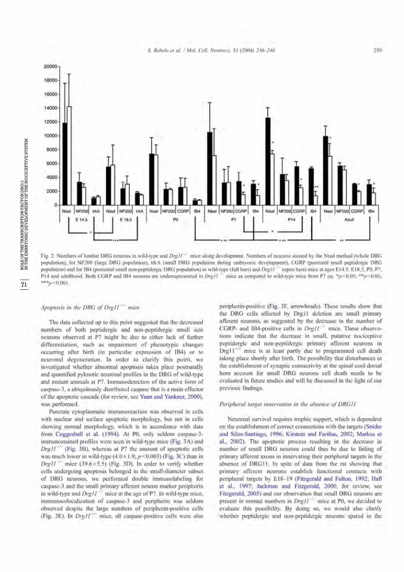
71
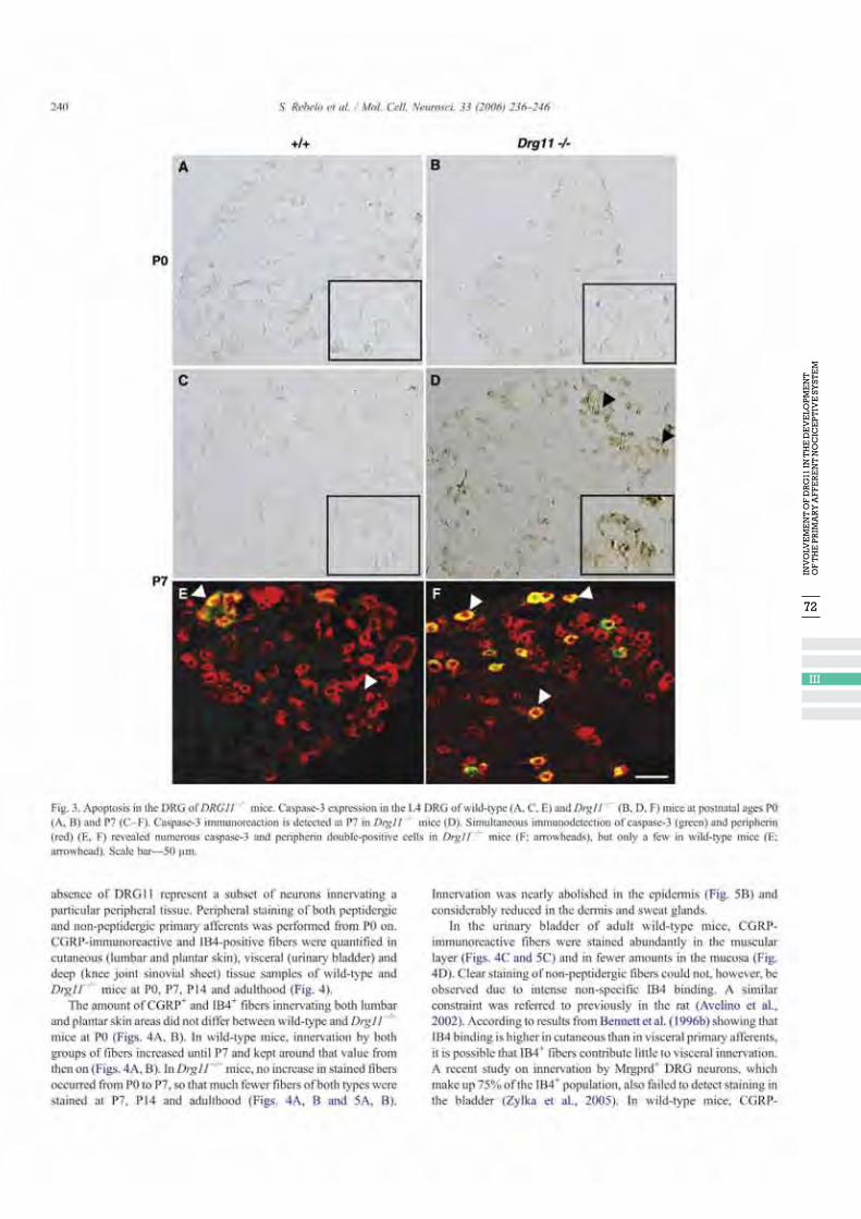
72
III
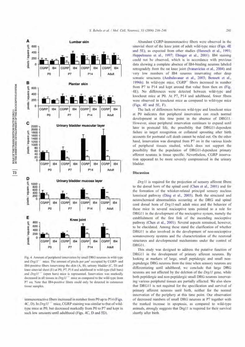
73

74
III
75
76
III
77
78
III

80
IV

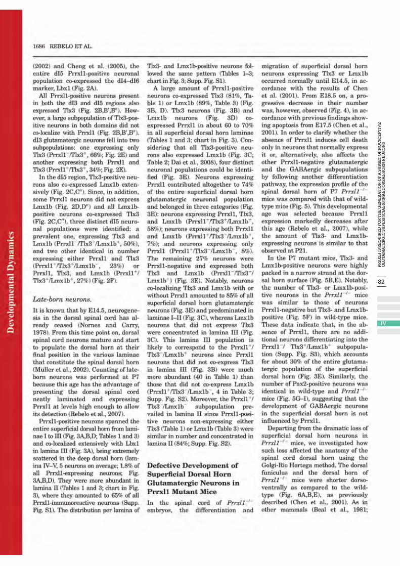
82
IV
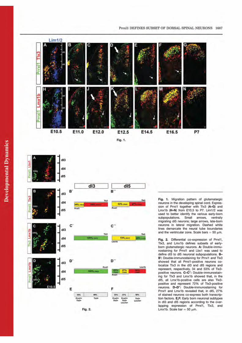

84
IV
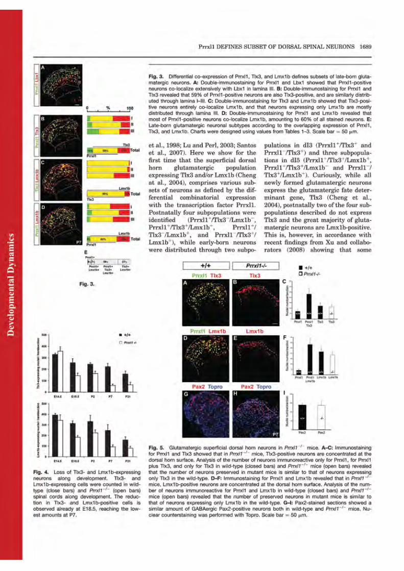
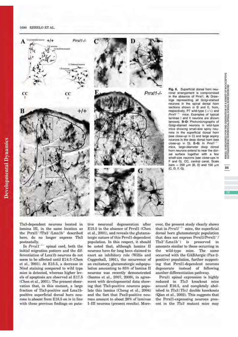
86
IV
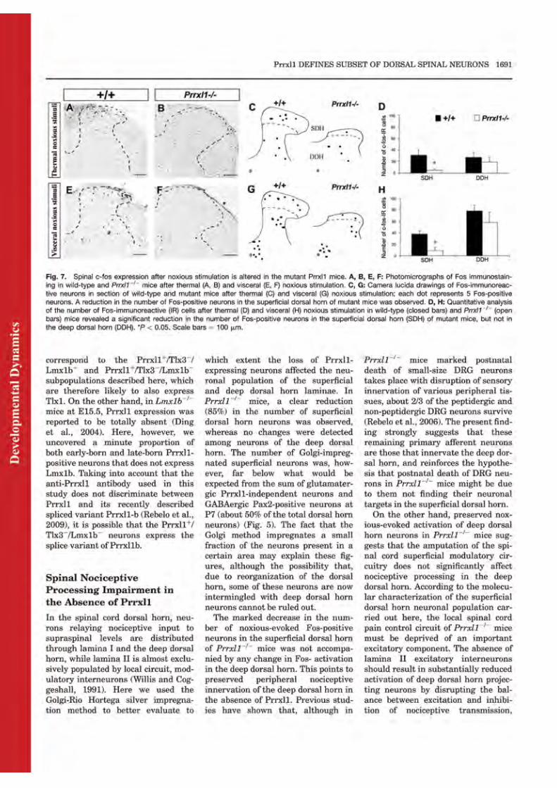
88
IV
90
IV

92
V
93
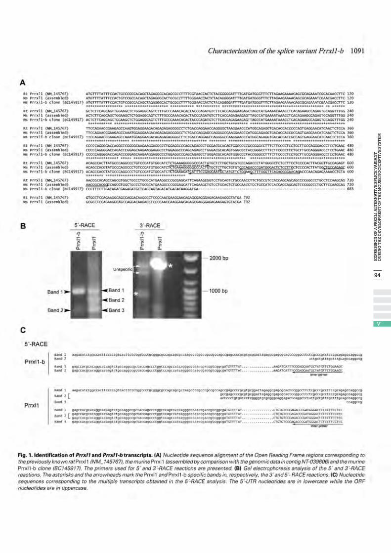
94
V
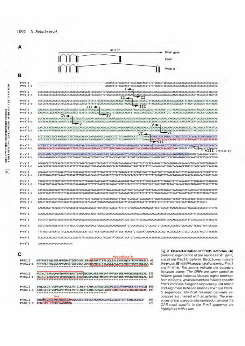
95
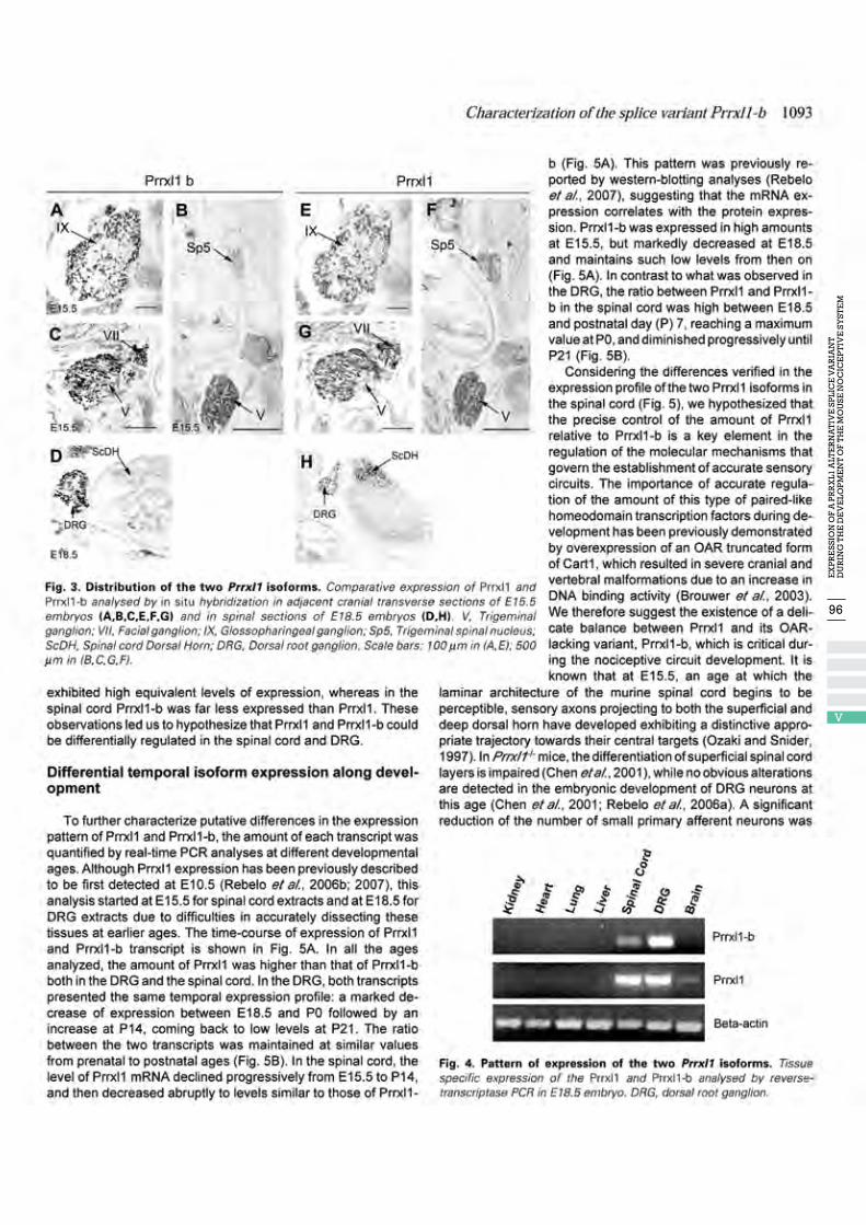
96
V
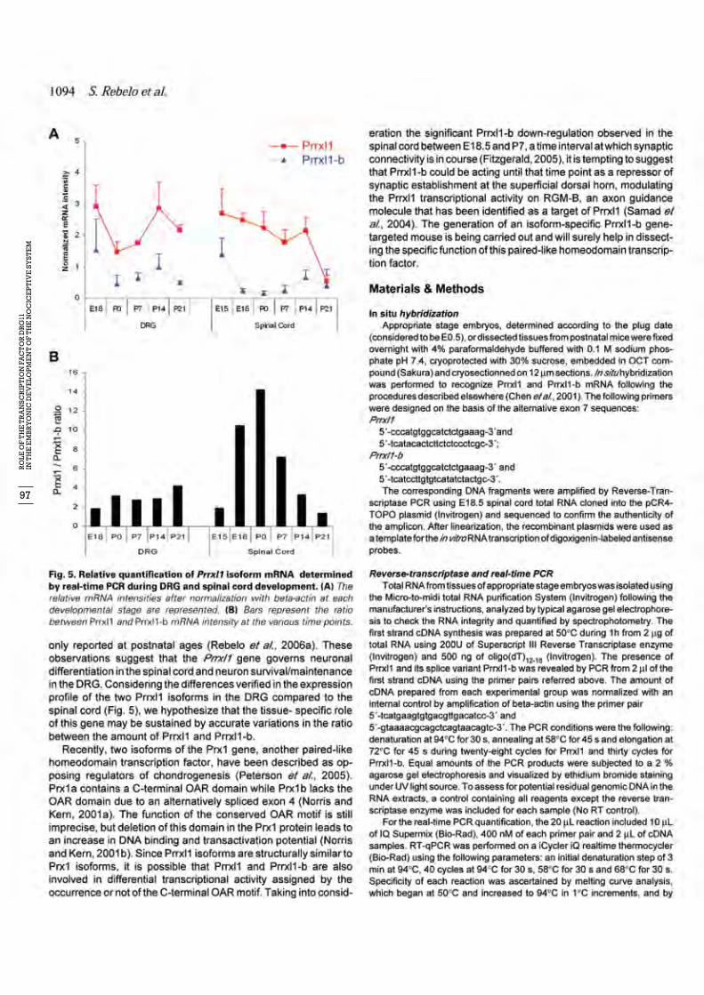
97
98
V
99


IIIDISCuSSION
This thesis uncovers Drg11 as the transcription factor responsible for the establishment of the first relay of the ascending nociceptive pathway. It shows that Drg11 plays a crucial role in the differen-tiation of various classes of superficial spinal dorsal horn neurons, but appears not to influence the normal embryonic development of small diameter, putative nociceptive DRG neurons. Such a differential role in the development of the first and second order nociceptive neurons is not ac-counted for by a Drg11 isoform, although the combinatorial expression of Drg11 and its isoform differs between the spinal cord and the DRG along development. Taken together, the results lead to the hypothesis that Drg11 directs the connection between primary afferents and second order nociceptive neurons by promoting the differentiation of the latter and signalling correct targeting for their primary afferent pathway. Throughout most of the thesis, a genetically modified mouse containing a deletion in the Drg11 gene (Drg11-/- or Drg11 knockout) was used.
DRG11 IS INVOLVED IN THE DEVELOPMENT OF THE NOCICEPTIVE SYSTEM
Drg11 is a paired-like homeodomain transcription factor first reported by Saito and collaborators (1995) to be expressed in the DRG and spinal cord dorsal horn, and later shown to be also present at the trigeminal complex (Ding et al., 2003). These findings were, however, based on RT-PCR and in situ hybridization approaches, and referred to isolated time points during either embryonic develo-pment or postnatal life. In this study, a novel antibody against Drg11 was produced, and its expres-sion along development and postnatal life was characterized at the spinal and supraspinal levels (Publication I). Drg11 was shown to be expressed throughout development from as early as E10.5 until shortly after birth in first and second order sensory structures along the entire neuroaxis. At the spinal level, Drg11 was expressed in laminae I-III of the superficial dorsal horn and in small diameter DRG neurons. At the supraspinal level, it was expressed in somatic and visceral sensory ganglia, namely the trigeminal, facial, vestibulocochlear, glossopharyngeal and vagus ganglia, and in the spinal trigeminal nucleus (Sp5) (subnucleus caudalis and oralis), principal trigeminal nu-cleus (Pr5), nucleus of the solitary tract (NTS) and nucleus prepositus (NP). No Drg11 expression was observed in cranial ganglia exclusively subserving motor function. Notably, both the location of Drg11 along the brain and the size and neurochemical signature of cranial and DRG primary afferent neurons agreed with the spinal superficial dorsal horn location as to a specific role for Drg11 in the development of the nociceptive system. However, Drg11 was also detected in a trigeminal area devoted to tactile sensation, the Pr5 (our own results; Qian et al., 2002; Ding et al., 2003), while at the spinal level a small fraction of Drg11-immunostained neurons was located in lamina III, the site of termination of Aδ D-hair follicle primary afferents (Light and Perl, 1979b; Willis et al., 2004). Hence, although the major sites of termination of tactile primary afferents did not express Drg11, the possibility of an involvement in the establishment of a particu-lar part of the sensory circuit processing innocuous input should be considered. In order to investigate whether Drg11 is required for the development of the primary nociceptive circuit, Drg11-/- mice were generated and characterized as to DRG and spinal cord morphological and neurochemical abnormalities, and nociceptive function was experimentally evaluated (Publication II). Drg11-/- mice exhibited a distorted spinal cord dorsal horn, with a reduction in the number of small dark Nissl stained neurons and in CGRP and TrkA immunostaining, and a complete absence of PKCγ staining. Additionally, an abnormal distribution of primary afferent fibers in the superficial dorsal horn was observed, with an apparent lateral-to-medial shift in their distribution. Such an aber-rant projection could be taken as indicative of a role for Drg11 in medio-lateral somatotopic organi-zation. However, the loss of PKCy (Publication II), Lmx1b and Tlx3 neurons (Publication IV) across the entire mediolateral extent, together with the almost complete amputation of the superficial dorsal horn, better revealed by the Golgi staining (Publication IV), is against this possibility.
103
As to nociceptive behaviour, Drg11-/- mice displayed higher response latencies in the hot plate, tail-flick and paw withdrawal tests for thermal sensitivity, and reduced withdrawal response to mecha-nical stimulation by von Frey filaments. In addition, they exhibited reduced responses to chemical nociceptive stimulation in both the capsaicin and formalin tests. These results indicate that, in the absence of Drg11, mice present reduced sensitivity to noxious stimuli across a broad range of mo-dalities, including mechano-, thermo-, and chemo-sensitivities. Although all the applied tests deal only with cutaneous nociception, it is very likely that similar nociceptive defects occur for visceral and deep tissue stimulation. Peripheral innervation of Drg11-/- mice was shown to be disrupted at postnatal ages in the three types of peripheral tissues (Publication III), while noxious evoked induction of the c-fos proto-oncogene was compromised at the superficial dorsal horn after stimu-lation of the three peripheral areas (Publication IV). Sensorimotor functions, mediated by muscle afferent sensory neurons innervating spindle fibers and Golgi tendon organs, were intact in the knockout mice, indicating that Drg11 is not required for the development of the proprioceptive system (Publication II). This is consistent with the ob-servation that no evident neuronal loss, morphological defects or abnormal central projections of IA muscle afferent fibers took place in the ventral spinal cord of these mice. It should be noted that disruption of proprioceptive functioning, with impaired hindlimb locomotion, is actually obser-ved in mice bearing mutations in genes required for proprioceptive sensory neuron development or survival (Ernfords et al., 1994; Fariñas et al., 1994; Klein et al., 1994; Arber et al., 2000).
DRG11 APPEARS NOT TO BE INVOLVED IN THE DIFFERENTIATION OF NOCICEPTIVE PRIMARY AFFERENT NEuRONS BuT IS REquIRED FOR THEIR POSTNATAL SuRVIVAL
Mouse embryos deficient in Drg11 exhibited abnormalities in the timing and position of the ini-tial ingrowth of primary afferent fiber projections to the spinal cord dorsal horn. Primary afferent fibers approached the spinal cord and entered the dorsal horn gray matter with a delay of 3 to 4 days, to then penetrate biased towards its medial region (Publication II). Nevertheless, the develo-pment of small size TrkA-positive, CGRP-positive (peptidergic) and IB4-positive (non-peptidergic) (Publication III) neurons was not affected until birth, ruling out a role for Drg11 in their diffe-rentiation. The data collected in publication III showed that, until neonatal age, the total numbers of sensory neurons did not differ, nor did the expression of markers for different primary afferent neuronal subtypes between wild-type and Drg11-/- mutant mice. Moreover, innervation of peri-pheral targets was preserved in Drg11-/- mice at P0, indicating that peripheral innervation can rea-ch normal development. Consistent with these results was the absence of TUNEL-positive cells in the DRG of Drg11-/- mice during embryonic development and at the neonatal stage (Publications II and III). Drg11 thus seems to be neither required for the generation, differentiation and survival of primary afferent neurons until birth, nor for the normal innervation of the various peripheral tissues at this time point. From P7 on (Publication III), there was a decrease to about half of the numbers of both pepti-dergic and non-peptidergic small-diameter primary afferent neurons in Drg11-/- mice. Accordin-gly, size-frequency distribution of DRG neurons led to the conclusion that neurons in the Aβ range were present in numbers similar to those occurring in wild type mice. However, it also revealed that small diameter neurons at the C fiber range were more affected than neurons at the Aδ fiber range. This finding, together with the fact that IB4 neurons comprise mostly C primary afferents (Zwick et al., 2002), implies that the larger fraction of the small diameter neurons preserved in the absence of Drg11 is Aδ peptidergic (CGRP). C and Aδ fiber neurons are known to convey nocicep-tive and innocuous thermal input from the periphery (McCarthy and Lawson, 1990; Fundin et al., 1997). Yet, innocuous thermal neurons belong mostly in the C unmyelinated group (for review, see
104

Willis and Coggeshall, 1991). Thus, being mostly Aδ, the non Drg11-dependent small size primary afferent neurons must mainly convey nociceptive input. Taking into account that the deep dorsal horn keeps its normal morphology in Drg11-/- mice whereas the superficial dorsal horn almost completely disappears (Publication IV), these neurons most likely make up the nociceptive inner-vation of lamina V (Light and Perl, 1979a,b; Cervero and Connell, 1984). It should be noted that, in the knockout mice, peripheral innervation was disrupted from P7 on in cutaneous, visceral and deep peripheral tissues, supporting that the population of Drg11-dependent primary afferent neurons is not tissue specific. However, visceral tissues were much more affected, which agrees with c-fibers making up the bulk of visceral sensory innervation (Cervero, 1985). Taken together, the data showed that Drg11 is required for the maintenance, immediately after birth, of a significant fraction of normally differentiated small-diameter, putative nociceptive pep-tidergic and non-peptidergic primary afferent neurons mainly belonging in the C-unmyelinated class and innervating all peripheral tissues. These neurons are likely to follow programmed cell death in the absence of Drg11, as indicated by an increase at P7 of the immunoreactivity for an active form of caspase-3, an ubiquitous caspase that is a main effector of the apoptotic cascade (for review, see Yuan and Yankner, 2000).
DRG11 COMMANDS THE DIFFERENTIATION OF NOCICEPTIVE SPINAL NEuRONS
The correct perception of noxious events relies on the activation of distinct sensory neurons spe-cifically organized in different laminae in the spinal cord dorsal horn. These neurons differentiate during development in a spatial-temporal order due to the expression of combinatorial sets of homeodomain transcription factors. Drg11-/- mice exhibit defects in the superficial dorsal horn similar to those observed in Tlx3/1 or Lmx1b knockout mice (Cheng et al., 2004, 2005; Ding et al., 2004), suggesting that the three transcription factors belong to a genetic cascade involved in buil-ding up the spinal cord superficial nociceptive circuit (Gross et al., 2002; Muller et al., 2002; Qian et al., 2002). The present thesis shows that various subpopulations of superficial dorsal horn neurons can be defined by the differential combination of Drg11, Tlx3 and Lmx1b (Publication IV). During em-bryonic development, Drg11-immunoreactivity was detected both in early-born (dI3 and dI5) and late-born glutamatergic (dILB) Tlx3/Lmx1b-positive neurons. All newly formed early-born Drg11-positive neurons expressed the glutamatergic fate determinant gene, Tlx3 (Cheng et al., 2004; our own results). Postnatally, four subpopulations were identified. Although the majority (85%) ex-pressed both Tlx3 and Lmx1b with (58%) or without (27%) Drg11, a small fraction (15%) did not express Tlx3. This is in line with the observation by Xu and collaborators (2008) of some lamina III neurons Tlx3-dependent during development that did not express Tlx3 after birth. Half (7%) of the Tlx3-negative neurons only expressed Drg11. Drg11-positive neurons spanned the entire super-ficial dorsal horn from laminae I to III, although prevailing in lamina II (65%). The subpopulation that expressed Lmx1b but not Tlx3 (7%) was however located in lamina III. In the Drg11-/- spinal cord, the majority of Tlx3- and Lmx1b-positive neurons was absent from E18.5 on. This observation agrees with the detection of abnormal cell death at E17.5 (Publication II) and further reveals the glutamatergic nature of Drg11-/- dependent neurons. However, it is wor-th noting that there is a significant fraction of glutamatergic neurons that are not Drg11-dependent, which is preserved in the Drg11-/- mice in amounts similar to those present in wild-type mice. The same is true for the GABAergic, Pax2-positive population. Both findings support the assumption that Drg11-dependent neurons degenerate instead of following another differentiation pathway. The non Drg11-dependent superficial dorsal horn neurons were confined to a narrow strand, which could be delineated in spinal slices silver impregnated by the Golgi-Rio Hortega method (Publication IV). In these preparations, a clear reduction in the number of small size, spiny super-
105
ficial dorsal horn neurons was observed, which was more marked than would be expected from the sum of glutamatergic non Drg11-dependent neurons and GABAergic, Pax2-dependent neurons. The deep dorsal horn did not exhibit any changes nor presented small spiny neurons, which is against the possibility that, due to reorganization of the dorsal horn, superficial dorsal horn neu-rons were relocated in the deep dorsal horn. That discrepancy in numbers is more probably due to the fact that spinal neurons stained by the Golgi method represent a very small fraction of the entire neuronal population. There was also a marked reduction in the numbers of Fos-positive neurons in the superficial dorsal horn following noxious stimulation (Publication IV). In the deep dorsal horn, c-fos induc-tion was identical to that observed in wild-type mice. This finding, while in line with the normal development of the deep dorsal horn, raises important questions as to the role of superficial gluta-matergic local circuit neurons in the nociceptive activation of deep dorsal horn neurons. Although the spared - mainly Aδ - primary afferent neurons can account for the activation of the deep dorsal horn, the spinal cord pain modulatory circuitry of Drg11-/- mice must be deprived of an impor-tant excitatory component. This should result on an imbalance favouring inhibition of nociceptive transmission, and consequently on the decrease of noxious-evoked c-fos activation in the deep dorsal horn. In this respect, it is worth noting that about 85% of lamina II local circuit neurons are glutamatergic (Santos et al., 2007, 2009), against 20% GABAergic neurons, as revealed by the present work. It is possible that the spinal and supraspinal pain control systems have adapted along development to re-establish the lost balance. On the other hand, in spite of normal nociceptive activation in the deep dorsal horn, nociceptive behaviour is seriously affected (Publication II), which poses interesting questions regarding the relative role of the lamina I and deep dorsal horn nociceptive ascending systems in pain processing (for review, see Lima, 2008).
THE RELATIVE CONCENTRATION OF DRG11 AND ITS SPLICE VARIANT ALONG DEVELOPMENT MAY CONTRIBuTE TO ITS DIFFERENTIAL ROLE IN THE DRG AND SPINAL CORD
Gene expression can be differentially regulated by splice variants, providing a mechanism for pre-cise control of diverse morphogenetic events (Grabowski and Black, 2001; Fagnani et al., 2007; Li et al., 2007; Irimia et al., 2009). Here, we have characterized a Drg11 alternative splice variant (also known as Prrxl1-b), which lacks the OAR domain (Publication V). To date, the function of the OAR domain is not properly known but it is believed to have a molecular function directly rela-ted to the transcriptional activity of the paired-like homeodomain proteins (Simeone et al. 1994; Galliot et al., 1999; Meijlink et al., 1999; Norris and Kerne, 2001). The Drg11 splice isoform presented the same regional distribution pattern along the entire neu-roaxis as Drg11, but differed as to its relative quantitative expression profile in the DRG and spinal cord at distinct developmental ages. The amount of Drg11 was higher than that of its splice variant at both sites. However, in the DRG the two isoforms exhibited relatively high levels of expression, with the same temporal profile and a similar ratio from prenatal to postnatal ages. In the spinal cord, the expression of the Drg11 splice variant was practically nil from E18.5 on, with a striking high ratio between the two isoforms, particularly at P0. These observations suggest that the two isoforms are differentially regulated in the DRG and spinal cord, and raise the hypothesis that tissue-specific control of the amount of Drg11 relative to its splice variant is a key factor for the re-gulation of the molecular mechanisms that govern the development of the nociceptive circuit, and may contribute to the differential role of this transcription factor in the two regions (see below). Considering the marked down-regulation of the Drg11 splice variant in the spinal cord between E18.5 and P7, a time interval that encompasses the embryonic age at which synaptic connectivity is
106

occurring (Fitzgerald, 2005), it is possible that it acts as a repressor of synaptic organization at the superficial dorsal horn.
THE ROLE OF DRG11 IN THE DEVELOPMENT OF THE PRIMARY AFFERENT - SPINAL NOCICEPTIVE CIRCuIT
The work developed in this thesis departed from the hypothesis, raised by the study of Saito and collaborators (1995), that Drg11 should be important for the normal differentiation and synaptic connection of primary afferent and spinal cord nociceptive neurons. The loss-of-function studies performed allowed us to conclude that indeed Drg11 is crucial for the formation of a major compo-nent of superficial dorsal horn glutamatergic neurons (Publication IV), but apparently irrelevant for the normal development of primary afferent nociceptive neurons (Publication III). Peripheral targeting of primary afferent neurons also developed normally in the absence of Drg11 (Publica-tion III), whereas spinal targeting appeared to be disrupted, since central projections had difficulty in entering the spinal grey and finding their proper termination area (Publication II). Normally, primary afferent fibers arrive at the dorsal root entry zone by E10.5 and begin to invade the spinal gray matter at E12.5 (Ozaki and Snider, 1997). In the absence of Drg11, primary afferent arrival at the entry zone and penetration into the spinal gray was delayed by 3 to 4 days, and their distri-bution in the superficial dorsal horn somehow disrupted, conforming the abnormal dorsal horn morphology installed at this age (Publication II). Later, shortly after birth, these primary afferent neurons, which until then were phenotypically normal, underwent apoptosis and their numbers were markedly reduced, with parallel impairment of peripheral innervation (Publication III). These data strongly support the hypothesis that primary afferent neurons died due to the fact that they did not find their target neurons in the dorsal horn. It is known that neuronal survival requires trophic support, which depends on the establishment of correct connections with the targets (Snider and Silos-Santiago, 1996; Kirstein and Fariñas, 2002; Markus et al., 2002). In the rat, primary afferent neurons establish functional contacts with spinal neurons and peripheral tissues next to birth (Fitzgerald and Fukton, 1992; Hall et al., 1997; Jackman and Fiztgerald, 2000; Fitzgerald, 2005). At neonatal age, peripheral innervation was apparently normal in the absence of Drg11, which leaves the observed loss of 2/3 of glutamatergic superficial dorsal horn neurons as the putative cause for target deprivation-induced primary afferent neuronal death. It is worth noting that, besides the sealed 1/3 glutamatergic neurons, superficial GABAergic neurons and deep dorsal horn nociceptive neurons were also preserved and may have accounted for the conservation of 2/3 of thin primary afferent peptidergic and non-peptidergic neurons in the knockout mice. However, although following normal differentiation (Publication III), and in spite of normal neurogenesis at the superficial dorsal horn until E14.5 (Publication IV), in the absence of Drg11, primary afferent neurons had trouble in finding their way to the spinal cord at ages as early as E10.5, which points to a role for this transcription factor in very early stages of guiding primary axons to their spinal targets. It is known that, during development, DRG neurons extend their axons toward the dorsolateral part of the spinal cord, enter the spinal cord at the dorsal root entry zone and then grow longitudinally inside the spinal cord to form the dorsal funiculus without penetrating the dorsal mantle layer (for review see, Masuda and Shiga, 2005). Only after a ‘waiting period’ of a few days do these axons project into the dorsal mantle layer in a ventral to dorsal order. Proprioceptive afferents are the first to send collaterals ventrally, followed by large-caliber sensory afferent and at last fine calibre nociceptive and thermoreceptive afferents (Ozaki and Snider, 1997). Inhibitory cues transiently expressed, such as Sema3a (Messersmith et al., 1995; Shepherd et al., 1997; Fu et al., 2000; Masuda et al., 2003) and Netrin-1 (Watanabe et al., 2006; Masuda et al., 2008; Masuda et al., 2009) are apparently required for the correct patterning of sensory afferents during this waiting period (Ozaki and Snider, 1997). Furthermore, there is increasing evidence that trans-
107
cription factors and cell surface molecules, such as Runx3, Er81, Pea3 and F11, are involved in the correct projection of proprioceptive DRG axons (Arber et al., 2000; Perrin et al., 2001; Inoue et al., 2002; Livet et al., 2002; Chen et al., 2006), supporting evidence that several molecules orchestrate in order to elaborate the waiting period for sensory afferents. Appropriate neuronal migration is a prerequisite for the normal projection of primary afferents to the developing spinal cord. Ding and collaborators (2005) have shown that migration of early-born neurons is essential for the central projection of primary afferents, since they repel nociceptive and chemoattract proprioceptive affe-rents probably through the activity of Sema3a. To our best knowledge, Drg11 is the sole transcription factor that was shown to be present in migrating superficial nociceptive neurons, to account for their normal differentiation and to be involved in the correct projection of nociceptive afferents into the spinal cord (present thesis). It is however very probable that other transcription factors contribute to this process. A careful analysis of the genetic program under the control of Drg11 is being carried out in our laboratory in order to identify new players that, together with Drg11, govern the establishment of the first relay of the ascending nociceptive system.In summary, the data collected in this thesis suggest that Drg11 plays a double role in the formation of the DRG-spinal nociceptive circuit. It is involved in the differentiation of a major subpopulation of excitatory nociceptive superficial dorsal horn neurons in one hand, while apparently comman-ding the guiding and synaptic connectivity of their primary afferent pathway, on the other. Future studies using conditional Drg11 mutations should be conducted to test this hypothesis.
108
REFERENCES
Arber S, Ladle DR, Lin JH, Frank E, Jessell TM (2000). ETS gene Er81 controls the formation of functional connections between group Ia sensory afferents and motor neurons. Cell 101: 485-498. Cervero F (1985). Visceral nociception: peripheral and central aspects of visceral nociceptive systems. Philos. Trans. R. Soc. Lond. B. Biol. Sci. 308: 325-337.Cervero F, Connell LA (1984). Fine afferent fibers from viscera do not terminate in the substancia gelatinosa of the thoracic spinal cord. Brain Res. 294: 370-374.Chen AI, de Nooij JC, Jessell TM (2006). Graded activity of transcription factor Runx3 specifies the laminar termination pattern of sensory axons in the developing spinal cord. Neuron 49: 395-408.Cheng L, Arata A, Mizuguchi R, Qian Y, Karunaratne A, Gray PA, Arata S, Shirasawa S, Bouchard M, Luo P, Chen C-L, Busslinger M, Goulding M, Onimaru H, Ma Q (2004). Tlx3 and Tlx1 are post-mitotic selector genes determining glutamatergic over GABAergic cell fates. Nat Neurosci 7: 510-517.Cheng L, Samad OA, Xu Y, Mizuguchi R, Luo P, Shirasawa S, Goulding M, Ma Q (2005). Lbx1 and Tlx3 are opposing switches in deter-mining GABAergic versus glutamatergic transmitter phenotypes. Nat Neurosci 8: 1510-1515.Ding YQ, Yin J, Xu HM, Jacquim MF, Chen ZF (2003). Formation of whisker related principal sensory nucleus-based lemniscal pathway requires a paired homeodomain transcription factor, Drg11. J. Neurosci. 23: 7246–7254.Ding YQ, Yin J, Kania A, Zhao ZQ, Johnson RL, Chen ZF (2004). Lmx1b controls the differentiation and migration of the superficial dorsal horn neurons of the spinal cord. Development 131: 3693-3703. Ding YQ, Kim JY, Xu YS, Rao Y, Chen ZF (2005). Ventral migration of early-born neurons requires Dcc and is essencial for the projec-tions of primary afferents in the spinal cord. Development 132: 2047-2056.Ernfors P, Lee KF, Kucera J, Jaenisch R (1994). Lack of neurotrophin-3 leads to deficiencies in the peripheral nervous system and loss of limb proprioceptive afferents. Cell 77: 503-512.Fagnani M, Barash Y, Ip JY, Misquitta C, Pan Q, Saltzman AL, Shai O, Lee L, Rozenhek A, Mohammad N, Willaime-Morawek S, Babak T, Zhang W, Hughes TR, van der Kooy D, Frey BJ, Blencowe BJ (2007). Functional coordination of alternative splicing in the mamma-lian central nervous system. Genome Biol. 8: R108. Fariñas I, Jones KR, Backus C, Wang XY, Reichardt LF (1994). Severe sensory and sympathetic deficits in mice lacking neurotrophin-3. Nature 369: 658-661.Fitzgerald M, Fulton BP (1992). The physiological properties of developing sensory neurons. In: Scott, S.A., (Ed.), Sensory Neurons: diversity, development and plasticity. Oxford University Press, NY, pp. 287-306.Fitzgerald M (2005). The development of nociceptive circuits. Nat. Rev. Neurosci. 6: 507-520.Fu SY, Sharma K, Luo Y, Raper JA, Frank E (2000). SEMA3A regulates developing sensory projections in the chicken spinal cord. J. Neurobiol. 45: 227-236.Fundin BT, Arvidsson J, Aldskogius H, Johansson O, Rice SN, Rice FL (1997). Comprehensive immunofluorescence and lectin binding analysis of intervibrissal fur innervation in the mystacial pad of the rat. J. Comp. Neurol. 385: 185-206.Galliot B, de Vargas C, Miller D (1999). Evolution of homeobox genes: Q50 Paired-like genes founded the Paired class. Dev. Genes Evol. 209: 186-197.Grabowski PJ, Black DL (2001). Alternative RNA splicing in the nervous system. Prog. Neurobiol. 65: 289-308.Gross MK, Dottori M, Goulding M (2002). Lbx1 specifies somatosensory association interneurons in the dorsal spinal cord. Neuron 34: 535-549.Hall AK, Ai X, Hickman GE, MacPhedran SE, Nduaguba CO, Robertson CP (1997). The generation of neuronal heterogeneity in a rat sensory ganglia. J. Neurosci. 17: 2775-2784.Inoue K, Ozaki S, Shiga T, Ito K, Masuda T, Okado N, Iseda T, Kawaguchi S, Ogawa M, Bae SC, Yamashita N, Itohara S, Kudo N, Ito Y (2002). Runx3 controls the axonal projection of proprioceptive dorsal root ganglion neurons. Nat. Neurosci. 5: 946-954.Irimia M, Rukov JL, Roy SW, Vinther J, Garcia-Fernandez J (2009). Quantitative regulation of alternative splicing in evolution and development. Bioessays 31: 40-50. Jackman A, Fitzgerald M (2000). Development of peripheral hindlimb and central spinal cord innervation by subpopulations of dorsal root ganglion cells in the embryonic rat. J. Comp. Neurol. 418: 281-298.Klein R, Silos-Santiago I, Smeyne RJ, Lira SA, Brambilla R,Bryant S, Zhang L, Snider WD, Barbacid M (1994). Disruption of the neuro-trophin-3 receptor gene trkC eliminates Ia muscle afferents and results in abnormal movements. Nature 368: 249-251.Kirstein M, Fariñas I (2002). Sensing life: regulation of sensory neuron survival by neurotrophins. Cell. Mol. Life Sci. 59: 1787-1802.Li Q, Lee JA, Black DL (2007). Neuronal regulation of alternative pre-mRNA splicing. Nat. Rev. Neurosci. 8: 819-831.Light AR, Perl ER (1979a). Reexamination of the dorsal root projection to the spinal dorsal horn including observation on the differen-tial termination of coarse and fine fibers. J. Comp. Neurol. 186: 117-132.Light AR, Perl ER (1979b). Spinal termination of functionally identified primary afferent neurons with slowly conducting myelinated fibers. J. Comp. Neurol. 186:133-150.Lima D (2008). Ascending pathways: Anatomy and Physiology. In The Senses: A comprehensive reference. Basbaum, A. et al., editors. pp.477-526, Elsevier, LondonLivet J, Sigrist M, Stroebel S, De Paola V, Price SR, Henderson CR, Jessell TM, Arber S (2002). ETS gene Pea3 controls the central posi-tion and terminal arborisation of specific motor neuron pools. Neuron 35: 877-892.Markus A, Patel TD, Snider WD (2002). Neurotrophic factors and axonal growth. Curr. Opin. Neurobiol. 12: 523-531.Masuda T, Shiga T (2005). Chemorepulsion and cell adhesion molecules in patterning initial trajectories of sensory axons. Neurosci. Res. 51:337-347.Masuda T, Tsuji H, Taniguchi M, Yagi T, Tessier-Lavigne M, Fujisawa H, Okado N, Shiga T (2003). Differential non-target-derived re-pulsive signals play a critical role in shaping initial axonal growth of dorsal root ganglion neurons. Dev. Biol. 254: 289-302. Masuda T, Watanabe K, Sakuma C, Ikenaka K, Ono K, Yaginuma H (2008). Netrin-1 acts as a repulsive guidance cue for sensory axonal projections towards the spinal cord. J. Neurosci. 28: 10380-10385.Masuda T, Yaginuma H, Sakuma C, Ono K (2009). Netrin-1 signaling for sensory axons. Cell Adhesion and Migration 3:171-173.McCarthy PW, Lawson SN (1990). Cell type and conduction velocity of rat primary sensory neurons with calcitonin gene related peptide-like immunoreactivity. Neuroscience, 34: 623-632.Meijlink F, Beverdam A, Brouwer A, Oosterveen TC, Berge DT (1999). Vertebrate aristaless-related genes. Int. J. Dev. Biol. 43: 651-663.
109
Messersmith EK, Leonardo ED, Shatz CJ, Tessier-Lavigne M, Goodman CS, Kolodkin AL (1995). Semaphorin III can function as a selective chemorepellent to pattern sensory projections in the spinal cord. Neuron 14: 949-959.Müller T, Brohmann H, Pierani A, Heppenstall PA, Lewin GR, Jessell TM, Birchmeier C (2002). The homeodomain factor Lbx1 distin-guishes two major programs of neuronal differentiation in the dorsal spinal cord. Neuron 34: 551-562.Norris RA, Kern MJ (2001). Identification of domains mediating transcription, repression, and inhibition in the paired-related homeo-box protein, Prx2 (S8). DNA Cell Biol. 20: 89-99.Ozaki S, Snider WD (1997). Initial trajectories of sensory axons toward laminar targets in the developing mouse spinal cord. J. Comp. Neurol. 380: 215-229. Perrin FE, Rathjen FG, Stoeckli ET (2001). Distinct subpopulations of sensory afferents require F11 or axonin-1 for growth to their target layers within the spinal cord of the chick. Neuron 30: 707-723. Qian Y, Shirasawa S, Chen C-L, Cheng L, Ma Q (2002). Proper development of relay somatic sensory neurons and D2/D4 interneurons requires homeobox genes Rnx/Tlx-3 and Tlx-1. Genes Dev 16: 1220-1233.Saito T, Greenwood A, Sun Q, Anderson DJ (1995). Identification by differential RT-PCR of a novel paired homeodomain protein spe-cifically expressed in sensory neurons and a subset of their CNS targets. Mol. Cell. Neurosci. 6: 280-292.Santos SF, Rebelo S, Derkach VA, Safronov BV (2007). Excitatory interneurons dominate sensory processing in the spinal substantia gelatinosa of rat. J Physiol. 581:241-254.Santos SF, Luz LL, Szucs P, Lima D, Derkach VA, Safronov BV (2009). Transmission efficacy and plasticity in glutamatergic synapses formed by excitatory interneurons of the substantia gelatinosa in the rat spinal cord. PLos One 4: e8047.Shepherd IT, Luo Y, Lefcort F, Reichardt LF, Raper JA (1997). A sensory axon repellent secreted from ventral spinal cord explants is neutralized by antibodies raised against collapsing-1. Development 124: 1377-1385. Snider WD, Silos-Santiago I (1996). Dorsal root ganglion neurons require functional neurotrophin receptors for survival during deve-lopment. Philos. Trans. R. Soc. Lond. B 351: 395-403.Simeone A, D’Apice MR, Nigro V, Casanova J, Graziani F, Acampora D, Avantaggiato V (1994). Orthopedia, a novel homeobox-contai-ning gene expressed in the developing CNS of both mouse and Drosophila. Neuron 13: 83-101.Watanabe K, Tamamaki N, Furuta T, Ackerman S, Ikenaka K, Ono K (2006). Dorsally derived netrin 1 provides an inhibitory cue and elaborates the ‘waiting period’ for primary sensory axons in the developing spinal cord. Development 133: 1379-1387.Willis WD, Coggeshall RE (1991). Sensory mechanisms of the spinal cord. Plenum Press, NY and London.Willis, WD, Westlund, KN, Carlton, SM (2004). Pain system. In: The rat nervous system, Third edition, Edited by Paxinos. Chapter 27:853-890.Xu Y, Lopes C, Qian Y, Liu Y, Cheng L, Goulding M, Turner EE, Lima D, Ma Q (2008). Tlx1 and Tlx3 coordinate specification of dorsal horn pain-modulatory peptidergic neurons. J Neurosci. 28: 4037-46.Yuan J, Yankner BA (2000). Apoptosis in the nervous system. Nature 407: 802-809.Zwick M, Davis BM, Woodbury J, Burkett JN, Koerber HR, Simpson JF, Albers KM (2002). Glial cell line-derived neurotrophic factor is a survival factor for isolectin B4-positive, but not vanilloid receptor 1-positive, neurons in the mouse. J. Neurosci. 22: 4057-4065.
110


IVSuMMARY AND CONCLuSIONS

In this work, we investigated the putative involvement of Drg11 in the development of the nociceptive system, in particular at the DRG-spinal level, as suggested by the study by Saito and Collaborators (1995) showing that this transcription factor is expressed in the DRG and spinal superficial dorsal horn. Through the manufacture of a polyclonal antibody against DGR11, we were able to demonstrate that Drg11 is expressed throughout embryonic development, from as early as the embryonic age 10.5 (E10.5) until early postnatal age, in first and second order sensory structures along the entire neuro-axis (Publication I). At the spinal level, DRG11 immunostainig was observed in small diameter DRG neurons and the superficial dorsal horn, while at the supraspinal level, it was observed in several cra-nial sensory ganglia and the respective relay nuclei in the brainstem. This distribution pointed out a putative role for DRG11 in the formation of the first link of the ascending sensory pathway, not only at the spinal level but also along the brain. In order to ascertain whether DRG11 specifically commands the development of the nociceptive system, we underwent the phenotypic characterization of a Drg11 knockout mice (Drg11-/-) (Publica-tion II). Drg11-/- mice exhibited reduced reflex responses to mechanical, thermal and chemical pain-ful stimuli, together with anatomical and neurochemical abnormalities in the superficial dorsal horn and misdistribution of their primary afferents, which entered the spinal gray with a marked delay. We then addressed the involvement of Drg11 in the development of primary afferent nociceptive neurons (Publication III) and observed that, in mice deprived of Drg11, primary afferent neurons follow normal differentiation and project normally to their peripheral targets until neonatal age. Shortly after birth, however, about 1/3 both of peptidergic and non-peptidergic, putative nociceptive neurons followed apoptosis, which paralleled marked impairment of peripheral innervation of the skin, viscera and deep tissues. These data indicate that Drg11 is not required for the normal diffe-rentiation of small diameter, putative nociceptive primary afferents, but essential for the survival of a significant fraction of those neurons immediately after birth. The analysis of the differentiation of spinal cord dorsal horn neurons in wild type and Drg11-/- mice followed (Publication IV). Drg11 was shown to be required for the differentiation, after E 14.5, of a subset, amounting to 73%, of nociceptive glutamatergic superficial dorsal horn neurons, which could be subdivided in 3 different categories expressing either (i) Drg11, Tlx3 and Lmx1b, (ii) Drg11 and Lmx1b, or (iii) Drg11 alone. Moreover, Golgi studies confirmed the absence, in Drg11-/-- mice, of a large amount of small, spiny neurons in the spinal superficial cord dorsal horn, while c-fos induc-tion studies revealed defective noxious-evoked activation at the superficial but not the deep dorsal horn. Besides demonstrating that Drg11 is required for the proper development of a major fraction of glutamatergic superficial dorsal horn neurons, these data indicate that the lack of this excitatory, mostly local circuit neuronal population does not result in decreased nociceptive activation at the deep dorsal horn, which, together with the depressed nociceptive behavior observed in these animals (Publication II), underlies the importance of the spinal superficial nociceptive relay in pain proces-sing. In addition, taking into account the normal embryonic development of primary afferents in the absence of Drg11, reported in publication IV, these data support the hypothesis that postnatal death of primary afferent neurons in this condition is accounted for by the lack of a neuronal target in the spinal gray. Lastly, we investigated the contribution of a Drg11 splice variant to the observed differential role of Drg11 in the DRG and spinal cord (Publication V). We verified that both Drg11 and its isoform are present in both regions along development, although a marked decrease in the relative concentration of the DRG11 isoform takes place after E18.5 in the spinal cord, but not in the DRG. This finding suggested a role for the Drg11 isoform as a repressor of the establishment of synaptic connections between primary afferents and superficial dorsal horn neurons, which is known to take place between E18.5 and birth. Altogether the studies that compose this thesis unravel a role for Drg11 in the development of the first arm of the ascending nociceptive pathway, and reveal that Drg11 may both command the di-fferentiation of a large fraction of glutamatergic nociceptive superficial dorsal horn neurons and the establishment of its afferent connections from the DRG.
114


VRESuMO E CONCLuSõES

Neste trabalho, investigámos o envolvimento do gene Drg11 no desenvolvimento do sistema nociceptivo, nomea-damente ao nível do gânglio raquidiano e medula espinhal, tal como sugerido pelo estudo de Saito e colaboradores (1995) que mostrava que este factor de transcrição é expresso no gânglio raquidiano e no corno dorsal superficial da medula espinhal. Através da produção de um anticorpo policlonal contra DGR11, demonstrámos que o Drg11 é expresso du-rante o desenvolvimento embrionário, logo a partir da idade embrionária 10,5 (E10.5) até a idade pós-natal pre-coce, em estruturas de primeira e segunda ordem sensorial ao longo da neuroeixo (Publicação I). Ao nível da medula espinhal, o Drg11 foi observado por imunohistoquímica em neurónios pequenos do gânglio raquidiano e nas lâminas superficiais da medula espinhal, enquanto que ao nível supra-espinhal, observou-se no gânglio sensorial do trigémio e em vários núcleos de projecção no tronco cerebral. Essa distribuição sugeriu um possível papel do Drg11 na formação da primeira ligação da via ascendente sensorial, não só a nível espinhal, como tam-bém ao longo do encéfalo. A fim de se verificar se Drg11 está envolvido especificamente no desenvolvimento do sistema nocicepti-vo, realizámos a caracterização fenotípica de ratinhos knockout Drg11 (Drg11-/-) (Publicação II). Ratinhos Drg11-/- apresentaram uma redução das respostas a estímulos dolorosos de natureza mecânica, térmica e química, juntamente com alterações anatómicas e neuroquímicas ao nível do corno dorsal superficial, para além uma má distribuição dos seus aferentes primários, que entravam na substância cinzenta da me-dula espinhal com um atraso significativo. Em seguida, observámos o papel do Drg11 no desenvolvimento de neurónios nociceptivos aferentes primários (Publicação III) e observámos que, em ratinhos Drg11-/-, os neurónios aferentes primários diferenciavam-se nor-malmente e projectavam normalmente para os seus alvos periféricos até a idade neonatal. No entanto, após o nasci-mento, cerca de 1/3 dos neurónios nociceptivos peptidérgicos e não peptidérgicos sofriam apoptose que era seguida de deficiente inervação periférica da pele, vísceras e dos tecidos profundos. Estes dados indicam que o Drg11 não é necessário para a diferenciação normal de neurónios nociceptivos aferentes primários, mas é essencial para a sobre-vivência de uma parcela significativa desses neurónios imediatamente após o nascimento. Seguiu-se a análise diferencial de neurónios das lâminas superficiais do corno dorsal da medula espinhal em ratinhos Drg11-/- e wildtype (Publicação IV). O Drg11 mostrou-se necessário para a diferenciação de uma subpopulação, depois de E14,5, que correspondia a 73% dos neurónios glutamatérgicos presentes no corno dorsal, e que podem ser subdivididos em três categorias diferentes que expressam tanto (i) Drg11, Tlx3 e Lmx1b, (ii) Drg11 e Lmx1b, (iii) apenas Drg11. Além disso, realizámos estudos com impregnação pelo mé-todo de Golgi Rio-Hortega que confirmaram a ausência, em ratinhos Drg11-/-, de uma grande quantidade de pequenos neurónios espinhosos no corno dorsal superficial da medula espinhal, enquanto que estudos de indução do protooncogene c-fos revelou uma diminuída activação no corno dorsal superficial mas não no corno dorsal profundo, após estimulação nóxica. Além de demonstrar que Drg11 é necessário para o bom desenvolvimento de uma grande fracção de neurónios glutamatérgicos do corno dorsal, estes dados indicam que a falta desta população excitatória não resulta em diminuição da activação nociceptiva no corno dorsal profundo, que juntamente com a resposta comportamental diminuída após estimulação dolorosa observada nestes animais (publicação II), reforça a importância das lâminas superficiais no processamento nociceptivo. Além disso, tendo em conta o desenvolvimento embrionário normal dos aferentes primários, na ausência de Drg11, mencionado na publicação IV, esses dados apoiam a hipótese de que a morte pós-natal dos neurónios aferentes primários nesta condição é explicada pela falta de um alvo neuronal espinhal. Finalmente, investigámos a contribuição de uma isoforma do Drg11 no papel diferencial do Drg11 observado no gânglio raquidiano e da medula espinhal (Publicação V). Verificámos que tanto o Drg11 como a sua isoforma estão presentes em ambas as regiões ao longo do desenvolvimento, apesar de uma diminuição acentuada na concentra-ção relativa da isoforma Drg11 que ocorre após E18.5 na medula espinhal, mas não no gânglio raquidiano. Este achado sugere um papel para a isoforma Drg11 como um repressor do estabelecimento de conexões sinápticas entre os aferentes primários e os neurónios superficiais do corno dorsal, que se sabe ocorrer entre E18.5 e o nascimento. No seu conjunto, os estudos que compõem esta tese, permitem revelar um papel para Drg11 no desenvolvi-mento do primeiro componente da via nociceptiva ascendente, e revelam que o Drg11 é necessário para a dife-renciação de uma grande fracção de neurónios glutamatérgicos nociceptivos presentes nas lâminas superficiais do corno dorsal e no estabelecimento de sinapses com o gânglio.
118


