Role of Ultrasonography in Thyroid Disease Sheila Sheth, MD Thyroid nodules are very common; autopsy studies show that nearly half the popula- tion of the United States harbors thyroid nodules. 1 However, only 4% to 8% of these nodules are palpable and detected clinically. Many more are discovered incidentally on a computed tomographic scan, magnetic resonance imaging, or ultrasound of the neck performed for an indication unrelated to thyroid disease. 2 In recent years, there has been an explosion of investigation generated by the discovery of these inci- dental thyroid nodules. Despite the high prevalence of thyroid nodules in the general population, only 5% to 10% of nodules are malignant. The overwhelming majority of thyroid nodules are not true neoplasms but rather represent nodular hyperplasia (also called adenomatoid or colloid nodule). Thyroid cancer is uncommon: in 2008, there were 37,340 new cases diagnosed and 1590 patients died from the disease. 3 Well-differentiated papillary thyroid carcinomas (PTCs) account for 75% to 90% of all thyroid cancers. It is clear from these statistics that one of the important challenges for imagers and clinicians is to identify potentially cancerous lesions and reassure the vast majority of patients harboring benign nodules. Ultrasonography (US) is the single-most valuable imaging modality in the evaluation of the thyroid gland. Indications for thyroid US include evaluation for a palpable thyroid nodule or suspected thyroid enlargement and workup of thyroid nodules discovered incidentally. It should not be used as a screening test for the detection of nodules. 4 In addition to nodule detection and characterization, US provides optimal guidance for fine-needle aspiration biopsy (FNAB), which, despite some limitations, remains the gold standard for the characterization of thyroid nodules. This review discusses the US appearances of thyroid nodules, emphasizing sono- graphic features associated with potentially malignant or, at the other end of the spec- trum, likely benign nodules. Diffuse thyroid abnormalities have also been reviewed. The technique of ultrasound-guided FNAB and the emerging role of elastography in characterizing thyroid nodules have also been addressed. Division of Ultrasound and Diagnostic Imaging, The Russell H. Morgan Department of Radi- ology and Radiological Science, Johns Hopkins Medical Institutions, Johns Hopkins University, 600 North Wolfe Street, Baltimore, MD 21287, USA E-mail address: [email protected]KEYWORDS Thyroid ultrasound Thyroid cancer Thyroid nodules Otolaryngol Clin N Am 43 (2010) 239–255 doi:10.1016/j.otc.2010.02.001 oto.theclinics.com 0030-6665/10/$ – see front matter ª 2010 Elsevier Inc. All rights reserved.
Transcript
Role ofUltrasonographyin Thyroid Disease
Sheila Sheth, MD
KEYWORDS
� Thyroid ultrasound � Thyroid cancer � Thyroid nodules
Thyroid nodules are very common; autopsy studies show that nearly half the popula-tion of the United States harbors thyroid nodules.1 However, only 4% to 8% of thesenodules are palpable and detected clinically. Many more are discovered incidentallyon a computed tomographic scan, magnetic resonance imaging, or ultrasound ofthe neck performed for an indication unrelated to thyroid disease.2 In recent years,there has been an explosion of investigation generated by the discovery of these inci-dental thyroid nodules. Despite the high prevalence of thyroid nodules in the generalpopulation, only 5% to 10% of nodules are malignant. The overwhelming majority ofthyroid nodules are not true neoplasms but rather represent nodular hyperplasia(also called adenomatoid or colloid nodule). Thyroid cancer is uncommon: in 2008,there were 37,340 new cases diagnosed and 1590 patients died from the disease.3
Well-differentiated papillary thyroid carcinomas (PTCs) account for 75% to 90% ofall thyroid cancers. It is clear from these statistics that one of the important challengesfor imagers and clinicians is to identify potentially cancerous lesions and reassure thevast majority of patients harboring benign nodules.
Ultrasonography (US) is the single-most valuable imaging modality in the evaluationof the thyroid gland. Indications for thyroid US include evaluation for a palpable thyroidnodule or suspected thyroid enlargement and workup of thyroid nodules discoveredincidentally. It should not be used as a screening test for the detection of nodules.4
In addition to nodule detection and characterization, US provides optimal guidancefor fine-needle aspiration biopsy (FNAB), which, despite some limitations, remainsthe gold standard for the characterization of thyroid nodules.
This review discusses the US appearances of thyroid nodules, emphasizing sono-graphic features associated with potentially malignant or, at the other end of the spec-trum, likely benign nodules. Diffuse thyroid abnormalities have also been reviewed.The technique of ultrasound-guided FNAB and the emerging role of elastography incharacterizing thyroid nodules have also been addressed.
Division of Ultrasound and Diagnostic Imaging, The Russell H. Morgan Department of Radi-ology and Radiological Science, Johns Hopkins Medical Institutions, Johns Hopkins University,600 North Wolfe Street, Baltimore, MD 21287, USAE-mail address: [email protected]
Otolaryngol Clin N Am 43 (2010) 239–255doi:10.1016/j.otc.2010.02.001 oto.theclinics.com0030-6665/10/$ – see front matter ª 2010 Elsevier Inc. All rights reserved.
The thyroid gland is imaged using high-frequency linear transducers, 8 to 15 MHz,depending on the thickness of the patient’s neck. Gray-scale transverse and sagittalimages are recorded for each lobe. Occasionally, in large patients, additional scanningwith a 6-MHz linear transducer may prove beneficial. If the thyroid gland is enlarged,a curvilinear transducer may be used for better measurements.
The normal thyroid has a homogeneous, medium gray echotexture (Fig. 1).Anatomic landmarks are best defined on transverse sections: the thyroid gland isfound between the common carotid artery laterally and the trachea medially.
Measurements of any detected thyroid nodule should be performed in sagittal,transverse, and anteroposterior dimensions with electronic calipers placed outsideany visible halo.
US EVALUATION OF THYROID NODULES
Once a thyroid nodule is discovered, the single-most important next step is to decidewhether an FNAB should be recommended. Although this procedure is relativelynoninvasive, it is desirable to limit its use for nodules that are suspicious or indetermi-nate to minimize unnecessary costs and anxiety to the patient. In addition, there isa documented 5% false-negative rate for FNAB.5
US CHARACTERISTICS OF THYROID NODULES: A SYSTEMATIC ANALYSIS
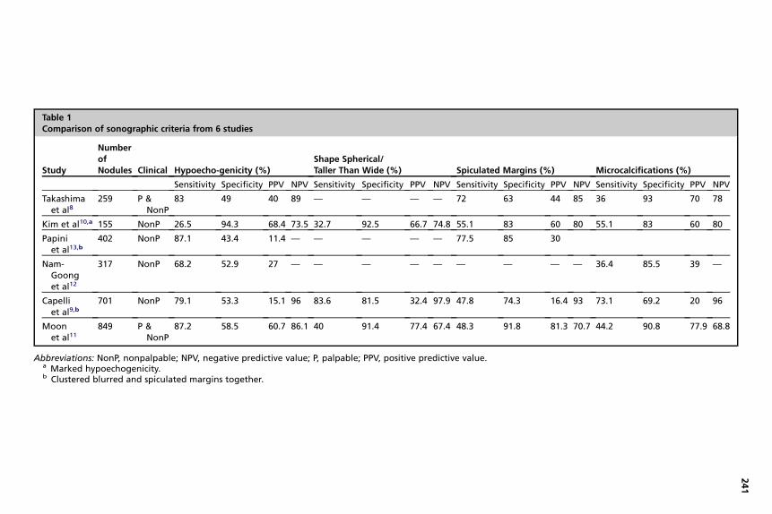
To encourage a rationale approach to the management of thyroid nodules detected onUS, several medical societies, including the American Thyroid Association (ATA), theSociety of Radiologists in Ultrasound (SRU), and the American Association of ClinicalEndocrinologists (AACE), have recently published a series of guidelines.4,6,7 The USfeatures of thyroid nodules that should be analyzed are summarized in the ConsensusStatement on thyroid nodules from the SRU7 and the AACE.4 They include nodule sizeand content (solid, complex, or cystic). For solid thyroid nodules, the following param-eters should be evaluated: nodule echotexture, shape, borders (smooth or nodular),the presence and quality of intranodular calcifications, and the presence of a perinod-ular halo. Table 1 compares the sensitivity, specificity, positive predictive values, andnegative predictive values of each of these sonographic criteria from 6 large studies,including a large retrospective study of 849 thyroid nodules recently conducted by the
Fig. 1. Normal thyroid gland. (A) Sagittal US right thyroid lobe shows a homogeneous glandwith medium gray echogenicity. (B) Transverse US shows both thyroid lobes. Note thehypoechoic strap muscle anteriorly (arrows). C, common carotid artery; T, trachea.
Table 1Comparison of sonographic criteria from 6 studies
Study
NumberofNodules Clinical Hypoecho-genicity (%)
Shape Spherical/Taller Than Wide (%) Spiculated Margins (%) Microcalcifications (%)
Korean Society of Neuro and Head and Neck Radiology Thyroid study group. Many ofthe statistics listed below stem from these articles and are summarized in Table 1.9–13
Nodule Content: Cystic Versus Solid Nodules
Before the availability of high-resolution high-frequency transducers, the role of USwas limited to distinguishing between cystic and solid nodules. Purely cystic nodulesare anechoic. They are almost invariably benign and represent colloid cysts. Somecolloid cysts contain echogenic foci with posterior reverberation or comet tail artifact(Fig. 2). A subset of cystic nodules shows a lacelike or honeycomb pattern of multiplesmall cysts separated by thin septations (Fig. 3). This pattern is strongly associatedwith a benign hyperplastic nodule11,14,15 and has been dubbed the ‘‘leave me alone’’lesion.15 Consequently, purely cystic nodules, with or without comet tails, and cysticnodules with a honeycomb appearance do not need FNAB.14
However, many cystic thyroid nodules have a solid-appearing component. Althoughthese complex nodules are often referred for biopsy for concern that they representa cystic papillary cancer, their most common underlying cause is a degeneratedcolloid nodule (Fig. 4). Because these nodules contain avascular debris and fibrosis,they tend to yield scant or no follicular cells and are associated with a higher number ofinconclusive FNAB.14,16
Careful analysis of any solid area within cystic nodules is imperative to identify therare papillary thyroid cancer (approximately 2.5%) with a large cystic component.Hatabu and colleagues17 described the ‘‘calcified nodule within a cyst,’’ a sign ofpapillary excrescences with microcalcifications protruding into the cyst, as specificfor papillary thyroid cancer (Fig. 5). Such an area should be specifically targeted duringfine-needle aspiration.
Echotexture
The echotexture (or shade of gray) of solid nodules is another important criterion takeninto consideration when analyzing nodules. The echotexture of the nodule iscompared with that of the surrounding thyroid parenchyma and the strap muscle(Fig. 6). Nodules are described as isoechoic (same shade of gray as the thyroid), hypo-echoic (darker than the thyroid) or markedly hypoechoic (darker than the strapmuscle). Hypoechoic and very hypoechoic nodules are classified as suspicious and
Fig. 2. Colloid cyst with comet tail artifact in a 33-year-old woman. Sagittal US of the rightthyroid lobe demonstrates a 7-mm cystic nodule (between calibers) with 2 echogenic fociwith comet tail artifact (arrowhead). This is a benign lesion. Note the second purely cysticnodule (arrow).
Fig. 3. Cystic nodule with a honeycomb pattern in a 54-year-old woman with hyperparathy-roidism. Transverse US of the right thyroid lobe shows a 9-mm nodule (between calibers)with multiple small cysts separated by thin echogenic septa, a classic honeycomb patternassociated with benign hyperplastic nodules.
Role of Ultrasonography in Thyroid Disease 243
referred for FNAB.10 Kim and colleagues10 found that 26.5% of malignant noduleswere markedly hypoechoic (see Fig. 6) compared with only 5.6% of benign nodules.The underlying histology for these nodules is usually PTC, and it is postulated that thedense cellularity of PTC produces very few interfaces to the sound beam and hencethe hypoechoic appearance. Follicular neoplasms, whether benign adenomas orfollicular carcinomas, contain colloid, have a microfollicular structure, and usuallydisplay an echogenic or mixed echotexture. Pathologically, they are typically encap-sulated and tend to be sharply demarcated from the surrounding thyroid parenchymaon US.14
Shape
Moon and colleagues11 reported that an elongated shape as compared with a wideshape, defined as an anteroposterior to transverse ratio of 1 or greater, is highlyspecific (91.4%) for malignancy. These results confirmed the reports publishedearlier.9,10 In another series, nodules with a spherical shape (ratio of long to shortaxis <1.5) were found to be associated with an 18% risk of cancer.18 By contrast,a ratio of long to short axis greater than 2.5 was found to have a 100% negative
Fig. 4. Adenomatoid nodule presenting as a complex cystic nodule in a 49-year-old woman.(A) Transverse US of the right thyroid lobe demonstrates a 3.5-cm complex cystic nodule(between calibers). The nodule is predominantly cystic with septations and a solid appearingcomponent (arrow). Note echogenic foci with comet tail artifact (arrowhead). (B) TransverseUS of the right thyroid lobe with color Doppler shows vascularity within the solid compo-nent. The diagnosis of adenomatoid nodule was confirmed with US-guided FNAB.
Fig. 5. Cystic PTC in a 51-year-old woman who presented with right neck swelling. (A)Sagittal US of the right thyroid lobe shows a 2-cm complex cystic nodule (between calibers).There is a large solid component (arrow) with tiny echogenic foci suspicious for microcalci-fications (arrowhead). The solid component was specifically targeted during the FNABprocedure. (B) Transverse US of the right thyroid lobe with color Doppler shows some vascu-larity (arrowheads) within the solid component. The diagnosis of PTC was confirmed withUS-guided FNAB and surgery.
Sheth244
predictive value for malignancy. It is speculated that cancers tend to grow acrosstissue planes and assume a spherical shape to maximize their oxygen supply,whereas benign lesions respect normal thyroid parenchyma.
Borders
Predictably, a spiculated or nodular border is associated with a higher probability ofmalignancy (see Fig. 6). Classically, PTC invades the surrounding thyroid tissue andis poorly encapsulated. In the series published by Moon and colleagues,11 48.3% ofthyroid cancers had spiculated margins and 32.5% had smooth borders, whereas75.9% of benign nodules had smooth margins and only 8.2% were spiculated. Theseresults confirm findings from previous studies.10 Demonstration of a refractive shadowfrom the edge of a solid nodule is another suspicious finding that warrants fine-needleaspiration.14
Fig. 6. Right PTC and left adenomatoid nodule in a 37-year-old woman. (A) Sagittal US ofthe right thyroid lobe shows a 1.6-cm markedly hypoechoic nodule (between calibers).Note that the nodule is more hypoechoic than the strap muscle (arrows) and that its bordersare spiculated. The diagnosis of PTC was confirmed by FNAB. (B) Transverse US of the leftthyroid lobe shows a 1-cm nodule that is nearly isoechoic to the thyroid gland (between cali-bers). The diagnosis of adenomatoid nodule was confirmed by fine-needle aspiration.
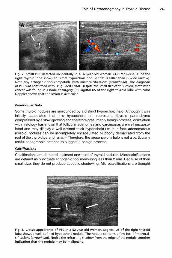
Fig. 7. Small PTC detected incidentally in a 32-year-old woman. (A) Transverse US of theright thyroid lobe shows an 8-mm hypoechoic nodule that is taller than is wide (arrow).Note tiny echogenic foci compatible with microcalcifications (arrowhead). The diagnosisof PTC was confirmed with US-guided FNAB. Despite the small size of this lesion, metastaticcancer was found in 1 node at surgery. (B) Sagittal US of the right thyroid lobe with colorDoppler shows that the lesion is avascular.
Role of Ultrasonography in Thyroid Disease 245
Perinodular Halo
Some thyroid nodules are surrounded by a distinct hypoechoic halo. Although it wasinitially speculated that this hypoechoic rim represents thyroid parenchymacompressed by a slow-growing and therefore presumably benign process, correlationwith histology has shown that follicular adenomas and carcinomas are well encapsu-lated and may display a well-defined thick hypoechoic rim.19 In fact, adenomatous(colloid) nodules can be incompletely encapsulated or poorly demarcated from therest of the thyroid parenchyma.20 Therefore, the presence of a halo is not a particularlyuseful sonographic criterion to suggest a benign process.
Calcifications
Calcifications are detected in almost one-third of thyroid nodules. Microcalcificationsare defined as punctuate echogenic foci measuring less than 2 mm. Because of theirsmall size, they do not produce acoustic shadowing. Microcalcifications are thought
Fig. 8. Classic appearance of PTC in a 52-year-old woman. Sagittal US of the right thyroidlobe shows a well-defined hypoechoic nodule. The nodule contains a few foci of microcal-cifications (arrowhead). Notice the refracting shadow from the edge of the nodule, anotherindication that the nodule may be malignant.
Sheth246
to represent the psammoma bodies or calcified laminated nidus that are frequentlyfound in PTC. The presence of microcalcifications in a solid nodule has a high spec-ificity of 91.3% to 96.3% and a positive predictive value of 74.8% for malignancy;unfortunately, the sensitivity is only 29% to 51.4% (Figs. 7 and 8).11,13
Coarse larger calcifications are caused by areas of degeneration and necrosiswithin thyroid nodules. On US, these calcifications appear as echogenic foci associ-ated with distal acoustic shadowing. These larger calcifications are found in bothbenign and malignant nodules, particularly in long-standing goiter and in medullarythyroid cancer (MTC). Thus the presence of coarse calcifications within a hypoechoicsolid nodule warrants FNAB.14
Vascularity
Color or power Doppler US provides useful additional information in the characteriza-tion of solid nodules by depicting nodular vascularity. It is postulated that malignantnodules are more likely to have disorganized internal vascularity and generally thyroidcancers tend to be hypervascular compared with the adjacent thyroid parenchyma(Fig. 9). Papini and colleagues13 defined an intranodular vascular pattern on colorDoppler US as most suspicious, with sensitivity of 74.2% and specificity of 80.8%.Benign nodules tend to be avascular or demonstrate only perinodular vascularity. Ithas also been postulated that power Doppler may be useful because it measuresthe amplitude of the Doppler signal rather than the mean velocity and is thus moresensitive to slow flow that may be present in small vessels.21 However, Frates andcolleagues22 found that 14% of malignant nodules were iso- or hypovascular to therest of the gland (see Fig. 7). We have observed that benign nodules may harborinternal vascularity and thus believe that the gray-scale appearance of nodules ismore helpful than their internal vascularity in predicting malignancy.
ULTRASOUND DIAGNOSIS OF THYROID NODULES: SUMMARY, CONTROVERSIES,AND CHALLENGES
In spite of the invaluable contribution of thyroid US in the assessment of thyroidnodules, there are many challenges, pitfalls, and controversies that need to beconsidered.
Multiple Suspicious Sonographic Findings
As shown in Table 1, individual sonographic signs are only moderately sensitive andspecific for predicting malignancy in solid thyroid nodules, as there can be significant
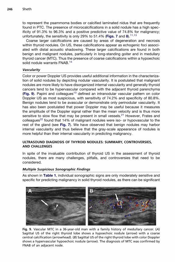
Fig. 9. Vascular MTC in a 36-year-old man with a family history of medullary cancer. (A)Sagittal US of the right thyroid lobe shows a hypoechoic nodule (arrow) with a coarsecentral calcification (arrowhead). (B) Sagittal US of the right thyroid lobe with color Dopplershows a hypervascular hypoechoic nodule (arrow). The diagnosis of MTC was confirmed byFNAB of an adjacent node.
Role of Ultrasonography in Thyroid Disease 247
overlap with benign processes because almost 70% of histologically proved benignnodules may exhibit 1 suspicious sonographic sign.23 However, malignant nodulestend to harbor multiple suspicious findings, for example, a mean of 2.6 abnormalitiesin the study by Kim and colleagues10 and the additional presence of microcalcifica-tions in a solid hypoechoic nodule that increased the odds ratio of this nodule beingmalignant from 6.5 to 13.1 in the series published by Nam-Goong and colleagues.12
Does Number Matter?
Even in patients referred for a palpable lesion, US can detect additional nodules in45% of cases.24 However, the prevalence of thyroid cancer is similar in patientswith solitary nodules or multinodular thyroid: in a large population of 1985 patientswith single or multiple nodules greater than 1 cm, the prevalence of thyroid cancerwas 14.8% in the solitary nodule group and 14.9% in the multinodular group.25
Too Small to Biopsy?
There is no correlation between the size of a thyroid nodule and the risk of underlyingcancer. The prevalence of thyroid cancer is similar in palpable and nonpalpablenodules, including incidentally discovered nodules measuring 10 mm or less.13
However, it is unclear whether diagnosis of micro-PTC affects life expectancy orwhether microcarcinomas as defined by a size smaller than 1 cm is less aggressive.26
In a series published by Nam-Goong and colleagues,12 69% of patients with incidentalPTC had extrathyroidal extension of the tumor or nodal metastases (see Fig. 7). Boththe ATA and the SRU recommend further workup of nodules greater than 1 cm andcareful analysis of the US characteristics listed earlier before formulating a decisionto perform FNAB.6,7 The AACE/Associazione Medici Endocrinologi (AME) guidelinesargue against an arbitrary cut off number to avoid missing very small but potentiallyaggressive tumors.4 Ultimately, the decision should be individualized, taking intoconsideration the age of the patient and the presence of comorbidity and risk factorsfor thyroid cancer, such as a positive family history, multiple endocrine neoplasia type2 syndrome, or previous head and neck irradiation.
The Special Case of Follicular Neoplasms
The suspicious sonographic appearances described earlier are usually associatedwith PTC, which represent approximately 70% of all thyroid malignancies. However,follicular neoplasms, which are less common, often do not exhibit classic malignantsonographic features. When comparing PTC with follicular carcinomas, Jeh andcolleagues19 found that 65.2% of follicular cancers were isoechoic to the thyroidparenchyma, 72.7% were oval, and 86.6% had a thin or thick hypoechoic rim.None of the cancers had microcalcifications. Benign follicular adenomas demon-strate similar sonographic findings on gray-scale US. Because a solid, homogeneousoval nodule with a hypoechoic capsule may represent a follicular neoplasm, sucha lesion should be referred for fine-needle aspiration (Fig. 10).14 Although somehave suggested that follicular carcinomas may exhibit more internal vascularity27
as compared with follicular adenomas, there is considerable overlap and it is notpossible to differentiate the two radiographically or based on cytologic specimen(see Fig. 10). Hurthle cell neoplasms are considered to be a variant of follicularcell neoplasms by some investigators, although more recent molecular analysessuggest that they are likely a distinct group of tumors. Their sonographic appearanceis similar to that of follicular neoplasms.28 The follicular variant of PTC, which repre-sents 9% to 22% of PTC, may have a sonographic appearance similar to follicularneoplasms (Fig. 11).29
Fig. 10. Follicular neoplasm in a 38-year-old woman. (A) Sagittal US of the left thyroid lobeshows an isoechoic solid nodule with well-defined borders and a hypoechoic rim (arrows).This appearance is suggestive of a follicular neoplasm and should prompt recommendationfor FNAB. (B) Transverse US of the left thyroid lobe confirms the findings. (C) Sagittal US ofthe left thyroid lobe with color Doppler shows increased vascularity in the nodule. Fine-nee-dle aspiration yielded the diagnosis of ‘‘suspicious for a follicular neoplasm.’’ Final pathologyafter surgical removal was follicular adenoma.
Sheth248
ULTRASOUND APPEARANCE OF RARE THYROID MALIGNANCIESMedullary Thyroid Cancer
MTC arises from the calcitonin-secreting C cells and account for approximately 5% ofthyroid malignancies. There is a familial form of medullary cancer, and it also affectspatients with multiple endocrine neoplasia type II. On US, the appearance of MTC isdifficult to differentiate from PTC; most present as a hypoechoic nodule. When calci-fications are present, they tend to be coarse and centrally located and may be relatedto underlying fibrosis and amyloid deposits (see Fig. 9).14 However, MTC can mimic
Fig. 11. Follicular variant of PTC in a 47-year-old woman with a history of hyperparathy-roidism. (A) Transverse US of the right thyroid lobe shows a well-defined oval nodule(arrow). The nodule is hypoechoic to the thyroid gland but less hypoechoic than the strapmuscle (arrowhead). (B) Sagittal US of the right thyroid lobe shows that the nodule has a hy-poechoic halo (arrows). The diagnosis of papillary thyroid cancer, follicular variant wasmade after surgical removal.
Role of Ultrasonography in Thyroid Disease 249
the far more common PTC with internal microcalcifications or appear as a solid non-calcified mass.
Anaplastic Thyroid Cancer
Anaplastic thyroid cancer accounts for 2% of all thyroid cancers, usually affectselderly patients, and is the most aggressive and lethal form of thyroid cancer, witha 5-year mortality of more than 95%. It often presents as a large hypoechoic mass,and invasion of adjacent muscles or the trachea is not uncommon.
Lymphoma and Metastases
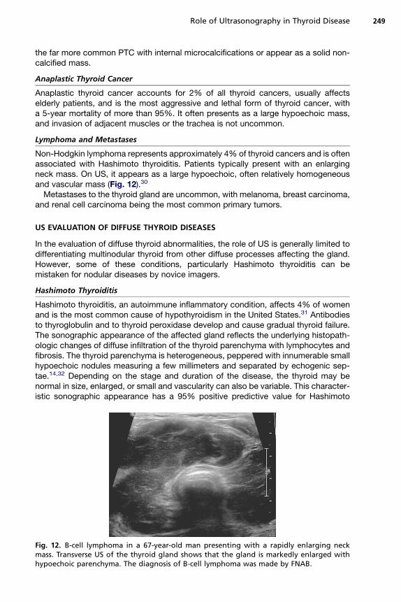
Non-Hodgkin lymphoma represents approximately 4% of thyroid cancers and is oftenassociated with Hashimoto thyroiditis. Patients typically present with an enlargingneck mass. On US, it appears as a large hypoechoic, often relatively homogeneousand vascular mass (Fig. 12).30
Metastases to the thyroid gland are uncommon, with melanoma, breast carcinoma,and renal cell carcinoma being the most common primary tumors.
US EVALUATION OF DIFFUSE THYROID DISEASES
In the evaluation of diffuse thyroid abnormalities, the role of US is generally limited todifferentiating multinodular thyroid from other diffuse processes affecting the gland.However, some of these conditions, particularly Hashimoto thyroiditis can bemistaken for nodular diseases by novice imagers.
Hashimoto Thyroiditis
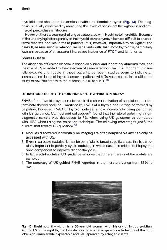
Hashimoto thyroiditis, an autoimmune inflammatory condition, affects 4% of womenand is the most common cause of hypothyroidism in the United States.31 Antibodiesto thyroglobulin and to thyroid peroxidase develop and cause gradual thyroid failure.The sonographic appearance of the affected gland reflects the underlying histopath-ologic changes of diffuse infiltration of the thyroid parenchyma with lymphocytes andfibrosis. The thyroid parenchyma is heterogeneous, peppered with innumerable smallhypoechoic nodules measuring a few millimeters and separated by echogenic sep-tae.14,32 Depending on the stage and duration of the disease, the thyroid may benormal in size, enlarged, or small and vascularity can also be variable. This character-istic sonographic appearance has a 95% positive predictive value for Hashimoto
Fig. 12. B-cell lymphoma in a 67-year-old man presenting with a rapidly enlarging neckmass. Transverse US of the thyroid gland shows that the gland is markedly enlarged withhypoechoic parenchyma. The diagnosis of B-cell lymphoma was made by FNAB.
Sheth250
thyroiditis and should not be confused with a multinodular thyroid (Fig. 13). The diag-nosis is usually confirmed by measuring the levels of serum antithyroglobulin and anti-thyroid peroxidase antibodies.
However, there are some challenges associated with Hashimoto thyroiditis. Becauseof the underlying heterogeneity of the thyroid parenchyma, it is more difficult to charac-terize discrete nodules in these patients. It is, however, imperative to be vigilant andcarefully assess any discrete nodules in patients with Hashimoto thyroiditis, particularlywomen, because of an apparent increased incidence of PTC31 and lymphoma.
Graves Disease
The diagnosis of Graves disease is based on clinical and laboratory abnormalities, andthe role of US is limited to the detection of associated nodules. It is important to care-fully evaluate any nodule in these patients, as recent studies seem to indicate anincreased incidence of thyroid cancer in patients with Graves disease. In a multicenterstudy of 557 patients with the disease, 3.8% had PTC.33
FNAB of the thyroid plays a crucial role in the characterization of suspicious or inde-terminate thyroid nodules. Traditionally, FNAB of a thyroid nodule was performed bypalpation; however, FNAB of thyroid nodules is now increasingly being performedwith US guidance. Carmeci and colleagues34 found that the rate of obtaining a non-diagnostic sample was decreased to 7% when using US guidance as comparedwith 16% when using the palpation technique. The following advantages justify thecurrent shift toward US guidance.35
1. Nodules discovered incidentally on imaging are often nonpalpable and can only beaccessed with US.
2. Even in palpable nodules, it may be beneficial to target specific areas; this is partic-ularly important in partially cystic nodules, in which case it is critical to biopsy thesolid component to improve diagnostic yield.
3. In large solid nodules, US guidance ensures that different areas of the nodule aresampled.
4. The accuracy of US-guided FNAB reported in the literature varies from 85% to94%.
Fig. 13. Hashimoto thyroiditis in a 38-year-old woman with history of hypothyroidism.Sagittal US of the right thyroid lobe demonstrates a heterogeneous echotexture of the rightlobe with innumerable hypoechoic nodules separated by echogenic septa.
Role of Ultrasonography in Thyroid Disease 251
Technique
Several techniques of US guidance and tissue sampling are used in standard prac-tice. Once the appropriate nodule is identified, some operators use the freehandtechnique, whereby the US transducer is placed over the nodule and the needle isinserted next to the transducer, whereas others use a fixed needle guide attachedto the transducer to introduce the needle (needle guide technique). Once the needleis inserted, the stylet is removed and the needle is moved rapidly within the target.Typically, 7 to 10 incursions are made till a flash of bloody fluid is seen within theneedle hub.
In addition to the various techniques used for needle insertion, 2 aspiration tech-niques can be used. In the capillary method, no suction is applied and the sampleis obtained by capillary action. Theoretically, this method minimizes trauma to thetissue and may decrease contamination by blood, especially if the nodule is veryvascular. Some operators favor the suction technique, applying 3 to 5 mL of suctionvia a syringe attached to the FNAB needle. This method may be slightly morecumbersome and allow slightly less control over needle motion but may yield morecells if the nodule is fibrotic. A recent study compared both techniques in 180samples and found no statistically significant difference in diagnostic accuracybetween the 2 methods.36
At our institution, we favor the needle guide technique and capillary samplingmethod. We use a 25-gauge 9-cm long needle and infiltrate the skin with 1% bufferedlidocaine before FNAB to minimize any patient discomfort, anticipating that a minimumof 3 samples will be required from each site. A trained cytopathologist or cytopathol-ogy technician is always available on-site to evaluate the adequacy of the sample atthe time of biopsy.
NEW HORIZONS: ELASTOGRAPHY AND THE ROLE OF PREOPERATIVE NECK USIN PATIENTS WITH THYROID CANCER
In recent years, 2 additional techniques have emerged in the armamentarium of eval-uating thyroid nodules and their utility is currently being debated in the literature.
Role of Preoperative Neck US in Patients with Thyroid Cancer
The value of neck US in the detection of local recurrences and cervical nodal metas-tases after total thyroidectomy for well-differentiated thyroid cancer is well estab-lished. Although PTC is a relatively indolent malignancy, it is associated witha relatively high incidence of local recurrences occurring in 30% of patients after initialthyroidectomy. In an attempt to diminish the risk of cervical metastases, several addi-tions to standard thyroidectomy have been proposed, including central compartmentnodal dissection and lateral neck dissection in patients with biopsy-proven cervicalnode metastases at the time of initial diagnosis. Thus, preoperative neck US hasa role in surgical planning, as was documented in 6% of patients in the series by Kou-varaki and colleagues.37 In addition, patients with positive nodes at the time of initialsurgery are at higher risk of recurrences in the future.38 Patients with MTC have aneven higher incidence of nodal metastases at the time of diagnosis, reported to reach32% to 80%.39 Based on these data, the ATA has recommended preoperative neckUS in patients with MTC in its most recent guidelines.40
The sonographic appearance of suspicious cervical nodes include a lack ofthe normal echogenic hilum, a round shape, the presence of intranodular micro-calcification or cystic areas, and increased vascularity by color Doppler(Fig. 14).41
Fig. 14. Lateral compartment nodal metastases in a 64-year-old patient with a suspiciousthyroid nodule. (A) Transverse US of the left thyroid lobe shows a large hypoechoic mass(arrows). The mass is taller than is wide, has nodular borders, and thus is highly suspiciousfor malignancy. (B) Transverse US of the left neck, level III shows 3 round heterogeneous no-des without a fatty hilum. FNAB of the thyroid nodule and one of the nodes yielded cellsdiagnostic for medullary thyroid carcinoma.
Sheth252
Elastography
Ultrasound elastography is used to evaluate tissue stiffness noninvasively. As a steadypressure is applied to the thyroid gland, the degree of deformity of the underlyingtissue is measured. This technique takes advantage of the fact that malignant nodulestend to be harder than benign nodules and thus deform less compared with thesurrounding normal thyroid parenchyma (Fig. 15). Two recent studies have showna statistically significant higher tissue stiffness index in malignant nodules ascompared with normal tissue and benign nodules, with a sensitivity of approximately88% and specificity of 77.5% to 90%.42,43 Applications of this technique may belimited to papillary thyroid cancer, which was the primary tumor evaluated in the priorstudies; thus, the findings may not be applicable to other thyroid tumor types. Anotherlimitation to this technique is that it is operator dependent and requires operatorexpertise. Larger studies are warranted before elastography can be routinely includedin the evaluation of thyroid nodules.
Fig. 15. Papillary cancer evaluated with elastography in a 56-year-old woman. Transverse USof the left thyroid lobe shows a hypoechoic nodule. Elastography shows that the noduledisplays predominantly a blue shade indicating that it is stiffer then the surrounding normalthyroid. FNAB of the thyroid nodule yielded cells diagnostic for papillary thyroid cancer.
Role of Ultrasonography in Thyroid Disease 253
SUMMARY
US remains the optimal imaging modality for the detection and characterization ofthyroid nodules. Careful analysis of key sonographic features as described earlierallow for more appropriate selection of indeterminate or suspicious nodules thatshould be referred for FNAB. US is also being increasingly used to guide FNAB, asit has been shown to improve diagnostic yield and is indispensable when obtainingbiopsies of an increasing number of nodules which are subcentimeter in size.
The larger question remains, however, as to whether the exhaustive workup of inci-dentally detected thyroid nodules leading to the diagnosis and treatment of asymp-tomatic micropapillary thyroid cancers ultimately affects survival and justifies thecosts and associated potential risks. With regards to thyroid incidentaloma, it isunclear if we have moved beyond, in the words of Dr Topliss, ‘‘the ignorant in thepursuit of the impalpable.’’44
REFERENCES
1. Mortensen JD, Woolner LB, Bennett WA. Gross and microscopic findings in clin-ically normal thyroid glands. J Clin Endocrinol Metab 1955;15(10):1270–80.
2. Hegedus L. Clinical practice. The thyroid nodule. N Engl J Med 2004;351(17):1764–71.
3. Jemal A, Siegel R, Ward E, et al. Cancer statistics, 2008. CA Cancer J Clin 2008;58(2):71–96.
4. Gharib H, Papini E, Valcavi R, et al. American Association of Clinical Endocrinol-ogists and Associazione Medici Endocrinologi medical guidelines for clinicalpractice for the diagnosis and management of thyroid nodules. Endocr Pract2006;12(1):63–102.
5. Gharib H, Goellner JR. Fine-needle aspiration biopsy of the thyroid: an appraisal.Ann Intern Med 1993;118(4):282–9.
6. Cooper DS, Doherty GM, Haugen BR, et al. Management guidelines for patientswith thyroid nodules and differentiated thyroid cancer. Thyroid 2006;16(2):109–42.
7. Frates MC, Benson CB, Charboneau JW, et al. Management of thyroid nodulesdetected at US: Society of Radiologists in Ultrasound consensus conferencestatement. Radiology 2005;237(3):794–800.
8. Takashima S, Fukuda H, Nomura N, et al. Thyroid nodules: re-evaluation withultrasound. J Clin Ultrasound 1995;23:179–84.
9. Cappelli C, Castellano M, Pirola I, et al. Thyroid nodule shape suggests malig-nancy. Eur J Endocrinol 2006;155(1):27–31.
10. Kim EK, Park CS, Chung WY, et al. New sonographic criteria for recommendingfine-needle aspiration biopsy of nonpalpable solid nodules of the thyroid. AJR AmJ Roentgenol 2002;178(3):687–91.
11. Moon WJ, Jung SL, Lee JH, et al. Benign and malignant thyroid nodules: USdifferentiation–multicenter retrospective study. Radiology 2008;247(3):762–70.
12. Nam-Goong IS, Kim HY, Gong G, et al. Ultrasonography-guided fine-needle aspi-ration of thyroid incidentaloma: correlation with pathological findings. Clin Endo-crinol (Oxf) 2004;60(1):21–8.
13. Papini E, Guglielmi R, Bianchini A, et al. Risk of malignancy in nonpalpablethyroid nodules: predictive value of ultrasound and color-Doppler features.J Clin Endocrinol Metab 2002;87(5):1941–6.
Sheth254
14. Reading CC, Charboneau JW, Hay ID, et al. Sonography of thyroid nodules:a ‘‘classic pattern’’ diagnostic approach. Ultrasound Q 2005;21(3):157–65.
15. Moon WJ, Kwag HJ, Na DG. Are there any specific ultrasound findings of nodularhyperplasia (‘‘leave me alone’’ lesion) to differentiate it from follicular adenoma?Acta Radiol 2009;50(4):383–8.
16. Layfield LJ, Cibas ES, Gharib H, et al. Thyroid aspiration cytology: current status.CA Cancer J Clin 2009;59(2):99–110.
17. Hatabu H, Kasagi K, Yamamoto K, et al. Cystic papillary carcinoma of the thyroidgland: a new sonographic sign. Clin Radiol 1991;43(2):121–4.
18. Alexander EK, Marqusee E, Orcutt J, et al. Thyroid nodule shape and predictionof malignancy. Thyroid 2004;14(11):953–8.
19. Jeh SK, Jung SL, Kim BS, et al. Evaluating the degree of conformity of papillarycarcinoma and follicular carcinoma to the reported ultrasonographic findings ofmalignant thyroid tumor. Korean J Radiol 2007;8(3):192–7.
20. Mazzaferri EL. Management of a solitary thyroid nodule. N Engl J Med 1993;328(8):553–9.
21. Cerbone G, Spiezia S, Colao A, et al. Power Doppler improves the diagnosticaccuracy of color Doppler ultrasonography in cold thyroid nodules: follow-upresults. Horm Res 1999;52(1):19–24.
22. Frates MC, Benson CB, Doubilet PM, et al. Can color Doppler sonography aid inthe prediction of malignancy of thyroid nodules? J Ultrasound Med 2003;22(2):127–31 [quiz: 132–4].
23. Wienke JR, Chong WK, Fielding JR, et al. Sonographic features of benign thyroidnodules: interobserver reliability and overlap with malignancy. J Ultrasound Med2003;22(10):1027–31.
24. Mandel SJ. Diagnostic use of ultrasonography in patients with nodular thyroiddisease. Endocr Pract 2004;10(3):246–52.
25. Frates MC, Benson CB, Doubilet PM, et al. Prevalence and distribution of carci-noma in patients with solitary and multiple thyroid nodules on sonography. J ClinEndocrinol Metab 2006;91(9):3411–7.
26. Kim JY, Lee CH, Kim SY, et al. Radiologic and pathologic findings of nonpalpablethyroid carcinomas detected by ultrasonography in a medical screening center. JUltrasound Med 2008;27(2):215–23.
27. Fukunari N, Nagahama M, Sugino K, et al. Clinical evaluation of color Dopplerimaging for the differential diagnosis of thyroid follicular lesions. World J Surg2004;28(12):1261–5.
28. Maizlin ZV, Wiseman SM, Vora P, et al. Hurthle cell neoplasms of the thyroid: sono-graphic appearance and histologic characteristics. J Ultrasound Med 2008;27(5):751–7 [quiz: 759].
29. Yoon JH, Kim EK, Hong SW, et al. Sonographic features of the follicular variant ofpapillary thyroid carcinoma. J Ultrasound Med 2008;27(10):1431–7.
30. Kwak JY, Kim EK, Ko KH, et al. Primary thyroid lymphoma: role of ultrasound-guided needle biopsy. J Ultrasound Med 2007;26(12):1761–5.
31. Repplinger D, Bargren A, Zhang YW, et al. Is Hashimoto’s thyroiditis a risk factorfor papillary thyroid cancer? J Surg Res 2008;150(1):49–52.
32. Yeh HC, Futterweit W, Gilbert P. Micronodulation: ultrasonographic sign of Hashi-moto thyroiditis. J Ultrasound Med 1996;15(12):813–9.
33. Kraimps JL, Bouin-Pineau MH, Mathonnet M, et al. Multicentre study of thyroidnodules in patients with Graves’ disease. Br J Surg 2000;87(8):1111–3.
34. Carmeci C, Jeffrey RB, McDougall IR, et al. Ultrasound-guided fine-needle aspi-ration biopsy of thyroid masses. Thyroid 1998;8(4):283–9.
Role of Ultrasonography in Thyroid Disease 255
35. Tublin ME, Martin JA, Rollin LJ, et al. Ultrasound-guided fine-needle aspirationversus fine-needle capillary sampling biopsy of thyroid nodules: does techniquematter? J Ultrasound Med 2007;26(12):1697–701.
36. Schoedel KE, Tublin ME, Pealer K, et al. Ultrasound-guided biopsy of the thyroid:a comparison of technique with respect to diagnostic accuracy. Diagn Cytopathol2008;36(11):787–9.
37. Kouvaraki MA, Shapiro SE, Fornage BD, et al. Role of preoperative ultrasonog-raphy in the surgical management of patients with thyroid cancer. Surgery2003;134(6):946–54 [discussion: 954–5].
38. Davidson HC, Park BJ, Johnson JT. Papillary thyroid cancer: controversies in themanagement of neck metastasis. Laryngoscope 2008;118(12):2161–5.
39. Ball DW. American Thyroid Association guidelines for management of medullarythyroid cancer: an adult endocrinology perspective. Thyroid 2009;19(6):547–50.
40. Kloos RT, Eng C, Evans DB, et al. Medullary thyroid cancer: management guide-lines of the American Thyroid Association. Thyroid 2009;19(6):565–612.
41. Sheth S, Hamper UM. Role of sonography after total thyroidectomy for thyroidcancer. Ultrasound Q 2008;24(3):147–54.
42. Hong Y, Liu X, Li Z, et al. Real-time ultrasound elastography in the differentialdiagnosis of benign and malignant thyroid nodules. J Ultrasound Med 2009;28(7):861–7.
43. Dighe M, Bae U, Richardson ML, et al. Differential diagnosis of thyroid noduleswith US elastography using carotid artery pulsation. Radiology 2008;248(2):662–9.
44. Topliss D. Thyroid incidentaloma: the ignorant in pursuit of the impalpable. ClinEndocrinol (Oxf) 2004;60(1):18–20.