Rom J Morphol Embryol 2011, 52(2):709–713 CASE REPORT Chondrosarcoma of the upper end of the femur ALINA MARIA ŞIŞU 1) , F. R. TATU 2) , LOREDANA GABRIELA STANA 1) , CODRUŢA ILEANA PETRESCU 1) , CARMEN TATU 3) , A. MOTOC 1) 1) Department of Anatomy and Embryology 2) Department of Orthopedics and Traumatology, Emergency Clinical Military Hospital 3) Department of Physiology “Victor Babeş” University of Medicine and Pharmacy, Timisoara Abstract Chondrosarcoma is a malignant tumor that produces cartilage matrix. Occurs in the fourth to sixth decades and has a male to female ratio of 2/1. It is most common in the long bones and on the surface of the pelvis. The authors present a case of chondrosarcoma of the upper end of the femur of a 50-year-old female patient who has come in our Department of Orthopedics and Traumatology two month ago, complaining of severe and persistent pain in the left hip joint and presenting limitation of adduction movement, limitation of internal-external rotation movements, and also could not be able to do thigh flexion on the abdomen. The woman presented a four-month history of persistent and severe pain, not assigned at anti-inflammatory drugs. Laboratory tests not had shown any significance. On radiographies and magnetic nuclear resonance the lesion was shown very clear, deciding for biopsy. The tumor had been large surgical excised with safe limits. Histopathology indicated the histological feature as a differentiated chondrosarcoma, grade III. Our patient has started the chemotherapy and radiation. Keywords: histopathology, malignant tumor, chondrosarcoma. Introduction Chondrosarcoma is a malignant tumor that originates from cartilage cells, maintaining its cartilaginous nature throughout its evolution. Is the second most common primary malignant bone tumor, occurring more commonly in men that in women, ratio 2/1. It is most frequently in the fourth to sixth decades (average age, 45 years). Chondrosarcoma occurs in three variants, classified according to location: central, peripheral and juxtacortical [1, 2]. Central chondrosarcoma arises in the medullary cavity of pelvic bones, ribs and long bones (femur, humerus), although any site may on occasion be affected (skull bones). Radiological, these tumors are characterized by poor defined borders, a thickened shaft, and perforation of the cortex. Although they may penetrate the cortex, extension beyond the periosteum is uncommon [3–5]. On gross examination, the neoplastic cartilaginous tissue is compressed inside the bone and exhibits areas of necrosis, cystic change and hemorrhage. The cortex of the bone is infiltrated by the tumor. Central chondrosarcoma begins with deep pain, which becomes more intense with time. In most cases, the tumor cannot be palpated, but in untreated cases, large masses may eventually form. Patient and Methods The 50-year-old woman, presented for severe and persisted pain in the left hip joint, with a four-month history of pain. Clinical signs showed a limitation of adduction thigh movement, limitation of internal external rotation thigh movements, and also could not be able to do thigh flexion on the abdomen. Peripheral pulse was present bilaterally. No swelling has been present on the left side. Laboratory tests were in normal ranges. Pelvic and lumbar column showed a malignant tumor. Magnetic Resonance Imaging (MRI) can be helpful in differentiating between benign and malignant lesions in several ways. Greater than 90% medullary involvement can be suggestive of chondrosarcoma, while the absence of 90% medullary involvement of non-contiguous areas of cartilage within the bone can suggest the presence of an enchondroma. In addition, the timing and progression of gadolinium contrast enhancement patterns may help direct a clinician toward or away from a diagnosis of malignancy [6, 7]. Many surgeons consider MRI critical for surgical planning because it can illustrate the extent of tumor involvement in bone and soft tissues. In our case, the patient had a tumor developed at the inferior surface of the femur neck (8/5/6 cm), with significant calcifications and highly irregular shape. It has been recommended the lesion biopsy [8–12]. Results Radioimaging results Pelvic and lumbar column radiographies showed bone demineralization, intervertebral space reduction R J M E Romanian Journal of Morphology & Embryology http://www.rjme.ro/
Transcript
Rom J Morphol Embryol 2011, 52(2):709–713
CCAASSEE RREEPPOORRTT
Chondrosarcoma of the upper end of the femur
ALINA MARIA ŞIŞU1), F. R. TATU2), LOREDANA GABRIELA STANA1), CODRUŢA ILEANA PETRESCU1), CARMEN TATU3), A. MOTOC1)
1)Department of Anatomy and Embryology 2)Department of Orthopedics and Traumatology,
Emergency Clinical Military Hospital 3)Department of Physiology
“Victor Babeş” University of Medicine and Pharmacy, Timisoara
Abstract Chondrosarcoma is a malignant tumor that produces cartilage matrix. Occurs in the fourth to sixth decades and has a male to female ratio of 2/1. It is most common in the long bones and on the surface of the pelvis. The authors present a case of chondrosarcoma of the upper end of the femur of a 50-year-old female patient who has come in our Department of Orthopedics and Traumatology two month ago, complaining of severe and persistent pain in the left hip joint and presenting limitation of adduction movement, limitation of internal-external rotation movements, and also could not be able to do thigh flexion on the abdomen. The woman presented a four-month history of persistent and severe pain, not assigned at anti-inflammatory drugs. Laboratory tests not had shown any significance. On radiographies and magnetic nuclear resonance the lesion was shown very clear, deciding for biopsy. The tumor had been large surgical excised with safe limits. Histopathology indicated the histological feature as a differentiated chondrosarcoma, grade III. Our patient has started the chemotherapy and radiation. Keywords: histopathology, malignant tumor, chondrosarcoma.
Introduction
Chondrosarcoma is a malignant tumor that originates from cartilage cells, maintaining its cartilaginous nature throughout its evolution. Is the second most common primary malignant bone tumor, occurring more commonly in men that in women, ratio 2/1. It is most frequently in the fourth to sixth decades (average age, 45 years). Chondrosarcoma occurs in three variants, classified according to location: central, peripheral and juxtacortical [1, 2].
Central chondrosarcoma arises in the medullary cavity of pelvic bones, ribs and long bones (femur, humerus), although any site may on occasion be affected (skull bones). Radiological, these tumors are characterized by poor defined borders, a thickened shaft, and perforation of the cortex. Although they may penetrate the cortex, extension beyond the periosteum is uncommon [3–5]. On gross examination, the neoplastic cartilaginous tissue is compressed inside the bone and exhibits areas of necrosis, cystic change and hemorrhage. The cortex of the bone is infiltrated by the tumor.
Central chondrosarcoma begins with deep pain, which becomes more intense with time. In most cases, the tumor cannot be palpated, but in untreated cases, large masses may eventually form.
Patient and Methods
The 50-year-old woman, presented for severe and persisted pain in the left hip joint, with a four-month
history of pain. Clinical signs showed a limitation of adduction thigh movement, limitation of internal external rotation thigh movements, and also could not be able to do thigh flexion on the abdomen. Peripheral pulse was present bilaterally. No swelling has been present on the left side. Laboratory tests were in normal ranges. Pelvic and lumbar column showed a malignant tumor. Magnetic Resonance Imaging (MRI) can be helpful in differentiating between benign and malignant lesions in several ways. Greater than 90% medullary involvement can be suggestive of chondrosarcoma, while the absence of 90% medullary involvement of non-contiguous areas of cartilage within the bone can suggest the presence of an enchondroma. In addition, the timing and progression of gadolinium contrast enhancement patterns may help direct a clinician toward or away from a diagnosis of malignancy [6, 7]. Many surgeons consider MRI critical for surgical planning because it can illustrate the extent of tumor involvement in bone and soft tissues. In our case, the patient had a tumor developed at the inferior surface of the femur neck (8/5/6 cm), with significant calcifications and highly irregular shape. It has been recommended the lesion biopsy [8–12].
Results
Radioimaging results
Pelvic and lumbar column radiographies showed bone demineralization, intervertebral space reduction
R J M ERomanian Journal of
Morphology & Embryologyhttp://www.rjme.ro/
Alina Maria Şişu et al.
710
L4/L5, L5/S1; reduction in bilateral hip joint space; in the lesser trochanter of the left femur bone was a tumor developed out without strict highlight the place of implantation, 8/7 cm, well-defined toward cranial pole, having an inhomogeneous feature and calcification areas, possible an osteochondroma (Figures 1 and 2) [13–17].
MRI revealed the tumor accompanied by a massive collection developed in the caudal adductors muscles group; significant and inhomogeneous gadolinium contrast of the tumor, without femoral inguinal or iliac adenopathies. The malignant criteria are present. Uterus, annexes and urinary bladder have had a normal aspect (Figures 3 and 4).
Figure 1 – X-ray: large hip joint tumor on the inferior surface of the lesser trochanter.
Figure 2 – X- ray: hip joint tumor, with net contour, intense, inhomogeneous.
Figure 3 – MRI transversal section: shows tumor. Figure 4 – MRI sagittal section: shows an important tumor, 80/50/60 mm with a lot of liquid.
Histopathologic results
On gross examination, chondrosarcoma is a grayish-white, lobulated mass. It has focal calcification and muriform aspect. The bigger one (2/1, 5 cm) had been decalcified (Figure 5).
Figure 5 – Gross examination; exophytic sarcoma with calcified areas and hemorrhage.
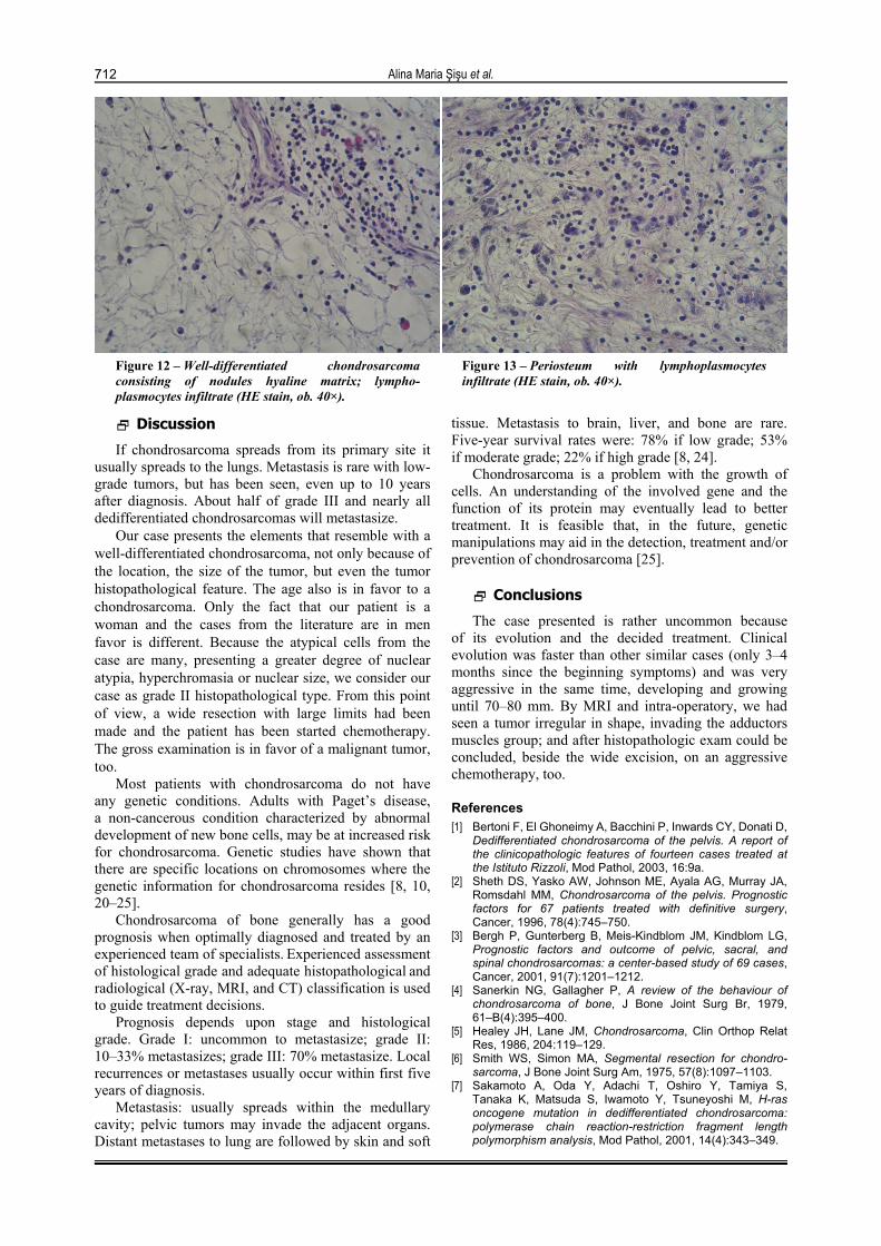
Histological, the tumor was HE-stained. Were found tumor fragments with lobulated pattern composed of cartilage matrix, which supports many chondroplasts congested with focal loss of arranging symmetrical character and containing not a strong pleomorphism. It is associated blades oblong of bone compact tissue. These are deformed and fragmented by the invasion of tumor tissue. Histological aspects are in favor for well-differentiated chondrosarcoma (Figures 6–13).
Wide range of differentiation and graded into: well differentiated; moderately differentiated; poorly differentiated (Table 1).
Grade I 60% painful Good Extended intralesional excision or wide resection
Grade II Up to 80% are painful Fair Wide resection
Grade III Up to 80% are painful Poor
Wide resection. Chemo-therapy and radiation
therapy in select cases
Chondrosarcoma of the upper end of the femur
711
Grade I (“low grade”) tumors most resemble normal cartilage, but may surround areas of lamellar bone (which is not seen in benign lesions), or show atypical cells including binucleate forms (cells with two nuclei instead of one).
Grade II (“intermediate grade”) is more cellular with a greater degree of nuclear atypia, hyperchromasia and nuclear size.
Grade III (“high grade”) tumors have significant areas of marked pleomorphism, large cells with more hyperchromatic nuclei.
Chondrosarcomas may also be classified by their histologic subtype. These subtypes include clear cell chondrosarcoma, mesenchymal chondrosarcoma, and dedifferentiated chondrosarcoma [18–20].
Figure 6 – Well-differentiated chondrosarcoma consisting of pale hyaline matrix (HE stain, ob. 40×).
Figure 7 – Tumor cells with atypical nuclei and peripherical necrosis (HE stain, ob. 40×).
Figure 8 – Malignant chondrocytes, large, atypical, with large nuclei (HE stain, ob. 40×).
If chondrosarcoma spreads from its primary site it usually spreads to the lungs. Metastasis is rare with low-grade tumors, but has been seen, even up to 10 years after diagnosis. About half of grade III and nearly all dedifferentiated chondrosarcomas will metastasize.
Our case presents the elements that resemble with a well-differentiated chondrosarcoma, not only because of the location, the size of the tumor, but even the tumor histopathological feature. The age also is in favor to a chondrosarcoma. Only the fact that our patient is a woman and the cases from the literature are in men favor is different. Because the atypical cells from the case are many, presenting a greater degree of nuclear atypia, hyperchromasia or nuclear size, we consider our case as grade II histopathological type. From this point of view, a wide resection with large limits had been made and the patient has been started chemotherapy. The gross examination is in favor of a malignant tumor, too.
Most patients with chondrosarcoma do not have any genetic conditions. Adults with Paget’s disease, a non-cancerous condition characterized by abnormal development of new bone cells, may be at increased risk for chondrosarcoma. Genetic studies have shown that there are specific locations on chromosomes where the genetic information for chondrosarcoma resides [8, 10, 20–25].
Chondrosarcoma of bone generally has a good prognosis when optimally diagnosed and treated by an experienced team of specialists. Experienced assessment of histological grade and adequate histopathological and radiological (X-ray, MRI, and CT) classification is used to guide treatment decisions.
Prognosis depends upon stage and histological grade. Grade I: uncommon to metastasize; grade II: 10–33% metastasizes; grade III: 70% metastasize. Local recurrences or metastases usually occur within first five years of diagnosis.
Metastasis: usually spreads within the medullary cavity; pelvic tumors may invade the adjacent organs. Distant metastases to lung are followed by skin and soft
tissue. Metastasis to brain, liver, and bone are rare. Five-year survival rates were: 78% if low grade; 53% if moderate grade; 22% if high grade [8, 24].
Chondrosarcoma is a problem with the growth of cells. An understanding of the involved gene and the function of its protein may eventually lead to better treatment. It is feasible that, in the future, genetic manipulations may aid in the detection, treatment and/or prevention of chondrosarcoma [25].
Conclusions
The case presented is rather uncommon because of its evolution and the decided treatment. Clinical evolution was faster than other similar cases (only 3–4 months since the beginning symptoms) and was very aggressive in the same time, developing and growing until 70–80 mm. By MRI and intra-operatory, we had seen a tumor irregular in shape, invading the adductors muscles group; and after histopathologic exam could be concluded, beside the wide excision, on an aggressive chemotherapy, too.
References [1] Bertoni F, El Ghoneimy A, Bacchini P, Inwards CY, Donati D,
Dedifferentiated chondrosarcoma of the pelvis. A report of the clinicopathologic features of fourteen cases treated at the Istituto Rizzoli, Mod Pathol, 2003, 16:9a.
[2] Sheth DS, Yasko AW, Johnson ME, Ayala AG, Murray JA, Romsdahl MM, Chondrosarcoma of the pelvis. Prognostic factors for 67 patients treated with definitive surgery, Cancer, 1996, 78(4):745–750.
[3] Bergh P, Gunterberg B, Meis-Kindblom JM, Kindblom LG, Prognostic factors and outcome of pelvic, sacral, and spinal chondrosarcomas: a center-based study of 69 cases, Cancer, 2001, 91(7):1201–1212.
[4] Sanerkin NG, Gallagher P, A review of the behaviour of chondrosarcoma of bone, J Bone Joint Surg Br, 1979, 61–B(4):395–400.
[6] Smith WS, Simon MA, Segmental resection for chondro-sarcoma, J Bone Joint Surg Am, 1975, 57(8):1097–1103.
[7] Sakamoto A, Oda Y, Adachi T, Oshiro Y, Tamiya S, Tanaka K, Matsuda S, Iwamoto Y, Tsuneyoshi M, H-ras oncogene mutation in dedifferentiated chondrosarcoma: polymerase chain reaction-restriction fragment length polymorphism analysis, Mod Pathol, 2001, 14(4):343–349.
Chondrosarcoma of the upper end of the femur
713[8] Sandberg AA, Bridge JA, Updates on the cytogenetics and
molecular genetics of bone and soft tissue tumors: chondro-sarcoma and other cartilaginous neoplasms, Cancer Genet Cytogenet, 2003, 143(1):1–31.
[9] Wehrli BM, Huang W, De Crombrugghe B, Ayala AG, Czerniak B, Sox9, a master regulator of chondrogenesis, distinguishes mesenchymal chondrosarcoma from other small blue round cell tumors, Hum Pathol, 2003, 34(3):263–269.
[10] Mitchell A, Rudan JR, Fenton PV, Juxtacortical dedifferent-iated chondrosarcoma from a primary periosteal chondro-sarcoma, Mod Pathol, 1996, 9(3):279–283.
[11] Rosenberg AE, Nielsen GP, Keel SB, Renard LG, Fitzek MM, Munzenrider JE, Liebsch NJ, Chondrosarcoma of the base of the skull: a clinicopathologic study of 200 cases with emphasis on its distinction from chordoma, Am J Surg Pathol, 1999, 23(11):1370–1378.
[12] Kalil RK, Inwards CY, Unni KK, Bertoni F, Bacchini P, Wenger DE, Sim FH, Dedifferentiated clear cell chondro-sarcoma, Am J Surg Pathol, 2000, 24(8):1079–1086.
[13] Gadwal SR, Fanburg-Smith JC, Gannon FH, Thompson LD, Primary chondrosarcoma of the head and neck in pediatric patients: a clinicopathologic study of 14 cases with a review of the literature, Cancer, 2000, 88(9):2181–2188.
[14] Björnsson J, McLeod RA, Unni KK, Ilstrup DM, Pritchard DJ, Primary chondrosarcoma of long bones and limb girdles, Cancer, 1998, 83(10):2105–2119.
[15] Bertoni F, Bacchini P, Hogendoorn PCW, Chondrosarcoma. In: Fletcher CDM, Unni KK, Mertens F (eds), World Health Organization Classification of Tumours. Pathology and Genetics of Tumours of Soft Tissue and Bone, IARC Press, Lyon, 2002, 247–251.
[16] Vanel D, De Paolis M, Monti C, Mercuri M, Picci P, Radiological features of 24 periosteal chondrosarcomas, Skeletal Radiol, 2001, 30(4):208–212.
[17] Skeletal Lesions Interobserver Correlation among Expert Diagnosticians (SLICED) Study Group, Reliability of histopathologic and radiologic grading of cartilaginous neoplasms in long bones, J Bone Joint Surg Am, 2007, 89(10):2113–2123.
[18] Veth R, Schreuder B, van Beem H, Pruszczynski M, de Rooy J, Cryosurgery in aggressive, benign, and low-grade malignant bone tumours, Lancet Oncol, 2005, 6(1):25–34.
[19] Bovée JVMG, Cleton-Jansen AM, Taminiau AHM, Hogendoorn PCW, Emerging pathways in the development of chondrosarcoma of bone and the implications for targeted treatment, Lancet Oncol, 2005, 6(8):599–607.
[20] Hameetman L, David G, Yavas A, White SJ, Taminiau AH, Cleton-Jansen AM, Hogendoorn PCW, Bovée JVMG, Decreased EXT expression and intracellular accumulation of heparan sulphate proteoglycan in osteochondromas and peripheral chondrosarcomas, J Pathol, 2007, 211(4):399–409.
[21] Bovée JVMG, van Royen M, Bardoel AF, Rosenberg C, Cornelisse CJ, Cleton-Jansen AM, Hogendoorn PCW, Near-haploidy and subsequent polyploidization characterize the progression of peripheral chondrosarcoma, Am J Pathol, 2000, 157(5):1587–1595.
[22] Bovée JVMG, van den Broek LJ, Cleton-Jansen AM, Hogendoorn PCW, Up-regulation of PTHrP and Bcl-2 expression characterizes the progression of osteochondroma
towards peripheral chondrosarcoma and is a late event in central chondrosarcoma, Lab Invest, 2000, 80(12):1925–1934.
[23] Rozeman LB, Hogendoorn PCW, Bovée JVMG, Diagnosis and prognosis of chondrosarcoma of bone, Expert Rev Mol Diagn, 2002, 2(5):461–472.
[24] Sandberg AA, Genetics of chondrosarcoma and related tumors, Curr Opin Oncol, 2004, 16(4):342–354.
[25] Rozeman LB, Hameetman L, Cleton-Jansen AM, Taminiau AH, Hogendoorn PCW, Bovée JVMG, Absence of IHH and retention of PTHrP signalling in enchondromas and central chondrosarcomas, J Pathol, 2005, 205(4):476–482.
Corresponding author Alina Maria Şişu, Assistant Professor, MD, PhD, Department of Anatomy and Embryology, “Victor Babeş” University of Medicine and Pharmacy, 2A Eftimie Murgu Square, 300041 Timişoara, Romania; Phone +40724–361 671, e-mail: [email protected], [email protected] Received: January 24th, 2011


![Rom J Morphol Embryol 2013, 54(1):205–210 R J M E CASE ... · PDF fileRom J Morphol Embryol 2013, 54(1):205–210 ISSN ... project report CEEX 68/2006 [3] shows that 650 000 ...](https://static.documents.pub/doc/80x56/5aae038b7f8b9a07498b87b2/rom-j-morphol-embryol-2013-541205210-r-j-m-e-case-j-morphol-embryol-2013.jpg)


