Page 1
ROSKILDE UNIVERSITY Deep brain
stimulation
A treatment for Parkinson’s disease
First semester project
Written by: Kevin von Heymann-Horan, Mie Primdahl Nielsen, Amir Zoet, and
Tina Becher Østerbøg
Supervisor: Ole Andersen
Submission date: December 21st 2012
Page 2
1. Semester project: Deep brain stimulation as a treatment for Parkinson’s disease
Kevin von Heymann-Horan, Mie Primdahl Nielsen, Amir Zoet, and Tina Becher Østerbøg Page 1
Table of contents
1. Abstract ......................................................................................................................... 3
2. Abbreviations ................................................................................................................ 4
3. Problem formulation ..................................................................................................... 5
4. Introduction................................................................................................................... 6
5. The Motor System ........................................................................................................ 8
5.1. The nigrostriatal pathway ...................................................................................... 9
5.2. Inter-neuron signalling in the basal ganglia ......................................................... 10
5.3. Excitotoxicity....................................................................................................... 14
5.4. Dopamine synthesis ............................................................................................. 16
5.5. Decomposition of monoamines ........................................................................... 17
5.5.1. Decomposition of dopamine ......................................................................... 17
6. Parkinson’s disease ..................................................................................................... 19
6.1. Lewy bodies ......................................................................................................... 20
6.2. Treatments for Parkinson's disease ...................................................................... 20
6.2.1. Ablation/Lesioning ....................................................................................... 21
6.2.2. Medicinal treatment ...................................................................................... 21
6.2.3. Stem Cell Therapy ........................................................................................ 25
6.2.4. Deep brain stimulation.................................................................................. 25
7. Deep brain stimulation................................................................................................ 27
7.1. History ................................................................................................................. 27
7.2. Use of deep brain stimulation .............................................................................. 30
7.3. Surgery ................................................................................................................. 32
8. Deep brain stimulation and Parkinson’s disease......................................................... 35
8.1. Oscillations: possible explanation of the mechanism of DBS? ........................... 35
8.2. Side-effects of deep brain stimulation ................................................................. 36
Page 3
1. Semester project: Deep brain stimulation as a treatment for Parkinson’s disease
Kevin von Heymann-Horan, Mie Primdahl Nielsen, Amir Zoet, and Tina Becher Østerbøg Page 2
8.2.1. Deep brain stimulation candidates ................................................................ 36
8.2.2. Deep brain stimulation and language/cognitive functions ............................ 37
9. Examination of case study and experiment ................................................................ 38
9.1. Close examination of experiment: Sunstedt et al, 2012 ...................................... 38
9.1.1. Introduction................................................................................................... 38
9.1.2. Method .......................................................................................................... 40
9.1.3. Results of the experiment. ............................................................................ 40
9.2. Close examination of a case study: Bejjani et al, 1999. ...................................... 41
9.2.1 Discussion of the case.................................................................................... 44
10. Discussion: future of deep brain stimulation ............................................................ 45
10.1. Should DBS be administered earlier in treatment? ............................................ 45
10.2. Closed-loop deep brain stimulation ................................................................... 47
10.3. Future of non-movement disorders.................................................................... 47
10.4. Motor cortex stimulation ................................................................................... 47
11. Conclusion ................................................................................................................ 49
12. Wordlist ..................................................................................................................... 51
13. Reference .................................................................................................................. 56
13.1. Articles ............................................................................................................... 56
13.2. Books ................................................................................................................. 60
13.3. Web pages .......................................................................................................... 61
13.4. Figure ................................................................................................................. 62
Page 4
1. Semester project: Deep brain stimulation as a treatment for Parkinson’s disease
Kevin von Heymann-Horan, Mie Primdahl Nielsen, Amir Zoet, and Tina Becher Østerbøg Page 3
1. Abstract
This project is an examination of the use of deep brain stimulation (DBS) in the
treatment of Parkinson's disease. The project contains an overview of the areas of the
brain that are affected by Parkinson's disease and that are targeted by deep brain
stimulation for treatment. The function and anatomy of these regions are discussed. The
history of deep brain stimulation is examined, as are new methods and theories as to
how deep brain stimulation can be used more effectively to treat not only Parkinson's
disease, but other diseases as well. Case studies are presented and examined. We
conclude that deep brain stimulation remains a valuable tool for treating Parkinson's
disease, recommend that deep brain stimulation be used more aggressively as a
treatment in the early stages of Parkinson’s disease, and call for deep brain stimulation
to be used as a treatment for a wider variety of illnesses.
Page 5
1. Semester project: Deep brain stimulation as a treatment for Parkinson’s disease
Kevin von Heymann-Horan, Mie Primdahl Nielsen, Amir Zoet, and Tina Becher Østerbøg Page 4
2. Abbreviations
ATP - adenosine triphosphate
COMT - catechol-O-methyl transferase
CT - computed tomography
cZI - caudal zona incerta
DA - dopamine
DBS - deep brain stimulation
GP - globus pallidus
GPi - internal globus pallidus
HVA - homovanilic acid
ICD - impulse control disorder
IPG - internal pulse generator
iPSC - induced pluripotent stem cell
MAO - monoamine oxidase
MAOI - monoamine oxidase inhibitor
MDD - major depressive disorder
MRI - magnetic resonance imaging
NO - nitric oxide
OCD - obsessive-compulsive disorder
PD - Parkinson's disease
PET - positron emission tomography
PPN - pedunculopontine nucleus
SN - substantia nigra
SNpc - substantia nigra pars compacta
TH - tyrosine hydroxylase
UPDRS - Unified Parkinson's Disease Rating Scale
USFDA - United States Food and Drug Administration
Vim - ventralis intermedius thalamus
Page 6
1. Semester project: Deep brain stimulation as a treatment for Parkinson’s disease
Kevin von Heymann-Horan, Mie Primdahl Nielsen, Amir Zoet, and Tina Becher Østerbøg Page 5
3. Problem formulation
With this project, we want to ask “how does deep brain stimulation work in the
treatment of Parkinson's disease, how effective is deep brain stimulation in treating
Parkinson's disease, and how can deep brain stimulation be more effective?” We plan to
answer these questions by studying published, clinical investigations of the use of deep
brain stimulation in Parkinson’s disease patients as well as primary research articles on
the molecular and physiological mechanisms by which deep brain stimulation works.
Page 7
1. Semester project: Deep brain stimulation as a treatment for Parkinson’s disease
Kevin von Heymann-Horan, Mie Primdahl Nielsen, Amir Zoet, and Tina Becher Østerbøg Page 6
4. Introduction
Parkinson's disease is a neurological condition that results from the death of
dopamine-producing neurons in the substantia nigra pars compacta (SNpc), a structure
in the basal ganglia. Those with Parkinson’s disease suffer from a variety of movement
related disorders, such as tremors, difficulty maintaining posture, and bradykinesia
(slow, labored movement). Parkinson’s disease is a degenerative disease; over time, cell
death in the SNpc becomes worse and worse, as do the resultant symptoms [Wichmann
et al, 2011]. It is a disease that typically affects older adults. There currently exists no
cure for Parkinson’s disease and, because of increased average longevity, Parkinson’s
disease is a disease that can be expected to appear with more frequency.
Parkinson’s disease is often treated with L-dopa, a precursor chemical to
dopamine that stimulates dopamine production in the SNpc. Over time, however, the
SNpc becomes so thoroughly destroyed that L-dopa ceases to work as a treatment.
Another treatment is deep brain stimulation (DBS), where electrodes are implanted in
the brain to stimulate the basal ganglia [Hickey & Stacy, 2011]. The areas to which deep
brain stimulation are most frequently applied are the internal globus pallidus, the
subthalamic nucleus and the ventral intermediate nucleus. The exact mechanism by
which deep brain stimulation works is not understood, but the areas that are stimulated
are those that are acted upon by the SNpc under normal conditions [Shah et al, 2010].
It is estimated that there are between 7 and 10 million people living with
Parkinson’s worldwide [PDF, 2012]. So far there are around 60.000 Parkinson’s patients
who have undergone deep brain stimulation surgery worldwide. It is expected that there
will be 8.000-10.000 new Parkinson’s patients who will undergo DBS surgery every
year worldwide. [Ponce & Lozano, 2010].
Deep brain stimulation has existed since the 19th century and has seen more
routine use in recent decades as a treatment for many neurological conditions, among
them Parkinson’s disease. Advances in neuroimaging technology coupled with a greater
understanding of the anatomy of the brain have led to experiments for how best to use
deep brain stimulation. It is known, for example, that deep brain stimulation can work
after the SNpc has been damaged to the point that L-dopa treatment is no longer
effective, but experiments now show that deep brain stimulation can be applied earlier,
Page 8
1. Semester project: Deep brain stimulation as a treatment for Parkinson’s disease
Kevin von Heymann-Horan, Mie Primdahl Nielsen, Amir Zoet, and Tina Becher Østerbøg Page 7
while L-dopa treatment is still effective, in order to increase the effectiveness of L-dopa.
deep brain stimulation may therefore be effective as a treatment for more patients than it
is currently used for [Hickey & Stacy, 2011]. Who is helped the most, how, and why are
all questions that remain to be answered.
Deep brain stimulation is not without drawbacks. While it can very effectively
and dramatically address the tremors and movement difficulties associated with
Parkinson’s disease, deep brain stimulation has been known to produce side-effects
ranging from worsened movement symptoms to compulsive gambling. The
dopaminergic system is delicate, but powerful and changes to it can result in dramatic
changes in behaviour. Deep brain stimulation has been proven to work to treat
Parkinson’s disease and is a technology that has the potential to be used more
effectively in the future. However, in order to develop it further it is necessary to study
deep brain stimulation and to better understand the precise effects that deep brain
stimulation has on the brain areas it targets, on the brain's dopamine pathways.
Page 9
1. Semester project: Deep brain stimulation as a treatment for Parkinson’s disease
Kevin von Heymann-Horan, Mie Primdahl Nielsen, Amir Zoet, and Tina Becher Østerbøg Page 8
5. The Motor System
From an evolutionary perspective, the ability to move is one of the oldest
abilities that vertebrates share. Many elements in the central nervous system work
together to allow organisms to move their bodies in a useful, coherent fashion. From
nerves in the spinal cord that control involuntary reflexes to complex, coordinated
movements that are planned in the higher brain areas, much of the brain consists of
neurons that ensure that the organism can move and function in its environment.
Some of the older structures involved in movement are the basal ganglia. In
humans, the basal ganglia are part of the midbrain, above the brain stem but buried
under the more evolutionarily recent cortices. The basal ganglia are a collection of
several structures – nuclei – that includes the globus pallidus, the putamen, and the
caudate nucleus. The latter two are also known collectively as the striatum. The nuclei
of the basal ganglia are connected to many other parts of the brain that also handle
movement, including the thalamus and the motor cortices, both of which are superior to
(ie above, towards the top of the skull when standing) the basal ganglia [Rosenzweig et
al, 2005]. Inputs to the basal ganglia from the motor cortex are received by the striatum.
The striatum passes along signals to the subthalamic nucleus and globus pallidus.
Excitatory pathways from the motor cortices extend directly to the subthalamic nucleus,
providing it with another source of input [Levy et al, 2002]. Signals passed to the
subthalamic nucleus are carried on to the substantia nigra [Lozano & Mahant, 2004].
Figure 5.1. Illustrates the basal ganglia
Page 10
1. Semester project: Deep brain stimulation as a treatment for Parkinson’s disease
Kevin von Heymann-Horan, Mie Primdahl Nielsen, Amir Zoet, and Tina Becher Østerbøg Page 9
5.1. The nigrostriatal pathway
The nuclei of the basal ganglia and, in particular, the striatum are stimulated by
dopamine that they receive from a nearby structure, the substantia nigra pars compacta
(SNpc) [Delong & Wichmann, 2010]. The flow of dopamine from the substantia nigra
to the striatum and the globus pallidus constitutes the first step of the nigrostriatal
pathway. The nigrostriatal pathway plays an important role in the motor system. The
steady flow of dopamine from the substantia nigra pars compacta through the pathway
is necessary for a healthy, functioning basal ganglia, which is in turn necessary for
modulating movement.
Figure 5.1.1. Diagram illustrating the connections of the structures of the nigrostriatal pathway.
In looking at the effects of the nigrostriatal pathway, it is illustrative to look at
Parkinson's disease. Parkinson's disease occurs due to a break-down of the nigrostriatal
pathway. The motor symptoms of Parkinson's disease (described in detail in section 6)
all arise from an insufficient supply of dopamine to the structures of the basal ganglia.
Under normal circumstances, the striatum and other basal ganglia structures are
constantly supplied with a steady stream of dopamine, regardless of whether the body is
in motion or at rest [Bergquist & Nissbrandt, 2005].
The neurotransmitters glutamate, which is excitatory, and GABA, which is
inhibitory, are regulators of dopamine release for the substantia nigra pars compacta
[Sherman, 2005]. Dopamine release in the striatum is heavily regulated by GABA and
Page 11
1. Semester project: Deep brain stimulation as a treatment for Parkinson’s disease
Kevin von Heymann-Horan, Mie Primdahl Nielsen, Amir Zoet, and Tina Becher Østerbøg Page 10
glutamate. Dopamine release in the substantia nigra pars compacta can be increased by
stimulating cells that produce/release glutamate [Bergquist & Nissbrandt, 2005], such as
those of the subthalamic nucleus [Chatha et al, 2000]. In the striatum, nitric oxide (NO)
produced endogenously from arginine, NADPH and O2 also stimulates dopamine
release [Harsing, 2008].
5.2. Inter-neuron signalling in the basal ganglia
To understand how signals pass from one structure of the brain to another it is
necessary to know how neurons function, and so a brief review of long-distance
communication between neurons will now be presented. Neurons are the cells in brain
tissue that work together to perform the various functions that occur in the brain. The
typical neuron has a star-shaped body, the soma, with many branch-like projections, the
dendrites. Most neurons also have an extension coming from the soma, called an axon.
The axon is partially sheathed in a substance called myelin, with small, exposed gaps.
At the end of the axon are the axon terminals. Human sensory and motor nerves may
have axons up to 1 m long.
Neurons communicate with chemical signals, neurotransmitters, over a small
gap between the two neurons called a synapse. The neuron that releases
neurotransmitters - that is, the neuron that is sending the signal - is the pre-synaptic
neuron. The neuron that receives the "message" is the post-synaptic neuron. The
synapse lies between the axon terminals of the pre-synaptic neuron and the dendrites of
the post-synaptic neuron, however, dendrite-to-dendrite signalling also takes place.
When signal transduction takes place between neurons, the dendrite or axon terminals
of the pre-synaptic neuron release neurotransmitters into the synaptic cleft. These
neurotransmitters then act as ligands and bind to receptors on the membrane of the post-
synaptic neuron.
Page 12
1. Semester project: Deep brain stimulation as a treatment for Parkinson’s disease
Kevin von Heymann-Horan, Mie Primdahl Nielsen, Amir Zoet, and Tina Becher Østerbøg Page 11
Figure 5.2.1. Illustrates how neurons communicate with chemical signals, neurotransmitters
The dopaminergic neurons that make up the substantia nigra are large, have a
large Golgi apparatus, and have dendrites with many receptors for the neurotransmitter
GABA. Synthesis of dopamine and the packaging of dopamine into vesicles for release
take place at the axon terminals, in the cytosol. Dopamine - also, adrenaline
(epinephrine) and noradenaline (norepinephrine) - is synthesized by the enzymes
tyrosine hydroxylase and Dopa decarboxylase. This process is explained in greater
detail in section 5.4. The axons of the dopaminergic neurons of the substantia nigra pass
through the globus pallidus en route to the striatum [McGeer et al, 1978].
Neurons signal to one another, release neurotransmitters, in response to changes
in their electrical charge. When a neuron is not being stimulated or inhibited, it will be
at its resting membrane potential. This potential - difference in charge between the
interior of the cell and the area just outside the plasma membrane - is between -70 and -
80 millivolts for neurons. Because the interior of the neuron has a negative charge
relative to the exterior, the potential is expressed as a negative number. There is a higher
concentration of sodium, Na+, ions outside of the neuron than in the cytosol; potassium,
K+, ions are found in higher concentration in the cytosol than outside the cell
[Gazzaniga et al, 2002]. These concentration differences are maintained via the ATP-
driven sodium-potassium pump.
Page 13
1. Semester project: Deep brain stimulation as a treatment for Parkinson’s disease
Kevin von Heymann-Horan, Mie Primdahl Nielsen, Amir Zoet, and Tina Becher Østerbøg Page 12
Figure 5.2.2. Time v. membrane voltage before, during, and after an action potential
Changes in the charge within the neuron can result in a so-called action
potential. An action potential occurs when sections of membrane on a neuron are rapidly
depolarized (the charge goes from negative to positive). The plasma membrane of a
neuron can be depolarized in multiple places simultaneously, if the neuron is receiving
multiple inputs. When a region of the membrane is depolarized, voltage-gated ion
channels in the plasma membrane open, allowing positively-charged sodium ions to
enter the neuron. The sodium ions are driven by passive transport along their electrical
and concentration gradients. As more sodium ions enter the neuron, the interior of the
neuron becomes even more depolarized (ie the cytosolic electrical charge becomes less
Page 14
1. Semester project: Deep brain stimulation as a treatment for Parkinson’s disease
Kevin von Heymann-Horan, Mie Primdahl Nielsen, Amir Zoet, and Tina Becher Østerbøg Page 13
negative/more positive). If the charge in the neuron reaches a certain threshold - around
-55 millivolts - an action potential will occur.
Once an action potential has been triggered, voltage-gated ion channels that had
been open, allowing in sodium ions, close. Potassium ions (K+) leave the neuron,
driving down the overall charge within the neuron (repolarization). Sodium-potassium
pumps in the plasma membrane exchange sodium ions in the cytosol for potassium ions
in the extracellular fluid. This energy-requiring pumping is fuelled by ATP. The charge
in the neuron returns to the resting state, with more K+ ions inside the neuron and more
Na+ ions outside the plasma membrane, and repolarization is complete. If the threshold
for activation is not reached while depolarisations are occurring, the neuron will use this
process to return itself to resting potential. For an action potential to occur, the
depolarization of the neuron must occur faster than the resting potential restoring
process.
When depolarization does occur rapidly enough to trigger an action potential,
the action potential travels down the axon of the neuron to the axon terminals. Upon the
arrival of the action potential, the axon terminals are depolarized. The depolarization of
the axon terminals causes neurotransmitter-containing vesicles in the axon terminals to
merge with the (pre-synaptic) neuron's plasma membrane (cell membrane), pushing the
neurotransmitters out of the axon and into the synapse. The neurotransmitters diffuse
throughout the synapse and bind to receptor proteins in the post-synaptic plasma
membrane via weak interactions (ionic, van der waal, hydrogen bonds).
There are two main classes of neurotransmitter receptors found on the post-
synaptic neuron, directly coupled and indirectly coupled receptors. Directly coupled
post-synaptic receptors are ion channels that open when a neurotransmitter molecule
binds with them. Indirectly coupled post-synaptic receptors involve G-proteins and
second messengers. With indirectly coupled receptors, second messengers are released
in the cytosol following the binding of neurotransmitter and receptor. Calcium ions,
Ca2+, are a common second messenger in this process, as are c-GMP and c-AMP
[Gazzaniga et al, 2002].
Dopamine receptors are indirectly coupled receptors that use c-AMP as a second
messenger. The many types of dopamine receptors are grouped into two large groups:
Page 15
1. Semester project: Deep brain stimulation as a treatment for Parkinson’s disease
Kevin von Heymann-Horan, Mie Primdahl Nielsen, Amir Zoet, and Tina Becher Østerbøg Page 14
D1-like receptors, which are excitatory, and D2-like receptors, which are inhibitory. The
neurons of the striatum, which receives the majority of the outputs from the substantia
nigra pars compacta, contains both D1-like and D2-like receptors [Bracci et al, 2002].
The receptor proteins of the post-synaptic neuron are eventually "switched off",
when the ligand leaves the receptor, either by diffusion or due to enzymatic breakdown.
between receptor and ligand (neurotransmitter). The neurotransmitters remain in the
synapse until they are either taken up again by the pre-synaptic neuron, are broken
down by enzymes, or until the neurotransmitters diffuse away from the synapse.
Catecholamines, including dopamine, may be taken up again by the pre-synaptic neuron
and/or degraded into simpler molecules by enzymes. The reuptake process uses active
transport, with transmembrane protein pumps. Enzymatic breakdown occurs when
either monoamine oxidase (MAO) or catechol-O-methyl transferase (COMT) is
released into the synapse [Gazzaniga et al, 2002]. This process is described in greater
detail in section 5.5.1.
Direct stimulation of the nuclei of the basal ganglia does not result in movement,
yet healthy basal ganglia are critical for the organism to move fluently. The basal
ganglia work to modulate and regulate an organism's movement rather than to direct and
control the movements. The basal ganglia's modulations are inhibitory. Another brain
structure, the cerebellum, performs the same function, but in the opposite direction; the
modulations on movement that come from the cerebellum are excitatory [Widmeier et
al, 2008]. An organism with a damaged cerebellum will have difficulties beginning
movements, while an organism with damaged basal ganglia will experience jerking,
out-of-control movements [McGeer et al, 1978]. In order to be able to execute fluid
movements, an organism must have both healthy basal ganglia and a healthy cerebellum
so that the two structures may work together harmoniously.
5.3. Excitotoxicity
Glutamate is not only the most abundant excitatory neurotransmitter in the
central nervous system, but it is also - potentially - a neurotoxin. High concentrations of
glutamate can lead to excitotoxicity, a condition wherein neurons become damaged and
die following excessive excitation. When glutamate binds to receptors on a (post-
synaptic) neuron, ion-channels open which allow in extracellular calcium ions, Ca2+.
Page 16
1. Semester project: Deep brain stimulation as a treatment for Parkinson’s disease
Kevin von Heymann-Horan, Mie Primdahl Nielsen, Amir Zoet, and Tina Becher Østerbøg Page 15
This is beneficial in rapidly depolarizing the neuron, but is harmful in excess. When too
many calcium ions flood the cytosol, serious harm can occur. The mitochondria are
damaged, causing them to stop ATP production, and cascading chemical reactions cause
damage to components of the cell, including DNA, culminating in cell death [Farooqui
et al, 2008].
Excitotoxicity leading to damage and death of neurons is of particular interest in
treating and preventing Parkinson's disease. The substantia nigra pars compacta receives
many excitatory glutamate inputs, for example from the subthalamic nucleus.
Overstimulation of the SNpc by these routes could explain the degradation of the
dopaminergic SNpc neurons. Treatment of Parkinson's disease by modulating the
glutamate pathway between the subthalamic nucleus and the substantia nigra is
discussed in section 6.2.2.
Page 17
1. Semester project: Deep brain stimulation as a treatment for Parkinson’s disease
Kevin von Heymann-Horan, Mie Primdahl Nielsen, Amir Zoet, and Tina Becher Østerbøg Page 16
5.4. Dopamine synthesis
The synthesis takes place in the cytoplasm of the nerve cell. Eventually
dopamine is formed out of tyrosine. Tyrosine is one of the non-essential amino acids
produced in our body. Non-essential amino acids are amino acids we do not necessarily
need to obtain from our food; we can synthesize them in our bodies.
Figure 5.4.1. Synthesis of the catecholamine neurotransmitters
Page 18
1. Semester project: Deep brain stimulation as a treatment for Parkinson’s disease
Kevin von Heymann-Horan, Mie Primdahl Nielsen, Amir Zoet, and Tina Becher Østerbøg Page 17
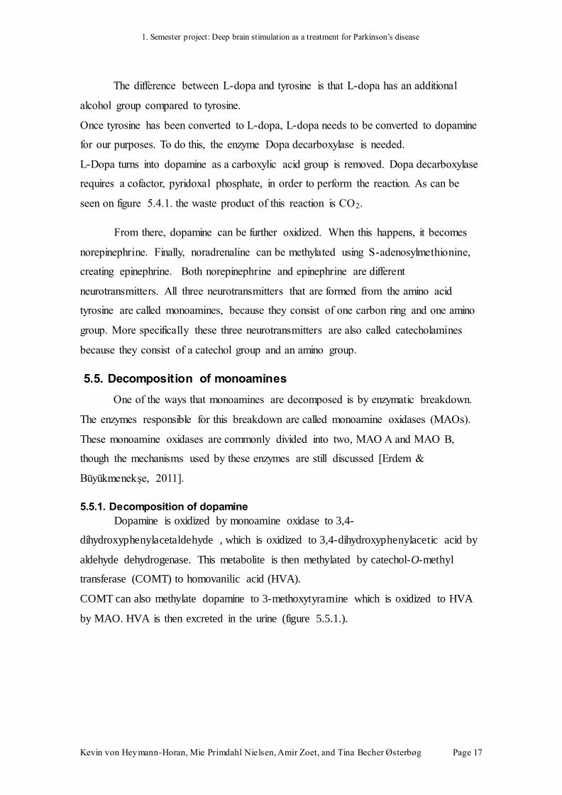
The difference between L-dopa and tyrosine is that L-dopa has an additional
alcohol group compared to tyrosine.
Once tyrosine has been converted to L-dopa, L-dopa needs to be converted to dopamine
for our purposes. To do this, the enzyme Dopa decarboxylase is needed.
L-Dopa turns into dopamine as a carboxylic acid group is removed. Dopa decarboxylase
requires a cofactor, pyridoxal phosphate, in order to perform the reaction. As can be
seen on figure 5.4.1. the waste product of this reaction is CO2.
From there, dopamine can be further oxidized. When this happens, it becomes
norepinephrine. Finally, noradrenaline can be methylated using S-adenosylmethionine,
creating epinephrine. Both norepinephrine and epinephrine are different
neurotransmitters. All three neurotransmitters that are formed from the amino acid
tyrosine are called monoamines, because they consist of one carbon ring and one amino
group. More specifically these three neurotransmitters are also called catecholamines
because they consist of a catechol group and an amino group.
5.5. Decomposition of monoamines
One of the ways that monoamines are decomposed is by enzymatic breakdown.
The enzymes responsible for this breakdown are called monoamine oxidases (MAOs).
These monoamine oxidases are commonly divided into two, MAO A and MAO B,
though the mechanisms used by these enzymes are still discussed [Erdem &
Büyükmenekşe, 2011].
5.5.1. Decomposition of dopamine
Dopamine is oxidized by monoamine oxidase to 3,4-
dihydroxyphenylacetaldehyde , which is oxidized to 3,4-dihydroxyphenylacetic acid by
aldehyde dehydrogenase. This metabolite is then methylated by catechol-O-methyl
transferase (COMT) to homovanilic acid (HVA).
COMT can also methylate dopamine to 3-methoxytyramine which is oxidized to HVA
by MAO. HVA is then excreted in the urine (figure 5.5.1.).
Page 19
1. Semester project: Deep brain stimulation as a treatment for Parkinson’s disease
Kevin von Heymann-Horan, Mie Primdahl Nielsen, Amir Zoet, and Tina Becher Østerbøg Page 18
Figure 5.5.1. Decomposition of dopamine
Page 20
1. Semester project: Deep brain stimulation as a treatment for Parkinson’s disease
Kevin von Heymann-Horan, Mie Primdahl Nielsen, Amir Zoet, and Tina Becher Østerbøg Page 19
6. Parkinson’s disease
Parkinson’s disease is mostly seen in elderly people over fifty years old who
have experienced cell death in the basal ganglia. The cause of the cell death that
characterizes Parkinson's disease is not necessarily always clear and most of the cases
are classified as idiopathic.
Parkinson’s disease is known for symptoms regarding movement. It can cause
tremors, uncontrolled shaking, bradykinesia (slow, labored movement), akinesia (lack of
movement), and other complications involved with movement. The symptoms of
Parkinson’s disease are caused by death of cells in the substantia nigra pars compacta
(SNpc), one of the basal nuclei. The substantia nigra pars compacta synthesizes the
neurotransmitter dopamine (DA) and releases it to nearby basal ganglia. When the
dopamine supply from the SNpc to these areas is reduced or eliminated completely, the
motor control problems that are characteristic to Parkinson’s disease emerge (tremors,
bradykinesia, dystonia, etc.) [Shah et al, 2010].
Figure 6.1.1. Illustrates the difference in the substantia nigra between a healthy person and a person with
Parkinson’s disease.
Besides the motor dysfunctions, neuropsychiatric complications in Parkinson’s
disease are nowadays also commonly recognised. Several disorders have been
diagnosed with significant frequency, including depression, psychosis and impulse
Page 21
1. Semester project: Deep brain stimulation as a treatment for Parkinson’s disease
Kevin von Heymann-Horan, Mie Primdahl Nielsen, Amir Zoet, and Tina Becher Østerbøg Page 20
control disorders. The specific effects of the neuropsychiatric disorder are unique to
each patient [Weintraub & Burn, 2011].
Depression is one of the disorders that is fairly often accompanied by
Parkinson’s disease with numbers varying from 5% to 20%. Up to 25% of all the
Parkinson’s disease patients have been treated with antidepressants at some time,
antidepressants does not only work against depression but also other diseases
[Weintraub & Burn, 2011].
Impulse control disorders (ICDs) are another type of disorder that is related to
Parkinson’s disease. ICDs include, for example, compulsive gambling, eating or buying.
In nearly 14% of the Parkinson’s disease patients ICDs were identified [Weintraub et al,
2010]. A patient using dopamine agonist was more likely to suffer from ICDs.
6.1. Lewy bodies
Patients with Parkinson’s disease are often characterised by Lewy bodies which
can be found in nerve cells in the brain. Lewy bodies consist primarily of α-synuclein, a
protein that may be linked to dopamine release, along with other proteins of its kind. In
α-synuclein knockout mice, mice genetically engineered not to produce α-synuclein,
increased release of dopamine was observed. This observation could mean that α-
synuclein is a negative regulator of dopamine [Tofaris & Spillantini, 2005].
Polymeropoulos et al found that there was a point mutation in the gene coding
for α-synuclein in a group of families [Polymeropoulos et al, 1997]. This group of
families had autosomal dominant inheritance of the Parkinson’s disease phenotype. This
also suggests the relationship of Lewy bodies and Parkinson’s disease. The point
mutation that the researchers found was located on the fourth exon of the α-synuclein
gene. This resulted in the insertion of threoninen instead of alanine in position 53 in the
protein.
6.2. Treatments for Parkinson's disease
Treatments for Parkinson's disease have focused primarily on the motor
problems that are the classic symptoms of the disease. There exist many treatments that
are effective to different degrees and that have a range of different side-effects. Some
Page 22
1. Semester project: Deep brain stimulation as a treatment for Parkinson’s disease
Kevin von Heymann-Horan, Mie Primdahl Nielsen, Amir Zoet, and Tina Becher Østerbøg Page 21
treatments are used in conjunction with one another to maximize the effectiveness of
each.
In Parkinson's disease, the relation between the motor function and the non-
motor function is complex. To find a good treatment for Parkinson’s disease is therefore
also rather complicated.
There are a few different treatments available to lower the severity of the Parkinson’s
disease symptoms.
6.2.1. Ablation/Lesioning
Surgical destruction of nuclei in the basal ganglia, also known as "ablation" or
"lesioning", is a well-known method for treating some parkinsonian symptoms. Tremor
and rigidity can both be effectively treated by ablation of the ventrolateral nuclei of the
thalamus. This treatment does not, however, lead to improvements across the board:
bradykinesia and akinesia are unaffected. Ablation fell out of favor as a treatment for
Parkinson's disease with the discovery of medicinal L-Dopa treatment, which has the
advantages of being non-invasive and of treating not only tremor and rigidity, but also
akinesia, bradykinesia [McGeer et al, 1978]. Ablation remains a common practice in
regions of the world where deep-brain stimulation is not available due to lack of
expertise [Wichmann & Delong, 2011]. Because ablation involves destroying brain
tissue, it is obviously not reversible.
6.2.2. Medicinal treatment
In the 1960s it was discovered that medicines could be administered to patients
suffering from Parkinson's disease that effectively treated the symptoms of the disease.
Although not without drawbacks, medicinal treatments were hailed as a great
improvement over ablation treatments. Medicinal treatments for Parkinson's disease
remain common and are effective for nearly all but the most advanced cases.
Levodopa (medicinal L-dopa) is the most popular medicine for the treatment of
Parkinson's disease, as it is converted into dopamine (DA) by the body itself. Dopamine
cannot easily be delivered to the body, as it is unable to cross the blood-brain barrier. L-
dopa, however, may be ingested and then cross into the brain, where it is converted into
dopamine in the substantia nigra pars compacta. Although the substantia nigra pars
compacta may be degraded because of cell death, the cells that do remain are able to
Page 23
1. Semester project: Deep brain stimulation as a treatment for Parkinson’s disease
Kevin von Heymann-Horan, Mie Primdahl Nielsen, Amir Zoet, and Tina Becher Østerbøg Page 22
produce a quantity of dopamine from the L-dopa that mimics the amount of dopamine
produced by a healthy substantia nigra when the medication is titrated correctly.
Medicinal L-Dopa is not without downsides, however. Levodopa can only be
used in the earlier stages of Parkinson’s disease and becomes less effective over time. In
very severe, advanced cases of Parkinson's disease, enough of the substantia nigra pars
compacta has died that L-dopa treatment is no longer effective. Without a sufficient
number of dopamine-making cells in the substantia nigra pars compacta, not enough
dopamine is created even with the help of extra L-dopa [Rosenzweig et al, 2005].
Below is a list of the side-effects of the use of L-Dopa:
Common side-effects
Abnormal thinking: holding false
beliefs that cannot be changed by
fact
Agitation
Anxiety
Clenching or grinding of teeth
Clumsiness or unsteadiness
Confusion
Difficulty swallowing
Dizziness
Excessive watering of mouth
False sense of well being
Feeling faint
General feeling of discomfort or
illness
Hallucinations (seeing, hearing, or
feeling things that are not there)
Hand tremor, increased
Nausea or vomiting
Numbness
Unusual and uncontrolled
movements of the body, including
the face, tongue, arms, hands, head,
and upper body
Unusual tiredness or weakness
Page 24
1. Semester project: Deep brain stimulation as a treatment for Parkinson’s disease
Kevin von Heymann-Horan, Mie Primdahl Nielsen, Amir Zoet, and Tina Becher Østerbøg Page 23
Not so common side-effects:
Blurred vision
Difficult urination
Difficulty opening mouth
Dilated (large) pupils
Dizziness or lightheadedness when
getting up from a lying or sitting
position
Double vision
Fast, irregular, or pounding
heartbeat
Hot flashes
Increased blinking or spasm of
eyelids
Loss of bladder control
Mental depression
Other mood or mental changes
Skin rash
Unusual weight gain or loss
Rare side-effects:
Back or leg pain
Bloody or black tarry stools
Chills
Convulsions (seizures)
Fever
High blood pressure
Inability to move eyes
Loss of appetite
Pain, tenderness, or swelling of foot
or leg
Pale skin
Prolonged, painful, inappropriate
penile erection
Sore throat
Stomach pain
Swelling of face
Swelling of feet or lower legs
Vomiting of blood or material that
looks like coffee grounds
[MAYO, 2012]
Some of the side-effects to taking the drug L-dopa are the effects encountered from
Parkinson’s disease.
Monoamine oxidase inhibitors (MAOIs) are also used to treat Parkinson’s
disease. Degradation of dopamine in the synapse by monoamine oxidase can be blocked
by monoamine oxidase inhibitors. By preventing monoamine oxidase from breaking
Page 25
1. Semester project: Deep brain stimulation as a treatment for Parkinson’s disease
Kevin von Heymann-Horan, Mie Primdahl Nielsen, Amir Zoet, and Tina Becher Østerbøg Page 24
down dopamine after it has been released into the synapse, the effects of dopamine are
amplified. Some of the side-effects by using MAOIs are sexual dysfunction,
sleeplessness, dizziness, gaining weight, muscle aches or tingling and difficulty
urinating. MAOI’s also interfere with the metabolism of vitamin B6 and can cause the
muscle ache or tingling [NBM, 2012].
Adenosine (A2A) antagonist is another type of medicine that has been studied,
because levodopa does not seem to work for longer terms. A2A does not focus on the
dopamine receptors, but rather on the adenosine receptors. Adenosine has, like
dopamine, an important role in the basal ganglia. The A2A receptors are mostly
localised near the dopamine receptors in the striatum, the basal nuclei that receives the
most projections from the substantia nigra pars compacta. Blocking the A2A receptors
should decrease the motor complications of Parkinson’s disease [Hickey & Stacy,
2012].
There are two kinds of adenosine receptors that are well researched: A1 and A2A
adenosine receptors. A1 receptors are common throughout the brain and prevent the
release of excitatory neurotransmitters when they are bound with adenosine. A2A
receptors are found in very high concentration in the basal ganglia and have a very
different effect when active. A2A receptors deactivate A1 adenosine receptors. Put
another way, A1 receptors inhibit the release of excitatory neurotransmitters; A2A de-
inhibits, removes the inhibition caused by A1 adenosine receptors. A2A receptors are
only activated when A1 adenosine receptors are highly active, when the neuron is
receiving many inhibitory signals. By blocking the A2A receptors, the check on the
amount of inhibition that adenosine can cause is removed [Cunha, 2008].
As discussed in section 5.3, cell death can occur from excitotoxicity.
Excitotoxicity stemming from over-active glutamate circuits is of particular concern in
Parkinson's disease. One potential way of mitigating this issue is to take full advantage
of the inhibitory effects of the A1 adenosine receptors by blocking the A2A adenosine
receptors in the basal ganglia. Blocking A2A receptors has been shown not only to
prevent further degeneration in patients with Parkinson's disease, but also to reduce the
motor symptoms of Parkinson's disease [Cunha, 2008].
Page 26
1. Semester project: Deep brain stimulation as a treatment for Parkinson’s disease
Kevin von Heymann-Horan, Mie Primdahl Nielsen, Amir Zoet, and Tina Becher Østerbøg Page 25
6.2.3. Stem Cell Therapy
Replacement of dead cells in the substantia nigra pars compacta with induced
pluripotent stem cells (iPSCs) is one of the newer, experimental treatments for
Parkinson's disease. Treatment with induced pluripotent stem cells is focused on
restoring the damage caused by the death of dopaminergic neurons. A number of studies
have found that mouse embryonic cells are able to be reprogrammed in mice to these
iPSCs. As of 2012, researchers have also succeeded in transforming human cells into
iPSCs [Pu et al, 2012]. The major issue with this treatment is how to turn these iPSCs
into exactly what is needed for the patient. Currently it is not possible to replicate the
dopaminergic neurons in the substantia nigra pars compacta, because the connections
between the neurons are too numerous. Transplanting such an amount of cells is too
risky to attempt in vivo.
6.2.4. Deep brain stimulation
Deep brain stimulation is a treatment that involves directly targeting certain
areas of the brain with electrical impulses that are administered via surgically implanted
electrodes. Although invasive, deep brain stimulation accompanied by pharmaceutical
treatment, such as levodopa, is known to be better than any pharmaceutical treatment
alone. A series of randomized control trials performed by Deuschl et al. have
demonstrated that deep-brain stimulation is more effective than medicinal treatment
[Deuschl et al, 2006]. Furthermore, deep brain stimulation is efficient even after
pharmaceuticals alone can no longer effectively treat the symptoms of Parkinson’s
disease. As such, deep brain stimulation has become a popular treatment for Parkinson’s
disease in the later stages of the disease.
Deep brain stimulation targets structures in the basal ganglia in order to
counteract the motor problems that are characteristic of Parkinson’s disease. Common
targets for deep brain stimulation for the treatment of Parkinson’s disease are the
subthalamic nucleus (STN), the globus pallidus (GPi), the ventralis intermedius (Vim)
thalamus. The subthalamic nucleus is the most common target, although different areas
are targeted depending upon the specifics of the symptoms in the patient. Stimulation of
the STN is helpful in treating all motor symptoms of Parkinson's disease. The ventralis
intermedius thalamus is often targeted when tremor is the main issue. Deep brain
stimulation of the GPi is effective for treating dyskinesias [Shah et al., 2010]. In the
Page 27
1. Semester project: Deep brain stimulation as a treatment for Parkinson’s disease
Kevin von Heymann-Horan, Mie Primdahl Nielsen, Amir Zoet, and Tina Becher Østerbøg Page 26
case of Parkinson's disease, deep brain stimulation is typically used as a treatment for
patients for whom pharmaceutical L-Dopa has worked in the past, but who are suffering
from motor problems despite the medication. Typical protocols stipulate that candidates
for deep brain stimulation should still respond to L-Dopa and should not show signs of
dementia. If patients do the effect of deep brain stimulation will not be efficient enough
compared to the risks of the surgery [Hickey & Stacy, 2011].
Page 28
1. Semester project: Deep brain stimulation as a treatment for Parkinson’s disease
Kevin von Heymann-Horan, Mie Primdahl Nielsen, Amir Zoet, and Tina Becher Østerbøg Page 27
7. Deep brain stimulation
In a healthy brain, the substantia nigra produces a steady stream of dopamine
directed at the striatum and the other basal ganglia, regardless of whether the body is
moving or at rest [Hickey & Stacy, 2011]. Deep brain stimulation is a treatment that
employs a surgically implanted neurostimulating device which sends electrical impulses
to interfere with the patient's neural impulses. The deep brain stimulation device
functions as a pacemaker which stimulates a target area of the brain. The impulses of
the deep brain stimulation device can, for example, help control movement and block
the abnormal neural signals that arise as a result of neural degradation in patients
suffering from Parkinson's disease [NINDS, 2012].
7.1. History
The development of deep brain stimulation started in 1870 when Eduard Hitzig
and Gustav Fritsch performed the first direct electrical stimulation of the motor cortex
in dogs that could not move their limbs due to anaesthesia [Fritsch & Hitzig, 2009].
Robert Bartholow used the same treatment in 1874, but this time on a human subject
[Harris & Almerigi, 2009]. Today, the deep brain stimulation system is widely used, but
it was not until the 1990s that it was adopted by mainstream neurosurgery for the
treatment of Parkinson's disease.
Timeline of deep brain stimulation
In 1870 Eduard Hitzig and Gustav Fritsch did an experiment on dogs' cerebrums
in Berlin, Germany. They experimented on the frontal part of the cerebrum, because the
motor part lies in that area. Anesthetized, the dogs could not move their limbs. By
Page 29
1. Semester project: Deep brain stimulation as a treatment for Parkinson’s disease
Kevin von Heymann-Horan, Mie Primdahl Nielsen, Amir Zoet, and Tina Becher Østerbøg Page 28
electrical stimulation of the frontal part of the cerebrum in one side of the brain, Hitzig
and Fritsch observed muscle contractions in the other side of the body. They also
discovered that there was a correlation between current and responding muscles,
wherein a higher current corresponded to larger groups of muscles responding [Fritsch
& Hitzig, 2009].
Robert Bartholow from Medical College of Ohio, USA worked in 1874 at Good
Samaritan Hospital where he was presented to Mary Rafferty. Rafferty had arrived at
the hospital with a 5 centimetre hole in her skull, which she had had for 13 months.
Figure 7.1.1. shows a drawing of Mary Rafferty's head, with exposed cortex [Harris & Almerigi, 2009]
Rafferty gave her permission to Bratholow to study and experiment on her brain.
This gave Bartholow the opportunity to redo and improve Hitzig and Fritsch’s
experiment from 1870, this time with a human subject. His experiment was to stimulate
different parts of the brain with different current strength to see the respond. The result
were contractions of leg and arm, dilated pupils, unpleasant tingling in arm and leg,
spasms of arm, loss of consciousness, 20 minutes in a coma and pain. After four days
and six tests, Rafferty died of unknown causes [Harris & Almerigi, 2009].
In 1954, James Olds and Peter Milner from McGill University, Canada
performed an experiment involving deep brain stimulation, this time of the midbrain.
Olds and Milner's experiment involved 15 male rats with electrodes surgically
implanted in their midbrains. Each rat was placed in a skinner box, a testing chamber
Page 30
1. Semester project: Deep brain stimulation as a treatment for Parkinson’s disease
Kevin von Heymann-Horan, Mie Primdahl Nielsen, Amir Zoet, and Tina Becher Østerbøg Page 29
for experiments in operant conditioning. From the top of the box, a lead connected the
electrodes placed in the rats' brains to a voltage source. The rats would receive a
stimulating current to the dopamine reward centre in the brain every time the rats
activated the lever in the Skinner box. This experiment showed that electrical
stimulation in primary reward centre increased the occurrence of the target behaviour,
lever pressing, while stimulation of the other sites had either no effect or the opposite
effect [Olds & Milner, 1954]. Although the dopamine pathway involved in this
experiment is not the same as in Parkinson’s disease, Olds and Milner demonstrated that
dopaminergic neurons can be stimulated with electrical impulses.
Figure 7.1.2 Shows the Skinner box Olds and Milner used in 1954
In 1972 Bechtereva et al, from the Institute for Experimental Medicine in
Leningrad, Russia performed a study on patients with parkinsonian hyperkinesia to find
connections between different brain parts and parkinsonian symptoms by using
electrical stimulation in caudate nucleus and globus pallidus. Because of this
experiment they found a lack of connection between caudate nucleus and medial globus
pallidus, where it would have existed in a normally functional brain. Therefore they
concluded that there had to be some kind of causal relation between parkinsonian
hyperkinesia and the medial globus pallidus [Bechtereva et al, 1972].
Page 31
1. Semester project: Deep brain stimulation as a treatment for Parkinson’s disease
Kevin von Heymann-Horan, Mie Primdahl Nielsen, Amir Zoet, and Tina Becher Østerbøg Page 30
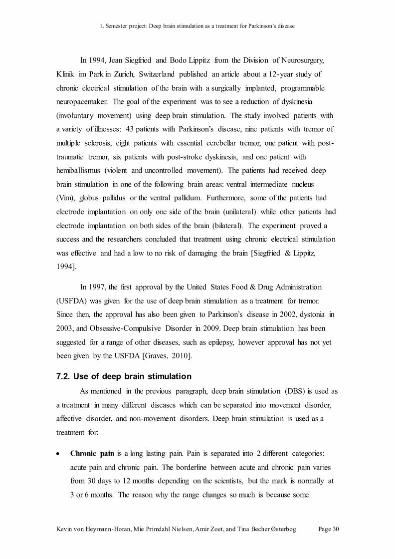
In 1994, Jean Siegfried and Bodo Lippitz from the Division of Neurosurgery,
Klinik im Park in Zurich, Switzerland published an article about a 12-year study of
chronic electrical stimulation of the brain with a surgically implanted, programmable
neuropacemaker. The goal of the experiment was to see a reduction of dyskinesia
(involuntary movement) using deep brain stimulation. The study involved patients with
a variety of illnesses: 43 patients with Parkinson’s disease, nine patients with tremor of
multiple sclerosis, eight patients with essential cerebellar tremor, one patient with post-
traumatic tremor, six patients with post-stroke dyskinesia, and one patient with
hemiballismus (violent and uncontrolled movement). The patients had received deep
brain stimulation in one of the following brain areas: ventral intermediate nucleus
(Vim), globus pallidus or the ventral pallidum. Furthermore, some of the patients had
electrode implantation on only one side of the brain (unilateral) while other patients had
electrode implantation on both sides of the brain (bilateral). The experiment proved a
success and the researchers concluded that treatment using chronic electrical stimulation
was effective and had a low to no risk of damaging the brain [Siegfried & Lippitz,
1994].
In 1997, the first approval by the United States Food & Drug Administration
(USFDA) was given for the use of deep brain stimulation as a treatment for tremor.
Since then, the approval has also been given to Parkinson’s disease in 2002, dystonia in
2003, and Obsessive-Compulsive Disorder in 2009. Deep brain stimulation has been
suggested for a range of other diseases, such as epilepsy, however approval has not yet
been given by the USFDA [Graves, 2010].
7.2. Use of deep brain stimulation
As mentioned in the previous paragraph, deep brain stimulation (DBS) is used as
a treatment in many different diseases which can be separated into movement disorder,
affective disorder, and non-movement disorders. Deep brain stimulation is used as a
treatment for:
Chronic pain is a long lasting pain. Pain is separated into 2 different categories:
acute pain and chronic pain. The borderline between acute and chronic pain varies
from 30 days to 12 months depending on the scientists, but the mark is normally at
3 or 6 months. The reason why the range changes so much is because some
Page 32
1. Semester project: Deep brain stimulation as a treatment for Parkinson’s disease
Kevin von Heymann-Horan, Mie Primdahl Nielsen, Amir Zoet, and Tina Becher Østerbøg Page 31
scientists use the expression "pain that extends beyond the expected period of
healing" to define when it is a chronic pain.
Parkinson’s disease is a disease where the dopamine-generating cells in the
substantia nigra are degenerated and destroyed. Dopamine makes muscle
movement smooth and continuous, and therefore will some of the effects of PD be
tremor, dystonia etc.
Tremor is involuntary shaking of a part of the body. Tremor is separated into
essential tremor and parkinsonian tremor.
Essential tremor is an inherited tremor which usually begins around the 50
years. It normally starts in the hands and the later to other parts of the body.
Parkinsonian tremor is the resting tremor seen in patients with Parkinsonism.
It is slow and regular movements, typically in the hands but also in other parts
of the body. The tremor normally stops by a voluntary movement.
Dystonia is a neurological disorder givinguncontrolled muscles contractions which
causes repeated twisting or abnormal postures. There can be several reasons for this
condition. It can be inherited, or it can be for example be caused by lack of oxygen
at birth, infection, poisoning or allergic reaction to pharmaceuticals.
Bipolar-disorder formerly known as manic-depressive disorder is a serious and
drastic mood change from mania to depression. Patients normally experience
changing episodes of mania and depression.
Spasmodic torticollis or cervical dystonia is a type of dystonia. Muscles
contractions in the neck make the neck have involuntarily turns. It is caused by an
increased transport of the neurotransmitter acetylcholine in the muscles of the neck
and it normally predominantly idiopathic but it can also develop as a reaction to
another disease.
Major depression or Major depressive disorder (MDD) is a diseasewhere the
patient has low self-esteem and has lost joy in normal activities. In the more severe
cases patients can have tendencies to get psychosis, delusions and hallucinations.
Patients with major depressions can also be suicidal.
Obsessive-Compulsive Disorder (OCD) is an anxiety disorder where the patient
have intrusive thoughts that create anxiety, fear or concern, and by doing a
repetitive behaviours they reduce their anxiety. OCD can also be a combination of
Page 33
1. Semester project: Deep brain stimulation as a treatment for Parkinson’s disease
Kevin von Heymann-Horan, Mie Primdahl Nielsen, Amir Zoet, and Tina Becher Østerbøg Page 32
obsessive and compulsive behaviour. Some of their repetitive behaviours can for
example be excessively washing, religious thoughts, sexual thoughts, and nervous
rituals.
Epilepsy is a disease where the patient has abnormal neural impulses, which can
result in seizures. Some scientists have different definitions of epilepsy. For some it
requires that seizures be recurring and unprovoked, and for others it requires one
seizure and a brain alteration, this alteration increases the risk of more seizures in
the future.
Tourette’s syndrome is a neuropsychiatric disorder. It is a genetic inherited
disorder which starts during childhood. The patient has several motoric tics and as a
minimum one vocal tic. These tics can be transient or chronic.
[Information about the diseases have been modified form Wikipedia and
TheFreeDictionary by Farlex]
Scientists have started experimental treatments with DBS on patients with post-
traumatic coma and Lesch-Nyhan syndrome. Post-traumatic coma is a condition
wherein the patient has been diagnosed with a persistent vegetative state due to brain
damage. Tsubokawa et al have done experimental treatment on patients with post-
traumatic coma where the patients have received electrical stimulation in many different
parts of the brain. The experiment showed impressive results: several patients awoke
from the coma and started to regain simple motor skills [Tsubokawa et al, 1990]. Lesch-
Nyhan syndrome is a genetic disorder where the patient will have self-harming
behaviour. In an experiment, Deon et al used deep brain stimulation to treat a boy with
severe self-harming behaviour. After two and a half years with deep brain stimulation
the boy showed almost no self-harming behaviour [Deon et al, 2012]. The results of
Deon et al's experiment is exciting because it shows that deep brain stimulation can be
used to treat neuropsychiatric diseases in addition to the neuromotor disorders for which
it is currently in common use.
7.3. Surgery
There are different stages in deep brain stimulation procedure. First, the patient
receives a magnetic resonance imaging (MRI) or a computed tomography (CT) scan to
locate the target area. There are different target areas depending on which of the
Page 34
1. Semester project: Deep brain stimulation as a treatment for Parkinson’s disease
Kevin von Heymann-Horan, Mie Primdahl Nielsen, Amir Zoet, and Tina Becher Østerbøg Page 33
diseases mentioned above (section 7.2) is to be treated. The target areas in deep brain
stimulation for movement disorders are the internal globus pallidus (GPi), subthalamic
nucleus (STN), ventral intermediate nucleus (Vim) of the thalamus [Wichmann &
Delong, 2011] and researchers have found a new target area, the pedunculopontine
nucleus (PPN) [Hamani et al, 2011]. The target areas in deep brain stimulation for
affective and non-movement disorders are the hypothalamus, inferior thalamic peduncle
(part of the thalamus), subgenual cingulate cortex (part of the cerebral cortex), anterior
internal capsule (part of the white matter, and separates thalamus and globus pallidus)
and GPi.
When the target area or areas have been located the surgery can begin. First, a
hole with a diameter of 14 mm is drilled in the skull so that the three components of the
deep brain stimulation device can be placed. First there is the lead, a thin electrode, the
tip of which is placed at the target area in the brain [NINDS, 2012]. Second, the
extension, a wire, is run under the skin of the head, neck, and shoulder either to the
collarbone or to just above the abdomen where the third component, the internal pulse
generator (IPG), is placed [NINDS, 2012]. The extension is connected the internal pulse
generator. The internal pulse generator sends electrical impulses through the extension,
to the lead, and into the brain to interfere with the patient’s neural impulses.
Figure 7.3.1 Shows how and where the deep brain stimulation device is placed, and how the lead
is connected to the extension and the implanted pulse generator
Page 35
1. Semester project: Deep brain stimulation as a treatment for Parkinson’s disease
Kevin von Heymann-Horan, Mie Primdahl Nielsen, Amir Zoet, and Tina Becher Østerbøg Page 34
There cannot be a standard for every installation of the electrode and the exact
placement must be tailored to each patient. Installations will be different because the
amount of decay that the brain cells (glial cells, myelinated axons) will have
experienced in each patient. The glial cells and myelinated axons of the neurons will
respond differently to the electrical current from the deep brain stimulation device and,
therefore, the precise placement will be unique for each patient. The patient is under
local anesthesia when the lead is implanted. When the patient is under local anesthesia
the surgeons can see right away if the lead is place correct or if it needs to be moved.
The surgeons test the placing of the lead, by turning on the deep brain stimulation
device; if it is placed correct there will be visible improvement of the patients
symptoms.
Page 36
1. Semester project: Deep brain stimulation as a treatment for Parkinson’s disease
Kevin von Heymann-Horan, Mie Primdahl Nielsen, Amir Zoet, and Tina Becher Østerbøg Page 35
8. Deep brain stimulation and Parkinson’s disease
Deep brain stimulation is offered to patients with severe, advanced Parkinson's
disease. Patients in Denmark with Parkinson’s disease can only get the deep brain
stimulation surgery if they have severe symptoms and if they are under 70-years-old
[Østergaard, 2012]. When treating Parkinson's disease, there are four different target
areas: the internal globus pallidus (GPi), subthalamic nucleus (STN), ventral
intermediate nucleus (Vim) of the thalamus and the pedunculopontine nucleus (PPN).
The most commonly used targeted area is the subthalamic nucleus. Stimulation of the
subthalamic nucleus and internal globus pallidus provides an improvement of
dyskinesia, bradykinesia and akinesia, while Vim-DBS only improves dyskinesia, and
PPN-DBS improves akinesia [Hickey & Stacy, 2011]. Typically, the improvements to
rigidity, tremor, and brady-/akinesia are nearly immediate and complete.
Stimulation of GPi and STN improve a wider range of symptoms but they
improve it differently. GPi-DBS makes the medication (for example L-dopa) more
effective. It does not work direct against the symptoms, but by making the medication
more effective it will improve the symptoms. The reason why STN is the most
commonly used target area is because it improves all the symptoms and it still works
after the medication has stopped [Krause et al., 2001].
8.1. Oscillations: possible explanation of the mechanism of DBS?
The exact mechanisms that underlie the salutary effects of deep brain stimulation
as a treatment for Parkinson's disease are not fully understood. It had previously been
thought that deep brain stimulation worked by simulating ablation (see section 6.2.1),
but the current consensus is that DBS works via a much different mechanism. The death
of the dopaminergic cells of the substantia nigra creates the initial disruption that leads
to the motor symptoms of Parkinson's disease. The lack of dopamine creates basal
ganglia-wide disruptions in the firing patterns of the various basal nuclei. It is
thought that deep brain stimulation overrides this arrhythmia or asynchrony in much
the same way that a pacemaker does for a heart.
The changes in the firing patterns, oscillations, in basal ganglia structures are
currently being studied by many neuroscientists. The oscillation patterns are thought to
be a key to understanding dysfunctional basal ganglia activity. The oscillations are
Page 37
1. Semester project: Deep brain stimulation as a treatment for Parkinson’s disease
Kevin von Heymann-Horan, Mie Primdahl Nielsen, Amir Zoet, and Tina Becher Østerbøg Page 36
different when the body is at rest and when performing voluntary movement. The
subthalamic nucleus is thought to be responsible for some of the synchronization of
oscillations in the basal ganglia. Stimulation of the subthalamic nucleus between 130
and 180 Hz typically provides effective treatment for Parkinson's disease. The exact
reason why this range of frequencies is effective is unknown, though it is possible that
the stimulation is interfering with the signals from the malfunctioning subthalamic
nucleus in a way that stops the outputs from the subthalamic nucleus from disrupting
the functions of other nuclei [Lozano & Mahant, 2004].
8.2. Side-effects of deep brain stimulation
The side-effects of deep brain stimulation can be separated into 16 issue classes,
which can be furthermore separated into 4 different groups. We are going to have our
focus on the 4 main groups. Group 1 is the “Therapeutic effects” where some of the
side-effects can be; abnormal motor effects or patients responding differently to their
medication as levodopa. Group 2 is the “Medical and technical intervention” and some
of the side-effects in this group can be surgery related side-effects like internal bleeding,
damaged tissue due to lowered blood flow and infections, it can also be problems with
the deep brain stimulation device. Group 3 is the “Affective, behavioural and cognitive
side-effects”. These side-effects can be compulsive shopping, gambling, hyper
sexuality, cognitive and memory decline, mood changes, depression, suicide attempts,
decline in speech abilities and a general lowering of life- and social qualities. Group 4 is
“Other issues”, which can be epilepsy, abnormal weight gain/loss, hallucinations,
sleeping disorders and drooling [Christen et al, 2012].
8.2.1. Deep brain stimulation candidates
Parkinson’s disease patients are carefully selected, for a deep brain stimulation
surgery to avoid or lower the risk of getting the side-effects mentioned in section 8.2.
Patients who have speech problem, neck rigidity and problems walking, as levodopa
non-responsive symptoms would not be good candidates to deep brain stimulation
because they would not respond that well to stimulation. There are also some
Parkinsonian symptoms which would be more effectively treated with medication than
deep brain stimulation, like hypophonia (soft speech, because of bad coordination in the
vocal musculature), apraxia of eyelid opening (difficulty initiating opening and closing
the eyelids) and depressions. Some patients with a severe case of cognitive dysfunctions
Page 38
1. Semester project: Deep brain stimulation as a treatment for Parkinson’s disease
Kevin von Heymann-Horan, Mie Primdahl Nielsen, Amir Zoet, and Tina Becher Østerbøg Page 37
(compared to the normal level in Parkinson’s patients) can worsen their cognitive
dysfunctions if they receive the deep brain stimulation surgery [Lozano & Mahant,
2004].
8.2.2. Deep brain stimulation and language/cognitive functions
Subthalamic nucleus deep brain stimulation has been shown to alleviate some
psychiatric disorders that are found with Parkinson's disease, namely anxiety and
depression [Wolz et al, 2012]. Subthalamic nucleus deep brain stimulation has been
shown to cause a decline in language abilities in experiments that compare patients to
healthy controls and to other Parkinson's patients who are being treated with
medications alone [Phillips et al, 2012]. Soon after treatment, STN deep brain
stimulation is linked with worsened verbal abilities when the device is switched on,
when compared to pre-operative conditions. This may be due, however, to a
decrease/cessation of L-Dopa treatment and not to the effects of DBS itself [Lefaucheur
et al, 2012]. Given how effective deep brain stimulation is in treating the motor
symptoms that are the primary, dominant feature of Parkinson's disease, it is unlikely
that the small effects found in the experiments by Phillips et al and Lefaucheur et al are
likely to dissuade patients with advanced Parkinson's from considering DBS as a
treatment for their condition.
Page 39
1. Semester project: Deep brain stimulation as a treatment for Parkinson’s disease
Kevin von Heymann-Horan, Mie Primdahl Nielsen, Amir Zoet, and Tina Becher Østerbøg Page 38
9. Examination of case study and experiment
As mentioned above several clinical studies have demonstrated the efficacy of DBS in
PD patients. Below two more specific and special investigations of the use of DBS are
described. We chose an experiment which focused on a specific symptom of
Parkinson’s disease. This experiment was interesting, because it was not just about
treating the normal symptoms like tremors, but it was also focusing on a specific
symptom which can have a big impact on Parkinson’s patients. For the case study, we
wanted to see the good things and the bad things of getting deep brain stimulation. Also
the case study shows the steps of the surgery, and the follow up. Which we thought was
interesting, because we have written so much about the deep brain stimulation surgery,
we then wanted to have a case where it was used, and then explained.
9.1. Close examination of experiment: Sunstedt et al, 2012
Recent experiments have tried new approaches to using deep brain stimulation
to treat certain side-effects of Parkinson’s disease more effectively. This experiment
tested stimulation of the caudal zona incerta to alleviate side effects problems that
sometimes arise from the more popular subthalamic nucleus DBS.
9.1.1. Introduction
Dysphagia, swallowing difficulty, is a symptom in patients with idiopathic
Parkinson’s increasing the mortality risk due to aAspiration pneumonia, inflammation
of the lungs caused by food or other material falling into the lungs. Dysphagia has been
reported to be the highest source of mortality amongst the side-effects of idiopathic
Parkinson’s. Dysphagia has been reported in some patients as a complication of STN
DBS. In a long term follow up, 15% of patients that received STN DBS showed this
adverse effect. [Liang G et al, 2006]. Accordingly, it is important to account for
dysphagia as a side effect when considering treatments for Parkinson’s.
The swallowing effect can be divided into four phases: Pre-oral, oral, pharyngeal
and esophageal, described in detail below:
Phase 1: Pre-Oral.
In the pre-oral stage, the food is in the mouth where it is chewed and moisturized with
Page 40
1. Semester project: Deep brain stimulation as a treatment for Parkinson’s disease
Kevin von Heymann-Horan, Mie Primdahl Nielsen, Amir Zoet, and Tina Becher Østerbøg Page 39
saliva. Here both muscles and tongue are involved, and therefore Parkinson’s disease
can impair this function.
Phase 2: Oral.
The food from the mouth gets transported to the pharynx (back of the throat), where the
soft palate elevates to avoid food going into the nose, and the back of the tongue pushes
the food to the back of the throat. In the oral stage, the tongue and the palate at are
important.
Phase 3: Pharyngeal.
The food moves into the esophagus. Breathing stops while this phase is in action, so that
food travel to the lungs/airways is prevented.
Phase 4: Esophageal.
The food goes through the esophagus and gastroesophageal junction into the stomach.
Aspiration pneumonia is mainly associated with deterioration in the pharyngeal phase.
The most common treatment for Parkinson’s disease is the medication levodopa. This
medication does not seem to affect the risk of Dysphagia as a side-effect. Deep brain
stimulation (DBS) in the subthalamic nucleus (STN) is the more common therapy used
for Parkinson’s patients, although the posterior subthalamic area including the caudal
zona incerta (cZI) has been suggested also as a therapy for parkinson’s patients. The
effect of STN DBS on swallowing function has been evaluated in a single study; cZI
DBS showed to improve some aspects of the pharyngeal stage of deglutition (i.e.
swallowing) where the oral stage was not affected. The effect of cZI DBS on
swallowing has not been described in the literature, except for a case where the patient
got transient dysphagia because of a misplaced electrode. Therefore it is also of great
importance to not only evaluate the effects of caudal zona incerta DBS on the primary
motor symptoms of Parkinson's disease (tremor, rigidity, etc.), but also to consider the
therapies’ effects on swallowing function.
The purpose of this study was to compare the post-operative status with regard to
dysphagia (6 and 12 months), with a baseline made before surgery, to see the negative
and the positive effects of the surgery on symptoms. This swallowing analysis is part of
Page 41
1. Semester project: Deep brain stimulation as a treatment for Parkinson’s disease
Kevin von Heymann-Horan, Mie Primdahl Nielsen, Amir Zoet, and Tina Becher Østerbøg Page 40
a larger randomized study, which is performed on patients who would normally be
treated with STN DBS but instead received cZI DBS.
9.1.2. Method
The cZI is located adjacent to the STN, it has excitatory glutaminergic
connections to the substantia nigra. A correct location for the electrodes was chosen
with a CT scan fused with the MRI. That means that the location was found, by looking
at the CT scans, and the MRI scans, so that they would find the best location. The post-
operative examinations were done with an optimized dose of levodopa. Any changes in
the patients’ devices or medication were only done if the situation and the specialist
called for it.
9.1.3. Results of the experiment.
There were no significant changes in swallowing function pre –operatively with
or without the normal medication. Pre-operative and post-operative comparisons
showed that there was a significant reduction of pre-swallow spillage, with and without
stimulation. 6 months after surgery there was found no difference in the swallowing
function. 12 months after surgery there was significantly more pre-swallow spillage
with the stimulator on than with it off.
This study examined the effect of cZI DBS on pharyngeal swallowing function in a
heterogeneous group of patients with Parkinson’s disease. Sundstedt et al mention that
they consider the number of participants acceptable, but low taking into consideration
the amount of patients having these surgeries done [Sundstedt et al, 2012]. The main
finding at the 1-year follow-up was that cZI DBS did not significantly increase the
prevalence of penetration (which is a pre stage of aspiration) or aspiration, nor was there
any increased occurrence of pharyngeal residue or decreased clearance because of the
stimulation or the operation itself. It is very important to know, that an operation like
cZI DBS therapy minimized the patient’s overall motor disturbances, but it does not
significantly impair these aspects of swallowing. Preswallow spillage was the only
swallowing parameter that was significantly affected by the surgery itself and by the
DBS. Since it was lower after 6 months, it seemed that the surgery had improved the
pre-swallow spillage symptoms. After 12 months, it seemed that the stimulation had a
negative effect on the pre-swallow spillage, since there was more with stimulation than
Page 42
1. Semester project: Deep brain stimulation as a treatment for Parkinson’s disease
Kevin von Heymann-Horan, Mie Primdahl Nielsen, Amir Zoet, and Tina Becher Østerbøg Page 41
without. It is difficult to explain why the pre-swallow spillage was lower after 6
months, but worse after 12 months. There was rather low prevalence of penetration and
no aspirations in the studied group. Another study has shown that aspiration does not
occur in Parkinson’s patients until around 10 years into the disease course. For patients
in the current study, the assessment of percentage deterioration in swallowing function
altered the subjective experience of swallowing related aspects of quality of life. With
all the results given by the small group, the conclusion is that cZI DBS did not have a
negative outcome on swallowing function. [Sundstedt et al, 2012]
9.2. Close examination of a case study: Bejjani et al, 1999.
The case report is about a 67-year-old, right-handed woman with a 30-year
history of Parkinson’s disease. The patient suffered from severe rigidity, severe akinesia
and moderate tremor while resting. These symptoms persisted despite daily treatment
with 900 mg of levodopa, 2.5 mg of pergolide, a dopamine receptor agonist, and 3 to 5
mg of lorazepam, a benzodiazepine that can be used as an anticonvulsant and muscle
relaxant. Furthermore, the patient suffered from disabling dyskinesias, a side-effect
induced by levodopa. She had no history of psychiatric disorders or depression, nor had
she had any mood fluctuations while receiving levodopa.
The patient underwent a bilateral implantation of electrodes in the subthalamic
nucleus. During surgery, four electrodes were connected by a cable under the scalp
extending to a programmable pulse generator placed under the skin in the subclavicular
area, similar to where a cardiac pacemaker would be placed. Each one of the electrodes
had four contacts numbered from 0 to 3, over a length of 7.5 mm. By having multiple
contacts on each, surgeons were able to place the electrode and then test each contact to
find which stimulated the target most effectively.
To identify the optimal sight, the effect of stimulation through each of the
four contacts of each electrode was evaluated 10 days after surgery, 12 hours after the
most recent dose of levodopa. The voltage of stimulation was increased in increments of
0,1 V, from 0 to 5 V, with a five minute plateau after every additional 0,5 V. This was
done until a satisfactory reduction in symptoms of Parkinson’s disease was obtained or
an adverse effect was discovered.
Page 43
1. Semester project: Deep brain stimulation as a treatment for Parkinson’s disease
Kevin von Heymann-Horan, Mie Primdahl Nielsen, Amir Zoet, and Tina Becher Østerbøg Page 42
Under the postoperative evaluation, the patient’s face showed profound sadness
within five seconds after a continuous monopolar 2.4-V rectangular current with a pulse
width of 60 microseconds and a frequency of 130Hz was delivered for seven minutes
through contact 0 of the electrode implanted on the left. The patient was still alert and
awake, and yet the patient leaned to the right and started to cry. The patient showed all
signs of a major depression. There were no hallucinations and no changes in her motor
or cognitive symptoms of Parkinson’s disease. The depression disappeared fewer than
90 seconds after the stimulation had stopped. The stimulation was performed at least
twice through the other contacts of each electrode, without eliciting the psychiatric,
depression response again.
Stimulation through upper contacts 1 and 2 on the left electrode and contact 2 on
the right electrode reduced motor symptoms of Parkinson’s disease on the contralateral
side. With this success, the patient was treated with continuous bilateral stimulation
through contact 1 and 2 of the left electrode and contact 2 of the right electrode. Drug
therapy was stopped one month after the surgery. The patient’s disability dropped from
a high disability to almost no Parkinson’s disease symptoms.
To verify the reproducibility of the depressive mode, the stimulation through
contact 0 of the left electrode was repeated twice on two successive days, 20 and 21
days after surgery. The stimulation was performed in the same manner as during the
original episode, where both the left and the right electrodes had been turned off for one
hour. On the first day of the experiment, stimulation was performed 12 hours after the
latest dose of levodopa. On the second day, a 200 mg dose of levodopa was
administered an hour before the study to obtain maximal improvement of motor skills.
To control for placebo effects the patient was not aware of whether the stimulation was
real or simulated.
On the first day, after the bilateral stimulation was stopped the patient’s
parkinsonian symptoms increased, but her mood remained the same. When current was
applied through contact 0 of the left electrode the patient’s facial expression changed
within seconds and the patient became extremely depressed, as during previous
stimulation through the same contact. After the stimulation had ended the patient’s
mood returned to normal within a minute. The same stimulation was done the day after
Page 44
1. Semester project: Deep brain stimulation as a treatment for Parkinson’s disease
Kevin von Heymann-Horan, Mie Primdahl Nielsen, Amir Zoet, and Tina Becher Østerbøg Page 43
and again the stimulation elicited symptoms of depression, without diminishing any of
the parkinsonian symptoms. The patient also cried more and moved her arms and head
more than on the previous day.
Eight months after the surgery, the patient was examined again. Two hours after
stimulation was turned off, positron emission tomography with oxygen-15-labeled water
(PET scan) was performed to obtain images of the patient's brain. Five images were
recorded after five minutes of stimulation through contact 0 of the left electrode. Five
more images were recorded eight minutes after stimulation was stopped. Stimulation
was done in a randomized fashion, so the patient was unaware of whether the
stimulation was real or simulated. During stimulation the patient showed both acute
sadness, although less severe than during previous sessions, and the sensation that her
body was being sucked into a hole. The illusion of bodily motion was not accompanied
by hallucinations or confusion. A significant increase of blood flow in the right parietal
lobe, in the left orbitofrontal cortex, in the left globus pallidus, and also in the left
amygdala and anterior thalamus were detected by the PET scan.
Figure 9.2.1. This picture shows the increased blood flow while stimulation is on.
The patient’s score for dyskinesia dropped from 9 to 0 on a scale of 0 to 13, her
motor skills score went from 5 to 0 on a scale from 0 to 7. Score 0 on the scales
indicates that there are no disabilities. The scale is called Unified Parkinson’s Disease
Rating Scale (UPDRS) [Bejjani et al, 1999].
Page 45
1. Semester project: Deep brain stimulation as a treatment for Parkinson’s disease
Kevin von Heymann-Horan, Mie Primdahl Nielsen, Amir Zoet, and Tina Becher Østerbøg Page 44
9.2.1 Discussion of the case
This case is fascinating in that it shows both the ways in which deep brain
stimulation can be effective for treating Parkinson's disease as well as demonstrating
some of the side-effects. The woman in the study is representative of the typical patient
who is referred to deep brain stimulation as a treatment for Parkinson's disease.
The rapidity with which the patient's parkinsonian symptoms vanished following
the activation of the deep brain stimulation device, after it had been properly adjusted to
her, is illustrative of how powerful a tool DBS is for managing Parkinson's disease. She
had been suffering from severe symptoms that persisted despite a regimen that included
three different drugs. Additionally, one of the drugs, levodopa, was causing serious
parkinsonian side-effects of its own. When activated, the stimulation caused the
patient's symptoms to drop to zero.
More interesting, perhaps, than the salutary effects are the side-effects that
occurred when the electrodes - specifically the 0 contact of the left electrode - did not
function as intended. Obviously, a sudden, severe depression is not what one wishes to
inflict on a patient already suffering from Parkinson's disease. However, the fact that a
sudden onset of depression did occur and was controllable gives many clues regarding
the role of different brain structures in eg. mood disorders. These findings suggest that
deep brain stimulation may be useful as a treatment for such disorders. If stimulation
can induce a depression it may also be able to be used to reverse a depression.
Page 46
1. Semester project: Deep brain stimulation as a treatment for Parkinson’s disease
Kevin von Heymann-Horan, Mie Primdahl Nielsen, Amir Zoet, and Tina Becher Østerbøg Page 45
10. Discussion: future of deep brain stimulation
As stated in section 7.1, deep brain stimulation has existed since the 19th
century. It was not until nearly the 21st century, however, that deep brain stimulation
came to be used as a normal part of treatment for illnesses. Research advances in
neurology, neurosurgery, and neuroimaging all point to the possibility that it will be in
the 21st century that deep brain stimulation begins to reach its full potential.
An unavoidable downside of deep brain stimulation is that implanting the device
causes damage to brain tissue. This is a complication that does show signs of improving,
however. Experiments by Constant et al (2012) have shown that resveratrol, a chemical
in red wine that is thought to have salutary effects, can be used to reduce the amount of
damage that is incurred when electrodes are placed for deep brain stimulation in rats.
Resveratrol is a strong antioxidant. Antioxidants are important for neutralizing
dangerous molecules in our cells. In the experiment, small tubes (cannulae) were used
in place of electrodes in order to mimic the sort of procedure used in subthalamic
nucleus deep-brain stimulation and to continually inject resveratrol into the rats' brains.
The results showed greatly diminished injury (scarring, cell death, apoptosis) around the
cannulae and a diminishment of side-effects associated with damage to the subthalamic
nucleus, suggesting that resveratrol had a neuroprotective effect. If similar results can be
found in resveratrol use on humans, the risk of injury from the implantation of deep-
brain stimulation devices could be greatly reduced [Constant et al, 2012].
10.1. Should DBS be administered earlier in treatment?
As mentioned in section 6.2.4., deep brain stimulation is a more effective
treatment for Parkinson's disease than any pharmaceutical treatments currently
available. Therefore, it may be worthwhile to consider using deep brain stimulation as a
treatment for Parkinson's disease earlier in the treatment regimen. Furthermore, deep
brain stimulation works very well as a complement to medicinal L-Dopa treatment,
increasing the efficacy of the drug. This is particularly true of DBS of the globus
pallidus [Krause et al., 2001].
Currently, deep brain stimulation is used late in the treatment of Parkinson’s
disease, as treatment of the disease with pharmaceuticals begins to lose effectiveness.
Deep brain stimulation is a dramatic invasive procedure with the potential for very
Page 47
1. Semester project: Deep brain stimulation as a treatment for Parkinson’s disease
Kevin von Heymann-Horan, Mie Primdahl Nielsen, Amir Zoet, and Tina Becher Østerbøg Page 46
harmful side-effects, particularly if mistakes in implantation occur during surgery. With
that in mind, there are experiments that point to the potential ofdeep brain stimulation to
be used as a protective measure in the brain, one that can slow the progression of
Parkinson’s disease. Although it is not yet well understood, experiments with laboratory
animals appear to show that cells in the substantia nigra pars compacta may become
damaged from chemical changes resulting from an increase in subthalamic nucleus
activity. The subthalamic nucleus is a common site for deep brain stimulation for
Parkinson’s disease treatment and, if it is the case that increased subthalamic nucleus
activity is killing dopaminergic cells in the substantia nigra, it may be possible to use
deep brain stimulation to modulate, to “turn down” the activity of the STN in such a
way as to protect the vulnerable dopamingergic cells [Hickey & Stacy, 2011].
By using the argument that deep brain stimulation can provide a protective
effect, such as the one just described, it is possible to argue that deep brain stimulation
should be used earlier in the treatment of Parkinson’s disease. Currently, deep brain
stimulation is a treatment that is not used until the patient's Parkinson’s disease has
advanced severely. If deep brain stimulation can be used to protect the cells that are
vulnerable in Parkinson’s disease, it may make sense to use deep brain stimulation
earlier in treatment in order to slow the progress of the disease.
Parkinson's disease is a degenerative condition and therefore there will be
patients whose symptoms are currently manageable with medications but, barring a
cure, will experience a worsening over time. Given increasing longevity in the world
today, many patients who receive a diagnosis of Parkinson's disease at a young or
relatively young age can expect to live until their condition worsens to the point that
treatment with DBS is necessary. They would, therefore, benefit greatly from any
positive health effects that come from early DBS use. It is likely, also, that there will be
those who are reluctant to undergo a costly and invasive procedure to treat a condition
for which they are already receiving effective treatment. Although the question of how
to lower the cost of deep brain stimulation is beyond the scope of this project, the
following section provides information about improvements that are occurring with
regard to deep brain stimulation and the future of DBS.
Page 48
1. Semester project: Deep brain stimulation as a treatment for Parkinson’s disease
Kevin von Heymann-Horan, Mie Primdahl Nielsen, Amir Zoet, and Tina Becher Østerbøg Page 47
10.2. Closed-loop deep brain stimulation
Local field potentials can potentially be used to regulate a closed-loop deep
brain stimulation device. The term, local field potentials, refers to the activity of the
neurons in a region. Action potentials, neurons firing electrical pulses to signal one
another, occur at certain frequencies in the subthalamic nucleus. The frequency of local
field potentials in the subthalamic nucleus for which deep brain stimulation is effective
remains stable even after years of stimulation [Giannicola et al., 2012]. This means that
it may be possible to develop a self-regulating deep brain stimulation device that adjusts
itself in response to any changes in local field stimulation. By measuring local field
potential frequency at the time of deep brain stimulation implantation, when the device
is adjusted to have the most salutary effect and the fewest side-effects, an optimal
frequency can be established for the patient. A self-regulating, closed-loop deep brain
stimulation device could conceivably be developed that adjusts itself to maintain
salutary effects using local field potentials as feedback information.
10.3. Future of non-movement disorders
In 2009 was the first US Food & Drug Administration approval given to deep
brain stimulation of a non-movement disorder. This non-movement disorder was
Obsessive-Compulsive Disorder. Scientists have proven that deep brain stimulation in
the nucleus accumbens is an effective and safe treatment for patients with Obsessive-
Compulsive Disorder. The nucleus accumbens is what in general talk is called dopamine
reward centre [Denys et al, 2010]. This is an important step for deep brain stimulation
and has opened up for broader use of deep brain stimulation in non-movement
disorders.
10.4. Motor cortex stimulation
A potential alternative to deep-brain stimulation is motor cortex stimulation.
Motor cortex stimulation is similar to deep-brain stimulation in that both procedures use
electrodes to stimulate regions of the brain. Motor cortex stimulation, like deep brain
stimulation, provides continuous, pacemaker-like stimulation to the areas to which it is
applied. Motor cortex stimulation is currently used in the treatment of chronic pain, and
it remains to be proven that motor cortex stimulation can effectively treat the symptoms
of Parkinson's disease [Zwartjes et al., 2012].
Page 49
1. Semester project: Deep brain stimulation as a treatment for Parkinson’s disease
Kevin von Heymann-Horan, Mie Primdahl Nielsen, Amir Zoet, and Tina Becher Østerbøg Page 48
If motor cortex stimulation can be used instead of deep-brain stimulation, the
amount of brain tissue that is damaged by the installation of the device can be reduced.
Motor cortex stimulation, as its name implies, is used to stimulate regions of the motor
cortex, which are superior to, and thus more accessible than, the basal ganglia.
Furthermore, motor cortex stimulation has the potential to side-step some of the
complications that arise from treatment with deep-brain stimulation. Motor cortex
stimulation targets areas that are not part of the nigrostriatal dopamine pathway, which
DBS does, and therefore treatment with motor cortex stimulation should be free of the
dopamine-and-mood side-effects (eg depression) that sometimes occur in treatments
that use deep-brain stimulation.
Page 50
1. Semester project: Deep brain stimulation as a treatment for Parkinson’s disease
Kevin von Heymann-Horan, Mie Primdahl Nielsen, Amir Zoet, and Tina Becher Østerbøg Page 49
11. Conclusion
Throughout history, treatments for Parkinson's disease have been informed by
the technology available at the time. Ablation was widespread until medicines that
provided superior effectiveness were discovered. Deep brain stimulation - not a new
technology when it came into use as a PD treatment in the 1990s, but one whose
potential had not been realized - provided further improvements, allowing for treatment
when medications ceased to work. Deep brain stimulation allowed for a very powerful
tool in the treatment of Parkinson's disease, but not one that was without risks. As our
understanding of the brain becomes more sophisticated, the mechanisms by which deep
brain stimulation works are becoming more clear, allowing it to be used more
effectively, with fewer side effects, and with less risk of harm to the patient during
surgery and after.
With this paper, we examined the literature on deep brain stimulation in order to
answer the following questions: how does deep brain stimulation work in the treatment
of Parkinson's disease, how effective is deep brain stimulation in treating Parkinson's
disease, and how can deep brain stimulation be more effective? We have gathered
information on clinical trials that show that deep brain stimulation is an effective
treatment for Parkinson's disease, particularly for cases of severe, advanced Parkinson's.
Review of contemporary literature on the treatment of Parkinson's disease and deep
brain stimulation has provided evidence to show that DBS has a bright future.
Furthermore, we have come to our own conclusions with regard to how deep brain
stimulation can be better used, which conclusions are discussed below.
If research showing that deep brain stimulation for Parkinson's disease provides
a neuroprotective effect that can slow the progression of the disease, there is a powerful
argument to be made for earlier and more regular use of deep brain stimulation in the
therapeutic regimen for Parkinson's disease. Advanced cases of Parkinson's disease can
render a patient debilitated and dependent on nursing care. In addition to severely
reducing the patient's quality of life, a situation requiring constant, attentive care is
extremely expensive and quickly overshadows the costs of DBS surgery. If deep brain
stimulation can provide a neuroprotective effect, it can extend the effectiveness of
Page 51
1. Semester project: Deep brain stimulation as a treatment for Parkinson’s disease
Kevin von Heymann-Horan, Mie Primdahl Nielsen, Amir Zoet, and Tina Becher Østerbøg Page 50
medicinal treatments, whose effects are enhanced by the use of DBS, and allow patients
to experience substantially better quality of life at lower cost over the long run.
It may also be possible to use deep brain stimulation on different structures of
the basal ganglia, other than the subthalamic nucleus and globus pallidus, the most
common locations, to increase the effectiveness of the therapy. Electrical stimulation of
other regions of the brain, such as the motor cortex, is not strictly "deep" brain
stimulation, but by using the same principles, more effective treatments for Parkinson's
disease can be found. Given that deep brain stimulation is already used not only for
Parkinson's disease, but also for epilepsy, it is likely that deep brain stimulation can also
be used to provide help for patients with other neurological disorders, such as chronic
depression, Tourette's syndrome, obsessive-compulsive disorder and bipolar disorder.
These treatments should be explored vigorously. Particularly important are
diseases such as chronic depression and bipolar disorder, which have a high risk of
suicide. DBS will remain a valuable treatment option, not only for Parkinson's disease,
but for other neurological disorders as well. The promise of advances in neuroimaging
and in understanding the functional anatomy of the brain gives DBS a bright future.
Following from our research, we recommend that deep brain stimulation be adopted in
the earlier stages of the treatment of Parkinson's disease for most patients. We also
conclude that much valuable knowledge can be gained by further research into the
effects of stimulation of other regions of the brain. This research should be given a high
priority, as it has the potential to improve lives. Research on the effectiveness of
combinations of different drugs used in conjunction with deep brain stimulation should
be pursued as well in order to make the most of this technology.
Page 52
1. Semester project: Deep brain stimulation as a treatment for Parkinson’s disease
Kevin von Heymann-Horan, Mie Primdahl Nielsen, Amir Zoet, and Tina Becher Østerbøg Page 51
12. Wordlist
Adenosine
Adenosine is a neurotransmitter in the brain that interacts with dopamine in the basal
ganglia [Hickey & Stacy, 2012]. Adenosine has an inhibitory (as opposed to excitatory)
effect in the central nervous system [Cunha, 2008].
Affect
“Affect” is a term used in psychology to describe emotion, mood, and outward,
physical expression of emotion.
Basal ganglia
The basal ganglia are a collection of structures, also known as basal ganglia or basal
nuclei, in the telencephelon and midbrain, below the cortex. The basal ganglia include
the globus pallidus, the putamen, and the caudate nucleus. These nuclei are important
in modulating motor functions, but do not directly stimulate movement [Rosenzweig et
al, 2005]. The basal ganglia are among the sites commonly targeted by deep brain
stimulation for treating Parkinson's disease.
Cerebellum
The cerebellum modulates motor functions and movements. The modulations of the
cerebellum are excitatory, in contrast to the inhibitory modulations of the basal ganglia.
Like the basal ganglia, the cerebellum helps to make movements fluid but does not
initiate movements [McGeer et al, 1978].
Cerebrum
Cerebrum is the forebrain and where the cerebral cortex and portions of the basal
ganglia are found. The cerebrum is generally separated in front (rostral) and back
(caudal) segments, where the front has the motor structures and the back contains the
non-motor structures.
Page 53
1. Semester project: Deep brain stimulation as a treatment for Parkinson’s disease
Kevin von Heymann-Horan, Mie Primdahl Nielsen, Amir Zoet, and Tina Becher Østerbøg Page 52
Dopamine
Dopamine, abbreviated DA, is a monoamine, a neurotransmitter in the catecholamine
family. Cells that synthesize dopamine are said to be ”dopaminergic.” Dopamine is
important in learning and movement. In the brain there exist several dopamine
pathways that regulate many aspects of behaviour [Rosenzweig et al, 2005].
Dopamine is synthesized from L-dopa in dopaminergic cells. Dopamine is itself a
precursor for other monoamine neurotransmitters, such as norepinephrine and
epinephrine [Rosenzweig et al, 2005].
GABA
Gamma-Aminobutyric acid. The primary inhibitory neurotransmitter in the brain.
Derived from glutamate.
Globus pallidus
The globus pallidus (GP) is a nucleus in the basal ganglia. It is, along with the
subthalamic nucleus, one of the most popular sites for deep-brain stimulation as a
treatment for Parkinson's disease [Follett et al, 2010].
Glutamate
Glutamate is the primary excitatory neurotransmitter in the brain. An amino acid.
Idiopathic
An idiopathic disease is a disease where the cause of the disease is unknown or the
disease arose spontaneously.
L-dopa
L-dihydroxy-phenylalanine, better known as L-dopa, is a precursor chemical in the
synthesis of dopamine (among other neurotransmitters). L-dopa is itself synthesized
from the amino acid, tyrosine [Widmeier et al, 2008].
Page 54
1. Semester project: Deep brain stimulation as a treatment for Parkinson’s disease
Kevin von Heymann-Horan, Mie Primdahl Nielsen, Amir Zoet, and Tina Becher Østerbøg Page 53
Lesch-Nyhan syndrome
Lesch-Nyhan syndrome is an X-chromosome linked genetic disorder and therefore
occurs mostly in males. It causes the lack of an enzyme “hypoxanthine guanine
phosphoribosyltransferase” which affects the metabolism of purines and that leads to a
high concentration of uric acid in the patient. But the hallmark of this disease is the
patients self-harming behaviour, where they can bite off their own fingertips and lips.
Levodopa
Levodopa is a name for L-dopa used as a medication for the treatment of Parkinson's
disease.
Monoamine oxidase inhibitor
Monoamine oxidase inhibitors (MAOIs) are a class of drug that prevent monoamine
oxidase from breaking down dopamine (among other monoamines) for pre-synaptic
reuptake. By so doing, MAOIs lengthen the effects of dopamine [Harsing, 2008].
Monoamine oxidase
Monoamine oxidases are a class of enzymes, abbreviated as MAO. For the purposes of
this paper, MAOs are important because they assist in breaking down dopamine for
reuptake into the pre-synaptic neuron [Rosenzweig et al, 2005].
Myelinated axons
Myelin is a substance that does not conduct electricity. It is found wrapped around
segments of the axons of neurons. An action potential “jumps” down the axon, over the
myelin towards the axon terminals. This is known as saltatory conduction and speeds
intracellular communication [Gazzaniga et al, 2002].
Nigrostriatal pathway
The nigrostriatal pathway is important in regulating motor functions and is one of the
important dopamine pathways. The nigrostriatal pathway begins with the dopamine-
synthesizing neurons in the substantia nigra in the midbrain. The pathway continues to
Page 55
1. Semester project: Deep brain stimulation as a treatment for Parkinson’s disease
Kevin von Heymann-Horan, Mie Primdahl Nielsen, Amir Zoet, and Tina Becher Østerbøg Page 54
the structures of the basal ganglia [Rosenzweig et al, 2005].
Nucleus Accumbens
A major structure of the mesolimbic dopamine pathway. The nucleus accumbens is
involved, notably, in reward.
Pedunculopontine nucleus
The pedunculopontine nucleus, abbreviated as PPN, is a structure that extends from the
brainstem to the midbrain. It is involved in movement and is connected to the basal
ganglia and the spinal cord. The pedunculopontine nucleus is among the structures
commonly targeted by deep brain stimulation for the treatment of Parkinson's disease
[Hamani et al, 2011].
Post-traumatic coma
Post-traumatic coma is unconsciousness for more than six hours, where the patient
cannot be awakened, and do not respond in any way to pain, light, sound or movement.
Skinner box
Skinner box is a tool in behavioural studies of operant conditioning, where the test
subject is rewarded for doing a specific action. In Olds and Milner's experiment, rats
had their brains' reward centres stimulated when they pressed a lever in the Skinner
box.
Striatum
The striatum consists of the putamen and the caudate nucleus. The striatum is a target
of the dopamine-producing neurons in the substantia nigra pars compacta. Outputs
from the striatum go to the thalamus and on to the cerebral cortex [Rosenzweig et al,
2005].
Substantia nigra
The ”black substance,” abbreviated SN. The substantia nigra is a structure closely
related to the nuclei in the basal ganglia [Rosenzweig et al, 2005]. Important to
Page 56
1. Semester project: Deep brain stimulation as a treatment for Parkinson’s disease
Kevin von Heymann-Horan, Mie Primdahl Nielsen, Amir Zoet, and Tina Becher Østerbøg Page 55
Parkinson's disease is a portion of the SN called the substantia nigra pars compacta
(SNpc). The SNpc consists of dopamine-synthesizing cells. In patients with Parkinson's
disease, the cells of the SNpc die, resulting in an insufficient supply of dopamine
[Widmeier et al, 2008].
Subthalamic nucleus
The subthalamic nucleus is closely related to the structures of the basal ganglia. It is
one of the preferred sites for deep-brain stimulation for the treatment of Parkinson's
disease [Follett et al, 2010]. The subthalamic nucleus provides excitatory,
glutaminergic input to the substantia nigra pars compacta [Chatha et al, 2000].
Superior
In the brain, if Structure A is superior to Structure B, that is another way of saying that
Structure A is “above” or “closer to the top of the organism than” Structure B.
Synapse
The gap between neurons where communication takes place. The neuron sending the
message is the pre-synaptic neuron and the neuron receiving the message is the post-
synaptic neuron.
Thalamus
The thalamus is a structure in the diencephalon, in the forebrain, that integrates
information from the senses, passes messages to the cerebral cortex, and helps to
regulate attention and arousal [Widmeier et al, 2008]. The thalamus receives inputs
from many parts of the brain; for the purposes of this paper, the thalamus is
important as a link in the chain between the systems that are affected by Parkinson's
disease.
Page 57
1. Semester project: Deep brain stimulation as a treatment for Parkinson’s disease
Kevin von Heymann-Horan, Mie Primdahl Nielsen, Amir Zoet, and Tina Becher Østerbøg Page 56
13. Reference
13.1. Articles
Bechtereva, NP, Bondartchuk, AN, Gretchin VB, Iliukhina VA, Kambarova DK,
Matveev YK, Petushkov EP, Pozdeev VK, Smirnov VM, Shandurina AN. Structural-
functional organization of the human brain and the pathophysiology of the
Parkinsonian type hyperkineses. Confinia Neurologica: Borderlands of neurology
1972, volume 34: 2, p: 14-17
Bejjani BP, Damier P, Arnulf I, Thivard L, Bonnet AM, Dormont D, Cornu P, Pidoux
B, Samson Y, Agid Y. Transient acute depression induced by high-frequency deep brain
stimulation. The New England journal of medicine 1999, 340: p: 1476 – 1480
Bergman H, Wichmann T, DeLong MR, Reversal of experimental parkinsonism by
lesions of the subthalamic nucleus. Science 1990, Volume 249, p: 1436–1438
Bracci E, Centonze D, Bernardi G, Calabresi P. Dopamine excites fast-spiking
interneurons in the striatum. Journal of neurophysiology 2002, 87, p: 2190-2194
Chatha BT, Bernard V, Streit P, Bolam JP. Synaptic localization of ionotropic
glutamate receptors in the rat substantia nigra. Neuroscience 2000, volume 101, p:
1037-1051.
Christen M, Bittlinger M, Walter H, Brugger P, Müller S. Dealing with side effects of
deep brain stimulation: Lessons learned from stimulating the STN. AJOB Neuroscience
2012, Volume 3, issue 1, p: 37-43
Constant JP, Fraley GS, Forbes E, Hallas BH, Leheste JR, Torres G. Reservatrol
protects neurons from cannulae implantation injury: implications for deep brain
stimulation. Neuroscience 2012, volume 222, p: 333-342
DeLong M & Wichmann T. Changing views of basal ganglia circuits and circuit
disorders. Clinical EEG and neuroscience 2010, 41: 61 - 67
Denys D, Mantione M, Figee M, van den Munckhof P, Koerselman F, Westenberg H,
Bosch A, Schuurman R. Deep brain stimulation of the nucleus accumbens for
treatment-refractory Obsessive-Compulsive Disorder. Arch Gen Psychiatry 2010,
Page 58
1. Semester project: Deep brain stimulation as a treatment for Parkinson’s disease
Kevin von Heymann-Horan, Mie Primdahl Nielsen, Amir Zoet, and Tina Becher Østerbøg Page 57
Volume 67, issue 10, p:1061-1068.
Deon LL, Kalichman MA, Booth CL, Slavin KV, Gaebler-Spira DJ. Pallidal deep-
brain stimulation associated with complete remission of self-injurious behaviors in a
patient with Lesch-Nyhan syndrome: a case report. Journal of Child Neurology 2012,
jan; 27, p: 117-120
Deuschl G, Schade-Brittinger C, Krack P, Volkmann J, Schäfer H, Bötzel K, Daniels C,
Deutschländer A, Dillmann U, Eisner W, Gruber D, Hamel W, Herzog J, Hilker R,
Klebe S, Kloss M, Koy J, Krause M, Kupsch A, Lorenz D, Lorenzl S, Mehdorn HM,
Moringlane JR, Oertel W, Pinsker MO, Reichmann H, Reuss A, Schneider GH,
Schnitzler A, Steude U, Sturm V, Timmermann L, Tronnier V, Trottenberg T, Wojtecki
L, Wolf E, Poewe W, Voges J. A randomized trial of deep-brain stimulation for
Parkinson's disease. The New England journal of medicine 2006, 355, p: 896-908.
Erdem SS, Büyükmenekşe B. Computational investigation on the structure–activity
relationship of the biradical mechanism for monoamine oxidase, Journal of Neural
Transmission 2011, Volume 118, Issue 7 , pp 1021-1029
Follett KA, Weaver FM, Stern M, Hur K, Harris CL, Luo P, Marks Jr. WJ, Rothlind J,
Sagher O, Moy C, Pahwa R, Burchiel K, Hogarth P, Lai EC, Duda JE, Holloway K,
Samii A, Horn S, Bronstein JM, Stoner G, Starr PA, Simpson R, Baltuch G, De Salles
A, Huang GD, Reda DJ. Pallidal versus subthalamic deep-brain stimulation for
Parkinson's disease. The New England journal of Medicine 2010, Volume 362, p:
2077-2091
Fritsch G, Hitzig E. Electric excitability of the cerebrum (Über die elektrische
Erregbarkeit des Grosshirns). Epilepsy & Behavior 2009, volume 15, p: 123–130
(Translated version of the original article from 1870)
Giannicola G, Rosa M, Servello D, Menghetti C, Carrabba G, Pacchetti C, Zangaglia
R, Cogiamanian F, Scelzo E, Marceglia S, Rossi L, Priori A. Subthalamic local field
potentials after seven-year deep brain stimulation in Parkinson's disease. Experimental
neurology 2012, 237: 312-317.
Hamani C, Moro E, Lozano AM. The pedunculopontine nucleus as a target for deep
brain stimulation. Journal of neural transmission 2011, 118: 1461 - 1468
Page 59
1. Semester project: Deep brain stimulation as a treatment for Parkinson’s disease
Kevin von Heymann-Horan, Mie Primdahl Nielsen, Amir Zoet, and Tina Becher Østerbøg Page 58
Harris LJ, Almerigi JB. Probing the human brain with stimulation electrodes: The
story of Roberts Bartholow’s (1874) experiment on Mary Rafferty. Brain and Cognition
2009, volume 70, p: 92-115
Hickey P, Stacy M. Available and emerging treatments for Parkinson’s disease: a
review. Drug design, development and therapy 2011, Volume 5, p: 241 – 254
Hickey P, Stacy M: Adenosine A2A Antagonists in Parkinson’s Disease: What’s next?,
Current neurology and neuroscience reports 2012, Volume 12(4), p: 376-85
Krause M, Fogel W, Heck A, Hacke W, Bonsanto M, Trenkwalder C, Tronnier V. Deep
brain stimulation for the treatment of Parkinson’s disease: subthalamic nucleus versus
globus pallidus internus. Journal of neurology, neurosurgery and psychiatry 2001, 70:
464-470
Lefaucheur R, Derrey S, Martinaud O, Wallon D, Chastan N, Gerardin E, Hannequin
D, Maltete D. Early verbal fluency decline after STN implantation: Is it a cognitive
microlesion effect? Journal of the neurological sciences 2012, 321: 96-99.
Levy R, Ashby P, Hutchinson WD, Lang AE, Lozano AM, Dostrovsky JO. Dependence
of subthalamic nucleus oscillations on movement and dopamine in Parkinson’s disease.
Brain 2002, volume 125, p: 1196-1209.
Liang G, Chou K, Baltuch G et al. Long-term outcomes of bilateral subthalamic
nucleus stimulation on patients with advanced Parkinson’s disease. Strerotact Funct
Neurosurg 2006, Volume 84, p: 221-227
Lozano AM & Mahant N. Deep brain stimulation surgery for Parkinson’s disease:
mechanisms and consequences. Parkinsonism and related disorders 2004 (10), p: 49-57
Nandi D, Liu X, Winter JL, Aziz TZ, Stein JF. Deep brain stimulation of the
pedunculopontine region in the normal non-human primate. Journal of clinical
neuroscience 2002, Volume 9, p: 170–174
Olds J, Milner P. Positive reinforcement produced by electrical stimulation of the
septal area and other regions of rat brain. Journal of Comparative Physiology and
Psychology 1954, volume 47: 6, p: 419–427
Phillips L, Litcofsky KA, Pelster M, Gelfand M, Ullman MT, Charles PD. Subthalamic
Page 60
1. Semester project: Deep brain stimulation as a treatment for Parkinson’s disease
Kevin von Heymann-Horan, Mie Primdahl Nielsen, Amir Zoet, and Tina Becher Østerbøg Page 59
nucleus deep brain stimulation impacts language in early Parkinson’s disease. PLoS
one 2012, 7: 1-11.
Polymeropoulos MH, Lavedan C, Leroy E, Ide SE, Dehejia A, Dutra A, Pike B,
Root H, Rubenstein J, Boyer R, Stenroos ES, Chandrasekharappa S, Athanassiadou
A, Papapetropoulos T, Johnson WG, Lazzarini AM, Duvoisin RC, Di Iorio G, Golbe
LI, Nussbaum RL. Mutation in the α-synuclein gene identified in families with
Parkinson's disease, Science 1997, Vol. 276 no. 5321 p: 2045-2047
Ponce FA, Lozano AM. Deep brain stimulation state of the art and novel stimulation
targets. Progress in brain research 2010, volume 184, p: 311-24.
Pu J, Jiang H, Zhang B, Feng J: Redefining Parkinson’s disease research using
induced pluripotent stem cells, Current neurology and neuroscience reports 2012,
volume 2(4), p: 392-398.
Shah RS, Chang SY, Min HK, Cho ZH, Blaha CD, Lee KH. Deep brain stimulation:
techonology at the cutting edge. Journal of clinical neurology 2010, 6: 167 – 182
Siegfried J, Lippitz B. Chronic electrical stimulation of the VL-VPL complex and of
the pallidum in the treatment of movement disorders: personal experience since 1982.
Stereotactic and Functional Neurosurgery 1994, Volume 62: 1-4, p: 71–75
Sundstedt S, Olofsson K, van Doorn J, Linder J, Nordh E, Blomstedt P. Swallowing
function in Parkinson's partients following Zona Incerta deep brain stimulation. Acta
neurologica scandivica 2012, Volume 126, p: 350-356.
Tofaris G.K. and Spillantini M.G. α-synuclein dysfunction in lewy body diseases,
Movement disorders: official journal of the Movement Disorder society 2005, Volume
20, supplement 12, p:37-44
Tsubokawa T, Yamamoto T, Katayama Y, Hirayama T, Maejima S, Moriya T. Deep-
brain stimulation in a persistent vegetative state: follow-up results and criteria for
selection of candidates. Brain Injury 1990, Volume 4, issue 4, p:315-327
Weintraub & Burn, Parkinson’s disease: The quintessential neuropsychiatric disorder,
Movement disorders: official journal of the Movement Disorder society 2011, Volume
26(6), p: 1022-1031
Wichmann T, DeLong MR, Guridi J, Obeso JA. Milestones in research on the
pathophysiology of Parkinson's disease. Movement disorders 2011, 26: 1032 – 1041
Page 61
1. Semester project: Deep brain stimulation as a treatment for Parkinson’s disease
Kevin von Heymann-Horan, Mie Primdahl Nielsen, Amir Zoet, and Tina Becher Østerbøg Page 60
Wichmann T, DeLong MR. Deep-brain stimulation for basal ganglia disorders. Basal
ganglia 2011, Volume 1: 65 – 77
Wolz M, Hauschild J, Fauser M, Klingelhofer L, Reichmann H, Storch A. Immediate
effects of deep brain stimulation of the subthalamic nucleus on nonmotor symptoms in
Parkinson’s disease. Parkinsonism and related disorders 2012, 18: 994-997.
Zwartjes DGM, Heida T, Feirabend HKP, Janssen MLF, Visser-Vanderwalle V,
Martens HCF, Veltink PH. Motor cortex stimulation for Parkinson's disease: a
modelling [sic] study. Journal of neural engineering 2012, 9, p: 1-12
13.2. Books
Bergquist P and Nissbrandt H. Dopamine release in substantia nigra: release
mechanisms and physiological function in motor control. In Ludwig M (Ed.). Dendritic
neurotransmitter release, 2005, p: 85-100. New York, USA: Springer.
Cunha RA. Adenosine neuromodulation and neuroprotection. In Lajtha A and Vizi ES
(Eds.). Handbook of neurochemistry and molecular neurobiology: neurotransmitter
system, 2008, p: 255 - 274. New York, USA: Springer.
Eccles JC, McGeer EG. Molecular neurobiology of the mammalian brain, 1978, p:
242-271, 439, 450. New York, USA: Plenum Press
Farooqui AA, Ong WY, Horrocks LA. Neurochemical aspects of excitotoxicity, 2008,
p: 137-143. New York, USA: Springer
Gazzaniga MS, Ivry RB, Mangun GR. Cognitive neuroscience: the biology of the mind
(second edition), 2002, p: 23-60. New York, USA: W. W. Norton & Company
Harsing Jr. LG. Dopamine and the dopaminergic systems of the brain. In Lajtha A and
Vizi ES (Eds.). Handbook of neurochemistry and molecular neurobiology:
neurotransmitter system, 2008, p: 149-170. New York, USA: Springer.
McGeer PL, Eccles JC, McGeer EG. Molecular neurobiology of the mammalian brain,
1978, p: 242-271. New York, USA: Plenum Press,
Rosenzweig MR, Breedlove SM, Watson NV Biological psychology, 2005, Ed. 4.
Sinauer Associates, Sunderland, MA, USA. p: 37-40, 92-98, 338-341, 345-347
Page 62
1. Semester project: Deep brain stimulation as a treatment for Parkinson’s disease
Kevin von Heymann-Horan, Mie Primdahl Nielsen, Amir Zoet, and Tina Becher Østerbøg Page 61
Sherman SM. The thalamic interneuron. In Ludwig M (Ed.). Dendritic
neurotransmitter release, 2005, p: 133-144. New York, USA: Springer.
Widmeier EP, Raff H, Strang KT Vander's human physiology: the mechanisms of
body function, 2008, Ed. 11. McGraw-Hill, New York. p: 176-177, 307
13.3. Web pages
Graves N. Medtronic dbs therapy for epilepsy. Fda: neurological devices panel March
2010
http://www.fda.gov/downloads/AdvisoryCommittees/CommitteesMeetingMaterials/Med
icalDevices/MedicalDevicesAdvisoryCommittee/NeurologicalDevicesPanel/UCM2044
43.pdf (PowerPoint from a presentation)
MAYO Clinic. Levodopa (Oral Route). (2012, November 1). Retrieved from
http://www.mayoclinic.com/health/drug- information/DR600837/DSECTION=side-
effects on 2012, December 02
NBM (Northwest Behavioural Medicine - Northwest Behavioural Research Centre).
Monoamine Oxidase Inhibitors. Retrieved from
http://www.psychatlanta.com/documents/maoi.pdf on December 1st 2012
NINDS (National Institute of Neurological Disorders and Stroke, National Institutes of
Health). NINDS deep brain stimulation for parkinson's disease information page. (2012,
February 07). Retrieved from
http://www.ninds.nih.gov/disorders/deep_brain_stimulation/deep_brain_stimulation.htm
on October 23th 2012
PDF (Parkinson’s Disease Foundation). Statistics on Parkinson's. Retrieved from
http://www.pdf.org/en/parkinson_statistics on October 17th 2012
Østergaard Karen. Operation i hjernen og dyb hjernestimulation (dbs). (2012, August
30). Retrieved from http://www.netdoktor.dk/sygdomme/operation_for_parkinson.htm
on 2012, November 29 (Danish)
Page 63
1. Semester project: Deep brain stimulation as a treatment for Parkinson’s disease
Kevin von Heymann-Horan, Mie Primdahl Nielsen, Amir Zoet, and Tina Becher Østerbøg Page 62
13.4. Figure
Front page picture: http://drugline.org/ail/pathography/543/ retrieved on 13/12/2012
Figure 5.1.: Bear MF, Connors BW, Paradiso MA. Neuroscience: exploring the brain
(2nd edition), 2001: 473-482. Baltimore, MD, USA: Lippincott Williams & Wilkins
Figure 5.1.1.: http://en.wikipedia.org/wiki/File:Basal-ganglia-classic.png retrieved on
23/11/2012
Figure 5.2.1.:
http://en.wikipedia.org/w/index.php?title=File:Synapse_Illustration2_tweaked.svg
retrieved on 14/12/2012
Figure 5.2.2.: http://en.wikipedia.org/wiki/File:Action_potential_vert.png
retrieved 19/12/2012
Figure 5.4.1.: http://en.wikipedia.org/wiki/File:Catecholamines_biosynthesis.svg
retrieved on 30/11/12
Figure 5.4.2.: http://en.wikipedia.org/wiki/Tetrahydrobiopterin retrieved on 23/11/2012
Figure 5.4.3.: http://en.wikipedia.org/wiki/Dihydrobiopterin retrieved on 23/11/2012
Figure 5.5.1.: http://en.wikipedia.org/wiki/File:Dopamine_degradation.svg retrieved
on 14/12/12
Figure 6.1.: http://www.nlm.nih.gov/medlineplus/ency/imagepages/19515.htm
retrieved on 14/12/12
Figure 7.1.1.: Harris LJ, Almerigi JB. Probing the human brain with stimulation
electrodes: The story of Roberts Bartholow’s (1874) experiment on Mary Rafferty.
Brain and Cognition 2009, volume 70, p: 98
Figure 7.1.2.: http://www.cerebromente.org.br/n18/history/stimulation_i.htm retrieved
on 23/11/2012
Figure 7.3.1.: http://www.med.unc.edu/neurology/divisions/movement-
disorders/copy_of_deep-brain-stimulation retrieved on 23/11/2012
Figure 9.1.1. Bejjani BP, Damier P, Arnulf I, Thivard L, Bonnet AM, Dormont D,
Cornu P, Pidoux B, Samson Y, Agid Y. Transient acute depression induced by high-
frequency deep brain stimulation. The New England journal of medicine 1999, 340: p:
1476 – 1480