i RPN6305PL Rev E 2004 instructions Warning For research use only. Not recommended or intended for diagnosis of disease in humans or animals. Do not use internally or externally in humans or animals. product code RPN6305 RPN6306 Deep Purple Total Protein Stain New Improved Protocol
Transcript
iR
PN
63
05
PL R
ev E 2
00
4
instructions
Warning
For research use only.
Not recom
mended or intended
for diagnosis of disease inhum
ans or animals.
Do not use internally or
externally in humans or
animals.
product code
RPN6305
RPN6306
Deep Purple TotalProtein Stain
New
Im
proved Protocol
RPN6305PL Rev E 10/14/04 9:53 AM Page 1
● 2
Handling
Storage O
n receipt, store in afreezer at -15 ºC
to -30 ºC.
Expiry The product is stable forat least 3 m
onths when
stored under therecom
mended conditions.
Deep P
urple total proteinstain is sensitive toprolonged exposure tolight. Long term
storage ofthe reagent should be inthe light tight container inw
hich it is provided.
Page finder Com
ponents 3
Safety warnings and precautions
3
Description 5
Procedure
6 A
pplications 6
Critical parameters
7
Solutions and reagents required 9
Protocol 11
1. Gel electrophoresis
11 2. Fixation
11 3. G
el staining 12
4. Blot staining
135. D
e-staining PVD
F blots14
6. De-staining nitrocellulose blots
147. Visualization
147.1. Flat-bed laser based fluorescence
imaging system
s 14
7.2. Imaging w
ith UV light sources
15 8 M
anual spot picking16
Additional information
17 R
e-staining of gels 17
Alternative staining trays
17 A
lternative imaging instrum
ents 17
Use of D
eep Purple w
ith Ettan DIG
E 18
Cleaning of im
aging instruments
18 C
leaning and preparation of Bind-Silane
coated plates 19
Troubleshooting guide 21
Quality control 24
Related products 25
References 25
Legal 26
Product information
26
RPN6305PL Rev E 10/14/04 9:53 AM Page 2
● 3
Components
This pack contains thefollow
ing ●
RP
N6305
Deep P
urple Protein
Stain, 5 ml R
econstitutesto 1 litre.
●R
PN
6306 D
eep Purple P
roteinStain, 25 m
lR
econstitutes to 5 litres.
Solution supplied containsD
eep Purple total protein
stain in 50% (v/v) D
MSO
and 50% (v/v) acetonitrile.
Safety warnings and
precautions W
arning: For research use only. Not
recomm
ended or intended for diagnosis ofdisease in hum
ans or animals. D
o not useinternally or externally in hum
ans or animals.
As all chem
icals should be considered aspotentially hazardous. W
e thereforerecom
mend that this product is handled only
by those persons who have been trained in
laboratory techniques, and that it is used inaccordance w
ith the principles of goodlaboratory practice. W
ear suitable protectiveclothing such as laboratory overalls, safetyglasses and gloves. C
are should be taken toavoid contact w
ith skin or eyes. In the case ofcontact w
ith skin or eyes wash im
mediately
with w
ater. See MSD
(s) and/or SS(s) forspecific com
ponent handling instructions.
Note: T
he protocol requires the use of aceticacid and m
ethanol or ethanol, sodiumhydrogen carbonate (N
aHC
O3 ) and sodium
carbonate (Na
2 CO
3 ).
Warning:A
cetic acid causes burns and is anirritant. Please follow
the manufacturer's
safety data sheet relating to the safe handlingand use of this m
aterial.
Warning:M
ethanol is toxic and flamm
able.Please follow
the manufacturer's safety data
RPN6305PL Rev E 10/14/04 9:53 AM Page 3
● 4
sheet relating to the safe handling and use ofthis m
aterial.
Warning:Sodium
carbonate (Na
2 CO
3 ) is anirritant. Please follow
the manufacturer's
safety data sheet relating to the safe handlingand use of this m
aterial.
Note: T
he protocol may involve the use of
UV
illumination devices and / or laser based
imaging instrum
ents.
Warning:In the case of both U
V illum
inationor laser scanning it should be ensured thatproper and effective safety regulations arefollow
ed. When using U
V illum
ination, a fullface U
V protective visor should be w
orn.
RPN6305PL Rev E 10/14/04 9:53 AM Page 4
● 5
Description D
eep Purple™ total protein stain is a naturally occurring,
biodegradable, fluorescent compound extracted from
a fungal species(1) and it has been developed as an ultra-sensitive fluorescent stain forthe detection of proteins in-gel and blots follow
ing electrophoreticseparation (2). D
eep Purple has been shown to be up to 8 tim
es more
sensitive than similar products although, as w
ith all protein stainingm
ethods certain individual proteins can exhibit different stainingcharacteristics. D
eep Purple is an environmentally friendly stain w
hichis diluted in w
ater for use and therefore allows for easy and convenient
disposal. The stain m
ay be used in conjunction with a range of
excitation sources including UV
light boxes, broad range visible lightsources and lasers (see figure 1).
Wavelength (nm
)
Relative intensity
Figure 1. Fluorescence excitation and emission spectra of D
eep Purplestained proteins in gel plugs taken from
a 2D gel.
RPN6305PL Rev E 10/14/04 9:53 AM Page 5
● 6
Procedure D
eep Purple is simple to use and reliable. G
els are run and then fixed instandard conditions using, for exam
ple, an acetic acid / methanol
solution. A staining protocol is provided for use w
ith both free-floatinggels, gels im
mobilized on glass plates and for proteins blotted onto
PVD
F or nitrocellulose mem
branes. The stain can therefore be used for
standard 1D and 2D
gel electrophoresis and for protein blots.
The stain is stable as supplied at –15 °C
to –30 ºC for at least 3 m
onthsand it has not been observed to generate particles or sedim
ent overtim
e. Working solution (1:200 dilution) is stable at 2–8 °C
for up to 1w
eek and 24 h at room tem
perature, while stained gels can be stored
for many w
eeks at 2–8 °C w
ithout significant loss in sensitivity. Inaddition, if som
e signal is lost, it is possible to re-stain gels and gainvirtually the sam
e intensity signal. Gels can also be stained using
Coom
assie post staining for manual spot picking.
The stain protocol allow
s for flexibility within individual experim
entalw
ork-flows, providing a rapid protocol of less than 3 hours for speedy
results or an overnight fix step for maxim
um sensitivity and w
ork-flowconvenience.
Applications T
he stain can be used on pre-cast or lab-cast, Bis-T
ris, Tris-A
cetate andT
ris-Glycine gels; w
ith gel systems containing low
or high SDS levels;
and is compatible w
ith other buffer systems such as M
ES, M
OPS or
tricine. Deep Purple is also suitable for protein blot staining using
PVD
F or nitrocellulose mem
branes.
Deep Purple stained gels can be im
aged on a variety of instruments.
Flat bed laser based fluorescence scanners are strongly recomm
endedfor optim
al imaging of D
eep Purple stained gels, for example using the
Typhoon™ scanner. H
owever the stain can also be visualized w
ith longw
avelength UV
illumination using - 365 nm
or blacklight blue UV
Asources and im
ages can be captured on a suitable CC
D or video cam
era
RPN6305PL Rev E 10/14/04 9:53 AM Page 6
● 7
system.
Deep Purple fits into the standard 2D
gel electrophoresis work-flow
and is particularly suitable for use with the E
ttan™ D
IGE
system. T
herecom
mended w
ork-flow for this system
involves the matching of D
eepPurple stained preparatory gels w
ith CyD
ye™ labeled analytical gels.
Deep Purple has been show
n to be compatible w
ith DeC
yder™difference analysis softw
are and the stain is compatible w
ith manual or
automated spot picking and m
ass spectrometry for protein
identification applications.
Critical parameters
●R
ead the entire protocol thoroughly before using the kit.
●E
nsure that the containers used for gels are clean and do not containany contam
inants. A w
ide variety of non-metallic containers can be
used with this stain, including polypropylene, polystyrene or Pyrex™
glass (for details see ‘Additional Inform
ation’, page 17).
●It is essential to ensure that plates to be coated w
ith Bind-Silane are
prepared to the highest standard (see ‘Additional Inform
ation’, page17).
●It is recom
mended to use gloves that are not pow
dered. Wash new
gloves prior to handling plates, containers or gels. Any pow
dertransferred to the gel m
ay show up as speckles on im
ages.
●D
uring preparation of plates for gel casting, it is advised to employ
methods that m
inimize generation of dust particles. T
he use of anytype of paper tow
el will generate particulate m
atter that will be
visualized as ‘speckles’. Plates should be cleaned using lint free cloths,such as C
rew™
Wipers.
●For gel staining, during the protein staining step a volum
e ofw
orking stain solution equivalent to at least a 10-fold excess of thegel volum
e should be used. For blot staining a volume of w
orking
RPN6305PL Rev E 10/14/04 9:53 AM Page 7
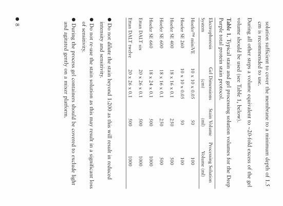
● 8 solution sufficient to cover the m
embrane to a m
inimum
depth of 1.5cm
is recomm
ended to use.
During all other steps a volum
e equivalent to ~20-fold excess of the gel
volume should be used (see Table 1, below
).
Table 1.Typical stain and gel processing solution volum
es for the Deep
Purple total protein stain protocol.
Electrophoresis
Gel D
imensions
Stain Volum
eProcessing Solution
System(cm
)(m
l)V
olume (m
l)
Hoefer™
miniV
E10 x 10 x 0.05
50100
Hoefer SE
26010 x 10 x 0.05
50100
Hoefer SE
40018 x 16 x 0.1
250500
Hoefer SE
60018 x 16 x 0.1
250500
Hoefer SE
66018 x 24 x 0.1
5001000
Ettan D
ALT
six20 x 26 x 0.1
5001000
Ettan D
ALT
twelve
20 x 26 x 0.1500
1000
●D
o not dilute the stain beyond 1:200 as this will result in reduced
intensity and sensitivity.
●D
o not re-use the stain solution as this may result in a significant loss
of sensitivity.
●D
uring the process gel containers should be covered to exclude lightand agitated gently on a m
ixer platform.
RPN6305PL Rev E 10/14/04 9:53 AM Page 8
● 9
Solutions and reagents required
Amersham
Biosciences reagents SD
S : PlusOne™
(code number 17-1313-01), U
SB reagent (U
S75819-100g or U
S85819-1kg) or similar high quality m
aterial (such as BD
HSpecially Pure grade).
Acrylam
ide gel and other related electrophoresis reagents are availablein the PlusO
ne range and are detailed in the current BioD
irectory™catalogue.
Additional reagents required Sodium
hydrogen carbonate (NaH
CO
3 ) and sodium carbonate
(Na
2 CO
3 ).
High purity w
ater (double distilled, RO
or equivalent).
Acetic acid, glacial.
Methanol/ethanol.
Note:H
igh purity water (R
O quality or better) should be used as a
diluent for Deep Purple total protein stain and for preparing all gel
processing solutions.
Fixation solution for gels7.5%
(v/v) acetic acid / 10% (v/v) m
ethanol or ethanol.
Add 75 m
l acetic acid and 100 ml m
ethanol to 825 ml w
ater.
Wash solution for large and backed gels
35 mM
sodium hydrogen carbonate
(NaH
CO
3 ) and 300 mM
sodiumcarbonate (N
a2 C
O3 ) in w
ater.
This can be achieved by dissolving 2.94 g of sodium
hydrogencarbonate (N
aHC
O3 ) and 31.8 g of sodium
hydrogen carbonate in 750m
l of water and adding w
ater to a final volume of 1000 m
l. The pH
of
RPN6305PL Rev E 10/14/04 9:53 AM Page 9
● 1
0
the solution should be pH 10-11 and should be verified. T
his solutioncan be stored for up to 2 w
eeks.
Wash solution for free floating gels and blots
200 mM
sodium carbonate (N
a2 C
O3 ) in w
ater.
This can be achieved by dissolving 21.2 g of sodium
carbonate(N
a2 C
O3 ) in 750 m
l of water and adding w
ater to a final volume of
1000 ml. T
he pH of the solution should be at least 11 and should be
verified. This solution can be stored for up to 2 w
eeks.
Working stain solution for gels and blots
1 in 200 dilution of Deep Purple in w
ater.
For the appropriate volume to use refer to Table 1 (see page 8). T
hissolution should be m
ade fresh at the time of use by adding an
appropriate aliquot of Deep Purple to w
ater in the gel staining tank. Ifnecessary it is possible to store this solution, protected from
exposureto light, for up to 1 w
eek at 2-8 °C or 24h at room
temperature.
Stabilization solution for gels7.5%
(v/v) acetic acid.
Make a 7.5%
acetic acid solution by adding 75 ml glacial acetic acid to
925 ml w
ater.
Methanol/Acetic acid solution for w
ashing of PVDF blots
10% (v/v) m
ethanol + 7.5% acetic acid.
Make a 10%
methanol / 7.5%
acetic acid solution by adding 75 ml of
glacial acetic and 100 ml of m
ethanol to 825 ml w
ater.
RPN6305PL Rev E 10/14/04 9:53 AM Page 10
● 1
1
Protocol
❶ Gel electrophoresis
1.1.Perform electrophoresis according to established techniques.
Note: If visual orientation is required on 1D
gels, Rainbow
™ M
arkers(R
PN800) m
ay be used. If a tracking dye is used, such as bromophenol
blue in the loading buffer, it is recomm
ended to run the dye front justoff the bottom
of the gel. For blotting it is particularly important that
the tracking dye and ion front are run off the base of the gel as ionscan interfere w
ith protein binding to the mem
brane.
1.2. If blotting, perform electro-transfer according to established
techniques and proceed to step 4.
❷ Fixation
2.1.Place an appropriate volume of 7.5%
(v/v) acetic acid / 10%(v/v)
methanol into the containers that w
ill be used to process gels. The
recomm
ended volume of fixation solution required is ~20 fold excess of
the gel volume (see Table 1, page 8).
Note:A
lternative fixation solutions that have been used successfullyw
ith Deep Purple total protein stain are:
●7.5%
acetic acid / 10% ethanol
●7.0%
acetic acid / 30% ethanol
2.2.Dism
antle the electrophoresis apparatus.
●For free floating gels rem
ove the gel from the plates by floating the
gel off with gentle agitation in the fix solution.
●For backed gels place the gel and plate directly into the fix solution.
Note:Place only one gel in each container. T
he stacking gel can be left
RPN6305PL Rev E 10/14/04 9:53 AM Page 11
● 1
2
attached to help with gel orientation.
2.3.Incubate in the fixation solution, for a minim
um of 1 hour, at
room tem
perature with gentle agitation.
Note:O
vernight fixation should be used for backed gels, large format
gels and thick gels (≥1.5 m
m) and it is also recom
mended for
applications where m
aximum
sensitivity is required.
❸Gel Staining
3.1.Take the stain out of the -15 °C to -30 °C
freezer and allow to
stand at room tem
perature for 5–10 minutes.
3.2.Pour off the fixation solution and replace with the w
ash solutionin ~20 fold excess (See Table 1, page 8 for all volum
es). Wash w
ithgentle agitation for 30 m
inutes.
Note: For backed gels and thick gels, the w
ash solution should be 35 m
M sodium
hydrogen carbonate (NaH
CO
3 ) and 300 mM
sodiumcarbonate (N
a2 C
O3 ) and for free floating gels the w
ash solution shouldbe 200 m
M sodium
carbonate (Na
2 CO
3 ).
3.3.Pour off the wash solution and replace w
ith water (10 fold excess
of the gel volume). B
riefly shake the stain concentrate and add Deep
Purple to make a 1:200 dilution. C
over the container to create a darkenvironm
ent and incubate for 1 hour at room tem
perature with gentle
agitation.
Note:T
he solution is light sensitive and should be kept out of brightlight.
Note:C
ontainers can be wrapped in foil or covered w
ith black plastic.It is not necessary to elim
inate light completely, only to ensure that
bright light is significantly reduced. Alternatively, containers, w
ith lids,that are a solid colored plastic m
ay be used.
3.4.Pour off the stain and replace with 7.5%
(v/v) acetic acid. Cover
RPN6305PL Rev E 10/14/04 9:53 AM Page 12
● 1
3
the container to create a dark environment and incubate w
ith gentleagitation for at least 15 m
inutes. Repeat the acetic acid step once. T
hegel can be im
aged at this stage.
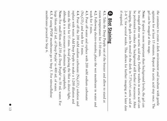
Note:
If speed is more im
portant than background levels, the gel canbe im
aged after one acetic acid step. Further washes in acetic acid can
be performed to reduce the background still further if necessary. A
fterim
aging, the gels can be stored in the dark in 7.5%(v/v) acetic acid at
2–8 ºC for several w
eeks. This allow
s for further imaging at a later date
if required.
➍Blot Staining
4.1.Take the Deep Purple out of the freezer and allow
to stand atroom
temperature for 5-10 m
ins.4.2. Follow
ing electro-transfer, place the wet m
embrane in w
ater andw
ash for 5 mins.
4.3. Pour off water and replace w
ith 200 mM
sodium carbonate
(Na
2 CO
3 ) and wash for 5 m
ins.4.4.Pour off the 200 m
M sodium
carbonate (Na
2 CO
3 ) solution andreplace w
ith water. A
dd Deep Purple to m
ake a 1:200 dilution andstain the blot for 15 m
ins. Avoid placing the blot in direct light,
although it is not necessary to eliminate light com
pletely. N
ote:For small blots add 250 µL
Deep Purple‘ to 50 m
L w
ater. Forlarge blots add 2 m
L of D
eep Purple‘ to 400 mL
of water.
4.5. If using PVD
F mem
branes go to Step 5. For nitrocellulosem
embranes proceed to Step 6.
RPN6305PL Rev E 10/14/04 9:53 AM Page 13
● 1
4
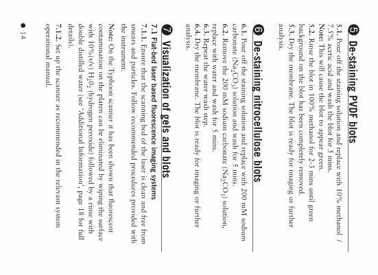
➎De-staining PVDF blots
5.1.Pour off the staining solution and replace with 10%
methanol /
7.5% acetic acid and w
ash the blot for 5 mins.
Note: T
his will cause the blot to appear green.
5.2.Rinse the blot in 100%
methanol for 2-3 m
ins until greenbackground on the blot has been com
pletely removed.
5.3.Dry the m
embrane. T
he blot is ready for imaging or further
analysis.
➏De-staining nitrocellulose blots
6.1.Pour off the staining solution and replace with 200 m
M sodium
carbonate (Na
2 CO
3 ) solution and wash for 5 m
ins.6.2.R
emove the 200 m
M sodium
carbonate (Na
2 CO
3 ) solution,replace w
ith water and w
ash for 5 mins.
6.3.Repeat the w
ater wash step.
6.4.Dry the m
embrane. T
he blot is ready for imaging or further
analysis.
➐Visualization of gels and blots
7.1
Flat-bed laser based fluorescence imaging system
s7.1.1.E
nsure that the scanning bed of the laser is clean and free fromsm
ears and particles. Follow recom
mended procedures provided w
iththe instrum
ent.
Note: O
n the Typhoon scanner it has been shown that fluorescent
contamination on the platen can be elim
inated by wiping the surface
with 10%
(v/v) H2 0
2(hydrogen peroxide) follow
ed by a rinse with
double distilled water (see ‘A
dditional Information’, page 18 for full
details).
7.1.2.Set up the scanner as recomm
ended in the relevant systemoperational m
anual.
RPN6305PL Rev E 10/14/04 9:53 AM Page 14
● 1
5
For example, the follow
ing settings are recomm
ended for use with a
Typhoon scanner;
Excitation : G
reen laser (532 nm)
Em
ission: 560LP or 610B
P filter.
Pre-scan using 1000 micron resolution and then scan using a 100
micron resolution.
Note:If the pre-scan show
s saturated bands/spots, reduce the PMT
voltage rating and pre-scan again. If the pre-scan shows too low
signalincrease the PM
T voltage rating and pre-scan again. D
eep Purple canalso be im
aged on a Typhoon scanner using the blue laser (457 nm or
488 nm). If using an alternate fluorescent scanner, for the best optim
alim
ages, scan using as similar settings as possible to those
recomm
ended.
7.1.3.Process the image according to experim
ental requirements and
the instructions for the relevant software program
.
7.2
Imaging w
ith UV light sources
7.2.1.Place the gel onto the UV
transilluminator or (blacklight blue
365 nm w
avelength emission is recom
mended) and follow
theoperating and safety instructions as relevant for the excitationinstrum
ent and imaging system
. Images can be captured using
appropriate camera system
s and filters (film, video, C
CD
).
Note: For long periods of illum
ination it is advisable to place the gelon a glass plate, raised on spacers above the transillum
inator, in orderto reduce heat dam
age to the stained proteins. Cooling the gel prior to
visualization can also help reduce fading.
RPN6305PL Rev E 10/14/04 9:53 AM Page 15
● 1
6
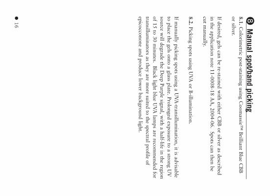
➑M
anual spot/band picking8.1.C
olorimetric post-staining using C
oomassie™
Brilliant B
lue CB
Bor silver.
If desired, gels can be re-stained with either C
BB
or silver as describedin the application note 11-0008-18 A
A, 2004-06. Spots can then be
cut manually.
8.2.Picking spots using UV
A or B
-illumination.
If manually picking spots using a U
VA
-transillumination, it is advisable
to place the gels onto a glass plate. Prolonged exposure to a strong UV
source will degrade the D
eep Purple signal, with a half-life in the region
of 15 to 30 minutes. B
lack light blue UV
A lam
ps are recomm
ended fortransillum
inators as they are more suited to the spectral profile of
epicocconone and produce lower background light.
RPN6305PL Rev E 10/14/04 9:53 AM Page 16
● 1
7
Additional information
Re-staining of gels G
els that have lost sensitivity over time or through incorrect storage
can be re-stained. In addition gels can be photo-bleached by prolongedexposure to U
V light and then re-stained. In all cases the m
ain protocolshould be follow
ed starting at the ‘Staining’ process (step 3, page 12).
Alternative staining trays Staining gels w
ith Deep Purple in stainless steel trays and the Processor
Plus gives comparable results. T
he limitation for large gels in Processor
Plus is that the maxim
um volum
e for the 29 x 35 cm tray is 400 m
l, sow
e recomm
end that the Processor Plus should be programm
ed with the
double number of steps. T
he use of Processor Plus gives higher andlow
er signal intensity for small and large gels, respectively. T
he use ofstainless steel trays as an alternative to the plastic trays results in tw
o-fold less sensitivity in a dilution series on a SD
S-PAG
E gel and few
erspots in 2-D
gel patterns.
Alternative imaging instrum
entsIm
aging is best performed using laser scanning fluorescence flat bed
imaging system
s, such as the Typhoon imager. T
his instrument can be
used to excite Deep Purple at 457 nm
, 488 nm or 532 nm
with
emission being collected through a 560 nm
long pass filter or a 610 nmband pass filter. W
hen using alternative laser based fluorescencescanners select laser w
avelength and emission filters that are closest to
those presented in the main protocol. O
ptimization of the scanning
process may be required to account for the relative pow
er of differentlasers and the use of alternative filter settings.
Many m
akes of UV
transilluminator are available that produce light of
UV
A (eg 365 nm
wavelength). B
lacklight blue UV
A sources are
particularly compatible w
ith gels stained with D
eep Purple.
RPN6305PL Rev E 10/14/04 9:53 AM Page 17
● 1
8
Gels excited by U
V light can be visualized in a num
ber of ways but if
further image analysis is required it is recom
mended that the im
age iscaptured on a C
CD
camera system
, such as the BioC
hemi™
Darkroom
(UV
P); the ethidium brom
ide (610 nm band pass) em
ission filter hasbeen used to give satisfactory im
ages from D
eep Purple stained gels.A
n alternative is imagers based on black light blue light sources. T
heseare sufficiently close to the excitation peak of D
eep Purple to produceuseful fluorescence.
Use of Deep Purple w
ith Ettan DIGE D
eep Purple fits into the standard 2D gel electrophoresis w
ork-flowand is particularly suitable for use w
ith the Ettan D
IGE
system. T
herecom
mended w
ork-flow involves the m
atching of Deep Purple post-
stained preparatory gels with C
yDye pre-labeled analytical gels.
How
ever, if it is necessary to post-stain analytical gels with D
eepPurple, it is recom
mended to im
age the protein stain using 457 nmlaser excitation in conjunction w
ith a 610 nm band pass em
ission filter(or equivalent if not using a Typhoon scanner). T
his will m
inimize any
potential cross-talk between D
eep Purple and the CyD
ye DIG
E fluors.
Deep Purple has been show
n to be compatible w
ith DeC
yder differenceanalysis softw
are and the stain is compatible w
ith manual or
automated spot picking and m
ass spectrometry for protein
identification applications.
Cleaning of imaging instrum
ents D
eep Purple total protein stain may leave a fluorescent residue on the
scanner platen. If the platen is not thoroughly cleaned, this residue caninterfere w
ith subsequent scans producing high background levels.
The follow
ing cleaning procedure has been shown to be com
patiblew
ith the Typhoon fluorescent imager to rem
ove contamination caused
by fluorescent products. Com
patibility of this procedure with
alternative instruments w
ould need to be investigated.
RPN6305PL Rev E 10/14/04 9:53 AM Page 18
● 1
9
●W
ipe the platen with 10%
H2 0
2(hydrogen peroxide).
●R
inse the platen with high purity w
ater.
●T
his operation should be carried out using lint free cloths, such asC
rew W
ipers. A pre-scan can be done to check for contam
inants thatm
ay affect results of scans.
●If the scanner is shared and used for scanning dyes/stains other than
Deep Purple, it is suggested that 1D
or free floating gels are placedonto a clean glass plate for scanning purposes. T
his will reduce the
possibility of cross contamination. If this m
ethod is used, the scanner‘Focal plane’ setting m
ay need to be set to +3mm
instead of platen (ifpossible).
Cleaning and preparation of Bind-Silane coated plates For com
plete removal of old gel and B
ind-Silane:
●T
horoughly scrape off any residual bound gel with a plastic scraper.
●W
ash the plates in freshly prepared 1%(v/v) D
econ™ (branded
Contrad™
in the USA
) and wash w
ith a soft sponge or brush tofurther rem
ove the gel.
●L
eave the plate to soak in 1%(v/v) D
econ overnight.
●T
he following day, w
ash the plate with a soft sponge.
●R
inse the plate with w
ater and leave the plate to soak in 1%(v/v)
HC
l for 1 hour.
●W
ash the plate in 1%(v/v) D
econ with a soft sponge or brush, then
rinse with high purity w
ater and leave to dry.
●B
efore using the plate, wipe over w
ith ethanol, using a lint free clothsuch as C
rew W
ipers.
●B
ind-Silane should be applied to plates using a lint free cloth so thatthe solution is spread evenly. L
ong strokes, from one edge of the plate
to the other, should be used, until the plate looks dry. The plate
RPN6305PL Rev E 10/14/04 9:53 AM Page 19
● 2
0
should then be covered with a dry cloth to prevent particulate
contamination and left for at least 2 hours to dry com
pletely.
RPN6305PL Rev E 10/14/04 9:53 AM Page 20
● 2
1
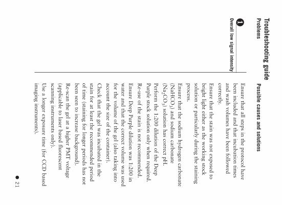
❶Overall low signal intensity
Ensure that all steps in the protocol have
been included and that incubation times
and wash volum
es have been followed
correctly.
Ensure that the stain w
as not exposed tobright light either as the w
orking stocksolution or particularly during the stainingprocess.
Ensure that the sodium
hydrogen carbonate(N
aHC
O3 ) and sodium
carbonate(N
a2 C
O3 ) solution has correct pH
.
Perform the 1:200 dilution of the D
eepPurple stock solution only w
hen required.
Re-use of the stain is not recom
mended.
Ensure D
eep Purple dilution was 1:200 in
water and that the correct volum
e was used
for the volume of the gel (also taking into
account the size of the container).
Check that the gel w
as incubated in thestain for at least the recom
mended period
of time (staining for longer periods has not
been seen to increase background).
Re-scan the gel at a higher PM
T voltage
(applicable to laser based fluorescentscanning instrum
ents only).
Use a longer exposure tim
e (for CC
D based
imaging instrum
ents).
Troubleshooting guideProblem
sPossible causes and solutions
RPN6305PL Rev E 10/14/04 9:53 AM Page 21
● 2
2
❷
Overall elevatedbackground
❸Localized patches of highbackground signal
Fading of stain in a stored gel – ensure gelsare stored in the recom
mended conditions
and protected from light. R
e-stain the gel ifnecessary.
Ensure correct fixation solution w
as used.
Prolong fixation to an overnight step.
Ensure that the sodium
hydrogen carbonate(N
aHC
O3 ) and sodium
carbonate(N
a2 C
O3 ) solution has correct pH
.
Perform extra w
ashes with 7.5%
(v/v) aceticacid after the staining step.
Ensure correct volum
es of all solutions havebeen used for the volum
e of gel (also takinginto account the size of the container).
Ensure that the platen of the im
agingsystem
is not contaminated w
ith fluorescentcom
pounds.
When using thicker gels or backed gels use
an overnight fixation step.
Use a high quality SD
S in the preparationand running of the gel (such as the PlusO
necode num
ber 17-1313-01 or USB
productU
S75819); replace pre-made running
buffers if necessary.
Platen of the imaging system
may be
contaminated w
ith fluorescent compounds
– follow the recom
mended cleaning
procedure.
RPN6305PL Rev E 10/14/04 9:53 AM Page 22
● 2
3
❹Spots and streaks clearlyvisible in the background
❺Small ‘speckles’ are seen
on the image
The SD
S front on gels will be stained by
Deep Purple and w
ill appear as a dark bandacross the gel. T
racker dyes, such asbrom
ophenol blue, can absorb fluorescentlight resulting in a band that appearsclearer than the background. To avoid thistracker dyes should be run off the bottomof the gel.
Handle gels w
ith care as physical damage
to gels may give a background stain in that
area. Use clean pow
der free gloves.
If a dark stained area is seen at low m
ass /high pI on a 2D
gel ensure that the fixationsolution is as recom
mended and the
fixation step is performed overnight.
If using backed gels ensure that the gelplates are properly cleaned before use andthat all gel m
aterial from previous
experiments is com
pletely removed.
Ensure that B
ind-Silane coated plates areevenly coated, dried efficiently and freefrom
particulate contamination.
Platen of the imaging system
may be
contaminated w
ith fluorescent compounds
– follow the recom
mended cleaning
procedure.
Use clean pow
der free gloves.
Ensure gel containers are perfectly clean
and rinsed before use. Ensure containers are
RPN6305PL Rev E 10/14/04 9:53 AM Page 23
● 2
4
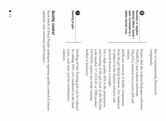
❻Boundary or negativestaining effects (bands /spots surrounded by alighter stained area)
➐Swelling of gels
free of contaminating fluorescent
compounds.
Ensure that the sodium
hydrogen carbonate(N
aHC
O3 ) and sodium
carbonate(N
a2 C
O3 ) solution has correct pH
andvolum
e.
Inefficient removal of buffer com
ponentsfrom
the gel during fixation. Use m
ethanolor ethanol in the fixation solution andperform
fixation overnight.
Use a high quality SD
S in the preparationand running of the gel (such as the PlusO
necode num
ber 17-1313-01 or USB
productU
S75819); replace pre-made running
buffers if necessary.
Swelling of free floating gels can be reduced
by adding 10% (v/v) m
ethanol in the finalacetic acid steps and the stabilizationsolution.
Quality control E
ach batch of Deep Purple undergoes rigorous quality control to ensure
optimum
and consistent performance.
RPN6305PL Rev E 10/14/04 9:53 AM Page 24
● 2
5
Related products A
mersham
Biosciences offers a com
prehensive range of electrophoresisreagents and hardw
are all with proven com
patibility to ensurereproducible high quality results. For a com
plete listing of productsavailable see the current A
urple total protein stain is exclusively licensedto A
mersham
Biosciences from
Fluorotechnics Pty
Ltd.
Deep P
urple total protein stain may only be used for
applications in life science research.
“CyD
ye: this product or portions thereof ism
anufactured under license from C
arnegie Mellon
University under patent num
ber 52
68
48
6 and other
patents pending. Som
e of these products may only be
available to collaborators and customers w
ithincertain of our technology access program
mes. The
purchase of CyD
ye DIG
EFluors includes a lim
itedlicense to use the C
yDye Fluors for internal research
and development, but not for any com
mercial
purposes. A license to use the C
yDye Fluors for
comm
ercial purposes is subject to a separate licenseagreem
ent with A
mersham
Biosciences’.
RPN6305PL Rev E 10/14/04 9:53 AM Page 26
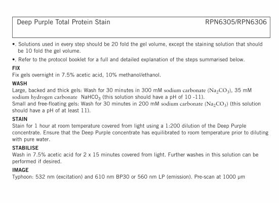
Deep Purple Total Protein Stain RPN6305/RPN6306
•. Solutions used in every step should be 20 fold the gel volume, except the staining solution that shouldbe 10 fold the gel volume.
•. Refer to the protocol booklet for a full and detailed explanation of the steps summarised below.
FIXFix gels overnight in 7.5% acetic acid, 10% methanol/ethanol.
WASHLarge, backed and thick gels: Wash for 30 minutes in 300 mM sodium carbonate (Na2CO3), 35 mMsodium hydrogen carbonate NaHCO3 (this solution should have a pH of 10 -11). Small and free-floating gels: Wash for 30 minutes in 200 mM sodium carbonate (Na2CO3) (this solutionshould have a pH of at least 11).
STAINStain for 1 hour at room temperature covered from light using a 1:200 dilution of the Deep Purpleconcentrate. Ensure that the Deep Purple concentrate has equilibrated to room temperature prior to dilutingwith pure water.
STABILISEWash in 7.5% acetic acid for 2 x 15 minutes covered from light. Further washes in this solution can beperformed if desired.
IMAGETyphoon: 532 nm (excitation) and 610 nm BP30 or 560 nm LP (emission). Pre-scan at 1000 µm
RPN6305PL Rev E 10/14/04 9:53 AM Page 27
RPN6305PC Rev E 2004
resolution to determine optimal PMT settings then scan at 100 µm resolution with optimal PMT. Otherimaging advices: For more details see Additional information in protocol booklet.
STORAGEAfter imaging, the gels can be stored in the dark in 7.5% acetic acid at 2-8 degrees for several weeks. Thisallows for further imaging at a later date if required.
Deep Purple is a trademark of Amersham Biosciences Limited.
Amersham and Amersham Biosciences are trademarks of Amersham plc.
GE and GE Monogram are trademarks of General Electric CompanyAll goods and services are sold subject to the terms and conditions of sale of the company within the AmershamBiosciences Group which supplies them. A copy of these terms and conditions is available on request
WarningFor research use only.Not recommended or intended for diagnosis of disease inhumans or animals. Do not use internally or externally in humans or animals.
http://www.amersham.com
Amersham Biosciences UKLimited Amersham Place Little Chalfont Buckinghamshire HP7 9NA UK
Amersham Biosciences AB SE-751 84 Uppsala Sweden
Amersham Biosciences Corp 800 Centennial Avenue PO Box 1327 Piscataway NJ 08855 USA
Amersham Biosciences Europe GmbH Munzinger Strasse 9 D-79111 Freiburg Germany