Running head: CALCIUM HANDLING PROTEINS IN THE HEART OF TUMOR BEARING MICE CALCIUM HANDLING PROTEINS IN THE HEART OF TUMOR BEARING MICE Kara C. Brooks The Ohio State University College of Nursing
Transcript
Running head: CALCIUM HANDLING PROTEINS IN THE HEART OF TUMOR
BEARING MICE
CALCIUM HANDLING PROTEINS IN THE HEART OF TUMOR BEARING MICE
Kara C. Brooks
The Ohio State University College of Nursing
CALCIUM HANDLING PROTEINS IN THE HEART OF TUMOR BEARING MICE
2
Calcium handling proteins in the heart of tumor bearing mice
Cachexia is a metabolic syndrome characterized by marked and unintentional
weight loss and muscle atrophy. Other complications of this syndrome include
fatigability, weakness, and loss of appetite. Cachexia can result from numerous sources
such as: burns, AIDS, COPD, and cancer among others. The occurrence of cachexia
reduces quality of life and indicates a poor prognosis with increased mortality. Currently,
the means by which cachexia develops are poorly understood and additionally, there are
no standard clinical methods approved to treat this syndrome. We hope the findings of
our research can be applied clinically to patients afflicted with cachexia. Preliminary
research in animal models of cachexia show depressed cardiac function, however, the
molecular mechanism of dysfunction remains unknown. Using the heart tissue of mice
injected with the Colon 26 adenocarcinoma, we looked at how the pathology affected the
calcium channel handling proteins in the heart. To do this, we initially performed qPCR
to identify gene expression changes that were significantly elevated or depressed in the
tumor mice. After identifying possible gene expression targets we looked at protein
levels using immunoblotting. Our results from the qPCR showed that Ryanodine
Receptor 2 (RyR2) was elevated in the tumor mice when compared to control mice.
Phospholamban (PLN) was also significantly depressed in the tumor mice when
compared to control mice. At the protein level, we found significantly increased
phosphorylated RyR2 (p-RyR2) with no change in total RyR2 levels. We also found no
significant changes in PLN in terms of phosphorylation or total protein. Our findings may
indicate that increased p-RyR2 is causing “leaky” Ca2+ channels, which may play a part
in cardiac dysfunction. This indicates calcium handling is altered in the tumor-bearing
CALCIUM HANDLING PROTEINS IN THE HEART OF TUMOR BEARING MICE
3
mice, which is possibly contributing to cardiac dysfunction. This information furthers
our understanding of the dysfunction in the heart with possible translational benefits.
Patients affected with cachexia that have increased levels of p-RyR2 may be at risk for
tachycardia or arrhythmia if calcium levels are unable to normalize.
Keywords: cancer, cachexia, calcium, proteins
CALCIUM HANDLING PROTEINS IN THE HEART OF TUMOR BEARING MICE
4
CHAPTER 1: STATEMENT OF THE PROBLEM
Introduction/Background of the Problem/Significance of the Study:
Cancer is a malignant neoplasm, a collection of uncontrollably dividing cells
forming a solid mass. It is one of the major health concerns in modern history. In the
United States, cancer is the second leading cause of death behind cardiovascular disease,
while in the United Kingdom, it is the leading cause of death. The morbidity rate of
cancer in the United States is approximately 1 in 3, meaning a third of the population will
develop cancer at some point in their lifetime. Of the people, one in four will die as a
direct result of cancer (Fearon, K. et al., 2012). Cancer, like other chronic disease, may
cause a syndrome of cachexia to develop in the patient. Cachexia is the rapid loss of both
adipose and muscle tissue that cannot be nutritionally prevented. Cancer patients have
approximately a 1 in 2 chance to develop cachexia, with some cancer having higher
incidences of cachexia. Of patients who develop cancer-induced cachexia (CIC)
approximately 1 in 4 will die as a direct result of cachexia. (Fearon, K. et al., 2012). CIC
has been poorly understood and misdiagnosed due to no common clinical standards to
measure cachexia. Cancer, and its treatment, is expected to produce mild weight loss and
anorexia, so often CIC was misinterpreted as a continuation of anorexia. A defining
feature of cachexia, when compared to anorexia, is that cachexia is not nutritionally
reversible. High caloric treatments such as Jevity, an Abbott produced 1.5-calories/ml
nutritional product, has only been shown to stabilize weight loss but not restore lost
weight (Fearon, K. et al., 2012). In an effort to address this treatment problem, in 2008,
a group of clinicians and scientists defined cachexia as a complex metabolic syndrome
associated with underlying illness and characterized by loss of muscle with or without
CALCIUM HANDLING PROTEINS IN THE HEART OF TUMOR BEARING MICE
5
loss of fat mass (Evans, W. et al., 2008, p. 794). A clinical guideline was also formulated
in that cachexia should be suspected if the patient lost 5% of the pre-morbid body mass
over a 6-month period, especially when treated with high caloric compounds (Evans, W.
et al., 2008, p. 794). The prominent clinical feature of cachexia is weight loss in adults
(corrected for fluid retention) or growth failure in children (excluding endocrine
disorders). Anorexia, inflammation, insulin resistance and increased muscle protein
breakdown are frequently associated with cachexia. Cachexia is distinct from starvation,
age-related loss of muscle mass (sarcopenia), primary depression, malabsorption and
hyperthyroidism, in that there is a loss of muscle mass, and is associated with increased
morbidity (Evans, W. et al., 2008, p. 794).
Cachexia is a metabolic syndrome characterized by dramatic and unintentional
body mass loss and muscle atrophy. Other symptoms of this disease include fatigue,
weakness and loss of appetite, which are thought to be a result of muscle atrophy. A
patient’s weight loss and other symptoms cannot be reversed nutritionally (Fearon, K., et
al., 2011, p. 490). Cachexia accompanies several chronic disease processes, including
cancer. Cachexia in the setting of cancer is referred to as cancer-induced cachexia, or
simply cancer cachexia. If the patient presents with cachectic symptoms, the likelihood
of death from the primary condition increases. Currently, the means by which chronic
diseases such as cancer cause cachexia are poorly understood. Treatment seeks to
stimulate the anabolic processes (muscle building), while inhibiting the catabolic
processes (muscle breakdown). There are no widely accepted drugs or treatment
methods to treat this syndrome. Treatment approaches such as caloric supplements,
prescribed diets, nutritional supplements, weight-bearing activities and medications
CALCIUM HANDLING PROTEINS IN THE HEART OF TUMOR BEARING MICE
6
(testosterone, Thalidomide and medical marijuana, among others), have demonstrated
limited effect, or have been ineffective in reversing the symptoms of cachexia
(Muliawati, Y., et al., 2012, p. 159-161).
Our study’s results have several limitations worthy of noting. Few other studies
have evaluated the effects of cachexia on cardiac muscle, so comparison is difficult. Also,
our experiments have a sample of 6 control mice and 6 colon26 tumor-bearing mice.
Although it does not affect the results of our study, there are limitations of applying our
findings in a clinical setting since animal models do not replicate all aspects of humans.
Continued replication and application to other models is necessary.
CHAPTER 2: REVIEW OF THE LITERATURE
Due to limited literature available on the effects of tumor induced cardiovascular
dysfunction, the review focused on the more understood mechanisms of heart failure.
Heart failure has been traditionally viewed and defined as a hemodynamic disease (Seta,
Y., et al., 1996). However, the inability of the hemodynamic hypothesis to explain the
progression of heart failure has lead to the development of the cytokine hypothesis (Seta,
Y., et al., 1996). Cytokines are a group of small molecular weight molecules that are
secreted by cells in response to a variety of stimuli, including environmental stress (Seta,
Y., et al., 1996). There are two major classes of cytokines that have been identified in
heart failure– vasoconstrictors and vasodepressors (Seta, Y., et al., 1996). According to
the cytokine hypothesis, heart failure progresses because cytokine cascades that are
activated following myocardial injury exert deleterious effects on the circulation (Seta,
Y., et al., 1996, p. 244). The cytokine hypothesis holds that the progression of heart
CALCIUM HANDLING PROTEINS IN THE HEART OF TUMOR BEARING MICE
7
failure begins because cytokine cascades exacerbate hemodynamic abnormalities or exert
direct toxic effects on the heart (Seta, Y., et al., 1996, p. 244). Cytokines do not
necessarily cause heart failure, but are responsible for its progression (Seta, Y., et al.,
1996). In fact, the initial cytokine release has an adaptive stress response that is
compensatory (Seta, Y., et al., 1996). However, each of these adaptive stress responses
has the potential to become overtly maladaptive with sustained overexpression (Seta, Y.,
et al., 1996).
Treatments have shifted with the emerging research of the cytokine hypothesis.
Most of the current standard-of-care drugs used for patients with chronic heart failure,
including β-adrenergic-receptor blockers (β-blockers), angiotensin-converting-enzyme
inhibitors (ACE inhibitors), and angiotensin II-receptor blockers (ARBS), are not
inotropic drugs; instead, they block the neurohumoral signaling by adrenergic and renin-
angiotensin pathways (Bers, D., 2007). As explained above, heart failure is accompanied
by neurohumoral changes that activate these pathways, probably as an initially adaptive
response that turns maladaptive by fuelling progressive remodeling and dysfunction
(Bers, D., 2007). Blocking these pathways can partially break this cycle and slow the
progression of heart failure (Bers, D., 2007). Treatments have moved away from
addressing the hemodynamic problems associated with heart failure and towards the
cytokine problems associated with heart failure.
Our research focuses on the Ca2+ handling proteins within the cardiomyocyte in
order to determine how the loss of Ca2+ is occurring. A normal action potential in the
heart muscle begins after depolarization of the cell. Upon membrane depolarization,
Ca2+ enters the myocyte through L-type Ca2+ channels. This influx triggers the
CALCIUM HANDLING PROTEINS IN THE HEART OF TUMOR BEARING MICE
8
Calcium-induced Calcium (CIC) release from the sarcoplasmic reticulum (SR) via
ryanodine receptors (RyRs) to amplify the Ca2+ current (Bers, D., 2005, p. 87). The
resultant rise in cytosolic Ca2+ activates the myofilaments – organized structures in the
cytoplasm composed of interdigitating filaments of either actin or myosin proteins – to
produce a cardiac contraction (Bers, D., 2005, p. 87). On activation, each myosin head
simultaneously grabs and pulls on an actin filament, in a process that uses the cellular
energy molecule ATP (Bers, D., 2007, p. 36). The coordinated contractile activity of the
myofilaments develops the forceful muscle contraction that ejects blood from the heart
(Bers, D., 2007, p. 36). To allow cardiac muscle to relax, cytosolic Ca2+ must decline
quickly (Bers, D., 2005, p. 87). This is accomplished by reestablishing the Ca2+ balance
within the myocyte (Bers, D., 2005, p. 87). The SR Ca2+ ATPase (SERCA) takes up the
amount of Ca2+ released from the SR back into the SR. The Ca2+ entering the cell via
the L-type Ca2+ current is removed via the Na/Ca2+ exchanger (NCX) (Bers, D., 2005,
p. 87). Ca2+ levels within the myocyte, therefore, influence the cardiac contraction.
Ca2+ levels can be affected by the stimulation rate (which affects total Ca2+ influx),
SERCA and NCX activity, and β-adrenergic stimulation (which phosphorylates
phospholamban and results in disinhibition of SERCA and increased SR Ca2+ uptake)
(Bers, D., 2005, p. 88). Acute malfunctions of these systems can result in both
mechanical and electrical dysfunction (e.g., reduced cardiac output and arrhythmias)
(Bers, D., 2007, p. 24).
In heart failure, a reduced amount of Ca2+ is available for release by the
sarcoplasmic reticulum, contributing to weaker myofilament activation and contraction
(Bers, D., 2007, p. 36). If the failing heart could be strengthened, patient outcomes
CALCIUM HANDLING PROTEINS IN THE HEART OF TUMOR BEARING MICE
9
would be more favorable (Bers, D., 2007, p. 36). Studies in animal models and from
failing human hearts have characterized the alterations in Ca2+ handling found in failing
myocardium (Bers, D., 2002, p. 198). Failing myocytes typically exhibit decreased Ca2+
transient amplitude, slowed decline, and a negative force-frequency relationship (Bers,
D., 2002, p. 203). Alterations in gene expression include down-regulation of SERCA and
ryanodine receptor and up-regulation of NCX (Bers, D., 2008, p. 32). These changes
play a significant role in decreasing the SR Ca2+ load available for release, the result of
which is the significant contractile dysfunction characteristic of HF myocytes (Bers, D.,
2002, p. 203). Increased diastolic leak of Ca2+ from the SR by altered ryanodine
receptor function could also contribute to the SR unloading seen in HF (Bers, D., 2002, p.
203).
CHAPTER 3: METHODOLOGY
Animal Husbandry
12-week-old female CD2F1 mice, obtained from Charles Rivers (Willmington,
MA), were housed in animal facilities at The Ohio State University. The mice were kept
in a 12 hour light and dark cycle under 37°C conditions. The mice were provided ad
libitum access to food and water and standard rodent chow. All protocols pertaining to
the mice were approved by the Institutional Animal Care and Use Committee (IACUC) at
The Ohio State University.
Murine Tumor Model
Briefly, the Colon-26 Adenocarcinoma (C26) cells were maintained in RPMI
1640 media supplemented with glutamine and penstrep. Before the injection, the cells
CALCIUM HANDLING PROTEINS IN THE HEART OF TUMOR BEARING MICE
10
were trypsinized, pelletized through centrifugation, and resuspended in PBS. Cells were
counted and a dilution was made for animal injection so that the amount of cells injected
subcutaneously was 5x10⁶. Before injection, the animals were induced under anesthesia
at 5% isoflurane and were maintained at 1% isoflurane during injection. Cells were
injected subcutaneously in between the scapula of the mouse. The mice were returned to
their home cage and observed to ensure their complete recovery from anesthesia. Tumor
growth was evident approximately 7-12 days post injection and the mice were moribund
by day 21 post injection. Mice were euthanized using a ketamine/xylazine cocktail and
left ventricular tissue was harvested and snap frozen in liquid nitrogen for further
processing.
RNA Isolation and qPCR
The tissue was briefly pulverized in Trizol Reagent (Sigma Aldrich, St. Louis,
MO) using a TissueLyzer (Qiagen, Valencia, CA) with 5mm stainless steel beads. After
pulverization, RNA was precipitated using chloroform to isolate RNA in the aqueous
phase. The RNA was further isolated and purified using the RNEasy Extraction Kit
(Qiagen, Valencia, CA). RNA was measured using a NanoDrop (ThermoScientific,
Waltham, MA) to standardize the amount of RNA for cDNA synthesis. CDNA was
synthesized using the iScript Reverse Transcription Supermix (Bio-Rad, Hercules, CA).
After cDNA synthesis, the cDNA was mixed with the appropriate template as well as
SYBR Green Master Mix (Bio-Rad, Hercules, CA) and placed into the CFX96 (Bio-Rad,
Hercules, CA) using a three step amplification protocol. The results were analyzed in
reference to the standard, GAPDH, using the Livak 2^(-ermix (Bio-Rad, Hercules, CA).
Western Blotting and Detection
CALCIUM HANDLING PROTEINS IN THE HEART OF TUMOR BEARING MICE
11
Proteins, except for RyR or PLN, were run using a pre-cast 4-15% TGX gel (Bio-
Rad, Herculues, CA) at 200 V for 30 minutes using the Bio-Rad Basic Power Pack (Bio-
Rad, Hercules, CA). After running, the gels were incubated in Tris-Glycine transfer
buffer for 30 minutes before being transferred onto methanol activated PVDF membranes
using the semi-dry system (Bio-Rad, Hercules, CA) at 10 V for 30 minutes.
RyR was run on an 8-12% Tris-Acetate Gradient Gel (Novus Biologicals,
Littleton, CO) at 200 V for 4 hours. Using a wet transfer system, the gels were
transferred onto menthol activated PVDF membranes at 10 mA overnight.
PLN was run on a 10-20% Tris-Tricine Gel (Bio-Rad, Hercules, CA) at 200 V for
1 hour. Using a semi-dry transfer system, the gels were transferred onto an activated
PVDF membrane at 10 V for 30 minutes.
All membranes were blocked using TBS Odyssey Blocking Buffer (Li-Cor,
Lincoln, NE) for 1 hour at room temperature. The blots were then incubated in primary
antibody suspended in 2.5% BSA at 4¹C overnight and washed 3 times for 5 minutes
using TBST. Near-infrared secondary antibodies (Li-Cor, Lincoln, NE) were suspended
in 2.5% BSA and the blot was incubated for 1 hour at room temperature. The blots were
then washed 3 times with TBST before being detected using the Li-Cor Odyssey XL
Laser Detection System (Li-Cor, Lincoln, NE). The emission values were recorded for
the detected bands and later used to quantify the relative amount of expression. A
separate 4-15% TGX (Bio-Rad, Hercules, CA) precast gel was run and stained with
Coomassie R250 for 2 hours at room temperature. The gel was then placed in destain
buffer until the actin band was clearly visible with minimal background. Using the 800
nm laser on the Li-Cor Odyssey XL Laser Detection System (Li-Cor, Lincoln, NE), the
CALCIUM HANDLING PROTEINS IN THE HEART OF TUMOR BEARING MICE
12
emission values were detected. These values were then used to calculate the fold change
for the protein of interest.
Assessment of Myosin Heavy Chain Composition
Left ventricular homogenates were prepared from control and tumor mice as
described subsequently. The myosin heavy chain isoforms in the left ventricular samples
were separated out using the SDS-PAGE. The separating gels consisted of 7%
acrylamide, with a 50:1 acrylamide:bis-acrylamide crosslinking ratio, and 5% (v/v)
glycerol as described previously. The gels were run in the Hoefer 630 Large Vertical
Slab Gel Electrophoresis unit for 21 hours at a constant voltage of 230. The unit was
cooled with circulating water to 8¹C. Then, the gels were stained using the sensitive
silver stain method as described previously. Once staining was finished, the reaction was
neutralized and a picture was taken for densitometry scanning in ImageJ in order to
compare the amount of each isoform.
Statistical Analyses
The protein fold changes and the results from the qPCR were put into the
statistical software Prism (GraphPad, La Jolla, CA) for statistical analysis and graphing.
The results from each study were analyzed using a Student’s T-Test. The results were
considered significant if p<0.05 and are indicated graphically by an asterisk.
CHAPTER 4: RESULTS
Reduced gene expression levels of PLN
The qPCR results showed a significant decrease of PLN gene expression in tumor
mice compared to control mice. There were no other calcium handling proteins found to
be significantly changed at the gene expression level. See Figure 5.1.
CALCIUM HANDLING PROTEINS IN THE HEART OF TUMOR BEARING MICE
13
Figure 5. 1: RNA expression changes of RyR, PLN, Cav1.2, and SERCA in the left ventricle of control and tumor mice. RNA expression levels were determined using the Livak method with GAPDH serving as the internal control. *p<0.05
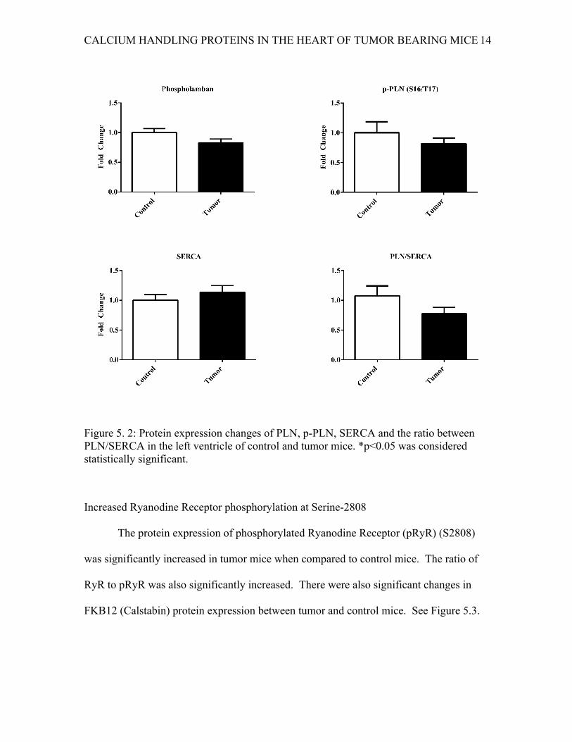
No change in PLN or phosphorylated-PLN (pPLN) or PLN/SERCA ratio
The PLN and pPLN protein expressions were not found to be significantly
changed in tumor mice compared to control mice. There was no change in the protein
expression of SERCA. The ratio of PLN to SERCA was also unchanged. See Figure 5.2.
CALCIUM HANDLING PROTEINS IN THE HEART OF TUMOR BEARING MICE
14
Figure 5. 2: Protein expression changes of PLN, p-PLN, SERCA and the ratio between PLN/SERCA in the left ventricle of control and tumor mice. *p<0.05 was considered statistically significant.
Increased Ryanodine Receptor phosphorylation at Serine-2808
The protein expression of phosphorylated Ryanodine Receptor (pRyR) (S2808)
was significantly increased in tumor mice when compared to control mice. The ratio of
RyR to pRyR was also significantly increased. There were also significant changes in
FKB12 (Calstabin) protein expression between tumor and control mice. See Figure 5.3.
CALCIUM HANDLING PROTEINS IN THE HEART OF TUMOR BEARING MICE
15
Figure 5. 3: Protein expression levels of RyR, pRyR, and FKB12 in the left ventricle of control and tumor mice as well as the ratio of pRyR/RyR. *p<0.05 was considered statistically significant.
Increased L-Type Calcium Channel protein expression
L-Type Calcium Channel protein expression was significantly increased in tumor
mice compared to controls. See Figure 5.4.
CALCIUM HANDLING PROTEINS IN THE HEART OF TUMOR BEARING MICE
16
Figure 5. 4: Protein expression change of the L-Type calcium channel in the left ventricle of control and tumor mice. *p<0.05 was consdiered statistically significant.
CALCIUM HANDLING PROTEINS IN THE HEART OF TUMOR BEARING MICE
17
Figure 5. 5: Representative immunoblots of calcium handling proteins as well as loading control in the left ventricle of control and tumor mice.
CHAPTER 5: CONCLUSIONS AND RECOMMENDATIONS/DISCUSSION
Our results indicate changes of the proteins involved in calcium induced calcium
release. We found significantly elevated protein expressions of both phosphorylated RyR
and L-Type calcium channel. We found significantly depressed RNA expression of PLN,
CALCIUM HANDLING PROTEINS IN THE HEART OF TUMOR BEARING MICE
18
but no changes in its protein expression. We did not find any changes in SERCA protein
expression, either.
RyR2 is the major mediator of CICR in animal cells. When activated RyR2
mediates the release of calcium ions from the SR. When RyR2 mediated Ca2+ release
increases, the intracellular Ca2+ increases and the contraction strengthens. However,
when RyR2 is hyper phosphorylated, it becomes “leaky.” Phosphorylated RyR2 causes
the channels to remain relaxed and in a semi-open state. This causes the SR to lose more
Ca2+, which means there is less Ca2+ in the SR for release and subsequent systolic
contractions are impaired. Diastolic (relaxation) Ca2+ release can trigger depolarization,
which results in cardiac arrhythmias (Bers, 2002, p. 203). Our findings may indicate that
hyper phosphorylated RyR2 is causing “leaky” Ca2+ channels, which may play a part in
the mice’s tachycardia.
Our results indicate that calcium handling may play a role in cardiac dysfunction
in cachexia. This information furthers our understanding of the dysfunction in the heart
with possible translational benefits. Patients affected by cachexia that have increased
levels of p-RyR2 may be at risk for tachycardia or arrhythmia if calcium levels are unable
to normalize.
CALCIUM HANDLING PROTEINS IN THE HEART OF TUMOR BEARING MICE
19
REFERENCES
Bers, D. (2002). Cardiac excitation-contraction coupling. Nature, 198-205.
Bers, D., & Harris, S. (2007). To the rescue of the failing heart. J. Am. Chem. Soc, 7–9.
http://doi.org/10.1038/473036a
Bers, D. M., & Guo, T. (2005). Calcium signaling in cardiac ventricular myocytes.
Annals of the New York Academy of Sciences, 1047, 86–98.
http://doi.org/10.1196/annals.1341.008
Evans, W. J., Morley, J. E., Argilés, J., Bales, C., Baracos, V., Guttridge, D., … Anker, S.
D. (2008). Cachexia: A new definition. Clinical Nutrition, 27(6), 793–799.
http://doi.org/10.1016/j.clnu.2008.06.013
Fearon, K. C. H., Glass, D. J., & Guttridge, D. C. (2012). Cancer cachexia: Mediators,
signaling, and metabolic pathways. Cell Metabolism, 16(2), 153–166.
http://doi.org/10.1016/j.cmet.2012.06.011
Fearon, K., Strasser, F., Anker, S. D., Bosaeus, I., Bruera, E., Fainsinger, R. L., …
Baracos, V. E. (2011). Definition and classification of cancer cachexia: An
international consensus. The Lancet Oncology, 12(5), 489–495.
http://doi.org/10.1016/S1470-2045(10)70218-7
Muliawati, Y., Haroen, L., Rotty, L. (2012). Cancer anorexia – cachexia syndrome. Acta
medica Indonesiana, 44 (2), 154-162.
CALCIUM HANDLING PROTEINS IN THE HEART OF TUMOR BEARING MICE
20
Seta, Y., Shan, K., Bozkurt, B., Oral, H., & Mann, D. L. (1996). Basic mechanisms in
heart failure: the cytokine hypothesis. Journal of Cardiac Failure, 2(3), 243–249.