Effects of Toxicants on Target MoleculesDysfunction of Target MoleculesDestruction of Target MoleculesNeoantigen Formation
Toxicity Not Initiated by Reaction with TargetMolecules
35
STEP 3—CELLULAR DYSFUNCTION AND RESULTANT TOXICITIES
Toxicant-Induced Cellular DysregulationDysregulation of Gene ExpressionDysregulation of Ongoing Cellular Activity
Toxic Alteration of Cellular MaintenanceImpairment of Internal Cellular Maintenance:
Mechanisms of Toxic Cell DeathImpairment of External Cellular Maintenance
STEP 4—REPAIR OR DYSREPAIR
Molecular RepairRepair of ProteinsRepair of LipidsRepair of DNA
Cellular Repair: A Strategy in Peripheral NeuronsTissue Repair
Apoptosis: An Active Deletion of Damaged CellsProliferation: Regeneration of TissueSide Reactions to Tissue Injury
When Repair FailsToxicity Resulting from Dysrepair
Tissue NecrosisFibrosisCarcinogenesis
CONCLUSIONS
Depending primarily on the degree and route of exposure, chemi-cals may adversely affect the function and/or structure of living or-ganisms. The qualitative and quantitative characterization of theseharmful or toxic effects is essential for an evaluation of the poten-tial hazard posed by a particular chemical. It is also valuable to un-derstand the mechanisms responsible for the manifestation of tox-icity—that is, how a toxicant enters an organism, how it interactswith target molecules, and how the organism deals with the insult.
An understanding of the mechanisms of toxicity is of bothpractical and theoretical importance. Such information provides arational basis for interpreting descriptive toxicity data, estimatingthe probability that a chemical will cause harmful effects, estab-lishing procedures to prevent or antagonize the toxic effects, de-signing drugs and industrial chemicals that are less hazardous, anddeveloping pesticides that are more selectively toxic for their tar-get organisms. Elucidation of the mechanisms of chemical toxic-ity has led to a better understanding of fundamental physiologicand biochemical processes ranging from neurotransmission (e.g.,curare-type arrow poisons) to deoxyribonucleic acid (DNA) repair
(e.g., alkylating agents). Pathologic conditions such as cancer andParkinson’s disease are better understood because of studies on themechanism of toxicity of chemical carcinogens and 1,2,3,6-tetrahydro-1-methyl-4-phenylpyridine (MPTP), respectively. Con-tinued research on mechanisms of toxicity will undoubtedly con-tinue to provide such insights.
This chapter reviews the cellular mechanisms that contributeto the manifestation of toxicities. Although such mechanisms aredealt with elsewhere in this volume, they are discussed in detail inthis chapter in an integrated and comprehensive manner. We pro-vide an overview of the mechanisms of chemical toxicity by re-lating a series of events that begins with exposure, involves a mul-titude of interactions between the invading toxicant and theorganism, and culminates in a toxic effect. This chapter focuses onmechanisms that have been identified definitively or tentatively inhumans or animals.
As a result of the huge number of potential toxicants and themultitude of biological structures and processes that can be im-paired, there are a tremendous number of possible toxic effects.
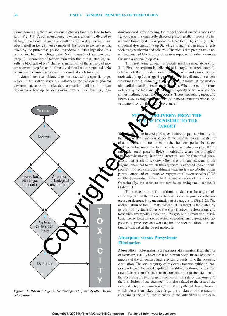
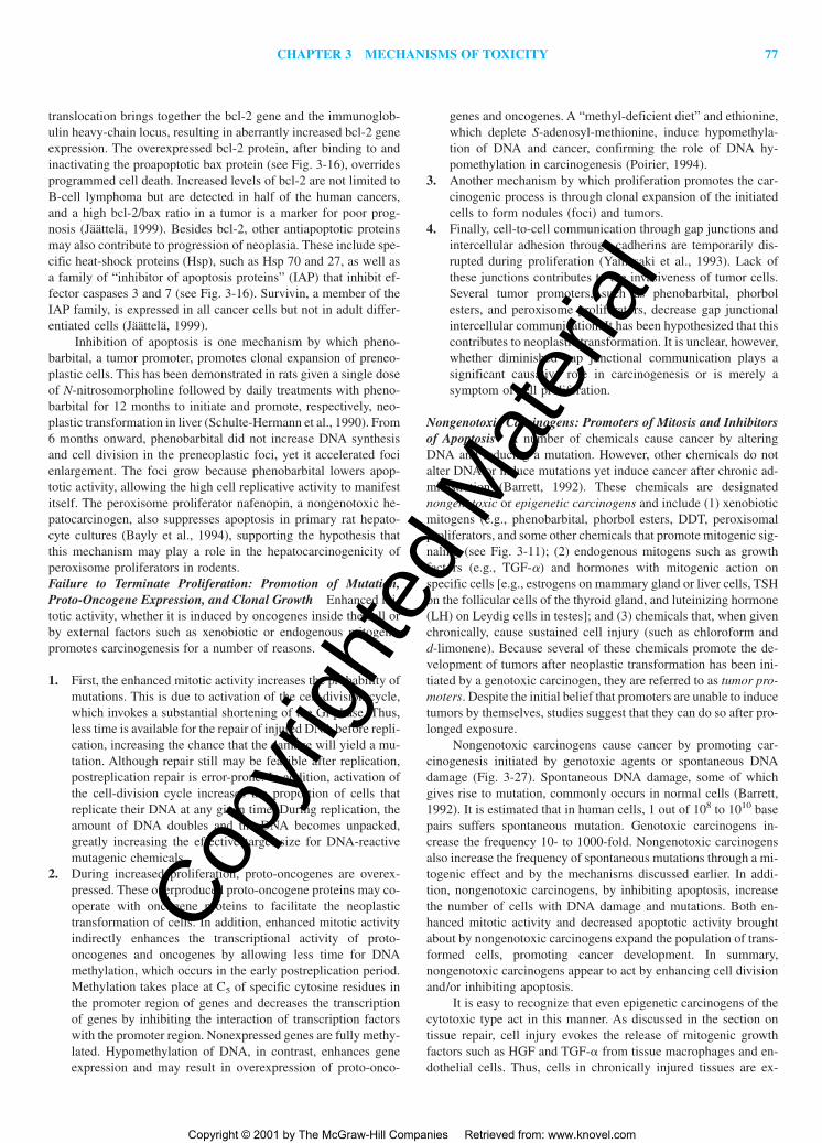
Correspondingly, there are various pathways that may lead to tox-icity (Fig. 3-1). A common course is when a toxicant delivered toits target reacts with it, and the resultant cellular dysfunction man-isfests itself in toxicity. An example of this route to toxicity is thattaken by the puffer fish poison, tetrodotoxin. After ingestion, thispoison reaches the voltage-gated Na� channels of motoneurons(step 1). Interaction of tetrodotoxin with this target (step 2a) re-sults in blockade of Na� channels, inhibition of the activity of mo-tor neurons (step 3), and ultimately skeletal muscle paralysis. Norepair mechanisms can prevent the onset of such toxicity.
Sometimes a xenobiotic does not react with a specific targetmolecule but rather adversely influences the biological (micro)environment, causing molecular, organellar, cellular, or organdysfunction leading to deleterious effects. For example, 2,4-
dinitrophenol, after entering the mitochondrial matrix space (step1), collapses the outwardly directed proton gradient across the in-ner membrane by its mere presence there (step 2b), causing mito-chondrial dysfunction (step 3), which is manifest in toxic effectssuch as hyperthemia and seizures. Chemicals that precipitate in re-nal tubules and block urine formation represent another examplefor such a course (step 2b).
The most complex path to toxicity involves more steps (Fig.3-1). First, the toxicant is delivered to its target or targets (step 1),after which the ultimate toxicant interacts with endogenous targetmolecules (step 2a), triggering perturbations in cell function and/orstructure (step 3), which initiate repair mechanisms at the molec-ular, cellular, and/or tissue levels (step 4). When the perturbationsinduced by the toxicant exceed repair capacity or when repair be-comes malfunctional, toxicity occurs. Tissue necrosis, cancer, andfibrosis are examples of chemically induced toxicities whose de-velopment follow this four-step course.
STEP 1—DELIVERY: FROM THESITE OF EXPOSURE TO THE
TARGET
Theoretically, the intensity of a toxic effect depends primarily onthe concentration and persistence of the ultimate toxicant at its siteof action. The ultimate toxicant is the chemical species that reactswith the endogenous target molecule (e.g., receptor, enzyme, DNA,microfilamental protein, lipid) or critically alters the biological(micro)environment, initiating structural and/or functional alter-ations that result is toxicity. Often the ultimate toxicant is the original chemical to which the organism is exposed (parent com-pound). In other cases, the ultimate toxicant is a metabolite of theparent compound or a reactive oxygen or nitrogen species (ROSor RNS) generated during the biotransformation of the toxicant.Occasionally, the ultimate toxicant is an endogenous molecule(Table 3-1).
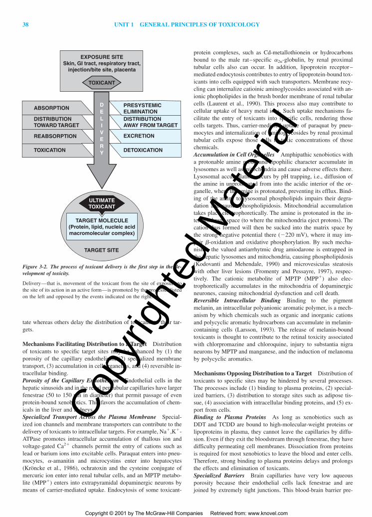
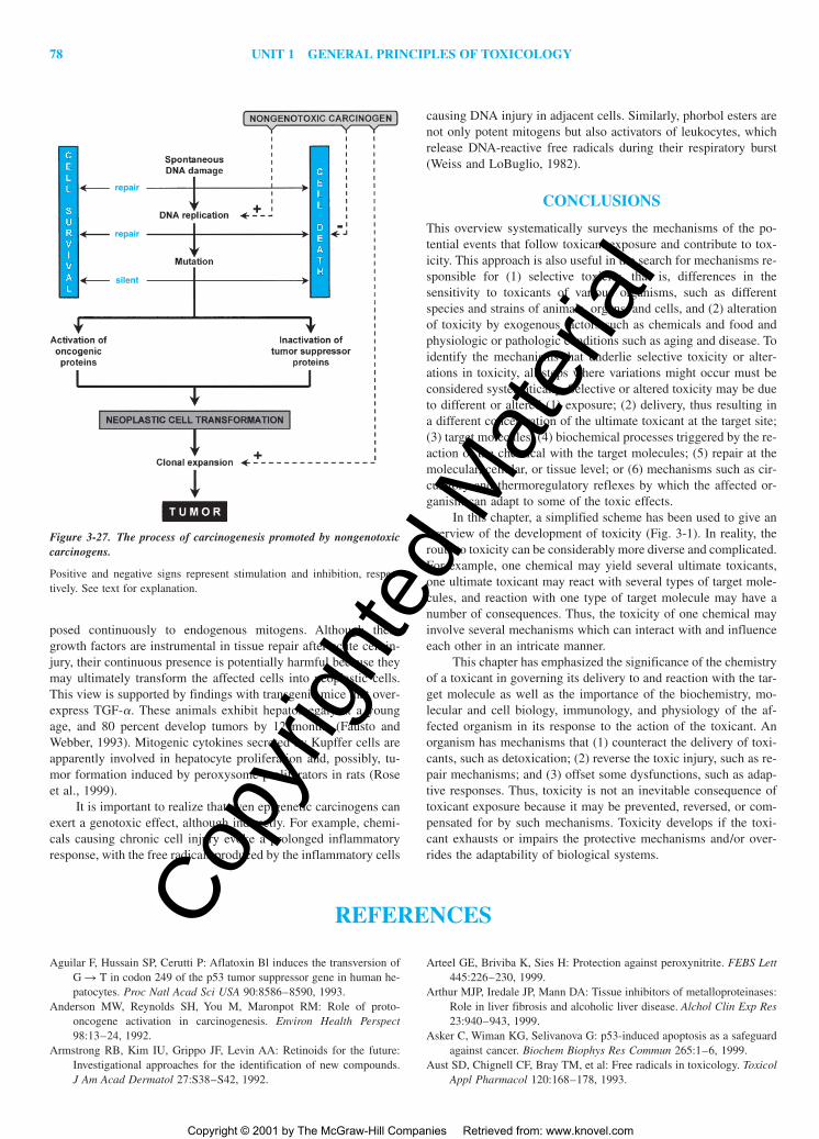
The concentration of the ultimate toxicant at the target mol-ecule depends on the relative effectiveness of the processes that in-crease or decrease its concentration at the target site (Fig. 3-2). Theaccumulation of the ultimate toxicant at its target is facilitated byits absorption, distribution to the site of action, reabsorption, andtoxication (metabolic activation). Presystemic elimination, distri-bution away from the site of action, excretion, and detoxication op-pose these processes and work against the accumulation of the ul-timate toxicant at the target molecule.
Absorption versus PresystemicElimination
Absorption Absorption is the transfer of a chemical from the siteof exposure, usually an external or internal body surface (e.g., skin,mucosa of the alimentary and respiratory tracts), into the systemiccirculation. The vast majority of toxicants traverse epithelial bar-riers and reach the blood capillaries by diffusing through cells. Therate of absorption is related to the concentration of the chemical atthe absorbing surface, which depends on the rate of exposure andthe dissolution of the chemical. It is also related to the area of theexposed site, the characteristics of the epithelial layer throughwhich absorption takes place (e.g., the thickness of the stratumcorneum in the skin), the intensity of the subepithelial microcir-
2a 2b
1
3
4
Delivery
Interactionwith targetmolecule
Alterationof biologicalenvironment
Cellulardysfunction,
injury
Dysrepair
Toxicant
TOXICITY
Figure 3-1. Potential stages in the development of toxicity after chemi-cal exposure.
culation, and the physicochemical properties of the toxicant. Lipidsolubility is usually the most important property influencing ab-sorption. In general, lipid-soluble chemicals are absorbed morereadily than are water-soluble substances.
Presystemic Elimination During transfer from the site of expo-sure to the systemic circulation, toxicants may be eliminated. Thisis not unusual for chemicals absorbed from the gastrointestinal (GI)tract because they must first pass through the GI mucosal cells,liver, and lung before being distributed to the rest of the body bythe systemic circulation. The GI mucosa and the liver may elimi-nate a significant fraction of a toxicant during its passage throughthese tissues, decreasing its systemic availability. For example,ethanol is oxidized by alcohol dehydrogenase in the gastric mucosa(Lim et al., 1993), cyclosporine is returned from the enterocyte intothe intestinal lumen by P-glycoprotein (an ATP-dependentxenobiotic transporter) and is also hydroxylated by cytochromeP450 (CP3A4) in these cells (Lin et al., 1999), morphine is glu-curonidated in the intestinal mucosa and the liver, and manganeseis taken up from the portal blood into the liver and excreted intobile. Such processes may prevent a considerable quantity of chem-icals from reaching the systemic blood. Thus, presystemic or first-pass elimination reduces the toxic effects of chemicals that reach
their target sites by way of the systemic circulation. In contrast,the processes involved in presystemic elimination may contributeto injury of the digestive mucosa, the liver, and the lungs by chem-icals such as ethanol, iron salts, �-amanitin, and paraquat becausethese processes promote their delivery to those sites.
Distribution to and Away from the Target
Toxicants exit the blood during the distribution phase, enter the ex-tracellular space, and may penetrate into cells. Chemicals dissolvedin plasma water may diffuse through the capillary endothelium viaaqueous intercellular spaces and transcelluar pores called fenestraeand/or across the cell membrane. Lipid-soluble compounds movereadily into cells by diffusion. In contrast, highly ionized and hy-drophilic xenobiotics (e.g., tubocurarine and aminoglycosides) arelargely restricted to the extracellular space unless specialized mem-brane carrier systems are available to transport them.
During distribution, toxicants reach their site or sites of action, usually a macromolecule on either the surface or the inte-rior of a particular type of cell. Chemicals also may be distributedto the site or sites of toxication, usually an intracellular enzyme,where the ultimate toxicant is formed. Some mechanisms facili-
Table 3-1Types of Ultimate Toxicants and Their Sources
Parent xenobiotics as ultimate toxicantsPb ionsTetrodotoxinTCDDMethylisocyanateHCNCO
tate whereas others delay the distribution of toxicants to their tar-gets.
Mechanisms Facilitating Distribution to a Target Distributionof toxicants to specific target sites may be enhanced by (1) theporosity of the capillary endothelium, (2) specialized membranetransport, (3) accumulation in cell organelles, and (4) reversible in-tracellular binding.Porosity of the Capillary Endothelium Endothelial cells in thehepatic sinusoids and in the renal peritubular capillaries have largerfenestrae (50 to 150 nm in diameter) that permit passage of evenprotein-bound xenobiotics. This favors the accumulation of chem-icals in the liver and kidneys.Specialized Transport Across the Plasma Membrane Special-ized ion channels and membrane transporters can contribute to thedelivery of toxicants to intracellular targets. For example, Na�,K�-ATPase promotes intracellular accumulation of thallous ion andvoltage-gated Ca2� channels permit the entry of cations such aslead or barium ions into excitable cells. Paraquat enters into pneu-mocytes, �-amanitin and microcystins enter into hepatocytes(Kröncke et al., 1986), ochratoxin and the cysteine conjugate ofmercuric ion enter into renal tubular cells, and an MPTP metabo-lite (MPP�) enters into extrapyramidal dopaminergic neurons bymeans of carrier-mediated uptake. Endocytosis of some toxicant-
protein complexes, such as Cd-metallothionein or hydrocarbonsbound to the male rat–specific �2u-globulin, by renal proximaltubular cells also can occur. In addition, lipoprotein receptor–mediated endocytosis contributes to entry of lipoprotein-bound tox-icants into cells equipped with such transporters. Membrane recy-cling can internalize catioinic aminoglycosides associated with an-ionic phopholipides in the brush border membrane of renal tubularcells (Laurent et al., 1990). This process also may contribute tocellular uptake of heavy metal ions. Such uptake mechanisms fa-cilitate the entry of toxicants into specific cells, rendering thosecells targets. Thus, carrier-mediated uptake of paraquat by pneu-mocytes and internalization of aminoglycosides by renal proximaltubular cells expose those cells to toxic concentrations of thosechemicals.Accumulation in Cell Organelles Amphipathic xenobiotics witha protonable amine group and lipophilic character accumulate inlysosomes as well as mitochondria and cause adverse effects there.Lysosomal accumulation occurs by pH trapping, i.e., diffusion ofthe amine in unprotonated from into the acidic interior of the or-ganelle, where the amine is protonated, preventing its efflux. Bind-ing of the amine to lysosomal phospholipids impairs their degra-dation and causes phospholipidosis. Mitochondrial accumulationtakes place electrophoretically. The amine is protonated in the in-termembrane space (to where the mitochondria eject protons). Thecation thus formed will then be sucked into the matrix space bythe strong negative potential there (�220 mV), where it may im-pair �-oxidation and oxidative phosphorylation. By such mecha-nisms, the valued antiarrhytmic drug amiodarone is entrapped inthe hepatic lysosomes and mitochondria, causing phospholipidosis(Kodovanti and Mehendale, 1990) and microvesiculas steatosiswith other liver lesions (Fromenty and Pessayre, 1997), respec-tively. The cationic metabolite of MPTP (MPP�) also elec-trophoretically accumulates in the mitochondria of dopaminergicneurones, causing mitochondrial dysfunction and cell death.Reversible Intracellular Binding Binding to the pigmentmelanin, an intracellular polyanionic aromatic polymer, is a mech-anism by which chemicals such as organic and inorganic cationsand polycyclic aromatic hydrocarbons can accumulate in melanin-containing cells (Larsson, 1993). The release of melanin-boundtoxicants is thought to contribute to the retinal toxicity associatedwith chlorpromazine and chloroquine, injury to substantia nigraneurons by MPTP and manganese, and the induction of melanomaby polycyclic aromatics.
Mechanisms Opposing Distribution to a Target Distribution oftoxicants to specific sites may be hindered by several processes.The processes include (1) binding to plasma proteins, (2) special-ized barriers, (3) distribution to storage sites such as adipose tis-sue, (4) association with intracellular binding proteins, and (5) ex-port from cells.Binding to Plasma Proteins As long as xenobiotics such asDDT and TCDD are bound to high-molecular-weight proteins orlipoproteins in plasma, they cannot leave the capillaries by diffu-sion. Even if they exit the bloodstream through fenestrae, they havedifficulty permeating cell membranes. Dissociation from proteinsis required for most xenobiotics to leave the blood and enter cells.Therefore, strong binding to plasma proteins delays and prolongsthe effects and elimination of toxicants.Specialized Barriers Brain capillaries have very low aqueousporosity because their endothelial cells lack fenestrae and arejoined by extremely tight junctions. This blood-brain barrier pre-
ABSORPTION PRESYSTEMICELIMINATIONDISTRIBUTIONAWAY FROM TARGET
EXCRETION
DETOXICATION
REABSORPTION
TOXICATION
DISTRIBUTIONTOWARD TARGET
DELIVERY
Figure 3-2. The process of toxicant delivery is the first step in the de-velopment of toxicity.
Delivery—that is, movement of the toxicant from the site of exposure tothe site of its action in an active form—is promoted by the processes listedon the left and opposed by the events indicated on the right.
vents the access of hydrophilic chemicals to the brain except forthose that can be actively transported. Water-soluble toxicants alsohave restricted access to reproductive cells, which are separatedfrom capillaries by multiple layers to cells. The oocyte is sur-rounded by the granulosa cells, and the spermatogenic cells aresurrounded by Sertoli cells that are tightly joined to form the blood-testis barrier (Chap. 20). Transfer of hydrophilic toxicants acrossthe placenta is also restricted. However, none of these barriers areeffective against lipophilic substances.Distribution to Storage Sites Some chemicals accumulate in tis-sues (i.e., storage sites) where they do not exert significant effects.For example, highly lipophilic substances such as chlorinated hy-drocarbon insecticides concentrate in adipocytes, whereas lead isdeposited in bone by substituting for Ca2� in hydroxyapatite. Suchstorage decreases the availability of these toxicants for their targetsites and acts as a temporary protective mechanism. However, in-secticides may return to the circulation and be distributed to theirtarget site, the nervous tissue, when there is a rapid lipid loss as aresult of fasting. This is thought to contribute to the lethality topesticide-exposed birds during migration or during the wintermonths, when food is restricted. The possibility that lead is mobi-lized from the bone during pregnancy is of concern.Association with Intracellular Binding Proteins Binding tonontarget intracellular sites also reduces the concentration oftoxicants at the target site, at least temporarily. Metallothionein, acysteine-rich cytoplasmic protein, serves such a function in acutecadmium intoxication (Goering et al., 1995).Export from Cells Intracellular toxicants may be transportedback into the extracellular space. This occurs in brain capillary en-dothelial cells. These cells contain in their luminal membrane anATP-dependent membrane transporter known as the multidrug-resistance (mdr) protein, or P-glycoprotein, which extrudes chem-icals and contributes to the blood-brain barrier (Schinkel, 1999).Compared to normal mice, mice with disrupted mdr 1a gene ex-hibit 100-fold higher brain levels of and sensitivity to ivermectin,a neurotoxic pesticide and human anthelmintic drug that is one ofmany P-glycoprotein substrates (Schinkel, 1999). The ooctye isalso equipped with the P-glycoprotein that provides protectionagainst chemicals that are substrates for this efflux pump (Elblinget al., 1993).
Excretion versus Reabsorption
Excretion Excretion is the removal of xenobiotics from the bloodand their return to the external environment. Excretion is a physi-cal mechanism whereas biotransformation is a chemical mecha-nism for eliminating the toxicant.
For nonvolatile chemicals, the major excretory structures inthe body are the renal glomeruli, which hydrostatically filter smallmolecules (�60 kDa) through their pores, and the proximal renaltubular cells and hepatocytes, which actively transport chemicalsfrom the blood into the renal tubules and bile canaliculi, respec-tively. These cells are readily exposed to blood-borne chemicalsthrought the large endothelial fenestrae; they have membrane trans-porters that mediate the uptake and luminal extrusion of certainchemicals (Chap. 5). Renal transporters have a preferential affin-ity for smaller (�300-Da), and hepatic transporters for larger(�400-Da), amphiphilic molecules. A less common “excretory”mechanism consists of diffusion and partition into the excreta onthe basis of their lipid content (see below) or acidity. For example,morphine is transferred into milk and amphetamine is transferred
into gastric juice by nonionic diffusion. This is facilitated by pHtrapping of those organic bases in those fluids, which are acidicrelative to plasma (Chap. 5).
The route and speed of excretion depend largely on thephysicochemical properties of the toxicant. The major excretoryorgans—the kidney and the liver—can efficiently remove onlyhighly hydrophilic, usually ionized chemicals such as organic acidsand bases. The reasons for this are as follows: (1) In the renalglomeruli, only compounds dissolved in the plasma water can befiltered; (2) transporters in hepatocytes and renal proximal tubularcells are specialized for the secretion of highly hydrophilic organicacids and bases; (3) only hydrophilic chemicals are freely solublein the aqueous urine and bile; and (4) lipid-soluble compounds arereadily reabsorbed by transcellular diffusion.
There are no efficient elimination mechanisms for nonvolatile,highly lipophilic chemicals such as polyhalogenated biphenyls andchlorinated hydrocarbon insecticides. If they are resistant to bio-transformation, such chemicals are eliminated very slowly and tendto accumulate in the body upon repeated exposure. Three ratherinefficient processes are available for the elimination of such chem-icals: (1) excretion by the mammary gland after the chemical isdissolved in the milk lipids; (2) excretion in bile in association withbiliary micelles and/or phospholipid vesicles; and (3) intestinal ex-cretion, an incompletely understood transport from the blood intothe intestinal lumen. Volatile, nonreactive toxicants such as gasesand volatile liquids diffuse from pulmonary capillaries into thealveoli and are exhaled.
Reabsorption Toxicants delivered into the renal tubules may dif-fuse back across the tubular cells into the peritubular capillaries.This process is facilitated by tubular fluid reabsorption, which in-creases the intratubular concentration as well as the residence timeof the chemical by slowing urine flow. Reabsorption by diffusionis dependent on the lipid solubility of the chemical. For organicacids and bases, diffusion is inversely related to the extent of ion-ization, because the nonionized molecule is more lipid-soluble. Theionization of weak organic acids such as salicylic acid and pheno-barbital and bases such as amphetamine, procainamide, and quini-dine is strongly pH-dependent in the physiologic range. Thereforetheir reabsorption is influenced significantly by the pH of the tubular fluid. Acidification of urine favors the excretion of weakorganic bases, while alkalinization favors the elimination of weak organic acids. Carriers for the physiologic oxyanions mediate the reabsorption of some toxic metal oxyanions in the kid-ney. Chromate and molybdate are reabsorbed by the sulfate trans-porter, whereas arsenate is reabsorbed by the phosphate trans-porter.
Toxicants delivered to the GI tract by biliary, gastric, and in-testinal excretion and secretion by salivary glands and the exocrinepancreas may be reabsorbed by diffusion across the intestinal mu-cosa. Because compounds secreted into bile are usually organicacids, their reabsorption is possible only if they are sufficientlylipophilic or are converted to more lipid-soluble forms in the in-testinal lumen. For example, glucuronides of toxicants such asdiethylstilbestrol and glucuronides of the hydroxylated metabolitesof polycyclic aromatic hydrocarbons, chlordecone, and halo-genated biphenyls are hydrolyzed by the �-glucuronidase of in-testinal microorganisms, and the released aglycones are reabsorbed(Gregus and Klaassen, 1986). Glutathione conjugates of hexa-chlorobutadiene and trichloroethylene are hydrolyzed by intestinaland pancreatic peptidases, yielding the cysteine conjugates, which
are reabsorbed and serve as precursors of additional metaboliteswith nephrotoxic properties (Dekant et al., 1989).
Toxication versus Detoxication
Toxication A number of xenobiotics (e.g., strong acids andbases, nicotine, aminoglycosides, ethylene oxide, methyliso-cyanate, heavy-metal ions, HCN, CO) are directly toxic, whereasthe toxicity of others is due largely to metabolites. Biotransforma-tion to harmful products is called toxication or metabolic activa-tion. With some xenobiotics, toxication confers physicochemicalproperties that adversely alter the microenvironment of biologicalprocesses or structures. For example, oxalic acid formed from eth-ylene glycol may cause acidosis and hypocalcemia as well as ob-struction of renal tubules by precipitation as calcium oxalate.Occasionally, chemicals acquire structural features and reactivityby biotransformation that allows for a more efficient interactionwith specific receptors or enzymes. For example, the organophos-phate insecticide parathion is biotransformed to paraoxon, an ac-tive cholinestrase inhibitor; the rodenticide fluoroacetate is con-verted in the citric acid cycle to fluorocitrate, a false substrate thatinhibits aconitase; and fialuridine, an antiviral drug withdrawn be-cause it produced lethal hepatotoxicity in patients, is phosphory-lated to the triphosphate, which inhibits DNA polymerase-� andthus impairs synthesis of mitochondrial DNA (Lewis et al., 1996).Most often, however, toxication renders xenobiotics and occasion-ally other molecules in the body, such as oxygen and nitric oxide(•NO), indiscriminately reactive toward endogenous moleculeswith susceptible functional groups. This increased reactivity maybe due to conversion into (1) electrophiles, (2) free radicals, (3)nucleophiles, or (4) redox-active reactants.Formation of Electrophiles Electrophiles are molecules contain-ing an electron-deficient atom with a partial or full positive chargethat allows it to react by sharing electron pairs with electron-richatoms in nucleophiles. The formation of electrophiles is involvedin the toxication of numerous chemicals (Table 3-2) (Chap. 6). Suchreactants are often produced by insertion of an oxygen atom, whichwithdraws electrons from the atom it is attached to, making thatelectrophilic. This is the case when aldehydes, ketones, epoxides,arene oxides, sulfoxides, nitroso compounds, phosphonates, andacyl halides are formed (Table 3-2). In other instances, conjugateddouble bonds are formed, which become polarized by the electron-withdrawing effect of an oxygen, making one of the double-bondedcarbons electron-deficient (that is, electrophilic). This occurs when�,�-unsaturated aldehydes and ketones as well as quinones andquinoneimines are produced (Table 3-2). Formation of many ofthese electrophilic metabolites is catalyzed by cytochrome P450.
Cationic electrophiles are produced as a result of heterolyticbond cleavage. For example, methyl-substituted aromatics such as7,12-dimethylbenzanthracene and aromatic amines (amides) suchas 2-acetylaminofluorene are hydroxylated to form benzylic alco-hols and N-hydroxy arylamines (amides), respectively (Miller andSurh, 1994). These substances are esterified, typically by sulfo-transferases. Heterolytic cleavage of the C—O or N—O bonds ofthese esters results in a hydrosulfate anion and the concomitant for-mation of a benzylic carbonium ion or arylnitrenium ion, respec-tively. The oxidation of metallic mercury to Hg2� and the reduc-tion of CrO4
2� to Cr3� as well as that of AsO43� to AsO3
2�/As3�
are examples of the formation of electrophilic toxicants from in-organic chemicals.Formation of Free Radicals A free radical is a molecule or mo-lecular fragment that contains one or more unpaired electrons in its
outer orbital. Radicals are formed by (1) accepting an electron or (2)losing an electron or by (3) homolytic fission of a covalent bond.
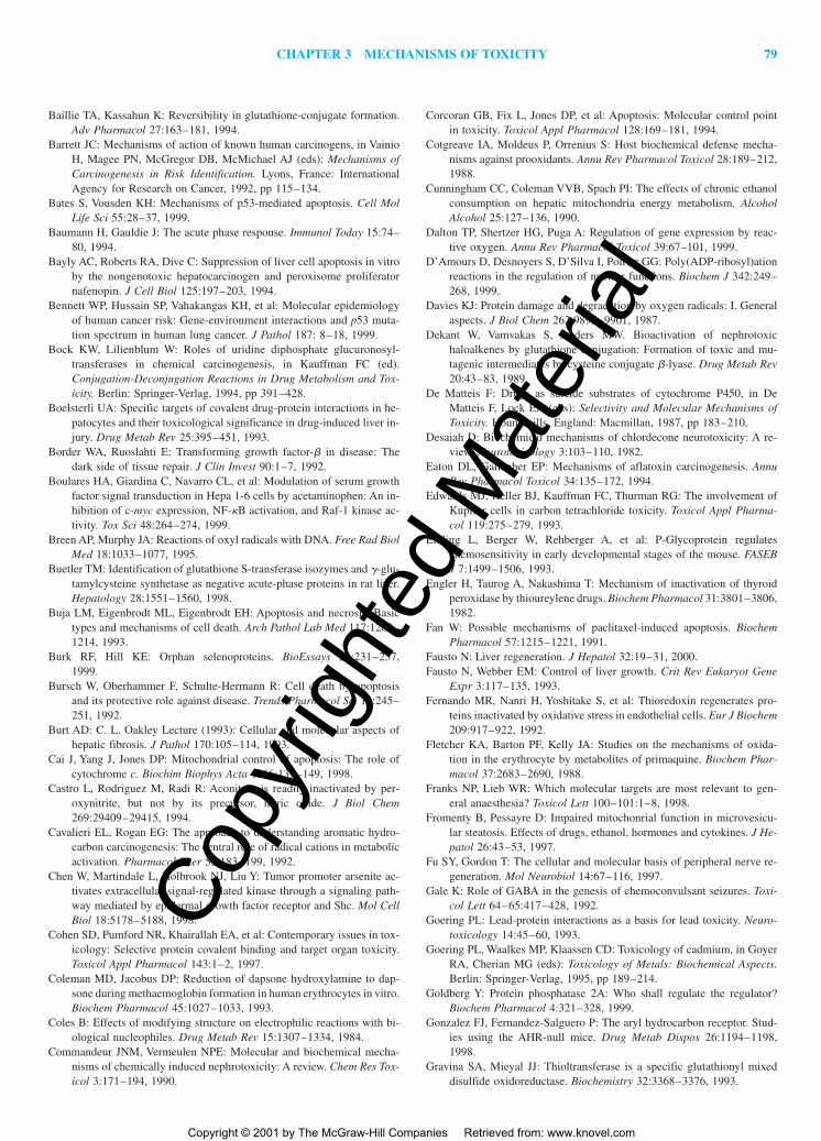
1. Xenobiotics such as paraquat, doxorubicin, and nitrofurantoincan accept an electron from reductases to give rise to radicals(Fig. 3-3). These radicals typically transfer the extra electronto molecular oxygen, forming a superoxide anion radical (O2
�•)and regenerating the parent xenobiotic, which is ready to gaina new electron (Kappas, 1986). Through this “redox cycling,”one electron acceptor xenobiotic molecule can generate manyO2
�• molecules. There are also endogenous sources of O2�• .
This radical is generated in large quantities by NAD(P)H ox-idase in activated macrophages and granulocytes during “res-piratory burst” and is also produced by the mitochondrial elec-tron transport chain, especially in the “uncoupled” state. Thesignificance of O2
�• stems to a large extent from the fact thatO2
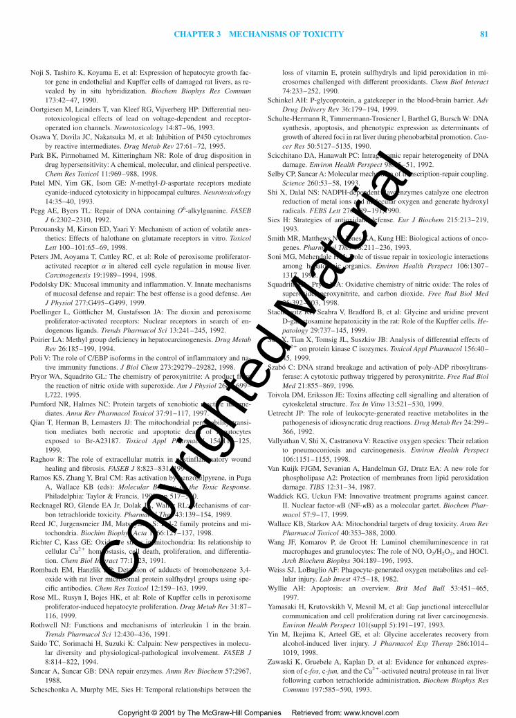
�• is a starting compound in two toxication pathways (Fig.3-4); one leading to formation of hydrogen peroxide (HOOH)and then hydroxyl radical (HO•), whereas the other producesperoxynitrite (ONOO�) and ultimately nitrogen dioxide(•NO2), and carbonate anion radical (CO3
�•).2. Nucleophilic xenobiotics such as phenols, hydroquinones,
aminophenols, amines, hydrazines, phenothiazines, and thiolsare prone to lose an electron and form free radicals in a reac-tion catalyzed by peroxidases (Aust et al., 1993). Some of thesechemicals, such as catechols and hydroquinones, may undergotwo sequential one-electron oxidations, producing first semi-quinone radicals and then quinones. Quinones are not only re-active electrophiles (Table 3-2) but also electron acceptors withthe capacity to initiate redox cycling or oxidation of thiols andNAD(P)H. Polycyclic aromatic hydrocarbons with sufficientlylow ionization potential, such as benzo[a]pyrene and 7,12-dimethylbenzanthracene, can be converted via one-electron ox-idation by peroxidases or cytochrome P450 to radical cations,which may be the ultimate toxicants for these carcinogens(Cavalieri and Rogan, 1992). Like peroxidases, oxyhemoglo-bin (Hb-FeII-O2) can catalyze the oxidation of aminophenolsto semiquinone radicals and quinoneimines. This is anotherexample of toxication, because these products, in turn, oxidizeferrohemoglobin (Hb-FeII) to methemoglobin (Hb-FeIII),which cannot carry oxygen.
3. Free radicals also are formed by homolytic bond fission, whichcan be induced by electron transfer to the molecule (reductivefission). This mechanism is involved in the conversion of CCl4to the trichloromethyl free radical (Cl3C•) by an electron trans-fer from cytochrome P450 or the mitochondrial electron trans-port chain (reductive dehalogenation) (Recknagel et al., 1989).The Cl3C• reacts with O2 to form the even more reactivetrichloromethylperoxy radical (Cl3COO•) (Hippeli and Elst-ner, 1999).
The hydroxyl radical (HO•), a free radical of paramounttoxicologic significance, also is generated by homolytic fis-sion. Such a process yields large amounts of HO• from waterupon ionizing radiation. Reductive homolytic fission of hy-drogen peroxide (HOOH) to HO• and HO� is called the Fen-ton reaction (Fig. 3-4). This is catalyzed by transition metalions, typically Fe(II) or, Cu(I), Cr(V), Ni(II), or Mn(II), andis a major toxication mechanism for HOOH and its precursorO2
�• as well as for transition metals. Moreover, the toxicity ofchemicals, such as nitrilotriacetic acid, bleomycin, and orel-lanin (Hippeli and Elstner, 1999), that chelate transition metalions is also based on Fenton chemistry because chelation in-
creases the catalytic efficiency of some transition metal ions.The pulmonary toxicity of inhaled mineral particles such asasbestos and silica is caused, at least in part, by the formationof HO• triggered by Fe ions on the particle surface (Vallyathanet al., 1998). Hydrogen peroxide is a direct or indirect by-prod-uct of several enzymatic reactions, including monoamine ox-
idase, xanthine oxidase, and acyl-coenzyme A oxidase. It isproduced in large quantities by spontaneous or superoxide dismutase-catalyzed dismutation of O2
�• .Homolytic cleavage is also thought to be involved in free
radical generation from ONOO� (Squadrito and Pryor, 1998)(Fig. 3-4). The facile reaction of ONOO� with the ubiquitous
Table 3-2Toxication by Formation of Electrophilic Metabolites
CO2 yields nitrosoperoxycarbonate (ONOOCO2�), which can
spontaneously homolyze into two radicals, the oxidant and ni-trating agent nitrogen dioxide (•NO2) and the oxidant carbon-ate anion radical (CO3
�•). Thus, formation of ONOO� and thelatter radicals represent a toxication mechanism for O2
�• and•NO. As •NO is the product of nitric oxide synthase (NOS),this mechanism is especially relevant in and around cells thatexpress NOS consitutively (i.e., neurons and endothelial cells)as well as in and around cells that express the inducible formof NOS in response to cytokines.
Formation of Nucleophiles The formation of nucleophiles is arelatively uncommon mechanism for activating toxicants. Exam-
ples include the formation of cyanide from amygdalin, which iscatalyzed by bacterial �-glucosidase in the gut; from acrylonitrileafter epoxidation and subsequent glutathione conjugation; and fromsodium mitroprusside by thiol-induced decomposition. Carbonmonoxide is a toxic metabolite of dihalomethanes that undergo ox-idative dehalogenation. Hydrogen selenide, a strong nucleophileand reductant, is formed from selenite by reaction with glutathioneor other thiols.Formation of Redox-Active Reactants There are specific mech-anisms for the creation of redox-active reactants other than thosealready mentioned. Examples include the formation of themethemoglobin-producing nitrite from nitrate by bacterialreduction in the intestine or from esters of nitrous or nitric acidsin reaction with glutathione. Dapsone hydroxylamine and 5-hydroxyprimaquine, hydroxylated metabolites of the respectivedrugs, produce methemoglobin by cooxidation (Fletcher et al.,1988). Reductants such as ascorbic acid and reductases such asNADPH-dependent flavoenzymes reduce Cr(VI) to Cr(V) (Shi andDalai, 1990). Xenobiotic radicals formed in redox cycling (e.g.,those depicted in Fig. 3-3) as well as O2
�• and •NO can reduceFe(III) bound to ferritin and consequently release it as Fe(II). Cr(V)and Fe(II) thus formed catalyze HO• formation (Fig. 3-4).
In summary, the most reactive metabolites are electron-deficient molecules and molecular fragments such as electrophilesand neutral or cationic free radicals. Although some nucleophilesare reactive (e.g., HCN, CO), many are activated by conversion toelectrophiles. Similarly, free radicals with an extra electron causedamage by giving rise to the neutral HO• radical after the forma-tion and subsequent homolytic cleavage of HOOH.
Detoxication Biotransformations that eliminate the ultimate tox-icant or prevent its formation are called detoxications. In some
Figure 3-3. Production of superoxide anion radical (O2•�) by paraquat
(PQ��), doxorubicin (DR), and nitrofurantoin (NF).
Note that formation of (O2•�) is not the final step in the toxication of these
xenobiotics, because O2•� can yield the much more reactive hydroxyl rad-
ical, as depicted in Fig. 3-4.
Figure 3-4. Two pathways for toxication of superoxide anion radical (O2•�) via nonradical products (ONOO–
and HOOH) to radical products (•NO2 , CO3•� and HO•).
In one pathway, conversion of (O2•�) to HOOH is spontaneous or is catalyzed by superoxide dismutase (SOD).
Homolytic cleavage of HOOH to hydroxyl radical and hydroxyl ion is called the Fenton reaction and is cat-alyzed by the transition metal ions shown. Hydroxyl radical formation is the ultimate toxication for xenobioticsthat form O2•
� (see Fig. 3-3) or for HOOH, the transition metal ions listed, and some chemicals that form com-plexes with these transiton metal ions. In the other pathway, O2•
� reacts avidly with nitric oxide (•NO), the prod-uct of •NO synthase (NOS), forming peroxynitrite (ONOO–). Spontaneous reaction of ONOO– with carbon diox-ide (CO2) yields nitrosoperoxy carbonate (ONOOCO2
–) that is homolytically cleaved to nitrogen dioxide (•NO2)and carbonate anion radical (CO3•
�). All three radical products indicated in this figure are oxidants, whereas•NO2 is also a nitrating agent.
cases, detoxication may compete with toxication for a chemical.Detoxication can take several pathways, depending on the chemi-cal nature of the toxic substance.Detoxication of Toxicants with No Functional Groups In gen-eral, chemicals without functional groups, such as benzene andtoluene, are detoxicated in two phases. Initially, a functional groupsuch as hydroxyl or carboxyl is introduced into the molecule, mostoften by cytochrome-P450 enzymes. Subsequently, an endogenousacid such as glucuronic acid, sulfuric acid, or an amino acid isadded to the functional group by a transferase. With some excep-tions, the final products are inactive, highly hydrophilic organicacids that are readily excreted.Detoxication of Nucleophiles Nucleophiles generally are detox-icated by conjugation at the nucleophilic functional group. Hy-droxylated compounds are conjugated by sulfation, glucuronida-tion, or rarely by methylation, whereas thiols are methylated orglucuronidated and amines and hydrazines are acetylated. Thesereactions prevent peroxidase-catalyzed conversion of the nucle-ophiles to free radicals and biotransformation of phenols,aminophenols, catechols, and hydroquinones to electrophilicquinones and quinoneomines. An alternative mechanism for theelimination of thiols and hydrazines is oxidation by flavin-containing monooxygenases (Jakoby and Ziegler, 1990). Some al-cohols, such as ethanol, are detoxicated by oxidation to carboxylicacids by alcohol and aldehyde dehydrogenases. A specificdetoxication mechanism is the biotransformation of cyanide to thio-cyanate by rhodanese.Detoxication of Electrophiles A general mechanism for thedetoxication of electrophilic toxicants is conjugation with the thiolnucleophile glutathione (Ketterer, 1988). This reaction may occurspontaneously or can be facilitated by glutathione S-transferases.Metal ions—such as Ag�, Cd2�, Hg2�, and CH3Hg� ions—read-ily react with and are detoxicated by glutathione. Specific mecha-nisms for the detoxication of electrophilic chemicals include epox-ide hydrolase-catalyzed biotransformation of epoxides and areneoxides to diols and dihydrodiols, respectively, and carboxyl-esterase-catalyzed hydrolysis of organophosphate ester pesticides.Others are two-electron reduction of quinones to hydroquinones byDT-diaphorase, reduction of �,�-unsaturated aldehydes to alcoholsby alcohol dehydrogenase or oxidation to acids by aldehyde de-hydrogenase, and complex formation of thiol-reactive metal ionsby metallothionein and the redox-active ferrous iron by ferrin. Co-valent binding of electrophiles to proteins can also be regarded asdetoxification provided that the protein has no critical function anddoes not become a neoantigen or otherwise harmful. Car-boxylesterases, for example, inactivate organophosphates not onlyby hydrolysis but also by covalent binding.Detoxication of Free Radicals Because O2
�• can be converted intomuch more reactive compounds (Fig. 3-4), its elimination is an im-portant detoxication mechanism. This is carried out by superoxidedismutases (SOD), high-capacity enzymes located in the cytosol(Cu,Zn-SOD) and the mitochondria (Mn-SOD), which convert O2
�•to HOOH (Fig. 3-5). Subsequently, HOOH is reduced to water bythe selenocysteine-containing glutathione peroxidase in the cytosolor by catalase in the peroxisomes (Fig. 3-5) (Cotgrave et al., 1988).
No enzyme eliminates HO•. While some relatively stableradicals, such as peroxyl radicals, can readily abstract a hydrogenatom from glutathione, �-tocopherol (vitamin E), or ascorbic acid(vitamin C), thus becoming nonradicals, these antioxidants aregenerally ineffective in detoxifying HO• (Sies, 1993). This is dueto its extremely short half-life (10�9 s), which provides little time
for the HO• to reach and react with antioxidants. Therefore theonly effective protection against HO• is to prevent its formationby elimination of its precursor, HOOH, via conversion to water(Fig. 3-5).
ONOO� (which is not a free radical oxidant) is significantlymore stable than HO• (half-life of about 1 s). Nevertheless, thesmall biological antioxidant molecules (glutathione, uric acid,ascorbic acid, �-tocopherol) are relatively inefficient in intercept-ing it because ONOO� rapidly reacts with CO2 (Squadrito andPryor, 1998) to form reactive free radicals (Fig. 3-4). More effi-cient is the selenocysteine-containing glutathione peroxidase,which can reduce ONOO� to nitrite (ONO�) the same way it re-duces HOOH to water (Arteel et al., 1999). Selenoprotein P, whichcontains 10 selenocysteine residues and coats the surface of en-dothelial cells, also reduces ONOO� and may serve as a protec-tant against this oxidant in blood (Arteel et al., 1999; Burk andHill, 1999). In addition, ONOO� reacts with oxyhemoglobin,heme-containing peroxidases and albumin, all of which could beimportant sinks for ONOO�. Furthermore, elimination of the twoONOO� precursors—i.e., •NO by reaction with oxyhemoglobin(to yield methemoglobin and nitrate) and O2
�• by SODs (seeabove)—is a significant mechanism in preventing ONOO� buildup(Squadrito and Pryor, 1998).
Peroxidase-generated free radicals are eliminated by electrontransfer from glutathione. This results in the oxidation ofglutathione, which is reversed by NADPH-dependent glutathionereductase (Fig. 3-6). Thus, glutathione plays an important role inthe detoxication of both electrophiles and free radicals.Detoxication of Protein Toxins Presumably, extra- and intracel-lular proteases are involved in the inactivation of toxicpolypeptides. Several toxins found in venoms, such as �- and�-bungaratoxin, erabutoxin, and phospholipase, contain intramol-ecular disulfide bonds that are required for their activity. These pro-teins are inactivated by thioredoxin, an endogenous dithiol proteinthat reduces the essential disulfide bond (Lozano et al., 1994).When Detoxication Fails Detoxication may be insufficient forseveral reasons:
1. Toxicants may overwhelm detoxication processes, leading toexhaustion of the detoxication enzymes, consumption of thecosubtrates, or depletion of cellular antioxidants such as glu-tathione, ascorbic acid, and �-tocopherol. This results in theaccumulation of the ultimate toxicant.
2. Occasionally, a reactive toxicant inactivates a detoxicating en-zyme. For example, ONOO� incapacitates Mn-SOD, whichnormally would counteract ONOO� formation (Murphy, 1999)(see Fig. 3-4).
Figure 3-5. Detoxication of superoxide anion radical (O2•�) by superox-
ide dismutase (SOD), glutathione peroxidase (GPO), and catalase (CAT).
3. Some conjugation reactions can be reversed. For example, 2-naphthylamine, a bladder carcinogen, is N-hydroxylated and glu-curonidated in liver, with the glucuronide excreted into urine.While in the bladder, the glucuronide is hydrolyzed, and the re-leased arylhydroxylamine is converted by protonation and de-hydration to the reactive electrophilic arylnitrenium ion (Bockand Lilienblum, 1994). Isocyanates and isothiocyanates form la-bile glutathione conjugates from which they can be released.Thus, methylisocyanate readily forms a glutathione conjugate inthe lung after inhalation. From there, the conjugate is distrib-uted to other tissues, where the reactive electrophilic parentcompound may be regenerated (Baillie and Kassahun, 1994).Such conjugates are considered transport forms of toxicants.
4. Sometimes detoxication generates potentially harmful by-products such as the glutathione thiyl radical and glutathionedisulfide, which are produced during the detoxication of freeradicals (Fig. 3-6). Glutathione disulfide can form mixed disul-fides with protein thiols, whereas the thiyl radical (GS•), afterreacting with thiolate (GS�), forms a glutathione disulfide rad-ical anion (GSSG•�), which can reduce O2 to O2•�.
STEP 2—REACTION OF THEULTIMATE TOXICANT WITH THE
TARGET MOLECULE
Toxicity is typically mediated by a reaction of the ultimate toxicantwith a target molecule (step 2a in Fig. 3-1). Subsequently, a seriesof secondary biochemical events occur, leading to dysfunction or in-jury that is manifest at various levels of biological organization, suchas at the target molecule itself, cell organelles, cells, tissues and or-gans, and even the whole organism. Because interaction of the ulti-mate toxicant with the target molecule triggers the toxic effect, con-sideration is given to (1) the attributes of target molecules, (2) thetypes of reactions between ultimate toxicants and target molecules,and (3) the effects of toxicants on the target molecules (Fig. 3-7).Finally, consideration is given to toxicities that are initiated not byreaction of the ultimate toxicant with target molecules but rather byalteration of the biological (micro)environment in which critical en-dogenous molecules, cell organelles, cells, and organs operate.
Attributes of Target Molecules
Practically all endogenous compounds are potential targets for tox-icants. The identification and characteristics of the target molecules
involved in toxicity constitute a major research priority, but a com-prehensive inventory of potential target molecules is impossible.Nevertheless, the most prevalent and toxicologically relevant tar-gets are macromolecules such as nucleic acids (especially DNA)and proteins. Among the small molecules, membrane lipids are fre-quently involved, whereas cofactors such as coenzyme A and pyri-doxal rarely are involved.
To be a target, an endogenous molecule must possess the ap-propriate reactivity and/or steric configuration to allow the ultimatetoxicant to enter into covalent or noncovalent reactions. For thesereactions to occur, the target molecule must be accessible to a suf-ficiently high concentration of the ultimate toxicant. Thus, endoge-nous molecules that are in the vicinity of reactive chemicals or areadjacent to sites where they are formed are frequently targets. Thefirst target for reactive metabolites is often the enzyme responsiblefor their production or the adjacent intracellular structures. For ex-ample, thyroperoxidase, the enzyme responsible for thyroid hor-mone synthesis, converts some nucleophilic xenobiotics (such asmethimazole, amitrole, and resorcinol) into reactive free radicalmetabolites that inactivate the thyroperoxidase (Engler et al., 1982).This is the basis for the antithyroid as well as the thyroid tumor–inducing effect of these chemicals. Carbon tetrachloride, which isactivated by cytochrome P450, destroys this enzyme as well as theneighboring microsomal membranes (Osawa et al., 1995). Severalmitochondrial enzymes—including pyruvate dehydrogenase, suc-cinate dehydrogenase, and cytochrome c oxidase—are convenienttargets for nephrotoxic cysteine conjugates such as dichlorovinylcysteine, because these conjugates are converted to electrophiles inthe same organelle by mitochondrial cysteine conjugate �-lyase(Dekant et al., 1989). Reactive metabolites that are unable to find
Figure 3-6. Detoxication of peroxidase (POD)–generated free radicalssuch as chlorpromazine free radical (CPZ�•) by glutathione (GSH).
The by-products are glutathione thiyl radical (GS•) and glutathione disul-fide (GSSG), from which GSH is regenerated by glutathione reductase(GR).
appropriate endogenous molecules in close proximity to their siteof formation may diffuse until they encounter such reactants. Forexample, hard electrophiles such as the arylnitrenium ion metabo-lite of N-methyl-4-aminoazobenzene react readily with hard nucle-ophilic atoms in nucleic acids, and thus target DNA in the nucleuseven though the electrophiles are produced in the cytoplasm.
Not all targets for chemicals contribute to the harmful effects.Thus, while carbon monoxide causes toxicity by binding to ferro-hemoglobin, it also associates with the iron in cytochrome P450with little or no consequence. Covalent binding of toxicants to var-ious intracellular proteins, including enzymes and structural pro-teins, has been demonstrated, yet it is often uncertain which pro-tein(s) is/are involved in binding that is toxicologically relevant(Cohen et al., 1997; Pumford and Halmes, 1997; Rombach andHanzlik, 1999). Arylation of some hepatic mitochondrial proteinsby acetaminophen might be causally related to the liver injury in-duced by this drug because the nonhepatotoxic regioisomer of acet-aminophen does not readily bind covalently to these proteins(Cohen et al., 1997). In contrast, arylation of a number of hepaticcytoplasmic proteins by acetaminophen is likely to be inconse-quential because a nonhepatotoxic regioisomer of this drug alsoarylates those proteins (Nelson and Pearson, 1990). Covalentbinding to proteins without adverse consequences may even rep-resent a form of detoxication by sparing toxicologically relevanttargets. This principle is best exemplified by covalent binding oforganophosphate insecticides to plasma cholinesterase, which is asignificant protective mechanism, as it counteracts phosphorylationof acetylcholinesterase, the target molecule. Thus, to conclusivelyidentify a target molecule as being responsible for toxicity, it shouldbe demonstrated that the ultimate toxicant (1) reacts with the tar-get and adversely affects its function, (2) reaches an effective con-centration at the target site, and (3) alters the target in a way thatis mechanistically related to the observed toxicity.
Types of Reactions
The ultimate toxicant may bind to the target molecules noncova-lently or covalently and may alter it by hydrogen abstraction, elec-tron transfer, or enzymatically.
Noncovalent Binding This type of binding can be due to apolarinteractions or the formation of hydrogen and ionic bonds and is typ-ically involved in the interaction of toxicants with targets such asmembrane receptors, intracellular receptors, ion channels, and some
enzymes. For example, such interactions are responsible for the bind-ing of strychnine to the glycine receptor on motor neurons in thespinal cord, TCDD to the aryl hydrocarbon receptor, saxitoxin tosodium channels, phorbol esters to protein kinase C, and warfarin tovitamin K 2,3-epoxide reductase. Such forces also are responsiblefor the intercalation of chemicals such as acridine yellow and dox-orubicin into the double helix of DNA. These chemicals are toxicbecause the steric arrangement of their atoms allows them to com-bine with complementary sites on the endogenous molecule more orless as a key fits into a lock. Noncovalent binding usually is re-versible because of the comparatively low bonding energy.
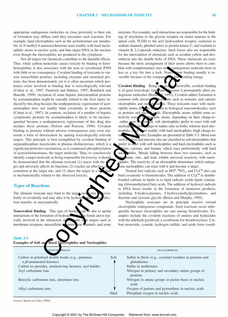
Covalent Binding Being practically irreversible, covalent bindingis of great toxicologic importance because it permanently alters en-dogenous molecules (Boelsterli, 1993). Covalent adduct formation iscommon with electrophilic toxicants such as nonionic and cationicelectrophiles and radical cations. These toxicants react with nucle-ophilic atoms that are abundant in biological macromolecules, suchas proteins and nucleic acids. Electrophilic atoms exhibit some se-lectivity toward nucleophilic atoms, depending on their charge-to-radius ratio. In general, soft electrophiles prefer to react with soft
nucleophiles (low charge-to-radius ratio in both), whereas hard elec-trophiles react more readily with hard nucleophiles (high charge-to-radius ratio in both). Examples are presented in Table 3-3. Metal ionssuch as silver and mercury also are classified as soft electrophiles thatprefer to react with soft nucleophiles and hard electrophiles such aslithium, calcium, and barium, which react preferentially with hardnucleophiles. Metals falling between these two extremes, such aschromium, zinc, and lead, exhibit universal reactivity with nucle-ophiles. The reactivity of an electrophile determines which endoge-nous nucleophiles can react with it and become a target.
Neutral free radicals such as HO•, •NO2, and Cl3C• also canbind covalently to biomolecules. The addition of Cl3C• to double-bonded carbons in lipids or to lipid radicals yields lipids contain-ing chloromethylated fatty acids. The addition of hydroxyl radicalsto DNA bases results in the formation of numerous products,including 8-hydroxypurines, 5-hydroxymethylpyrimidines, andthymine and cytosine glycols (Breen and Murphy, 1995).
Nucleophilic toxicants are in principle reactive towardelectrophilic endogenous compounds. Such reactions occur infre-quently because electrophiles are rare among biomolecules. Ex-amples include the covalent reactions of amines and hydrazideswith the aldehyde pyridoxal, a cosubstrate for decarboxylases. Car-bon monoxide, cyanide, hydrogen sulfide, and azide form coordi-
Table 3-3Examples of Soft and Hard Electrophiles and Nucleophiles
ELECTROPHILES NUCLEOPHILES
Carbon in polarized double bonds (e.g., quinones, Soft Sulfur in thiols (e.g., cysteinyl residues in proteins and�,�-unsaturated ketones) glutathione)
Carbon in epoxides, strained-ring lactones, aryl halides Sulfur in methionineAryl carbonium ions Nitrogen in primary and secondary amino groups of
proteinsBenzylic carbonium ions, nitrenium ions Nitrogen in amino groups in purine bases in nucleic
acidsAlkyl carbonium ions Oxygen of purines and pyrimidines in nucleic acids
nate covalent bonds with iron in various hemeproteins. Other nu-cleophiles react with hemoglobin in an electron-transfer reaction(see below).
Hydrogen Abstraction Neutral free radicals, such as those gen-erated in reactions depicted in Fig. 3-4, can readily abstract H atomsfrom endogenous compounds, converting those compounds intoradicals. Abstraction of hydrogen from thiols (R—SH) creates thiylradicals (R—S•), which are precursors of other thiol oxidationproducts, such as sulfenic acids (R—SOH) and disulfides (R—S—S—R). Radicals can remove hydrogen from CH2 groups of freeamino acids or from amino acid residues in proteins and convertthem to carbonyls. These carbonyls react with amines, formingcross-links with DNA or other proteins. Hydrogen abstraction fromdeoxyribose in DNA yields the C-4�-radical, the first step to DNAcleavage (Breen and Murphy, 1995). Abstraction of hydrogen fromfatty acids produces lipid radicals and initiates lipid peroxidation.As depicted in Fig. 3-8, nitration of tyrosine residues in proteinspurportedly involves H abstraction followed by covalent bindingbetween the resultant tyrosyl radical and •NO2 (Squadrito andPryor, 1998).
Electron Transfer Chemicals can oxidize Fe(II) in hemoglobinto Fe(III), producing methemoglobinemia. Nitrite can oxidizehemoglobin, whereas N-hydroxyl arylamines (such as dapsonehydroxylamine), phenolic compounds (such as 5-hydroxy pri-maquine), and hydrazines (such as phenylhydrazine) are cooxi-dized with oxyhemoglobin, forming methemoglobin and hydrogenperoxide (Coleman and Jacobus, 1993).
Enzymatic Reactions A few toxins act enzymatically on spe-cific target proteins. For example, ricin induces hydrolytic frag-mentation of ribosomes, blocking protein synthesis. Several bac-terial toxins catalyze the transfer for ADP-ribose from NAD� tospecific proteins. For example, diphtheria toxin blocks the func-
tion of elongation factor 2 in protein synthesis and cholera toxinactivates a G protein through such a mechanism. Snake venomscontain hydrolytic enzymes that destroy biomolecules.
In summary, most ultimate toxicants act on endogenous mol-ecules on the basis of their chemical reactivity. Those with morethan one type of reactivity may react by different mechanisms withvarious target molecules. For example, quinones may act as elec-tron acceptors and initiate thiol oxidation or free radical reactionsthat lead to lipid peroxidation, buy they may also act as soft elec-trophiles and bind covalently to protein thiols. The lead ion acts asa soft electrophile when it forms coordinate covalent bonds withcritical thiol groups in �-aminolevulinic acid dehydratase, its major target enzyme in heme synthesis (Goering, 1993). However,it behaves like a hard electrophile or an ion when it binds to pro-tein kinase C or blocks calcium channels, substituting for the nat-ural ligand Ca2� at those target sites.
Effects of Toxicants on Target Molecules
Reaction of the ultimate toxicant with endogenous molecules maycause dysfunction or destruction; in the case of proteins, it mayrender them foreign (i.e., an antigen) to the immune system.
Dysfunction of Target Molecules Some toxicants activateprotein target molecules, mimicking endogenous ligands. Forexample, morphine activates opiate receptors, clofibrate is an ag-onist on the peroxisome proliferator–activated receptor, and phor-bol esters and lead ions stimulate protein kinase C.
More commonly, chemicals inhibit the function of target mol-ecules. Several xenobiotics—such as atropine, curare, and strych-nine—block neurotransmitter receptors by attaching to the ligand-binding sites or by interfering with the function of ion channels.Tetrotodoxin and saxitoxin, for example, inhibit opening of thevoltage-activated sodium channels in the neuronal membrane,whereas DDT and the pyrethroid insecticides inhibit their closure.Some toxicants block ion transporters, others inhibit mitochon-drial electron transport complexes, and many inhibit enzymes.Chemicals that bind to tubulin (e.g., vinblastine, colchicine, pacli-taxel, trivalent arsenic) or actin (e.g., cytochalasin B, phalloidin)impair the assembly (polymerization) and/or disassembly (depoly-merization) of these cytoskeletal proteins.
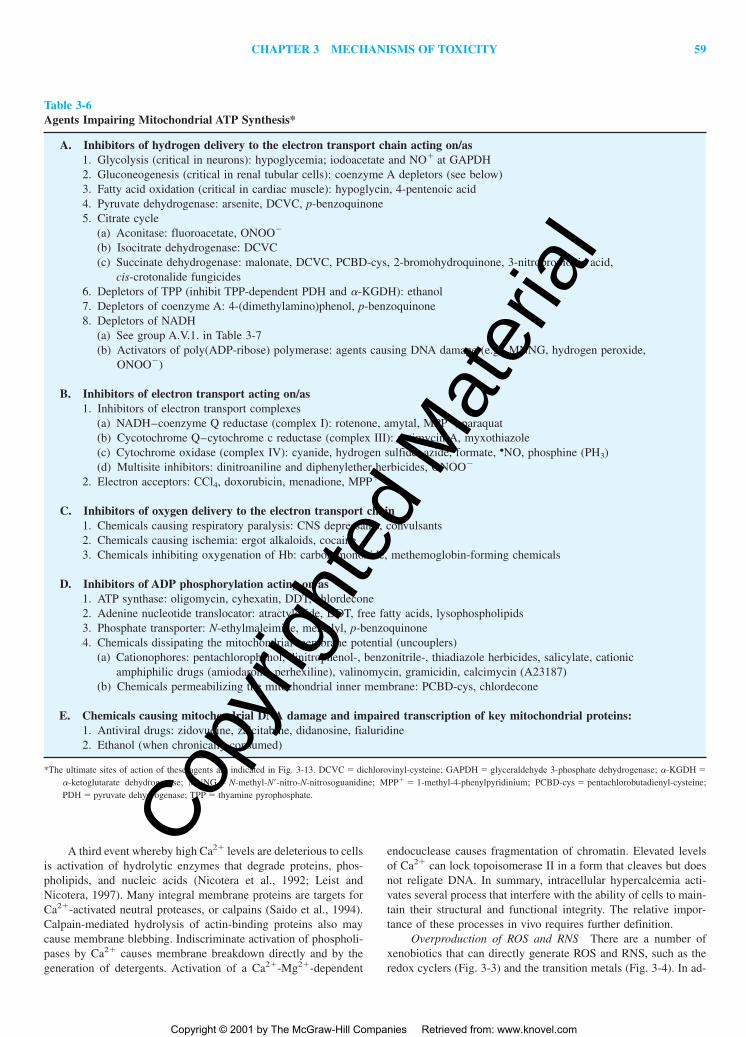
Protein function is impaired when conformation or structureis altered by interaction with the toxicant. Many proteins possesscritical moities, especially thiol groups, that are essential for cat-alytic activity or assembly to macromolecular complexes. Proteinsthat are sensitive to covalent and/or oxidative modification of theirthiol groups include the enzymes protein tyrosine phosphatases(Herrlich et al., 1998), glyceraldehyde 3-phosphate dehydrogenase(see Table 3-6), and pyruvate dehydrogenase (see Fig. 3-13), theCa2� pumps (see Fig. 3-14, Table 3-7), and the transcription fac-tor AP-1, just to name a few. The activity of these and many otherproteins is impaired by thiol-reactive chemicals, triggering aber-rant signal transduction and/or impaired maintenance of the cell’senergy and metabolic homeostasis. Protein tyrosine nitration (seeFig. 3-8) may alter also protein function or may interfere with sig-naling pathways that involve tyrosine kinases and phosphatases(Arteel et al., 1999).
Toxicants may interfere with the template function of DNA.The covalent binding of chemicals to DNA causes nucleotide mispairing during replication. For example, covalent binding of
Figure 3-8. Formation of 3-nitrotyrosine residues in proteins by reactionwith nitrogen dioxide ( •NO2).
(•NO2) is the nitrating species generated from ONOO� (Fig. 3-4). In ad-dition, (•NO2) is a contaminant in cigarette smoke, exhaust of gas enginesand stoves, as well as the causative agent of “silo-filler’s disease.’’
aflatoxin 8,9-oxide to N-7 of guanine results in pairing of theadduct-bearing guanine with adenine rather than cytosine, leadingto the formation of an incorrect codon and the insertion of an in-correct amino acid into the protein. Such events are involved in theaflatoxin-induced mutation of the ras proto-oncogene and the p53tumor suppressor gene (Eaton and Gallagher, 1994). 8-Hydroxy-guanine and 8-hydroxyadenine are mutagenic bases produced byHO• that can cause mispairing with themselves as well as withneighboring pyrimidines, producing multiple amino acid substitu-tions (Breen and Murphy, 1995). Chemicals such as doxorubicin,that intercalate between stacked bases in the double-helical DNA,push adjacent base pairs apart, causing an even greater error in thetemplate function of DNA by shifting the reading frame.
Destruction of Target Molecules In addition to adduct forma-tion, toxicants alter the primary structure of endogenous mole-cules by means of cross-linking and fragmentation. Bifunctionalelectrophiles such as 2,5-hexanedione, carbon disulfide, acrolein,4-hydroxynonenal, and nitrogen mustard alkylating agents cross-link cytoskeletal proteins, DNA, or DNA with proteins. Hydroxylradicals also can induce cross-linking by converting these macro-molecules into either reactive electrophiles (e.g., protein carbonyls),which react with a nucleophilic site in another macromolecule, orradicals, which react with each other. Cross-linking imposes bothstructural and functional constraints on the linked molecules.
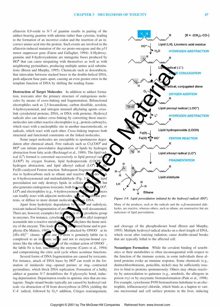
Some target molecules are susceptible to spontaneous degra-dation after chemical attack. Free radicals such as Cl3COO• andHO• can initiate peroxidative degradation of lipids by hydrogenabstraction from fatty acids (Recknagel et al., 1989). The lipid rad-ical (L•) formed is converted successively to lipid peroxyl radical(LOO•) by oxygen fixation, lipid hydroperoxide (LOOH) byhydrogen abstraction, and lipid alkoxyl radical (LO•) by the Fe(II)-catalyzed Fenton reaction. Subsequent fragmentation givesrise to hydrocarbons such as ethane and reactive aldehydes suchas 4-hydroxynonenal and malondialdehyde (Fig. 3-9). Thus, lipidperoxidation not only destroys lipids in cellular membranes butalso generates endogenous toxicants, both free radicals (e.g., LOO•,LO•) and electrophiles (e.g., 4-hydroxynonenal). These substancescan readily react with adjacent molecules, such as membrane pro-teins, or diffuse to more distant molecules such as DNA.
Apart from hydrolytic degradation by toxins and radiolysis,toxicant-induced fragmentation of proteins is not well documented.There are, however, examples for destruction of the prosthetic groupin enzymes. For instance, cytochrome P450 converts allyl isopropylacetamide into a reactive metabolite, which alkylates the heme moi-ety of the enzyme. This leads to loss of the altered heme and to por-phyria (De Matteis, 1987). Aconitase is attacked by ONOO� at its[4Fe-4S]2� cluster, whose one Fe atom is genuinely labile (as iscomplexed to an inorganic sulfur and not to enzyme-bound cys-teines like the others). As a result of the oxidant action of ONOO�,the labile Fe is lost, inactivating the enzyme (Castro et al., 1994)and compromising the citric acid cycle where aconitase functions.
Several forms of DNA fragmentation are caused by toxicants.For instance, attack of DNA bases by HO• can result in the for-mation of imidazole ring – opened purines or ring – contractedpyrimidines, which block DNA replication. Formation of a bulkyadduct at guanine N-7 destabilizes the N-glycosylic bond, induc-ing depurination. Depurination results in apurinic sites that are mu-tagenic. Single-strand breaks typically are caused by hydroxyl rad-icals via abstraction of H from desoxyribose in DNA yielding theC-4 radical, followed by O2
�• addition, Criegee rearrangement,
and cleavage of the phosphodiester bond (Breen and Murphy,1995). Multiple hydroxyl radical attacks on a short length of DNA,which occur after ionizing radiation, cause double-strand breaksthat are typically lethal to the affected cell.
Neoantigen Formation While the covalent binding of xenobi-otics or their metabolites is often inconsequential with respect tothe function of the immune system, in some individuals these al-tered proteins evoke an immune response. Some chemicals (e.g.,dinitrochlorobenzene, penicillin, nickel) may be sufficiently reac-tive to bind to proteins spontaneously. Others may obtain reactiv-ity by autooxidation to quinones (e.g., urushiols, the allergens inpoison ivy) or by enzymatic biotransformation (Park et al., 1998).For example, cytochrome P450 biotransforms halothane to an elec-trophile, trifluoroacetyl chloride, which binds as a hapten to vari-ous microsomal and cell surface proteins in the liver, inducing
HYDROGEN ABSTRACTION
HYDROGEN ABSTRACTION
FENTON REACTION
FRAGMENTATION
DIENE CONJUGATION
OXYGEN ADDITION
Figure 3-9. Lipid peroxidation initiated by the hydroxyl radical (HO•).
Many of the products, such as the radicals and the �,�-unsaturated alde-hydes, are reactive, whereas others, such as ethane, are nonreactive but areindicators of lipid peroxidation.
antibody production. The immune reaction is thought to be re-sponsible for the hepatitis-like syndrome seen in sensitive patients.Drug-induced lupus and possibly many cases of drug-inducedagranulocytosis are mediated by immune reactions triggered bydrug-protein adducts. The causative chemicals are typically nucle-ophiles, such as aromatic amines (e.g., aminopyrine, clozapine,procainamide, and sulfonamides), hydrazines (e.g., hydralazine andisoniazid), and thiols (e.g., propylthiouracil, methimazole, and cap-topril). These substances can be oxidized by myeloperoxidase dis-charged from activated granulocytes or by the ROS/RNS such cellsproduce (HO•, ONOO�, HOCl, see Fig. 3-22) to reactive metabo-lites that bind to the surface proteins of these cells, making themantigens (Uetrecht, 1992). Unfortunately, some proteins that bearan adduct can mimic some normal proteins, which thus also canbe attacked by the antibodies.
Toxicity Not Initiated by Reaction with Target Molecules
Some xenobiotics do not or do not only interact with a specific en-dogenous target molecule to induce toxicity but instead alter thebiological microenvironment (see step 2b in Fig. 3-1). Includedhere are (1) chemicals that alter H� ion concentrations in the aque-ous biophase, such as acids and substances biotransformed to acids,such as methanol and ethylene glycol, as well as protonophoricuncouplers such as 2,4-dinitrophenol and pentachlorophenol,which dissociate their phenolic protons in the mitochondrial ma-trix, thus dissipating the proton gradient that drives ATP synthesis;(2) solvents and detergents that physicochemically alter the lipidphase of cell membranes and destroy transmembrane solute gradi-ents that are essential to cell functions; and (3) other xenobioticsthat cause harm merely by occupying a site or space. For exam-ple, some chemicals (e.g., ethylene glycol) form water-insolubleprecipitates in the renal tubules. By occupying bilirubin binding
sites on albumin, compounds such as the sulfonamides inducebilirubin toxicity (kernicterus) in neonates. Carbon dioxide dis-places oxygen in the pulmonary alveolar space and causesasphyxiation.
STEP 3—CELLULARDYSFUNCTION AND RESULTANT
TOXICITIES
The reaction of toxicants with a target molecule may result in im-paired cellular function as the third step in the development of tox-icity (Fig. 3-1). Each cell in a multicellular organism carries outdefined programs. Certain programs determine the destiny ofcells—that is, whether they undergo division, differentiation (i.e.,express proteins for specialized functions), or apoptosis. Other pro-grams control the ongoing (momentary) activity of differentiatedcells, determining whether they secrete more or less of a substance,whether they contract or relax, and whether they transport and me-tabolize nutrients at higher or lower rates. For regulations of thesecellular programs, cells possess signaling networks (such as thoseshown in Figs. 3-11 and 3-12) that can be activated and inactivatedby external signaling molecules. To execute the programs, cells areequipped with synthetic, metabolic, kinetic, transport, and energy-producing system as well as structural elements, organized intomacromolecular complexes, cell membranes, and organelles, bywhich they maintain their own integrity (internal functions) andsupport the maintenance of other cells (external functions).
As outlined in Fig. 3-10, the nature of the primary cellulardysfunction caused by toxicants, but not necessarily the ultimateoutcome, depends on the role of the target molecule affected. Ifthe target molecule is involved in cellular regulation (signaling),dysregulation of gene expression and/or dysregulation of momen-tary cellular function occurs primarily. However, if the target mol-ecule is involved predominantly in the cell’s internal maintenance,
the resultant dysfunction can ultimately compromise the survivalof the cell. The reaction of a toxicant with targets serving externalfunctions can influence the operation of other cells and integratedorgan systems. The following discussion deals with these conse-quences.
Toxicant-Induced CellularDysregulation
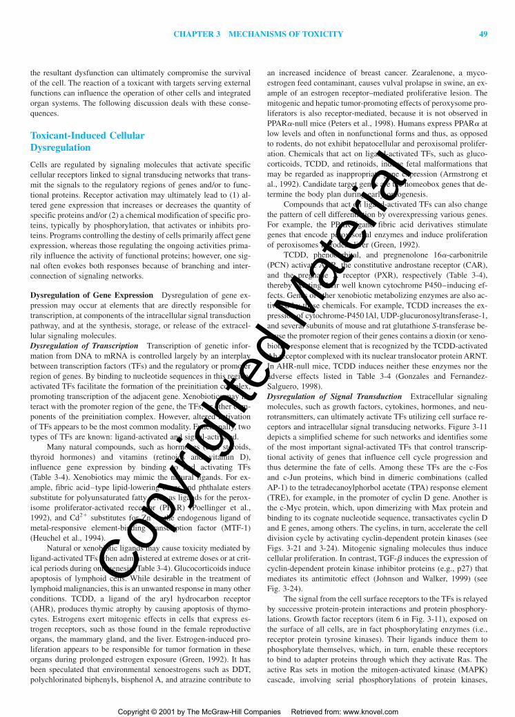
Cells are regulated by signaling molecules that activate specificcellular receptors linked to signal transducing networks that trans-mit the signals to the regulatory regions of genes and/or to func-tional proteins. Receptor activation may ultimately lead to (1) al-tered gene expression that increases or decreases the quantity ofspecific proteins and/or (2) a chemical modification of specific pro-teins, typically by phosphorylation, that activates or inhibits pro-teins. Programs controlling the destiny of cells primarily affect geneexpression, whereas those regulating the ongoing activities prima-rily influence the activity of functional proteins; however, one sig-nal often evokes both responses because of branching and inter-connection of signaling networks.
Dysregulation of Gene Expression Dysregulation of gene ex-pression may occur at elements that are directly responsible fortranscription, at components of the intracellular signal transductionpathway, and at the synthesis, storage, or release of the extracel-lular signaling molecules.Dysregulation of Transcription Transcription of genetic infor-mation from DNA to mRNA is controlled largely by an interplaybetween transcription factors (TFs) and the regulatory or promoterregion of genes. By binding to nucleotide sequences in this region,activated TFs facilitate the formation of the preinitiation complex,promoting transcription of the adjacent gene. Xenobiotics may in-teract with the promoter region of the gene, the TFs, or other com-ponents of the preinitiation complex. However, altered activationof TFs appears to be the most common modality. Functionally, twotypes of TFs are known: ligand-activated and signal-activated.
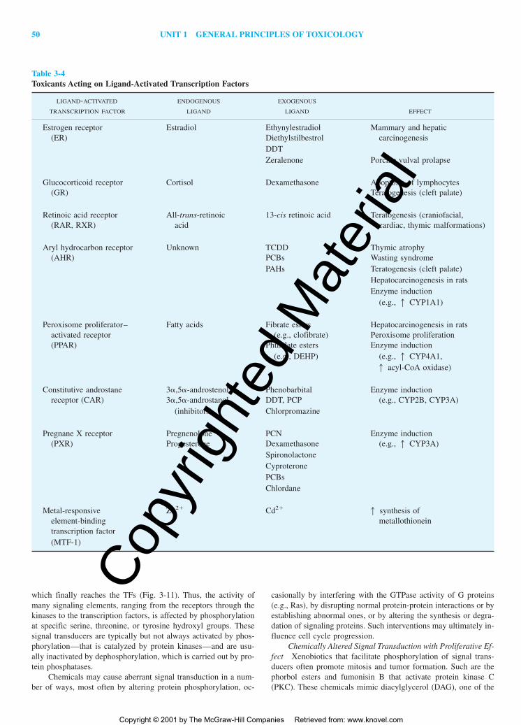
Many natural compounds, such as hormones (e.g., steroids,thyroid hormones) and vitamins (retinoids and vitamin D),influence gene expression by binding to and activating TFs(Table 3-4). Xenobiotics may mimic the natural ligands. For ex-ample, fibric acid–type lipid-lowering drugs and phthalate esterssubstitute for polyunsaturated fatty acids as ligands for the perox-isome proliferator-activated receptor (PPAR) (Poellinger et al.,1992), and Cd2� substitutes for Zn2�, the endogenous ligand ofmetal-responsive element-binding transcription factor (MTF-1)(Heuchel et al., 1994).
Natural or xenobiotic ligands may cause toxicity mediated byligand-activated TFs when administered at extreme doses or at crit-ical periods during ontogenesis (Table 3-4). Glucocorticoids induceapoptosis of lymphoid cells. While desirable in the treatment oflymphoid malignancies, this is an unwanted response in many otherconditions. TCDD, a ligand of the aryl hydrocarbon receptor(AHR), produces thymic atrophy by causing apoptosis of thymo-cytes. Estrogens exert mitogenic effects in cells that express es-trogen receptors, such as those found in the female reproductiveorgans, the mammary gland, and the liver. Estrogen-induced pro-liferation appears to be responsible for tumor formation in theseorgans during prolonged estrogen exposure (Green, 1992). It hasbeen speculated that environmental xenoestrogens such as DDT,polychlorinated biphenyls, bisphenol A, and atrazine contribute to
an increased incidence of breast cancer. Zearalenone, a myco-estrogen feed contaminant, causes vulval prolapse in swine, an ex-ample of an estrogen receptor–mediated proliferative lesion. Themitogenic and hepatic tumor-promoting effects of peroxysome pro-liferators is also receptor-mediated, because it is not observed inPPAR�-null mice (Peters et al., 1998). Humans express PPAR� atlow levels and often in nonfunctional forms and thus, as opposedto rodents, do not exhibit hepatocellular and peroxisomal prolifer-ation. Chemicals that act on ligand-activated TFs, such as gluco-corticoids, TCDD, and retinoids, induce fetal malformations thatmay be regarded as inappropriate gene expression (Armstrong etal., 1992). Candidate target genes are the homeobox genes that de-termine the body plan during early ontogenesis.
Compounds that act on ligand-activated TFs can also changethe pattern of cell differentiation by overexpressing various genes.For example, the PPAR-ligand fibric acid derivatives stimulategenes that encode peroxisomal enzymes and induce proliferationof peroxisomes in rodent liver (Green, 1992).
TCDD, phenobarbital, and pregnenolone 16�-carbonitrile(PCN) activate AHR, the constitutive androstane receptor (CAR),and the pregnane X receptor (PXR), respectively (Table 3-4),thereby exerting their well known cytochrome P450–inducing ef-fects. Genes of other xenobiotic metabolizing enzymes are also ac-tivated by these chemicals. For example, TCDD increases the ex-pression of cytochrome-P450 lAl, UDP-glucuronosyltransferase-1,and several subunits of mouse and rat glutathione S-transferase be-cause the promoter region of their genes contains a dioxin (or xeno-biotic) response element that is recognized by the TCDD-activatedAh receptor complexed with its nuclear translocator protein ARNT.In AHR-null mice, TCDD induces neither these enzymes nor theadverse effects listed in Table 3-4 (Gonzales and Fernandez-Salguero, 1998).Dysregulation of Signal Transduction Extracellular signalingmolecules, such as growth factors, cytokines, hormones, and neu-rotransmitters, can ultimately activate TFs utilizing cell surface re-ceptors and intracellular signal transducing networks. Figure 3-11depicts a simplified scheme for such networks and identifies someof the most important signal-activated TFs that control transcrip-tional activity of genes that influence cell cycle progression andthus determine the fate of cells. Among these TFs are the c-Fosand c-Jun proteins, which bind in dimeric combinations (calledAP-1) to the tetradecanoylphorbol acetate (TPA) response element(TRE), for example, in the promoter of cyclin D gene. Another isthe c-Myc protein, which, upon dimerizing with Max protein andbinding to its cognate nucleotide sequence, transactivates cyclin Dand E genes, among others. The cyclins, in turn, accelerate the celldivision cycle by activating cyclin-dependent protein kinases (seeFigs. 3-21 and 3-24). Mitogenic signaling molecules thus inducecellular proliferation. In contrast, TGF-� induces the expression ofcyclin-dependent protein kinase inhibitor proteins (e.g., p27) thatmediates its antimitotic effect (Johnson and Walker, 1999) (seeFig. 3-24).
The signal from the cell surface receptors to the TFs is relayedby successive protein-protein interactions and protein phosphory-lations. Growth factor receptors (item 6 in Fig. 3-11), exposed onthe surface of all cells, are in fact phosphorylating enzymes (i.e.,receptor protein tyrosine kinases). Their ligands induce them tophosphorylate themselves, which, in turn, enable these receptorsto bind to adapter proteins through which they activate Ras. Theactive Ras sets in motion the mitogen-activated kinase (MAPK)cascade, involving serial phosphorylations of protein kinases,
which finally reaches the TFs (Fig. 3-11). Thus, the activity ofmany signaling elements, ranging from the receptors through thekinases to the transcription factors, is affected by phosphorylationat specific serine, threonine, or tyrosine hydroxyl groups. Thesesignal transducers are typically but not always activated by phos-phorylation—that is catalyzed by protein kinases—and are usu-ally inactivated by dephosphorylation, which is carried out by pro-tein phosphatases.
Chemicals may cause aberrant signal transduction in a num-ber of ways, most often by altering protein phosphorylation, oc-
casionally by interfering with the GTPase activity of G proteins(e.g., Ras), by disrupting normal protein-protein interactions or byestablishing abnormal ones, or by altering the synthesis or degra-dation of signaling proteins. Such interventions may ultimately in-fluence cell cycle progression.
Chemically Altered Signal Transduction with Proliferative Ef-fect Xenobiotics that facilitate phosphorylation of signal trans-ducers often promote mitosis and tumor formation. Such are thephorbol esters and fumonisin B that activate protein kinase C(PKC). These chemicals mimic diacylglycerol (DAG), one of the
Table 3-4Toxicants Acting on Ligand-Activated Transcription Factors
LIGAND-ACTIVATED ENDOGENOUS EXOGENOUS
TRANSCRIPTION FACTOR LIGAND LIGAND EFFECT
Estrogen receptor Estradiol Ethynylestradiol Mammary and hepatic(ER) Diethylstilbestrol carcinogenesis
DDTZeralenone Porcine vulval prolapse
Glucocorticoid receptor Cortisol Dexamethasone Apoptosis of lymphocytes(GR) Teratogenesis (cleft palate)
Figure 3-11. Signal transduction pathways from cell membrane receptors to signal-activated nuclear tran-scription factors that influence transcription of genes involved in cell-cycle regulation.
The symbols of cell membrane receptors are numbered 1–9 and some of their activating ligands are indicated.Circles represent G proteins, oval symbols protein kinases, rectangles transcription factors, wavy lines genes,and diamond symbols inhibitory proteins, such as protein phosphatases (PTP, PP2A), the GTPase-activating pro-tein GAP, and the inhibitory binding protein IkB. Arrowheads indicate stimulation or formation of second mes-sengers (e.g., DAG, IP3, cAMP, Ca2�), whereas blunt arrows indicate inhibition. Phosphorylation and dephos-phorylation are indicated by �P and �P, respectively. Abbreviations for interfering chemicals are printed inblack (As arsenite; CALY calyculin A; FA fatty acids; FB1 fumonisin B; MC-LR microcystin-LR;OKA okadaic acid; MMS methylmethane sulfonate; PMA phorbol miristate acetate; ROS reactiveoxygen species; SHR SH-reactive chemicals, such as iodoacetamide; STAU staurosporin).
In the center of the depicted networks is the pathway activated by growth factors, such as EGF, that actson a tyrosine kinase receptor (#6) which uses adaptor proteins (Shc, Grb2 and SOS; not shown) to convert theinactive GDP-bound Ras to active GTP-bound form, which in turn activates the MAP-kinase phosphorylationcascade (Raf, MAPKK, MAPK). The phosphorylated MAPK moves into the nucleus and phosphorylates tran-scription factors thereby enabling them to bind to cognate sequences in the promoter regions of genes to facil-itate transcription. There are numerous interconnections between the signal transduction pathways. Some ofthese connections permit the use of the growth factor receptor (#6)-MAPK “highway’’ for other receptors (e.g.,4, 5, 7) to send mitogenic signals. For example, receptor (#4) joins in via its G protein �/� subunits and tyro-sine kinase Src; the integrin receptor (#5), whose ligands are constituents of the extracellular matrix (ECM),possibly connects via G-protein Rho (not shown) and focal adhesion kinase (FAK); and the G-protein-coupledreceptor (#7) via phospholipase C (PLC)-catalyzed formation of second messengers and activation of protein-kinase C (PKC). The mitogenic stimulus relayed along the growth factor receptor (#6)-MAPK axis can be am-plified by, for example, the Raf-catalyzed phosphorylation of I�B, which unleashes NF-�B from this inhibitoryprotein, and by the MAPK-catalyzed inhibitory phosphorylation of Smad that blocks the cell-cycle arrest sig-nal from the TGF-� receptor (#9). Activation of protein kinases (PKC, CaMK, MAPK) by Ca2� can also trig-ger mitogenic signaling. Several xenobiotics that are indicated in the figure may dysregulate the signaling net-work. Some may induce cell proliferation either by activating mitogenic protein kinases (e.g., PKC), or inhibitinginactivating proteins, such as protein phosphatases (PTP, PP2A), GAP or I�B. Others, e.g., inhibitors of PKC,oppose mitosis and facilitate apoptosis.
This scheme is oversimplified and tentative in several details. Virtually all components of the signalingnetwork (e.g., G proteins, PKCs, MAPKs) are present in multiple, functionally different forms whose distribu-tion may be cell specific. The pathways depicted are not equally relevant for all cells. In addition, these path-ways regulating gene expression determine not only the fate of cells, but also control certain aspects of the on-going cellular activity. For example, NF-�B induces synthesis of acute phase proteins.
physiologic activators of PKC (Fig. 3-11). The other physiologicPKC activator Ca2� is mimicked by Pb2�, whose effect on PKC�is concentration-dependent: stimulatory at picomolar concentra-tion, when Pb2� occupies only high-affinity binding sites on PKC,and inhibitory at micromolar concentration, where the low affinitysites are also occupied (Sun et al., 1999). Lead acetate does inducemarked hepatocellular proliferation in rats. The activated PKCpromotes mitogenic signaling at least in two ways: (1) byphosphorylating Raf, the first protein kinase in the MAPK path-way (Fig. 3-11), and (2) by phosphorylating a protein phosphatasethat dephosphorylates the transcription factor c-Jun at specific sites(Thr 231, Ser 234, and Ser 249), thereby permitting its binding toDNA. Protein kinases may also be activated by interacting proteinsthat had been altered by a xenobiotic. For example, the TCDD-liganded AHR binds to MAPK. This may contribute to the TCDD-induced overexpression of cyclins and cyclin-dependent kinases inguinea pig liver (Ma and Babish, 1999).
Abberant phosphorylation of proteins may result not onlyfrom increased phosphorylation by kinases but also from decreaseddephosphorylation by phosphatases. Inhibition of phosphatases ap-pears to be the underlying mechanism of the mitogenic effect ofvarious chemicals, oxidative stress, and ultraviolet (UV) irradia-tion (Herrlich et al., 1999). Protein tyrosine phosphatases and dual-specificity phosphatases (i.e., enzymes that remove phosphate fromphosphorylated tyrosine as well as serine and threonine residues)contain a catalytically active cysteine and are susceptible to inac-tivation by oxidation and covalent reaction with SH-reactive chem-icals. Indeed, xenobiotics such as the SH-reactive iodoacetamide,the organometal compound tributyltin, arsenite, and oxidants (e.g.,HOOH) cause phosphorylation of the epidermal growth factor(EGF) receptor (item 6 in Fig. 3-11) by interfering with the pro-tein tyrosine phosphatase that would dephosphorylate and thus“silence” this receptor (Herrlich et al., 1999; Chen et al., 1998).Arsenite may also inactivate the dual-specificity phosphatase thatdephosphorylates and “silences” certain MAPKs (JNK, p38),whereas methylmethane sulfonate (MMS) appears to inhibit aprotein phosphatase that inactivates Src, a protein tyrosine kinase(Herrlich et al., 1999). The thiol oxidizing agent diamide (whichincreases phosphorylation of MAPKs) and phenolic antioxidants(which form phenoxyl radicals and increase c-Fos and c-Jun ex-pression) (Dalton et al., 1999) may also act by incapacitating pro-tein tyrosine phosphatases. Protein phosphatase 2A (PP2A) is themajor soluble ser/thr phosphatase in cells and is likely responsi-ble, at least in part, for reversing the growth factor–induced stim-ulation of MAPK, thereby keeping the extent and duration ofMAPK activity under control (Goldberg, 1999). PP2A also re-moves an activating phosphate from a mitosis-triggering proteinkinase (p34cdc2). Several natural toxins are extremely potent in-hibitors of PP2A; including the blue-green algae poisonmicrocystin-LG and the dinoflagellate-derived okadaic acid(Toivola and Eriksson, 1999), which are tumor promoters in ex-perimental animals subjected to prolonged low-dose exposure. Itis to be noted, however, that acute high-dose exposure to micro-cystin induces severe liver injury, whereas such exposure to okadaicacid is the underlying cause of the diarrhetic shellfish poisoning.In these conditions, hyperphosphorylation of proteins other thanthose involved in proliferative signaling (e.g. hepatocellular mi-crofilaments in microcystin poisoning) may be primarily responsi-ble for the pathogenesis.
Apart from phosphatases, there are also inhibitory bindingproteins that can keep signaling under control. Such is I�B, which
binds to NF-�B, preventing its transfer into the nucleus and itsfunction as a TF. Upon phosphorylation, I�B becomes degradedand NF-�B is set free. Because phosphorylation of I�B can be cat-alyzed by Raf, a protein kinase in the MAPK cascade (Fig. 3-11),and because the released NF-�B can transactivate the c-Myc gene,NF-�B is an important contributor to proliferative and prolife sig-naling. In addition, because NF-�B also targets the genes of sev-eral cytokines (e.g., TNF-�, IL-1�) and acute phase proteins (e.g.,C-reactive protein, �1-acid glycoprotein), and because such cy-tokines acting on their receptors (items 2 and 3 in Fig. 3-11) alsoactivate NF-�B, this TF plays a leading role also in inflammatoryand acute phase reactions (Lee et al., 1998; Waddick and Uckun,1999). I�B degradation and NF-�B activation can also be inducedby oxidative stress, and it appears that peroxides are the reactiveoxygen species (ROS) that mediate this effect (Dalson et al., 1999).Activated NF-�B probably contributes to the proliferative and in-flammatory response to oxidative stress. NF-�B also protects cellsfrom apoptosis by maintaining c-Myc transcription, which is re-quired for survival (Waddick and Uckun, 1999), and by transacti-vating the genes of antiapoptotic IAP proteins, which inhibit cas-pases (Jäättelä, 1999). Another site from which abberant mitogenicsignals may originate is the GTP/GDP binding protein Ras, whichis active in GTP-bound form but inactive in GDP-bound form. Theactivity of Ras is normally terminated via stimulation of its ownGTPase activity by a GTPase-activating protein (GAP) (Fig. 3-11)that returns Ras into its inactive GDP-bound state. Fatty acids,which may accumulate, for example, in response to phospholipaseA activation and exposure to peroxysome proliferators (Rose et al.,1999), inhibit GAP and can delay the turning off of Ras. As dis-cussed in more detail later in the chapter, genotoxic carcinogensmay mutate Ras, and if the mutation leads to a loss of its GTPaseactivity, this would result in a permanent signaling for the MAPKpathway—a condition that contributes to malignant transformationof the affected cell population.
Chemically Altered Signal Transduction with AntiproliferativeEffect Downturning of increased proliferative signaling after cellinjury may compromise replacement of injured cells. This predic-tion has been made from a recent study on cultured Hepa 1-6 cellsthat exhibited the following, seemingly consequential alterationsupon exposure to acetaminophen (follow the path in Fig. 3-11): in-hibition of Raf � diminished degradation of I�B � diminishedbinding of NF-�B to DNA � diminished expression of c-MycmRNA (Boulares et al., 1999). Down-regulation of a normal mi-togenic signal is a step away from survival and toward apoptosis.Indeed, staurosporin, an inhibitor of PKC, and gliotoxin, an in-hibitor of I�B degradation (Waddick and Uckun, 1999), are potentapoptosis inducers. TGF-� and glucocorticoids increase I�B syn-thesis and, in turn, decrease NF-�B activation and c-Myc expres-sion (Waddick and Uckun, 1999). These mechanisms may con-tribute to the apoptotic effect of TGF-� and glucocorticoids, thelatter in lymphoid cells.Dysregulation of Extracellular Signal Production Hormones ofthe anterior pituitary exert mitogenic effects on endocrine glandsin the periphery by acting on cell surface receptors. Pituitary hor-mone production is under negative feedback control by hormonesof the peripheral glands. Perturbation of this circuit adversely af-fects pituitary hormone secretion and, in turn, the peripheral gland.For example, xenobiotics that inhibit thyroid hormone production(e.g., the herbicide amitrole and the fungicide metabolite ethyl-enethiourea) or enhance thyroid hormone elimination (e.g., phe-nobarbital) reduce thyroid hormone levels and increase the secre-
tion of thyroid-stimulating hormone (TSH) because of the reducedfeedback inhibition. The increased TSH secretion stimulates celldivision in the thyroid gland, which is responsible for the goitersor thyroid tumors caused by such toxicants (Chap. 21). Decreasedsecretion of pituitary hormone produces the opposite adverse af-fect, with apoptosis followed by involution of the peripheral targetgland. For example, estrogens produce testicular atrophy in malesby means of feedback inhibition of gonadotropin secretion. Thelow sperm count in workers intoxicated with the xenoestrogenchlordecone probably results from such a mechanism.
Dysregulation of Ongoing Cellular Activity Ongoing controlof specialized cells is exerted by signaling molecules acting onmembrane receptors that transduce the signal by regulating Ca2�
entry into the cytoplasm or stimulating the enzymatic formation ofintracellular second messengers. The Ca2� or other second mes-
sengers ultimately alter the phosphorylation of functional proteins,changing their activity and, in turn, cellular functions almost in-stantly. Toxicants can adversely affect ongoing cellular activity bydisrupting any step in signal coupling.Dysregulation of Electrically Excitable Cells Many xenobioticsinfluence cellular activity in excitable cells, such as neurons, skele-tal, cardiac, and smooth muscle cells. Cellular functions such asthe release of neurotransmitters and muscle contraction are con-trolled by transmitters and modulators synthesized and released byadjacent neurons. The major mechanisms that control such cellsare shown schematically in Fig. 3-12, and chemicals that interferewith these mechanisms are listed in Table 3-5.
Altered regulation of neural and/or muscle activity is the ba-sic mechanism of action of many drugs and is responsible fortoxicities associated with drug overdosage, pesticides, and micro-bial, plant, and animal toxins (Herken and Hucho, 1992). As neu-
Figure 3-12. Signaling mechanisms for neurotransmitters..
This simplified scheme depicts major cellular signaling mechanisms that are operational in many neurons andmuscle and exocrine cells. Chemicals acting on the numbered elements are listed in Table 3-5. Fast signaling isinitiated by the opening of ligand-gated Na�/Ca2� channels (1,2). The resultant cation influx decreases the in-side negative potential (i.e., evokes depolarization) and thus triggers the opening of the voltage-gated Na� andCa2� channels (7,8). As a second messenger, the influxed Ca2� activates intracellular Ca2�-binding proteinssuch as calmodulin (CM) and troponin C (TC), which, in turn, enhance the phosphorylation of specific proteins,causing activation of specific cellular functions. The signal is terminated by channels and transporters (e.g., 9,10)that remove cations from the cells and thus reestablish the inside negative resting potential (i.e., cause repolar-ization) and restore the resting Ca2� level. Fast signaling can be suppressed by opening the ligand-activated Cl�
or K� channels (3-6), which increases the inside negativity (i.e., induces hyperpolarization) and thus counter-acts opening of the voltage-gated Na� and Ca2� channels (7,8). Signal transduction from other receptors (11–14)is slower because it involves enzymatic generation of second messengers: inositol 1,4,5-trisphosphate (IP3) anddiacylglycerol (DAG) by phospholipase C (PLC) and cyclic AMP (cAMP) by adenylyl cyclase (AC). Thesemessengers influence cellular activities by activating protein kinases directly or by mobilizing Ca2� from thesarcoplasmic or endoplasmic reticulum (SR and ER), as IP3 does. Ach acetylcholine; Glu glutamate;GABA �-aminobutyric acid; Gly glycine; Op opioid peptides; NE norepinephrine; E epinephrine;5HT 5-hydroxytryptamine; G G protein; PIP2 phosphatidylinositol 4,5-bisphosphate. Encircled positiveand negative signs indicate activation and inhibition, respectively.
Table 3-5Agents Acting on Signaling Systems for Neurotransmitters and Causing Dysregulation of the Momentary Activity of Electrically Excitable Cells Such as Neurons and Muscle Cells*
*Numbering of the signaling elements in this table corresponds to the numbering of their symbols in Fig. 3-12. This tabulation is simplified and incomplete. Virtually all receptors and channels listed occur in multiple forms with dif-ferent sensitivity to the agents. The reader should consult the pertinent literature for more detailed information. CNS central nervous system; ChE cholinesterase; Ind indirectly acting (i.e., by altering neurotransmitter level);TCAD tricyclic antidepressant.
rons are signal-transducing cells, the influence of chemicals on neu-rons is seen not only on the neuron affected by the toxicant butalso on downstream cells influenced by the primary target. Thus,tetrodotoxin, which blocks voltage-gated Na� channels (item 7 inFig. 3-12) in motor neurons, causes skeletal muscle paralysis. Incontrast, cyclodiene insecticides, which block GABA receptors(item 3 in Fig. 3-12) in the central nervous system, induce neu-ronal excitation and convulsions (Narahashi, 1991).
Perturbation of ongoing cellular activity by chemicals may bedue to an alteration in (1) the concentration of neurotransmitters,(2) receptor function, (3) intracellular signal transduction, or (4)the signal-terminating processes.
Alteration in Neurotransmitter Levels Chemicals may altersynaptic levels of neurotransmitters by interfering with their syn-thesis, storage, release, or removal from the vicinity of the recep-tor. The convulsive effect of hydrazides is due to their ability todecrease the synthesis of the inhibitory neurotransmitter GABA(Gale, 1992). Reserpine causes its several adverse effects by in-hibiting the neuronal storage of norepinephrine, 5-hydroxytrypta-mine, and dopamine, thereby depleting these transmitters. Skeletalmuscle paralysis caused by botulinum toxin is due to inhibition ofacetylcholine release from motor neurons and the lacking stimula-tion of the acethylcholine receptors at the neuromuscular junction(receptor 1 in Fig. 3-12). In contrast, inhibition of acetyl-cholinesterase by organophosphate or carbamate insecticides orchemical warfare agents (e.g., soman) prevents the hydrolysis ofacetylcholine, resulting in massive stimulation of cholinergic re-ceptors (receptors 1, 5, and 10 in Fig. 3-12) and a cholinergic cri-sis (Table 3-5). Inhibition of the neuronal reuptake of norepineph-rine by cocaine or tricyclic antidepressants is responsible foroverexcitation of alpha1-adrenergic receptors on vascular smoothmuscles, resulting in nasal mucosal ulceration and myocardial in-farction in heavy cocaine abusers, whereas overstimulation ofbeta1-adrenergic receptors contributes to life-threatening arryhth-mias. Similar cardiac complications may result from amphetamineabuse, because amphetamine enhances the release of norepineph-rine from adrenergic neurons and competitively inhibits neuronalreuptake of this transmitter. A hypertensive crisis can occur withthe combined use of tricyclic antidepressants and monoamine ox-idase inhibitors, drugs that block different mechanisms of norepi-nephrine elimination (Hardman et al., 1995).
Toxicant–Neurotransmitter Receptor Interactions Somechemicals interact directly with neurotransmitter receptors, in-cluding (1) agonists that associate with the ligand-binding site onthe receptor and mimic the natural ligand, (2) antagonists that oc-cupy the ligand-binding site but cannot activate the receptor, (3)activators, and (4) inhibitors that bind to a site on the receptor thatis not directly involved in ligand binding. In the absence of otheractions, agonists and activators mimic, whereas antagonists and in-hibitors block, the physiologic responses characteristic of endoge-nous ligands. For example, muscimol, a mushroom poison, is anagonist at the inhibitory GABAA receptor (item 3 in Fig. 3-12),whereas barbiturates, benzodiazepines, general anaesthetics, andalcohols are activators (Narahashi, 1991). Thus, all these agentscause inhibition of central nervous system activity, resulting in se-dation, general anesthesia, coma, and ultimately blockade of themedullary respiratory center, depending on the dose administered.There are also similarities in the responses evoked by agonist/activators on excitatory receptors and those elicited by antagonists/inhibitors on inhibitory sites. Thus, glutamate receptor agonists andmuscarinic receptor agonists cause neuronal hyperactivity in the
brain and ultimately convulsions, as do inhibitors of GABAA re-ceptor. It is also apparent that chemicals acting as agonists/activators on inhibitory receptors and those acting as antagonists/inhibitors on excitatory receptors may exert similar effects. More-over, general anesthetic solvents induce general anesthesia not onlyby activating the inhibitory ligand-gated chloride-ion channels (i.e.,GABAA and glycine receptors; see items 3 and 4, respectively, inFig. 3-12) but also by inhibiting the excitatory ligand-gated cationchannels (i.e., neuronal nicotinic acethylcholine receptor and glu-tamate receptors; see items 1 and 2, respectively, in Fig. 3-12)(Franks and Lieb, 1998; Perouansky et al., 1998). Because thereare multiple types of receptors for each neurotransmitter, these re-ceptors may be affected differentially by toxicants. For example,the neuronal nicotinic acetylcholine receptor is extremely sensitiveto inhibition by lead ions, whereas the muscular nicotine receptorsubtype is not (Oortgiesen et al., 1993). Other chemicals that pro-duce neurotransmitter receptor–mediated toxicity are listed inTable 3-5.
Some sensory neurons have receptors that are stimulated bychemicals, such as the capsaicin receptor, which is a ligand-gatedcation channel (Herken and Hucho, 1992). This receptor mediatesthe burning sensation of the tongue and reflex stimulation of thelacrimal gland associated with exposure to red pepper and other ir-ritants. Lacrimators in tear gas, which are typically thiol-reactivechemicals, also stimulate these neurons, though their precise modeof action is unclear.
Toxicant–Signal Transducer Interactions Many chemicalsalter neuronal and/or muscle activity by acting on signal-trans-duction processes. Voltage-gated Na� channels (item 7 in Fig. 3-12), which transduce and amplify excitatory signals generatedby ligand-gated cation channels (receptors 1 and 2 in Fig. 3-12),are activated by a number of toxins derived from plants and ani-mals (Table 3-5) as well as by synthetic chemicals such as DDT,resulting in overexcitation (Narahashi, 1992). In contrast, agentsthat block voltage-gated Na� channels (such as tetrodotoxin andsaxitoxin) cause paralysis. The Na� channels are also important insignal transduction in sensory neurons; therefore, Na�-channel ac-tivators evoke sensations and reflexes, whereas Na�-channel in-hibitors induce anesthesia. This explains the reflex bradycardia andburning sensation in the mouth that follow the ingestion of monks-hood, which contains the Na�-channel activator aconitine, as wellas the use of Na�-channel inhibitors such as procaine and lido-caine for local anesthesia.
Toxicant–Signal Terminator Interactions The cellular signalgenerated by cation influx is terminated by removal of the cationsthrough channels or by transporters (Fig. 3-12). Inhibition of cationexport may prolong excitation, as occurs with the inhibition ofCa2�-activated K� channels (item 9 in Fig. 3-12) by Ba2�, whichis accompanied by potentially lethal neuroexcitatory and spasmo-genic effects. Glycosides from digitalis and other plants inhibitNa�,K�-ATPase (item 10 in Fig. 3-12) and thus increase the in-tracellular Na� concentration, which, in turn, decreases Ca2� ex-port by Ca2�/Na� exchange (Fig. 3-12). The resultant rise in theintracellular concentration of Ca2� enhances the contractility andexcitability of cardiac muscle. Inhibition of brain Na�,K�-ATPaseby chlordecone may be responsible for the tremor observed inchlordecone-exposed workers (Desaiah, 1982). Lithium salts, al-though used therapeutically, have the potential to produce hyper-reflexia, tremor, convulsions, diarrhea, and cardiac arrhythmias(Hardman et al., 1995). Lithium also markedly potentiates cholin-ergically mediated seizures. A possible reason for these toxic ef-
fects is inefficient repolarization of neurons and muscle cells in thepresence of Li�. Whereas Li� readily enters these cells throughNa� channels, contributing to the signal-induced depolarization, itis not a substrate for the Na�,K� pump. Therefore, the cells failto repolarize properly if a fraction of intracellular Na� is replacedby Li�.
Failure of the Na�,K� pump also is believed to contribute tothe neuronal damage resulting from hypoxia, hypoglycemia, andcyanide intoxication. Inasmuch as 70 percent of the ATP producedin neurons is used to drive the Na�,K� pump; cessation of ATPsynthesis causes a cell to become or remain depolarized. Thedepolarization-induced release of neurotransmitters such as gluta-mate from such neurons is thought to be responsible for the hy-poxic seizures and further amplification of neuronal injury by theneurotoxic actions of glutamate (Patel et al., 1993).Dysregulation of the Activity of Other Cells While many sig-naling mechanisms also operate in nonexcitable cells, disturbanceof these processes is usually less consequential. For example, ratliver cells possess alpha1-adrenergic receptors (item 12 in Fig. 3-12) whose activation evokes metabolic changes, such as increasedglycogenolysis and glutathione export, through elevation of intra-cellular Ca2�, which may have toxicologic significance.
Many exocrine secretory cells are controlled by muscarinicacetylcholine receptors (item 11 in Fig. 3-12). Salivation, lacrima-tion, and bronchial hypersecretion after organophosphate insecti-cide poisoning are due to stimulation of these receptors. In con-trast, blockade of these receptors contributes to the hyperthermiacharacteristic of atropine poisoning. Kupffer cells, residentmacrophages in the liver, secrete inflammatory mediators (see Fig.3-22) that can harm the neighboring cells. Because Kupffer cellspossess glycine receptors, i.e., glycine-gated Cl� channels (item 4in Fig. 3-12), the secretory function of these macrophages (e.g., se-cretion of inflammatory mediators) can be blocked by administra-tion of glycine, which induces hyperpolarization via influx of Cl�. Such intervention alleviates ethanol-induced liver injury (Yinet al., 1998).
The discovery that some sulfonamides produce hypoglycemiain experimental animals led to the development of oral hypo-glycemic agents for diabetic patients. These drugs inhibit K�
channels in pancreatic beta cells, inducing sequentially depolar-ization, Ca2� influx through voltage-gated Ca2� channels, and ex-ocytosis of insulin (Hardman et al., 1995). The antihypertensivediazoxide acts in the opposite fashion on K� channels and im-pairs insulin secretion. While this effect is generally undesirable,it is exploited in the treatment of inoperable insulin-secreting pan-creatic tumors.
Toxic Alteration of CellularMaintenance
Numerous toxicants interfere with cellular maintenance functions.In a multicellular organism, cells must maintain their own struc-tural and functional integrity as well as provide supportive func-tions for other cells. Execution of these functions may be disruptedby chemicals, resulting in a toxic response.
Impairment of Internal Cellular Maintenance: Mechanisms of Toxic Cell Death For survival, all cells must synthesize en-dogenous molecules; assemble macromolecular complexes, mem-branes, and cell organelles; maintain the intracellular environment;
and produce energy for operation. Agents that disrupt these func-tions, especially the energy-producing function of mitochondriaand protein synthesis controlling function of the genome, jeopard-ize survival and may cause toxic cell death.
There are three critical biochemical disorders that chemicalsinflicting cell death may initiate, namely ATP depletion, sustainedrise in intracellular Ca2�, and overproduction of ROS and RNS. Inthe following discussion, these events and the chemicals that maycause them are individually characterized. Then it is pointed outhow their concerted action may induce a bioenergetic catastrophe,culminating in necrosis. Finally, there follows a discussion of thecircumstances under which the cell can avoid this disordered de-cay and how it can execute death by activating catabolic processesthat bring about an ordered disassembly and removal of the cell,called apoptosis.Primary Metabolic Disorders Jeopardizing Cell Survival: ATPDepletion, Ca2� Accumulation, ROS/RNS Generation Deple-tion of ATP ATP plays a central role in cellular maintenance bothas a chemical for biosynthesis and as the major source of energy.It is utilized in numerous biosynthetic reactions, activating en-dogenous compounds by phosphorylation and adenylation, and isincorporated into cofactors as well as nucleic acids. It is requiredfor muscle contraction and polymerization of the cytoskeleton, fu-eling cellular motility, cell division, vesicular transport, and themaintenance of cell morphology. ATP drives ion transporters suchas the Na�,K�-ATPase in the plasma membrane, the Ca2�-ATPasein the plasma and the endoplasmic reticulum membranes, and H�-ATPase in the membrane of lysosomes and neurotransmitter-containing vesicles. These pumps maintain conditions essential forvarious cell functions. For example, the Na� concentration gradi-ent across the plasma membrane generated by the Na�,K� pumpdrives Na�-glucose and Na�-amino acid cotransporters as well asthe Na�/Ca2� antiporter, facilitating the entry of these nutrientsand the removal of Ca2�.
Chemical energy is released by hydrolysis of ATP to ADP orAMP. The ADP is rephosphorylated in the mitochondria by ATPsynthase (Fig. 3-13). Coupled to oxidation of hydrogen to water,this process is termed oxidative phosphorylation. In addition toATP synthase, oxidative phosphorylation requires the (1) deliveryof hydrogen in the form of NADH to the initial electron transportcomplex; (2) delivery of oxygen to the terminal electron transportcomplex; (3) delivery of ADP and inorganic phosphate to ATP syn-thase; (4) flux of electrons along the electron transport chain to O2,accompanied by ejection of protons from the matrix space acrossthe inner membrane; and (5) return of protons across the innermembrane into the matrix space down an electrochemical gradientto drive ATP synthase (Fig. 3-13).
Several chemicals impede these processes, interfering withmitochondrial ATP synthesis (Commandeur and Vermeuien, 1990;Wallace and Starkow, 2000). These chemicals are divided into fivegroups (Table 3-6). Substances in class A interfere with the deliv-ery of hydrogen to the electron transport chain. For example, flu-oroacetate inhibits the citric acid cycle and the production of re-duced cofactors. Class B chemicals such as rotenone and cyanideinhibit the transfer of electrons along the electron transport chainto oxygen. Class C agents interfere with oxygen delivery to the ter-minal electron transporter, cytochrome oxidase. All chemicals thatcause hypoxia ultimately act at this site. Chemicals in class D in-hibit the activity of ATP synthase, the key enzyme for oxidativephosphorylation. At this site, the synthesis of ATP may be inhib-ited in one of four ways: (1) direct inhibition of ATP synthase, (2)
interference with ADP delivery, (3) interference with inorganicphosphate delivery, and (4) deprivation of ATP synthase from itsdriving force, the controlled influx of protons into the matrix space.Protonophoric chemicals (uncouplers) such as 2,4-dinitrophenoland pentachlorophenol import protons into the mitochondrial ma-trix, dissipating the proton gradient that drives the controlled in-flux of protons into the matrix, which, in turn, drives ATP syn-thase. Finally, chemicals causing mitochondrial DNA injury, andthereby impairing synthesis of specific proteins encoded by the mi-tochondrial genome (e.g., subunits of complex I and ATP synthase),are listed in group E. These include the dideoxynucleoside antivi-ral drugs used against AIDS, such as zidovudine. Table 3-6 listsother chemicals that impair ATP synthesis.
Impairment of oxidative phosphorylation is detrimental tocells because failure of ADP rephosphorylation results in the ac-cumulation of ADP and its breakdown products as well as deple-tion of ATP. Accordingly, hepatocytes exposed to KCN and iodoac-etate exhibit a rapid rise in cytosolic H� and Mg2� as a result ofthe hydrolysis of adenosine di- and triphosphates (existing as Mgsalts) and the release of phosphoric acid and Mg2� (Herman et al.,1990). The increased conversion of pyruvate to lactate also maycontribute to the acidosis. The lack of ATP compromises the op-eration of ATP-requiring ion pumps, leading to the loss of ionicand volume-regulatory controls (Buja et al., 1993). Shortly afterintracellular acidosis and hypermagnesemia, liver cells exposed toKCN and iodoacetate exhibit a rise in intracellular Na�, probablyas a result of failure of the Na� pump, after which plasma mem-brane blebs appear. The intracellular phosphoric acidosis is bene-ficial for the cells presumably because the released phosphoric acidforms insoluble calcium phosphate, preventing the rise of cytoso-lic Ca2�, with its deleterious consequences (see below). In addi-tion, a low pH also directly decreases the activity of phospholi-pases and inhibits mitochondrial permeability transition (see later).Terminally, the intracellular pH rises, increasing phospholipase ac-
tivity, and this contributes to irreversible membrane damage (i.e.,rupture of the blebs) not only by degrading phospholipids but alsoby generating endogenous detergents such as lysophospholipidsand free fatty acids. The lack of ATP aggravates this condition be-cause the reacylation of lysophospholipids with fatty acids is im-paired.
Sustained Rise of Intracellular Ca2� Intracellular Ca2� lev-els are highly regulated (Fig. 3-14). The 10,000-fold difference be-tween extracellular and cytosolic Ca2� concentration is maintainedby the impermeability of the plasma membrane to Ca2� and bytransport mechanisms that remove Ca2� from the cytoplasm(Richter and Kass, 1991). Ca2� is actively pumped from the cy-tosol across the plasma membrane and is sequestered in the endo-plasmic reticulum and mitochondria (Fig. 3-14). Because they areequipped with a low-affinity transporter, the mitochondria play asignificant role in Ca2� sequestration only when the cytoplasmiclevels rise into the micromolar range. Under such conditions, alarge amount of Ca2� accumulates in the mitochondria, where itis deposited as calcium phosphate.
Toxicants induce elevation of cytoplasmic Ca2� levels by pro-moting Ca2� influx into or inhibiting Ca2� efflux from the cyto-plasm (Table 3-7). Opening of the ligand- or voltage-gated Ca2�
channels or damage to the plasma membrane causes Ca2� to movedown its concentration gradient from extracellular fluid to thecytoplasm. Toxicants also may increase cytosolic Ca2� inducingits leakage from the mitochondria or the endoplasmic reticulum.They also may diminish Ca2� efflux through inhibition of Ca2�
transporters or depletion of their driving forces. Several chemicalsthat can cause a sustained rise in cytoplasmic Ca2� levels are listedin Table 3-7. Sustained elevation of intracellular Ca2� is harmfulbecause it can result in (1) depletion of energy reserves, (2) dys-function of microfilaments, (3) activation of hydrolytic enzymes,and (4) generation of ROS and RNS.
There are at least three mechanisms by which sustained ele-vations in intracellular Ca2� unfavorably influence the cellular en-ergy balance. First, high cytoplasmic Ca2� levels cause increasedmitochondrial Ca2� uptake by the Ca2� “uniporter,” which, likeATP synthase, utilizes the inside negative mitochondrial mem-brane potential (m) as the driving force. Consequently, mito-chondrial Ca2� uptake dissipates m and inhibits the synthesisof ATP. Moreover, agents that oxidize mitochondrial NADH acti-vate a transporter that extrudes Ca2� from the matrix space(Richter and Kass, 1991). The ensuing continuous Ca2� uptakeand export (“Ca2� cycling”) by the mitochondria further com-promise oxidative phosphorylation. Second, Ca2� may also im-pair ATP synthesis by causing oxidative injury to the inner mem-brane by mechanisms described later. Third, a sustained rise incytoplasmic Ca2� not only impairs ATP synthesis but also in-creases ATP consumption by the Ca2�-ATPases working to elim-inate the excess Ca2�.
A second mechanism by which an uncontrolled rise in cyto-plasmic Ca2� causes cell injury is microfilamental dissociation(Nicotera et al., 1992; Leist and Nicotera, 1997). The cellwide net-work of actin filaments maintains cellular morphology by attach-ment of the filaments to actin-binding proteins in the plasma mem-brane. An increase of cytoplasmic Ca2� causes dissociation of actinfilaments from �-actinin and fodrin, proteins that promote an-choring of the filament to the plasma membrane. This representsa mechanism leading to plasma membrane blebbing, a conditionthat predisposes the membrane to rupture.
Products: 2H2O ATP
PDH �OX
Citrate Cycle
Electron Transport Chain
Figure 3-13. ATP synthesis (oxidative phosphorylation) in mitochondria.
Arrows with roman numerals point to the ultimate sites of action of fourcategories of agents that interfere with oxidative phosphorylation(Table 3-6). For simplicity, this scheme does not indicate the outer mito-chondrial membrane and that protons are extruded from the matrix spacealong the electron transport chain at three sites. �OX � beta-oxidation offatty acids; e� � electron; Pi � inorganic phosphate; ANT � adenine nu-cleotide translocator; ATP SYN � ATP synthase (FoF1ATPase).
A third event whereby high Ca2� levels are deleterious to cellsis activation of hydrolytic enzymes that degrade proteins, phos-pholipids, and nucleic acids (Nicotera et al., 1992; Leist andNicotera, 1997). Many integral membrane proteins are targets forCa2�-activated neutral proteases, or calpains (Saido et al., 1994).Calpain-mediated hydrolysis of actin-binding proteins also maycause membrane blebbing. Indiscriminate activation of phospholi-pases by Ca2� causes membrane breakdown directly and by thegeneration of detergents. Activation of a Ca2�-Mg2�-dependent
endocuclease causes fragmentation of chromatin. Elevated levelsof Ca2� can lock topoisomerase II in a form that cleaves but doesnot religate DNA. In summary, intracellular hypercalcemia acti-vates several process that interfere with the ability of cells to main-tain their structural and functional integrity. The relative impor-tance of these processes in vivo requires further definition.
Overproduction of ROS and RNS There are a number ofxenobiotics that can directly generate ROS and RNS, such as theredox cyclers (Fig. 3-3) and the transition metals (Fig. 3-4). In ad-
Table 3-6Agents Impairing Mitochondrial ATP Synthesis*
A. Inhibitors of hydrogen delivery to the electron transport chain acting on/as1. Glycolysis (critical in neurons): hypoglycemia; iodoacetate and NO� at GAPDH2. Gluconeogenesis (critical in renal tubular cells): coenzyme A depletors (see below)3. Fatty acid oxidation (critical in cardiac muscle): hypoglycin, 4-pentenoic acid4. Pyruvate dehydrogenase: arsenite, DCVC, p-benzoquinone5. Citrate cycle
cis-crotonalide fungicides6. Depletors of TPP (inhibit TPP-dependent PDH and �-KGDH): ethanol7. Depletors of coenzyme A: 4-(dimethylamino)phenol, p-benzoquinone8. Depletors of NADH
(a) See group A.V.1. in Table 3-7(b) Activators of poly(ADP-ribose) polymerase: agents causing DNA damage (e.g., MNNG, hydrogen peroxide,
ONOO�)
B. Inhibitors of electron transport acting on/as1. Inhibitors of electron transport complexes
2. Electron acceptors: CCl4, doxorubicin, menadione, MPP�
C. Inhibitors of oxygen delivery to the electron transport chain1. Chemicals causing respiratory paralysis: CNS depressants, convulsants2. Chemicals causing ischemia: ergot alkaloids, cocaine3. Chemicals inhibiting oxygenation of Hb: carbon monoxide, methemoglobin-forming chemicals
D. Inhibitors of ADP phosphorylation acting on/as1. ATP synthase: oligomycin, cyhexatin, DDT, chlordecone2. Adenine nucleotide translocator: atractyloside, DDT, free fatty acids, lysophospholipids3. Phosphate transporter: N-ethylmaleimide, mersalyl, p-benzoquinone4. Chemicals dissipating the mitochondrial membrane potential (uncouplers)
(b) Chemicals permeabilizing the mitochondrial inner membrane: PCBD-cys, chlordecone
E. Chemicals causing mitochondrial DNA damage and impaired transcription of key mitochondrial proteins:1. Antiviral drugs: zidovudine, zalcitabine, didanosine, fialuridine2. Ethanol (when chronically consumed)
*The ultimate sites of action of these agents are indicated in Fig. 3-13. DCVC dichlorovinyl-cysteine; GAPDH glyceraldehyde 3-phosphate dehydrogenase; �-KGDH
dition, overproduction of ROS and RNS can be secondary to theintracellular hypercalcemia, as Ca2� activates enzymes that gen-erate ROS and/or RNS in the following ways:
1. Activation of the dehydrogenases in the citric acid cycle byCa2� accelerates the hydrogen output from the citrate cycleand, in turn, the flux of electrons along the electron transportchain (see Fig. 3-13). This, together with the suppressed ATPsynthase activity (owing to the Ca2�-induced uncoupling), in-creases the formation of O2
�• by the mitochondrial electrontransport chain.
2. Ca2�-activated proteases proteolytically convert xanthine de-hydrogenase into xanthine oxidase, whose byproducts are O2
�•and HOOH.
3. Neurons and endothelial cells constitutively express NOS thatis activated by Ca2�. Given the extremely high reactivity of•NO with O2
�• , co-production of these radicals will inevitablylead to formation of ONOO�, a highly reactive oxidant(Murphy, 1999) (Fig. 3-4). Moreover, ONOO� can increaseits own formation by incapacitating the highly sensitive Mn-SOD, which would eliminate O2
�• , a precursor of ONOO�.
Interplay between the Primary Metabolic Disorders Spells Cel-lular Disaster The primary derailments in cellular biochemistrydiscussed above do not remain isolated but interact and amplifyeach other in a number of ways (Fig. 3-15):
Table 3-7Agents Causing Sustained Elevation of Cytosolic Ca2�
A. Chemicals inducing Ca2� influx into the cytoplasm
B. Chemicals inhibiting Ca2� export from the cytoplasm (inhibitors of Ca2�-ATPasein cell membrane and/or endoplasmic reticulum)
VI. From the endoplasmic reticulum:1. IP3 receptor activators: �-HCH (lindan), IP3 formed during “excitotoxicity”2. Ryanodine receptor activators: �-HCH
IV. Chemicals impairing mitochondrial ATP synthesis (see Table 3-6)
Figure 3-14. Four mechanisms for the elimination of Ca2� from the cy-toplasm: Ca2�-ATPase-mediated pumping into (1) the extracellular spaceas well as (2) the endoplasmic reticulum (ER) and ion-gradient–driventransport into (3) the extracellular space (by the Ca2�/Na� exchanger)as well as (4) the mitochondria (M; by the Ca2� uniporter).
Some chemicals that inhibit these mechanisms are listed in Table 3-7, groupB.
1. Depletion of cellular ATP reserves deprives the endoplasmicand plasma membrane Ca2� pumps of fuel, causing elevationof Ca2� in the cytoplasm. With the influx of Ca2� into the mi-tochondria, ��m declines, hindering ATP synthase.
2. As stated above, intracellular hypercalcemia facilitates forma-tion of ROS and RNS, which oxidatively inactivate the thiol-dependent Ca2� pump, which, in turn, aggravates the hyper-calcemia.
3. The ROS and RNS can also drain the ATP reserves. •NO is areversible inhibitor of cytochrome oxidase, NO� (nitrosoniumcation, a product of •NO) S-nitrosylates and thus inactivatesglyceraldehyde 3-phosphate dehydrogenase, impairing glycol-ysis, whereas ONOO� irreversibly inactivates respiratorychain complexes I, II, III, and aconitase (by reacting with theirFe-S center) (Murphy, 1999). Therefore, •NO and ONOO� in-hibit cellular ATP synthesis.
4. Furthermore, ONOO� can induce DNA single-strand breaks,which activate poly(ADP-ribose) polymerase (PARP) (Szabó,1996). As part of the repair strategy, activated PARP transfersmultiple ADP-ribose moieties from NAD� to nuclear proteinsand PARP itself (D’Amours et al., 1999). Consumption ofNAD� severely compromises ATP synthesis (see Fig. 3-13),whereas resynthesis of NAD� consumes ATP. Hence a majorconsequence of DNA damage by ONOO� is a cellular energydeficit (Murphy, 1999).
The chain of events and their contribution to the worseningmetabolic conditions are somewhat cell-specific. For example,cyanide toxicity in neurons is associated with depolarization andglutamate release (Patel et al., 1993), followed by Ca2� influxthrough voltage-gated as well as glutamate-gated channels (seeitems 8 and 12, respectively, in Fig. 3-12). As they express Ca2�-activated NOS, neurons are also prone to generate “nitrosativestress,” which affects not only themselves but perhaps more sig-nificantly the neighboring astrocytes (Szabó, 1996). In contrast, incyanide- and iodoacetate-poisoned liver cells, the increase in cy-toplasmic Ca2� is not an early event (Herman et al., 1990) and
•NO formation is less likely involved. Nevertheless, the interplayof ATP depletion, intracellular hypercalcemia, and overproductionof ROS and RNS, involving multiple vicious cycles (Fig. 3-15),can progressively aggravate the biochemical disorder until it be-comes a disaster.Mitochondrial Permeability Transition (MPT) and the Worst Out-come: Necrosis Mitochrondrial Ca2� uptake, decreased ��m,generation of ROS and RNS, depletion of ATP, and consequencesof the primary metabolic disorders (e.g., accumulation of inorganicphosphate, free fatty acids, and lysophosphatides) are all consid-ered as causative factors of an abrupt increase in the mitochondr-ial inner-membrane permeability, termed MPT, believed to becaused by opening of a proteinaceous pore (“megachannel”) thatspans both mitochondrial membranes (Lemasters et al., 1998;Kroemer et al., 1998). As this pore is permeable to solutes ofsize �1500 Da, its opening permits free influx into the matrix spaceof protons, causing rapid and complete dissipation of ��m andcessation of ATP synthesis as well as osmotic influx of water, re-sulting in mitochondrial swelling. Ca2� that had accumulated inthe matrix space effluxes through the pore, flooding the cytoplasm.Such mitochondria are not only incapable of synthesizing ATP buteven waste the remaining sources because depolarization of the in-ner membrane forces the ATP synthase to operate in the reversemode, as an ATPase, hydrolyzing ATP. Then even glycolysis maybecome compromised by the insufficient ATP supply to the ATP-requiring glycolytic enzymes (hexokinase, phosphofructokinase).A complete bioenergetic catastrophe ensues in the cell if the meta-bolic disorders evoked by the toxic agent (such as one listed inTables 3-6 and 3-7) is so extensive that most or all mitochondriain the cell undergo MPT, causing depletion of cellular ATP (seeFig. 3-17). Degradative processes already outlined (e.g., oxidativeand hydrolytic degradation of macromolecules and membranes aswell as disintegration of intracellular solute and volume home-ostasis) will go to completion, causing a complete failure in main-tenance of cellular structure and functions and culminating in celllysis or necrosis.An Alternative Outcome of MPT: Apoptosis The chemicals thatadversely affect the cellular energy metabolism, Ca2� homeostasisand redox state and ultimately cause necrosis, may also induce apop-tosis, another form of demise. While the necrotic cell swells and ly-ses, the apoptotic cell shrinks; its nuclear and cytoplasmic materialscondense, and then it breaks into membrane-bound fragments (apop-totic bodies) that are phagocytosed (Wyllie, 1997).
As discussed above, the multiple metabolic defects that a cellsuffers in its way to necrosis are causal yet rather random in se-quence. In contrast, the routes to apoptosis are ordered, involvingcascade-like activation of catabolic processes that finally disas-semble the cell. Many details of the apoptotic pathways have beenuncovered in recent years, some of which are presented schemat-ically in Fig. 3-16.
It appears that most if not all chemical-induced cell death willinvolve the mitochondria, and the resulting mitochondrial dys-function (such as Ca2� accumulation, dissipation of ��m, over-production of ROS/RNS) may ultimately trigger either necrosis orapoptosis, and that MPT is a crucial event in both. Another relatedevent is release into the cytoplasm of cytochrome c (cyt c), a smallhemeprotein that normally resides in the mitochondrial intermem-brane space attached to the surface of inner membrane.
The significance of cyt c release is twofold (Cai et al., 1998):(1) As cyt c is the penultimate link in the mitochondrial electrontransport chain, its loss will block ATP synthesis, increase forma-
ATP
ROSRNS
Ca++
Figure 3-15. Interrelationship between the primary metabolic disorders(ATP depletion, intracellular hypercalcemia, and overproduction ofROS/RNS) that ultimately cause necrosis or apoptosis.
See text for details. ATP-SYN � ATP synthase, MET � mitochondrialelectron transport; NOS � nitric oxide synthase; PARP � poly(ADP-ribose) polymerase; ROS � reactive oxygen species; RNS � reactive ni-trogen species; XO � xanthine oxidase; ��m � mitochondrial membranepotential.
HYDROLYSIS OF SPECIFIC CELLULAR PROTEINS(e.g., PARP, DFF, �-fodrin, actin, lamins, FAK, SERBP)
effector caspases
signaling caspases
Figure 3-16. Apoptotic pathways initiated by mitochondrial insult, nuclear DNA insult and Fas or TNF re-ceptor-1 stimulation.
The figure is a simplified scheme of three pathways to apoptosis. (1) Mitochondrial insult (see text) ultimatelyopens the permeability transition pore spanning both mitochondrial membranes and/or causes release of cy-tochrome c (Cyt c) from the mitochondria. Cyt c release is facilitated by Bax or Bid proteins and opposed byBcl-2 protein. (2) DNA insult, especially double-strand breaks, activates p53 protein which increases the ex-pression of Bax (that mediates Cyt c release) and the membrane receptor protein Fas. (3) Fas ligand or tumornecrosis factor binds to and activates their respective receptor, Fas and TNF1 receptor. These ligand-bound re-ceptors and the released Cyt c interact with specific adapter proteins (i.e., FADD, RAIDD and Apaf-1) throughwhich they proteolytically activate procaspases (PC) to active caspases (C). The latter in turn cleave and acti-vate other proteins (e.g., the precursor of Bid, P-Bid) and PC-3, a main effector procaspase. The active effectorcaspase-3 activates other effector procaspases (PC-6, PC-7). Finally, C-3, C-6, and C-7 clip specific cellular pro-teins, whereby apoptosis occurs. These pathways are not equally relevant in all types of cells and other path-ways, such as those employing TGF-� as an extracellular signaling molecule, and ceramide as an intracellularsignaling molecule, also exist. DFF DNA fragmentation factor; FAK focal adhesion kinase; PARP
poly(ADP-ribose) polymerase; SREBP sterol regulatory element binding protein.
4� as shown in Fig. 3-13), and poten-tially thrust the cell toward necrosis. (2) Simultaneously, the un-leashed cyt c (and perhaps other proteins set free from the mito-chondria) represents a signal or an initial link in the chain of eventsdirecting the cell to the apoptotic path (Fig. 3-16). Upon binding,together with ATP, to an adapter protein (Apaf-1), cyt c can induceproteolytic cleavage of the Apaf-1-bound latent procaspase-9 to ac-tive caspase-9.
Caspases are cysteine proteases (that is, they possess catalyt-ically active cysteine) that clip proteins at specific asparagineresidues (Nicholson and Thornberry, 1997). They reside in the cy-toplasm in inactive forms, as procaspases, which are proteolyti-cally converted to the active proteases. Some caspases (e.g., 2, 8,and 9) cleave and activate procaspases. Thereby these signalingcaspases carry the activation wave to the so-called effector cas-pases (e.g., 3, 6, and 7), which clip specific cellular proteins, acti-vating or inactivating them. It is the caspase-catalyzed hydrolysisof these specific proteins that accounts directly or indirectly for themorphologic and biochemical alterations in apoptotic cells. For ex-ample, proteolytic inactivation of PARP prevents futile DNA re-pair and wasting of ATP; hydrolytic activation of DNA fragmen-tation factor induces fragmentation of nuclear DNA; clipping ofstructural proteins (�-fodrin, actin, lamins) aids in disassembly ofthe cell; incapacitation of focal adhesion kinase (see Fig. 3-11) per-mits detachment of the cell from the extracellular matrix; and hy-drolytic activation of sterol regulatory element–binding proteinsmay contribute to accumulation of sterols and externalization ofphosphatidylserine in the plasma membrane that identify the apop-totic cell to phagocytes.
The decisive mitochondrial events of cell death, i.e., MPT andrelease of cyt c, are controlled by the Bcl-2 family of proteins,which includes members that facilitate (e.g., Bax, Bad, Bid) andthose that inhibit (e.g., Bcl-2, Bcl-XL) these processes. While thedeath-promoting members probably act directly in the mitochon-drial membranes, their death-suppressor counterparts are thoughtto act predominantly by dimerizing with the death agonists andtherefore neutralizing them. Thus, the relative amount of these an-tagonistic proteins functions as a regulatory switch between cellsurvival and death (Reed et al., 1998).
The proapoptotic Bax and Bid proteins also represent linkswhereby death programs initiated extramitochondrically, e.g., byDNA damage in the nucleus or by stimulation of Fas receptors atthe cell surface, can engage the mitochondria into the apoptoticprocess (Green, 1998) (Fig. 3-16). DNA damage (evoked byionizing and UV radiations, alkylating chemicals, doxorubicin(Adriamycin), and topoisomerase II inhibitors) induces stabiliza-tion and activation of p53 protein, a transcription factor, whichincreases expression of Bax protein (Bates and Vousden, 1998) (seealso Fig. 3-25). As discussed further on, DNA damage is poten-tially mutagenic and carcinogenic, therefore apoptosis of cells withdamaged DNA is an important self-defense of the body againstoncogenesis. Furthermore, the antitumor drugs targeting thenuclear DNA exert their desirable toxic effects against tumor cells(and also their undesirable cytotoxic effects against rapidly dividingnormal cells such as hematopoietic cells and small intestinal mu-cosal cells) by inducing apoptosis primarily via a p53-dependentmechanism. Stimulation of TNF receptor-1 or Fas can directly ac-tivate caspases, nevertheless Fas activation can also engage themitochondria into the death program via caspase-mediated activa-tion of Bid (Fig. 3-16). The Fas system is involved in cell-mediatedcytotoxicity, as cytotoxic T lymphocytes express the Fas ligand thatactivates Fas in the membrane of potential target cells, such as those
of the liver, heart, and the lung. The Fas system also mediates germcell apoptosis in the testes of rodents exposed to mono-(2-ethyl-hexyl)phthalate or 2,5-hexanedione, the ultimate toxicant formedfrom hexane. These chemicals damage the microtubules in the Ser-toli cells that normally nurse the germ cells. Unable to support thegerm cells, Sertoli cells overexpress the Fas ligand to limit the num-ber of germ cells (which upregulate their Fas receptor) by deletingthem via apoptosis (Cohen et al., 1997; Lee et al., 1997).
Thus, apoptosis can be executed via multiple pathways, all in-volving caspase activation. The route preferred will depend amongothers on the initial insult (Fig. 3-16) as well as on the type andstate of the cell. For example, T lymphocytes lacking the Bax genecan still undergo p53-dependent death in response to ionizing ra-diation, probably by increasing Fas expression (Fig. 3-16), whereasBax-null fibroblasts cannot.ATP Availability Determines the Form of Cell Death There areseveral common features in the process of apoptosis and necrosis.First of all, many xenobiotics—such as the hepatotoxin acetamin-ophen, 1,1-dichloroethylene, thioacetamide, and cadmium as wellas the nephrotoxin ochratoxin—can cause both apoptosis andnecrosis (Corcoran et al., 1994). Toxicants tend to induce apopto-sis at low exposure levels or early after exposure at high levels,whereas they cause necrosis later at high exposure levels. In addi-tion, induction of both forms of cell death by cytotoxic agents mayinvolve similar metabolic disturbances and most importantly MPT(Lemasters et al., 1998; Kroemer et al., 1998; Quian et al., 1999),and blockers of the latter (e.g., cyclosporin A, Bcl-2 overexpres-sion) prevent both apoptosis and necrosis. What determines, then,whether the injured cell undergoes apoptosis or necrosis—which,as emphasized further on, may have a significant impact on thesurrounding tissue?
Recent findings suggest that the availability of ATP is criticalin determining the form of cell death. In experimental models sodifferent as Ca2�-exposed hepatocytes, Fas-stimulated T lympho-cytes, and HOOH-exposed endothelial cells, necrosis occurred in-stead of apoptosis when cells were depleted of ATP, but apoptosistook place rather than necrosis when ATP depletion was alleviatedby providing substrates for ATP generation (Leist et al., 1997;Lemasters et al., 1998; Lelli et al., 1998).
Lemasters et al. (1998) used confocal microscopy to visual-ize mitochondria in cells exposed to an apoptogenic stimulus andfound that MPT does not occur uniformly in all mitochondria. Theyproposed a model in which the number of mitochondria undergo-ing MPT (which probably depends on the degree of chemical ex-posure) determines the severity of cellular ATP depletion and, inturn, the fate of the cell. According to this model (Fig. 3-17), whenonly a few mitochondria develop MPT, they, and with them theproapoptotic signals (e.g., externalized cyt c), are removed by lyso-somal autophagy. When MPT involves more mitochondria, the au-tophagic mechanism becomes overwhelmed and the released cyt cinitiates caspase activation and apoptosis (Fig. 3-16). When MPTinvolves virtually all mitochondria, ATP becomes severely depletedfor reasons discussed above. Lack of ATP prevents execution ofthe apoptotic program, which involves ATP-requiring steps, one ofwhich is formation of the complex between Apaf-1, cyt c, andpocaspase-9 (Fig. 3-16). Then cytolysis occurs before the caspasescome into action.Induction of Cell Death by Unknown Mechanisms In additionto chemicals that ultimately injure mitochondria by disruptingoxidative phosphorylation and/or control of intracellular Ca2�,there are toxicants that cause cell death by affecting other func-tions or structures primarily. Included here are (1) chemicals that
directly damage the plasma membrane, such as lipid solvents, de-tergents, and venom-derived hydrolytic enzymes; (2) xenobioticsthat damage the lysosomal membrane, such as aminoglycoside an-tibiotics and hydrocarbons binding to a2u-globulin; (3) toxins thatdestroy the cytoskeleton, such as the microfilamental toxins phal-loidin and cytochalasins and the microtubular toxins colchicine and2,5-hexanedione; (4) the protein phosphatase inhibitor hepatotoxinmicrocystin, which causes hyperphosphorylation of microfilamentsand other cellular proteins (Toivola and Eriksson, 1999); and (5) toxins that disrupt protein synthesis, such as �-amanitin andricin.
The events leading to cell death after exposure to thesechemicals are generally unknown. It is likely that cell deathcaused by these chemicals is ultimately mediated by impairmentof oxidative phosphorylation, sustained elevation of intracellu-lar Ca2�, and/or overproduction of ROS/RNS and that it takesthe form of necrosis if these processes are abrupt but apoptosisif they are protracted. For example, direct injury of the plasmamembrane would lead rapidly to increased intracellular Ca2�
levels. Neurofilamental toxins that block axonal transport causeenergy depletion in the distal axonal segment. More subtlechanges may also underlie the cell death. For example, pacli-taxel, an antimicrotubule agent, purportedly causes hyperphos-phorylation and inactivation of Bcl-2, which favors opening theMPT pore (Fan, 1999).
Impairment of External Cellular Maintenance Toxicants alsomay interfere with cells that are specialized to provide support toother cells, tissues, or the whole organism. Chemicals acting onthe liver illustrate this type of toxicity. Hepatocytes produce andrelease into the circulation a number of proteins and nutrients. Theyremove cholesterol and bilirubin from the circulation, converting
them into bile acids and bilirubin glucuronides, respectively, forsubsequent excretion into bile. Interruption of these processes maybe harmful to the organism, the liver, or both. For example, inhi-bition of the hepatic synthesis of coagulation factors by coumarinsdoes not harm the liver but may cause death by hemorrhage (Hard-man et al., 1995). This is the mechanism of the rodenticidal ac-tion of warfarin. In the fasting state, inhibitors of hepatic gluco-neogenesis such as hypoglycin may be lethal by limiting the supplyof glucose to the brain. Similarly, Reye’s syndrome, which isviewed as a hepatic mitochondrial injury caused by a combinationof a viral disease (which may induce hepatic NOS) and intake ofsalicylate (which provokes MPT) (Fromenty and Pessayre, 1997;Lemasters et al., 1998), causes not only hepatocellular injury butalso severe metabolic disturbances (hypoglycemia, hyperam-monemia) that affect other organs as well. Chemical interferencewith the �-oxidation of fatty acids or the synthesis, assembly, andsecretion of lipoproteins overloads the hepatocytes with lipids,causing hepatic dysfunction (Fromenty and Pessayre, 1997). �-Naphthylisothiocyanate causes separation of the intercellular tightjunctions that seal bile canaliculi (Knell et al., 1987), impairingbiliary secretion and leading to the retention of bile acids andbilirubin; this adversely affects the liver as well as the entire organism.
STEP 4—REPAIR OR DYSREPAIR
The fourth step in the development of toxicity is inappropriaterepair (Fig. 3-1). As noted previously, many toxicants altermacromolecules, which, if not repaired, cause damage at higherlevels of the biological hierarchy in the organism. Because re-pair influences the progression of toxic lesions, mechanisms ofrepair are categorized in Fig. 3-18 and discussed below in detail.
Molecular Repair
Damaged molecules may be repaired in different ways. Somechemical alterations, such as oxidation of protein thiols and methy-lation of DNA, are simply reversed. Hydrolytic removal of the mol-ecule’s damaged unit or units and insertion of a newly synthesizedunit or units often occur with chemically altered DNA and perox-idized lipids. In some instances, the damaged molecule is totallydegraded and resynthesized. This process is time-consuming but
CELLSURVIVAL APOPTOSIS NECROSIS
Figure 3-17. “Decision plan’’ on the fate of injured cell.
See the text for details. MPT � mitochondrial permeability transition;RO(N)S � reactive oxygen or nitrogen species.
Figure 3-18. Repair mechanisms.
Dysfunction of these mechanisms results in dysrepair, the fourth step inthe development of numerous toxic injuries. ECM � extracellular matrix.
unavoidable in cases such as the regeneration of cholinesterases af-ter organophosphate intoxication.
Repair of Proteins Thiol groups are essential for the function ofnumerous proteins, such as receptors, enzymes, cytoskeletal pro-teins, and TFs. Oxidation of protein thiols (Prot-SHs) to proteindisulfides (Prot-SS, Prot1-SS-Prot2), protein-glutathione mixeddisulfides, and protein sulfenic acids (Prot-SOH) as well as oxi-dation of methionine in proteins to methionine sulfoxide can be re-versed by enzymatic reduction (Fernando et al., 1992; Gravina andMieyal, 1993; Maskovitz et al., 1999) (Fig. 3-19). The endogenousreductants are thioredoxin and glutaredoxin, small, ubiquitous pro-teins with two redox-active cysteines in their active centers.Because the catalytic thiol groups in these proteins are oxidized,they are recycled by reduction with NADPH generated by glucose-6-phosphate dehydrogenase and 6-phosphogluconate dehydroge-nase in the pentose phosphate pathway.
Repair of oxidized hemoglobin (methemoglobin) occurs bymeans of electron transfer from cytochrome b5, which is then re-generated by a NADH-dependent cytochrome b5 reductase (alsocalled methemoglobin reductase). Soluble intracellular proteins aresusceptible to denaturation by physical or chemical insults. Mole-cular chaperones such as the heat-shock proteins are synthesizedin large quantities in response to protein denaturation and are im-portant in the refolding of altered proteins (Morimoto, 1993). Dam-aged proteins can be eliminated also by proteolytic degradation.For example, the immunogenic trifluoroactylated proteins that areformed in the liver during halothane anesthesia are degraded bylysosomal proteases (Cohen et al., 1997). Although the ATP/ubiq-
uitin–dependent proteolytic system is specialized in controlling thelevel of regulatory proteins (e.g., p53, I�B, cyclins), it can alsoeliminate damaged or mutated intracellular proteins (Hershko andCiechanover, 1998). These proteins are first conjugated with ubiq-uitin, allowing their recognition by proteasomes—large proteasecomplexes in the cytosol that proteolytically degrade them. Re-moval of damaged and aggregated proteins is especially critical in the eye lens for maintenance of its transparency. Erythrocyteshave ATP-independent, nonlysosomal proteolytic enzymes that rap-idly and selectively degrade proteins denatured by HO• (Davies,1987).
Repair of Lipids Peroxidized lipids are repaired by a complexprocess that operates in concert with a series of reductants as wellas with glutathione peroxidase and reductase (Fig. 3-20).Phospholipids containing fatty acid hydroperoxides are preferen-tially hydrolyzed by phospholipase A2, with the peroxidized fattyacids replaced by normal fatty acids (van Kuijk et al., 1987). Again,NADPH is needed to “repair” the reductants that are oxidized inthe process.
Repair of DNA Despite its high reactivity with electrophiles andfree radicals, nuclear DNA is remarkably stable, in part because itis packaged in chromatin and because several repair mechanismsare available to correct alterations (Sancar and Sancar, 1988). Themitochondrial DNA, however, lacks histones and efficient repairmechanisms and therefore is more prone to damage.Direct Repair Certain covalent DNA modifications are directlyreversed by enzymes such as DNA photolyase, which cleaves ad-
Prot-[SH]2
Prot1-SH + Prot2-SHProt-SH + H2OProt-Met + H2O
Prot-SSProt1-SS-Prot2
Prot-SOHProt-Met=O
Prot-SSG Prot-SH + GSH
Figure 3-19. Repair of proteins oxidized at their thiol groups.
Protein disulfides (Prot-SS, Prot1-SS-Prot2), protein sulfenic acids (Prot-SOH) and protein methionine sulfox-ides (Prot-MetO) are reduced by thioredoxin (TR-[SH]2) with methionine sulfoxide reductase catalyzing thelatter process. Protein-glutathione mixed disulfides (Prot-SSG) are reduced by glutaredoxin (GRO-[SH]2), whichis also called thioltransferase. The figure also indicates how TR-[SH]2 and GRO-[SH]2 are regenerated fromtheir disulfides (TR-SS and GRO-SS, respectively). In the mitochondria, TR-SS also can be regenerated by thedithiol dihydrolipoic acid, a component of the pyruvate- and �-ketoglutarate dehydrogenase complexes. GSH
glutathione; GSSG glutathione disulfide; GR-[SH]2 and GR-SS glutathione reductase (dithiol and disulfideforms, respectively); TRR-[SH]2 and TRR-SS thioredoxin reductase (dithiol and disulfide forms, respectively).
jacent pyrimidines dimerized by UV light. Inasmuch as thischromophore-equipped enzyme uses the energy of visible light tocorrect damage, its use is restricted to light-exposed cells. Minoradducts, such as methyl groups, attached to the O6 position of gua-nine are removed by O6-alkylguanine-DNA-alkyltransferase (Peggand Byers, 1992). While repairing the DNA, this alkyltransferasedestroys itself, transferring the adduct onto one of its cysteineresidues. This results in its inactivation and eventual degradation.Thus, like glutathione, which is depleted during detoxication ofelectrophiles, O6-alkylguanine-DNA-alkyltransferase is consumedduring the repair of DNA.Excision Repair Base excision and nucleotide excision are twomechanisms for removing damaged bases from DNA (Chaps. 8and 9). Lesions that do not cause major distortion of the helix typ-ically are removed by base excision, in which the altered base isrecognized by a relatively substrate-specific DNA-glycosylase thathydrolyzes the N-glycosidic bond, releasing the modified base andcreating an apurinic or apyrimidinic (AP) site in the DNA. Forexample, 8-hydroxyguanine (8-OH-Gua), a major mutagenicproduct of oxidative stress, is removed from the DNA by specific8-OH-Gua DNA glycosylase. The AP site is recognized by the APendonuclease, which hydrolyzes the phosphodiester bond adjacentto the abasic site. After its removal, the abasic sugar is replaced
with the correct nucleotide by a DNA polymerase and is sealed inplace by a DNA ligase.
Bulky lesions such as adducts produced by aflatoxins oraminofluorene derivatives and dimers caused by UV radiation areremoved by nucleotide-excision repair. An ATP-dependent nucle-ase recognizes the distorted double helix and excises a number ofintact nucleotides on both sides of the lesion together with the onecontaining the adduct. The excised section of the strand is restoredby insertion of nucleotides into the gap by DNA polymerase andligase, using the complementary strand as a template. This phe-nomenon, designated “unscheduled DNA synthesis,” can be de-tected by the appearance of altered deoxynucleosides in urine. Ex-cision repair has a remarkably low error rate of less than 1 mistakein 109 bases repaired.
Poly(ADP-ribose)polymerase (PARP) appears to be an im-portant contributor in excision repair. Upon base damage or single-strand break, PARP binds to the injured DNA and becomes acti-vated. The active PARP cleaves NAD� to use the ADP-ribosemoiety of this cofactor for attaching long chains of polymeric ADP-ribose to nuclear proteins, such as histones. Because one ADP-ribose unit contains two negative charges, the poly(ADP-ribosyl)ated proteins accrue negativity and the resultant electrore-pulsive force between the negatively charged proteins and DNA
FA-CoA
AQUEOUS PHASE AQUEOUS PHASELIPID PHASE
LFTFPL
lipidperoxidation
PL-OO•
TOC-OH
TOC-O•
LPLPL-OOH
PLase
FA-OOH
FA-OH
•O-ASC-OH
HO-ASC-OH O=ASC=O
GRO-SS
2GSH2GSH
H2OGR-SS
GR-SS
NADPH + H+
NADPH + H+
NADP+
NADP+
GSSG
GSSG
GPX
GRO-[SH]2
GR-[SH]2
GR-[SH]2
Figure 3-20. Repair of peroxidized lipids.
Phospholipid peroxyl radicals (PL-OO•) formed as a result of lipid peroxidation (Fig. 3-9) may abstract hydro-gen from alpha-tocopherol (TOC-OH) and yield phospholipid hydroperoxide (PL-OOH). From the latter, thefatty acid carrying the hydroperoxide group is eliminated via hydrolysis catalyzed by phospholipase (PLase),yielding a fatty acid hydroperoxide (FA-OOH) and a lysophospholipid (LPL). The former is reduced to a hydroxy-fatty acid (FA-OH) by glutathione peroxidase (GPX), utilizing glutathione (GSH), whereas the latter is reacy-lated to phospholipid (PL) by lysophosphatide fatty acyl-coenzyme A transferase (LFTF), utilizing long-chainfatty acid-coenzyme A (FA-CoA). The figure also indicates regeneration of TOC-OH by ascorbic acid (HO-ASC-OH), regeneration of ascorbic acid from dehydroascorbic acid (OASCO) by glutaredoxin (GRO-[SH]2),and reduction of the oxidized glutaredoxin (GRO-SS) by GSH. Oxidized glutathione (GSSG) is reduced by glu-tathione reductase (GR-[SH]2), which is regenerated from its oxidized form (GR-SS) by NADPH, the ultimatereductant. Most NADPH is produced during metabolism of glucose via the pentose phosphate shunt. TOC-O•
causes decondensation of the chromatin structure. It is hypothe-sized that PARP-mediated opening of the tightly packed chromatinallows the repair enzymes to access the broken DNA and fix it.Thereafter, poly(ADP-ribose) glycohydrolase gains access to thenucleus from its perinuclear localization and reverses the PARP-mediated modification of nuclear proteins (D’Amours et al., 1999).Other features of PARP that are relevant in toxicity—such as de-struction of PARP by caspases during apoptosis as well as the sig-nificance of NAD� (and consequently ATP) wasting by PARP innecrosis—have been discussed earlier in this chapter.
Surveillance for damage by repair systems is not equally vig-ilant on the two DNA strands, and repair rates are not uniform forall genes (Scicchitano and Hanawalt, 1992). Actively transcribedgenes are more rapidly repaired than are nontranscribed genes, andlesions in the transcribed strand that block RNA polymerase aremore rapidly repaired than are lesions in the nontranscribed or cod-ing strand. A protein termed transcription-repair coupling factorin Escherichia coli recognizes and displaces the RNA polymerasethat has stalled at a DNA lesion, allowing access by the excisionrepair enzymes to the damage (Selby and Sancar, 1993).Recombinational (or Postreplication) Repair Recombinationalrepair occurs when the excision of a bulky adduct or an intrastrandpyrimidine dimer fails to occur before DNA replication begins(Sancar and Sancar, 1988). At replication, such a lesion preventsDNA polymerase from polymerizing a daughter strand along a siz-able stretch of the parent strand that carries the damage. The repli-cation results in two homologous (“sister”) yet dissimilar DNA du-plexes: one that has a large postreplication gap in its daughter strandand an intact duplex synthesized at the opposite leg of the repli-cation fork. This intact sister duplex is utilized to complete thepostreplication gap in the damaged sister duplex. This is accom-plished by recombination (“crossover”) of the appropriate strandsof the two homologous duplexes. After separation, the sister du-plex that originally contained the gap carries in its daughter stranda section originating from the parent strand of the intact sister,which in turn carries in its parent strand a section originating fromthe daughter strand of the damaged sister. This strand recombina-tion explains the phenomenon of “sister chromatid exchange,”which is indicative of DNA damage corrected by recombinationalrepair. This process also repairs double breaks, which can also berepaired by the so-called DNA nonhomologous end-joining sys-tem that ligates DNA ends and employs several proteins includingDNA-dependent protein kinase. A combination of excision and re-combinational repairs occurs in restoration of DNA with interstrandcross-links. The process of recombinational repair at the molecu-lar level has been partially characterized in E. coli. Much less isknown about this process in eukaryotes.
Cellular Repair: A Strategy in Peripheral Neurons
Repair of damaged cells is not a widely applied strategy in over-coming cellular injuries. In most tissues, injured cells die, with thesurvivors dividing to replace the lost cells. A notable exception isnerve tissue, because mature neurons have lost their ability to mul-tiply. In peripheral neurons with axonal damage, repair does occurand requires macrophages and Schwann cells. Macrophages re-move debris by phagocytosis and produce cytokines and growthfactors, which activate Schnwann cells to proliferate and transdif-ferentiate from myelinating operation mode into a growth-supporting mode. Schwann cells play an indispensable role in pro-moting axonal regeneration by increasing their synthesis of cell
adhesion molecules (e.g., N-CAM), by elaborating extracellularmatrix proteins for base membrane construction, and by producingan array of neurotrophic factors (e.g., nerve growth factor, glial–cell line – derived growth factor) and their receptors (Fu andGordon, 1997). While comigrating with the regrowing axon,Schwann cells physically guide as well as chemically lure the axonto reinnervate the target cell.
In the mammalian central nervous system, axonal regrowth isprevented by growth inhibitory glycoproteins (e.g., NI 35, myelin-associated glycoprotein) and chondroitin sulfate proteoglycansproduced by the oligodendrocytes and by the scar produced by as-trocytes (Johnson, 1993). Thus, damage to central neurons is irre-versible but is compensated for in part by the large number of re-serve nerve cells that can take over the functions of lost neurons.For example, in Parkinson’s disease, symptoms are not observeduntil there is at least an 80 percent loss of nigrostriatal neurons.
Tissue Repair
In tissues with cells capable of multiplying, damage is reversed bydeletion of the injured cells and regeneration of the tissue byproliferation. The damaged cells are eliminated by apoptosis ornecrosis.
Apoptosis: An Active Deletion of Damaged Cells Apoptosisinitiated by cell injury can be regarded as tissue repair for two rea-sons, the first of which is that it may intercept the process leadingto necrosis, as discussed earlier (see Fig. 3-17). Necrosis is a moreharmful sequala than apoptosis for the tissue in which the injuredcell resides. A cell destined for apoptosis shrinks; its nuclear andcytoplasmic materials condense, and then it breaks into membrane-bound fragments (apoptotic bodies) that are phagocytosed (Burschet al., 1992). During necrosis, cells and intracellular organellesswell and disintegrate with membrane lysis. While apoptosis is or-derly, necrosis is a disorderly process that ends with cell debris inthe extracellular environment. The constituents of the necrotic cellsattract aggressive inflammatory cells, and the ensuing inflamma-tion amplifies cell injury (see further on). With apoptosis, deadcells are removed without inflammation. Second, apoptosis mayintercept the process leading to neoplasia by eliminating the cellswith potentially mutagenic DNA damage. This function of apopto-sis is discussed in more detail in the final section of this chapter.
It must be emphasized, however, that apoptosis of damagedcells has a full value as a tissue repair process only for tissues thatare made up of constantly renewing cells (e.g., the bone marrow,the respiratory and gastrointestinal epithelium, and the epidermisof the skin), or of conditionally dividing cells (e.g., hepatic and re-nal parenchymal cells), because in these tissues the apoptotic cellsare readily replaced. The value of apoptosis as a tissue repair strat-egy is markedly lessened in organs containing nonreplicating andnonreplaceable cells, such as the neurons, cardiac muscle cells, andfemale germ cells, because deletion of such cells, if extensive, cancause a deficit in the organ’s function.
Proliferation: Regeneration of Tissue Tissues are composed ofvarious cells and the extracellular matrix. Tissue elements are an-chored to each other by transmembrane proteins. Cadherins allowadjacent cells to adhere to one other, whereas connexins connectneighboring cells internally by association of these proteins intotubular structures (gap junctions). Integrins link cells to the extra-cellular matrix. Therefore, repair of injured tissues involves not
only regeneration of lost cells and the extracellular matrix but alsoreintegration of the newly formed elements. In parenchymal organssuch as liver, kidney, and lung, various types of cells are involvedin the process of tissue restoration. Nonparenchymal cells of mesenchymal origin residing in the tissue, such as residentmacrophages and endothelial cells, and those migrating to the siteof injury, such as blood monocytes, produce factors that stimulateparenchymal cells to divide and stimulate some specialized cells(e.g., the stellate cells in the liver) to synthesize extracellular ma-trix molecules.Replacement of Lost Cells by Mitosis Soon after injury, cells ad-jacent to the damaged area enter the cell division cycle (Fig. 3-21).
Enhanced DNA synthesis is detected experimentally as an increasein the labeling index, which is the proportion of cells that incor-porate administered 3H-thymidine or bromodeoxyuridine into theirnuclear DNA during the S phase of the cycle. Also, mitotic cellscan be observed microscopically. As early as 2 to 4 h after ad-ministration of a low dose of carbon tetrachloride to rats, the mi-totic index in the liver increases dramatically, indicating that cellsalready in the G2 phase progress rapidly to the M phase. The mi-totic activity of the hepatocytes culminates at 36 to 48 h, after afull transit through the cycle, indicating that quiescent cells resid-ing in G0 enter and progress to mitosis (M). Peak mitosis of non-parenchymal cells occurs later, after activation and replication ofparenchymal cells (Burt, 1993). In some tissues, such as intestinalmucosa and bone marrow, stem cells first divide to provide self-renewal and then differentiate to replace more mature cells lostthrough injury. Stem cells are also located in the liver, in the bileductules. In toxic liver injury, when hepatocyte replication is im-paired, the stem cells proliferate to form the so-called oval cells,which can differentiate into both hepatocytes and biliary epithelialcells (Fausto, 2000). In an ozone-exposed lung, the nonciliatedClara cells and type II pneumocytes undergo mitosis and terminaldifferentiation to replace, respectively, the damaged ciliatedbronchial epithelial cells and type I pneumocytes (Mustafa 1990).
Sequential changes in gene expression occur in the cells thatare destined to divide. Early after injury, intracellular signalingturns on, as indicated by activation of protein kinases (e.g., theMAP kinase homolog JNK) as well as transcription factors (e.g.,NF-�B, AP-1, C/EBP; see Fig. 3-11), and expression of numerousgenes are increased (Fausto, 2000). Among these so-calledimmediate-early genes are those that code for transcription factorssuch as c-fos, c-jun and c-myc as well as cytokine-like secretedproteins (Mohn et al., 1991; Zawaski et al., 1993). These primarygene products amplify the initial gene-activation process by stim-ulating other genes directly or through cell surface receptors andthe coupled transducing networks (Fausto and Webber, 1993). Afew hours later the so-called delayed-early genes are expressed,such as the Bcl-XL,which encodes an antiapoptotic protein fromthe Bcl-2 family (see Fig. 3-16), followed by the genes whose prod-ucts regulate the cell-division cycle (Fausto, 2000). Not only genesfor the cell cycle accelerator proteins (e.g., cyclin D and mdm2;see Fig. 3-24), but also genes whose products decelerate the cellcycle (e.g., p53 and p21; see Fig. 3-24) become temporarily over-expressed, suggesting that this duality keeps tissue regenerationprecisely regulated. Thus, genetic expression is reprogrammed sothat DNA synthesis and mitosis gain priority over specialized cel-lular activities. For example, as a result of dedifferentiation, re-generating hepatocytes underexpress cytochrome P450 and hepaticstellate cells cease to accumulate fat and vitamin A.
It has been speculated that the regenerative process is initi-ated by the release of chemical mediators from damaged cells. Thenonparenchymal cells, such as resident macrophages and endothe-lial cells, are receptive to these chemical signals and produce a hostof secondary signaling molecules, cytokines, and growth factorsthat promote and propagate the regenerative process (Fig. 3-22).The cytokines TNF-� and IL-6 purportedly promote transition ofthe quiescent cells into cell cycle (“priming”), whereas the growthfactors, especially the hepatocyte growth factor (HGF) and trans-forming growth factor-� (TGF-�), initiate the progression of the“primed” cells in the cycle toward mitosis (Fausto, 2000). Despiteits name, neither the formation nor the action of HGF is restrictedto the liver. It is produced by resident macrophages and endothe-lial cells of various organs—including liver, lung, and kidney—
4/6 4/6 2 2 1 1
M
Figure 3-21. The cell division cycle and the participating cyclins andcyclin-dependent protein kinases.
Areas representing phases of the cycle are meant to be proportional to thenumber of cells in each phase. Normally, most cells are in G0 phase, a dif-ferentiated and quiescent state. After receiving signals to divide, theyprogress into the G1 phase of the cell division cycle. G0�G1 transition in-volves activation of immediate early genes so that cells acquire replicativecompetence. Now increasingly responsive to growth factors, these cellsprogress to the phase of DNA synthesis (S). If this progression is blocked(e.g., by the accumulated p53 protein), the cells may undergo apoptosis(A). After DNA replication, the cells prepare further for mitosis in the G2
phase. Mitosis (M) is the shortest phase of the cell cycle (approximately40 min out of the 40-h-long cycle of hepatocytes) and most likely requiresthe largest energy expenditure per unit of time. The daughter cells producedmay differentiate and enter into the pool of quiescent cells (G0), substitut-ing for those which had been lost. During the cycle, the levels of variouscyclins surge (see figure). These proteins bind to and activate specific cy-clin-dependent protein kinases (Cdk, see figure), which, in turn, phospho-rylate and thus activate enzymes and other proteins required for DNA repli-cation and cell division (Johnson and Walker, 1999) (see Fig. 3-24). Aftertissue necrosis, the number of cells entering the cell division cycle markedlyincreases at areas adjacent to the injury. The proportion of cells that are inS phase in a given period is reflected by the labeling index, whereas thepercentage of cells under going mitosis is the mitotic index (see text).
and in a paracrine manner activates receptors on neighboringparenchymal cells (Fig. 3-22). In rats intoxicated with carbon tetra-chloride, the synthesis of HGF in hepatic and renal nonparenchy-mal cells increases markedly (Noji et al., 1990) and HGF levels inblood rise rapidly (Lindroos et al., 1991). The communication be-tween parenchymal and nonparenchymal cells during tissue repairis mutual. For example, TGF-�, a potent mitogen produced by re-generating hepatocytes, acts both as an autocrine and a paracrinemediator on liver cells as well as on adjacent nonparenchymal cells(Fig. 3-22).
Besides mitosis, cell migration also significantly contributesto restitution of certain tissues. The mucosa of the gastrointestinal
tract is an important barrier; therefore replacement of mortally in-jured epithelial cells is an urgent need. Cells of the residual ep-ithelium rapidly migrate to the site of injury as well as elongateand thin to reestablish the continuity of the surface even before thiscould be achieved by cell replication. Mucosal repair is dictatednot only by growth factors and cytokines operative in tissue repairelsewhere but also by specific factors such as trefoil peptides thatare associated with the mucous layer of the gastrointestinal tractand become overexpressed at sites of mucosal injury (Podolsky,1999).Replacement of the Extracellular Matrix The extracellular ma-trix is composed of proteins, glycosaminoglycans, and the glyco-protein and proteoglycan glycoconjugates (Gressner, 1992). Inliver, these molecules are synthesized by stellate or fat storing cellslocated in the space of Disse, between the hepatic sinusoid and thehepatocytes (Fig. 3-22). The stellate cells become activated duringliver regeneration, undergoing mitosis and major phenotypicchanges. The latter changes include not only increased synthesisand secretion of extracellular matrix constituents but also loss offat and vitamin A content and expression of actin. Thus, restingstellate cells become transdifferentiated into myofibroblast-likecontractile and secretory cells. Activation of stellate cells is medi-ated chiefly by two growth factors—platelet-derived growth fac-tor (PDGF) and transforming growth factor-� (TGF-�) (Fig. 3-22).Both may be released from platelets (that accumulate and degran-ulate at sites of injury) and later from the activated stellate cellsthemselves. The main sources of TGF-�, however, are the neigh-boring tissue macrophages residing in the hepatic sinusoids (Gress-ner, 1992). A dramatic increase in TGF-� mRNA levels in Kupf-fer cells is observed with in situ hybridization after carbontetrachloride–induced hepatic necrosis (Burt, 1993). Proliferationof stellate cells is induced by the potent mitogen PDGF, whereasTGF-� acts on the stellate cells to stimulate the synthesis of ex-tracellular matrix components, including collagens, fibronectin,tenascin, and proteoglycans. This effect of TGF-� is mediatedthrough activation of JNK (a MAPK homolog) and not through thetranscription factor Smad proteins, that relay the signal for the anti-proliferative and pro-apoptotic effects of TGF-� (see Fig. 3-11).TGF-� also plays a central role in extracellular matrix formationin other tissues. In the kidney and the lung, for example, TGF-�targets the mesangial cells and the septal fibroblasts, respectively(Border and Ruoslahti, 1992). Remodeling of the extracellular ma-trix is aided by matrix metalloproteinases, which hydrolyase spe-cific components of the matrix, as well as by tissue inhibitors ofmatrix metalloproteinases. The former group of these proteins orig-inates from various types of nonparenchymal cells, including in-flammatory cells; however, their inhibitors are mainly produced bystellate cells (Arthur et al., 1999).
The way in which tissue regeneration is terminated after re-pair is unclear, but the gradual dominance of TGF-�, which is apotent antimitogen and apoptogen, over mitogens is a contributingfactor in the termination of cell proliferation. Extracellular matrixproduction may be halted by products of the proliferative responsethat bind and inactivate TGF-�. The proteoglycan decorin and thepositive acute phase protein alpha2-macroglobulin are examples ofsuch products (Gressner, 1992).
Side Reactions to Tissue Injury In addition to mediators thataid in the replacement of lost cells and the extracellular matrix,resident macrophages and endothelial cells activated by cell injuryalso produce other mediators that induce ancillary reactions withuncertain benefit or harm tissues (Fig. 3-22). Such reactions in-
Figure 3-22. Mediators of tissue repair and side reactions to tissue in-jury in liver: (1) growth factors promoting replacement of cells and theextracellular matrix; (2) mediators of inflammation, acute-phase protein(AAP) synthesis, and fever; and (3) cytotoxic mediators of inflammatorycells.
leukotriene C4; IL interleukin; LTB4 leukotriene B4; PAF platelet-activating factor; CINC (the rat homolog of IL-8) cytokine-induced neu-trophil chemoattractant; MCP-1 monocyte chemotactic protein; TNF
tumor necrosis factor. Cells presented are E endothelial cells; G gran-ulocyte; H hepatocyte; M macrophage (Kupffer cell); S stellate cell(also called perisinusoidal, Ito or fat-storing cell). *Rather than the en-dothelial cells, other stromal cells are the main sources of chemokines (e.g.,stellate cells for MCP-1). Solid arrows represent effects of growth factorson cell division, whereas the dashed arrow shows the effect on extracellu-lar matrix formation. Positive and negative signs indicate stimulation andinhibition, respectively. See text for further details.
clude inflammation, altered production of acute-phase protein, andgeneralized reactions such as fever.Inflammation Cells and Mediators Alteration of the microcir-culation and accumulation of inflammatory cells are the hallmarksof inflammation. These processes are largely initiated by residentmacrophages secreting cytokines such as TNF-� and interleukin-1(IL-1) in response to tissue damage (Baumann and Gauldie, 1994)(Fig. 3-22). These cytokines, in turn, stimulate neighboring stro-mal cells, such as the endothelial cells and fibroblasts, to releasemediators that induce dilation of the local microvasculature andcause permeabilization of capillaries. Activated endothelial cellsalso facilitate the egress of circulating leukocytes into the injuredtissue by releasing chemoattractants and expressing cell-adhesionmolecules, which are cell surface glycoproteins (Jaeschke, 1997).One group of cell-adhesion molecules, called selectins, located onthe membrane of endothelial cells, interact with their ligands onthe surface of leukocytes, thereby slowing down the flow of thesecells and causing them to “roll” on the capillary surface. Subse-quently a stronger interaction (adhesion) is established between theendothelial cells and leukocytes with participation of intercellularadhesion molecules (e.g., ICAM-1) expressed on the endothelialcell membrane and integrins expressed on the membrane of leuko-cytes. This interaction is also essential for the subsequenttransendothelial migration of leukocytes. This is facilitated by gra-dients of chemoattractants that induce expression of leukocyte in-tegrins. Chemoattractants originate from various stromal cells andinclude chemotactic cytokines (or chemokines), such as the mono-cyte chemotactic protein-1 (MCP-1) and IL-8 (whose rat homologis the cytokine-induced neutrophil chemoattractant or CINC), aswell as lipid-derived compounds, such as platelet-activating factor(PAF) and leukotriene B4 (LTB4). Ultimately all types of cells inthe vicinity of injury express ICAM-1, thus promoting leukocyteinvasion; the invading leukocytes also synthesize mediators, thuspropagating the inflammatory response. Production of most in-flammatory mediators is induced by signaling, turned on byTNF-� and IL-1, which results in activation of transcription fac-tors, notably NF-�B and C/EBP (Poli, 1998) (see Fig. 3-11). Genesof many of the proteins mentioned above (e.g., selectins, ICAM-1, MCP-1, IL-8) and below (e.g., inducible nitric oxide synthase,acute phase proteins) as well as the genes of TNF-� and IL-1 them-selves contain binding sites for the NF-�B (Lee and Burckart,1998).
Inflammation Produces Reactive Oxygen and NitrogenSpecies Macrophages, as well as leukocytes, recruited to the siteof injury undergo a respiratory burst, producing free radicals andenzymes (Weiss and LoBuglio, 1982) (Fig. 3-22). Free radical areproduced in the inflamed tissue in three ways, each of which in-volves a specific enzyme: NAD(P)H oxidase, nitric oxide synthase,or myeloperoxidase.
During the respiratory burst, membrane-bound NAD(P)H ox-idase is activated in both macrophages and granulocytes and pro-duces superoxide anion radical (O2
�•) from molecular oxygen:
NAD(P)H � 2O2 � NAD(P)� � H� � 2O2•�
The O2•� can give rise to the hydroxyl radical (HO•) in twosequential steps: The first is spontaneous or is catalyzed by super-oxide dismutase, and the second, the Fenton reaction, is catalyzedby transition metal ions (see also Fig. 3-4):
Macrophages, but not granulocytes, generate another cyto-toxic free radical, nitric oxide (•NO). This radical is produced fromarginine by nitric oxide synthase (Wang et al., 1993), which is in-ducible in macrophages by bacterial endotoxin and the cytokinesIL-1 and TNF:
L-arginine � O2 � L-citrulline � •NO
Subsequently, O2•� and •NO, both of which are products of acti-vated macrophages, can react with each other, yielding peroxyni-trite anion; upon reaction with carbon dioxide, this decays into tworadicals, nitrogen dioxide and carbonate anion radical (Fig. 3-4):
O2•� � •NO � ONOO�
ONOO� � CO2 � ONOOCO2�
ONOOCO2� � •NO2 � CO3•�
Granulocytes, but not macrophages, discharge the lysosomalenzyme myeloperoxidase into engulfed extracellular spaces, thephagocytic vacuoles (Wang et al., 1993). Myeloperoxidase cat-alyzes the formation of hypochlorous acid (HOCl), a powerful ox-idizing agent, from hydrogen peroxide (HOOH) and chloride ion:
HOOH � H� � Cl� � HOH � HOCl
Like HOOH, HOCl can form HO• as a result of electron trans-fer from Fe2� or from O2•� to HOCl:
HOCl � O2•� � O2 � Cl� � HO•
All these reactive chemicals, as well as the discharged lysosomalproteases, are destructive products of inflammatory cells. Althoughthese chemicals exert antimicrobial activity at the site of microbialinvasion, at the site of toxic injury they can damage the adjacenthealthy tissues and thus contribute to propagation of tissue injury(see “Tissue Necrosis,” below). Moreover, in some chemically in-duced injuries, inflammation plays the leading role. For example,�-naphthyl-isothiocyanate (ANIT), a cholestatic chemical, causesneutrophil- dependent hepatocellular damage. ANIT apparentlyacts on bile duct epithelial cells, causing them to release chemoat-tractants for neutrophil cells, which upon invading the liver, injurehepatocytes (Hill et al., 1999). Kupffer cell activation, TNF-� re-lease, and subsequent inflammation are also prominent andcausative events in galactosamine-induced liver injury in rats(Stachlewitz et al., 1999).Altered Protein Synthesis: Acute-Phase Proteins Cytokines re-leased from macrophages and endothelial cells of injured tissuesalso alter protein synthesis, predominantly in the liver (Baumannand Gauldie, 1994) (Fig. 3-18). Mainly IL-6 but also IL-1 and TNFact on cell surface receptors and increase or decrease the tran-scriptional activity of genes encoding certain proteins called posi-tive and negative acute-phase proteins, respectively, utilizing pri-marily the transcription factors NF-�B, C/EBP, and STAT (Poli,1998; see Fig. 3-12). Many of the hepatic acute-phase proteins,such as C-reactive protein, are secreted into the circulation, andtheir elevated levels in serum are diagnostic of tissue injury, in-flammation, or neoplasm. Increased sedimentation of red bloodcells, which is also indicative of these conditions, is due to en-richment of blood plasma with positive acute-phase proteins suchas fibrinogen.
Apart from their diagnostic value, positive acute-phase pro-teins may play roles in minimizing tissue injury and facilitating re-
pair. For example, many of them, such as alpha2-macroglobulin andalpha1-antiprotease, inhibit lysosomal proteases released from theinjured cells and recruited leukocytes. Haptoglobin binds hemo-globin in blood, metallothionein complexes metals in the cells, hemeoxygenase oxidizes heme to biliverdin, and opsonins facilitatephagocytosis. Thus, these positive acute-phase proteins may be in-volved in the clearance of substances released upon tissue injury.
Negative acute-phase proteins include some plasma proteins,such as albumin, transthyretin, and transferrin, as well as severalforms of cytochrome P450 and glutathione S-transferase (Buetler,1998). Because the latter enzymes play important roles in the tox-ication and detoxication of xenobiotics, the disposition and toxic-ity of chemicals may be altered markedly during the acute phaseof tissue injury.
Although the acute-phase response is phylogenetically pre-served, some of the acute-phase proteins are somewhat species-specific. For example, during the acute phase of tissue injury orinflammation, C-reactive protein and serum amyloid A levels dra-matically increase in humans but not in rats, whereas the concen-trations of alpha1-acid glycoprotein and alpha2-macroglobulin in-crease markedly in rats but only moderately in humans.Generalized Reactions Cytokines released from activatedmacrophages and endothelial cells at the site of injury also mayevoke neurohormonal responses. Thus IL-1, TNF, and IL-6 alterthe temperature set point of the hypothalamus, triggering fever. IL-1 possibly also mediates other generalized reactions to tissueinjury, such as hypophagia, sleep, and “sickness behavior”(Rothwell, 1991). In addition, IL-1 and IL-6 act on the pituitary toinduce the release of ACTH, which in turn stimulates the secretionof cortisol from the adrenals. This represents a negative feedbackloop because corticosteroids inhibit cytokine gene expression.
When Repair Fails
Although repair mechanisms operate at molecular, cellular, and tis-sue levels, for various reasons they often fail to provide protectionagainst injury. First, the fidelity of the repair mechanisms is notabsolute, making it possible for some lesions to be overlooked.However, repair fails most typically when the damage overwhelmsthe repair mechanisms, as when protein thiols are oxidized fasterthan they can be reduced. In other instances, the capacity of repairmay become exhausted when necessary enzymes or cofactors areconsumed. For example, alkylation of DNA may lead to con-sumption of O6-alkylguanine-DNA-alkyltransferase (Pegg andByers, 1992), and lipid peroxidation can deplete alpha-tocopherol.Sometimes the toxicant-induced injury adversely affects the repairprocess itself. Thus, after exposure to necrogenic chemicals, mito-sis of surviving cells may be blocked and restoration of the tissuebecomes impossible (Soni and Mehendale, 1998). Finally, sometypes of toxic injuries cannot be repaired effectively, as occurswhen xenobiotics are covalently bound to proteins. Thus, toxicityis manifested when repair of the initial injury fails because the re-pair mechanisms become overwhelmed, exhausted, or impaired orare genuinely inefficient.
It is also possible that repair contributes to toxicity. This mayoccur in a passive manner, for example, if excessive amounts ofNAD� are cleaved by PARP when this enzyme assists in repair-ing broken DNA strands, or when too much NAD(P)H is consumedfor the repair of oxidized proteins and endogenous reductants. Ei-ther event can compromise oxidative phosphorylation, which is alsodependent on the supply of reduced cofactors (see Fig. 3-13), thuscausing or aggravating ATP depletion that contributes to cell in-
jury. Excision repair of DNA and reacylation of lipids also con-tribute to cellular deenergization and injury by consuming signif-icant amounts of ATP. However, repair also may play an active rolein toxicity. This is observed after chronic tissue injury, when therepair process goes astray and leads to uncontrolled proliferationinstead of tissue remodeling. Such proliferation of cells may yieldneoplasia whereas overproduction of extracellular matrix results infibrosis.
Toxicity Resulting from Dysrepair
Like repair, dysrepair occurs at the molecular, cellular, and tissuelevels. Some toxicities involve dysrepair at an isolated level. Forexample, hypoxemia develops after exposure to methemoglobin-forming chemicals if the amount of methemoglobin produced over-whelms the capacity of methemoglobin reductase. Because this re-pair enzyme is deficient at early ages, neonates are especiallysensitive to chemicals that cause methemoglobinemia. Formationof cataracts purportedly involves inefficiency or impairment oflenticular repair enzyme, such as the endo- and exopeptidases,which normally reduce oxidized crystalline and hydrolyze dam-aged proteins to their constituent amino acids. Dysrepair also isthought to contribute to the formation of Heinz bodies, which areprotein aggregates formed in oxidatively stressed and aged redblood cells. Defective proteolytic degradation of the immunogenictrifluoroacetylated proteins may make halothane-anesthetized pa-tients victims of halothane hepatitis.
Several types of toxicity involve failed and/or derailed repairsat different levels before they become apparent. This is true for themost severe toxic injuries, such as tissue necrosis, fibrosis, andchemical carcinogenesis.
Tissue Necrosis As discussed above, several mechanisms maylead to cell death. Most or all involve molecular damage that is po-tentially reversible by repair mechanisms. If repair mechanisms op-erate effectively, they may prevent cell injury or at least retard itsprogression. For example, prooxidant toxicants cause no lipid frag-mentation in microsomal membranes until alpha-tocopherol is de-pleted in those membranes. Membrane damage ensues when thisendogenous antioxidant, which can repair lipids containing peroxylradical groups (Fig 3-20), becomes unavailable (Scheschonka etal., 1990). This suggests that cell injury progresses toward cellnecrosis if molecular repair mechanisms are inefficient or the mo-lecular damage is not readily reversible.
Progression of cell injury to tissue necrosis can be interceptedby two repair mechanisms working in concert: apoptosis and cellproliferation. As discussed above, injured cells can initiate apop-tosis, which counteracts the progression of the toxic injury. Apop-tosis does this by preventing necrosis of injured cells and the con-sequent inflammatory response, which may cause injury byreleasing cytotoxic mediators. Indeed, the activation of Kupffercells, the source of such mediators in the liver, by the administra-tion of bacterial lipopolysaccharide (endotoxin) greatly aggravatesthe hepatotoxicity of galactosamine. In contrast, when the Kupffercells are selectively eliminated by pretreatment of rats with gadolin-ium chloride, the necrotic effect of carbon tetrachloride is markedlyalleviated (Edwards et, al., 1993). Blockade of Kupffer cell func-tion with glycine (via the inhibitory glycine receptor; see item 4in Fig. 3-12) also protects the liver from alcohol-induced injury(Yin et al., 1998).
Another important repair process that can halt the propaga-tion of toxic injury is proliferation of cells adjacent to the injured
cells. This response is initiated soon after cellular injury. A surgein mitosis in the liver of rats administered a low (nonnecrogenic)dose of carbon tetrachloride is detectable within a few hours. Thisearly cell division is thought to be instrumental in the rapid andcomplete restoration of the injured tissue and the prevention ofnecrosis. This hypothesis is corroborated by the finding that in ratspretreated with chlordecone, which blocks the early cell prolifer-ation in response to carbon tetrachloride, a normally nonnecrogenicdose of carbon tetrachloride causes hepatic necrosis (Soni andMehendale, 1998). The sensitivity of a tissue to injury and the ca-pacity of the tissue for repair are apparently two independent vari-ables, both influencing the final outcome of the effect of injuriouschemical—that is, whether tissue restitution ensues with survivalor tissue necrosis occurs with death. For example, variations in tis-sue repair capacity among species and strains of animals appear tobe responsible for certain variations in the lethality of hepatotoxi-cants (Soni and Mehandale, 1998).
It appears that the efficiency of repair is an important deter-minant of the dose-response relationship for toxicants that causetissue necrosis. Following chemically induced liver injury, the in-tensity of tissue repair increases up to a threshold dose, restraininginjury, whereupon it is inhibited, allowing unrestrained progressionof injury (Soni and Mehendale, 1998). Impaired signaling to mi-tosis (see Fig. 3-11) caused by high concentrations of acetamino-phen may account for lagging repair of the liver damaged by thisdrug (Boulares et al., 1999), but maintenance of DNA synthesis,mitotic machinery, and energy supply may also be impaired at high-dose chemical exposures. That is, tissue necrosis is caused by acertain dose of a toxicant not only because that dose ensures suf-ficient concentration of the ultimate toxicant at the target site toinitiate injury but also because that quantity of toxicant causes adegree of damage sufficient to compromise repair, allowing for pro-gression of the injury. Experimental observations with hepatotox-icants indicate that apoptosis and cell proliferation are operativewith latent tissue injury caused by low (nonnecrogenic) doses oftoxicants but are inhibited with severe injury induced by high(necrogenic) doses. For example, 1,1-dichloroethylene, carbontetrachloride, and thioacetamide all induce apoptosis in the liver atlow doses but cause hepatic necrosis after high-dose exposure (Cor-coran et al., 1994). Similarly, there is an early mitotic response inthe liver to low-dose carbon tetrachloride, but this response is ab-sent after administration of the solvent at necrogenic doses (Soniand Mehendale, 1998). This suggests that tissue necrosis occursbecause the injury overwhelms and disables the repair mechanisms,including (1) repair of damaged molecules, (2) elimination of dam-aged cells by apoptosis, and (3) replacement of lost cells by celldivision.
Fibrosis Fibrosis is a pathologic condition characterized by ex-cessive deposition of an extracellular matrix of abnormal compo-sition. Hepatic fibrosis, or cirrhosis, results from chronic con-sumption of ethanol or intoxication with hepatic necrogens suchas carbon tetrachloride and iron. Pulmonary fibrosis is induced bydrugs such as bleomycin and amiodarone and prolonged inhalationof oxygen or mineral particles. Doxorubicin may cause cardiac fi-brosis, whereas exposure to ionizing radiation induces fibrosis inmany organs. Most of these agents generate free radicals and causechronic cell injury.
Fibrosis is a specific manifestation of dysrepair of the injuredtissue. As discussed above, cellular injury initiates a surge in cel-lular proliferation and extracellular matrix production, which nor-
mally ceases when the injured tissue is remodeled. If increased pro-duction of extracellular matrix is not halted, fibrosis develops.
The cells that manufacture the extracellular matrix during tis-sue repair (e.g., stellate cells in liver, fibroblasts-like cells in lungsand skin) are the ones that overproduce the matrix in fibrosis. Thesecells are controlled and phenotypically altered (“activated”) by cy-tokines and growth factors secreted by nonparenchymal cells, in-cluding themselves (see Fig. 3-22). TGF-� appears to be the ma-jor mediator of fibrogenesis, although other factors, such as TNFand platelet-derived growth factor, are also involved (Border andRuoslahti, 1992). Indeed, subcutaneous injection of TGF-� in-duces local fibrosis, whereas TGF-� antagonists such as anti-TGF-� immunoglobulin and decorin ameliorate experimental fibrogen-esis. In several types of experimental fibrosis and in patients withactive liver cirrhosis, overexpression of TGF-� in affected tissueshas been demonstrated. The increased expression of TGF-� is acommon response mediating regeneration of the extracellular ma-trix after an acute injury. However, while TGF-� productionceases when repair is complete, this does not occur when tissueinjury leads to fibrosis. Failure to halt TGF-� overproduction couldbe caused by continuous injury or a defect in the regulation ofTGF-�.
The fibrotic action of TGF-� is due to (1) stimulation of thesynthesis of individual matrix components by specific target cellsand (2) inhibition of matrix degradation by decreasing the synthe-sis of matrix metalloproteinases and increasing the level of tissueinhibitors of metalloproteinases (Burt, 1993; Arthur et al., 1999).Interestingly, TGF-� induces transcription of its own gene in tar-get cells, suggesting that the TGF-� produced by these cells canamplify in an autocrine manner the production of the extracellularmatrix. This positive feedback may facilitate fibrogenesis (Borderand Ruoslahti, 1992).
Fibrosis involves not only excessive accumulation of the ex-tracellular matrix but also changes in its composition. The base-ment membrane components, such as collagen IV and laminin, aswell as the fibrillar type collagens (collagen I and III), which con-fer rigidity to tissues, increase disproportionately, during fibroge-nesis (Gressner, 1992).
Fibrosis is detrimental in a number of ways:
1. The scar compresses and may ultimately obliterate theparenchymal cells and blood vessels.
2. Deposition of basement membrane components between thecapillary endothelial cells and the parenchymal cells presentsa diffusional barrier which contributes to malnutrition of thetissue cells.
3. An increased amount and rigidity of the extracellular matrixunfavorably affect the elasticity and flexibility of the wholetissue, compromising the mechanical function of organs suchas the heart and lungs.
4. Furthermore, the altered extracellular environment is sensedby integrins. Through these transmembrane proteins and thecoupled intracellular signal transducing networks (see Fig.3-11) fibrosis may modulate several aspects of cell behavior,including polarity, motility, and gene expression (Burt, 1993;Raghow, 1994).
Carcinogenesis Chemical carcinogenesis involves insufficientfunction of various repair mechanisms, including (1) failure ofDNA repair, (2) failure of apoptosis, and (3) failure to terminatecell proliferation.
Failure of DNA Repair: Mutation, the Initiating Event in Car-cinogenesis. Chemical and physical insults may induce neoplas-tic transformation of cells by genotoxic and nongenotoxicmechanisms. Chemicals that react with DNA may cause damagesuch as adduct formation, oxidative alteration, and strand break-age (Fig. 3-23). In most cases, these lesions are repaired or injuredcells are eliminated. If neither event occurs, a lesion in the parentalDNA strand may induce a heritable alteration, or mutation, in thedaughter strand during replication. The mutation may remain silentif it does not alter the protein encoded by the mutant gene or if themutation causes an amino acid substitution that does not affect thefunction of the protein. Alternatively, the genetic alteration may beincompatible with cell survival. The most unfortunate scenario forthe organism occurs when the altered genes express mutant pro-teins that reprogram cells for multiplication. When such cells un-dergo mitosis, their descendants also have a similar propensity forproliferation. Moreover, because enhanced cell division increasesthe likelihood of mutations, these cells eventually acquire addi-tional mutations that may further increase their growth advantageover their normal counterparts. The final outcome of this processis a nodule, followed by a tumor consisting of transformed, rap-idly proliferating cells (Fig. 3-23).
The critical role of DNA repair in preventing carcinogenesisis attested by the human heritable disease xeroderma pigmento-sum. Affected individuals exhibit deficient excision repair and agreatly increased incidence of sunlight-induced skin cancers. Cellsfrom these patients are also hypersensitive to DNA-reactive chem-icals, including aflatoxin Bl, aromatic amines, polycyclic hydro-carbons, and 4-nitroquinoline-1-oxide (Lehmann and Dean, 1990).Also, mice with ablated PARP gene are extremely sensitive to�-rays and N-methylnitrosourea and show genomic instability, asindicated by increases in the levels of both sister chromatid ex-changes and chromatid breaks following DNA damage (D’Amourset al., 1999).
A small set of cellular genes are the targets for genetic alter-ations that initiate neoplastic transformations. Included are proto-oncogenes and tumor-suppressor genes (Barrett, 1992).
Mutation of Proto-oncogenes Proto-oncogenes are highlyconserved genes encoding proteins that stimulate the progressionof cells through the cell cycle (Smith et al., 1993). The productsof proto-oncogenes include (1) growth factors; (2) growth factorreceptors; (3) intracellular signal transducers such as G proteins,protein kinases, cyclins, and cyclin-dependent protein kinases; and(4) nuclear transcription factors. Figure 3-24 depicts several proto-oncogene products that are closely involved in initiating the cell-division cycle. The legend of that figure outlines some importantdetails on the function of these proteins and their interaction withtumor suppressor proteins (to be discussed below). Transient in-creases in the production or activity of proto-oncogene proteins arerequired for regulated growth, as during embryogenesis, tissue re-generation, and stimulation of cells by growth factors or hormones.In contrast, permanent activation and/or overexpression of theseproteins favors neoplastic transformation. One mechanism wherebygenotoxic carcinogens induce neoplastic cell transformation is byproducing an activating mutation of a proto-oncogene. Such a mu-tation is so named because the altered gene (then called an onco-gene) encodes a permanently active protein that forces the cell intothe division cycle. An example of mutational activation of an onco-gene protein is that of the Ras proteins.
Ras proteins represent a family of G-proteins with GTP/GDPbinding capacity as well as GTPase activity (Anderson et al., 1992).
They are localized on the inner surface of the plasma membraneand function as crucial mediators in responses initiated by growthfactors (see Figs. 3-11 and 3-24). Ras is located downstream fromgrowth factor receptors and nonreceptor protein tyrosine kinasesand upstream from mitogen-activated protein kinase (MAPK) cas-cade whose activation finally upregulates the expression of cyclinD and initiates the mitotic cycle (Fig. 3-24). In this pathway, Rasserves as a molecular switch, being active in the GTP-bound formand inactive in the GDP-bound form. Some mutations of the rasgene (e.g., a point mutation in codon 12) dramatically lowers theGTPase activity of the protein. This in turn locks Ras in the per-manently active GTP-bound form. Continual rather than signal-de-pendent activation of Ras can lead eventually to uncontrolled pro-liferation and transformation. Indeed, microinjection ofRas-neutralizing monoclonal antibodies into cells blocks the mi-togenic action of growth factors as well as cell transformation byseveral oncogenes. Numerous carcinogenic chemicals induce mu-tations of ras proto-oncogenes that lead to constitutive activationof Ras proteins (Anderson et al., 1992). These include N-methyl-N-nitrosourea, polycyclic aromatic hydrocarbons, benzidine, afla-toxin Bl, and ionizing radiation. Most of these agents induce pointmutations by transversion of G35 to T in codon 12.
While mutation-induced constitutive activation of oncogeneproteins is a common mechanism in chemical carcinogenesis, over-expression of such proteins also can contribute to neoplastic celltransformation. This may result from (1) sustained transactivationof the promoter region of a proto-oncogene (e.g., the promoter of
repair
repair
silent
CELL
SURVIVAL
CELL
DEATH
GENOTOXIC CARCINOGEN
NEOPLASTIC CELL TRANSFORMATION
Figure 3-23. The process of carcinogenesis initiated by genotoxic car-cinogens (see text for explanation).
Figure 3-24. Key regulatory proteins controlling the cell division cycle with some signaling pathways andxenobiotics affecting them.
Proteins on the left, represented by gray symbols, accelerate the cell cycle and are oncogenic if permanently ac-tive or expressed at high level. In contrast, proteins on the right, represented by blue symbols, decelerate or ar-rest the cell cycle and thus suppress oncogenesis, unless they are inactivated (e.g., by mutation).
Accumulation of cyclin D (cD) is a crucial event in initating the cell division cycle. cD activates cyclin-dependent protein kinases 4 and 6 (cdk4/6), which in turn phosphorylate the retinoblastoma protein (pRb) caus-ing dissociation of pRb from transcription factor E2F (Johnson and Walker, 1999). Then the unleashed E2F isable to bind to and transactivate genes whose products are essential for DNA synthesis, such as dihydrofolatereductase (DHFR), thymidine kinase (TK), thymidylate synthetase (TS), and DNA polymerase (POL), or areregulatory proteins, such as cyclin E (cE), cyclin A (cA) and cyclin-dependent protein kinase 1 (cdk1), that pro-mote further progression of the cell cycle. Expression of cD is increased, for example, by signals evoked bygrowth factors (GFs) via ras proteins and by transcription factors, such as myc and �-catenin (�-cat). Some car-cinogens, e.g., benzpyrene (BP) and reactive oxygen species (ROS), may cause mutation of the ras gene thatresults in permanently active mutant ras protein, but BP as well as TCDD may also induce simple overexpres-sion of normal ras protein.
Cell cycle progression is counteracted, for example, by pRb (which inhibits the function of E2F), by cy-clin-dependent protein kinase inhibitors (such as p16, p21, and p27), by p53 (that transactivates the p21 gene),and by ARF (also called p19 that binds to mdm2, thereby neutralizing the antagonistic effect of mdm2 on p53).
ras gene by TCDD- or benzpyrene-ligated Ah receptor; Ramos etal., 1998), (2) an alteration of the regulatory region of proto-oncogenes (e.g., by hypomethylation or translocation) and (3) am-plification of the proto-oncogene (Anderson et al., 1992). Gene am-plification (i.e., the formation more than one copy) may be initiatedby DNA strand breaks, and therefore often observed after expo-sure to ionizing radiation
Mutation of Tumor-Suppressor Genes Tumor-suppressorgenes encode proteins that inhibit the progression of cells in thedivision cycle. Figure 3-24 depicts such proteins, which include,for example, cyclin-dependent protein kinase inhibitors (e.g., p16,p21, and p27), transcription factors (e.g., p53) that transactivategenes encoding cyclin-dependent protein kinase inhibitors, andproteins (e.g., pRb) that block transcription factors involved inDNA synthesis and cell division. Uncontrolled proliferation canoccur when the mutant tumor-suppressor gene encodes a proteinthat cannot suppress cell division. Inactivating mutations of spe-cific tumor suppressor genes in germ cells are responsible for theinherited predisposition to cancer, as in familial retinoblastoma,Wilms’ tumor, familial polyposis, and Li-Fraumeni syndrome(Gennett et al., 1999). Mutations of tumor-suppressor genes in so-matic cells contribute to nonhereditary cancers. The best-knowntumor suppressor gene involved in both spontaneous and chemi-cally induced carcinogenesis is p53.
The p53 tumor suppressor gene encodes a 53,000-dalton pro-tein with multiple functions (Fig. 3-25). Acting as a transcriptionfactor, the p53 protein (1) transactivates genes whose products ar-rest the cell cycle (e.g., p21 and gadd 45) or promote apoptosis(e.g., bax and fas receptor) and (2) represses genes that encode an-tiapoptotic proteins (e.g., bcl-2 and IGF1 receptor) (Asker et al.,1999; Bennett et al., 1999). DNA damage and illegitimate expres-sion of oncogenes (e.g., c-myc) stabilizes the p53 protein, causingits accumulation (Fig. 3-25). The accumulated p53 induces cell cy-cle arrest (permitting DNA repair) or even apoptosis of the affectedcells. Thus, p53 eliminates cancer-prone cells from the replicativepool, counteracting neoplastic transformation (Fig. 3-23); thereforeit is commonly designated as guardian of the genome.
Indeed, cells that have no p53 are a million times more likelyto permit DNA amplification than are cells with a normal level ofthe suppressor gene. Furthermore, genetically engineered micewith the p53 gene deleted develop cancer by 6 to 9 months of age.These observations attest to the crucial role of the p53 tumor-sup-pressor gene in preventing carcinogenesis.
Mutations in the p53 gene are found in 50 percent of humantumors and in a variety of induced cancers. The majority are “mis-sense mutations” that change an amino acid and result in a faultyor altered protein (Bennett et al., 1999). The faulty p53 proteinforms a complex with endogenous wild-type p53 protein and in-activates it. Thus, the mutant p53 not only is unable to function asa tumor suppressor protein but also prevents tumor suppression bythe wild-type p53. Moreover, some observations suggest that the
mutant p53 can actively promote cell proliferation, much as anoncogene protein does.
Different carcinogens cause different mutations in the p53tumor-suppressor gene. An example is the point mutation in codon249 from AGG to AGT, which changes amino acid 249 in the p53protein from arginine to serine. This mutation predominates inhepatocellular carcinomas in individuals in regions where food iscontaminated with aflatoxin B1 (Bennett et al., 1999). Because afla-toxin B1 induces the transversion of G to T in codon 249 of thep53 tumor-suppressor gene in human hepatocytes (Aguilar et al.,
Signals evoked by DNA damage and TGF-� will ultimately result in ac-cumulation of p53 and p27 proteins, respectively, and deceleration of thecell cycle. In contrast, mutations that disable the tumor suppressor proteinsfacilitate cell cycle progression and neoplastic conversion and are commonin human tumors. Aflatoxin B1 (ATX), BP and UV light cause such muta-tions of the p53 gene (Bennet et al., 1999), whereas pRb mutations occurinvariably in methylcholanthrene (MC)-induced transplacental lung tumorsin mice (Miller, 1999).
APOPTOSISCELL CYCLEARREST
p53
illegitimateoncogeneactivation
DNAdamage
Figure 3-25. The guardian of the genome: p53 tumor suppressor pro-tein—its role and regulation.
The p53 protein is chiefly a gene transcription modulator. For example, ittransactivates p21 (or waf1) and gadd45 genes whose products are inhibitorsof cyclin–cyclin-dependent protein kinase complexes and arrest the cell cy-cle in G1 and G2 phases, respectively. p53 also transactivates the genes ofpro-apoptotic proteins (e.g., bax and fas; see Fig. 3-16) and transrepressesthe genes of anti-apoptotic proteins (e.g., bcl-2 and insulin-like growth fac-tor-1 [IGF-1] receptor), whereby it promotes apoptosis. These (and other)p53-induced pro-apoptotic mechanisms may be cell specific, i.e., all arenot necessarily occurring in the same cell at the same time.
The intracellular level and activity of p53 depends primarily on thepresence of mdm2 protein, which inactivates p53 and promotes its proteo-somal degradation. The influence of mdm2 on p53 may be disrupted byDNA damage (possibly via phosphorylation of p53) and by “illegitimate’’oncogene activation. The latter results in overexpression of the ARF (orp19) protein, which in turn, binds to mdm2, releasing p53 from its inacti-vator mdm2. Both mechanisms thus stabilize p53 protein, thereby greatlyincreasing its abundance and activity.
By arresting division of cells with potentially mutagenic DNA dam-age and eliminating such cells, p53 protein counteracts neoplastic devel-opment. p53-null mice, like ARF-null mice, develop tumors with high in-cidence. Mutational inactivation of the p53 protein is thought to contributeto the carcinogenic effect of aflatoxin B1, sunlight and cigarette smoke inhumans. Overexpression of mdm2 can lead to constitutive inhibition of p53and thereby promotes oncogenesis even if the p53 gene is unaltered. Seethe text for more details.
1993), it appears likely that this mutation is indeed induced by thismycotoxin. Although the detected mutation in patients presumablycontributes to the hepatocarcinogenicity of aflatoxin B1 in humans,it is not required for aflatoxin B1–induced hepatocarcinogenesis inrats, as rats do not show this aberration in the transformed livercells.
Cooperation of Proto-oncogenes and Tumor-SuppressorGenes in Carcinogenesis The accumulation of genetic damage inthe form of (1) mutant proto-oncogenes (which encode activatedproteins) and (2) mutant tumor-suppressor genes (which encode in-activated proteins) is the main driving force in the transformationof normal cells with controlled proliferative activity to malignantcells with uncontrolled proliferative activity. Because the numberof cells in a tissue is regulated by a balance between mitosis andapoptosis, the uncontrolled proliferation results from perturbationof this balance (Fig. 3-26).Failure of Apoptosis: Promotion of Mutation and ClonalGrowth In response to DNA damage caused by UV or gammairradiation or genotoxic chemicals, the levels of p53 protein in cellsincrease dramatically (5- to 60-fold) (Levine et al., 1994). As dis-
cussed above, the high p53 protein levels block the progression ofcells in the G1 phase and allow DNA repair to occur before repli-cation or induce cell death by apoptosis (Fig. 3-25). Consequently,apoptosis eliminates cells with DNA damage, preventing mutation,the initiating event in carcinogenesis.
Preneoplastic cells, or cells with mutations, have muchhigher apoptotic activity than do normal cells (Bursch et al.,1992). Therefore apoptosis counteracts clonal expansion of theinitiated cells and tumor cells. In fact, facilitation of apoptosiscan induce tumor regression. This occurs when hormone-dependent tumors are deprived of the hormone that promotesgrowth and suppresses apoptosis. This is the rationale for the useof tamoxifen, an antiestrogen, and gonadotropin-releasing hor-mone analogs to combat hormone-dependent tumors of the mam-mary gland and the prostate gland, respectively (Bursch et al.,1992).
Thus, the inhibition of apoptosis is detrimental because it fa-cilitates both mutations and clonal expansion of preneoplastic cells.Indeed, inhibition of apoptosis plays a role in the pathogenesis ofhuman B-cell lymphomas. In this malignancy, chromosomal
CONTROLLEDPROLIFERATION
UNCONTROLLEDPROLIFERATION
Figure 3-26. A model of cooperation between a proto-oncogene (1) and a tumor suppressor gene (2) beforeand after mutation.
The model shows that the normal proteins encoded by the cellular proto-oncogenes and the tumor suppressorgenes [(3) and (4), respectively] reciprocally influence mitosis and apoptosis (5) and thus ensure controlled cellproliferation (6). However, the balance between the effects of these two types of proteins is offset by a toxicant-induced mutation of their genes (7) if the mutant prot-oncogene (oncogene) (8) encodes a constitutively (i.e.,permanently) active oncogene protein (9) and the mutant tumor suppressor gene (10) encodes an inactive tumorsuppressor protein (11). Under this condition, the effect of the oncogene protein on mitosis and apoptosis is un-opposed (12), resulting in uncontrolled proliferation. Such a scenario may underlie the carcinogenicity of afla-toxin B1, which can induce mutations in ras proto-oncogenes and the p53 tumor suppressor gene (see text fordetails). Positive and negative signs represent stimulation and inhibition, respectively; � means “no effect.”
translocation brings together the bcl-2 gene and the immunoglob-ulin heavy-chain locus, resulting in aberrantly increased bcl-2 geneexpression. The overexpressed bcl-2 protein, after binding to andinactivating the proapoptotic bax protein (see Fig. 3-16), overridesprogrammed cell death. Increased levels of bcl-2 are not limited toB-cell lymphoma but are detected in half of the human cancers,and a high bcl-2/bax ratio in a tumor is a marker for poor prog-nosis (Jäättelä, 1999). Besides bcl-2, other antiapoptotic proteinsmay also contribute to progression of neoplasia. These include spe-cific heat-shock proteins (Hsp), such as Hsp 70 and 27, as well asa family of “inhibitor of apoptosis proteins” (IAP) that inhibit ef-fector caspases 3 and 7 (see Fig. 3-16). Survivin, a member of theIAP family, is expressed in all cancer cells but not in adult differ-entiated cells (Jäättelä, 1999).
Inhibition of apoptosis is one mechanism by which pheno-barbital, a tumor promoter, promotes clonal expansion of preneo-plastic cells. This has been demonstrated in rats given a single doseof N-nitrosomorpholine followed by daily treatments with pheno-barbital for 12 months to initiate and promote, respectively, neo-plastic transformation in liver (Schulte-Hermann et al., 1990). From6 months onward, phenobarbital did not increase DNA synthesisand cell division in the preneoplastic foci, yet it accelerated focienlargement. The foci grow because phenobarbital lowers apop-totic activity, allowing the high cell replicative activity to manifestitself. The peroxisome proliferator nafenopin, a nongenotoxic he-patocarcinogen, also suppresses apoptosis in primary rat hepato-cyte cultures (Bayly et al., 1994), supporting the hypothesis thatthis mechanism may play a role in the hepatocarcinogenicity ofperoxisome proliferators in rodents.Failure to Terminate Proliferation: Promotion of Mutation,Proto-Oncogene Expression, and Clonal Growth Enhanced mi-totic activity, whether it is induced by oncogenes inside the cell orby external factors such as xenobiotic or endogenous mitogens,promotes carcinogenesis for a number of reasons.
1. First, the enhanced mitotic activity increases the probability ofmutations. This is due to activation of the cell-division cycle,which invokes a substantial shortening of the Gl phase. Thus,less time is available for the repair of injured DNA before repli-cation, increasing the chance that the damage will yield a mu-tation. Although repair still may be feasible after replication,postreplication repair is error-prone. In addition, activation ofthe cell-division cycle increases the proportion of cells thatreplicate their DNA at any given time. During replication, theamount of DNA doubles and the DNA becomes unpacked,greatly increasing the effective target size for DNA-reactivemutagenic chemicals.
2. During increased proliferation, proto-oncogenes are overex-pressed. These overproduced proto-oncogene proteins may co-operate with oncogene proteins to facilitate the neoplastictransformation of cells. In addition, enhanced mitotic activityindirectly enhances the transcriptional activity of proto-oncogenes and oncogenes by allowing less time for DNAmethylation, which occurs in the early postreplication period.Methylation takes place at C5 of specific cytosine residues inthe promoter region of genes and decreases the transcriptionof genes by inhibiting the interaction of transcription factorswith the promoter region. Nonexpressed genes are fully methy-lated. Hypomethylation of DNA, in contrast, enhances geneexpression and may result in overexpression of proto-onco-
genes and oncogenes. A “methyl-deficient diet” and ethionine,which deplete S-adenosyl-methionine, induce hypomethyla-tion of DNA and cancer, confirming the role of DNA hy-pomethylation in carcinogenesis (Poirier, 1994).
3. Another mechanism by which proliferation promotes the car-cinogenic process is through clonal expansion of the initiatedcells to form nodules (foci) and tumors.
4. Finally, cell-to-cell communication through gap junctions andintercellular adhesion through cadherins are temporarily dis-rupted during proliferation (Yamasaki et al., 1993). Lack ofthese junctions contributes to the invasiveness of tumor cells.Several tumor promoters, such as phenobarbital, phorbolesters, and peroxisome proliferators, decrease gap junctionalintercellular communication. It has been hypothesized that thiscontributes to neoplastic transformation. It is unclear, however,whether diminished gap junctional communication plays asignificant causative role in carcinogenesis or is merely asymptom of cell proliferation.
Nongenotoxic Carcinogens: Promoters of Mitosis and Inhibitorsof Apoptosis A number of chemicals cause cancer by alteringDNA and inducing a mutation. However, other chemicals do notalter DNA or induce mutations yet induce cancer after chronic ad-ministration (Barrett, 1992). These chemicals are designatednongenotoxic or epigenetic carcinogens and include (1) xenobioticmitogens (e.g., phenobarbital, phorbol esters, DDT, peroxisomalproliferators, and some other chemicals that promote mitogenic sig-naling (see Fig. 3-11); (2) endogenous mitogens such as growthfactors (e.g., TGF-�) and hormones with mitogenic action onspecific cells [e.g., estrogens on mammary gland or liver cells, TSHon the follicular cells of the thyroid gland, and luteinizing hormone(LH) on Leydig cells in testes]; and (3) chemicals that, when givenchronically, cause sustained cell injury (such as chloroform andd-limonene). Because several of these chemicals promote the de-velopment of tumors after neoplastic transformation has been ini-tiated by a genotoxic carcinogen, they are referred to as tumor pro-moters. Despite the initial belief that promoters are unable to inducetumors by themselves, studies suggest that they can do so after pro-longed exposure.
Nongenotoxic carcinogens cause cancer by promoting car-cinogenesis initiated by genotoxic agents or spontaneous DNAdamage (Fig. 3-27). Spontaneous DNA damage, some of whichgives rise to mutation, commonly occurs in normal cells (Barrett,1992). It is estimated that in human cells, 1 out of 108 to 1010 basepairs suffers spontaneous mutation. Genotoxic carcinogens in-crease the frequency 10- to 1000-fold. Nongenotoxic carcinogensalso increase the frequency of spontaneous mutations through a mi-togenic effect and by the mechanisms discussed earlier. In addi-tion, nongenotoxic carcinogens, by inhibiting apoptosis, increasethe number of cells with DNA damage and mutations. Both en-hanced mitotic activity and decreased apoptotic activity broughtabout by nongenotoxic carcinogens expand the population of trans-formed cells, promoting cancer development. In summary,nongenotoxic carcinogens appear to act by enhancing cell divisionand/or inhibiting apoptosis.
It is easy to recognize that even epigenetic carcinogens of thecytotoxic type act in this manner. As discussed in the section ontissue repair, cell injury evokes the release of mitogenic growthfactors such as HGF and TGF-� from tissue macrophages and en-dothelial cells. Thus, cells in chronically injured tissues are ex-
posed continuously to endogenous mitogens. Although thesegrowth factors are instrumental in tissue repair after acute cell in-jury, their continuous presence is potentially harmful because theymay ultimately transform the affected cells into neoplastic cells.This view is supported by findings with transgenic mice that over-express TGF-�. These animals exhibit hepatomegaly at a youngage, and 80 percent develop tumors by 12 months (Fausto andWebber, 1993). Mitogenic cytokines secreted by Kupffer cells areapparently involved in hepatocyte proliferation and, possibly, tu-mor formation induced by peroxysome proliferators in rats (Roseet al., 1999).
It is important to realize that even epigenetic carcinogens canexert a genotoxic effect, although indirectly. For example, chemi-cals causing chronic cell injury evoke a prolonged inflammatoryresponse, with the free radicals produced by the inflammatory cells
causing DNA injury in adjacent cells. Similarly, phorbol esters arenot only potent mitogens but also activators of leukocytes, whichrelease DNA-reactive free radicals during their respiratory burst(Weiss and LoBuglio, 1982).
CONCLUSIONS
This overview systematically surveys the mechanisms of the po-tential events that follow toxicant exposure and contribute to tox-icity. This approach is also useful in the search for mechanisms re-sponsible for (1) selective toxicity, that is, differences in thesensitivity to toxicants of various organisms, such as differentspecies and strains of animals, organs, and cells, and (2) alterationof toxicity by exogenous factors such as chemicals and food andphysiologic or pathologic conditions such as aging and disease. Toidentify the mechanisms that underlie selective toxicity or alter-ations in toxicity, all steps where variations might occur must beconsidered systematically. Selective or altered toxicity may be dueto different or altered (1) exposure; (2) delivery, thus resulting ina different concentration of the ultimate toxicant at the target site;(3) target molecules; (4) biochemical processes triggered by the re-action of the chemical with the target molecules; (5) repair at themolecular, cellular, or tissue level; or (6) mechanisms such as cir-culatory and thermoregulatory reflexes by which the affected or-ganism can adapt to some of the toxic effects.
In this chapter, a simplified scheme has been used to give anoverview of the development of toxicity (Fig. 3-1). In reality, theroute to toxicity can be considerably more diverse and complicated.For example, one chemical may yield several ultimate toxicants,one ultimate toxicant may react with several types of target mole-cules, and reaction with one type of target molecule may have anumber of consequences. Thus, the toxicity of one chemical mayinvolve several mechanisms which can interact with and influenceeach other in an intricate manner.
This chapter has emphasized the significance of the chemistryof a toxicant in governing its delivery to and reaction with the tar-get molecule as well as the importance of the biochemistry, mo-lecular and cell biology, immunology, and physiology of the af-fected organism in its response to the action of the toxicant. Anorganism has mechanisms that (1) counteract the delivery of toxi-cants, such as detoxication; (2) reverse the toxic injury, such as re-pair mechanisms; and (3) offset some dysfunctions, such as adap-tive responses. Thus, toxicity is not an inevitable consequence oftoxicant exposure because it may be prevented, reversed, or com-pensated for by such mechanisms. Toxicity develops if the toxi-cant exhausts or impairs the protective mechanisms and/or over-rides the adaptability of biological systems.
repair
repair
silent
Figure 3-27. The process of carcinogenesis promoted by nongenotoxiccarcinogens.
Positive and negative signs represent stimulation and inhibition, respec-tively. See text for explanation.
REFERENCES
Aguilar F, Hussain SP, Cerutti P: Aflatoxin Bl induces the transversion ofG � T in codon 249 of the p53 tumor suppressor gene in human he-patocytes. Proc Natl Acad Sci USA 90:8586–8590, 1993.
Anderson MW, Reynolds SH, You M, Maronpot RM: Role of proto-oncogene activation in carcinogenesis. Environ Health Perspect98:13–24, 1992.
Armstrong RB, Kim IU, Grippo JF, Levin AA: Retinoids for the future:Investigational approaches for the identification of new compounds.J Am Acad Dermatol 27:S38–S42, 1992.
Arthur MJP, Iredale JP, Mann DA: Tissue inhibitors of metalloproteinases:Role in liver fibrosis and alcoholic liver disease. Alchol Clin Exp Res23:940–943, 1999.
Asker C, Wiman KG, Selivanova G: p53-induced apoptosis as a safeguardagainst cancer. Biochem Biophys Res Commun 265:1–6, 1999.
Aust SD, Chignell CF, Bray TM, et al: Free radicals in toxicology. ToxicolAppl Pharmacol 120:168–178, 1993.
Baillie TA, Kassahun K: Reversibility in glutathione-conjugate formation.Adv Pharmacol 27:163–181, 1994.
Barrett JC: Mechanisms of action of known human carcinogens, in VainioH, Magee PN, McGregor DB, McMichael AJ (eds): Mechanisms ofCarcinogenesis in Risk Identification. Lyons, France: InternationalAgency for Research on Cancer, 1992, pp 115–134.
Bayly AC, Roberts RA, Dive C: Suppression of liver cell apoptosis in vitroby the nongenotoxic hepatocarcinogen and peroxisome proliferatornafenopin. J Cell Biol 125:197–203, 1994.
Bennett WP, Hussain SP, Vahakangas KH, et al: Molecular epidemiologyof human cancer risk: Gene-environment interactions and p53 muta-tion spectrum in human lung cancer. J Pathol 187: 8–18, 1999.
Bock KW, Lilienblum W: Roles of uridine diphosphate glucuronosyl-transferases in chemical carcinogenesis, in Kauffman FC (ed).Conjugation-Deconjugation Reactions in Drug Metabolism and Tox-icity. Berlin: Springer-Verlag, 1994, pp 391–428.
Boelsterli UA: Specific targets of covalent drug-protein interactions in he-patocytes and their toxicological significance in drug-induced liver in-jury. Drug Metab Rev 25:395–451, 1993.
Border WA, Ruoslahti E: Transforming growth factor-� in disease: Thedark side of tissue repair. J Clin Invest 90:1–7, 1992.
Boulares HA, Giardina C, Navarro CL, et al: Modulation of serum growthfactor signal transduction in Hepa 1-6 cells by acetaminophen: An in-hibition of c-myc expression, NF-�B activation, and Raf-1 kinase ac-tivity. Tox Sci 48:264–274, 1999.
Breen AP, Murphy JA: Reactions of oxyl radicals with DNA. Free Rad BiolMed 18:1033–1077, 1995.
Buetler TM: Identification of glutathione S-transferase isozymes and �-glu-tamylcysteine synthetase as negative acute-phase proteins in rat liver.Hepatology 28:1551–1560, 1998.
Buja LM, Eigenbrodt ML, Eigenbrodt EH: Apoptosis and necrosis: Basictypes and mechanisms of cell death. Arch Pathol Lab Med 117:1208–1214, 1993.
Burk RF, Hill KE: Orphan selenoproteins. BioEssays 21:231–237,1999.
Bursch W, Oberhammer F, Schulte-Hermann R: Cell death by apoptosisand its protective role against disease. Trends Pharmacol Sci 13:245–251, 1992.
Burt AD: C. L. Oakley Lecture (1993): Cellular and molecular aspects ofhepatic fibrosis. J Pathol 170:105–114, 1993.
Cai J, Yang J, Jones DP: Mitochondrial control of apoptosis: The role ofcytochrome c. Biochim Biophys Acta 1366:139–149, 1998.
Castro L, Rodriguez M, Radi R: Aconitase is readily inactivated by per-oxynitrite, but not by its precursor, nitric oxide. J Biol Chem269:29409–29415, 1994.
Cavalieri EL, Rogan EG: The approach to understanding aromatic hydro-carbon carcinogenesis: The central role of radical cations in metabolicactivation. Pharmacol Ther 55:183–199, 1992.
Chen W, Martindale L, Holbrook NJ, Liu Y: Tumor promoter arsenite ac-tivates extracellular signal-regulated kinase through a signaling path-way mediated by epidermal growth factor receptor and Shc. Mol CellBiol 18:5178–5188, 1998.
Cohen SD, Pumford NR, Khairallah EA, et al: Contemporary issues in tox-icology: Selective protein covalent binding and target organ toxicity.Toxicol Appl Pharmacol 143:1–2, 1997.
Coleman MD, Jacobus DP: Reduction of dapsone hydroxylamine to dap-sone during methaemoglobin formation in human erythrocytes in vitro.Biochem Pharmacol 45:1027–1033, 1993.
Coles B: Effects of modifying structure on electrophilic reactions with bi-ological nucleophiles. Drug Metab Rev 15:1307–1334, 1984.
Commandeur JNM, Vermeulen NPE: Molecular and biochemical mecha-nisms of chemically induced nephrotoxicity: A review. Chem Res Tox-icol 3:171–194, 1990.
Corcoran GB, Fix L, Jones DP, et al: Apoptosis: Molecular control pointin toxicity. Toxicol Appl Pharmacol 128:169–181, 1994.
Cotgreave IA, Moldeus P, Orrenius S: Host biochemical defense mecha-nisms against prooxidants. Annu Rev Pharmacol Toxicol 28:189–212,1988.
Cunningham CC, Coleman VVB, Spach PI: The effects of chronic ethanolconsumption on hepatic mitochondria energy metabolism. AlcoholAlcohol 25:127–136, 1990.
Dalton TP, Shertzer HG, Puga A: Regulation of gene expression by reac-tive oxygen. Annu Rev Pharmacol Toxicol 39:67–101, 1999.
D’Amours D, Desnoyers S, D’Silva I, Poirier GG: Poly(ADP-ribosyl)ationreactions in the regulation of nuclear functions. Biochem J 342:249–268, 1999.
Davies KJ: Protein damage and degradation by oxygen radicals: I. Generalaspects. J Biol Chem 262:9895–9901, 1987.
Dekant W, Vamvakas S, Anders MW. Bioactivation of nephrotoxichaloalkenes by glutathione conjugation: Formation of toxic and mu-tagenic intermediates by cysteine conjugate �-lyase. Drug Metab Rev20:43–83, 1989.
De Matteis F: Drugs as suicide substrates of cytochrome P450, in DeMatteis F, Lock EA (eds): Selectivity and Molecular Mechanisms ofToxicity. Houndmills, England: Macmillan, 1987, pp 183–210.
Desaiah D: Biochemical mechanisms of chlordecone neurotoxicity: A re-view. Neurotoxicology 3:103–110, 1982.
Elbling L, Berger W, Rehberger A, et al: P-Glycoprotein regulateschemosensitivity in early developmental stages of the mouse. FASEBJ 7:1499–1506, 1993.
Engler H, Taurog A, Nakashima T: Mechanism of inactivation of thyroidperoxidase by thioureylene drugs. Biochem Pharmacol 31:3801–3806,1982.
Fan W: Possible mechanisms of paclitaxel-induced apoptosis. BiochemPharmacol 57:1215–1221, 1991.
Fausto N: Liver regeneration. J Hepatol 32:19–31, 2000.Fausto N, Webber EM: Control of liver growth. Crit Rev Eukaryot Gene
teins inactivated by oxidative stress in endothelial cells. Eur J Biochem209:917–922, 1992.
Fletcher KA, Barton PF, Kelly JA: Studies on the mechanisms of oxida-tion in the erythrocyte by metabolites of primaquine. Biochem Phar-macol 37:2683–2690, 1988.
Franks NP, Lieb WR: Which molecular targets are most relevant to gen-eral anaesthesia? Toxicol Lett 100–101:1–8, 1998.
Fromenty B, Pessayre D: Impaired mitochonrial function in microvesicu-lar steatosis. Effects of drugs, ethanol, hormones and cytokines. J He-patol 26:43–53, 1997.
Fu SY, Gordon T: The cellular and molecular basis of peripheral nerve re-generation. Mol Neurobiol 14:67–116, 1997.
Gale K: Role of GABA in the genesis of chemoconvulsant seizures. Toxi-col Lett 64–65:417–428, 1992.
Goering PL: Lead-protein interactions as a basis for lead toxicity. Neuro-toxicology 14:45–60, 1993.
Goering PL, Waalkes MP, Klaassen CD: Toxicology of cadmium, in GoyerRA, Cherian MG (eds): Toxicology of Metals: Biochemical Aspects.Berlin: Springer-Verlag, 1995, pp 189–214.
Goldberg Y: Protein phosphatase 2A: Who shall regulate the regulator?Biochem Pharmacol 4:321–328, 1999.
Gonzalez FJ, Fernandez-Salguero P: The aryl hydrocarbon receptor. Stud-ies using the AHR-null mice. Drug Metab Dispos 26:1194–1198,1998.
Gravina SA, Mieyal JJ: Thioltransferase is a specific glutathionyl mixeddisulfide oxidoreductase. Biochemistry 32:3368–3376, 1993.
Herken H, Hucho F (eds): Selective Neurotoxicity. Berlin: Springer-Verlag,1992.
Herman B, Gores GJ, Nieminen AL, et al: Calcium and pH in anoxic andtoxic injury. Crit Rev Toxicol 21:127–148, 1990.
Herrlich P, Rahmsdorf HJ, Bender, K: Signal transduction induced by ad-verse agents: “Activation by inhibition.’’ The UV response 1997, inPuga A. Wallace KB (eds): Molecular Biology of the Toxic Response.Philadelphia: Taylor & Francis, 1999, pp 479–492.
Hershko A, Ciechanover A: The ubiquitin system. Annu Rev Biochem67:425–479, 1998.
Heuchel R, Radtke F, Georgiev O, et al: The transcription factor MTF-1 isessential for basal and heavy metal-induced metallothionein gene ex-pression. EMBO J 13:2870–2875, 1994.
Hill DA, Jean PA, Roth RA: Bile duct epithelial cells exposed to alpha-naphthylisothiocyanate produce a factor that causes neutrophil-dependent hepatocellular injury in vitro. Tox Sci 47:118–125,1999.
Hippeli S, Elstner EF: Transition metal ion-catalyzed oxygen activationduring pathogenic processes. FEBS Lett 443:1–7, 1999.
Jaeschke H: Cellular adhesion molecules: Regulation and functional sig-nificance in the pathogenesis of liver diseases. Am J Physiol273:G602–G611, 1997.
Jakoby WB, Ziegler DM: The enzymes of detoxication. J Biol Chem265:20715–20718, 1990.
Johnson AR: Contact inhibition in the failure of mammalian CNS axonalregeneration. Bioessays 15:807–813, 1993.
Johnson DG, Walker CL: Cyclins and cell cycle checkpoints. Annu RevPharmacol Toxicol 39:295–312, 1999.
Kappus H: Overview of enzyme systems involved in bio-reduction of drugsand in redox cycling. Biochem Pharmacol 35:1–6, 1986.
Ketterer B: Protective role of glutathione and glutathione transferases inmutagenesis and carcinogenesis. Mutat Res 202:343–361, 1988.
Krell H, Metz J, Jaeschke H, et al: Drug-induced intrahepatic cholestasis:Characterization of different pathomechanisms. Arch Toxicol 60:124–130, 1987.
Kroemer G, Dallaporta B, Resche-Rigon M: The mitochondrial death/liferegulator in apoptosis and necrosis. Annu Rev Physiol 60:619–642,1998.
Kröncke KD, Fricker G, Meier PJ, et al: �-Amanitin uptake into hepato-cytes: Identification of hepatic membrane transport systems used byamatoxins. J Biol Chem 261:12562–12567, 1986.
Larsson BS: Interaction between chemicals and melanin. Pigment Cell Res6:127–133, 1993.
Lee J, Richburg JH, Younkin SC, Boekelheide K: The Fas system is a keyregulator of germ cell apoptosis in the testis. Endocrinology 138:2081–2088, 1997.
Lee JI, Burckart GJ: Nuclear factor kappa B: important transcription fac-tor and therapeutic target. J Clin Pharmacol 38:981–993, 1998.
Lehmann AR, Dean SW: Cancer-prone human disorders with defects in
DNA repair, in Cooper CS, Grover PL (eds): Chemical Carcinogene-sis and Mutagenesis II. Berlin: Springer-Verlag, 1990, pp 71–101.
Leist M, Nicotera P: Calcium and neuronal death. Rev Physiol BiochemPharmacol 132:79–125, 1997.
Leist M, Single B, Castoldi AF, et al: Intracellular adenosine triphosphate(ATP) concentration: a switch in the decision between apoptosis andnecrosis. J Exp Med 185:1481–1486, 1997.
Lelli JL, Becks LL, Dabrowska MI, Hinshaw DB: ATP converts necrosisto apoptosis in oxidant-injured endothelial cells. Free Rad Biol Med25:694–702, 1998.
Lemasters JJ, Nieminen AL, Qian T, et al: The mitochondrial permeabil-ity transition in cell death: A common mechanism in necrosis, apop-tosis and autophagy. Biochim Biophys Acta 1366:177–196, 1998.
Lewis W, Levine ES, Griniuviene B, et al: Fialuridine and its metabolitesinhibit DNA polymerase � at sites of multiple adjacent analog incor-poration, decrease mtDNA abundance, and cause mitochondrial struc-tural defects in cultured hepatoblasts. Proc Natl Acad Sci 93:3592–3597, 1996.
Lim RT Jr, Gentry RT, Ito D, et al: First-pass metabolism of ethanol is pre-dominantly gastric. Alcohol Clin Exp Res 17:1337–1344, 1993.
Lin JH, Chiba M, Baillie TA: Is the role of the small intestine in first-passmetabolism overemphasized? Pharmacol Rev 51:135–137, 1999.
Lindroos PM, Zarnegar R, Michalopoulos GK: Hepatocyte growth factor(hepatopoietin A) rapidly increases in plasma before DNA synthesisand liver regeneration stimulated by partial hepatectomy and carbontetrachloride administration. Hepatology 13:743–750, 1991.
Ma X, Babish JG: Activation of signal transduction pathways by dioxins.“Activation by inhibition.’’ The UV response 1997. Puga A, WallaceKB (eds): Molecular Biology of the Toxic Response. Philadelphia: Tay-lor & Francis, 1999, pp 493–516.
Meyer M, Schreck R, Baeuerle PA: H2O2 and antioxidants have oppositeeffects on activation of NF-�B and AP-1 in intact cells: AP-1 as sec-ondary antioxidant-responsive factor. EMBO J 12:2005–2015, 1993.
Miller JA, Surh Y-J: Sulfonation in chemical carcinogenesis, in KauffmanFC (ed): Conjugation-Deconjugation Reactions in Drug Metabolismand Toxicity. Berlin: Springer-Verlag, 1994, pp 429–457.
Miller MS: Tumor suppressor genes in rodent lung carcinogenesis. Muta-tion of p53 does not appear to be an early lesion in lung tumor patho-genesis. Toxicol Appl Pharmacol 156:70–77, 1999.
Mohn KL, Laz TM, Hsu JC, et al: The immediate-early growth responsein regenerating liver and insulin-stimulated H-35 cells: Comparisonwith serum-stimulated 3T3 cells and identification of 41 novel im-mediate-early genes. Mol Cell Biol 11:381–390, 1991.
Morimoto RI: Cells in stress: Transcriptional activation of heat shock genes.Science 259:1409–1410, 1993.
Mustafa MG: Biochemical basis of ozone toxicity. Free Radic Biol Med9:245–265, 1990.
Narahashi T: Nerve membrane Na� channels as targets of insecticides.Trends Pharmacol Sci 13:236–241, 1992.
Narahashi T: Transmitter-activated ion channels as the target of chemicalagents, in Kito S et al (eds): Neuroreceptor Mechanisms in the Brain.New York: Plenum Press, 1991, pp 61–73.
Nelson SD, Pearson PG: Covalent and noncovalent interactions in acutelethal cell injury caused by chemicals. Annu Rev Pharmacol Toxicol30:169–195, 1990.
Noji S, Tashiro K, Koyama E, et al: Expression of hepatocyte growth fac-tor gene in endothelial and Kupffer cells of damaged rat livers, as re-vealed by in situ hybridization. Biochem Biophys Res Commun173:42–47, 1990.
Oortgiesen M, Leinders T, van Kleef RG, Vijverberg HP: Differential neu-rotoxicological effects of lead on voltage-dependent and receptor-operated ion channels. Neurotoxicology 14:87–96, 1993.
Osawa Y, Davila JC, Nakatsuka M, et al: Inhibition of P450 cytochromesby reactive intermediates. Drug Metab Rev 27:61–72, 1995.
Park BK, Pirmohamed M, Kitteringham NR: Role of drug disposition indrug hypersensitivity: A chemical, molecular, and clinical perspective.Chem Res Toxicol 11:969–988, 1998.
Pegg AE, Byers TL: Repair of DNA containing O6-alkylguanine. FASEBJ 6:2302–2310, 1992.
Perouansky M, Kirson ED, Yaari Y: Mechanism of action of volatile anes-thetics: Effects of halothane on glutamate receptors in vitro. ToxicolLett 100–101:65–69, 1998.
Peters JM, Aoyama T, Cattley RC, et al: Role of peroxisome proliferator-activated receptor � in altered cell cycle regulation in mouse liver.Carcinogenesis 19:1989–1994, 1998.
Podolsky DK: Mucosal immunity and inflammation. V. Innate mechanismsof mucosal defense and repair: The best offense is a good defense. AmJ Physiol 277:G495–G499, 1999.
Poellinger L, Göttlicher M, Gustafsson JA: The dioxin and peroxisomeproliferator-activated receptors: Nuclear receptors in search of en-dogenous ligands. Trends Pharmacol Sci 13:241–245, 1992.
Poirier LA: Methyl group deficiency in hepatocarcinogenesis. Drug MetabRev 26:185–199, 1994.
Poli V: The role of C/EBP isoforms in the control of inflammatory and na-tive immunity functions. J Biol Chem 273:29279–29282, 1998.
Pryor WA, Squadrito GL: The chemistry of peroxynitrite: A product fromthe reaction of nitric oxide with superoxide. Am J Physiol 268:L699–L722, 1995.
Pumford NR, Halmes NC: Protein targets of xenobiotic reactive interme-diates. Annu Rev Pharmacol Toxicol 37:91–117, 1997.
Qian T, Herman B, Lemasters JJ: The mitochondrial permeability transi-tion mediates both necrotic and apoptotic death of hepatocytes exposed to Br-A23187. Toxicol Appl Pharmacol 154:117–125,1999.
Raghow R: The role of extracellular matrix in postinflammatory woundhealing and fibrosis. FASEB J 8:823–831, 1994.
Ramos KS, Zhang Y, Bral CM: Ras activation by benzo[a]pyrene, in PugaA, Wallace KB (eds): Molecular Biology of the Toxic Response.Philadelphia: Taylor & Francis, 1999, pp 517–530.
Recknagel RO, Glende EA Jr, Dolak JA, Waller RL: Mechanisms of car-bon tetrachloride toxicity. Pharmacol Ther 43:139–154, 1989.
Reed JC, Jurgensmeier JM, Matsuyama S: Bcl-2 family proteins and mi-tochondria. Biochim Biophys Acta 1366:127–137, 1998.
Richter C, Kass GE: Oxidative stress in mitochondria: Its relationship tocellular Ca2� homeostasis, cell death, proliferation, and differentia-tion. Chem Biol Interact 77:1–23, 1991.
Rombach EM, Hanzlik RP: Detection of adducts of bromobenzene 3,4-oxide with rat liver microsomal protein sulfhydryl groups using spe-cific antibodies. Chem Res Toxicol 12:159–163, 1999.
Rose ML, Rusyn I, Bojes HK, et al: Role of Kupffer cells in peroxisomeproliferator-induced hepatocyte proliferation. Drug Metab Rev 31:87–116, 1999.
Rothwell NJ: Functions and mechanisms of interleukin 1 in the brain.Trends Pharmacol Sci 12:430–436, 1991.
Saido TC, Sorimachi H, Suzuki K: Calpain: New perspectives in molecu-lar diversity and physiological-pathological involvement. FASEB J8:814–822, 1994.
Sancar A, Sancar GB: DNA repair enzymes. Annu Rev Biochem 57:2967,1988.
Scheschonka A, Murphy ME, Sies H: Temporal relationships between the
loss of vitamin E, protein sulfhydryls and lipid peroxidation in mi-crosomes challenged with different prooxidants. Chem Biol Interact74:233–252, 1990.
Schinkel AH: P-glycoprotein, a gatekeeper in the blood-brain barrier. AdvDrug Delivery Rev 36:179–194, 1999.
Schulte-Hermann R, Timmermann-Trosiener I, Barthel G, Bursch W: DNAsynthesis, apoptosis, and phenotypic expression as determinants ofgrowth of altered foci in rat liver during phenobarbital promotion. Can-cer Res 50:5127–5135, 1990.
Scicchitano DA, Hanawalt PC: Intragenomic repair heterogeneity of DNAdamage. Environ Health Perspect 98:45–51, 1992.
Shi X, Dalal NS: NADPH-dependent flavoenzymes catalyze one electronreduction of metal ions and molecular oxygen and generate hydroxylradicals. FEBS Lett 276:189–191,1990.
Sies H: Strategies of antioxidant defense. Eur J Biochem 215:213–219,1993.
Smith MR, Matthews NT, Jones KA, Kung HE: Biological actions of onco-genes. Pharmacol Ther 58:211–236, 1993.
Soni MG, Mehendale HM: Role of tissue repair in toxicologic interactionsamong hepatotoxic organics. Environ Health Perspect 106:1307–1317, 1998.
Squadrito GL, Pryor WA: Oxidative chemistry of nitric oxide: The roles ofsuperoxide, peroxynitrite, and carbon dioxide. Free Rad Biol Med25:392–403, 1998.
Stachlewitz RF, Seabra V, Bradford B, et al: Glycine and uridine preventD-galactosamine hepatoxicity in the rat: Role of the Kupffer cells. He-patology 29:737–145, 1999.
Sun X, Tian X, Tomsig JL, Suszkiw JB: Analysis of differential effects ofPb2� on protein kinase C isozymes. Toxicol Appl Pharmacol 156:40–45, 1999.
Szabó C: DNA strand breakage and activation of poly-ADP ribosyltrans-ferase: A cytotoxic pathway triggered by peroxynitrite. Free Rad BiolMed 21:855–869, 1996.
Toivola DM, Eriksson JE: Toxins affecting cell signalling and alteration ofcytoskeletal structure. Tox In Vitro 13:521–530, 1999.
Uetrecht JP: The role of leukocyte-generated reactive metabolites in thepathogenesis of idiosyncratic drug reactions. Drug Metab Rev 24:299–366, 1992.
Vallyathan V, Shi X, Castranova V: Reactive oxygen species: Their relationto pneumoconiosis and carcinogenesis. Environ Health Perspect106:1151–1155, 1998.
Van Kuijk FJGM, Sevanian A, Handelman GJ, Dratz EA: A new role forphospholipase A2: Protection of membranes from lipid peroxidationdamage. TIBS 12:31–34, 1987.
Waddick KG, Uckun FM: Innovative treatment programs against cancer.II. Nuclear factor-�B (NF-�B) as a molecular gartet. Biochem Phar-macol 57:9–17, 1999.
Wallace KB, Starkov AA: Mitochondrial targets of drug toxicity. Annu RevPharmacol Toxicol 40:353–388, 2000.
Wang JF, Komarov P, de Groot H: Luminol chemiluminescence in ratmacrophages and granulocytes: The role of NO, O2/H2O2, and HOCl.Arch Biochem Biophys 304:189–196, 1993.
Wyllie AH: Apoptosis: an overview. Brit Med Bull 53:451–465,1997.
Yamasaki H, Krutovskikh V, Mesnil M, et al: Gap junctional intercellularcommunication and cell proliferation during rat liver carcinogenesis.Environ Health Perspect 101(suppl 5):191–197, 1993.
Yin M, Ikejima K, Arteel GE, et al: Glycine accelerates recovery from alcohol-induced liver injury. J Pharmacol Exp Therap 286:1014–1019, 1998.
Zawaski K, Gruebele A, Kaplan D, et al: Evidence for enhanced expres-sion of c-fos, c-jun, and the Ca2�-activated neutral protease in rat liverfollowing carbon tetrachloride administration. Biochem Biophys ResCommun 197:585–590, 1993.