Sami A. Sanjad, MD Diagnostic and Therapeutic Approach to Hematuria PEDIATRIC RENAL DISEASE: SELECTED TOPICS Baylor College of Medicine Houston, Texas Page 1 of 26 Slide 1 I will proceed and talk about hematuria and the diagnostic approach to hematuria. I thought it was appropriate to show a paper published by Leighton Hill, several years ago. This was about recurrent hematuria in childhood. His name may be sandwiched between two pathologists, but I’m sure that he was the major contributor to the article. I’ll refer to this article later on as we move on, but I think it was a pivotal article in many ways because a lot of the stuff we know about hematuria today really goes back to this article.
Transcript
Sami A. Sanjad, MD Diagnostic and Therapeutic Approach to Hematuria
PEDIATRIC RENAL DISEASE: SELECTED TOPICS Baylor College of Medicine Houston, Texas Page 1 of 26
Slide 1
I will proceed and talk about hematuria and the diagnostic approach to hematuria. I thought it was appropriate to show a paper published by Leighton Hill, several years ago. This was about recurrent hematuria in childhood. His name may be sandwiched between two pathologists, but I’m sure that he was the major contributor to the article. I’ll refer to this article later on as we move on, but I think it was a pivotal article in many ways because a lot of the stuff we know about hematuria today really goes back to this article.
Sami A. Sanjad, MD Diagnostic and Therapeutic Approach to Hematuria
PEDIATRIC RENAL DISEASE: SELECTED TOPICS Baylor College of Medicine Houston, Texas Page 2 of 26
Slide 2
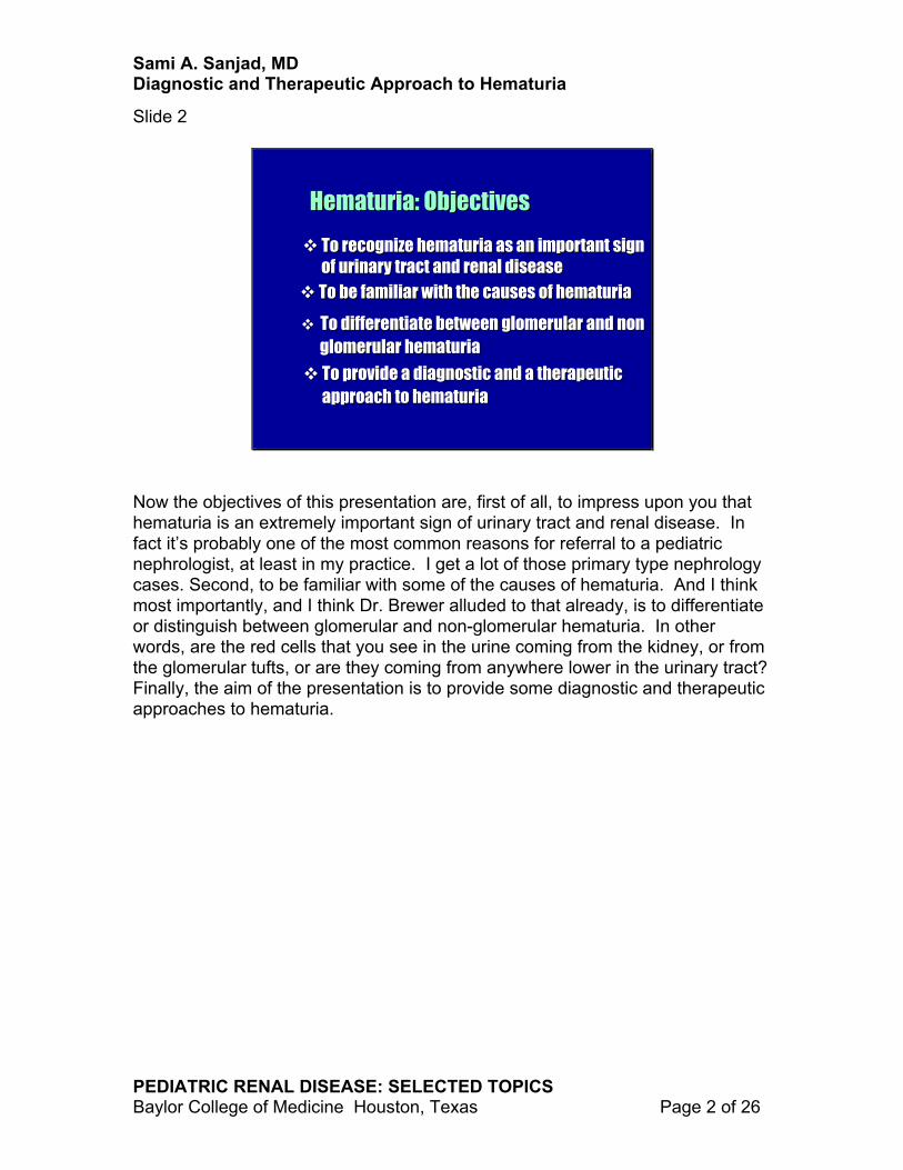
Hematuria: ObjectivesHematuria: Objectives
To provide a diagnostic and a therapeutic To provide a diagnostic and a therapeutic approach to approach to hematuriahematuria
To recognizeTo recognize hematuriahematuria as an important sign as an important sign of urinary tract and renal diseaseof urinary tract and renal disease
To be familiar with the causes ofTo be familiar with the causes of hematuriahematuria
To differentiate betweenTo differentiate between glomerularglomerular and non and non glomerular hematuriaglomerular hematuria
Now the objectives of this presentation are, first of all, to impress upon you that hematuria is an extremely important sign of urinary tract and renal disease. In fact it’s probably one of the most common reasons for referral to a pediatric nephrologist, at least in my practice. I get a lot of those primary type nephrology cases. Second, to be familiar with some of the causes of hematuria. And I think most importantly, and I think Dr. Brewer alluded to that already, is to differentiate or distinguish between glomerular and non-glomerular hematuria. In other words, are the red cells that you see in the urine coming from the kidney, or from the glomerular tufts, or are they coming from anywhere lower in the urinary tract? Finally, the aim of the presentation is to provide some diagnostic and therapeutic approaches to hematuria.
Sami A. Sanjad, MD Diagnostic and Therapeutic Approach to Hematuria
PEDIATRIC RENAL DISEASE: SELECTED TOPICS Baylor College of Medicine Houston, Texas Page 3 of 26
Slide 3
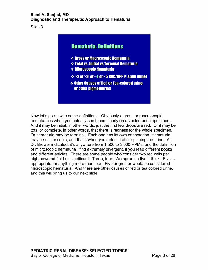
Hematuria: DefinitionsHematuria: Definitions
Other Causes of Red or TeaOther Causes of Red or Tea--colored urine colored urine or other or other pigmenturiaspigmenturias
Gross or MacroscopicGross or Macroscopic Hematuria Hematuria Total vs. InitialTotal vs. Initial vsvs TerminalTerminal HematuriaHematuriaMicroscopic Microscopic HematuriaHematuria
>2 or >3 or> >2 or >3 or> 44 or> 5 RBC/HPF ? (spun urine)or> 5 RBC/HPF ? (spun urine)
Now let’s go on with some definitions. Obviously a gross or macroscopic hematuria is when you actually see blood clearly on a voided urine specimen. And it may be initial, in other words, just the first few drops are red. Or it may be total or complete, in other words, that there is redness for the whole specimen. Or hematuria may be terminal. Each one has its own connotation. Hematuria may be microscopic, and that’s when you detect it after spinning the urine. As Dr. Brewer indicated, it’s anywhere from 1,500 to 3,000 RPMs, and the definition of microscopic hematuria I find extremely divergent, if you read different books and different articles. There are some people who consider two red cells per high-powered field as significant. Three, four. We agree on five, I think. Five is appropriate, or anything more than four. Five or greater would be considered microscopic hematuria. And there are other causes of red or tea colored urine, and this will bring us to our next slide.
Sami A. Sanjad, MD Diagnostic and Therapeutic Approach to Hematuria
PEDIATRIC RENAL DISEASE: SELECTED TOPICS Baylor College of Medicine Houston, Texas Page 4 of 26
Slide 4
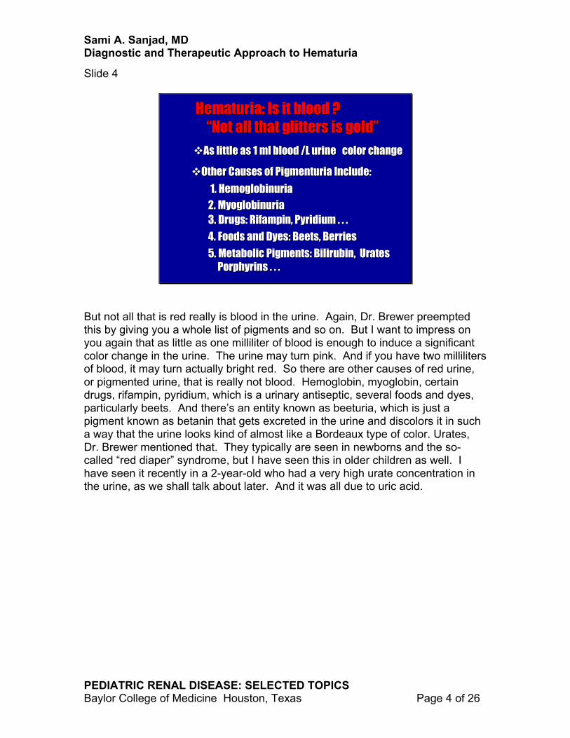
Hematuria: Is it blood ? Hematuria: Is it blood ? “Not all that glitters is gold”“Not all that glitters is gold”
4. Foods and Dyes: Beets, Berries4. Foods and Dyes: Beets, Berries
But not all that is red really is blood in the urine. Again, Dr. Brewer preempted this by giving you a whole list of pigments and so on. But I want to impress on you again that as little as one milliliter of blood is enough to induce a significant color change in the urine. The urine may turn pink. And if you have two milliliters of blood, it may turn actually bright red. So there are other causes of red urine, or pigmented urine, that is really not blood. Hemoglobin, myoglobin, certain drugs, rifampin, pyridium, which is a urinary antiseptic, several foods and dyes, particularly beets. And there’s an entity known as beeturia, which is just a pigment known as betanin that gets excreted in the urine and discolors it in such a way that the urine looks kind of almost like a Bordeaux type of color. Urates, Dr. Brewer mentioned that. They typically are seen in newborns and the so-called “red diaper” syndrome, but I have seen this in older children as well. I have seen it recently in a 2-year-old who had a very high urate concentration in the urine, as we shall talk about later. And it was all due to uric acid.
Sami A. Sanjad, MD Diagnostic and Therapeutic Approach to Hematuria
PEDIATRIC RENAL DISEASE: SELECTED TOPICS Baylor College of Medicine Houston, Texas Page 5 of 26
Now what is the differential? How do you approach a child that has a red or a brown urine? The first thing you do is centrifuge the urine according to the methods we already described. If the sediment is red, or the supernatant is red, you follow the following procedures. If the sediment is red, it means you are dealing with blood. If the supernatant is red, then you go ahead and dipstick the urine. And if it’s positive, you are dealing with myoglobin or hemoglobin. If it’s negative, you are dealing with the dyes we talked about, beets, bilirubin, and so on.
Sami A. Sanjad, MD Diagnostic and Therapeutic Approach to Hematuria
PEDIATRIC RENAL DISEASE: SELECTED TOPICS Baylor College of Medicine Houston, Texas Page 6 of 26
Slide 6
Hematuria: TestingHematuria: Testing
Urine sediment exam is the gold standard Urine sediment exam is the gold standard and should always be performed to confirm and should always be performed to confirm a positive a positive heme heme testtest
Urine dipsticks forUrine dipsticks for hemeheme can detect 1can detect 1--2 2
RBCsRBCs/HPF/HPF
May give false positive valuesMay give false positive values
Now the urine dipsticks are extremely sensitive, and they can detect as little as 1 to 2 red cells per high-powered field. They may give false positive results even if there is a little bacteria that have peroxidase production, or even a little fiber of meat or something in a diaper. Anything that can discolor the urine might give you a false positive result with the dipstick. So once you have a positive dipstick in the urine, the gold standard is to go ahead and perform the microscopic examination.
Sami A. Sanjad, MD Diagnostic and Therapeutic Approach to Hematuria
PEDIATRIC RENAL DISEASE: SELECTED TOPICS Baylor College of Medicine Houston, Texas Page 7 of 26
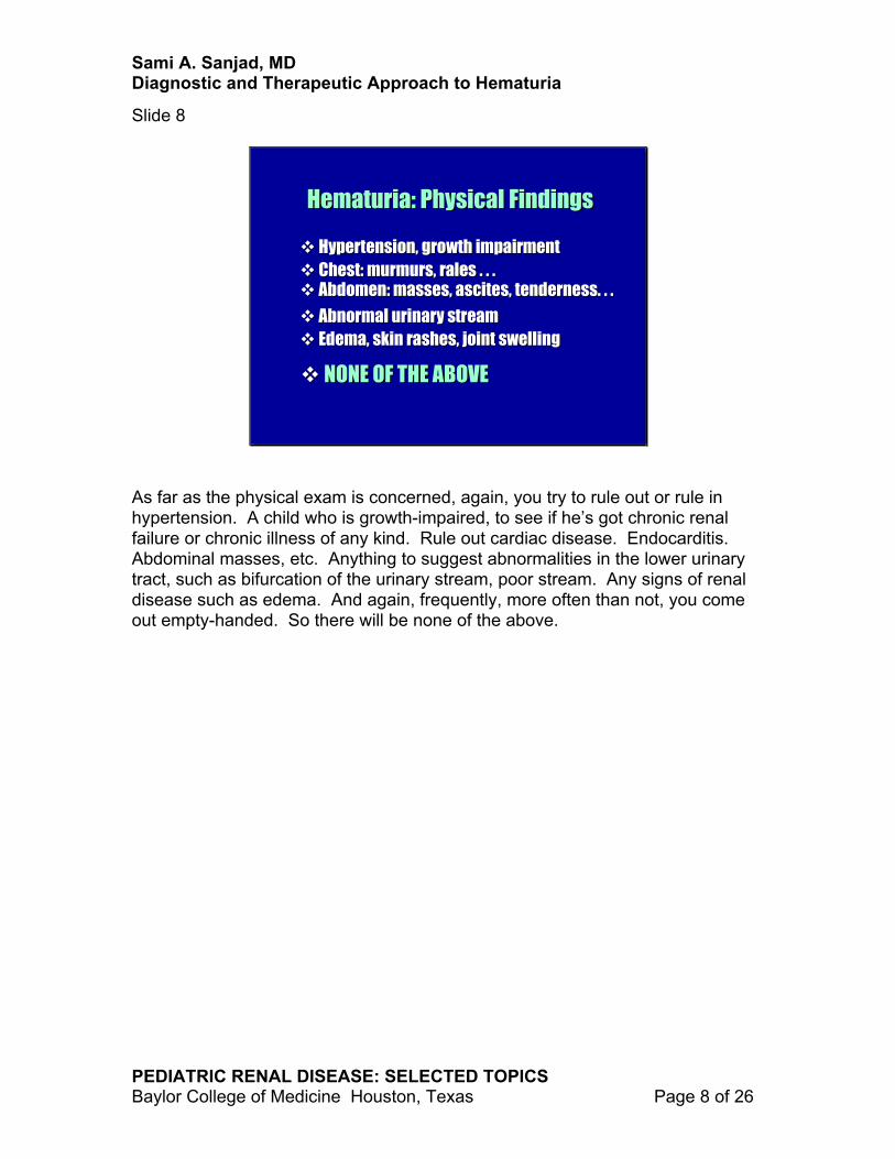
HxHx of skin or joint abnormalitiesof skin or joint abnormalities
Weight gain, swelling, recent URIWeight gain, swelling, recent URIDrug history, allergiesDrug history, allergiesFamily Family HxHx:: hematuriahematuria, deafness, CRF, deafness, CRF
NONE OF THE ABOVENONE OF THE ABOVE
Now once you have confirmed that you are dealing with hematuria, you have to get the differential diagnosis as to what might be causing it. So you go into the history. Frequently you start with inquiring about things that might give you hematuria of a nonspecific nature, such as fever, abdominal pain, dysuria. Things that would give you a clue as to what might have caused the hematuria. Sore throat and impetigo suggest an antecedent infection that might give you a nephritic type of syndrome. Skin or joint abnormalities suggest vasculitis. Weight gain, swelling, a recent upper respiratory infection. Drug history, allergies, and so on. All these are things that are frequently asked. Family history of renal disease. Deafness. Think of Alport’s disease. But more often than not, you come out empty-handed. Usually when we talk about hematuria, you find no or very little by way of historical clues to help you in the diagnosis.
Sami A. Sanjad, MD Diagnostic and Therapeutic Approach to Hematuria
PEDIATRIC RENAL DISEASE: SELECTED TOPICS Baylor College of Medicine Houston, Texas Page 8 of 26
As far as the physical exam is concerned, again, you try to rule out or rule in hypertension. A child who is growth-impaired, to see if he’s got chronic renal failure or chronic illness of any kind. Rule out cardiac disease. Endocarditis. Abdominal masses, etc. Anything to suggest abnormalities in the lower urinary tract, such as bifurcation of the urinary stream, poor stream. Any signs of renal disease such as edema. And again, frequently, more often than not, you come out empty-handed. So there will be none of the above.
Sami A. Sanjad, MD Diagnostic and Therapeutic Approach to Hematuria
PEDIATRIC RENAL DISEASE: SELECTED TOPICS Baylor College of Medicine Houston, Texas Page 9 of 26
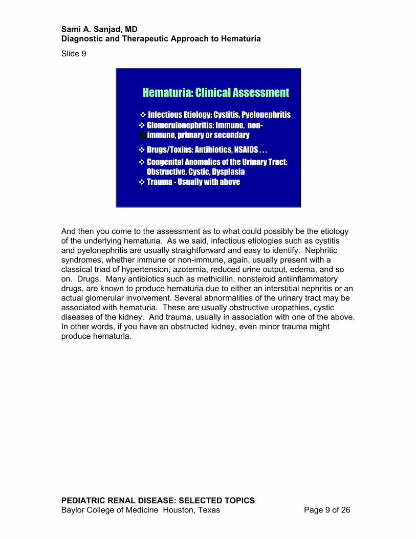
Congenital Anomalies of the Urinary Tract: Congenital Anomalies of the Urinary Tract: Obstructive, Cystic,Obstructive, Cystic, DysplasiaDysplasia
And then you come to the assessment as to what could possibly be the etiology of the underlying hematuria. As we said, infectious etiologies such as cystitis and pyelonephritis are usually straightforward and easy to identify. Nephritic syndromes, whether immune or non-immune, again, usually present with a classical triad of hypertension, azotemia, reduced urine output, edema, and so on. Drugs. Many antibiotics such as methicillin, nonsteroid antiinflammatory drugs, are known to produce hematuria due to either an interstitial nephritis or an actual glomerular involvement. Several abnormalities of the urinary tract may be associated with hematuria. These are usually obstructive uropathies, cystic diseases of the kidney. And trauma, usually in association with one of the above. In other words, if you have an obstructed kidney, even minor trauma might produce hematuria.
Sami A. Sanjad, MD Diagnostic and Therapeutic Approach to Hematuria
PEDIATRIC RENAL DISEASE: SELECTED TOPICS Baylor College of Medicine Houston, Texas Page 10 of 26
Now neoplastic diseases are rare in children as a cause of hematuria. The only thing you could think of would be a Wilms tumor in a younger child with an abdominal mass. This would raise the index of suspicion to a tumor. Hemoglobinopathies, particularly sickle cell disease, should be suspected in African-Americans if there are no other clear-cut etiologies. These are rare causes of hematuria. Vascular, such as hemangioma, A-V malformations. Renal vein thrombosis is usually self-evident. You have a usually newborn, or a severely dehydrated child who has an abdominal mass and hematuria, and this is usually fairly clear-cut as far as diagnosis goes. There are other causes which are nonspecific, such as fever, exercise. Exercise is a relatively common cause of hematuria. Regardless of the etiology or the underlying background, exercise seems to accentuate or increase the frequency of hematuria. And again, you may end up empty-handed and come up with none of the above. And this is what we refer to as monosymptomatic hematuria. And this is what I’ll be talking about for the remaining of the session.
Sami A. Sanjad, MD Diagnostic and Therapeutic Approach to Hematuria
PEDIATRIC RENAL DISEASE: SELECTED TOPICS Baylor College of Medicine Houston, Texas Page 11 of 26
1.1. Recurrent grossRecurrent gross hematuriahematuria with normal with normal urinalyses between episodesurinalyses between episodes
So, monosymptomatic hematuria, by definition, is hematuria that is not self-evident. In other words, you cannot rule in or rule out any of the causes we talked about earlier. It’s usually somebody who usually presents to the clinic or to the hospital or the emergency room either with a grossly bloody urine, or it may be something that is picked up on a routine urinalysis, and the child may have several red cells picked up microscopy. Now the prevalence of gross hematuria was described to be around .13%. Julie Ingelfinger several years ago, performed a retrospective study of emergency room visits, and she came up with this figure. Interestingly, in that the majority of these patients, the diagnosis was fairly straightforward. More than 60% were due – or about 60% – were due to either urinary tract infections, cystitis, trauma, perineal irritation, and so on. The remaining 40% or so were not very straightforward, but there was a high suspicion of an underlying infection. In other words, there was no positive culture detected, but it could have been viral cystitis and so on. Microscopic hematuria, on the other hand, is probably three to ten times as prevalent as gross hematuria. Now the figure of 4% is from Dr. Dodge from Galveston County who performed screening on about 12,000 schoolchildren, again several years ago. And interestingly in one urinalysis they detected microscopic hematuria defined as greater than five RBCs per high-powered field in 4% of the children. When they repeated the urinalysis one month later or one week later - I don’t recall – the prevalence dropped to about 1%. When they repeated this a third time, the prevalence dropped to about .5%. In other words, only a small percentage had three positive urinalyses, three consecutive positive urinalyses. And the more urinalyses you do down the line, the more attrition you get from this group. Now the patterns of hematuria may be a recurrent gross hematuria with normal urinalysis in between. Or it might be persistent microscopic hematuria.
Sami A. Sanjad, MD Diagnostic and Therapeutic Approach to Hematuria
PEDIATRIC RENAL DISEASE: SELECTED TOPICS Baylor College of Medicine Houston, Texas Page 12 of 26
Now the differential diagnosis of monosymptomatic hematuria has been narrowed down in the last several years to the following. Familial hypercalciuria, which is an autosomal dominant condition. Familial hyperuricosuria more recently has been identified as, I would say, a relatively common cause of hematuria. IgA or immunoglobin A nephropathy. Hereditary nephritis, or Alport syndrome. And the last is benign familial hematuria, also known as thin basement membrane disease. Again, this is not a very good term because benign familial hematuria occasionally may not be so benign, and sometimes it may not be familial as well. But in the majority of patients, or subjects, this is an autosome dominant condition as well. Occasionally hematuria might be due to unrecognized urinary tract abnormalities, infections, stones, and so on, that were missed during the initial workup. Now the first five I’ll consider as the most likely things you’ll find in a child who has microscopic unexplained hematuria. Interestingly, in the article that Dr. Hill co-authored years ago, if the article had been published maybe just a few years later, I think all of this would have fit into that article because the entity of immunoglobin A nephropathy had not been described yet. In other words, there were no immunofluorescent studies done in that article. And so I think, from the description a lot of the descriptive changes fit very well with IgA nephropathy. Some of them fit, again, very well with what we refer to now as benign familial hematuria or thin basement membrane disease, even though it was not recognized as an entity per se.
Sami A. Sanjad, MD Diagnostic and Therapeutic Approach to Hematuria
PEDIATRIC RENAL DISEASE: SELECTED TOPICS Baylor College of Medicine Houston, Texas Page 13 of 26
Slide 13
MonosymtomaticMonosymtomatic Hematuria: Hematuria: Glomerular vs. Extraglomerular BleedingGlomerular vs. Extraglomerular Bleeding
Now how do we differentiate between glomerular and extraglomerular type of hematuria? The patients who have extraglomerular hematuria, the urine is going to be red or pink, whereas in patients who have glomerular disease, it’s more likely to be reddish to brown. Clots are almost never found in patients with glomerular disease, whereas you might find clots in patients who have bleeding from the lower urinary tract. Proteinuria is usually minimal in patients who have extraglomerular disease or nonexistent at all, and it’s usually in excess of half a gram per day. The morphology of the red cells is normal in patients with urinary tract bleeding, whereas it is usually dysmorphic in patients with glomerular bleeding. Casts, of course, would be absent in patients with urinary tract bleeding but are likely to be positive in patients with glomerular bleeding.
Sami A. Sanjad, MD Diagnostic and Therapeutic Approach to Hematuria
PEDIATRIC RENAL DISEASE: SELECTED TOPICS Baylor College of Medicine Houston, Texas Page 14 of 26
Slide 14
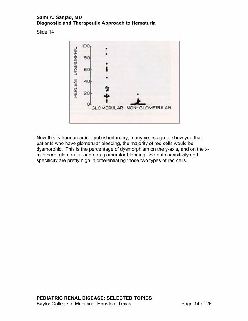
Now this is from an article published many, many years ago to show you that patients who have glomerular bleeding, the majority of red cells would be dysmorphic. This is the percentage of dysmorphism on the y-axis, and on the x-axis here, glomerular and non-glomerular bleeding. So both sensitivity and specificity are pretty high in differentiating those two types of red cells.
Sami A. Sanjad, MD Diagnostic and Therapeutic Approach to Hematuria
PEDIATRIC RENAL DISEASE: SELECTED TOPICS Baylor College of Medicine Houston, Texas Page 15 of 26
Slide 15
This is a good example of dysmorphic red cells. You can see, as Dr. Brewer is trying to show you, they are squished there. Some of these are normal. These are what normal RBCs look like, and these are dysmorphic red cells. This from a patient with IgA nephropathy.
Sami A. Sanjad, MD Diagnostic and Therapeutic Approach to Hematuria
PEDIATRIC RENAL DISEASE: SELECTED TOPICS Baylor College of Medicine Houston, Texas Page 16 of 26
Slide 16
And this is what normal RBCs look like. They have a nice halo around them. They have the typical round appearance. And this is from a child with cystitis.
Sami A. Sanjad, MD Diagnostic and Therapeutic Approach to Hematuria
PEDIATRIC RENAL DISEASE: SELECTED TOPICS Baylor College of Medicine Houston, Texas Page 17 of 26
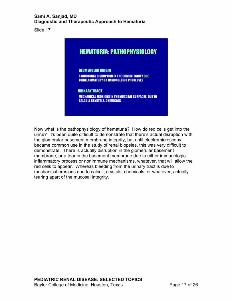
STRUCTURAL DISRUPTION IN THE GBM INTEGRITY DUE TOINFLAMMATORY OR IMMUNOLOGIC PROCESSES
MECHANICAL EROSIONS IN THE MUCOSAL SURFACES DUE TO CALCULI, CRYSTALS, CHEMICALS...
Now what is the pathophysiology of hematuria? How do red cells get into the urine? It’s been quite difficult to demonstrate that there’s actual disruption with the glomerular basement membrane integrity, but until electromicroscopy became common use in the study of renal biopsies, this was very difficult to demonstrate. There is actually disruption in the glomerular basement membrane, or a tear in the basement membrane due to either immunologic inflammatory process or nonimmune mechanisms, whatever, that will allow the red cells to appear. Whereas bleeding from the urinary tract is due to mechanical erosions due to calculi, crystals, chemicals, or whatever, actually tearing apart of the mucosal integrity.
Sami A. Sanjad, MD Diagnostic and Therapeutic Approach to Hematuria
PEDIATRIC RENAL DISEASE: SELECTED TOPICS Baylor College of Medicine Houston, Texas Page 18 of 26
Slide 18
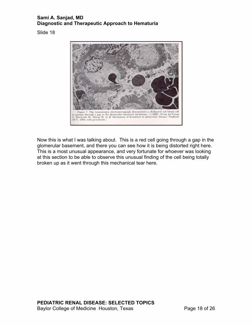
Now this is what I was talking about. This is a red cell going through a gap in the glomerular basement, and there you can see how it is being distorted right here. This is a most unusual appearance, and very fortunate for whoever was looking at this section to be able to observe this unusual finding of the cell being totally broken up as it went through this mechanical tear here.
Sami A. Sanjad, MD Diagnostic and Therapeutic Approach to Hematuria
PEDIATRIC RENAL DISEASE: SELECTED TOPICS Baylor College of Medicine Houston, Texas Page 19 of 26
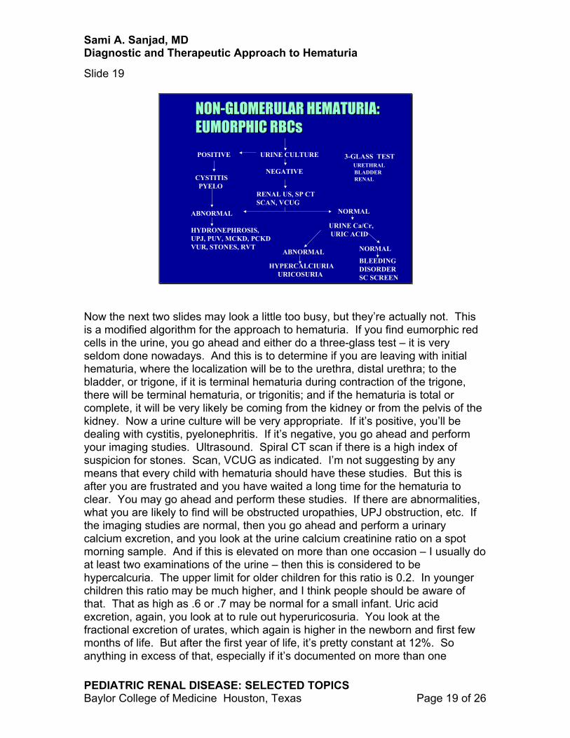
Now the next two slides may look a little too busy, but they’re actually not. This is a modified algorithm for the approach to hematuria. If you find eumorphic red cells in the urine, you go ahead and either do a three-glass test – it is very seldom done nowadays. And this is to determine if you are leaving with initial hematuria, where the localization will be to the urethra, distal urethra; to the bladder, or trigone, if it is terminal hematuria during contraction of the trigone, there will be terminal hematuria, or trigonitis; and if the hematuria is total or complete, it will be very likely be coming from the kidney or from the pelvis of the kidney. Now a urine culture will be very appropriate. If it’s positive, you’ll be dealing with cystitis, pyelonephritis. If it’s negative, you go ahead and perform your imaging studies. Ultrasound. Spiral CT scan if there is a high index of suspicion for stones. Scan, VCUG as indicated. I’m not suggesting by any means that every child with hematuria should have these studies. But this is after you are frustrated and you have waited a long time for the hematuria to clear. You may go ahead and perform these studies. If there are abnormalities, what you are likely to find will be obstructed uropathies, UPJ obstruction, etc. If the imaging studies are normal, then you go ahead and perform a urinary calcium excretion, and you look at the urine calcium creatinine ratio on a spot morning sample. And if this is elevated on more than one occasion – I usually do at least two examinations of the urine – then this is considered to be hypercalcuria. The upper limit for older children for this ratio is 0.2. In younger children this ratio may be much higher, and I think people should be aware of that. That as high as .6 or .7 may be normal for a small infant. Uric acid excretion, again, you look at to rule out hyperuricosuria. You look at the fractional excretion of urates, which again is higher in the newborn and first few months of life. But after the first year of life, it’s pretty constant at 12%. So anything in excess of that, especially if it’s documented on more than one
Sami A. Sanjad, MD Diagnostic and Therapeutic Approach to Hematuria
PEDIATRIC RENAL DISEASE: SELECTED TOPICS Baylor College of Medicine Houston, Texas Page 20 of 26
occasion, would be suspicious or almost diagnostic of hyperuricosuria. If none of these are present, then you look for rarer causes of hematuria such as bleeding disorders, sickle cell screen, and so on. I must say that hyperuricosuria, at least in my experience in the last couple of years, has been a relatively common cause of microscopic hematuria. I don’t know what to make of it. We usually don’t treat. But we’ll talk about this in just a second.
Sami A. Sanjad, MD Diagnostic and Therapeutic Approach to Hematuria
PEDIATRIC RENAL DISEASE: SELECTED TOPICS Baylor College of Medicine Houston, Texas Page 21 of 26
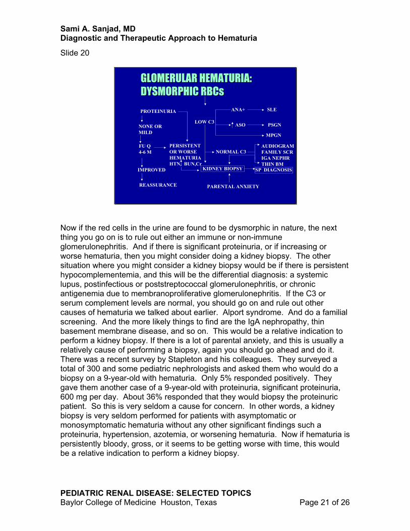
Now if the red cells in the urine are found to be dysmorphic in nature, the next thing you go on is to rule out either an immune or non-immune glomerulonephritis. And if there is significant proteinuria, or if increasing or worse hematuria, then you might consider doing a kidney biopsy. The other situation where you might consider a kidney biopsy would be if there is persistent hypocomplementemia, and this will be the differential diagnosis: a systemic lupus, postinfectious or poststreptococcal glomerulonephritis, or chronic antigenemia due to membranoproliferative glomerulonephritis. If the C3 or serum complement levels are normal, you should go on and rule out other causes of hematuria we talked about earlier. Alport syndrome. And do a familial screening. And the more likely things to find are the IgA nephropathy, thin basement membrane disease, and so on. This would be a relative indication to perform a kidney biopsy. If there is a lot of parental anxiety, and this is usually a relatively cause of performing a biopsy, again you should go ahead and do it. There was a recent survey by Stapleton and his colleagues. They surveyed a total of 300 and some pediatric nephrologists and asked them who would do a biopsy on a 9-year-old with hematuria. Only 5% responded positively. They gave them another case of a 9-year-old with proteinuria, significant proteinuria, 600 mg per day. About 36% responded that they would biopsy the proteinuric patient. So this is very seldom a cause for concern. In other words, a kidney biopsy is very seldom performed for patients with asymptomatic or monosymptomatic hematuria without any other significant findings such a proteinuria, hypertension, azotemia, or worsening hematuria. Now if hematuria is persistently bloody, gross, or it seems to be getting worse with time, this would be a relative indication to perform a kidney biopsy.
Sami A. Sanjad, MD Diagnostic and Therapeutic Approach to Hematuria
PEDIATRIC RENAL DISEASE: SELECTED TOPICS Baylor College of Medicine Houston, Texas Page 22 of 26
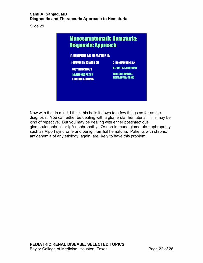
Now with that in mind, I think this boils it down to a few things as far as the diagnosis. You can either be dealing with a glomerular hematuria. This may be kind of repetitive. But you may be dealing with either postinfectious glomerulonephritis or IgA nephropathy. Or non-immune glomerulo-nephropathy such as Alport syndrome and benign familial hematuria. Patients with chronic antigenemia of any etiology, again, are likely to have this problem.
Sami A. Sanjad, MD Diagnostic and Therapeutic Approach to Hematuria
PEDIATRIC RENAL DISEASE: SELECTED TOPICS Baylor College of Medicine Houston, Texas Page 23 of 26
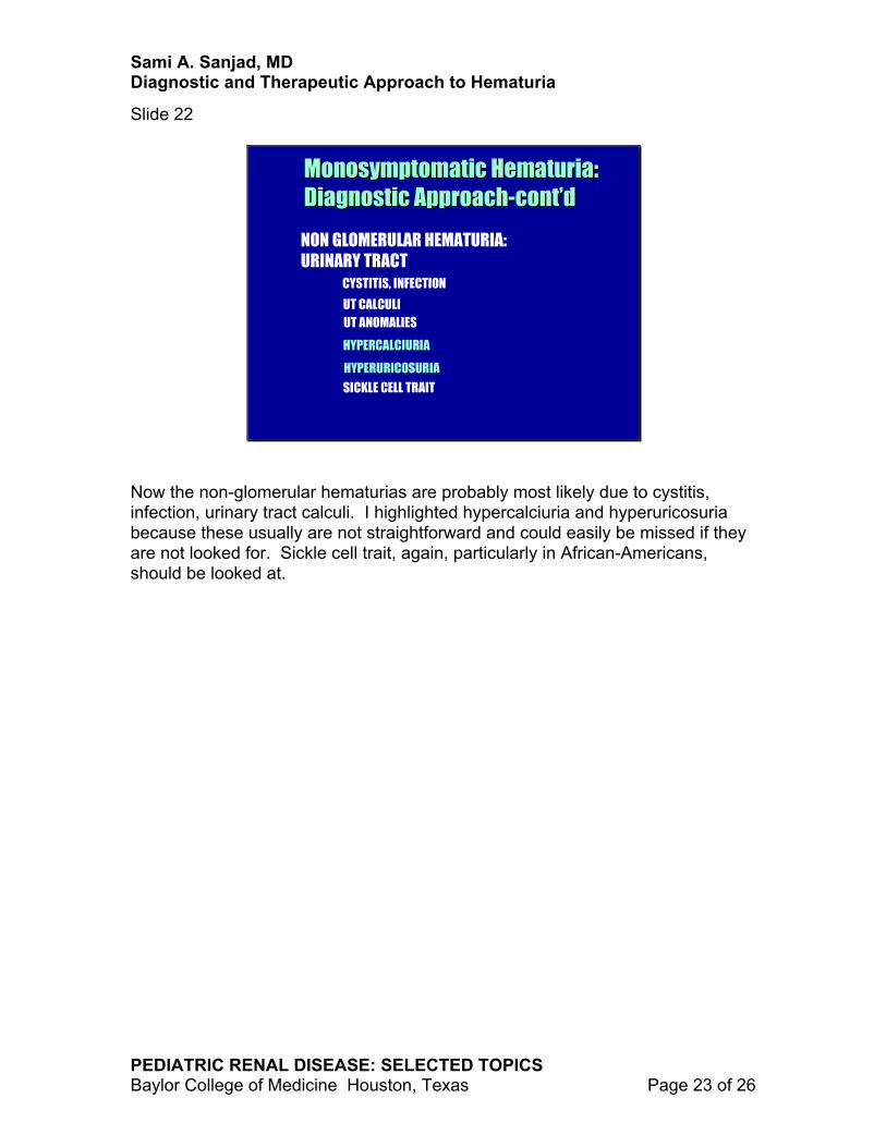
Now the non-glomerular hematurias are probably most likely due to cystitis, infection, urinary tract calculi. I highlighted hypercalciuria and hyperuricosuria because these usually are not straightforward and could easily be missed if they are not looked for. Sickle cell trait, again, particularly in African-Americans, should be looked at.
Sami A. Sanjad, MD Diagnostic and Therapeutic Approach to Hematuria
PEDIATRIC RENAL DISEASE: SELECTED TOPICS Baylor College of Medicine Houston, Texas Page 24 of 26
Slide 23
Monosymptomatic Hematuria Monosymptomatic Hematuria and and HypercalciuriaHypercalciuria//UricosuriaUricosuria
30-35% OF CHILDREN WITH HEMATURIA HAVE HYPERCALCIURIA
HYPERCALCIURIA DEFINED BY UCa/Cr OR BY 24-HRUCa > 4 MG/KG
HYPERURICOSURIA FOUND IN 5-20% OF CHILDREN WITH UNEXPLAINED HEMATURIA. FE UA>12%
Now the reason I mention hypercalciuria is that, again, it has been shown by Stapleton and his group that this accounts for about 30 to 35% of children with hematuria. Hyperuricosuria, accounts for 5 to 20% of children with unexplained hematuria. This is diagnosed by uric acid excretion or a fractional excretion in excess of 12%. These are two relatively common causes, easy to diagnose without much ado. All you need to do is a spot urine and look at the urine calcium creatinine. Or if you want to do a 24-hour urine, anything in excess of 4mg/kg/day would be diagnostic.
Sami A. Sanjad, MD Diagnostic and Therapeutic Approach to Hematuria
PEDIATRIC RENAL DISEASE: SELECTED TOPICS Baylor College of Medicine Houston, Texas Page 25 of 26
1-REASSURANCE, LONG TERM FOLLOW UP HEMATURIA DOESN’T CAUSE ANEMIA
2-DIETARY THERAPY: LOW Na FOR HYPERCALCIURIA,LOW PURINE FOR URICOSURIA
3-DRUG THERAPY: THIAZIDES FOR HYPERCALCIURIA,ALKALINIZING AGENTS FOR HYPERURICOSURIA+/- ALLOPURINOL
4-SPECIFIC THERAPY FOR NEPHRITIS
Now I think the most important aspect of the approach to hematuria is reassurance that this is not a serious condition unless you have diagnosed something serious. But with this asymptomatic hematuria, it’s very important to reassure the family that this is not cancer. Many of them worry about cancer. Many of them worry about anemia due to a few red cells in the urine. I tell them this doesn’t happen. Dietary therapy may be of some benefit in patients who have hypercalciuria. Patients put on a low sodium diet might benefit if the hypercalciuria is typically renal in origin. A low purine diet may help patients with hyperuricosuria. Drug therapy. We seldom resort to drug therapy, but I have used thiazides in a few patients with hypercalciuria, and it usually reduces urinary calcium excretion quite appreciably. Hyperuricosuria, some people have recommended allopurinol. What I have done is just ask them to stay away from high purines in their diets. And occasionally alkalinize their urine with citrates. And if you run into a rare case of surreptitious nephritis, or undiagnosed silent nephritis, then again, a kidney biopsy would have been performed and the treatment would have been specifically addressed to that.
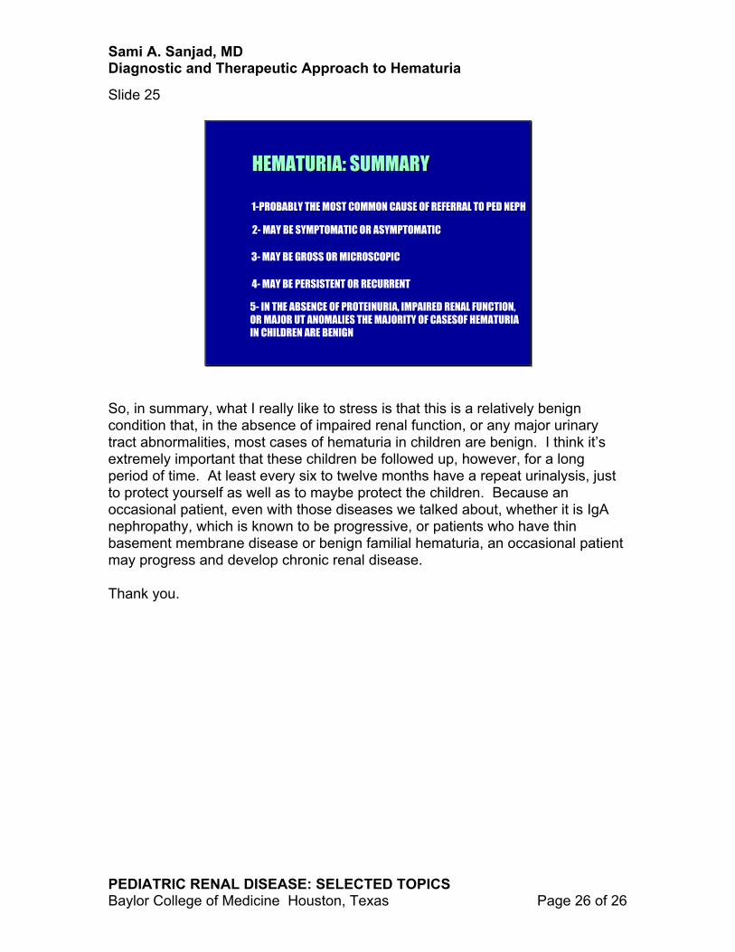
Sami A. Sanjad, MD Diagnostic and Therapeutic Approach to Hematuria
PEDIATRIC RENAL DISEASE: SELECTED TOPICS Baylor College of Medicine Houston, Texas Page 26 of 26
Slide 25
HEMATURIA: SUMMARYHEMATURIA: SUMMARY
1-PROBABLY THE MOST COMMON CAUSE OF REFERRAL TO PED NEPH
3- MAY BE GROSS OR MICROSCOPIC
4- MAY BE PERSISTENT OR RECURRENT
2- MAY BE SYMPTOMATIC OR ASYMPTOMATIC
5- IN THE ABSENCE OF PROTEINURIA, IMPAIRED RENAL FUNCTION, OR MAJOR UT ANOMALIES THE MAJORITY OF CASESOF HEMATURIAIN CHILDREN ARE BENIGN
So, in summary, what I really like to stress is that this is a relatively benign condition that, in the absence of impaired renal function, or any major urinary tract abnormalities, most cases of hematuria in children are benign. I think it’s extremely important that these children be followed up, however, for a long period of time. At least every six to twelve months have a repeat urinalysis, just to protect yourself as well as to maybe protect the children. Because an occasional patient, even with those diseases we talked about, whether it is IgA nephropathy, which is known to be progressive, or patients who have thin basement membrane disease or benign familial hematuria, an occasional patient may progress and develop chronic renal disease.