SARM1-specific motifs in the TIR domain enable NAD + loss and regulate injury-induced SARM1 activation Daniel W. Summers a,b , Daniel A. Gibson b , Aaron DiAntonio b,c,1 , and Jeffrey Milbrandt a,c,1 a Department of Genetics, Washington University in St. Louis, St. Louis, MO 63110; b Department of Developmental Biology, Washington University in St. Louis, St. Louis, MO 63110; and c Hope Center for Neurological Disorders, Washington University in St. Louis, St. Louis, MO 63110 Edited by Richard H. Goodman, Vollum Institute, Portland, OR, and approved August 19, 2016 (received for review January 27, 2016) Axon injury in response to trauma or disease stimulates a self- destruction program that promotes the localized clearance of damaged axon segments. Sterile alpha and Toll/interleukin re- ceptor (TIR) motif-containing protein 1 (SARM1) is an evolution- arily conserved executioner of this degeneration cascade, also known as Wallerian degeneration; however, the mechanism of SARM1-dependent neuronal destruction is still obscure. SARM1 possesses a TIR domain that is necessary for SARM1 activity. In other proteins, dimerized TIR domains serve as scaffolds for in- nate immune signaling. In contrast, dimerization of the SARM1 TIR domain promotes consumption of the essential metabolite NAD + and induces neuronal destruction. This activity is unique to the SARM1 TIR domain, yet the structural elements that enable this activity are unknown. In this study, we identify fundamental properties of the SARM1 TIR domain that promote NAD + loss and axon degeneration. Dimerization of the TIR domain from the Caenorhabditis elegans SARM1 ortholog TIR-1 leads to NAD + loss and neuronal death, indicating these activities are an evolutionarily conserved feature of SARM1 function. Detailed analysis of sequence homology identifies canonical TIR motifs as well as a SARM1-specific (SS) loop that are required for NAD + loss and axon degeneration. Furthermore, we identify a residue in the SARM1 BB loop that is dispensable for TIR activity yet required for injury-induced activa- tion of full-length SARM1, suggesting that SARM1 function requires multidomain interactions. Indeed, we identify a physical interaction between the autoinhibitory N terminus and the TIR domain of SARM1, revealing a previously unrecognized direct connection be- tween these domains that we propose mediates autoinhibition and activation upon injury. SARM | NAD | cell death | axon degeneration | sarmoptosis A xon degeneration in response to injury or disease is a hall- mark of neurological disorders of the peripheral and central nervous systems. Akin to programmed cell death pathways, such as apoptosis, axon injury in response to disease or trauma stim- ulates a local signaling cascade that executes destruction of the injured axon segment (1). Despite growing attention, the molec- ular details of this prodegenerative cascade are still unclear. A more substantial understanding of the molecular factors that participate in this axon destructive process will likely provide new therapeutic strategies for peripheral neuropathies and other neurodegenerative conditions. Genetic screens in invertebrate and vertebrate model systems identified the Sterile alpha and Toll/interleukin receptor (TIR) motif-containing protein 1 (SARM1) as a conserved, fundamental executioner of pathological axon destruction (2, 3). After nerve injury, SARM1 is required for the precipitous loss of the me- tabolite NAD + (4). Augmenting NAD + biosynthetic pathways protects injured axons from degeneration, suggesting this step is crucial in axonal breakdown (5, 6). In addition to local axon degeneration, SARM1 promotes neuronal cell death in response to mitochondrial toxins, oxygen glucose deprivation, and viral infection (7–10). Activated SARM1 can induce cell death in- dependent of other known programmed cell death programs in a pathway termed sarmoptosis. Hence, SARM1 exerts a major role in pathological neuron destruction, so a deeper understanding of the molecular mechanism of SARM1 function may identify novel targets for the inhibition of this prodestructive factor. Domain analysis of the SARM1 protein generated a model of SARM1 activation and function (3, 11, 12). SARM1 possesses a Toll/interleukin receptor (TIR) domain that is required for SARM1-dependent axon degeneration and cell death. Recently, others and we demonstrated that enforced dimerization of the SARM1 TIR domain is sufficient to induce local axon de- generation (3, 4, 13). Tandem sterile alpha motif (SAM) do- mains mediate SARM1 multimerization, and hence are required for SARM1 activity. The SARM1 N-terminal domain contains Heat/Armadillo repeats and restrains SAM-TIR activity, main- taining SARM1 in an autoinhibited state (3, 11, 12). Although SARM1 could possess constitutive activity that is countered in a healthy axon downstream of the pathway, our current model is that axon injury triggers a release of SARM1 autoinhibition, thus enabling TIR-dependent neuronal destruction (1). Whether the N terminus, SAM, or TIR domains act independently or if there is interdomain cross-talk in response to injury is unknown. TIR domains are platforms for homodimerization as well as heterodimerization with other TIR domains (14). Enforced homodimerization of the human SARM1 TIR (HsTIR) domain stimulates rapid consumption of NAD + followed by axon de- generation and cell death (4). The capacity of dimerized SARM1 TIR to stimulate NAD + loss and cell death is unique, because dimerization of other human TIR domains does not induce axon degeneration or cell death (4, 15, 16). Hence, the TIR domain of SARM1 possesses distinct qualities that enable SARM1- dependent NAD + loss after neuronal injury. Indeed, phylogenetic Significance Axon degeneration is an important pathological event in mul- tiple neurodegenerative disorders. Axon injury stimulates the prodestructive factor SARM1, leading to the precipitous loss in the metabolite NAD + . Remarkably, enforcing dimerization of the Toll/interleukin receptor (TIR) domain from SARM1 is suf- ficient to promote NAD + loss and axon degeneration. In this study, we uncover fundamental elements within the SARM1 TIR domain responsible for this activity, including a unique motif that is highly specific to SARM1. In addition, we discover a role for the SARM1 TIR domain in injury-induced activation of the SARM1 protein, suggesting that this domain contributes to SARM1 regulation in addition to the execution of axon degenera- tion. These studies identify potential avenues for therapeutic in- tervention in SARM1-dependent axon destruction pathways. Author contributions: D.W.S. and D.A.G. designed research; D.W.S. and D.A.G. performed research; D.W.S., D.A.G., A.D., and J.M. analyzed data; and D.W.S., D.A.G., A.D., and J.M. wrote the paper. The authors declare no conflict of interest. This article is a PNAS Direct Submission. 1 To whom correspondence may be addressed. Email: [email protected] or diantonio@ wustl.edu. This article contains supporting information online at www.pnas.org/lookup/suppl/doi:10. 1073/pnas.1601506113/-/DCSupplemental. www.pnas.org/cgi/doi/10.1073/pnas.1601506113 PNAS | Published online September 26, 2016 | E6271–E6280 NEUROSCIENCE PNAS PLUS
Transcript
SARM1-specific motifs in the TIR domain enable NAD+
loss and regulate injury-induced SARM1 activationDaniel W. Summersa,b, Daniel A. Gibsonb, Aaron DiAntoniob,c,1, and Jeffrey Milbrandta,c,1
aDepartment of Genetics, Washington University in St. Louis, St. Louis, MO 63110; bDepartment of Developmental Biology, Washington University in St.Louis, St. Louis, MO 63110; and cHope Center for Neurological Disorders, Washington University in St. Louis, St. Louis, MO 63110
Edited by Richard H. Goodman, Vollum Institute, Portland, OR, and approved August 19, 2016 (received for review January 27, 2016)
Axon injury in response to trauma or disease stimulates a self-destruction program that promotes the localized clearance ofdamaged axon segments. Sterile alpha and Toll/interleukin re-ceptor (TIR) motif-containing protein 1 (SARM1) is an evolution-arily conserved executioner of this degeneration cascade, alsoknown as Wallerian degeneration; however, the mechanism ofSARM1-dependent neuronal destruction is still obscure. SARM1possesses a TIR domain that is necessary for SARM1 activity. Inother proteins, dimerized TIR domains serve as scaffolds for in-nate immune signaling. In contrast, dimerization of the SARM1TIR domain promotes consumption of the essential metaboliteNAD+ and induces neuronal destruction. This activity is uniqueto the SARM1 TIR domain, yet the structural elements that enablethis activity are unknown. In this study, we identify fundamentalproperties of the SARM1 TIR domain that promote NAD+ loss andaxon degeneration. Dimerization of the TIR domain from theCaenorhabditis elegans SARM1 ortholog TIR-1 leads to NAD+ lossand neuronal death, indicating these activities are an evolutionarilyconserved feature of SARM1 function. Detailed analysis of sequencehomology identifies canonical TIR motifs as well as a SARM1-specific(SS) loop that are required for NAD+ loss and axon degeneration.Furthermore, we identify a residue in the SARM1 BB loop that isdispensable for TIR activity yet required for injury-induced activa-tion of full-length SARM1, suggesting that SARM1 function requiresmultidomain interactions. Indeed, we identify a physical interactionbetween the autoinhibitory N terminus and the TIR domain ofSARM1, revealing a previously unrecognized direct connection be-tween these domains that we propose mediates autoinhibition andactivation upon injury.
SARM | NAD | cell death | axon degeneration | sarmoptosis
Axon degeneration in response to injury or disease is a hall-mark of neurological disorders of the peripheral and central
nervous systems. Akin to programmed cell death pathways, suchas apoptosis, axon injury in response to disease or trauma stim-ulates a local signaling cascade that executes destruction of theinjured axon segment (1). Despite growing attention, the molec-ular details of this prodegenerative cascade are still unclear. Amore substantial understanding of the molecular factors thatparticipate in this axon destructive process will likely providenew therapeutic strategies for peripheral neuropathies and otherneurodegenerative conditions.Genetic screens in invertebrate and vertebrate model systems
identified the Sterile alpha and Toll/interleukin receptor (TIR)motif-containing protein 1 (SARM1) as a conserved, fundamentalexecutioner of pathological axon destruction (2, 3). After nerveinjury, SARM1 is required for the precipitous loss of the me-tabolite NAD+ (4). Augmenting NAD+ biosynthetic pathwaysprotects injured axons from degeneration, suggesting this step iscrucial in axonal breakdown (5, 6). In addition to local axondegeneration, SARM1 promotes neuronal cell death in responseto mitochondrial toxins, oxygen glucose deprivation, and viralinfection (7–10). Activated SARM1 can induce cell death in-dependent of other known programmed cell death programs in apathway termed sarmoptosis. Hence, SARM1 exerts a major role
in pathological neuron destruction, so a deeper understanding ofthe molecular mechanism of SARM1 function may identify noveltargets for the inhibition of this prodestructive factor.Domain analysis of the SARM1 protein generated a model of
SARM1 activation and function (3, 11, 12). SARM1 possesses aToll/interleukin receptor (TIR) domain that is required forSARM1-dependent axon degeneration and cell death. Recently,others and we demonstrated that enforced dimerization of theSARM1 TIR domain is sufficient to induce local axon de-generation (3, 4, 13). Tandem sterile alpha motif (SAM) do-mains mediate SARM1 multimerization, and hence are requiredfor SARM1 activity. The SARM1 N-terminal domain containsHeat/Armadillo repeats and restrains SAM-TIR activity, main-taining SARM1 in an autoinhibited state (3, 11, 12). AlthoughSARM1 could possess constitutive activity that is countered in ahealthy axon downstream of the pathway, our current model isthat axon injury triggers a release of SARM1 autoinhibition, thusenabling TIR-dependent neuronal destruction (1). Whether theN terminus, SAM, or TIR domains act independently or if thereis interdomain cross-talk in response to injury is unknown.TIR domains are platforms for homodimerization as well as
heterodimerization with other TIR domains (14). Enforcedhomodimerization of the human SARM1 TIR (HsTIR) domainstimulates rapid consumption of NAD+ followed by axon de-generation and cell death (4). The capacity of dimerized SARM1TIR to stimulate NAD+ loss and cell death is unique, becausedimerization of other human TIR domains does not induce axondegeneration or cell death (4, 15, 16). Hence, the TIR domainof SARM1 possesses distinct qualities that enable SARM1-dependent NAD+ loss after neuronal injury. Indeed, phylogenetic
Significance
Axon degeneration is an important pathological event in mul-tiple neurodegenerative disorders. Axon injury stimulates theprodestructive factor SARM1, leading to the precipitous loss inthe metabolite NAD+. Remarkably, enforcing dimerization ofthe Toll/interleukin receptor (TIR) domain from SARM1 is suf-ficient to promote NAD+ loss and axon degeneration. In thisstudy, we uncover fundamental elements within the SARM1TIR domain responsible for this activity, including a uniquemotif that is highly specific to SARM1. In addition, we discovera role for the SARM1 TIR domain in injury-induced activation ofthe SARM1 protein, suggesting that this domain contributes toSARM1 regulation in addition to the execution of axon degenera-tion. These studies identify potential avenues for therapeutic in-tervention in SARM1-dependent axon destruction pathways.
Author contributions: D.W.S. and D.A.G. designed research; D.W.S. and D.A.G. performedresearch; D.W.S., D.A.G., A.D., and J.M. analyzed data; and D.W.S., D.A.G., A.D., and J.M.wrote the paper.
analysis of almost 2,000 TIR domains suggests the TIR domainsfrom human SARM1 and its orthologs are more closely relatedto TIR domains found in bacteria rather than other metazoans(17). Consequently, we suspected that unique motifs in theSARM1 TIR domain promote SARM1 activity in injury-inducedaxon degeneration.In this study, we demonstrate that the ability of the SARM1
TIR domain to induce NAD+ loss and neuronal death is con-served from humans to Caenorhabditis elegans. Using sequencehomology and structural prediction, we identify putative loopmotifs in the SARM1 TIR domain, including a highly conservedSARM1-specific (SS) loop motif that we show is required forSARM1 function. These motifs are required for TIR-mediatedNAD+ loss as well as axon degeneration and neuronal cell death.Finally, we identify a residue within the SARM1 TIR domainthat is dispensable for TIR function but is required for injury-induced activation of SARM1, revealing the importance ofinterdomain cross-talk in SARM1 regulation.
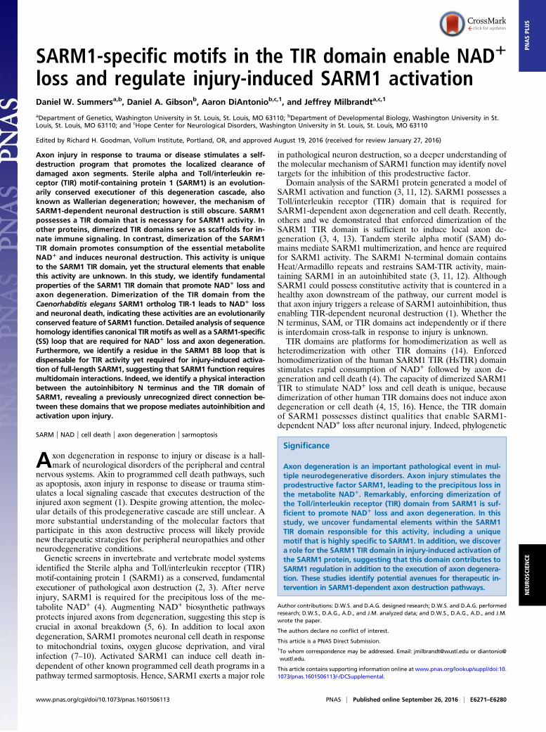
ResultsDimerization of the TIR Domain from C. elegans TIR-1 PromotesNeuronal Cell Death. To identify elements within the SARM1TIR domain responsible for NAD+ loss and cell death, we firstinvestigated whether this TIR activity is conserved in a distantSARM1 relative. The SARM1 ortholog in C. elegans, tir-1, is
implicated in nonapoptotic cell death in development and motorneuron degeneration in a model of ALS (18, 19), suggesting aconserved role in neurodegeneration. However, tir-1 regulatesdevelopmental patterning in the nervous system as well asstimulating innate immune signaling in response to pathogens(11, 20, 21), two activities that appear inconsistent with theneurodestructive properties of human SARM1. The TIR do-mains from SARM1 and TIR-1 are the most homologous regionsof these proteins (44% identity); hence, we sought to examinewhether the capacity of HsTIR to deplete NAD+ and promoteneuronal cell death is conserved in TIR-1 (CeTIR). The TIRdomain of TIR-1 (amino acids 757–917) was fused to Frb/Fkbpmoieties expressed in dorsal root ganglion (DRG) sensory neu-rons, and dimerization was stimulated with rapamycin (Fig. 1A).As previously reported, dimerizing HsTIR causes robust celldeath beginning as early as 8 h after rapamycin addition. Inneurons expressing CeTIR, rapamycin treatment also leads tocell death (Fig. 1 B and C). Cell death induced by dimerization ofCeTIR is slightly delayed compared with HsTIR, becoming ap-parent 12 h after rapamycin addition. Similar to dimerizationof HsTIR, plasma membrane blebbing preceded the ethidiumhomodimer nuclear labeling that signifies cell death.The Frb/Fkbp system enables enforced heterodimerization of
select TIR combinations. We investigated whether SARM1 TIRsfrom humans and C. elegans could functionally dimerize to induce
Fig. 1. Dimerization of CeTIR stimulates NAD+ lossand neuronal cell death. (A) Scheme for TIR di-merization via fusing Frb and Fkbp moieties to theTIR domain. Rapamycin stimulates TIR dimerizationand cell death. (B) Images of embryonic DRGsexpressing the indicated Frb/Fkbp TIR constructs afterrapamycin addition. Dying cells are labeled withethidium homodimer and costained with Hoescht tolabel nuclei. (C) Quantification of cellular death afterrapamycin addition. (D) ΔΨ was monitored with tet-ramethylrhodamine methyl ester in DRG neuronsexpressing the indicated Frb/Fkbp TIR construct afterrapamycin addition. (E and F) NAD+ and ATP levelswere measured from DRGs expressing the indicatedFrb/Fkbp TIR construct after rapamycin addition.(G and H) NAD+ and ATP were measured at moreextended time points after rapamycin addition to as-sess metabolite levels after long-term TIR dimer-ization. Error bars reflect ± SEM (n = 3). (Scalebars, 25 μM.)
E6272 | www.pnas.org/cgi/doi/10.1073/pnas.1601506113 Summers et al.
cell death. Reciprocal combinations of Frb/Fkbp TIRs wereexpressed in neurons and then treated with rapamycin. Humanand nematode TIR heterodimers induce robust cell death withsimilar kinetics to HsTIR pairs (Fig. 1 B and C), indicating thatexchanging one partner in the complex is sufficient to overcomethe slower kinetics of CeTIR homodimers. We investigated thespecificity of this phenomenon by evaluating the TIR domainfrom Myd88, which does not induce cell death as a homodimer(4, 15, 16). Enforced dimerization between HsTIR and the TIRdomain from Myd88 did not induce cell death (Fig. 1C), in-dicating that two SARM1 TIR domains are required for func-tional dimerization. The finding that CeTIR forms functionaldimers with HsTIR to stimulate rapid neuronal cell death isconsistent with the model that the destructive function of thisdomain is conserved across SARM1 orthologs.Dimerization of HsTIR leads to a precipitous drop in mito-
chondrial potential (ΔΨ) before cell blebbing and death (4). Weinvestigated whether CeTIR dimerization caused a similar effect.The ΔΨ was monitored with the live-cell dye tetramethylrhod-amine methyl ester. CeTIR dimerization stimulates a dramaticloss of ΔΨ ∼4 h after rapamycin treatment (Fig. 1D). Again, thekinetics of ΔΨ loss upon CeTIR dimerization were slightlydelayed compared with HsTIR dimerization, although ΔΨ pre-cedes the appearance of membrane blebs and labeling withethidium homodimer as observed with HsTIR.An important function of the HsTIR domain is its capacity to
trigger rapid NAD+ consumption upon dimerization (4). Similarto ΔΨ, NAD+ loss precedes axon degeneration and morpho-logical indicators of cell death by several hours. Because CeTIRdimerization stimulates neuronal cell death, we examinedwhether NAD+ levels were also reduced before the detection ofneuronal cell death. NAD+ and ATP levels were measured inDRG neurons expressing HsTIR or CeTIR dimerization con-structs at the indicated times after rapamycin addition (Fig. 1 E–H). In DRGs expressing HsTIRs, NAD+ levels decline within30 min of rapamycin treatment and have dropped over 90% by2 h (Fig. 1 E and F). ATP levels also decline, although substantialATP loss is observed only after NAD+ levels are barely detect-able. Within this time course, CeTIR dimerization did not triggera change in NAD+ or ATP. Because CeTIR dimerization stim-ulates death at a much later time point than HsTIR di-merization, we analyzed NAD+ and ATP levels at later timepoints (Fig. 1 G and H). Indeed, NAD+ levels begin to decreaseby 4 h after rapamycin treatment and continued to decline afterprolonged CeTIR dimerization. Similar to HsTIR dimerization,ATP decline is delayed compared with NAD+, because ATPlevels remain stable for up to 8 h but are largely undetectable24 h after rapamycin addition. Hence, the capacity of HsTIRdimerization to induce energetic failure and neuronal death isconserved between humans and nematodes. This functionalconservation, as well as the ability for the human and worm TIRdomains to heterodimerize functionally, suggests that structuralmotifs that enable these unique activities are present in SARM1and TIR-1.
The SARM1 TIR Domain Possesses a Unique Loop Motif.Conservationof the core functions of the SARM1 TIR domain encouraged usto search for sequence elements within this TIR domain thatcontribute to its unique activity. TIR domains are defined by astereotypical secondary structure architecture composed of al-ternating β-strands and α-helices (14). Surface-exposed loopmotifs between β-strand and α-helix elements are highly di-vergent between TIR domains and contribute to selective TIR/TIR interaction and function. For example, loop motifs calledBB and DD loops (named for their adjacent β-strand andα-helix) are especially important in TIR signaling. Mutagenesisof these loops inactivates signaling of many well-studied TIRdomains (22–25). However, each TIR domain uses a particular
combination of motifs to mediate unique TIR/TIR interactions.As such, we sought to define canonical TIR loop motifs as well asidentify unique features in the SARM1 TIR domain that mightenable its specialized functions (e.g., triggering NAD+ con-sumption). We used multiple structure prediction algorithms toidentify the putative secondary structure architecture of theSARM1 TIR domain (Fig. 2A). The predicted secondary struc-ture of the C. elegans TIR domain closely matches the predictedsecondary structure of HsTIR. These predicted structures werecompared with the structures obtained through crystallizationstudies of numerous TIR domains, including those TIR domainsfrom TLR1 and Myd88 (14). Using this approach, we identifiedthe putative BB and DD loops in the SARM1 TIR domain basedon the location of these loop regions in relation to adjacentsecondary structure elements. The BB loop in our analysis closelymatches the sequence identified by a recent study investigatinghow the BB loop from the SARM1 TIR domain impacts bind-ing to the TIR-containing adaptors Myd88 and TIR domain-containing adaptor inducing interferon beta (TRIF) (26).We also identified a major difference between the predicted
secondary structure of SARM1 TIR domains and the predictedsecondary structure of other crystallized TIR domains, be-cause we found an extended loop region between the βc andαc elements (Fig. 2B). Sequence alignments highlight a motifpresent in the TIR domain from SARM1 and its functional rel-ative TIR-1, whereas this loop is much shorter or missing fromother human TIR domains. This motif shows strong homol-ogy in diverse SARM1 orthologs with numerous amino acidshighly conserved across distant SARM1 relatives (Fig. 2C). Incontrast, the DD loop was quite divergent, with only a fewamino acids showing strong conservation. Similar to the SSloop, the BB loop was extensively conserved across manySARM1 relatives.
Functional Analysis of TIR Motifs in SARM1-Dependent AxonDegeneration. Upon identifying the BB and DD loops as wellas a previously unstudied TIR motif (the SS loop) in theSARM1 TIR domain, we investigated the contribution of thesemotifs to SARM1 function in injury-induced axon degeneration.Individual point mutations in highly conserved residues weregenerated in full-length human SARM1 fused with Venus at theC terminus and expressed in SARM1−/− neurons. The Venus tagallowed us to evaluate localization of each SARM1 mutant aswell as expression levels relative to WT SARM1 (Figs. S1 andS2). Injured axons do not degenerate in SARM1-deficient neu-rons (2, 3). However, reexpression of human SARM1 restoresinjury-induced axon degeneration, thus providing an assay withwhich to identify important functional residues. Neurons ex-pressing SARM1 with inactivating mutations will not undergoaxon degeneration, whereas innocuous mutations will supportdegeneration similar to WT SARM1 (3). SARM1 mutants wereexpressed in SARM1−/− primary sensory DRGs using lentivirusinfection, axons were transected, and the degeneration of distalaxons was measured using an in-house–generated algorithm inwhich degenerated axons manifest in a score of 0.35 or higher(Fig. 2D). Several point mutations led to very low SARM1 ex-pression, and these point mutations were not evaluated further(noted with asterisks in Fig. 2C). The SARM1 mutants shown inFig. 2E have expression levels comparable to WT SARM1 and,as with WT human SARM1, localize predominantly to mito-chondria and are present in axons (3) (Fig. S1). However, theprecise location of endogenous SARM1 is still unclear, becausepools of nonmitochondrial SARM1 are also reported (2, 3).In the BB loop, two highly conserved charged residues (E596K
and K597E) as well as a highly conserved glycine (G601P) weretargeted for point mutagenesis. Individual point mutations inthese residues completely abolish SARM1 activity, becauseaxons were preserved for 24 h after axotomy (Fig. 2 E and F).
Summers et al. PNAS | Published online September 26, 2016 | E6273
However, mutation of a residue outside the predicted BB loop(E604K) has no effect on SARM1 function. These observa-tions confirm the functional importance of the BB loop toSARM1 activity.To investigate the function of the SS loop, we performed ex-
tensive point mutagenesis of this motif targeting both highlyand poorly conserved residues. Individual mutagenesis of threesequential residues (D627K, K628D, and C629S) in the middleof the SS loop strongly disrupted SARM1 function in axon de-generation (Fig. 2 E and F). Notably, these residues are particularlywell conserved among SARM1 orthologs (Fig. 2C). Mutagenesis ofadjacent residues did not affect SARM1 activity or strongly reducedSARM1 expression (e.g., L626M, D632K). These studies clearlyshow that residues within the SS loop are essential for the ability ofSARM1 to promote axon degeneration.The DD loop is far less conserved in SARM1 orthologs than
the BB and SS loops. We mutated several residues to investigatethe function of this motif (Fig. 2 E and F). Two individual pointmutations disrupted SARM1 function; however, these mutants(D658K and G659P) expressed poorly. Other mutations exam-ined did not affect SARM1 activity. Although this loop regionmight contribute to SARM1 activity, we are currently unable todraw concrete conclusions based on these observations, and sofocus the remaining studies on the BB and SS loops.
Mutation of SARM1 Motifs Impairs TIR Activity. We identified TIRmotifs that are necessary for SARM1-dependent axon de-generation in response to traumatic axon injury. These residuesimpact activity of full-length SARM1, and we hypothesized thatthese motifs would be necessary for activity of the isolated TIRdomain, particularly the capacity to stimulate NAD+ loss. Toexplore this hypothesis, we tested a subset of mutations de-scribed above in the homodimerizable SARM1 TIR model sys-tem (4) (Fig. 3A). The SARM1 TIR domain is fused to an Fkbpvariant (DmrB) at the N terminus that undergoes homodimeri-zation in the presence of a rapamycin analog (AP20187). Afterenforcing TIR dimerization with AP20187, we are able tomonitor parameters such as NAD+ loss, ΔΨ loss, and neuronalcell death. DmrB-TIR constructs also contained a C-terminalVenus tag to monitor expression (Fig. S3). We tested three loss-of-function mutations in the BB loop and two loss-of-functionmutations in the SS loop. Wild-type SARM1 TIR stimulatesneuronal cell death within 8 h of AP20187 induction. Two ofthree point mutations in the BB loop (E596K and G601P) andboth SS loop mutations (D627K and C629S) completely blockTIR-induced cell death as long as 24 h after AP20187 addition(Fig. 3A). As a second measure of neuronal health, we examinedΔΨ in DRGs after AP20187 addition and found that ΔΨ was alsopreserved after dimerization of these mutants (Fig. 3B).
Fig. 2. Residues within the TIR domain are requiredfor SARM1-dependent axon degeneration. (A) Sec-ondary structure of TIR domains from human SARM1and C. elegans TIR-1 was predicted with threealgorithms. The secondary structures of other hu-man TIR domains are shown below. Secondarystructures are derived from solved crystal structures.The β-sheets and α-helices are labeled using con-ventional nomenclature applied to these elementsin TIR domains. (B) Extended loop region betweenthe βc and αc elements in the TIR domain fromSARM1 and TIR-1 is largely absent in other humanTIR domains. We are calling this motif the SS loop.(C) Sequence alignments of TIR motifs in SARM1orthologs. Residues highlighted in yellow are con-served. Residues highlighted in blue are amino acidchanges to similar residues. Residues that nothighlighted are poorly conserved. Residues chosenfor mutagenesis are indicated in the alignment.*SARM1-Venus expression was very low. (D) Diagramof rescue strategy in SARM1−/− DRGs. SARM1−/−
DRGs expressing full-length SARM1 restore axondegeneration, whereas SARM1 containing loss-of-function mutations (mut) does not rescue axon de-generation. (E and F) Axons from SARM1−/− DRGsexpressing the indicated SARM1-Venus constructwere transected with a razor blade. Axon degenera-tion was measured 24 h later. Degenerated axonsmanifest in an axon degeneration score of 0.35 orgreater (indicated by dashed line). Error bars reflect± SEM (n = 3). (Scale bar, 25 μM.)
E6274 | www.pnas.org/cgi/doi/10.1073/pnas.1601506113 Summers et al.
Dimerization of the SARM1 TIR domain stimulates rapidNAD+ loss and subsequent collapse of ATP levels. We examinedwhether the BB or SS loops contribute to this important SARM1activity. NAD+ and ATP were measured from neuronal extracts10 h after AP20187 addition. Although NAD+ and ATP levelsare largely depleted in the presence of WT TIR, dimerizationof the four TIR mutants that failed to promote cell death alsofailed to reduce NAD+ or ATP levels (Fig. 3 E and F). Hence,the BB loop and the newly identified SS loop are required forTIR activity in SARM1-dependent NAD+ destruction andaxonal degeneration.In these studies, we identified one mutant, the BB loop mu-
tation (K597E), that stimulates cell death as effectively as theWT TIR domain (Fig. 3 A and B). This finding was surprisingbecause full-length SARM1 containing this mutation did notsupport injury-induced axon degeneration when it was expressedin SARM1−/− neurons (Fig. 2E). We compared the kinetics ofearly morphological indicators of cell death (plasma membraneblebbing) and ΔΨ loss between WT and K597E mutant SARM1TIR domains. Time course analysis revealed that dimerization ofmutant TIR (K597E) induced ΔΨ loss and plasma membraneblebbing with the same kinetics as WT TIR (Fig. 3 C and D).Furthermore, dimerization of TIR (K597E) also led to NAD+
and ATP decline (Fig. 3 E and F), demonstrating that this mu-tant TIR domain is equivalent to WT TIR in these assays. Theseresults indicate that mutating this residue abolishes activity offull-length SARM1 in response to axon injury but that this
residue is dispensable for TIR domain activity in an enforceddimerization assay.
SARM1 K597E Does Not Affect Toxicity of the SAM-TIR Fragment. Thediscovery that mutation of K597 within the SARM1 TIR BBloop abolishes the activity of full-length SARM1 but does notaffect TIR activities is quite striking. This result implies thatK597 might influence interdomain communication between theTIR domain and another domain in SARM1: either the SAMdomains or the N-terminal autoinhibitory domain. To discrimi-nate between these two possibilities, we investigated how theK597E mutation impacts the function of the constitutively activeSAM-TIR fragment of SARM1. This fragment lacks the auto-inhibitory N terminus, and its expression triggers spontaneousneuronal cell death (3, 27) (Fig. 4A); however, like injury-induced axon degeneration, its effects can be suppressed bythe NAD+ precursor nicotinamide riboside (NR) (4). Hence, ifthe K597E mutation within the TIR domain impacts SAM-dependent multimerization, then it will block SAM-TIR–medi-ated cell destruction. However, we found that expression of WTSAM-TIR or the mutant SAM-TIR (K597E) in DRG neuronsinduced cell death to a similar extent. In contrast, mutagenesis ofresidues in the BB loop (E596K) and SS loop (D627K) that arerequired for TIR function in the dimerization assay also inacti-vates SAM-TIR (Fig. 4 B and C and Fig. S4). To ensure thatSAM-TIR (K597E) is stimulating sarmoptosis as we have pre-viously characterized with WT SAM-TIR (4, 7), DRG neurons
Fig. 3. Mutagenesis of TIR motifs abrogates NAD+
loss upon TIR dimerization. (A) Point mutations weremade in homodimerizable (DmrB) TIR, whereinAP20187 induces dimerization and neuronal death.Nonfunctional point mutations in the BB loop and SSloop prevent DmrB-TIR–dependent death 24 h afterAP20187 addition. Images of ethidium homodimer-labeled DRG neurons (Right) and quantification(Lower) are shown. (B) Tetramethylrhodamine methylester is preserved in DRGs expressing loss-of-functionTIR mutations 24 h after AP20187 addition. DmrB-TIR (K597E) induces cell death as well as WT, eventhough this mutation abolishes activity of full-lengthSARM1. (C and D) Kinetics of membrane blebbingand TMRM loss upon dimerization of WT TIR orK597E TIR. (E and F ) NAD+ and ATP levels are sub-stantially reduced 10 h after AP20187 treatment inDRG cells expressing WT and K597E DmrB-TIR; how-ever, levels are preserved in the presence of loss-of-function TIR mutants. Error bars reflect ± SEM (n = 3).(Scale bars, 25 μM.) **P < 0.01.
Summers et al. PNAS | Published online September 26, 2016 | E6275
were pretreated with NR. Similar to WT SAM-TIR, cell deathtriggered by SAM-TIR (K597E) was potently suppressed by NR(Fig. 4 B and C and Fig. S4). These results are consistent withour findings above in which K597E does not alter the execu-tioner activity of the TIR domain. Furthermore, because this BBloop mutation does not impact putative interactions between theSAM and TIR domains, we suspected this residue influencesactivity of the N terminus.
Mutant SARM1 (K597E) Acts as a Dominant Negative and BlocksInjury-Induced Axon Degeneration. SARM1 containing a K597Emutation within the BB loop is inactive, yet fragments ofSARM1 with this mutation that lacks the N-terminal domainretain full cell destructive activity. Studies from our group andothers suggest that the SARM1 N terminus maintains SARM1 inan autoinhibited state until injury provokes activation of themolecule (3, 11, 12). These results suggest that the K597E TIRmutation might influence this N-terminal function. To gain fur-ther insight, we took advantage of the fact that functionalSARM1 is a multimer. This requirement for multimerization isdemonstrated by experiments showing that SARM1 mutantswith inactivating TIR domain mutations act as dominant nega-tives and block axon degeneration in WT neurons (3). Becauseexpression of the SARM1 (K597E) mutant in SARM1−/− neu-rons is unable to support injury-induced axon degeneration (Fig.3), we explored whether it might act as a dominant negative inWT neurons. Indeed, SARM1 (K597E) expression potentlyinhibited axon degeneration for at least 36 h postaxotomy (Fig.5A). We also examined whether the injury-induced loss in axonalΔΨ was altered by SARM1 (K597E) expression. Twenty-fourhours after transection, ΔΨ was preserved in DRG axonsexpressing SARM1 (K597E) (Fig. S5), indicating that the mor-phologically intact severed axons are metabolically active.We explored whether SARM1 (K597E) expression could block
other forms of SARM1-dependent axon degeneration and celldeath. Axon degeneration in response to the chemotherapeuticagent vincristine is strongly attenuated in SARM1−/− axons (3).Indeed, SARM1 (K597E) expression blocked axon degenerationin response to the chemotherapeutic agent vincristine (Fig. 5B).Furthermore, SARM1 (K597E) expression suppressed axon de-generation and cell death after treatment with carbonyl cyanide3-chlorophenylhydrazone (CCCP) (Fig. 5 C and D), a mitochon-drial toxin that stimulates SARM1-dependent axon degeneration
and cell death (7). In contrast, SARM1 (K597E) did not suppresscell death in response to trophic factor withdrawal (Fig. S6), a formof neuronal cell death that is independent of SARM1 (3).Based on studies with the isolated TIR domain, SARM1
(K597E) is competent to deplete NAD+. However, if SARM1(K597E) cannot be activated in response to injury, then wepredict that NAD+/ATP levels would not plummet in transectedaxons from neurons expressing this mutant, because its TIR ac-tivity would still be restrained by the N terminus. To test thishypothesis, we examined axonal NAD+ and ATP levels afteraxon transection. In SARM1−/− DRGs, axonal NAD+ and ATPremain stable after transection and reexpression of WT SARM1leads to a rapid decline in axonal NAD+ and ATP after injury(Fig. 6 A and B). However, in the presence of SARM1 (K597E),there is no significant decrease in axonal NAD+ in the transectedaxon within 10 h after injury. In SARM−/−DRGs, axonal NAD+ levelseventually decline to ∼40% of control. We attribute this decrease tothe loss of the NAD+ biosynthetic enzyme NMNAT2, which isdegraded in both WT and SARM1−/− neurons (28). We seerobust preservation of axonal ATP levels in SARM1−/− DRGsexpressing SARM1 (K597E), because ATP levels persist at 70%of control for 18 h after axotomy (Fig. 6B). Because SARM1(K597E) expression delays axon degeneration in WT DRGs,we also examined NAD+/ATP levels in transected axons fromWT DRGs expressing this mutant. Overexpression of SARM1(K597E) delays NAD+ and ATP decline in WT DRGs (Fig. 6 Cand D), consistent with its dominant negative activity. Intriguingly,SARM1 (K597E) expression preserves severed axons for over 36 hafter injury; however, axonal NAD+ levels decline to below 20%within 18 h after injury. Because ATP levels are still maintained(∼75% of uninjured axons), delaying NAD+ loss is sufficient tomaintain ATP levels for a prolonged period, and thus extend axonhealth. Such findings are consistent with other reports suggestingthat NAD+ must be depleted below 5% to impact cell viability (29).These observations collectively demonstrate that SARM1 (K597E)impairs injury-induced activation of the SARM1 protein and re-veal a surprising link between autoinhibition and N-terminal andSARM1 TIR domains.
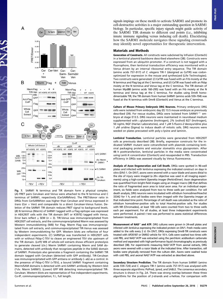
The TIR Domain Physically Interacts with the N Terminus of SARM1.The functional interaction between the SARM1 N-terminal andTIR domains described above led us to investigate whether ornot these domains also have a physical interaction. The TIRdomain from the cytosolic adaptor TRIF is regulated by directphysical interaction with the N-terminal domain (30), and asimilar mechanism regulates activity of TIR-containing re-sistance proteins from plants (31). To investigate whether theSARM1 TIR and N-terminal domains are in close proximitywithin the full-length molecule, we used Förster resonance en-ergy transfer (FRET), a powerful approach to investigate intra-molecular interactions. FRET pairs Cerulean and Venus werefused to the N terminus and TIR domain at the C terminus offull-length SARM1, respectively (CerSARMVen). Expression ofCerSARMVen rescues axon degeneration in severed SARM1−/−
(Fig. S7), indicating that the addition of fluorescent tags did notimpair function. A strong FRET signal is detected in DRG axonsexpressing this CerSARMVen that is comparable to a directfusion of Cerulean and Venus alone (Fig. 7A). Furthermore,deletion of the TIR domain reduces FRET to background levelsthat are comparable to FRET obtained following the coex-pression of Cerulean and Venus as separate proteins. Hence,FRET between the N and C termini of SARM1 is dependentupon the presence of the TIR domain. We did not observe achange in FRET in severed axons upon axotomy (Fig. S7),suggesting that conformational changes in activated SARM1 donot disrupt Cerulean/Venus interaction or that only a smallfraction of SARM1 is activated in response to axonal injury.
Fig. 4. K597E does not impair SAM-TIR–dependent neuronal death.(A) Expression of fragment containing the SAM domains and TIR domainfrom SARM1 induces neuronal cell death. (B) Quantification of neuronaldeath in cells expressing the indicated SAM-TIR protein. DRGs were alsopreincubated with 1 mM NR, which suppresses SARM1-dependent cell death.(C) Images of DRG neurons labeled with ethidium homodimer. *P < 0.05.Error bars reflect ± SEM (n = 3). Ctrl, control. (Scale bar, 25 μM.)
E6276 | www.pnas.org/cgi/doi/10.1073/pnas.1601506113 Summers et al.
These studies suggest the SARM1 N terminus is in closeenough association to the TIR domain to elicit a FRET signal.Because the above experiment was performed in the context offull-length SARM1, we wondered whether the N terminus canphysically interact with the TIR of SARM1 even when thesedomains are not present in the same molecule. Thus, we exam-ined whether these fragments form a complex when expressedtogether in trans. Differentially tagged forms of the SARM1N-terminal domain and TIR domain were expressed in HEK293Tcells, and coimmunoprecipitation studies were performed. Indeed,the N terminus and TIR domain of SARM1 coimmunoprecipitatewith one another, suggesting these two SARM1 domains physi-cally interact. TIR (K597E) likewise physically associated withthe N terminus (Fig. 7B). The association of the mutant TIRdomain with the N terminus is consistent with our functionalstudies that showed that autoinhibition is maintained in theTIR (K597E) mutant.Because the K597E mutation converts SARM1 into a domi-
nant negative, we wondered if this mutation might enhancebinding between the TIR domain and the N terminus in full-length, natively folded SARM1. To test this hypothesis, we useda protease-sensitive (ps) form of SARM1 that possesses a to-bacco etch virus (TEV)-protease site between SAM2 and theTIR domain (4). SARM1ps is functional; however, upon TEVproteolysis, the N terminus/SAM domains are separated from
the TIR domain and SARM1 is rendered inactive. Using aCerulean tag on the C terminus, the TIR domain was immuno-precipitated from HEK293T cell extracts in the presence orabsence of TEV proteolysis and the coprecipitated N terminus/SAM fragment was assessed using a SARM1 antibody generatedagainst a peptide in the SAM2 domain. Consistent with thefindings above, the SARM1 N terminus/SAM fragment coim-munoprecipitated with the TIR domain. Notably, the interactionis strongly enhanced (threefold; P < 0.05) in the presence of theK597E mutation in the TIR domain (Fig. 7C and Fig. S8). Thesefindings support a model wherein N-terminal autoinhibition ofTIR activity is mediated by direct binding between the N ter-minus and the TIR domain.
DiscussionSARM1 activation in response to neuronal injury evokes a rapidconsumption of NAD+ followed by a decline in ATP that ulti-mately leads to local axonal degeneration or neuronal cell death(4, 13). Although the molecular signaling pathways underlyingSARM1-dependent neuronal destruction are still being defined,dimerization of the TIR domain of SARM1 is sufficient tostimulate NAD+ loss and neuronal death. Our study identifiesimportant motifs in the SARM1 TIR domain that enable SARM1-specific functions (e.g., NAD+ loss), including a conserved loopmotif that is only present in SARM1 orthologs. Furthermore, weuncover a surprising connection between the SARM1 TIR domainand autoinhibition mediated by the N terminus and provide evi-dence for a physical interaction between these two domains. Ourstudies have important mechanistic implications for SARM1 acti-vation as well as the execution of neuronal self-destruction uponinjury.
SARM1-Unique Motifs Confer TIR-Dependent Axon Degeneration.TIR domains across diverse phyla, including animals, plants,and fungi, contribute to cellular defense by stimulating innateimmune signaling pathways in response to pathogens (32).SARM1 is one of five TIR-containing adaptor proteins in thecytosol, although unlike other TIR adaptors, including Myd88 orTRIF, human SARM1 inhibits innate immune signaling (33). Atthe sequence level, TIR domains in the SARM1 family showgreater similarity to bacterial TIR domains than the TIR do-mains found in metazoans (17). We predicted that unique motifswithin the SARM1 TIR domain would contribute to its specificfunctions. Structural prediction identified a loop motif that isunique to the SARM1 family that we call the SS loop. Threeconserved sequential residues in the SS loop were required forSARM1 function in injury-induced axon degeneration. In par-ticular, this motif is necessary for TIR-dependent NAD+ lossupon TIR dimerization. We hypothesize that this motif enablesSARM1-specific functions, including NAD+ consumption, inresponse to axonal injury. Whether this motif is a platform forprotein interaction with a downstream executioner molecule orcontributes to TIR/TIR dimerization is an important questionfor future studies.We also identified putative BB and DD loops in the SARM1
TIR. The BB loop contributes to TIR function in many proteins(14); in some cases, it serves as the dimerization interface in aTIR/TIR complex. The BB loop is highly conserved across dis-tant SARM1 relatives and is essential for SARM1-dependentaxon degeneration. Compared with TIR domains from otherproteins, the SARM1 BB loop is largely distinct, with the ex-ception of a highly conserved glycine (G601). Mutagenesis of thisresidue strongly inhibited SARM1 activity in injury-induced axondegeneration as well as TIR-dependent NAD+ loss and death.Mutagenesis of this residue also interfered with SARM1-dependent suppression of innate immune signaling (26). Hence,the BB loop contributes to diverse SARM1 functions, potentiallyas an interface for TIR/TIR dimerization as seen in other TIR
Fig. 5. Sarm1 (K597E) is a potent dominant negative, blocking axon de-generation after axotomy. (A) Axons from WT DRG neurons expressing theindicated Sarm1 construct were transected, and the degeneration of distalaxons monitored over time. Expression of Sarm1 (K597E) delays axon de-generation for ∼36 h postaxotomy. (B) Expression of SARM1 (K597E) alsopreserves axon integrity in response to the chemotherapeutic agent vin-cristine. Axons were treated with 40 nM vincristine, and axon degenerationwas measured 36 h postexposure. Expression of SARM1 (K597E) suppressesaxon degeneration (C) and cell death (D) in response to the mitochondrialuncoupling agent CCCP (50 μM, 24 h). *P < 0.05; **P < 0.01. Error barsreflect ± SEM (n = 3). (Scale bars, 25 μM.)
Summers et al. PNAS | Published online September 26, 2016 | E6277
domains. The DD loop is another prominent TIR motif thatcontributes to TIR/TIR interactions (23, 34, 35), but it is nothighly conserved in SARM1 orthologs. In accord, our muta-genesis of the DD loop did not identify functionally importantresidues, suggesting that this motif contributes to SARM1 ac-tivity in a minor fashion.
The SARM1 TIR Domain Is a Conserved Executioner of NeuronalDestruction. The function of SARM1 in neurodegeneration isconserved from mammals to invertebrates (2, 3). In our study, wefind that the capacity of the SARM1 TIR domain to promoteneuronal cell death is also conserved in the nematode orthologTIR-1. Furthermore, neuronal cell death is preceded by loss ofΔΨ and NAD+/ATP collapse. However, the prodestructive ac-tivity of TIR-1 poses a conundrum, because in a number of sit-uations, C. elegans tir-1 mediates nondestructive cellularsignaling. For example, tir-1 promotes survival in response topathogenic infection by stimulating gene expression of innateimmune pathways (20, 21) and participates in specification ofleft/right symmetry in the developing nervous system (11). Inaddition, axon degeneration can occur through mechanisticallydistinct pathways from Wallerian degeneration (36). Impor-tantly, tir-1 acts upstream of a MAPK signaling pathway, afunction that is also conserved, because vertebrate SARM1 ac-tivates a MAPK signal during injury-induced axon degeneration(11, 13, 20, 36). Hence, both signaling and NAD+-depletingfunctions of the SARM1 TIR domain are conserved. It is notknown how these two activities of SARM1 TIR domains arerelated, or how the same molecule can promote survival signal-ing as well as cellular demise.Activity of the TIR domain from TIR-1 is attenuated com-
pared with the HsTIR. If TIR motifs such as the SS loop interactwith a NAD+ consuming enzyme, then differential kinetics ofTIR-induced cell death may be due to lower affinity for potentialbinding partners within the mouse cellular environment. Alter-natively, the SARM1 TIR domain itself might be an enzyme thatdirectly consumes NAD+. As such, lower activity might permitTIR-1 to carry out signaling functions without compromising cellviability. In this model, following traumatic injury, TIR-1 activityis stimulated to such an extent that it evokes cell death.
TIR Motifs Modulate Injury-Induced SARM1 Activation. Others andwe previously demonstrated that deletion of the SARM1 N termi-nus generates a constitutively active protein (3, 11), indicating thatthe N terminus autoinhibits SARM1 activity in the absence of in-jury. Although the TIR domain is clearly important for execution ofSARM1-dependent axon degeneration, we identify here a role forthe SARM1 TIR domain in injury-induced activation of SARM1.Mutagenesis of Lysine597 in the SARM1 BB loop preventsSARM1 activation; however, this residue is completely dispensablefor the prodegenerative activity of the dimerized TIR domain. Thisresidue is highly conserved in SARM1 relatives, and substitutionsoccurring at this position in any species are limited to the similarlycharged arginine. Furthermore, using the BB loop for two func-tions, activation and execution, would allow for direct coupling ofSARM1 injury-induced activation to downstream axon destruction.TIR-containing proteins use numerous strategies to regulate
TIR/TIR interactions. For example, TLRs couple ligand bindingto conformational changes that are transduced through theplasma membrane to TIR domains in the cytosol and activatesignaling pathways (37). The cytosolic adaptor TRIF/TICAM1uses a mechanism wherein the N-terminal domain folds backonto the TIR domain to hide essential effector residues in theunstructured region adjacent to the TIR domain (30). Interest-ingly, TIR-containing proteins in plants use a similar mechanismto suppress the activity of plant immune resistance proteins thatpromote cellular death (31). Our studies support a modelwhereby direct physical interaction between the N terminus ofSARM1 with its TIR domain is necessary for SARM1 auto-inhibition. We observe a strong intramolecular FRET signalbetween the N terminus and TIR domain in the context of full-length SARM1. These two domains also form a physical complexwhen expressed in trans. The N terminus might physically restrictTIR domains from dimerizing or block association with anauxiliary factor required for TIR activity. Interestingly, mutationof Lysine597 enhances physical association between the TIRdomain and the rest of the SARM1 protein, suggesting residuesin the BB loop might contribute to this physical interaction.How axonal injury stimulates physical release of N-terminal
autoinhibition is unknown. We anticipate that other residues inthe SARM1 TIR domain and N terminus (and perhaps SAMdomain) contribute to SARM1 regulation. Furthermore, how injury
Fig. 6. SARM1 (K597E) expression prevents axonalNAD+ loss after axotomy. (A and B) Axonal NAD+
and ATP levels were measured from transectedSARM1−/− DRGs expressing an empty vector, WTSARM1, or SARM1 (K597E). There is a significantdrop in axonal NAD+ in the presence of SARM1;however, there is no significant decrease in thepresence of SARM1 (K597E). (C and D) Axonal NAD+
and ATP levels were measured from transected WTDRGs expressing the same constructs as described inA and B. Expression of SARM1 (K597E) delays axonalloss of NAD+ and ATP after axotomy. *P < 0.05. Errorbars reflect ± SEM (n = 3). N.D, not detectable.
E6278 | www.pnas.org/cgi/doi/10.1073/pnas.1601506113 Summers et al.
signals impinge on these motifs to activate SARM1 and promote itscell-destructive activities is a major outstanding question in SARM1biology. In particular, specific injury signals might guide activity ofthe SARM1 TIR domain to different end points (i.e., inhibitinginnate immune signaling versus inducing cell death). Elucidatinghow the SARM1 molecule navigates these signaling crossroadsmay identify novel opportunities for therapeutic intervention.
Materials and MethodsGeneration of Constructs. All constructs were subcloned by InFusion (Clontech)in a lentiviral plasmid backbone described elsewhere (38). Constructs wereexpressed from an ubiquitin promoter. If a construct is not tagged with afluorophore, then lentiviral transduction efficiency was monitored with aVenus driven by an internal ribosomal entry sequence. The TIR domain(amino acids 757–917) of C. elegans TIR-1 (designed as CeTIR) was codon-optimized for expression in the mouse and synthesized (Life Technologies).Two constructs were generated: (i) CeTIR was fused with an Frb moiety at theN terminus and Flag tag at the C terminus, and (ii) CeTIR was fused with an Fkbpmoiety at the N terminus and Venus tag at the C terminus. The TIR domain ofhuman Myd88 (amino acids 160–295) was fused with an Frb moiety at the Nterminus and Venus tag at the C terminus. For studies using DmrB homo-dimerizable TIR, the TIR domain from human SARM1 (amino acids 559–704) wasfused at the N terminus with DmrB (Clontech) and Venus at the C terminus.
Culture of Mouse Primary Embryonic DRG Neurons. Primary embryonic DRGcells were isolated from embryonic day (E) 13.5 mouse embryos as previouslydescribed (39). For rescue studies, DRGs were isolated from SARM1−/− em-bryos at stage E13.5. DRG neurons were maintained in neurobasal mediumsupplemented with L-glutamine (Invitrogen), 2% (vol/vol) B27 (Invitrogen),50 ng/mL NGF (Harlan Laboratories), and 1 μM 5-fluoro-2′deoxyuridine plus1 μM uridine (Sigma) to induce death of mitotic cells. DRG neurons wereseeded on plates precoated with poly-D-lysine and laminin.
Lentiviral Transduction. Lentiviral particles were generated from HEK293Tcells as previously described (38). Briefly, expression constructs for the in-dicated SARM1 mutant were cotransfected with plasmids containing lenti-viral packaging proteins and vesicular stomatitis virus glycoprotein. After48 h posttransfection, lentiviral particles in the media were concentratedusing Lenti-X concentrator (Clontech) and resuspended in PBS. Transductionefficiency in DRGs was assessed visually by Venus fluorescence.
Analysis of Axon Degeneration and Cell Death. DRGs were spotted in 96-wellplates and infected with lentivirus expressing the indicated construct on days invitro (DIV) 1. On DIV7, axons were severed with a razor blade and axons distal tothe site of injury were imaged (a 20× objective was used in all imaging experi-ments) using a high-content Operetta imager (PerkinElmer). Axon degenerationwas quantified from bright-field images using an ImageJ macro (38) that definesthe ratio of fragmented axon area to total axon area. For an individual exper-iment, six fields were analyzed from two to three wells per condition. For celldeath analysis, DRG neurons were incubated with ethidium homodimer/Hoechst33342 for 1 h, and cell bodies were then imaged with the Operetta imager atthat indicated time point. Percentage of cell death was calculated as the ratio ofethidium homodimer-positive cells to total Hoechst-positive cells. For studieswith NR (Chromadex), at least 100 cells were counted from two to three wellseach per experiment. For all studies, at least three independent experimentswere performed. A paired t test was performed to assess statistical differencesbetween treatments.
Measurement of NAD+ and ATP. DRG cultures were grown in 24-well plates andinfected with lentivirus expressing the indicated protein on DIV1. Fresh media wereadded to the cells every 2 d. On DIV7, DRGs expressing DmrB-TIR constructs weretreated with AP20187 or DMSO vehicle for 10 h. Cells were washed two times withcold PBS, and NAD+/ATP was extracted using the perchloric acid/sodium carbonatemethod and separated with high-performance liquid chromatography as previouslydescribed (38). For experiments measuring NAD+/ATP from axonal extracts, DRGaxons were severed with a razor blade at the indicated time. After transection, cellbodies were physically removed from the culture dish. Axons were washed twicewith cold PBS, and axonal NAD+/ATP was extracted as described above.
Secondary Structure Prediction. The TIR domain from human SARM1 (aminoacids 559–724) and C. elegans TIR-1 (amino acids 757–917) was analyzed withthree separate algorithms: PsiPred, Jpred, and SABLE. The consensus secondarystructure is shown in Fig. 2A. There was strong overlap between these threealgorithms for the position of β-strands and α-helices. For comparison with
Fig. 7. SARM1 N terminus and TIR domain form a physical complex.(A) FRET pairs Cerulean and Venus were attached to the N terminus and Cterminus of SARM1, respectively (CerSARMVen). The FRET/donor ratio inDRGs from CerSARMVen was higher than Cerulean and Venus expressed intrans (Cer + Ven) and comparable to a direct Cerulean-Venus fusion. De-letion of the SARM1 TIR domain reduces FRET signal to background levels.(B) N terminus (Nterm) of SARM1 tagged with a Flag epitope was expressedin HEK293T cells with the TIR domain (WT or K597E) tagged with Venus.Error bars reflect ± SEM (n = 3). TIR-Venus was immunoprecipitated fromHEK293T cell extracts, and the coimmunoprecipitated Nterm was assessed byWestern immunoblotting (WB) for Flag. Nterm Flag was immunoprecipi-tated from cell extracts, and coimmunoprecipitated TIR-Venus was assessedby Western immunoblotting for GFP. Western blots are reflective of fourindependent experiments. (C) SARM1ps was transfected in HEK293T cellswith or without Fkbp-C-TEV to cleave an engineered TEV site upstream ofthe TIR domain. (Left) WB of whole-cell extracts shows efficient proteolysisto generate cleaved (clv.) Nterm SARM1 containing Nterm and SAM do-mains, detected with antibody that recognizes peptide in the SAM2 domainof SARM1. Proteolysis also generates a fragment containing the SARM1 TIRdomain tagged with Cerulean (detected with GFP antibody). TIR-Ceruleanwas immunoprecipitated with GFP antisera or antibody (−ab) as a control. Inthe presence of Fkbp-C-TEV (+TEV), a bound SARM1 fragment containingNterm/SAM domains is detected with anti-SARM1 antibody by Western blot(*clv. Nterm SARM1). (Lower) GFP WB detecting immunoprecipitated TIR-Cerulean. Western blots are representative of five independent experiments.Co-IP, coimmunoprecipitation; f.l., full length.
Summers et al. PNAS | Published online September 26, 2016 | E6279
NEU
ROSC
IENCE
PNASPL
US
other TIR domains, the secondary structures from the following crystal struc-tures were used: TLR1 (1FYV), TLR6 (4OM7), TLR10 (2J67), TLR2 (1FYW), TRAM(2M1W), and Myd88 (2Z5V).
Sensitized Emission FRET. Measurements of FRET intensity using the sensitizedemission method were performed in live embryonic DRG neurons infected onDIV1 with lentivirus expressing the SARM1-FRET construct. Imaging was per-formed on DIV7 using the Operetta high-content imager. All measurementswere taken from single axons. The SARM1-FRET construct consists of a cytosolicversion of SARM1 (amino acids 1–27 deleted) in which the N terminus is taggedwith the FRET donor mCerulean and the C-terminal TIR domain is tagged withthe FRET acceptor Venus; the donor and acceptor are both attached with ashort peptide linker. Images of mCerulean intensity were collected using a430/25 excitation filter and a 470/30 emission filter, and FRET images werecollected using a 430/25 excitation filter and a 525/20 emission filter. Images ofneurons expressing only mCerulean were used to calculate donor bleed-through (BT) using a 430/25 excitation filter and a 525/20 emission filter.Neurons expressing only Venus imaged using a 430/25 excitation filter anda 525/20 emission filter showed no appreciable cross-excitation of Venus.The donor BT value was calculated for each experiment performed.
After background subtraction, relative FRET efficiency was determined bymeasuring both the mCerulean and FRET intensity in the same regions of singleaxons. The corrected FRET intensity was determined by subtracting the percentageof the donor signal that is BT, and thus contributes to the raw FRET intensity:
FRETcorrected = FRETraw −�mCeruleanintensity ×BT
�.
The corrected FRET intensity value is then divided by the donor intensity valueto yield the relative FRET efficiency:
Relative FRET efficiency= FRETcorrected=Donor.
All image processing and measurements for FRET experiments were per-formed using ImageJ (NIH).
Coimmunoprecipitation. HEK293T cells were transfected with the indicatedplasmids using XtremeGene transfection reagent (Roche). Cells were washedwith fresh media 24 h later. Cells were lysed 36 h posttransfection in non-denaturing lysis buffer [1× PBS, 2 mM EDTA, 10 mM Hepes (pH 7.4), 0.5%Triton X]. Cell extracts were briefly sonicated, and cell debris was collected ina pellet by centrifugation at 1,500 × g for 4 min. Supernatants were col-lected and incubated with 0.15% (wt/vol) BSA and the indicated antisera/resin, anti-GFP (Rockland Immunochemicals) or anti-Flag resin (preblockedin 1.5% (wt/vol) BSA; BioTool), overnight at 4 °C. Aliquots were saved insample buffer as whole-cell extract samples. For GFP coimmunoprecipita-tion samples, Protein G agarose [preblocked in 1.5% (wt/vol) BSA] wasadded to the sample and incubated for another hour. Beads were washedthree times with cold lysis buffer and resuspended in sample buffer. Pre-cipitates and whole-cell extract samples were analyzed by SDS/PAGE andWestern immunoblotting for the indicated proteins. For studies with SARM1ps,transfected cells were treated with 100 nM rapamycin for 3 h before cell lysis toinduce SARM1ps proteolysis. Western blots were quantified with ImageJ.
ACKNOWLEDGMENTS. D.W.S. is supported by a Development Grant fromthe Muscular Dystrophy Association. D.A.G. is supported by the NIH underRuth L. Kirschstein National Research Service Award NS092447 from theNational Institute of Neurological Disorders and Stroke. A.D. and J.M aresupported by funds from the NIH (Grants NS087632, AG013730, andNS065053).
1. Gerdts J, Summers DW, Milbrandt J, DiAntonio A (2016) Axon self-destruction: Newlinks among SARM1, MAPKs, and NAD+ metabolism. Neuron 89(3):449–460.
2. Osterloh JM, et al. (2012) dSarm/Sarm1 is required for activation of an injury-inducedaxon death pathway. Science 337(6093):481–484.
3. Gerdts J, Summers DW, Sasaki Y, DiAntonio A, Milbrandt J (2013) Sarm1-mediated axondegeneration requires both SAM and TIR interactions. J Neurosci 33(33):13569–13580.
4. Gerdts J, Brace EJ, Sasaki Y, DiAntonio A, Milbrandt J (2015) SARM1 activation triggersaxon degeneration locally via NAD⁺ destruction. Science 348(6233):453–457.
5. Sasaki Y, Araki T, Milbrandt J (2006) Stimulation of nicotinamide adenine di-nucleotide biosynthetic pathways delays axonal degeneration after axotomy.J Neurosci 26(33):8484–8491.
6. Wang J, et al. (2005) A local mechanism mediates NAD-dependent protection of axondegeneration. J Cell Biol 170(3):349–355.
7. Summers DW, DiAntonio A, Milbrandt J (2014) Mitochondrial dysfunction inducesSarm1-dependent cell death in sensory neurons. J Neurosci 34(28):9338–9350.
8. Kim Y, et al. (2007) MyD88-5 links mitochondria, microtubules, and JNK3 in neuronsand regulates neuronal survival. J Exp Med 204(9):2063–2074.
9. Mukherjee P, Woods TA, Moore RA, Peterson KE (2013) Activation of the innatesignaling molecule MAVS by bunyavirus infection upregulates the adaptor proteinSARM1, leading to neuronal death. Immunity 38(4):705–716.
10. Mukherjee P, Winkler CW, Taylor KG, Woods TA, Nair V, et al. (2015) SARM1, not MyD88,mediates TLR7/TLR9-induced apoptosis in neurons. J Immunol 195(10):4913–4921.
11. Chuang CF, Bargmann CI (2005) A Toll-interleukin 1 repeat protein at the synapsespecifies asymmetric odorant receptor expression via ASK1 MAPKKK signaling. GenesDev 19(2):270–281.
12. Panneerselvam P, et al. (2013) T-cell death following immune activation is mediatedby mitochondria-localized SARM. Cell Death Differ 20(3):478–489.
13. Yang J, et al. (2015) Pathological axonal death through a MAPK cascade that triggersa local energy deficit. Cell 160(1-2):161–176.
14. Ve T, Williams SJ, Kobe B (2015) Structure and function of Toll/interleukin-1 receptor/resistance protein (TIR) domains. Apoptosis 20(2):250–261.
15. Fekonja O, Bencina M, Jerala R (2012) Toll/interleukin-1 receptor domain dimers asthe platform for activation and enhanced inhibition of Toll-like receptor signaling.J Biol Chem 287(37):30993–31002.
16. Panter G, Jerala R (2011) The ectodomain of the Toll-like receptor 4 prevents con-stitutive receptor activation. J Biol Chem 286(26):23334–23344.
17. Zhang Q, Zmasek CM, Cai X, Godzik A (2011) TIR domain-containing adaptor SARM is alate addition to the ongoing microbe-host dialog. Dev Comp Immunol 35(4):461–468.
18. Vérièpe J, Fossouo L, Parker JA (2015) Neurodegeneration in C. elegans models of ALSrequires TIR-1/Sarm1 immune pathway activation in neurons. Nat Commun 6:7319.
19. Blum ES, Abraham MC, Yoshimura S, Lu Y, Shaham S (2012) Control of nonapoptoticdevelopmental cell death in Caenorhabditis elegans by a polyglutamine-repeat pro-tein. Science 335(6071):970–973.
20. Liberati NT, et al. (2004) Requirement for a conserved Toll/interleukin-1 resistancedomain protein in the Caenorhabditis elegans immune response. Proc Natl Acad SciUSA 101(17):6593–6598.
21. Couillault C, et al. (2004) TLR-independent control of innate immunity in Caeno-rhabditis elegans by the TIR domain adaptor protein TIR-1, an ortholog of humanSARM. Nat Immunol 5(5):488–494.
22. Xu Y, et al. (2000) Structural basis for signal transduction by the Toll/interleukin-1receptor domains. Nature 408(6808):111–115.
23. Nyman T, et al. (2008) The crystal structure of the human toll-like receptor 10 cyto-plasmic domain reveals a putative signaling dimer. J Biol Chem 283(18):11861–11865.
24. Loiarro M, et al. (2013) Mutational analysis identifies residues crucial for homo-dimerization of myeloid differentiation factor 88 (MyD88) and for its function inimmune cells. J Biol Chem 288(42):30210–30222.
25. Loiarro M, et al. (2005) Peptide-mediated interference of TIR domain dimerization inMyD88 inhibits interleukin-1-dependent activation of NF-kappaB. J Biol Chem280(16):15809–15814.
27. Panneerselvam P, Singh LP, Ho B, Chen J, Ding JL (2012) Targeting of pro-apoptoticTLR adaptor SARM to mitochondria: Definition of the critical region and residues inthe signal sequence. Biochem J 442(2):263–271.
28. Gilley J, Orsomando G, Nascimento-Ferreira I, Coleman MP (2015) Absence of SARM1 rescuesdevelopment and survival of NMNAT2-deficient axons. Cell Reports 10(12):1974–1981.
29. Del Nagro C, Xiao Y, Rangell L, Reichelt M, O’Brien T (2014) Depletion of the centralmetabolite NAD leads to oncosis-mediated cell death. J Biol Chem 289(51):35182–35192.
30. Tatematsu M, et al. (2010) A molecular mechanism for Toll-IL-1 receptor domain-con-taining adaptormolecule-1-mediated IRF-3 activation. J Biol Chem 285(26):20128–20136.
31. Bernoux M, et al. (2011) Structural and functional analysis of a plant resistance pro-tein TIR domain reveals interfaces for self-association, signaling, and autoregulation.Cell Host Microbe 9(3):200–211.
32. Brown J, Wang H, Hajishengallis GN, Martin M (2011) TLR-signaling networks: Anintegration of adaptor molecules, kinases, and cross-talk. J Dent Res 90(4):417–427.
33. Carty M, et al. (2006) The human adaptor SARM negatively regulates adaptor proteinTRIF-dependent Toll-like receptor signaling. Nat Immunol 7(10):1074–1081.
34. Snyder GA, et al. (2014) Crystal structures of the Toll/Interleukin-1 receptor (TIR)domains from the Brucella protein TcpB and host adaptor TIRAP reveal mechanismsof molecular mimicry. J Biol Chem 289(2):669–679.
35. Bovijn C, et al. (2012) Identification of interaction sites for dimerization and adapterrecruitment in Toll/interleukin-1 receptor (TIR) domain of Toll-like receptor 4. J BiolChem 287(6):4088–4098.
36. Hayakawa T, et al. (2011) Regulation of anoxic death in Caenorhabditis elegans by mam-malian apoptosis signal-regulating kinase (ASK) family proteins. Genetics 187(3):785–792.