Page 1
i
“I hereby declare that I have read this dissertation and in my opinion this thesis has
satisfied the scope and quality for the award of the degree of Master of Science
(Biotechnology).”
Signature: ……..…………………………….
Name of supervisor: Dr SHAHIR SHAMSIR
Date: …………………………………...
Page 2
ii
STRUCTURAL ANALYSIS AND MODELING COMPARISON OF PRIMATES’
DYSTROPHIN
FAEZE SADAT MOHAJER
A dissertation submitted in partial fulfilment of the
requirements for the award of the degree of
Master of Science (Biotechnology
Faculty of Bioscience and Bioengineering
Universiti Technologi Malaysia
January 2013
Page 3
iii
I declare that this thesis entitled “Structural analysis and modelling comparison of
primates’ Dystrophin” is the result of my own research except as cited in the
references. The thesis has not been accepted for any degree and is not concurrently
submitted in candidature of any other degree.
Signature: ....................................................
Name: Faeze Sadat Mohajer..................
Date: Dec .2012.....................................
Page 4
iv
To my beloved parents, sisters and brother
Page 5
v
ACKNOWLEDGMENT
I would like to begin by sincerely thanking my supervisor, Associated Prof Dr.
Shahir Shamsir for his kindness and constant support, guidance and mentorship over the
course of this thesis. He gave me the freedom to define my thesis statements. I am very
grateful to having the opportunity for work on this thesis.
I am heartily thankful to my parents, for their unconditional supports and
encouraging me to continue my education.
Page 6
vi
ABSTRACT
Dystrophin is a rod-shaped cytoplasmic protein which is an essential piece of
a protein complex that binds the cytoskeleton of a muscle fiber to the
nearby extracellular matrix within the cell membrane. Lack of this protein in the muscle
leads muscular dystrophy. Currently, Dystrophin structure has just been detected in
human being. In fact, it has not been recognized in other types of primates. However,
bioinformatics’ software has presented a big opportunity to predict its structure in other
species on based on its structure in human being. The data for this project will be
sourced by both bioinformatics databases and experimental information. The used
databases were NCBI, PDB (protein data bank), UniProt, and Gene bank. The data that
is gathered from these databases were analyzed using multiple sequence analysis
software such as BLAST, Jalview to find conserved regions in the protein among the
species of interest (different kinds of primates). In addition, the tertiary structure of the
protein will be predicted by Swiss-model and will be observed and compared by 3-d
structure viewing software such as: VMD and DeepView. In conclusion, the analysis of
this study illustrates that Dystrophin has been changed slightly through evolution.
However, the small changes that have been occurred have not affected the 3D structure
and function of the protein.
Page 7
vii
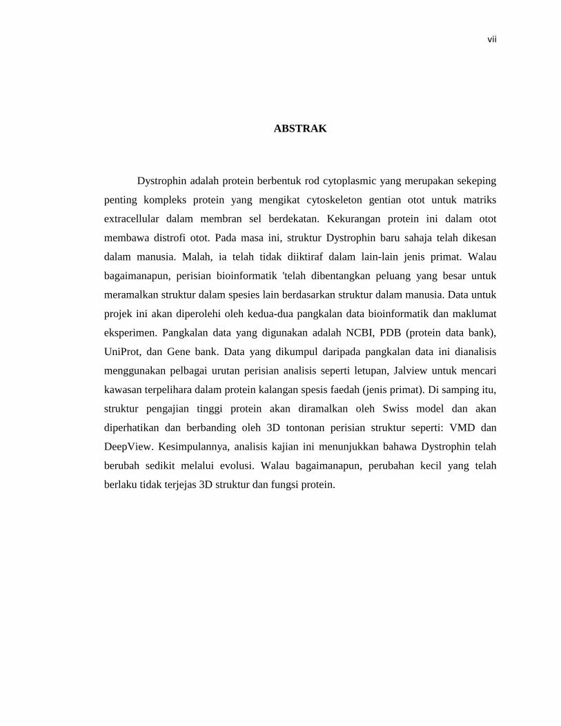
ABSTRAK
Dystrophin adalah protein berbentuk rod cytoplasmic yang merupakan sekeping
penting kompleks protein yang mengikat cytoskeleton gentian otot untuk matriks
extracellular dalam membran sel berdekatan. Kekurangan protein ini dalam otot
membawa distrofi otot. Pada masa ini, struktur Dystrophin baru sahaja telah dikesan
dalam manusia. Malah, ia telah tidak diiktiraf dalam lain-lain jenis primat. Walau
bagaimanapun, perisian bioinformatik 'telah dibentangkan peluang yang besar untuk
meramalkan struktur dalam spesies lain berdasarkan struktur dalam manusia. Data untuk
projek ini akan diperolehi oleh kedua-dua pangkalan data bioinformatik dan maklumat
eksperimen. Pangkalan data yang digunakan adalah NCBI, PDB (protein data bank),
UniProt, dan Gene bank. Data yang dikumpul daripada pangkalan data ini dianalisis
menggunakan pelbagai urutan perisian analisis seperti letupan, Jalview untuk mencari
kawasan terpelihara dalam protein kalangan spesis faedah (jenis primat). Di samping itu,
struktur pengajian tinggi protein akan diramalkan oleh Swiss model dan akan
diperhatikan dan berbanding oleh 3D tontonan perisian struktur seperti: VMD dan
DeepView. Kesimpulannya, analisis kajian ini menunjukkan bahawa Dystrophin telah
berubah sedikit melalui evolusi. Walau bagaimanapun, perubahan kecil yang telah
berlaku tidak terjejas 3D struktur dan fungsi protein.
Page 8
viii
TABLE OF CONTENT
CHAPTER TITLE PAGE
DECLARATION III
DEDICATION IV
ACKNOWLEDGEMENT V
ABSTRACT VI
ABSTRAK VII
TABLE OF CONTENTS VIII
LIST OF TABLES XI
LIST OF FIGURES XII
LIST OF ABBREVIATION XIV
LIST OF SYMBOLS XV
LIST OF APPENDIX XVI
1 INTRODUCTION
1.1 Background 1
1.1.1 Duchenne Muscular Dystrophy (DMD) 2
1.2 Problem Statement 3
1.3 Objectives 3
Page 9
ix
1.4 Research Scope 4
2 LITERATURE REVIEWS
2.1 Duchenne Muscular Dystrophy (DMD) 5
2.2 Evolutionary Studies between humans and primates 6
2.3 Evolutionary comparison of proteins between human and primates 6
2.4 Previous Studies on Dystrophin’s structure 9
2.5 Comparison of Proteins by different Bioinformatics Tools 9
3 RESEARCH METHODOLOGY
3.1 Experimental design 11
3.2 Identification of Dystrophin sequence in primate species 12
3.3 Selection of dystrophin structures 14
3.4 Multiple sequence alignment (MSA) analysis 14
3.5 Prediction and Modeling of tertiary Structure 15
4 RESULTS AND DISCUSSION
4.1 Identification of Dystrophin sequence in primate species 17
4.2 Selection of 3D structure 19
4.3 Multiple sequence alignment 21
4.3.1 Comparison of Hydrophobicity 24
4.4 Prediction and Modeling of 3DStructure 25
4.4.1 Prediction and Modeling of Callithrix jacchus 26
4.4.2 Prediction and Modeling of Nomascus leucogenys 27
Page 10
x
4.4.3 Prediction and Modeling of Otolemur garnettii 28
4.4.4 Prediction and Modeling of dystrophin in Pan paniscus 29
4.4.5 Prediction and Modeling of dystrophin in Saimiri 30
4.5 Evaluation of Homology Modeling 31
4.6 Comparison of primary and Tertiary Structures 32
5 CONCLUSION
5.1 Conclusion 37
5.2 Future Work 38
REFERENCES 39
Appendix A~B 44~65
Page 11
xi
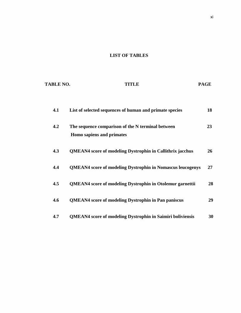
LIST OF TABLES
TABLE NO. TITLE PAGE
4.1 List of selected sequences of human and primate species 18
4.2 The sequence comparison of the N terminal between 23
Homo sapiens and primates
4.3 QMEAN4 score of modeling Dystrophin in Callithrix jacchus 26
4.4 QMEAN4 score of modeling Dystrophin in Nomascus leucogenys 27
4.5 QMEAN4 score of modeling Dystrophin in Otolemur garnettii 28
4.6 QMEAN4 score of modeling Dystrophin in Pan paniscus 29
4.7 QMEAN4 score of modeling Dystrophin in Saimiri boliviensis 30
Page 12
xii
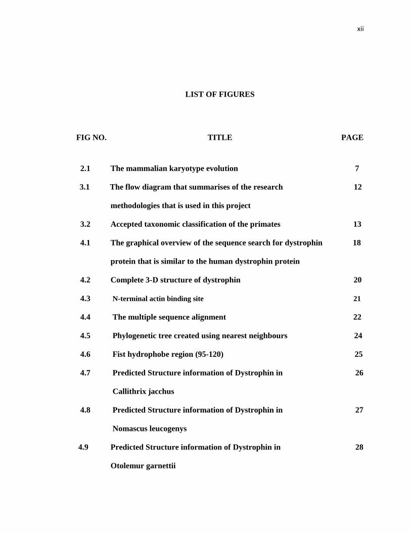
LIST OF FIGURES
FIG NO. TITLE PAGE
2.1 The mammalian karyotype evolution 7
3.1 The flow diagram that summarises of the research 12
methodologies that is used in this project
3.2 Accepted taxonomic classification of the primates 13
4.1 The graphical overview of the sequence search for dystrophin 18
protein that is similar to the human dystrophin protein
4.2 Complete 3-D structure of dystrophin 20
4.3 N-terminal actin binding site 21
4.4 The multiple sequence alignment 22
4.5 Phylogenetic tree created using nearest neighbours 24
4.6 Fist hydrophobe region (95-120) 25
4.7 Predicted Structure information of Dystrophin in 26
Callithrix jacchus
4.8 Predicted Structure information of Dystrophin in 27
Nomascus leucogenys
4.9 Predicted Structure information of Dystrophin in 28
Otolemur garnettii
Page 13
xiii
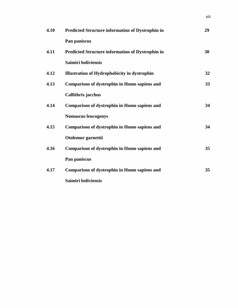
4.10 Predicted Structure information of Dystrophin in 29
Pan paniscus
4.11 Predicted Structure information of Dystrophin in 30
Saimiri boliviensis
4.12 Illustration of Hydrophobicity in dystrophin 32
4.13 Comparison of dystrophin in Homo sapiens and 33
Callithrix jacchus
4.14 Comparison of dystrophin in Homo sapiens and 34
Nomascus leucogenys
4.15 Comparison of dystrophin in Homo sapiens and 34
Otolemur garnettii
4.16 Comparison of dystrophin in Homo sapiens and 35
Pan paniscus
4.17 Comparison of dystrophin in Homo sapiens and 35
Saimiri boliviensis
Page 14
xiv
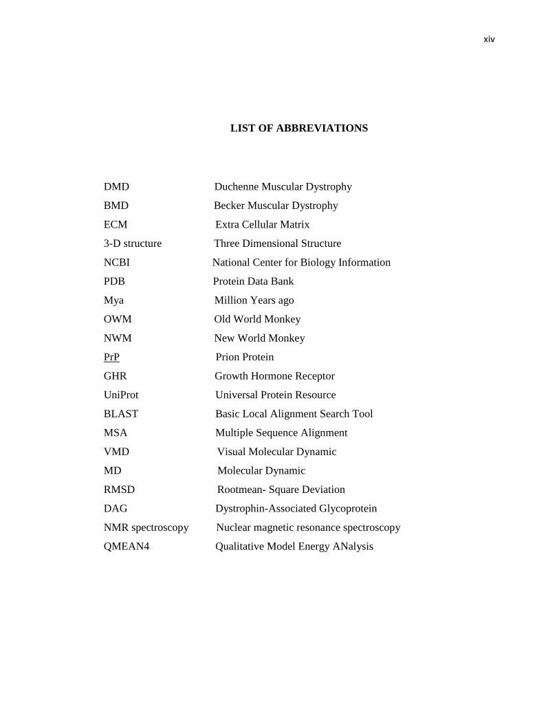
LIST OF ABBREVIATIONS
DMD Duchenne Muscular Dystrophy
BMD Becker Muscular Dystrophy
ECM Extra Cellular Matrix
3-D structure Three Dimensional Structure
NCBI National Center for Biology Information
PDB Protein Data Bank
Mya Million Years ago
OWM Old World Monkey
NWM New World Monkey
PrP Prion Protein
GHR Growth Hormone Receptor
UniProt Universal Protein Resource
BLAST Basic Local Alignment Search Tool
MSA Multiple Sequence Alignment
VMD Visual Molecular Dynamic
MD Molecular Dynamic
RMSD Rootmean- Square Deviation
DAG Dystrophin-Associated Glycoprotein
NMR spectroscopy Nuclear magnetic resonance spectroscopy
QMEAN4 Qualitative Model Energy ANalysis
Page 15
xv
LIST OF SYMBOLS
aa Amino Acid
Asp Aspartic acid
Cys Cysteine
Ser Serine
Ala Alanine
Gly Glycine
Glu Glutamic acid
Gln Glutamine
Asn Asparagine
Lys Lysine
Arg Arginine
Met Methionine
Ala Alanine
Page 16
xvi
LIST OF APPENDICES
APPENDIX TITLE PAGE
A FASTA Format of Dystrophin in selected primates 45
B Alignment between Homo sapiens (Query) with 57
Callithrix jacchus (Sbjct)
Page 17
1
CHAPTER 1
INTRODUCTION
1.1 Introduction
Dystrophin is a rod-shaped protein that is found in both skeletal and cardiac
muscles. It is mainly a structural protein with different variety that is suited to different
types of organs in the human body. Dystrophin is mostly located in muscles that are
used for movement (skeletal muscles) and the muscles of the heart (cardiac muscles).
There are small amount of dystrophin in nerve cells of the brain (Chelly, et al., 1990).
Dystrophin also works as a part of a group of proteins that interact together in a protein
complex called the costamere or the dystrophin-associated protein complex. This protein
complex is responsible for the strengthening of muscle fibres and protects them from
injury during muscles contraction and relaxation. The dystrophin protein is encoded in
the DMD gene and is sometimes called the DMD protein due to its association with the
Duchenne Muscular Syndrome (DMD) disease. In muscle, the DMD complex acts as an
anchor that connects each one of the muscle cell's structural framework (cytoskeleton)
with the lattice of proteins and other molecules outside the cell (extracellular matrix). In
fact, it connects actin of cytoskeleton by its N-terminal (action binding site) to ECM
(Extra Cellular Matrix). (Ozawa, et al., 1995). In addition, it may also have a
Page 18
2
responsibility in cell signalling with its interaction with proteins that send and receive
chemical signals. Currently, there is only limited knowledge on the role of this protein in
nerve cells. Research has proposed that dystrophin in nerve cells are necessary for
normal structure and function of synapses, which are important for specializing
connections between nerve cells in the place that cell-to-cell communication
happens.(Ahn & Kunkel, 1993)
1.1.1 Duchenne Muscular Dystrophy (DMD)
Normal skeletal muscle is composed of muscle fibres that are equally spaced,
sharp, and has a moderately uniform size. They are symmetric, and also are
multinucleated with nuclei’s which are placed at the periphery of the skeletal fibre. In
DMD, patient that lacks dystrophin shows the muscle cells of patients who are suffered
from Duchenne Muscular Dystrophy (DMD) are abnormally defenceless. These
suggests that dystrophin plays a significant role in muscle rigidity, making muscle cells
possessing dystrophin being far stiffer than cells which lack dystrophin (Pasternak,
Wong, & Elson, 1995). Fatal DMD muscle is quite normal except for the occasions that
fibres are eosinophilic hypercontracted (Blake, Weir, Newey, & Davies, 2002). DMD
gene locus is on X chromosome. Thus, Duchenne Muscular Dystrophy (DMD) is an X-
linked recessive mutation where approximately 1 in 3,500 boys are suffered from this
disease (Blake, et al., 2002). Patients suffer from muscle waste, usually lose their ability
for walking and are paralyzed before the age of 12 and die in their late teens or early
twenties typically because of respiratory failure. A milder type for this disease is called
the Becker muscular dystrophy (BMD), which symptoms are detected later and the
patient survival time is longer. The most extensive mutations can cause the complete
dystrophin deficiency, while a truncated protein presents in a very limited level are
found in BMD patients. In addition, mutations in the genes can affect the encoding of
Page 19
3
many constituents of the dystrophin associated protein complex which can cause other
forms of this disease like the limb-girdle muscular dystrophies and congenital muscular
dystrophy (Blake, et al., 2002).
1.2 Problem Statement
Currently, research has only determined the dystrophin structure for humans.
None of the structure from primates has been determined either experimentally or
computationally using protein structure modelling. A comparison between human and
primate dystrophin will elucidate its evolutionary path, especially in its most significant
domain (the first 246 amino acids of its N-terminal (actin-binding site) of the protein)
(Pasternak, et al., 1995). The aim of this study is to compare the dystrophin structure
between human and primates by first modelling the structure and analysing the
differences in the amino acid composition, 3D structure and evolutionary changes.
1.3 Objectives
The objectives of this study are:
1. To model the structure of dystrophin from primates for comparison with human
dystrophin.
2. To analyse the differences in the amino acid sequences and composition between
different primates and human.
Page 20
4
3. To analyse the 3D structure and investigation the implication of the differences
in amino acid sequence of dystrophin between human and primate.
1.4 Scope of the Study
The data for this project will be sourced by bioinformatics databases that stores
experimental information such as the NCBI (http://www.ncbi.nlm.nih.gov ), the Protein
Data Bank (www.rcsb.org), the UniProt (http://www.uniprot.org ) and the Genbank
(http://www.ncbi.nlm.nih.gov/genbank/ ).
Page 21
39
REFRENCES
Ahn, A. H., & Kunkel, L. M. (1993). The structural and functional diversity of
dystrophin. Nature genetics, 3(4), 283-291.
Baumbach, L., Chamberlain, J., Ward, P., Farwell, N., & Caskey, C. (1989).
Molecular and clinical correlations of deletions leading to Duchenne and
Becker muscular dystrophies. Neurology, 39(4), 465-465.
Blake, D. J., Weir, A., Newey, S. E., & Davies, K. E. (2002). Function and genetics
of dystrophin and dystrophin-related proteins in muscle. Physiological
reviews, 82(2), 291-329.
Chelly, J., Hamard, G., Koulakoff, A., Kaplan, J. C., Kahn, A., & Berwald-Netter,
Y. (1990). Dystrophin gene transcribed from different promoters in
neuronal and glial cells, 344, 64 - 65
Cheng, Z., Gengbin, S., Zhuolin, L., Xiulin, L., & Jianhua, C. Correlation between
the hydrophobic structure of dystrophin and DMD/BMD, 12(6):341-344]
Page 22
40
Diogo, R., Richmond, B. G., & Wood, B. (2012). Evolution and homologies of
primate and modern human hand and forearm muscles, with notes on
thumb movements and tool use. Journal of human evolution, 32(7), 64-78
Gilad, Y., Oshlack, A., Smyth, G. K., Speed, T. P., & White, K. P. (2006).
Expression profiling in primates reveals a rapid evolution of human
transcription factors. Nature, 440(7081), 242-245.
Hasegawa, M., Kishino, H., & Yano, T. (1985). Dating of the human-ape splitting
by a molecular clock of mitochondrial DNA. Journal of molecular evolution,
22(2), 160-174.
Hu, X., Burghes, A., Ray, P. N., Thompson, M. W., Murphy, E., & Worton, R. G.
(1988). Partial gene duplication in Duchenne and Becker muscular
dystrophies. Journal of medical genetics, 25(6), 369-376.
Labuda, D., Zietkiewicz, E., & Yotova, V. (2000). Archaic lineages in the history of
modern humans. Genetics, 156(2), 799-808.
Lenk, U., Hanke, R., Kräft, U., Grade, K., Grunewald, I., & Speer, A. (1993). Non-
isotopic analysis of single strand conformation polymorphism (SSCP) in the
exon 13 region of the human dystrophin gene. Journal of medical genetics,
30(11), 951-954.
Page 23
41
Luther, P. K., Squire, J. M., & Forey, P. L. (1996). Evolution of myosin filament
arrangements in vertebrate skeletal muscle. Journal of morphology, 229(3),
325-335.
Muthu, M., Richardson, K. A., & Sutherland-Smith, A. J. (2012). The Crystal
Structures of Dystrophin and Utrophin Spectrin Repeats: Implications for
Domain Boundaries. PloS one, 7(7), 23-56.
Norwood, F., Sutherland-Smith, A. J., Keep, N. H., & Kendrick-Jones, J. (2000).
The structure of the N-terminal actin-binding domain of human dystrophin
and how mutations in this domain may cause Duchenne or Becker muscular
dystrophy. Structure (London, England: 1993), 8(5), 481-564.
Oshima, J., Magner, D. B., Lee, J. A., Breman, A. M., Schmitt, E. S., White, L. D.,
et al. (2009). Regional genomic instability predisposes to complex dystrophin
gene rearrangements. Human genetics, 126(3), 411-423.
Ozawa, E., Yoshida, M., Suzuki, A., Mizuno, Y., Hagiwara, Y., & Noguchi, S.
(1995). Dystrophin-associated proteins in muscular dystrophy. Human
molecular genetics, 4, 1711-2345.
Pantel, J., Machinis, K., Sobrier, M. L., Duquesnoy, P., Goossens, M., & Amselem,
S. (2000). Species-specific Alternative Splice Mimicry at the Growth
Hormone Receptor Locus Revealed by the Lineage of Retroelements during
Primate Evolution a novel mechanism acounting for protein diversity
Page 24
42
between and within species. Journal of Biological Chemistry, 275(25),
18664-18669.
Pasternak, C., Wong, S., & Elson, E. L. (1995). Mechanical function of dystrophin
in muscle cells. The Journal of cell biology, 128(3), 355-361.
Schätzl, H. M., Da Costa, M., Taylor, L., Cohen, F. E., & Prusiner, S. B. (1995).
Prion protein gene variation among primates. Journal of molecular biology,
245(4), 362-564.
Sun, X., Zhulin,I. & Wartell, R. M. (2002). Predicted structure and phyletic
distribution of the RNA‐binding protein Hfq. Nucleic acids research, 30(17),
3662-3671.
Takahata, N., & Satta, Y. (1997). Evolution of the primate lineage leading to
modern humans: phylogenetic and demographic inferences from DNA
sequences. Proceedings of the National Academy of Sciences, 94(9), 4811-
4815-5985.
Takeshima, Y., Yagi, M., Okizuka, Y., Awano, H., Zhang, Z., Yamauchi, Y., et al.
(2010). Mutation spectrum of the dystrophin gene in 442 Duchenne/Becker
muscular dystrophy cases from one Japanese referral center. Journal of
human genetics, 55(6), 379-388.
Yan, J., Wen, W., Xu, W., Long, J., Adams, M. E., Froehner, S. C., et al. (2005).
Structure of the split PH domain and distinct lipid-binding properties of the
Page 25
43
PH–PDZ supramodule of α-syntrophin. The EMBO journal, 24(23), 3985-
3995.
Zhang, J., Webb, D. M., & Podlaha, O. (2002). Accelerated protein evolution and
origins of human-specific features: Foxp2 as an example. Genetics, 162(4),
1825-1835.