Page 1
SATURDAY SCIENCE SLAM
1:00 PM - 1:03 PM
RM17 High Definition Micro-Imaging of the Lymphatic Vessels in Secondary
Lymphedema Patients Using ULTRA High-Frequency Ultrasound Kameda Medical Center, Kamogawa
Presenter: Akitatsu Hayashi, MD
Akitatsu Hayashi, MD(1), Giuseppe Visconti, M.D.(2), Takumi Yamamoto, M.D., Ph.D.(3),
Hidehiko Yoshimatsu, MD(4), Nobuko Hayashi, MD(5) and Marzia Salgarello, MD(6)
(1)Kameda Medical Center, Kamogawa, Japan, (2)University Hospital “Agostino Gemelli”
Università Cattolica del Sacro Cuore, Roma, Italy, (3)Plastic and Reconstructive Surgery, Center
Hospital of National Center for Global Health and Medicine, Tokyo, Japan, (4)Plastic and
Reconstructive Surgery, Cancer Institute Hospital of the Japanese Foundation for Cancer
Research, Tokyo, Japan, (5)Plastic and Reconstructive Surgery, Taiyo-kai Social Welfare
Awachiiki Iryo Center, Tateyama, Japan, (6)Università Cattolica del “Sacro Cuore”, University
Hospital “A. Gemelli”, Roma, Italy
Background: As technology advances, ultrasound could become progressively more powerful
tool in diagnostic procedure and surgery. Recent development of ultra-high resolution ultrasound
systems, with frequencies as high as 70 MHz and capability resolution as fine as 30 ƒÊm, could
permit more precise detection of small size anatomical structures. We present new capabilities of
ultra high-frequency ultrasound (UHFUS) for imaging of the lymphatic vessels for diagnostic
procedure and surgery of secondary lymphedema, which may overcome the weakness of the
conventional imaging technique.
Methods: 58 extremities in 53 patients with secondary extremity lymphedema were examined
using UHFUS and ICG lymphography preoperatively. UHFUS was performed on affected
extremities at the following three sites: medial thigh, medial leg and posterior leg (in lower
extremity), volar upper arm, volar forearm and dorsal forearm (in upper extremity). UFHUS
findings of the lymphatic vessels were classified into the four patterns with aspect ratio and
echogenic texture around the lymphatic vessel. ICG lymphography findings were classified into
the following four patterns: linear, splash, stardust and diffuse patterns. The association between
UHFUS findings of the lymphatic vessels and ICG lymphography were examined. In addition,
before lymphaticovenous anastomosis, the correlation between intraoperative UHFUS findings
of the lymphatic vessels (direct detction) and histology of the lymphatic vessels was investigated.
Page 2
Results: The association between preoperative UHFUS findings of the lymphatic vessels and
ICG lymphography was strong (Cramer's V: 0.573).
As the hyperechoic region in direct UHFUS findings of the lymphatic vessels grows, the
thickness of smooth muscle cell of the lymphatic vessels in histology was likely to increase
(correlation coefficient: 0.973).
Page 3
Conclusion s: UHFUS provides images with extremely high resolution, demonstrating new
characteristics of the lymphatic vessels in diagnostic procedure and surgery of secondary
lymphedema. This advanced technology for treatment of lymphedema may open new frontiers
and has infinite possibilities.
Page 4
1:03 PM - 1:06 PM
RM18 Simultaneous Ventral Hernia Repair and Panniculectomy: A Systematic Review and
Meta-Analysis of Outcomes Georgetown University School of Medicine, Washington
Presenter: Kareem M Termanini, MS
Kareem M Termanini, MS(1), Michael Sosin, MD(2), Cara K Black, BA(1), Vishal D Thanik,
MD(2), Pierre B Saadeh, MD(2) and Jamie P. Levine, MD(2)
(1)Georgetown University School of Medicine, Washington, DC, (2)NYU Langone Health, New
York, NY
Background: Simultaneous ventral hernia repair and panniculectomy (SVHRP) is a procedure
that is more commonly being offered to patients with excess skin and subcutaneous tissue in
need of a ventral hernia repair. However, concerns for developing surgical site complications and
uncertainty regarding the durability of repair may deter surgeons from performing a SVHRP.
Outcomes vary within the literature of SVHRP. The purpose of this study was to assess the
durability, complication profile, ad safety of SVHRP.
Methods: The current literature on SVHRP was queried using MEDLINE, PubMed and
Cochrane databases. Predefined selection criteria yielded 76 relevant titles were identified and 16
articles were ultimately included for analysis. A meta-analysis random effects model was used to
analyze primary outcomes identified as surgical site occurrence and hernia recurrence.
Secondary outcomes including techniques employed and systemic complications were analyzed
via a weighted mean pooled analysis from the systematically collected data.
Results: This study captured 917 patients that underwent a SVHRP with a mean age of 52.2
years (±7.02), BMI of 36.1 (±5.83), and pannus weight of 3.18 kg. The surgical site occurrence
rate was 27.9% (95% CI 15.6-40.2, I2 = 70.9%) and a hernia recurrence rate of 4.9% (95% CI
2.4-7.3, I2 =70.1%), Figure 1, with a mean follow up of 17.8 months (±7.7 months). Specific to
SSO, the most common complication was a superficial surgical site infection (15.8%) and
seroma formation (11.2%). Systemic complications were less common (7.8%) with a
thromboemobolic event rate of 1.2%. Overall mortality rate was .43% (Table 1).
Conclusion: Performing an SVHRP is associated with a high rate of SSO but SSI seems to be
less prominent than may be anticipated. The low hernia recurrence rate and the safety of this
procedure supports its current implementation in abdominal wall reconstruction.
Table 1. Complication Rates by Study
First Author,
Year
SSI
(%)
Skin
Dehiscence
(%)
Delayed
Wound
Healing/Skin
Necrosis
(%)
Hematoma/
Seroma
(%)
Hernia
Recurrence
(%)
Non-Surgical
Site
Complications
(%)
DVT/PE
(%)
McNichols,
2018
36.8 25.5 41.5 22.6 20.1 16.0 2.8
Mazzocchi, 13.5 N/A N/A 18.1 4.5 0 0
Page 5
2011
Robertson,
2003
18.3 N/A 14.6 4.0 9.8 0 0
Saxe, 2007 N/A N/A N/A 0 N/A 0 N/A
Cheesborough,
2015
0 0 3.13 6.3 0 6.3 0
Shermak, 2006 10.0 N/A 20.0 12.5 2.5 5 0
Dumanian,
2005
13.0 12.5 N/A 8.0 16.7 8.3 2.5
Berry, 2007 16.0 6.0 19.2 2.0 8 53.0 0
Zemlyak, 2012 28.4 28.4 N/A 20.6 N/A N/A 13.0
Espinosa-De-
Los-Monteros,
2016
5.2 8.6 5.2 5.2 5.2 10.3 0
Okusanya,
2014
30.0 10.0 0 0 10.0 10.0 10.0
Harth, 2011 30.0 40.0 N/A 0 10.0 80.0 N/A
Moreno-Egea,
2016
1.9 0 0 5.9 0 2.0 0
Bang, 1997 4.0 N/A 8.1 2.4 4.8 0 0
Warren, 2015 16.3 2.3 21.0 16.3 11.6 N/A N/A
Downey, 2005 18.0 22.0 0 40.0 0 N/A N/A
Figure 1. Surgical Site Occurrence (SSO) Forest Plot
Page 6
1:06 PM - 1:09 PM
RM19 Utilization of a Co-Surgeon for Bilateral Microvascular Breast Reconstruction
Decreases Operative Duration and Associated Costs University of Texas M.D. Anderson Cancer Center, Houston
Presenter: Geoffroy C. Sisk, M.D.
Geoffroy C. Sisk, M.D.(1), Carrie K. Chu, M.D.(1), Rene D Largo, M.D.(2), Mark V.
Schaverien, M.D. M.Ed. M.Sc. F.R.C.S.(1), Margaret S. Roubaud, M.D.(1), Sahil K. Kapur,
M.D.(1), Mark T. Villa, M.D.(1), Patrick B. Garvey, M.D.(1) and Alexander F. Mericli, M.D.(1)
(1)University of Texas M.D. Anderson Cancer Center, Houston, TX, (2)MD Anderson Cancer
Center, Houston, TX
Background: Bilateral microvascular breast reconstruction is a long and demanding procedure,
requiring sustained focus throughout. Prolonged operative times can be detrimental to both the
surgeon and the patient. The use of a surgeon/co-surgeon team may improve efficiency;
however, this has not been robustly studied. We hypothesized that the use of a co-surgeon for
bilateral microvascular breast reconstruction would decrease operative times and associated
costs. Methods: We retrospectively reviewed all patients who underwent bilateral autologous
breast reconstruction at a single institution over an 18-month period. We excluded patients
requiring >2 flaps for breast reconstruction and/or undergoing simultaneous lymphovenous
bypass or vascularized lymph node transfer. Primary outcome measures included operative
duration, operating room charges, and complications. Statistical analyses (Chi-square,
independent t-test, FisherÕs exact test) identified differences in outcomes between the single-
surgeon and surgeon/co-surgeon team subgroups. Results: We included 104 bilateral
microvascular breast reconstructions (44 single-surgeon versus 60 surgeon/co-surgeon) with a
median follow-up of 15 months. There were no differences in demographics, comorbidities,
BMI, or follow-up between the two groups. Co-surgeon cases required an average of 63.2 fewer
minutes than single-surgeon cases (612 vs. 675.2 minutes, p=0.002), resulting in a $940 mean
cost savings per case (p=0.002) based on operating room charges as calculated from hospital-
generated bills. Surgeons in the first three yeas of practice were more likely to utilize a co-
surgeon (p<0.0001), but the choice to use a co-surgeon did not correlate with lower
microsurgical case volumes (p=0.63). Complications were equivalent for the single-surgeon
versus surgeon/co-surgeon groups. Subgroup analysis of frequent surgeon/co-surgeon pairs
revealed an even greater time and cost advantage versus the single-surgeon group (86 fewer
minutes, p=0.001; $1270 savings, p=0.001). Conclusion: Bilateral microvascular breast
reconstruction is quicker and more cost-effective when performed using a two-surgeon team as
compared to a single operating surgeon. Complications are equivalent in co-surgeon and single
surgeon subgroups. Our data suggest that the described cost benefit is even more significant for
two-surgeon teams who work together frequently. These findings support a collaborative
approach to bilateral autologous breast reconstruction when feasible. Further investigation may
identify patient or surgeon subgroups for whom this benefit is most pronounced.
Page 7
1:09 PM - 1:12 PM
RM20 Free Flap Transfer with Distraction Osteogenesis and Induced Membrane
Technique Is an Effective Method of Limb Salvage in Gustilo Iiib Patients Hansjörg Wyss Department of Plastic Surgery, NYU Langone Health, New York
Presenter: Salma A Abdou, BA
Salma A Abdou, BA(1), John T. Stranix, M.D.(2), David A Daar, MD, MBA(3), Z-Hye Lee,
MD(3), Devan D Mehta, MD(3), Philipp Leucht, MD, PhD(4), Pierre B. Saadeh, MD(5), Jamie
P. Levine, MD(3) and Vishal D Thanik, MD(3)
(1)NYU School of Medicine, New York, NY, (2)NYU Langone Medical Center, New York,
NY, (3)NYU Langone Health, New York, NY, (4)NYU School of Medicine, new York, NY,
(5)Institute of Reconstructive Plastic Surgery, New York University, New York, NY
Background: Osteocutaneous reconstruction for segmental long bone defects is challenging due
to concomitant injuries and limited donor sites. While they remain the gold standard for these
defects, osteocutaneous free flaps may not always be feasible. There is a paucity of data on limb
salvage outcomes following combined soft tissue reconstruction and bone transport or induced
membrane technique (Masquelet) procedures.
Methods: We reviewed a consecutive series of open tibia fracture patients undergoing soft tissue
reconstruction at our institution. Only patients with critical size bone defects undergoing
distraction osteogenesis or the Masquelet technique were included. Information on patient
demographics, method of bone and soft tissue reconstruction, and postoperative outcomes were
included.
Results: Fourteen patients were identified, with the majority (12, 85.7%) being male. The
average age was 35.9 ± 12.6 years old and BMI was 22.7 ± 8.4. All patients suffered Gustilo
type IIIB type fractures, with the majority (13, 92.9%) of etiologies being traumatic in nature.
Half of the group received muscle flaps and the rest received fasciocutaneous flaps. Among the
seven fasciocutaneous flaps, the majority were anterolateral thigh flaps (6, 85.7%). The rectus
abdominis muscle flap was the most common muscle flap type (5, 71.4%). Ten (71.4%%)
patients underwent distraction osteogenesis (DO) and the remaining patients underwent the
induced membrane technique.
Five patients required re-exploration for venous insufficiency, of which the majority (4, 80%)
were salvaged. The number of patients with soft tissue infection, hardware infection, and
osteomyelitis was 3 (21.4%), 3 (21.4%), and 2 (14.3%), respectively. The average bone gap
length was 65.7 ± 31.3 mm (range, 20-120 mm). In the DO group, average external fixation
duration was 245 days (range, 47-686 days). In the induced membrane technique group, the
average duration of the first stage (i.e., time from antibiotic cement spacer placement to bone
grafting) was 95 days (range, 42-181). Bone union rate, as determined by radiographs, was
85.7%. There was one complete flap failure (7.1%). One patient underwent below the knee
amputation after failing DO and developing chronic osteomyelitis and infected nonunion.
Page 8
Conclusion: Non-osteocutaneous flap methods of limb reconstruction are a viable option in
patients with segmental long bone defects, with a bone union rate of 85% and limb salvage rate
over 90% in patients with Gustilo IIIB type fractures.
Case
No. Age Sex
Mechanism of
Injury Cause of Bone Gap Type of flap
Bone gap
length (mm)
Bone gap
reconstruction
External fixation
duration, d Complications
1 30 M Unspecified trauma Infected nonunion Rectus abdominis 40 DO 105
2 37 M MVA Infected nonunion Rectus abdominis 80 DO 453
3 49 M MVA Tibial nonunion Rectus abdominis 30 DO 686
4 27 M Snow mobile accident Tibial nonunion Rectus abdominis 50 DO Unknown
5 58 M GSW Tibial malunion Anterolateral thigh 20 DO 212 STI, OST, venous insufficiency POD3, flap failure 6 27 M Motorcycle accident Infected nonunion Anterolateral thigh 100 DO 91 Venous insufficiency POD1, BKA
7 54 F Pedestrian Struck Infected nonunion Rectus abdominis 120 DO Unknown Partial necrosis due to venous insufficiency POD6
8 16 F Motorcycle accident Acute bone loss Anterolateral thigh 60 DO 47 Venous insufficiency POD 1 9 24 M MVA Infected nonunion Latissimus dorsi 120 DO 378 Venous insufficiency POD 0
10 43 M Motorcycle accident Infected nonunion Reverse sural 85 DO 231 OST, hardware infection, STI
11 37 M Pedestrian struck Acute bone loss Anterolateral thigh 42 Masquelet 42* 12 39 M Work injury Acute bone loss Anterolateral thigh 60 Masquelet 43*
13 24 M Pedestrian struck Infected nonunion Anterolateral thigh 53 Masquelet 114* STI, hardware infection, OST
14 32 M Rhabdosarcoma Tumor excision Rotational soleus 60 Masquelet 181* * Indicates duration of first stage of induced membrane technique, ie time from placement of antibiotic cement spacer to bone graft
MVA, motor vehicle accident; GSW, gunshot wound; DO, distraction osteogenesis; BKA, below knee amputation; POD, post-operative day; OST, osteomyelitis; STI, soft tissue infection
Page 9
1:12 PM - 1:15 PM
RM21The Development of an Affordable Microsurgery Model and an Assessment of
Latency, Back Wall and Front Wall Suturing in Microsurgery Training NYP Hospital - Weill Cornell Medical Center, New York
Presenter: Nicholas D. Brownstone, MD
Nicholas D. Brownstone, MD(1), Connie Lu, BS(2), Jay Rosenberg, DVM(2), Aleks Karnick,
MPH(1) and David Otterburn, MD(3)
(1)NewYork-Presbyterian/Weill Cornell Medical Center, New York, NY, (2)Weill Cornell
Medicine, New York, NY, (3)Department of Surgery, NewYork-Presbyterian/Weill Cornell
Medical College, New York, NY
Background: Microsurgery training usually entails expensive operative microscopes and animal
models of limited availability. No study in the plastic surgery literature has sought to develop a
simple skills examination model as a method of assessment in microsurgery during residency.
Therefore, the aim of this study was to create an easily reproducible and affordable operative
model with the goal of assessing and improving resident operative skills. Methods: A
microanastamosis operative model was designed emphasizing reproducibility, external validity,
cost and verisimilitude using a 3mm synthetic vessel (LifeLike BioTissue, London, ON, Canada)
stapled to a suture pad. The anastomosis was performed under 2.5x loupe magnification with 7-0
prolene sutures, a set of micro-instruments and LED light source. The exercise, requiring 6
knots, was recorded and blinded for review by an Attending using the validated, Objective
Structure Assessment of Technical Skills (O.S.A.T.S.) measure. The performance of senior
residents defined as P.G.Y. 4 through 6 and having received formal microsurgery training were
compared to the performance of junior residents, defined as P.G.Y. 1 through 3. A Likert scale
survey was given afterwards to assess the ability of the model to improve skills in the O.R.
Results: 9 integrated residents, representing all P.G.Y. levels, participated. A t-test was used to
compare scores of senior versus junior residents. Significant values were observed for overall
time to completion (100.27s vs. 60.13s, p=0.46) and overall quality of knots (3.83 vs. 4.75,
p=0.013) with seniors performing better. Latency, defined as total time to completion subtracted
from time to complete each separate knot, was less for senior residents (142.6 vs. 61.5, p=0.03).
Senior residents performed better in quality of knots on both the front wall (4.05 vs. 4.75,
p=0.002) and back wall sutures (3.4 vs. 4.75, p=0.01). On the front wall suture, seniors also
performed better in respect for tissue (3.55 vs. 4.31, p=0.04) and time to completion (103.05s vs.
58.44s, p=0.006). Participants consistently rated between some improvement and significant
improvement when asked how this exercise would lead to improvement in the O.R. Conclusion:
Participants acknowledged these exercises will lead to improvement in their O.R. skills.
Furthermore, latency and suture location are two ways to reliably differentiate junior and senior
residents. These two skills can be focused on to help junior residents with little training achieve
proficiency quicker and to help track progress. It is our hope that this model will become integral
in assessing and improving resident microsurgery education.
Page 10
1:15 PM - 1:18 PM
RM22 Propeller Perforator Flaps Versus Keystone Flaps in Trunk Reconstruction University of Medicine Iuliu Hatieganu, Cluj Napoca
Presenter: Alexandru Valentin Georgescu, Prof, MD, PhD
Alexandru Valentin Georgescu, Prof, MD, PhD Plastic Surgery and Reconstructive Microsurgery Clinic, UMF Iuliu Hatieganu, Cluj Napoca,
Romania
Background: Is well known that there are a lot of possibilities for covering soft tissue defects in
the anterior and posterior trunk, from local random advancement flaps, to free flaps and local
perforator flaps.We present our series of propeller perforator flaps versus keystone flaps used in
covering soft tissue defect of the trunk.
Material and Methods: We compare our results in covering trunk tissue defects by using a
propeller perforator flap in 41 cases versus a keystone flap in 23 cases. Our series include
patients with posttraumatic conditions, postsurgical conditions, oncologic pathologies or cardio-
thoracic surgery, which, after excision of the lesion needed soft tissue covering for medium and
large tissue defects. In our experimental studies in vivo in pigs and using hand held Doppler, we
found numerous perforators able to support harvesting and displacing large quantities of regional
soft tissue. They were 53 males and 11 females, aged between 27 and 82 years.
Results: All the flaps were viable, and in only 5 cases (2 keystone flaps and 3 propeller flaps) we
noted some small wound dehiscence, which healed spontaneously.
Conclusion s: Based on the study of the literature and by our own studies and clinical series, we
can consider that both the propeller flaps and the keystone flaps represent a very good option for
covering medium to large trunk defects. However, as a multiperforators flap, the keystone flap
seems to be more reliable in covering very big defects.
Page 11
1:18 PM - 1:21 PM
RM23 A Prospective Clinical Trial Comparing Visible Light Spectroscopy to Handheld
Doppler for Postoperative Free Tissue Transfer Monitoring The University of Texas M.D. Anderson Cancer Center, Houston
Presenter: Alexander F. Mericli, M.D.
Alexander F. Mericli, M.D.(1), James Wren, M.P.H.(2), Patrick B. Garvey, MD(3), Jun Liu,
PhD(4), Charles E Butler, MD(4) and Jesse C Selber, MD, MPH(5)
(1)University of Texas M.D. Anderson Cancer Center, Houston, TX, (2)The University of Texas
M.D. Anderson Cancer Center, Houston, TX, (3)Department of Plastic Surgery, MD Anderson
Cancer Center, Houston, TX, (4)Plastic Surgery, The University of Texas MD Anderson Cancer
Center, Houston, TX, (5)Department of Plastic Surgery, The University of Texas MD Anderson
Cancer Center, Houston, TX
Background: Early detection of compromised free flap perfusion is critical. A common
modality of thrombosis detection, physical exam augmented with hand-held Doppler, only
provides intermittent data and is insensitive to venous compromise. Visible light spectroscopy
(VLS) provides continuous, non-invasive evaluation of tissue perfusion. We hypothesized that
VLS is a more sensitive and specific monitoring method for early detection of post-operative flap
compromise than intermittent, hand-held Doppler and clinical exam.
Methods: We prospectively conducted a controlled study evaluating the sensitivity, specificity,
and accuracy of the T-Stat Model 303 VLS oximeter (Spectros Corp., Portola Valley, CA) versus
that of intermittent Doppler and clinical exam. We prospectively collected and analyzed patient
data, complications, reoperations, flap failures, and tissue oxygen saturation (StO2).
Results: Sixty-eight patients with 81 flaps completed the study. The majority of flaps (86.4%)
were either transverse rectus abdominis musculocutaneous or deep inferior epigastric artery
perforator flaps. The mean StO2 for all flaps in the study was 56.7±7.1% (range = 39.4-72.1%)
and did not differ significantly with patient comorbidity or flap type. During the course of the
study, three flaps were returned to the operating room for exploration due to a perfusion
abnormality, and the resulting salvage rate was 100%. The sensitivity, specificity, and accuracy
of VLS were found to be greater than both intermittent Doppler and clinical exam.
Conclusion: VLS is a reliable, continuous adjunct to free tissue transfer monitoring with
advantages over intermittent hand-held Doppler and clinical exam.
Page 12
1:21 PM - 1:24 PM
RM24 Retrospective Review of Free Fibula Transfer in Paediatric Sarcoma: A Single
Centre Study of Epiphyseal Transfers Guy's & St.Thomas Hospital, London
Presenter: Maleeha Mughal, MBBS MRCS (Eng) MSc FRCS Plast
Maleeha Mughal, MBBS MRCS (Eng) MSc FRCS Plast(1), Victoria Rose, MBBS FRCS
Plast(1), Jian Farhadi, MD FMH(Plast) EPOBRAS(2), Rob Pollock, MBBS BSc FRCS(Orth)(3)
and Paul Roblin, FRCS(4)
(1)Guy's & St.Thomas Hospital, London, United Kingdom, (2)Plastic Surgery Department, St
Thomas' Hospital, London, United Kingdom, (3)Royal National orthopaedic Hospital, Stanmore,
United Kingdom, (4)Department of Plastic Surgery, St.Thomas Hospital, London, United
Kingdom
Background
Surgical resection remains the first choice of management in primary osseous tumours to obtain
local control. The concept of skeletal reconstruction in paediatric cases encompasses replacement
of tissue defects and the potential for osseous growth. The free vascularised fibular graft (FVFG)
provides rapid autograft integration and the potential for a physeal transfer in paediatric patients.
We present our experience in management of paediatric skeletal reconstruction with the fibular
flap.
Methods
A retrospective review of all paediatric extremity cases undertaken at our unit by the senior
authors was carried out.
Data collection parameters included patient demographics, diagnosis, tumour location, size of
resection, size of skeletal reconstruction, flap ischaemia time, vessels used for anastomosis,
postoperative complications, time to union, time to ambulation/mobilisation and measurement of
limb growth.
Results
A total of 6 cases were reviewed with a mean age of 5.8 years. Four male and two female
patients were identified.83.3% had neoadjuvant chemo and radiotherapy. Tumour site included 4
humerus, one ulna and one tibia. All patients underwent epiphyseal and diaphyseal transfer with
a dual anastomosis utilising the recurrent branch of anterior tibial artery for the physeal transfer.
We also include clinical results of osteomyelitis involving the humerus which was managed with
the same technique.
Rate of primary union at six months follow up was 100% with an annual longitudinal growth of
17mm on average. Complications included a periprosthetic fracture following trauma which was
treated conservatively. One patient required management of donor site wound dehiscence.
None of our patients undergoing epiphyseal transfer had residual peroneal nerve palsy.
Page 13
Conclusion
Enhanced microsurgical and oncological advances have resulted in improved prognosis and
survival rates in paediatric sarcoma. In addition, limb salvage surgery has been associated with a
more prolonged survival than the historically accepted primary amputation. In the younger
patient skeletal reconstruction post resection should meet demands of high function and
cosmesis.
Anatomical studies confirm the independence of the diaphyseal and epiphyseal blood supplies
till growth is complete therefore it is imperative that the reconstructive surgeon isolate the
physeal vessels and anastomose at the recipient site in addition to the fibular vessels. We
emphasise the need for a separate anastomosis of the physeal blood supply from the recurrent
branch of the anterior tibial artery as this would prevent future limb length discrepancy.
Page 14
1:24 PM - 1:27 PM
RM25 Composite Tissue Preservation with a Technique Called Persufflation Thomas M Suszynski, Dallas
Presenter: Thomas Mark Suszynski, MD PhD
Thomas Mark Suszynski, MD PhD(1), William E Scott, PhD(2), Nicholas T. Haddock,
MD(1), Tolga Turker, MD(3) and Papas K Papas, PhD(3)
(1)University of Texas Southwestern Medical Center, Dallas, TX, (2)Newcastle University,
Newcastle upon Tyne, United Kingdom, (3)University of Arizona, Tucson, AZ
Page 16
1:27 PM - 1:30 PM
RM26 Indications, Complications and Long-Term Outcomes for Free Vascularized Fibula
Grafts in the Pediatric Population: A 17-Year Experience Children's Hospital of Los Angeles, Los Angeles
Presenter: Meghan C McCullough, MD, MS
Meghan C McCullough, MD, MS(1), Alexandre Arkader, MD(2), Rojine Ariani, MS(1), Nina
Lightdale-Miric, MD(3), Vernon Tolo, MD(1) and Milan Stevanovic, MD, PhD(4)
(1)Keck School of Medicine, University of Southern California, Los Angeles, CA, (2)Children's
Hospital of Philadelphia, Philadelphia, PA, (3)Children's Orthopaedic Center, Children's
Hospital Los Angeles, Los Angeles, CA, (4)Keck School of Medicine of USC, Los Angeles, CA
Background:
Bone defects of the extremities due to trauma, tumors, infection or congenital anomalies can
present a difficult treatment challenge in pediatric patients. Microsurgical reconstruction using
free vascularized fibular bone grafts (FVFG) provides a biologic reconstruction with immediate
structural support, long-term viability and the potential for longitudinal growth.
Methods:
The operative logs for a single surgeon at a tertiary pediatric center in a large metropolitan area
were retrospectively reviewed for cases of free vascularized fibula graft between January 2000
and January 2017. Demographic characteristics, medical history, surgical indications,
complications, functional outcomes and follow-up duration were recorded.
Results:
The study consisted of twenty-nine patients with 12 females and 17 males. Mean patient age was
9.65 years (1-21 years). Indications for reconstruction included malignant bone tumor (n=10),
osteomyelitis (n=10), congenital pseudoarthrosis of tibia (n=6), and osteofibrous dysplasia (n=3).
Twenty patients (68.9%) underwent lower extremity reconstruction and nine (31.1%) underwent
upper extremity reconstruction. There were no deaths in the series and mean follow-up time for
patients was 5.1 years (2 years to 12 years). Long-term graft survival was achieved in 27 patients
(93.1%), and 24 patients (82.7%) had full functional recovery. Donor site complications occurred
in three patients (10.3%), all with implant fracture of syndesmotic screws with subsequent valgus
deformity and all of whom required surgery. Recipient site complications occurred in twelve
patients (41.3%) with all requiring operative intervention. Graft fracture occurred in five patients
(17.2%), implant failure in five (17.2%) and non-union in eight (27.6%).
Conclusion:
This case series demonstrates the versatility of FVFG in the pediatric population when applied to
a wide variety of pathologic conditions. Though technically challenging, it is an extremely useful
salvage option and can facilitate complex limb reconstruction. While donor site complications
are uncommon, treatable recipient site complications are relatively frequent, and parents should
Page 17
be counseled regarding this expectation. Excellent long-term graft viability and functional
recovery can be achieved in the large majority of patients.
Page 18
1:30 PM - 1:33 PM
RM27 A Two-Stage Excision of Complex Arteriovenous Malformations: The Pineapple
Technique China Medical University Hospital, Taichung
Presenter: Tony Chieh-Ting Huang, MD, MSc
Tony Chieh-Ting Huang, MD, MSc(1), Oscar J Manrique, MD(1), Pedro Ciudad, MD, PhD(2)
and Hung-chi Chen, MD, PhD, FACS(3)
(1)Mayo Clinic, Rochester, MN, (2)China Medical University Hospital, Taichung, Taiwan,
(3)Department of Plastic Surgery, China Medical University, Taichung, Taiwan
Background: Large and complex arteriovenous malformations (AVM’s) are of high risk for
massive bleeding during excision. Various methods have been described, such as
pre/postoperative embolization, hypotension during general anesthesia, etc. Our goal is to
describe the outcomes of one-stage vs. two-stage approach during the resection of large AVM’s.
Methods: Retrospective review of patients that required excision of large AVMs. Preoperative
CT scan and duplex ultrasound were performed to determine size, depth and tissue involvement.
Patients were divided in two groups: One-stage vs. two-stage excision. One-stage approach was
simple excision of the AVM. A two-stage approach consisted of placing several 2-0 PDS sutures
around and within the AVM, followed by a second operation (excision of the AVM).
Demographics, complications, re-interventions and outcomes were analyzed and compare
between the two groups.
Results: From 2004 to 2016, a total of 41 cases of large AVM’s (21 and 20 patients for the one-
stage vs. the two-stage technique respectively) were analyzed. Average follow-up was 2 years.
Average age was 32 (range 19-35). 60% were women and 40% men. The average size of AVMs
was 4 x 15 cm in the head and neck region (62% of cases), 6 x 25 cm in the extremities (27% of
cases), and 5 x 14 cm in the trunk (11% of cases). There was no significant size difference
between intervention groups. Tissues involved were skin, subcutaneous tissue and muscle in 38
patients and 3 patients involved bone. Complete excision of the AVM was achieved in 65% of
the one-stage patients vs. 95% of the two-stage patients (p<0.005). Two-patients in the one-stage
group required admission to the ICU due to massive bleeding from the oral mucosa (4.5 and 5
liters). 60% of the patients in the one stage group vs. 10% of the two-stage group required re-
intervention (embolization and re-excision) due to incomplete excision (p<0.03). Within a 2-year
follow-up, 3 patients had recurrence (one-stage group). There was no mortality in this series, and
excision of AVM followed by reconstruction was achieved in all patients.
Conclusion: Surgical excision of AVM’s can be challenging. When it is not excised
appropriately, significant complications may develop. Based on this data, we believe that a two-
stage excision avoids massive bleeding, minimizes re-interventions and allows for a better
excision. However, further studies with a higher number of patients are required to better
understand the outcomes.
Page 19
1:33 PM - 1:36 PM
RM28 Surgical Versus Non-Surgical Treatment in Unilateral Extremity Lymphedema:
Outcome Based on Objective Clinical Findings and Lymphoscintigraphy Marco Pappalardo, Taoyuan
Presenter: Marco Pappalardo, M.D.
Marco Pappalardo, M.D.(1), Chia-Yu Lin, MSc(2) and Ming-Huei Cheng, MD, MBA(3)
(1)Plastic and Reconstructive Surgery, Chang Gung Memorial Hospital, Chang Gung University
College of Medicine, Taoyuan, Taiwan, (2)Plastic and Reconstructive surgery, Chang Gung
Memorial Hospital, Taoyuan, Taiwan, (3)Center for Tissue Engineering, Taoyuan, Taiwan
Background: Modern lymphedema treatment is an emerging field, and there is still a relative
paucity of consistent outcomes data following lymphedema treatment from which to draw
definitive conclusions. The senior author developed Cheng’s Lymphedema Grading (CLG) with
the aim to provide an objective tool to assess the severity of extremity lymphedema based on
objective clinical findings. Recently, the Taiwan Lymphoscintigraphy Staging (TLS) has been
proposed as a further step towards a comprehensive clinico-imaging grading system aiding
information for patient’s selection regarding the most appropriate treatment. The aim of this
study was to investigate the treatment outcome of a large series of unilateral extremity
lymphedema patients based on the management proposed by the CLG and TLS using objective
measurements.
Methods: A review of a prospective database was performed for patients with extremity
lymphedema who underwent one of 3 treatment modalities including complete decongestive
therapy, lymphovenous anastomosis, and vascularized lymph node flap transfer between 2012
and 2017. Patients were divided into two groups: “Surgical” group who underwent the
lymphedema microsurgeries suggested by the CLG and TLS and “Non-Surgical” group who
refused the treatment proposed and instead chose to continue with complete decongestive
therapy. The outcome of treatments was assessed with the improvement of the CT volumetric
difference, volumetric reduction rate, episodes of cellulitis and lymphoscintigraphy. CT scans
and lymphoscintigraphy were performed one year postoperatively to evaluate the volumetric
difference and improvement of lymphatic drainage.
Results: Two-hundred-forty-one patients with unilateral extremity lymphedema were included.
In the Surgical group, 130 patients underwent the lymphatic microsurgery treatment suggested
by CLG and TLS and 111 patients composed the Non-Surgical group. There were no statistical
differences in TLS, CLG and symptom duration (P= 0.06, 0.08, 0.09 respectively) between the
two groups. At a mean follow-up of 29.1±5.3 months, the Surgical group showed statistically
significant improvements in postoperative CT volumetric difference, volumetric reduction rate,
and episodes of cellulitis than the Non-Surgical group (P <0.01 for all). The post-operative
lymphoscintigraphy showed improvement on the TLS in 75% of the patients in the Surgical
group with a statistically significant difference than Non-Surgical group (P< 0.01).
Conclusion: Appropriate diagnosis, staging, grading and tracking of postoperative outcome
represent key factors in extremity lymphedema management. CLG and TLS provide a
comprehensive decision-making and treatment evaluation tools with significantly improved
outcome for unilateral extremity lymphedema.
Page 20
1:36 PM - 1:39 PM
RM29 Complications after Jejunal Mesenteric Vascularized Lymph Node Transfer: A 3-
Year Experience Ohio State University Wexner Medical Center, Columbus
Presenter: Casey T Kraft, MD
Casey T Kraft, MD(1), Steven Schulz, MD(2), Sumanas W Jordan, MD, PhD(1), Daniel
Eiferman, MD(3) and Roman J. Skoracki, MD(4)
(1)Ohio State University, Columbus, OH, (2)Ohio State University Wexner Medical Center,
Columbus, OH, (3)The Ohio State University, Columbus, OH, (4)Department of Plastic Surgery,
The Ohio State University, Columbus, OH
Background
Vascularized lymph node transfer (VLNT) is a well-known method of surgical management for
refractory extremity lymphedema. Generally, donor lymph nodes are harvested from the axilla,
groin, or supraclavicular area. However, these sites offer their own disadvantages and introduce
risk for inducing lymphedema at the surgical site. In our experience, the jejunal mesentery can be
an excellent source of lymph nodes for transfer, without the risk of inducing lymphedema.
However, long term complications are unknown.
Methods
A retrospective review was performed for all patients at our institution undergoing surgical
treatment of lymphedema using jejunal VLNT from February 2015 to February 2018.
Demographic data, length of follow up, surgical complications, subjective results, and limb
volume measurements were reviewed.
Results
Twenty-nine patients have undergone jejunal VLNT at our institution in the last three years, with
a total of 30 transfers. Five patients had a concurrent omental flap. All patients had MDACC
stage 3 or 4 lymphedema based on ICG lymphogram or had failed previous surgical treatments.
The majority of patients (89.6%) had cancer related lymphedema from previous lymph node
dissection. Average length of follow up was 17.6 months (range 1.0-36.8 months). Average age
at time of surgery was 58.7 years, average BMI was 31.6. There was 1 flap loss in this time
frame (3.3%). Four patients developed hernias post-operatively (13.8%), and 3 had non-
operative small bowel obstructions (10.3%), although in one patient it is unclear if the
obstruction was from surgical adhesions or newly diagnosed inflammatory bowel disease. One
patient had a post-operative wound infection (3.4%). Pre-operative and post-operative volume
measurements were available for 13 flaps (43.3%). On average, there was minimal change in
limb volume after transfer, with an average volume reduction of 1.1%, and a concurrent BMI
decrease of 1.3%. Seven patients (24.1%) underwent later liposuction for volume reduction,
which we usually only perform if there is clinical improvement in the patient’s lymphedema. The
majority (58.6%) of patients had subjective improvement in their symptoms, even if limb volume
was unchanged, and 9 (31.0%) are no longer wearing compression garments.
Page 21
Conclusion
Jejunal VLNT can be an effective option for patients requiring surgical treatment of extremity
lymphedema, without the risk of post-operative lymphedema. The majority of patients report
subjective improvement in symptoms, with a low complication rate. Patients and surgeons
should be aware of the risk of hernia and small bowel obstruction with this method compared to
other lymph node sources.
Page 22
1:39 PM - 1:42 PM
RM30 Analysis of the Microsurgery Fellowship Match from 2014-2018 Indicates Increased
Competition for Positions Alex Wong, Los Angeles
Presenter: Alex K Wong, M.D.
Michael Cooper, B.S.(1), Giulia Daneshgaran, B.S.(1), Emma Vartanian, MD(2), Meghana G
Shamsunder, MPH(3), Babak J Mehrara, MD(4), Evan Matros, MD MMSc MPH(4) and Alex K
Wong, M.D.(5)
(1)Keck School Of Medicine of USC, Los Angeles, CA, (2)USC, Los Angeles, CA, (3)Memorial
Sloan Kettering Cancer Center, New York, NY, (4)Plastic and Reconstructive Surgery,
Memorial Sloan Kettering Cancer Center, New York, NY, (5)Division of Plastic and
Reconstructive Surgery, Keck School of Medicine of the University of Southern California, Los
Angeles, CA
Background:
Although microsurgery fellowship programs have existed in the US for many years, the
microsurgery fellowship match was started in 2010 by the American Society of Reconstructive
Microsurgery and is currently administered by SF Match. Its purpose is to coordinate fellowship
appointments and relieve the pressure of forced early choices. There is a paucity of data on the
outcomes of the match process for this subspecialty.
Methods:
Retrospective data was requested from SF Match administration. De-identified information from
programs and applicants from 2014-2018 was provided for analysis. Data fields included (by
year): # of participating fellowship programs, # of applicants registered, # of applicants
withdrawn, # of applicants who submitted a rank list, # of unmatched applicants, # programs
with unfilled positions, and # of applicants matched by rank order. Pearson's Chi-squared test
was used for statistical analysis.
Results:
From 2014-2018, the number of fellowship programs has increased but did not change
significantly (range 20-23, mean 22.2) however the number of total positions increased from 35
to 41. The majority of fellowship programs filled all positions. There was an average of 4.4
programs with a post-match vacancy. There were also unmatched applicants every year. The %
of applicants getting their first or second choice has been trending down and was highest in 2014
(67%) and lowest in 2018 (48%). There was a significant rise in the % of unmatched applicants
when 2014-15 was compared to 2016-18 with the highest non-match rate of 33% in 2018
(p=0.017). Also, there was a significant decrease in the % of applicants matching to their first or
second choice when 2014-15 was compared to 2016-18 (p=0.048).
Conclusion:
Page 23
The microsurgery fellowship match is an efficient and organized mechanism for participants
seeking training and programs seeking fellowship candidates. The number of participating
programs is constant and the majority of applicants match to one of their top choices.
However,% of unmatched applicants is increasing and the % of applicants matching to their first
or second choice is decreasing, both of which imply that microsurgery fellowship has become
more competitive for applicants over the study period.
Page 24
1:42 PM - 1:45 PM
RM31 Surgical Results, Postoperative Complications, and Patient-Reported Outcomes
Following Free Flap Reconstruction of Severe Upper Extremity Injuries: A Long-Term
Retrospective Follow-up Study Erasmus MC, University Medical Center Rotterdam, Rotterdam
Presenter: Sara Van Bekkum, MD
Sara Van Bekkum, MD(1), Tim de Jong, MD, PhD(2), Michiel Zuidam, MD, PhD(2) and Marc
A.M. Mureau, MD, PhD(2)
(1)Erasmus MC, University Medical Center Rotterdam, Rotterdam, Netherlands, (2)Plastic and
Reconstructive Surgery, Erasmus MC, University Medical Center Rotterdam, Rotterdam,
Netherlands
Background
Reconstruction of severe upper extremity injuries is challenging, specifically in case of soft
tissue loss or amputation(s) requiring microsurgical techniques. The resulting impact on daily
and working life may be significant. However, there is a lack of knowledge on the long-term
impact of microsurgical reconstruction of these injuries on patient-reported upper extremity
function and quality of life (QoL).
Methods
In a retrospective, long-term follow-up study, all consecutive patients who had received a free
flap for a severe upper extremity injury between 1993 and 2014 were identified. Patients were
divided into five severity of injury groups: soft tissue only injury, open fracture, (sub)total
amputation, any injury with motor nerve injury, and closed fracture. Patient-reported upper
extremity function and QoL were assessed using the 36-item short-form health-survey (SF-36),
the disabilities of the arm, should, and hand questionnaire (DASH), and the Michigan hand
outcomes questionnaire (MHQ).
Results
Eighty patients were identified, of whom the majority was treated for an open fracture or
(sub)total amputation (75%). 30% had a complication; flap loss occurred in 6% of the patients
and a secondary amputation in 5% with no significant relationships with the severity of injury.
Twenty-eight patients (35%) responded to the questionnaires with a mean follow-up time of
10.1±6.3 years. No significant differences in patient characteristics, severity, and complications
were found between responders and non-responders. Patients reported significantly poorer SF-36
“Physical Component Score”, “Bodily Pain”, and “Role Limitations due to Physical Health”
scores compared to Dutch standard norms (p=0.005, p=0.017, and p=0.002, respectively).
Severity of injury, timing of operation and level of injury were not related to patient-reported
outcomes. Patients with complications reported poorer outcomes, although this was not
statistically significant. Patient-reported pain had a negative correlation with the total scores of
the DASH, MHQ and SF-36. There were large differences in MHQ-scores between the injured
and non-injured side; mean overall ADL-score 67.8±30.0 vs. 86.5±14.2 (p=0.008) and mean
Page 25
total hand score 68.0±21.3 vs. 87.3±12.7 (p<0.001). Still, 58% of the patients were (very)
satisfied with the overall function of their injured hand.
Conclusion
Free flap reconstruction for severe upper extremity injuries is an effective method to repair these
challenging defects. At 10 years follow-up, despite a significantly poorer hand function of the
injured side, the majority of patients was satisfied with this. However, the injury and its
treatment continued to have a significant impact on daily QoL with chronic pain being an
important factor negatively affecting functional outcomes and QoL.
Page 26
1:45 PM - 1:48 PM
RM32 Chimeric Multiple Perforator Fibula Flap (CMPF): Expanding Single Flap
Reconstruction and Optimizing Donor Site Morbidity State University of Campinas - Brazil, Campinas
Presenter: Guilherme Cardinali Barreiro, MD, PhD
Guilherme Cardinali Barreiro, MD, PhD(1,2), Chelsea C. Snider, MD(3) and Luiz Henrique
Silva Borges, MD(4)
(1)Medical Assistance Institute for the Public Server, Sao Paulo, Brazil, (2)Plastic Surgery, State
University of Campinas, Campinas, Brazil, (3)Institute for Plastic Surgery, Southern Illinois
University School of Medicine, Springfield, IL, (4)State University of Campinas, Campinas,
Brazil
Chimeric Multiple Perforator Fibula Flap (CMPF): Expanding Single Flap Reconstruction and
Optimizing Donor Site Morbidity Background The free osteocutaneous fibula is the flap of
choice for complex composite mandibular and maxillary reconstruction. However, recipient
defect size, flap volume, and donor site morbidity pose individual challenges. We present an
anatomic study and clinical application of a perforator-preserving free fibula flap with multiple
individual skin islands and a lateral hemisoleus. The chimeric multiple perforator free fibula
(CMPF) design increases the versatility of the flap, obviates the need for secondary free tissue
transfer, and improves donor site morbidity. Methods Thirty-eight flaps in 17 fresh cadavers
were dissected using a perforator preserving technique. A total of 138 cutaneous perforators were
isolated, averaging 3.64 perforators per leg. Twenty-six percent were located in the proximal
third, with an average length of 6.8 cm. Seventy-eight percent were musculocutaneous
perforators. The tibioperonal trunk gave rise to the proximal peroneal perforator in 15.2% of
cases and was not included in the single-pedicle free fibula flap dissection. From April 2011 to
May 2016, the CMPF flap was utilized in 117 composite mandibular and maxillary
reconstructions. Age ranged from 7 to 74 years. Flap design was based on defect size and
isolated cutaneous and muscular perforators. Results All patients were reconstructed using the
CMPF flap technique. The flap contained multiple skin islands in 87 cases (74.4%) and a single
proximal skin island in 25 cases (21.3 %). In 101 (86.3%) patients, the proximal perforator was a
direct branch of the peroneal artery. It followed a musculocutaneous course in 88% of cases and
had an average length of 7.4 cm. In 16 patients (13.7%), the proximal perforator was a direct
branch of the tibioperoneal trunk and the flap was subsequently designed distally. The lateral
hemisoleus was harvested in 72 cases (61.5%). The proximal leg defect was primarily closed in
86 patients (76.8%) and skin grafted in 26 (23.2%); distal leg donor site was grafted in 80 (87%)
cases. There were no proximal donor site dehiscences and all skin grafts healed well. The distal
donor site dehiscence rate was 72.8% and required local wound care or regrafting. Nine flaps
(7.7%) were lost due to salivary fistula or infection, all of which occurred in complex tertiary
cases. Conclusion The CMPF flap is based on a single peroneal vascular pedicle and provides
independent skin and muscle components for large volume complex head and neck
reconstructions. Utilizing the proximal perforator and associated skin island improves donor site
morbidity. Figure 1. Double skin island osteomyocutaneous free fibula flap. Peroneal trunk
(white arrow), proximal perforator to proximal skin island (black arrow), perforator to lateral
hemisoleous (yellow arrow), vascular pedicle to fibula (red arrow), distal perforators to distal
skin island (small white arrow).
Page 27
1:48 PM - 1:51 PM
RM33Training Effects of Visual Stroboscopic Impairment on Surgical Performance: A
Randomized-Controlled vishwanath chegireddy, houston
Presenter: Vishwanath R. Chegireddy, M.D.
Vishwanath R. Chegireddy, M.D.(1), Dmitry Zavlin, MD(1), John J Nguyen-Lee, M.D.(2),
Linden Shih, B.S.(3), Anna M Nia, M.S.(4), Jeffrey D. Friedman, MD(1) and Anthony Echo,
MD(1)
(1)Houston Methodist Hospital, Weill Cornell Medicine, Houston, TX, (2)Houston Methodist
Hospital, Houston, TX, (3)Houston Methodist Hospital, houston, TX, (4)University of Texas
Medical Branch, Galveston, TX
Background
There have been numerous advances to accelerate and improve quality of surgical training to
meet the growing US demand of graduating surgeons. In this randomized-controlled study, the
authors investigated the learning effects of limited visual input with stroboscopic glasses on
surgical proficiency in untrained novice surgeons.
Methods
Medical students were randomized into either the experimental group wearing stroboscopic
eyewear (n=11) or the reference group (n=11). For five weeks, the subjects were scored during
standardized surgical tasks from the ACS/APDS Resident Skills Curriculum: knot tying, simple
interrupted sutures, and a running stitch. In addition, we preoperatively employed the State-Trait
Anxiety Inventory and postoperatively, the NASA Task Load Index.
Results
The demographic characteristics of our study participants were uniformly distributed: each group
had 7 males and 4 females. Average ages were 23.6 and 24.2 years (p=0.471). The anxiety was
low during all five sessions and indifferent between both groups. At the end of the study, no
changes were observed in the stroboscopic group for the knot tying task (p=0.619). However, for
the simple interrupted and the running stitch, the students with stroboscopic glasses performed
significantly better (p=0.001 and p=0.024, respectively). The stroboscopic students also had
significantly lower workload scores (p=0.001).
Conclusion
Regular training with stroboscopic glasses has a significant positive effect on the technical skills
of novice surgical trainees with regards to more complex tasks such as multiple simple
interrupted suturing or running suture. Intermittently impaired vision is beneficial in the early
education of surgical students and residents.
Page 28
1:51 PM - 1:54 PM
RM34 Breast Reconstruction Practices and Barriers in Sub-Saharan Africa: A Survey of
West African Surgeons University of Michigan, Ann Arbor
Presenter: Adeyemi A Ogunleye, MD, SM
Adeyemi A Ogunleye, MD, SM(1), Kavitha Ranganathan, MD(2), Joe Habbouche, MD(3),
Oluseyi Aliu, MD(4), Pius Agbenorku, MD, MPCS, PhD, FPCS(Plast), FICS, FWACS,
FGCS(5) and Adeyiza O. Momoh, MD(6)
(1)Medical University of South Carolina, Charleston, SC, (2)University of Michigan, Ann arbor,
MI, (3)University of Michigan, Ann Arbor, MI, (4)Johns Hopkins University, Baltimore, MD,
(5)Kwame Nkrumah University of Science & Technology, Kumasi, Ghana, (6)Department of
Surgery, Section of Plastic Surgery, University of Michigan, Ann Arbor, MI
Background
Breast reconstruction improves self-esteem and quality of life of breast cancer patients
undergoing mastectomy. However, breast reconstruction is infrequently performed in most
resource-poor settings such as sub-Saharan Africa. Defining barriers that preclude widespread
use of breast reconstruction in resource-poor settings can educate future interventions focused on
improving access to care. We aimed to understand perceptions of breast reconstruction among
surgeons in sub-Saharan Africa and define current barriers to care.
Methods
Questionnaires in English and French were administered to general and plastic surgeons
attending the annual meeting of the West African College of Surgeons in February 2018. The
questionnaire elicited information on surgeon demographics, practice setting and experience with
mastectomies and post-mastectomy breast reconstruction. Information on barriers to breast
reconstruction focused on patient-related factors and surgeon-related factors was also obtained
using 13 statements with a Likert scale response. Demographic data was analyzed and univariate
analysis was performed with use of t-tests and chi square tests as appropriate.
Results
Thirty-eight surgeons completed the questionnaires. Ten of the respondents were plastic
surgeons (27%) and the remainder were general surgeons. 78% of the surgeons practiced in
university/teaching hospital settings and 50% had practiced for more than 10 years. Mastectomy
and reconstruction operations were mostly funded through patient self-pay (52.6% and 91.4%
respectively). Only 65.8% of the surgeons worked at centers with multidisciplinary tumor
boards. A high proportion of responding surgeons reported that the following factors limit breast
reconstruction in their patients: patient lack of knowledge about breast reconstruction (81%),
patient concerns about reconstruction cost (81.3%), lack of patient referral for reconstruction
(75%), limited reconstruction resources (76.3%) and limited expertise of surgeons (72.9%).
Close to half of the surgeons (44.4%) considered breast reconstruction essential care. These
surgeons were more likely to consider ‘lack of breast reconstruction resources’ as a barrier to
Page 29
breast reconstruction than those who did not consider breast reconstruction essential care
(p=0.03).
Conclusion
Breast reconstruction is self-funded by most patients and rarely performed by West African
surgeons, although a significant percentage of surgeons considers reconstruction essential care.
Surgeons perceive several patient and systemic factors as well as lack of reconstruction resources
and expertise as a significant barrier to reconstruction. Strategies to improve on funding and
resources for breast reconstruction as a part of cancer care in sub-Saharan Africa are critical to
provide an opportunity to optimize the quality of life experienced by breast cancer survivors.
Page 30
1:54 PM - 1:57 PM
RM35 Acute Vascular Compromise Risk Factors and Management in Vascularized Lymph
Node Transfer for Breast Cancer Related Lymphedema: A 10 Year Review Chang Gung Memorial Hospital, Linkou
Presenter: Nicholas Thu Khoa Do, M.D.
Nicholas Thu Khoa Do, M.D.(1) and Ming-Huei Cheng, MD, MBA(2)
(1)Chang Gung Memorial Hospital, Taoyuan, Taiwan, (2)Center for Tissue Engineering,
Taoyuan, Taiwan
Background: Acute vascular compromise is a potential early complication of any free flap but
differences exist specific to the flap and the pathology prompting free tissue transfer. This study
seeks to identify risk factors for early vascular compromise specific to vascularized lymph node
transfer (VLNT) for breast cancer related lymphedema.
Methods: All patients undergoing VLNT for upper extremity lymphedema between 2008 and
2018 were retrospectively reviewed for episodes of vascular compromise. Demographics,
medical history, lymphedema history, recipient & donor site characteristics, intra-operative
events, and post-operative events were evaluated for potential association with vascular
compromise. Rate of VLNT vascular compromise was compared to breast reconstruction free
flaps from the same time period.
Results: Fifty-five patients received 56 upper extremity VLNT between 2008 and 2018. The
mean age of the patients was 56.8 ± 8.6 years and a mean body mass index of 27 ± 4.1 kg/m2.
Fifty-three (96.4%) patients had received prior radiation therapy. On average, patients had
experienced 43.3 ± 30.5 months of lymphedema symptoms with increased circumferential
differentiation in the affected arm compared with the unaffected arm. The mean of Cheng’s
lymphedema grading was grade 2 with cellulitis episodes of 2.5 ±1.9 times per year. There were
9 vascular compromises: 2 arterial occlusions, 5 venous occlusions, 0 hematomas, and 2 skin
paddle necroses. All VLNT flaps survived. The re-exploration rate was 12.7%, statistically
higher than the re-exploration rate for DIEP flaps at 6.2% (p=0.04). Compared to the non-
vascular compromise patients, vascular compromise patients had more frequent cellulitis
episodes (3.4 vs 2.4), fewer second venous anastomoses (1.2 vs 1.3), smaller skin paddles (29.4
cm2vs 33.3cm2), longer ischemia times (56.8 mins vs 49.2 mins), and twice the number of intra-
operative salvages (0.67 vs 0.28).
Conclusion s: Vascular compromise has an incidence of 12.7% in VLNT. Potential associated
risk factors may include number of cellulitis episodes, number of venous anastomoses, skin
paddle size, ischemia time, and number of intra-operative salvages.
Page 31
1:57 PM - 2:00 PM
RM36 Single-Institution, Multidisciplinary Experience of Soft Tissue Sarcomas in the
Chest Wall. Department of Plastic Surgery, Helsinki University Hospital, Helsinki
Presenter: Erkki Tukiainen, Professor
Erkki Tukiainen, Professor(1), Hiroaki Kuwahara, M.D(2), Riikka Nevala, M, D(2) and Juho
T K Salo, M.D.(3)
(1)Department of Plastic and Reconstructive Surgery, Helsinki University Hospital, Helsinki,
Finland, (2)Helsinki University Hospital, Helsinki, Finland, (3)helsinki University Hospital,
Helsinki, Finland
Background . Soft tissue sarcoma of the chest wall is uncommon, about 10-15% of sarcomas are
located in the chest wall. Deep or extensive tumors demand resection on several tissue layer or
full thickness resection. Proper reconstruction is important when indicated. The aim of this study
was to evaluate the clinical outcome and survival. Single institution, tertiary referral center with
19 years experience is presented.
Methods . There were 49 patients, the mean age was 57 years. Most (63.3%) were high grade
tumors.
There were 19 full-thickness and 30 partial thickness resections. Reconstruction was performed
37 cases. Microvascular free flap was necessary in 6 patients. Pedicled or local flaps were used
in 18 patients.
Chest wall stabilization comprised of 21 cases using a mesh, 3 cases using a sandwich-technique
(methyl-methacrylate between two meshes), one case using free avascular rib grafts and a mesh,
and one case using titanium bars.
The mean follow-up time was 7 years 2 months.
Results . There was no 30-day mortality. By the end follow-up of 35 patients were alive, 9 had
died from sarcoma and 5 from other causes. Local recurrence developed in 8 and metastasis in 9
patients.
The 1-, 5- and 10-year survival rates were 93.8%, 76.0% and 71.6%. The overall recurrence free
rates were 84.4%, 70.7% and 70.7%, indicating that the first two years are most critical in this
aspect.
Favourable prognostic variables for survival included age under 50 years, radical treatment
(resection with wide margins or with marginal margins and adjuvant radiotherapy). Patients who
had undergone non-radical treatment had a 3.1-fold lower change of survival than those who had
undergone radical treatment.
Conclusion . Surgical resection with wide margins should be the mainstay for chest wall
sarcoma patients. If wide are not achieved adjuvant therapy should be given. Even extensive
chest wall resections and reconstructions are safe operations.
Page 32
2:00 PM - 2:03 PM
RM37 Survival Outcomes of Osseointegrated Dental Implants in Vascularised Bone Flap
Used for Reconstruction Following Maxillary and Mandibular Resection: A Systematic
Review Memorial Sloan Kettering Cancer Center, New York
Presenter: Hina Panchal, MD MPH
Hina Panchal, MD MPH(1), Ivana Petrovic, DMD(1), Evan Rosen, DMD(1), Marisol
Hernandez, MLA, MA(1), Joseph J. Disa, MD(2), Robert J Allen, Jr., MD(3), Babak J Mehrara,
MD(4), Evan Matros, MD MMSc MPH(3) and Jonas A Nelson, MD(3)
(1)Memorial Sloan Kettering Cancer Center, New York, NY, (2)Surgery, Memorial Sloan
Kettering Cancer Center, New York, NY, (3)Plastic and Reconstructive Surgery, Memorial
Sloan Kettering Cancer Center, New York, NY, (4)Division of Plastic Surgery, Memorial Sloan
Kettering Cancer Center, New York, NY
Background: Oromaxillofacial defects among patients with surgical resection of mandible or
maxilla can substantially affect health-related quality of life. Reconstruction using microvascular
bone-transfer restores facial contour, structural support, and provides an opportunity to establish
dental occlusion allowing mastication. While literature maintains high free-flap survival,
osseointegration of implants in the reconstructed jaw is still a matter of debate. Herein, we
performed a systemic review examining implant survival in patients who underwent
reconstruction with vascularized bone-flap following mandibular or maxillary resection.
Methods: Following PRISMA guidelines, English language papers were queried on PubMed,
Embase, and Cochrane platforms using MeSH terms. Studies involving segmental resection of
mandible or maxilla followed by reconstruction using osseous/composite free-flap(s) implanted
with the dental fixture(s) were included. Studies involving single case reports, non-human
subjects, and inadequate information about implant survival were excluded. Weighted implant
survivals were calculated for the entire cohort and sub-cohorts stratified by radiotherapy: non-
radiation, pre-implant radiation, and post-implant radiation.
Results: From 3965 publications identified, 42 studies were reviewed (Figure-1). Of the total
2136 patients studied, 1092 patients received 3627 dental implants (Table-1). Most studies
utilized fibula free-flaps(n=27), followed by deep-circumflex-iliac-artery-flap (n=12), scapular
(n=5), and radial artery osteocutaneous forearm-free-flap (n=4). Overall implant survival was
95% at median follow-up of 36 months (range: 71–100%; Table-2). Non-radiation patients
experienced the best survival (94 % at 37 months vs. 85% in radiated patients at 42 months).
Weighted survival was significantly greater in patients with radiations following implant
placement compared to those who had radiations before implantation (88 vs. 81%; p=0.012).
Conclusion: In this most up-to-date and comprehensive review of implant survival in
reconstructed mandible or maxilla, the majority of dental implants osseointegrated in
vascularized bone-flap demonstrating 95% median survival at three-years. Implants placed
before the radiation of free-flaps had superior survival rate compared to those placed after
radiations. These findings can be valuable to enhance surgical decision-making in patients who
need implant-placement after reconstruction.
Page 36
2:03 PM - 2:06 PM
RM38 Full Facial Allotransplantation Including the Temporomandibular Joints: An
Anatomical Study and Surgical Protocol Cleveland Clinic Foundation, Cleveland
Presenter: Vahe Fahradyan, MD
Vahe Fahradyan, MD, Teresa Nunez-Villaveiran, MD, Edoardo Dalla Pozza, MD, Majid
Rezaei, DDS, Bahar Bassiri Gharb, MD, PhD, Francis A. Papay, MD and Antonio Rampazzo,
MD, PhD
Cleveland Clinic, Cleveland, OH
Background: Face allotransplantation cardinally shifted the reconstruction paradigm of
unmanageable facial defects. Incorporation of temporomandibular joint (TMJ) in total face
allotransplant can further improve functional outcome in these patients. The purpose of our study
was to evaluate the vascular supply of TMJ and develop a surgical protocol of a total face
allotransplantation including TMJ.
Methods: 100 skulls and mandibles from the Hamann-Todd collection (Cleveland Museum of
Natural History) and 100 facial CT scans were examined to evaluate the variability of TMJ
dimensions. Intercondylar distance, mandibular ramus and body dimensions, gonial (GA),
intercondylar (ICA) and condyle-symphysis (CSA) angles were measured on dry skulls. Bilateral
frontal ramal inclination (FRI) and lateral ramal inclination (LRI) were measured in 3D
reconstructed CT scans. Injection of the common carotid arteries with red-colored latex was
performed in eight fresh cadavers for visualization of the vascular branches entering the TMJ.
Four fresh cadavers were used to perform a TMJ-included total face transplantation. Donor
allografts were harvested using an extracranial Le Fort III approach combined with a temporal
craniotomy to include the total TMJ. A bilateral sagittal split osteotomy was performed to
address any anticipated discrepancy between donor and recipient intercondylar distance. The
allograft was transferred to the recipient's face and secured with miniplates at the orbital rims and
nasal bridge. TMJs were secured on the zygomatic processes of temporal bones by long
miniscrews.
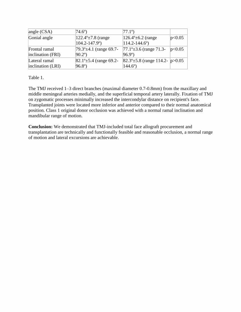
Results: Statistically significant difference between genders were observed in all parameters
measured on dry skulls except for ICA and CSA. There was a statistically significant difference
of FRI between genders in CT measurements. The mean values, standard deviation and the range
of all measurement are provided in Table 1.
Male Female
Intercondylar distance 118.1mm±5 (range
104.6-128mm)
112.2mm±6.5 (range
98-123.3mm)
p<0.05
Gonion-gonion distance 99.0mm±6.3 (range
86.6-115.3mm)
91.2mm±4.9 (range
82.7-104.5mm)
p<0.05
Lateral fossa-lateral
fossa points
120.1mm±4.6 (range
109.5-129.1mm)
113.3mm±4.9 (range
101.7-123.2mm)
p<0.05
Intercondylar angle
(ICA)
141.1º ±10.2 (range
118.9-162.5º)
139.7º±10.0 (range
119.3-160.3º)
p>0.05
Condyle-symphysis 64.8º±5.6 (range 53.4- 64.1º±5.6 (range 54- p>0.05
Page 37
angle (CSA) 74.6º) 77.1º)
Gonial angle 122.4º±7.8 (range
104.2-147.9º)
126.4º±6.2 (range
114.2-144.6º)
p<0.05
Frontal ramal
inclination (FRI)
79.3º±4.1 (range 69.7-
90.2º)
77.1º±3.6 (range 71.3-
96.9º)
p<0.05
Lateral ramal
inclination (LRI)
82.1º±5.4 (range 69.2-
96.8º)
82.3º±5.8 (range 114.2-
144.6º)
p>0.05
Table 1.
The TMJ received 1–3 direct branches (maximal diameter 0.7-0.8mm) from the maxillary and
middle meningeal arteries medially, and the superficial temporal artery laterally. Fixation of TMJ
on zygomatic processes minimally increased the intercondylar distance on recipient's face.
Transplanted joints were located more inferior and anterior compared to their normal anatomical
position. Class 1 original donor occlusion was achieved with a normal ramal inclination and
mandibular range of motion.
Conclusion: We demonstrated that TMJ-included total face allograft procurement and
transplantation are technically and functionally feasible and reasonable occlusion, a normal range
of motion and lateral excursions are achievable.
Page 38
2:06 PM - 2:09 PM
RM39 Lower Eyelid Reconstruction: A New Defect Classification to Predict Outcomes. Northwestern University Feinberg School of Medicine, Chicago
Presenter: Jonathan T Bricker, BA
Jonathan T Bricker, BA(1), Chad A Purnell, M.D.(2), Elbert E Vaca, MD(3) and Mohammed S
Alghoul, MD(4)
(1)Northwestern University, Chicago, IL, (2)Northwestern Feinberg School of Medicine,
Chicago, IL, (3)Division of Plastic and Reconstructive Surgery, Northwestern Memorial
Hospital, Chicago, IL, (4)Northwestern Memorial Hospital, Chicago, IL
Background:
Traditional teaching in lower eyelid reconstruction utilizes a classification system based on depth
and 25% increments in defect width. A third critical component is the vertical dimension; defects
may be limited to the pretarsal segment or extend into the preseptal and orbital segments. This
study proposes a new classification system that includes the vertical missing component to
predict functional and aesthetic outcomes.
Methods:
A retrospective review of patients who underwent lower lid reconstruction by a single surgeon
was performed. Defect size, type of reconstruction, number of surgeries, and post-operative
lower lid retraction were analyzed. Defects were classified into four categories based on the
vertical anatomic segment involved: I. Pretarsal; II. Preseptal; III. Eyelid-Cheek junction; and
IV. Complex Pretarsal + Preseptal. Aesthetic evaluation was performed by three blinded
reviewers based on objective criteria. Post-operative functional outcomes were evaluated by
measuring the pre-operative and post-operative central and lateral marginal reflex distance-2
(MRD-2). Outcome measures were compared among the 4 defect types using one-way ANOVA,
with Tukey's HSD post-hoc test comparisons.
Results:
Thirty-four patients underwent reconstruction of lower eyelid defects from 2013 to 2017, 16
females and 18 males, with a mean age of 65 (23-87). Etiology included basal cell carcinoma
(65%), squamous cell carcinoma (21%), melanoma (6%), sebaceous cell carcinoma (3%),
osteosarcoma (3%) and sebaceous hyperplasia (3%). The mean defect size was 3.8 cm2, ranging
from 0.2 to 23 cm2. There were 12 "Pretarsal" defects [Class I], 9 "Preseptal" defects [Class II], 9
"Eyelid-Cheek" defects [Class III], and 4 "Complex Pretarsal + Preseptal" defects [Class IV].
The “Complex Pretarsal + Preseptal" group had the worst aesthetic outcomes and highest
incidence of post-operative retraction at 75%, with a significantly greater change from pre- to
post-operative central and lateral MRD-2 (mean = 3.06 and 3.17 mm, respectively) compared to
each of the other groups (p < .01). Consequently, Group IV had significantly more revision
surgeries (mean = 5.5) compared to the other groups (p < .001).
Conclusion:
Page 39
Tarsal defects that extend into the preseptal segment are at a higher risk for post-operative lower
lid retraction. As the vertical height of the defect increases, the ability to replace the missing
tissue with local flaps while maintaining low tension on the lid margin becomes more
challenging. A new classification is therefore proposed for better planning and patient
counseling. Strategies to prevent complications and maximize outcomes in each group will be
discussed based on outcomes.
Page 40
2:09 PM - 2:12 PM
RM40 Reinnervation of the Orbicularis Oculi Muscle in Addition to Static Lid Support
Confers Corneal Protective Advantages over Static Interventions Alone in the Subacute
Facial Palsy Patient University of Texas Southwestern Medical Center, Dallas
Presenter: Ahneesh J Mohanty, BA
Ahneesh J Mohanty, BA(1), Justin Lee Perez, MD(1), Austin Hembd, MD(2), Nikhitha
Thrikutam, BS(1), Jeremy Bartley, MD(1) and Shai Rozen, MD(3)
(1)University of Texas Southwestern Medical Center, Dallas, TX, (2)University of Texas
Southwestern Medical Center Department of Plastic Surgery, Dallas, TX, (3)Department of
Plastic Surgery, UT Southwestern Medical Center, Dallas, TX
Background
Corneal protection is crucial in flaccid facial palsy patients. Prolonged orbicularis oculi muscle
(OOM) denervation without effective oculo-protective measures results in exposure keratopathy
and in severe cases, loss of vision. Traditional protective measures include corneal lubrication
and static procedures of both upper and lower eyelids. Yet, a small group of patients with
subacute palsy durations may benefit from reinnervation of the OOM via several methods. The
goal of this study, the first of its kind, was to objectively compare degrees of corneal protection
between solely static and combined static and dynamic approaches.
Methods
Retrospective review was performed on two patient groups of complete palsy patients: 1) long
standing facial palsy patients who underwent solely static support procedures of the lower and
upper eyelid, and 2) subacute facial palsy patients who underwent OOM reinnervation in
addition to static lid procedures. Only patients with available detailed ophthalmologic exams
were included. In addition to review of demographics and detailed medical history, statistical
analysis of mean corneal punctate epithelial erosion scores was performed using SigmaPlot 14
software. Static and dynamic palpebral measurements were assessed pre and post-operatively
using MEEI Emotrics software and frame-by-frame video analysis for objective quantification of
palpebral aperture and closure dynamics.
Results
Nine patients underwent combined periorbital reinnervation and static eye procedures while 15
underwent solely static procedures. The average age was 37 and 50, respectively. The mean
ophthalmology follow-up period for the combined versus static only groups was 10 and 24
months, respectively. Corneal analysis at 9+ months post-operatively revealed a 57% average
reduction in corneal punctate epithelial erosion scores in the OOM reinnervated group compared
to the static only group (p<.05). This was consistent with improvements in eye closure dynamics:
the static only group revealed a mean post-operative palpebral aperture during voluntary
maximal closure of 3.29 mm, and complete closure in 6.67%. Comparatively, the combined
reinnervated-static group revealed a mean post-operative palpebral aperture during voluntary
maximal closure of 0.78 mm, complete closure in 70%, and a rapidity of closure of 0.697
Page 41
seconds. This represents a 56.3% improvement in voluntary maximal closure from baseline
(p<.01) and a 76.2% improvement over periorbital static intervention alone (p<.01).
Conclusion
In a subgroup of patients with subacute paralysis in whom orbicularis oculi muscle reinnervation
is feasible, dynamic periorbital reanimation combined with static procedures is superior to static
procedures alone in conferring both corneal protection as well as improving dynamic and static
palpebral closure variables.
Page 42
2:12 PM - 2:15 PM
RM41 Comparing Reconstructive Outcomes in Patients with Gustilo Type Iiib Fractures
and Concomitant Arterial Injuries New York University, New York
Presenter: Joseph A. Ricci, MD
Joseph A. Ricci, MD(1), John T. Stranix, MD(2), Z-Hye Lee, MD(2), Lavinia Anzai, MD(2),
Vishal D Thanik, MD(2), Pierre B. Saadeh, MD(2) and Jamie P. Levine, MD(2)
(1)Albany Medical Center, Albany, NY, (2)New York University, New York, NY
Background: The Gustilo Classification serves as a proxy for injury severity, but recent data
suggests rising complications with decreasing arterial runoff. This study aims to compare to
evaluate different microsurgical anastomosis options based on the number of patent vessels in
the lower extremity.
Methods: A single-center retrospective review of 806 lower extremity free flaps from 1976 –
2016 was performed. Patients with Gustilo IIIB injuries were grouped based on the number of
patent vessels in the leg (3-2-1). Patients were compared based on the type of anastomosis
performed, evaluating for perioperative complications and flap failures.
Results: Perioperative complications occurred in 111 flaps (27%): 71 takebacks (17%); 45
partial losses (11%); 37 complete losses (9%). Among patients with 3-vessel runoff (61.8%)
there was no difference in takebacks or flap loss between those with end-to-end versus end-to-
side anastomoses. In 68 patients (18.7%) with 2-vessel runoff, no difference between takebacks
or flap loss was noted when comparing any anastomosis (end-to-end into an injured vessel, end-
to-end into an uninjured vessel, or end-to-side into an uninjured vessel), although vein grafts
were required more often in the end-to-side groups (p <0.01). Finally, in 39 patients (10.7%)
with single-vessel runoff, no difference was seen between end-to-end anastomosis into an injured
vessel or end-to-side anastomosis into an uninjured vessel in terms of takebacks or flap loss.
Conclusion: Higher rates of flap failure correlated with decreasing numbers of patent vessels in
the leg, but neither type of microvascular anastomosis, nor vessel selection demonstrated any
impact on reconstructive outcomes.
Page 43
2:15 PM - 2:18 PM
RM42 Indocyanine Green Fluorescence Angiography Reduces Fat Necrosis and Resection
Volumes in DIEP Flap Breast Reconstruction UT Southwestern Medical Center, DALLAS
Presenter: Austin Hembd, MD
Austin Hembd, MD(1), Nicholas T. Haddock, MD(2) and Sumeet S. Teotia, MD(1)
(1)University of Texas Southwestern Medical Center Department of Plastic Surgery, Dallas, TX,
(2)University of Texas Southwestern Medical Center, Dallas, TX
Background: This study aims to determine if the intraoperative use of laser-assisted indocyanine
green (ICG) fluorescence angiography affects postoperative fat necrosis or flap failure in a
multivariate analysis of DIEP free flap breast reconstructions.
Methods: A retrospective review was performed on 866 free flaps for breast reconstruction at a
single center from 2010-2016. ICG was used after the flap had been anastomosed on the chest to
subjectively assess for areas of hypoperfusion if there was clinical concern.
A univariable analysis was conducted on the use of ICG angiography along with 27 other patient
demographic and surgical factors. From this, an odds ratio (OR) with 95% confidence intervals
of the effect on flap fat necrosis and failure was derived for each variable. Variables with a p<.15
in the univariable analysis were entered in a backward selection algorithm to yield a
multivariable logistic regression model, in which a p<.05 cutoff was used.
Results: 409 total single pedicle DIEP hemi-abdominal free flaps were included. The average
age of the patients was 50.5 years old. The average follow up for these patients was 18.5 months,
with a median of 15.75 months. 14.4% of flaps had fat necrosis in total.
Intraoperative ICG angiography was used for 130 flaps (31.8%) and was independently
associated with a decrease in the odds of fat necrosis (OR .46, p-value= .04). ICG angiography
directly guided excision of hypoperfused areas in 50 flaps (38.5%), ensured the presence of
adequate perfusion in 78 flaps (60%), and identified a pedicle kink after anastomosis in 2 flaps
(1.5%).
There was flap failure in 7 cases. There was no reduction in flap failure rates when utilizing ICG
angiography (2.3% failure with versus 1.4% without, p-value=.68).
Prophylactic excisions of the flap without using ICG angiography were done in 107 flaps (26%)
and did not affect fat necrosis rates (OR 1.74, p-value =.1). The average weight of the resected
portion of flaps without ICG angiography was 250.8 grams, whereas the average weight of the
resected portion of flaps with ICG angiography was 152.3 grams. This 98.5-gram per flap
difference was significant (p-value=.01).
Conclusion: Our results indicate that intraoperative laser-assisted ICG fluorescence angiography
decreases the odds of fat necrosis in DIEP flap breast reconstruction by guiding a more accurate
flap debulking at inset. This can potentially save an average of 98.5 grams of tissue per flap
when compared to excising areas of hypoperfusion by clinical signs alone.
Page 44
2:18 PM - 2:21 PM
RM43 A Strategy to Alleviate Ethnic Disparities in Breast Reconstruction: Improving
Health Literacy through Community Engagement University of Pennsylvania, Philadelphia
Presenter: Olatomide T. Familusi, MD
Olatomide T. Familusi, MD(1), Irfan Rhemtulla, MD, MS(2), Justin P. Fox, MD, MHS(3),
Fabiola A. Enriquez, BA(1) and Paris Butler, MD, MPH(4)
(1)University of Pennsylvania, Philadelphia, PA, (2)university of Pennsylvania, philadelphia,
PA, (3)Plastic and Reconstructive Surgery, University of Pennsylvania, Philadelphia, PA,
(4)Plastic Surgery, University of Pennsylvania, Philadelphia, PA
Background
After oncologic breast surgery, women of color undergo reconstruction at disproportionately
lower rates than their Caucasian counterparts. In this study we address health literacy, a
modifiable contributor to this disparity, through community engagement.
Methods
In collaboration with a large church in West Philadelphia PA, The Abramson Cancer Center, The
Division of Plastic Surgery at the University of Pennsylvania, and with funding from the Plastic
Surgery Foundation, the authors developed a health awareness symposium centered on breast
reconstruction. The program included lectures, patient testimonials and a Q&A session.
Participants completed pre and post-symposium surveys focusing on the availability, timing and
options for breast reconstruction to assess knowledge.
Results
A total of 63 community members attended the 4-hour symposium. Participants were mostly
female (88.9%), and of African American descent (87.3%). Almost half (46%) possessed a
college degree or higher. Half were current breast cancer patients/survivors while 24% identified
as family members/friends of a breast cancer patient. Prior to the session, 12.7% of participants
were unaware of breast reconstruction as a treatment option after mastectomy while 54% and
71.4% were unaware of insurance coverage for breast reconstruction and contralateral balancing
procedures respectively. There were statistically significant increases in the number of
participants responding correctly to questions regarding insurance coverage (p<0.0001), timing
of reconstruction (p=0.002), and reconstruction options (p=0.002) after the program as compared
to before. The percentage of participants agreeing that breast reconstruction could improve a
woman’s self image increased from 55.6% to 76.2% (p=0.004).
Conclusion
The etiology of the existing disparity in breast reconstruction is complex and multifactorial.
Noting that health literacy is critical to a patient’s decision-making process about their diagnosis
and treatment, this study demonstrates that targeted community based educational programs can
improve awareness about breast reconstruction.
Page 45
2:21 PM - 2:24 PM
RM44 Improved Diagnostic Accuracy of Periprosthetic Breast Infection: Alpha-Defensin 1 Brown University and Rhode Island Hospital, Providence
Presenter: Marten N Basta, MD
Marten N Basta, MD(1), Adnan Prsic, MD(2), Daniel Kwan, MD(3), Karl H Breuing, MD,
FACS(1), Charles C Jehle, MD(1), Paul Liu, MD, FACS(3), Rachel Sullivan, MD(1) and Scott
Schmidt, MD(1)
(1)Brown University & Rhode Island Hospital, Providence, RI, (2)University of Washington,
Seattle, WA, (3)Brown University, Providence, RI
Background
Implant-based breast reconstruction comprises 75% of all breast reconstructions today. Prompt,
accurate diagnosis of breast implant infection is critical to minimizing patient morbidity. The
current standard of care, bacterial culture, is falsely negative in 25-30% of untreated patients and
substantially more patients receiving antibiotics. A critical need exists for better testing
modalities that remain cost-effective. Alpha defensin-1 (AD-1), an antimicrobial peptide released
from neutrophils in response to local pathogen invasion, serves as a marker for infection. With
sensitivity/specificity of 97% & 96%, it has replaced bacterial culture as the preferred diagnostic
modality for orthopedic periprosthetic infection. If AD-1 behaves similarly in periprosthetic
breast infections, it may substantially alter practice patterns throughout plastic and reconstructive
surgery, with rapid and cheap diagnostic confirmation within 24 hours. We evaluate and
compare the diagnostic performance of AD-1 to bacterial culture in suspected periprosthetic
breast infection.
Methods
An IRB-approved prospective study including all adults with prior prosthetic breast
reconstruction (expander or implant) and suspected periprosthetic infection requiring operative
washout was conducted. Patients missing gram stain and culture data were excluded.
Demographics, operative history, prosthetic characteristics, and antibiotic exposure were
collected. Implant pocket fluid was sent for gram stain, bacterial culture, AD-1 assay, and
surgical pathology. Based upon average sample AD-1 and C-Reactive Protein (CRP) levels, the
AD-1 assay reports presence/absence of infection. Sample lactate, human neutrophil elastase,
and cell differentials were also collected. Summary statistics and student’s T-test or Fisher tests
of association were performed (p<0.05=significant).
Results
10 breasts with suspected periprosthetic infection met criteria and were included, 7 of which
were acutely infected with purulent pocket fluid. Gram stain correctly identified 1 of 7 infections
(accuracy–40%) while culture correctly identified 6 of 7 infections (accuracy–90%). AD-1,
however, identified all 7 infections (accuracy-100%). AD-1 sensitivity was significantly better
than gram stain (100% vs. 14.3%, p=0.02) and more sensitive than culture (100% vs. 85.7%),
though the difference was insigificant (p=1.0). Infected breasts averaged higher levels of
Page 46
inflammatory markers (CRP: 15.8 mg/dL vs. 2.9 mg/dL, p=0.08), (lactate: 122 mg/dL vs. 74
mg/dL, p=0.09), both trending towards significance.
Conclusion
This prospective study demonstrates the utility of AD-1 in diagnosing periprosthetic breast
infection. Combining AD-1 with adjunctive inflammatory markers may allow more accurate and
prompt detection of implant infection in order to reduce morbidity and reconstructive failures.
Page 47
2:24 PM - 2:27 PM
RM45 Immediate Postoperative Application of VAC Dressing over Free Muscle Flaps to
the Lower Extremity Does Not Compromise Flap Survival and Has Improved Aesthetic
Outcomes University of Florida College of Medicine, Gainesville
Presenter: Harvey Chim, MD
Harvey Chim, MD(1), Rachel Cohen-Shohet, MD(1), Patrick J Buchanan, MD(1), Wrood
Kassira, MD(2), Morad Askari, MD(2) and Christopher John Salgado, MD(2)
(1)University of Florida College of Medicine, Gainesville, FL, (2)University of Miami, Miami,
FL
Background: Free muscle flaps are a mainstay for reconstruction of distal third leg wounds and
for large lower extremity wounds with exposed bone. However a major problem is the
significant postoperative flap swelling, which may take months to resolve. We studied the
efficacy and safety of immediate application of a vacuum assisted closure (VAC) dressing after a
free muscle flap to the lower extremity. A prospective study was performed in a subcohort of
patients to objectively compare free muscle flaps treated in the immediate postoperative period
with a VAC dressing compared to exposed flaps.
Materials and Methods: Over a 4 year period, 24 consecutive free split latissimus muscle flaps
were performed for lower extremity coverage by a single surgeon, all of which had a VAC
dressing applied immediately postoperative and for 5 consecutive days. Flap monitoring was
through an implantable doppler probe. A subcohort of these patients over a 19-month period was
included in an analysis where all consecutive free muscle flaps for lower extremity
reconstruction at a Level I trauma center were evaluated prospectively for postoperative flap
thickness, complications and flap survival. Immediate application of a VAC dressing was
performed in 9 patients, while the flap was left exposed for monitoring in 8 patients.
Results: In the consecutive cohort of 24 patients treated with postoperative VAC dressing, flap
survival was 95.8%, with the sole flap failure due to venous congestion from previously
undiagnosed extensive deep venous thrombosis. In the subcohort analysis comparing a separate
group of flaps left exposed, there was no statistically significant difference in flap survival
between both cohorts. Mean flap thickness at postoperative day 5 for the VAC group was 6.4 ±
6.4 mm, while flap thickness for the exposed flap group was 29.6 ± 13.5 mm. Flap thickness was
significantly decreased at postoperative day 5 for the VAC dressing group.
Conclusion: Immediate application of VAC dressing following free muscle flaps to the lower
extremity does not compromise flap survival or outcomes and results in decreased flap thickness
and a better aesthetic outcome.
Page 48
Fig. 1. POD6 flap treated with VAC dressing. Flap is flat and has nice contour.
Fig. 2. POD6 flap left exposed for monitoring. Flap is thick and swollen.
Page 49
2:27 PM - 2:30 PM
RM46 Can We Effectively Reduce the Operative Time of Deep Inferior Epigastric Artery
Perforator (DIEAP) Flaps' Breast Reconstruction in Lower Volume Centers? One
Surgeon's Experience King Fahad Medical City, Riyadh
Presenter: Abdulaziz K Alhujayri, MBBs
G. Althubaiti, MBBs, MHA, FRCSC(1), Abdulaziz K Alhujayri, MBBs(1) and Feras
Alshomer, MBBs, MSc(2)
(1)King Fahad Medical City, Riyadh, Saudi Arabia, (2)King Fahad medical city, Riyadh, Saudi
Arabia
Background:
In breast cancer reconstruction with DIEP flaps, long operative time carries a physical burden on
the surgeon, in addition to the financial burden that may be associated with longer hospital stay
and slower recovery.
The average time for this procedure is reported to be up to 6 to 8 hours in many centers around
the world with a hospital stay the could extend to 5 days.
Few authors published their series from large volume centers in which they managed to reduce
the operative time to 3.5 hours or even less. However, these series came from large volume
centers and may not be applicable to lower volume centers because they demand higher
experience that is refined by rapid turnover. In addition to that, most authors came up with
certain recommendations to reduce OR time, but there is little written to demonstrate the before-
and-after effect of implementing the recommended measures.
In this paper we demonstrate the "before and after" effect of using selected well-structured
strategies in reducing operative time of breast reconstruction with DIEP flaps in our lower
volume center (less than 50 flaps/year). The measures we use are applicable to all centers
including lower volume center.
Methods:
We chose certain efficiency strategies that include preoperative planning, identifying tasks
boundaries, simultaneous timed task execution, minimizing instruments requirement, a “no idle
strategy” and economy of motion during wound closure.
To reduce the variables and to have better standardization, we only included unilateral delayed
DIEP flaps done by the main author and looked at the effect of implementing these measures on
his operative time.
In addition to looking at the operative time, we reviewed surgical outcomes including hospital
stay, and the financial impact before and after using these efficiency measures.
Results:
Page 50
A total of 51 cases were enrolled in the study over 4 years. Using efficiency measures, the
average procedure time was reduced from 8.2 hours to 2.99 hours. Similarly, there was an
associated reduction of postoperative hospital stay from 4.5 days to 3 days. This is reflected also
financially as reducing the OR time and hospital stay significantly reduced the total cost of the
procedure by almost half of the initial costs.
Conclusion:
Implementing efficiency strategies translated into a meaningful consistent reduction in operative
time, hospital stay and costs in our experiencewithout the need to do high volume of cases.