58
Studies on Hog Cholera and Preventive Treatment. BY
Studies on Hog Cholera and Preventive Treatment.
BY

Studies on Hog Cholera and Preventive Treatment.
BY WALTER E. KING, Bacteriologist.
ROBT. H. WILSON, Assistant in Bacteriology.
INTRODUCTION.
In November, 1908, this department issued Bulletin No. 157, entitled “Studies on Hog Cholera and Preventive Treatment,’’ in which were presented the results of the preliminary experi- ments relative to the work on “horse serum hog cholera vac- cine.” Since that time the work has been actively continued. The present bulletin, therefore, is written as a continuation of Bulletin No. 157, and while it presents the results obtained to date at this Station, it does not conclude the investigation.
Attempts are being made in this work to approach the prob- lem from the following points of view:
1. The nature of the causative factor of hog cholera. 2. Methods of immunization, While the use of hyperimmune serum, as developed accord-
ing to the method employed by the Bureau of Animal Industry, United States Department of Agriculture, has passed far be- yond the experimental stage and is, without doubt, of great value to the hog industry, yet the method has one serious dis- advantage relative to the cost involved in its preparation. The success of the hyperimmune serum, as an effective preventive agent, is quite universally recognized, but the actual prepara- tion and distribution of the product, a t present, largely depends upon the various state legislatures. This fact alone tends to justify the statement that the cost of its preparation may seri- ously curtail its usefulness. Therefore, aside from the scientific interest pertaining to the subject of hog cholera, it appears important that aggressive work should be conducted relative to the many unsolved problems involved in the study of this animal disease. All honest and intelligent experimental ef- forts toward the solution of some of these problems constantly add to the knowledge the disease, and are therefore of practical significance.
(139)

PART I.PREVENTIVE TREATMENT.
As a brief review of the experiments outlined in the pre- ceding bulletin, it may be stated that the character of this ex- perimental work involved the use of the horse as a medium in attempting to modify hog cholera virus. It was found that a short residence (one to two hours) of the hog cholera serum in the circulatory system of the horse did not change the in- fectious nature of the material. Indeed, experiments tended t o show that a residence of one or two hours in the blood system of the horse sometimes apparently raised the virulence of the hog cholera virus. Experiments also showed that during a residence of from four t o twenty-four hours in the circulatory system of the horse there was a gradual modification of the hog cholera virus, the degree of modification depending, as a rule, upon the number of hours allowed for this purpose. Theoretically, therefore, there seemed to be some period of time, a t some point between four and twenty-four hours after the horse had received the intravenous injection of hog cholera serum, when the virus was sufficiently modified to act as a vaccine under all conditions, yet was devoid of infectious qualities.
The constant aim of the work for the past two years has been to collect experimental evidence bearing upon the above points. The chief emphasis has been placed upon the following attempted determinations :
1. To attempt to find after what period of residence in the horse, hog cholera virus becomes so modified (if a modification really does take place) that it may become an efficient vac- cine. In other words, a t what time after the horse is injected intravenously with approximately 150 cc. of hog cholera serum will the resulting horse serum produce a mild reaction, when injected subcutaneously into healthy hogs, and actively im- munize, and a t the same time possess no dangerous infectious qualities.
2. To study the possible virulent qualities of the horse serum drawn a relatively short time after the intravenous injection of the horse with hog cholera serum, and to determine, if pos- sible, whether or not the horse serum virus is a mere dilution of hog cholera virus in normal horse blood.

3. To determine the approximate minimal fatal dose of highly virulent hog cholera serum.
4. To study the possibilities of hyperimmunizing hogs and producing protective hyperimmune serum by injecting im- mune hogs with the horse serum virus.
While no absolutely definite conclusions have been reached, on the whole the results have been fairly constant. It is recognized that some of the results lead us deeper into the jungle of unsolved questions, and emphasize the importance of repeated and persistent study in fields in which animal ex- perimentation is involved. The statement should be made that one-half-hour horse serum virus (blood drawn one-half hour after the animal has received intravenously approximately 150 cc. of hog cholera serum) may not produce virulent hog cholera under all conditions when injected into healthy hogs. Neither may 24-hour horse serum (drawn twenty-four hours after the animal has received an injection of hog cholera virus) never produce symptoms of cholera. “The great variation among in- dividual animals, and the peculiar idiosyncracies of swine particularly, preclude such a definite assertion.”¹ It must be explained, therefore, that in attempting to verify the following experiments, one test or a few tests will not suffice. By those who are familiar with work of this nature, it is readily under- stood that only the average of a series of duplicate tests pre- sents reliable results.
Field Work in 1908 with Experimental Horse Serum Hog Cholera Vaccine.
The results of a comparatively large number of duplicate tests a t the Station indicate that 24-hour horse serum² pos- sessed no pathogenic properties when injected into healthy hogs, but that it did contain enough modified hog cholera virus to impart t o them some protective qualities. Moreover, it was quite definitely determined that the 24-hour horse serum was modified to such a degree that its vaccinating properties were not constant. A general survey of the whole work indicated that 24-hour horse serum might be used in the field with
ventive Treatment,” page 58. 1. Bulletin 157, Kansas State Experiment Station, “Studies on Hog Cholera and Pre-
serum, or “24-hour horse serum,” will be used to indicate that the serum in question 2. Hereafter in this bulletin the expression “½-hour horse serum,” “6-hour horse
was obtained from the horse ½ hour, 6 hours or 24 hours after the animal had been in- jected intravenously with hog cholera serum. safety, but considerable doubt was entertained concerning its

activity. The average of the results a t the Station showed that the 24-hour horse serum might successfully protect from 15 to 45 per cent of the treated hogs. However, this was a point to be determined in the field, and accordingly several herds of healthy hogs were experimentally vaccinated with the serum.
The first test was made in a herd of 485 hogs, at Brough- ton, Kan. In this herd 222 pigs, weighing approximately 50 pounds each, received, individually, from one to five cubic centimeters of fresh 24-hour horse serum, 88 received Bru- schettini’s vaccine, and 175 were unvaccinated. Three weeks later this herd contracted hog cholera through natural ex- posure from an adjacent farm, and at the completion of the outbreak the following animals were dead: College vacci- nated, 73.4 per cent; Bruschettini’s vaccine treatment, 90 per cent; unvaccinated, 89.1 per cent. Thus it was found that in 222 susceptible pigs, situated under practical conditions, the 24-hour horse serum did not produce even a noticable re- action, and furthermore that the serum was modified to such a degree that i t protected only 15.7 per cent of the treated animals.
Three other small herds of hogs in the field, consisting of a total of 28 treated animals, were vaccinated with 24-hour horse serum. In none of these herds did the vaccinated hogs show any disturbances, nor did any of the three herds subse- quently become infected through natural exposure.
In the meantime, a number of preliminary duplicate tests with 6-hour horse serum, both a t the Station and in the field, appeared to warrant an extensive field test with that experi- mental product. In these preliminary tests, including the treatment of a relatively large number of hogs with small sub- cutaneous injections of 6-hour horse serum, no serious symp- toms had occurred. It was also made evident by careful test that the protection following the use of the 6-hour horse serum was greater than that resulting from the treatment with the 24-hour horse serum. During the months of October, Novem-ber and December of 1908, and January of 1909, a total of 809 hogs in twenty-eight different herds³ were experimentally
3. Four other herds, making a total of 32, were treated during the spring months of 1909 with 6-hour horse serum. From these no reports were sent in, and they are there- fore not included.

Sept. 1910.] Hog Cholera and Preventive Treatment. 143
vaccinated with 6-hour horse serum vaccine4. These hogs, with the exception of one herd, were situated on various farms in the state of Kansas. Among the twenty-eight treated herds 612 unvaccinated, or control hogs were kept in the same en- closures with the vaccinated animals, under the same practical conditions.
In eleven of these herds the experimental 6-hour horse serum vaccine appeared to produce the disease. In some of these in- stances the disease appeared in a mild form, but in several TABLE 1. Results of field work with experimental horse serum vaccine.
cases acute hog cholera was present. Of the total of vaccinated hogs, 7.1 per cent gave evidence of contracting the disease from the vaccine. With almost no exceptions these were corn-fed hogs which were being fattened for market. Nearly all of these eleven herds were in close proximity to hog cholera a t the time of vaccination; therefore, in some cases the disease may have gained access before the time of treatment.
Herd No. 28 (table 1>, situated a t Jewell City, Kan., was vaccinated on November 27, 1908. Forty-two of these hogs, weighing about 225 pounds per head, each received subcuta- neously 5½ cc. of 6-hour horse serum vaccine. In this herd a severe reaction followed the vaccination. Six of the treated animals became sick and 3 died in a few days. The other 3 re- covered soon after the signs of the reaction disappeared in the remainder of the 42 treated animals. During this experiment, hog cholera existed in a neighboring herd within about sixty rods of the vaccinated animals; however, after the results from vaccination had occurred these hogs remained in a normal con- dition.
In eleven of the total twenty-eight herds, hog cholera ap- parently existed at the time of vaccination. In these herds an average of about 45 per cent of the vaccinated hogs and from 55 to 60 per cent of the unvaccinated animals ultimately suc- cumbed to the disease.
In herd No. 14 (table 1 ) , located a t Frankfort, Kan., 28 hogs were vaccinated and 36 were left as controls. A few ani- mals in this herd were evidently infected by natural exposure previous to the time of vaccination. On the next day after treatment 2 of the vaccinated hogs were sick, and on the second day after, 2 additional animals. On the third day these 4 treated animals died. The injection of the modified virus evi- dently accelerated the disease. None of the remaining 24 vaccinated hogs showed the slightest symptoms of the disease, while over 50 per cent of the 38 controls became sick and 19.4 per cent died. Lung lesions were prominent in this outbreak. Excluding the 4 animals which died three days after vaccina- tion, 100 per cent of the treated animals in this herd were saved. These hogs were lightly fed, before and after vaccina- tion, on alfalfa and small rations of corn.
On December 24, 1908, herd No. 5 (table I), consisting of 268 hogs, near Dallas, Tex., showed symptoms of hog cholera.

During that morning 3 of the animals died, and on that day 125of the hogs were vaccinated with 6-hour horse serum vaccine; and 140 were left unvaccinated. By the latter part of Feb- ruary, 1909, the disease had evidently run its course and only 20 per cent of the vaccinated hogs were dead, while 90 per cent of the controls had succumbed. This herd was fed hotel refuse and received very little heavy diet such as corn.
Table 1 shows the average result of the field work with the experimental horse serum vaccine. By placing all of the re- sults together, without regard to different conditions to which references are made above, it is found that death from hog cholera resulted in 32.13 per cent of the hogs treated with 6- hour horse serum vaccine, as against 46.65 per cent of the un- treated animals.
From an experimental point of view these results were satis- factory. Individual losses occurred to a few of the farmers who volunteered to cooperate with the Station in this work. Such losses have occurred because the Station had insufficient means to carry on extensive experiments in this line of in- vestigation without such cooperation. Perhaps it should be stated that those farmers who cooperated in this work did so voluntarily after clearly understanding the nature of the in- vestigation. While no specific contract existed between the individual farmer and the Station, yet in every single instance the experimental nature of the work was carefully explained through correspondence. By virtue of this, the Station was not responsible, either legally or morally, for individual losses which occurred.
The results of the field work may be summed up as follows: 1. Twenty-four-hour horse serum vaccine, as a rule, is
lacking in infectious properties. It possesses some degree of protection, which, however, is not constant enough to prove of practical use.
2. Six-hour horse serum vaccine, under certain conditions relative to character of diet and individual variation in sus- ceptibility, produces virulent hog cholera. It possesses greater protective properties than does 24-hour horse serum vaccine, and under certain conditions in the field it may save from 80 to 100 per cent of the treated animals. For practical field condi- tions, however, 6-hour horse serum vaccine is not safe.
3. After the intravenous injection of the horse with ap-

proximately 150 cc. of hog cholera virus, the virus becomes modified, the degree of modification depending, as a rule, upon the period of residence in the circulatory system of the horse. It is possible that 10- t o 15-hour horse serum vaccine might possess both safety and activity.
4. In addition to the observations of various investigators and the common knowledge of those familiar with hog cholera, a general survey of the field work in this investigation em- phasizes the following conditions :
( a ) That the character of the diet has great influence upon susceptibility and resistance to the disease, heavy rations of corn being especially conducive to increased susceptibility.
( b ) That, while hog cholera is usually much more prevalent during the fall months, hogs are easily infected in cold weather.
(c) That lung lesions may predominate in one locality while intestinal lesions may be more frequent in another locality.
Infectious Nature of Horse Serum Virus.
After the completion of the field work, it became necessary to study the nature of the hog cholera virus when modified by passage through the horse. Among the points which de- manded careful attention, it was especially important that some tests should be made to show whether or not horse serum virus is a mere dilution of hog cholera virus. Therefore, in addition t o the suggestive data already obtained, it was deemed advisable to make a series of duplicate tests upon susceptible pigs with horse serum drawn a t varying intervals of time, representing short periods of residence after the horse had received intravenous injections of hog cholera virus. To de- termine, if possible, whether or not the horse serum so treated represented nothing other than a dilution of the original hog cholera serum, plans were made to estimate the approximate dilution of virus when the virus was injected into the blood of the living horse, and compare the results from the injection of this into susceptible pigs, with the corresponding dilutions of the same virus in normal horse blood outside the body of the animal, and in sterile physiological salt solution. It was neces- sary, therefore, in each series of experiments to weigh the horse under observation, estimate the amount of blood con- tained in his body by taking one-fifteenth of the body weight, then compute the dilution of hog cholera serum in vivo, when

the animal received intravenously a given quantity of the virus. When the blood was drawn from the horse from a half to nine hours after injection of the virus, the resulting horse serum was tested as to its infectious properties by injecting it subcutaneously into susceptible pigs, using an approximate dosage of 4 cc. of the horse serum per 50 lbs. weight. In each case, duplicate tests were made of the same lot of hog cholera virus, placed in the same dilution in normal, citrated or de- fibrinated horse blood, immediately after drawing, and in ster-

ile, physiological salt solution, both being kept a t a tempera- ture of 37º C. for a period of time exactly corresponding to that, which represented the residence of the hog cholera virus in the circulatory system of the horse. At the expiration of this period the dilutions were injected subcutaneously into sus- ceptible pigs, using the same rate of dosage as in the case of the horse serum virus. In this work all possible precautions were exercised to avoid extraneous exposure. At first, the tempera- tures of the experimental animals were taken from the date of inoculation until the test was completed. Later this was dis- continued, owing to possibilities of exposure to the disease.
These test pigs were kept in the experimental stable, each in- dividual test, of course, being confined to an isolated pen. The pens were provided with cement floors and with continuous three-foot sides of cement. Each pen was separated from the others by a two-foot cement alley. As a control for these ex- periments, a susceptible 50-pound pig was kept in one of these pens for several months during the investigation. This animal remained in a normal condition throughout. In order to de- termine whether or not it was protected by natural immunity, it was later placed with hogs suffering from hog cholera. This

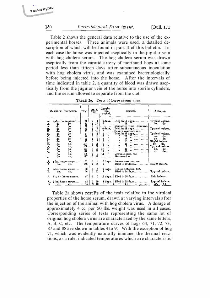
Table 2 shows the general data relative to the use of the ex- perimental horses. Three animals were used, a detailed de- scription of which will be found in part II of this bulletin. In each case the horse was injected aseptically in the jugular vein with hog cholera serum. The hog cholera serum was drawn aseptically from the carotid artery of moribund hogs a t some period less than fifteen days after subcutaneous inoculation with hog cholera virus, and was examined bacteriologically before being injected into the horse. After the intervals of time indicated in table 2, a quantity of blood was drawn asep- tically from the jugular vein of the horse into sterile cylinders, and the serum allowed to separate from the clot.
Table 2a shows results of the tests relative to the virulent properties of the horse serum, drawn a t varying intervals after the injection of the animal with hog cholera virus. A dosage of approximately 4 cc. per 50 Ibs. weight was used in all cases. Corresponding series of tests representing the same lot of original hog cholera virus are characterized by the same letters, A, B, C, etc. The temperature curves of hogs 64, 71, 72, 73, 87 and 88 are shown in tables 4 to 9. With the exception of hog 71, which was evidently naturally immune, the thermal reac- tions, as a rule, indicated temperatures which are characteristic
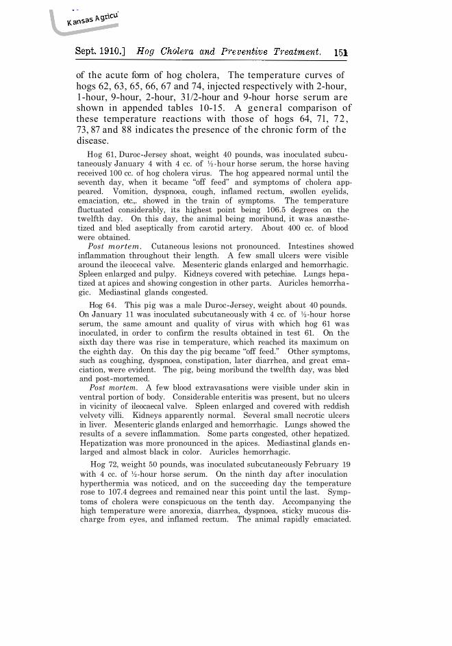
of the acute form of hog cholera, The temperature curves of hogs 62, 63, 65, 66, 67 and 74, injected respectively with 2-hour, 1-hour, 9-hour, 2-hour, 31/2-hour and 9-hour horse serum are shown in appended tables 10-15. A general comparison ofthese temperature reactions with those of hogs 64, 71, 72 , 73, 87 and 88 indicates the presence of the chronic form of the disease.
Hog 61, Duroc-Jersey shoat, weight 40 pounds, was inoculated subcu- taneously January 4 with 4 cc. of ½-hour horse serum, the horse having received 100 cc. of hog cholera virus. The hog appeared normal until the seventh day, when it became “off feed” and symptoms of cholera app-peared. Vomition, dyspnoea, cough, inflamed rectum, swollen eyelids, emaciation, etc.,. showed in the train of symptoms. The temperature fluctuated considerably, its highest point being 106.5 degrees on the twelfth day. On this day, the animal being moribund, it was anæsthe- tized and bled aseptically from carotid artery. About 400 cc. of blood were obtained.
Post mortem. Cutaneous lesions not pronounced. Intestines showed inflammation throughout their length. A few small ulcers were visible around the ileocecal valve. Mesenteric glands enlarged and hemorrhagic. Spleen enlarged and pulpy. Kidneys covered with petechiae. Lungs hepa- tized at apices and showing congestion in other parts. Auricles hemorrha- gic. Mediastinal glands congested.
Hog 64. This pig was a male Duroc-Jersey, weight about 40 pounds. On January 11 was inoculated subcutaneously with 4 cc. of ½-hour horse serum, the same amount and quality of virus with which hog 61 was inoculated, in order to confirm the results obtained in test 61. On the sixth day there was rise in temperature, which reached its maximum on the eighth day. On this day the pig became “off feed.” Other symptoms, such as coughing, dyspnoea, constipation, later diarrhea, and great ema- ciation, were evident. The pig, being moribund the twelfth day, was bled and post-mortemed.
Post mortem. A few blood extravasations were visible under skin in ventral portion of body. Considerable enteritis was present, but no ulcers in vicinity of ileocaecal valve. Spleen enlarged and covered with reddish velvety villi. Kidneys apparently normal. Several small necrotic ulcers in liver. Mesenteric glands enlarged and hemorrhagic. Lungs showed the results of a severe inflammation. Some parts congested, other hepatized. Hepatization was more pronounced in the apices. Mediastinal glands en- larged and almost black in color. Auricles hemorrhagic.
Hog 72, weight 50 pounds, was inoculated subcutaneously February 19 with 4 cc. of ½-hour horse serum. On the ninth day after inoculation hyperthermia was noticed, and on the succeeding day the temperature rose to 107.4 degrees and remained near this point until the last. Symp- toms of cholera were conspicuous on the tenth day. Accompanying the high temperature were anorexia, diarrhea, dyspnoea, sticky mucous dis-charge from eyes, and inflamed rectum. The animal rapidly emaciated.


A decubitus was maintained after the tenth day, the animal occasionally getting up to drink. On the twelfth day, the hog being moribund, it was anaesthetized and bled aseptically from the right carotid artery.
Post mortem. No cutaneous lesions were visible. An enteritis was present in the large intestine, especially the caecum. A few small un- developed ulcers were present near the ileocaecal valve. The mesenteric glands were slightly involved, being somewhat enlarged and hemorrhagic. Spleen practically normal in size, but very dark and friable. Liver ap- parently normal. Kidneys showed numerous petechiae on surface, and on section showed congestion and inflammation. Lungs normal, both in color and consistency. Auricles of heart thickly studded with hemorrhagic foci. Mediastinal lymph glands black in color and very hard.
Hog 87, a 30-pound Yorkshire, received, March 18, 4 cc. of ½-hour horse serum. Several days after receiving this serum, hyperthermia was noticeable, followed later by anorexia, dyspnea, and vomition. These symptoms became intensified. The animal, being moribund on the four- teenth day, was bled and post-mortemed.
Post mortem. Pinkish discoloration of the skin on abdomen. A slight inflammatory condition in vicinity of ileocaecal valve, remainder of intes- tines practically normal. Lungs showed hemorrhagic foci; some hepatiza- tion at apices. Auricles of heart ecchymotic.
Hog 88, a Duroc-Jersey, weight 45 pounds, injected March 18 with 4 cc. of ½-hour horse serum. Five days after injection hog refused feed and showed symptoms of cholera. Bled and post-mortemed April 14.
Post mortem. Cutaneous lesions absent. Intestines inflamed; a button- shaped ulcer in caecum. Kidneys petechiated. Liver normal. Spleen en- larged and hemorrhagic. Lungs hepatized and ecchymotic. Mediastinal glands enlarged and dark in color.
Hog 94, Duroc-Jersey, weight 40 pounds, received 4 cc. of ½-hour horse serum. Animal refused feed on ninth day and showed a general depression. Gradually became worse, and died April 18.
Post mortem. Skin on abdomen and inner side of thighs reddish purple in color. Considerable enteritis present. A few small ulcers at junction of ileum and cæcum. Kidneys thickly studded with petechiae. Spleen enlarged, black and pulpy. Liver apparently normal. Mesenteric glands enlarged and hemorrhagic. Mucosa of stomach inflamed, especially that of pyloric portion. Lungs covered with large hemorrhages, apices of lungs hepatized. Auricles hemorrhagic. Mediastinal glands enlarged and hemorrhagic.
Hog 95. This test was a duplicate of that upon hog 94. Incubation period, symptoms and lesions identical with those of 94.
Hog 107, Duroc-Jersey, weight 70 pounds, was injected May 11 with 4 cc. of ½-hour horse serum. Sickened the eighth day, showing symp- toms of cholera. Was bled May 24, thirteen days after injection.
Post mortem. Slight cutaneous lesions. Intestines normal, with the exception of a small area near ileocaecal valve, which was inflamed. Spleen enlarged and friable. Liver practically normal. Kidneys slightly
- 2-171


petechiated. Lungs showed the results of a severe inflammation, being hemorrhagic and hepatized in many places.
Hog 109, Duroc-Jersey shoat, weight 90 pounds, on May 21, 1910, re- ceived 4 cc. of ½-hour horse serum, the horse having received 40 CC. of hog cholera virus intra-jugular and bled one-half hour after injection. Incubation period, six days. Symptoms and lesions typical of hog cholera, although the lesions not as pronounced as usual. This hog received ap- proximately 1/225 of a cc.
Hog 126, Yorkshire, weight 60 pounds, injected with 4 cc. of ½-hour horse serum, on June 18. Ten days after injection feed was refused and animal appeared listless. Other symptoms appeared, such as dyspnoea, great thirst, constipation, later diarrhea and emaciation. . Died during night of July 7.
Post mortem. Lesions typical of cholera.
Hog 133, Duroc-Jersey, weight 50 pounds, injected June 28 with 4 cc. of ½-hour horse serum. Sickened on the twelfth day. Symptoms char- acteristic of cholera. Died July 17.
Post mortem. Skin on ventral portion of body purplish in color. In- testines inflamed and dotted with hemorrhagic foci. A few small unde- veloped ulcers near ileocæcal valve. Mesenteric glands enlarged and gorged with blood. Kidneys petechiated. Liver contained several necrotic areas. Spleen pulpy and very dark. Numerous blood extravasations in lungs. Auricles ecchymotic. Mediastinal glands involved.
Hog 65, a Poland China, weight 65 pounds, was inoculated subcuta- neously with 5 cc. of 9-hour horse serum, on January 20, 1910. The hog was apparently healthy until the thirteenth day, when it gradually went off feed and took on a listless appearance. The temperature fluctuated considerably between normal, 103.5 degrees, and 106 degrees. Would have chills, usually one or two a day, lasting about an hour at a time. Had a severe cough. At times the animal would seem to be improving, leaving its bed and walking around the pen; would also stand at the feed trough with the other hogs and eat ravenously. These periods of seeming convalescence would not last long, however, the pig returning to its bed and resuming its usual decubitus and appearance. The patient lingered along in this condition for over thirty days, before dying. To- wards the end it experienced great difficulty in breathing, every breath seeming to be painful. The feces appeared to be normal throughout the course of the disease.
Post mortem. A slight discoloration of the skin was noticeable on the ventral portion of the body and on the inner side of the thighs. The intestines were practically normal, a slight inflammation being present around the ileocæcal valve. The contents of the intestines appeared to be normal in consistency and color. Mesenteric glands but slightly in- volved. Spleen and liver practically normal. Kidneys slightly ecchy- motic near pelvis. The thoracic cavity was the seat of very pronounced lesions. The lungs were practically one solid mass of hepatized, necrotic tissue. Upon section with a bistoury, a fatty, cheesy-like appearance was revealed. In interstices between the lungs and pleura, when they


were not adherent, was present a clear jelly-like liquid, partly organized in places. Probably not more than one-fifth of the total lung tissue was functional, this undoubtedly accounting for the dyspnoea and other grave respiratory troubles which the animal experienced. The pericardial fluid was almost solidified, encasing the heart in a mold. Auricles very dark in color and hemorrhagic. Mediastinal glands slightly enlarged and showing blood extravasations.
Hog 66, A Poland China sow, weight 85 pounds, was inoculated sub- cutaneously January 20 with 5 cc. 2-hour horse serum. The normal tem- perature was 103.8 degrees. The temperature remained normal af ter inoculation until the sixth day, when i t rose to 104.5 degrees, and dropped to 102.8 degrees on the 8th day. The highest mark recorded in the temperature chart was on the tenth day, when the temperature was 106.5 degrees, fluctuating between this point and normal until the death of the animal. The animal refused feed on the tenth day and showed other signs of illness. Probably the most pronounced symptom in the early stage, aside from being “off feed,” was a dry hacking cough. The coat became rough and staring, eyes congested and swollen. It was almost impossible for i t to move from its bed. Limbs cold, the posterior ones seeming to be partially paralyzed. The animal lingered along, gradually becoming weaker, until the twenty-fourth day after inoculation, when i t died.
Post mortem. The autopsy was not held until two days after death, but the weather being very cold, the cadaver was preserved in good con- dition. The body was not badly emaciated, in spite of the fact that the animal had not eaten for nearly two weeks previous to death. The skin on the ears was slightly discolored, being a purplish hue. The intestinal walls showed hemorrhagic foci in places. No ulceration was visible in the caecum, nor at the ileocaecal valve. The mesenteric glands were almost black in color and greatly enlarged. Spleen normal in size and color, but very soft and friable. Kidneys showed petechtiae, the petechia- tion being more pronounced in the hilus of the organ. Liver showed a few ulcerative, necrotic areas. Extensive lesions were found in the lungs, the lower portions of the lobes being in the stage of gray hepatization, the consistency being almost solid; other portions showing the results of various stages of inflammation. Evidence of severe pleuritis was notice- able, the lungs being adherent to pleura in places. The heart showed petechiae, the petechiae being more extensive in the auricles. Mediastinal glands enlarged and hemorrhagic. A striking feature in this case was the extent of the glandular involvement—every set of glands from the retropharyngeal to the inguinal showed affection.
Hog 67 was a Poland China male, in good thrifty condition, weighing 95 pounds. On January 20 was inoculated subcutaneously with 6 cc. of 3½-hour horse serum. On the morning of the ninth day the animal missed its first feed, and gave a temperature of 107 degrees. It maintained this degree of temperature almost throughout the course of the disease. The animal would lie in its bed almost continuously, occasionally getting up to drink. Gave evidence of great thirst, naturally arising from the high
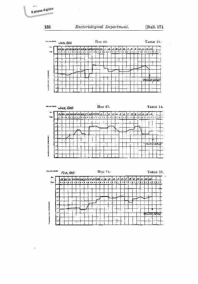

fever. Had a very severe cough, almost going into convulsions at times. Eyes became dull and listless, coat rough and staring, had a watery mucous discharge from rectum. The animal did not seem to convalesce or to get worse until the twenty-third day, when the symptoms became aggravated, and the patient rapidly weakened, finally succumbing the night of February 13, twenty-five days af ter inoculation.
Post mortem. Did not show any cutaneous lesions. Intestines slightly inflamed in region o f cæcum, but no ulcerations. Mesenteric glands in- volved. Spleen normal in size, but dark and soft. Liver normal. Mucosa of stomach inflamed and pyloric portion covered with a diphtheretic-like membrane. Kidneys showed petechiae on surface, Apices of lungs hepa- tized and base o f right lung congested and inflamed. Lungs adherent topleura in places. Chambers of heart gorged with black, jelly-like clots. Auricles ecchymotic. Mediastinal lymph nodes enlarged and hemorrhagic.
Hog 74, a Poland China male, weight 50 pounds, received subcutane- ously, February 19, 4 cc. o f 9-hour horse serum. On the eighth day after injection i t had a rise in temperature of two degrees. On the eleventh day the temperature was 106 degrees and the hog showed signs of illness-loss o f appetite, dyspnea, constipation and a marked general depression. Later diarrhea appeared, the feces being very watery and tinged with blood. The hog rapidly emaciated and weakened, dying the night of March 14, 1910.
Post mortem. No cutaneous lesions. Spleen practically normal. In- testines inflamed, more particularly the small intestines. Kidneys showed hemorrhagic foci. Liver studded with numerous small necrotic centers, which on section were found to contain thick caseous pus. Lungs were pneumonic in appearance, more especially the left lung. Pericardial fluid almost solidified. Auricles hemorrhagic. Mediastinal and bronchial glands showed involvement, being enlarged and hemorrhagic.
Hog 110, Duroc-Jersey shoat, weight 85 pounds, received, May 21,4 cc. of l-hour horse serum. Incubation period six days. Symptoms and lesions characteristic of hog cholera.
From the above tests it may be observed that ½-hour horse serum, as a rule, is highly infectious in nature. With the ex- ception of natural immune No. 71, a subcutaneous injection of4 cc. per 50 pounds weight produced in a series of twelve pigs (hogs 61, 64, 72, 73, 87, 88, 94, 95, 107, 109, 126 and 133) acute cholera, with an average incubation period of 6.8 days and an average termination of the disease in 14.8 days, of 91.6 per cent of the cases, hog 73 having recovered. On the whole, from the limited observations afforded by the experi- ments with 1- to 9-hour horse serum, as indicated in table 2a, previous results were verified.

C C
Hog 68, weight 65 pounds, received in the subcutem 4 cc. of a mixture of normal horse blood and hog cholera virus. One per cent sodium citrate was added to the blood immediately upon drawing from the horse, to prevent coagulation. The mixture was kept at a temperature of 37 degrees C. for one-half hour before injection. The animal was appar- ently normal until the twelfth day, when i t refused feed. Other symp- toms gradually appeared, such as hyperthermia, constipation, followed by diarrhea, dyspnea, coughing, gummed eyelids, and a general debility. The animal died the twenty-first day after injection.
Post mortem. The intestinal tract showed evidence of inflammation, more particularly the stomach, the mucosa of which was badly con- gested and inflamed. No intestinal ulcers were present. Right kidney slightly ecchymotic near hilus. Spleen and liver practically normal. Lower portions of lungs badly hepatized. Some pleuritis. Heart muscle hemorrhagic.
Hog 80, a 30-pound Yorkshire shoat, received on March 8 3½ cc. of ½-hour dilution (1 cc. hog cholera virus and 350 cc. horse serum). Thirteen days after injection the animal seemed to be suffering from a paraplegia, a t times being unable to rise without assistance. Had con- siderable fever and appeared to be greatly depressed. No typical symp- toms of cholera were in evidence. The hog was bled March 23, 1910.
Post mortem. The only apparent pathological condition was a great distention of the bladder, the animal apparently not having urinated for several days. The bladder occupied the greater portion of the pelvic cavity and extended well into the abdominal cavity. The pressure ex- erted by the distended bladder on the surrounding nerves was probably the cause of the paralysis of the hind parts. All the other viscera seemed to be normal.
Hog 100, a Duroc-Jersey, weight 50 pounds, received on April 15 4 cc. of a mixture of horse blood and hog cholera virus (1 cc. virus and 240 cc. blood). Ten days after injection feed was refused and animal became listless. A profuse, watery diarrhea occurred the twelfth day and con- tinued until the end. Would lie in a corner of the pen continually, seem- ing to be semiconscious. Animal weakened gradually, and died May 2.

ture of hog cholera virus and normal salt solution (1 cc. virus and 350 cc. normal salt). Fourteen days after injection symptoms of cholera were in evidence. These symptoms gradually became more pronounced, the ani- mal dying March 22, 1910.
Post mortem. A slight skin discoloration on ventral portion of body. Spleen enlarged and pulpy. Kidneys petechiated. Liver normal. A few small ulcers a t ileocsæcal valve. Lungs slightly hemorrhagic.
a
Hog 93, Poland China, weight 55 pounds. On March 26 injected 4 cc. of mixture of virus and physiological salt solution (1 cc. virus and 240 cc. salt solution). Sickened fourteen days after injection. Symptoms those of cholera. Died April 13.
Post mortem. Lesions typical of cholera. Hog 108, Poland China shoat, weight 100 pounds. Injected May 11
with 4 cc. dilution of virus (hog 89) in normal salt solution in proportion of 1 cc. virus to 350 cc. normal salt. Animal sickened on sixth day, re- fusing feed and having the general depression characteristic of cholera. Died May 21.
Post mortem. A slight discoloration of skin on ventral portion of body. Intestines inflamed throughout their entirety, and thickly studded with petechiæ. Two small undeveloped ulcers near i l e o c a l valve. Spleen enlarged and hemorrhagic. Extensive extravasation of blood in kidney tissue. Liver apparently normal. Lungs thickly covered with hem- orrhagic foci. Auricles hemorrhagic. Mediastinal glands enlarged andhemorrhagic.
Hog 112, Poland China shoat, weight 90 pounds, received on May 264 cc. of dilution of virus and normal salt solution in proportion of 1 cc. virus to 172 cc. of normal salt solution. On the ninth day feed was re- fused, and other signs of illness were in evidence. Had a rough, staring coat, and a general listless appearance. Would seldom leave its bed;

when it did, would walk with a tottering gait. On the eighteenth day a profuse diarrhea set in, which continued until the end. Being in a dying condition on June 18, was bled and post-mortemed.
Post mortem. Cutaneous lesions quite extensive. Blood extravasa- tion in intestines, kidneys, lungs and heart muscle. No intestinal ulcers. This hog received approximately 1/43 of a cc. of hog cholera virus.
Hog 131, Duroc-Jersey, weight 40 pounds. Injected with 4 cc. of a mixture of virus and physiological salt solution, in proportion of 1 cc. virus to 244 cc. of the salt solution. Hog was apparently normal until the eighteenth day. At this time symptoms of cholera appeared, al- though not as pronounced as usual. Died July 11.
Post mortem. Fair lesions.
Tables 16 and 16a show results obtained when susceptible hogs were injected subcutaneously, using a dosage of approx- imately 4 cc. per 50 lbs. weight, with dilutions of hog cholera virus in normal horse blood, in vitro, and in sterile physiological salt solution corresponding to those dilutions indicated in table 2a with horse serum virus. Attention is again called to the statement that the symbols, A, B, C, etc., in the first column of each of tables 16 and 16a, refer to the respective lots of the original hog cholera serum, A, B, C, etc., which were injected in the horses, as shown in table 2. Thus, in tables 16a and 16, hogs 75 and 79 received dilutions of the same virus in physio- logical salt solution, and in normal horse blood, respectively, which hogs 72 and 73, table 2a, received in the form of horse serum virus.

these below. It is necessary to state that in determining the average results in table 16b, relative to the virus diluted in normal horse blood, no account was taken of hog 113, which was probably naturally immune ; also, in determining the per cent of normal horse blood dilution, in vitro, hog 80 was not considered, as death did not result from hog cholera.
It may be noted in table 2 that horse serum virus from hog cholera virus, lot F, contained an approximate dilution of 1part of virus to 861.8 parts of diluting fluid. This dilution produced the following results (tables 2a, 16 and 16a) : Horse serum virus, after six days’ incubation, the death of pig 109 in nine days, with typical lesions ; normal horse blood dilution, after eighteen days’ incubation, the death of pig 132, with slight lesions ; physiological salt solution dilution, after seven- teen days’ incubation, mild reaction and recovery in the case of hog 118. It was decided, therefore, to increase the dilution of hog cholera virus in the horse serum virus, in normal horse blood, in vitro and in physiological salt solution, until the minimal fatal dose was reached. The purpose of this was

twofold, in that the results might aid in determining whether or not horse serum virus represented mere dilution as well as indicating the minimal fatal dose of hog cholera virus. The tests with lots of serum F, H and I refer to the attempts in establishing the minimal fatal dose. Just what is the smallest amount of hog cholera virus that is capable of producing the disease is a point well worth knowing. Of course, the min- imal fatal dose will vary within certain limits, depending upon the age and susceptibility of the animal and the degree of virulence of hog cholera serum. In this work the above con- ditions of variation have been controlled as well as possible by using 50-pound susceptible pigs from the same locality, many of which belonged to the same litters, and by making the dilutions with the same strain of serum, so activated that it produced by the subcutaneous injection of 2 cc. the death of 50-pound test pigs within fifteen days.
For the purpose of clearness, portions of tables 2, 2a, 16 and 16a, covering the tests with lots of serum F, H and I, are pre-

sented in me appended tables 16c and 16d. A study of these tables shows the approximate dilution of virulent hog cholera serum in sterile physiological salt solution which may be taken as the minimal fatal dose. Using the results obtained from test pigs 118, 136 and 138 (table 16c), it is suggested that per- haps the minimal fatal dose of hog cholera virus lies some- where between 1/215 and 1/300 cc. per 50 pounds weight. It will be noticed, however, that when this dilution was used in the form of horse serum virus different results were obtained. With virus lot F, using a dilution of 1 to 861.8 parts horse blood, in the form of horse serum virus, hog 109, after an in- cubation period of six days, promptly died from acute hog cholera, while the corresponding normal blood dilution test pig, No. 132, after eighteen days’ incubation, died in twenty- one days with slight lesions, and the corresponding physio-logical salt dilution test pig, No. 118, showed only a mild re- action after an incubation period of seventeen days.
The results of the test of virus lot H, in a dilution of 1 to 1202, and virus lot I, dilution 1 to 1724, conform with those obtained from the tests with virus lot F. In the case of the horse serum virus, having an estimated dilution of virus, lot H, of 1 to 1202, pig 135 became sick after fourteen days and finally recovered, after having been affected with the chronic form of cholera for a number of days. Pigs 140 and 136, in- jected with corresponding dilutions of virus, lot H, in normal horse blood, in vitro, and physiological salt solution, respec- tively, showed no reactions. Finally, of pigs 137, 141 and 138, each injected with 1/431 cc. of virus, lot I, in the form of horse serum virus, normal blood dilution, in vitro, and physiological salt solution, respectively, none showed subsequent symptoms. From these results the evidence is strong that, notwithstanding certain disadvantages in attempting to accurately estimate the dilution of any given amount of fluid placed in the blood of a living animal, the horse serum virus retains a greater degree of activity as the dilution of hog cholera virus in the blood in vivo becomes higher, than do the corresponding increasing dilutions in normal horse blood, in vitro and in physiological salt solution.
Experimental evidence points to the fact that hog cholera virus undergoes some modification when passed through the horse. Results indicate that there is at first a process of

activation of the virus, which process begins immediately after the intravenous injection of the horse, and continues for a relatively short period. Observations tend to show that at the expiration of one-half hour's residence in the blood of the horse the virus attains its highest degree of activation. It is scarcely possible, therefore, that this process of activation represents a multiplication of the theoretical invisible micro- organisms. Experimental evidence also shows that, after this period of activation, a continued residence of four to twenty- four hours or longer of the virus in the horse will result in a gradual decline in virulence. This phase of the process of the modification may be due to a disintegration of the virus or an accumulation of substances deleterious to the virus. The de- cline in the activity of the virus would, in all probability, take place to some extent in the presence of normal horse blood out- side of the living body.
The above-suggested conclusions are based upon the results of the dilution tests, supplemented by the results of the field work and previous observations. From a practical point of view, perhaps the most important results of this incomplete investigation pertain to the virulent nature of ½-hour horse serum. The positive results of the series of dilution tests are almost preponderantly in favor of the dilution in the form of horse serum virus. It was recognized at the beginning that a comparison of the dilution of the virus in normal horse blood in vivo with that in normal horse blood in vitro was perhaps of slight value. The results of various investigations in other lines of work show that in normal blood outside the body the leucocytes may immediately give off inactivating materials. Other factors in the normal horse blood in vitro may also exert some influence, thus impairing the direct comparison of re- sults of normal horse blood dilutions outside the animal body with those of horse serum virus. On the other hand, the re- sults of the series of comparative tests of the corresponding dilutions of hog cholera virus, in the form of horse serum virus and physiological salt solution, are especially valuable in showing the possible activated pathogenic properties of hog cholera virus, after a residence of one-half hour in the circu- latory system of the horse. When a given amount of virus is injected intravenously into the horse and a dilution of a given ratio is estimated in the blood of the living animal, it

does not appear reasonable to believe that a given portion of the blood of the horse represents a like dilution when drawn one- half hour or more after the injection of the animal. Some of the inoculated virus may be carried into other channels of the circulatory system, some may be situated in the peripheries, some may have been absorbed, and finally lytic action may have depleted the hog cholera virus, or foreign serum, by the time the virus had maintained a residence of one-half hour or longer in the living body of the animal. When a given dilution of the virus is prepared in sterile physiological salt solution, how- ever, it is reasonable to suppose that the dilution remains the same, and that if any change might take place after an incu- bation at body temperature of one-half hour or longer, that change would be a multiplication of virulent properties.
It therefore seems apparent that these preliminary tests tend to show that horse serum virus represents an activated form of hog cholera virus, and not a mere dilution, because of the following reasons :
1. In a series of 12 hogs, ½-hour horse serum virus pro- duced death in 91.6 per cent of the animals, with an average incubation period of 6.8 days and an average period of duration of the disease of 14.8 days. In comparison with the above, cor- responding dilutions with the same lots of virus in normal horse blood in vitro, and in sterile physiological salt solution, in series of 8 and 6 hogs, respectively, produced death in 37.5 per cent and 83.4 per cent of the animals, with average incubation periods of 11.4 days and 13.3 days, and average periods of duration of disease of 18.2 days and 19.2 days, respectively.
2. The symptoms of disease and the autopsies of the hogs injected with the ½-hour horse serum virus were, as a rule, characteristic of typical acute hog cholera, while those of the corresponding dilutions in normal horse blood and physio-logical salt solution were more chronic and less marked in character.
3. From a limited amount of data indicated in table 16c,it is shown that the minimal fatal dose of virulent serum, as represented by a dilution of the virus in physiological salt solution, does not indicate the minimal fatal dose when the dilution is maintained in the form of horse serum virus.
4. During extensive field work covering the experimental vaccination of approximately 850 hogs in about eleven herds,

approximately 60 animals were infected with typical hog cholera by the subcutaneous inoculation of ½ to 5 cc. of 6-hour horse serum.
From the data which is at hand, it is suggested that the fol- lowing summarized comments may be made, relative to the character of experimental horse serum, hog cholera virus, and horse serum hog cholera vaccine :
1. Horse serum virus ½-hour horse serum) does not repre- sent a mere dilution of the given hog cholera virus.
2. A residence of hog cholera virus for a half hour in the circulatory system of the horse appears to activate the virus.
3. Half-hour horse serum virus is capable of producing typ- ical acute hog cholera when injected subcutaneously, in rela-
4. The minimal fatal dose of highly virulent hog cholera serum may perhaps be found at some point between 1/215 and 1/300 cc.
tively small doses (4 cc.), into healthy hogs.
Experimental Hyperimmunitization with Half-hour Horse Serum Virus.
The possibility of successfully producing hyperimmune serum by substituting horse serum virus for virulent hog cholera serum in the hyperimmunizing process was sug- gested by some of the results obtained during the laboratory and field work in 1908.5
It was deemed advisable, however, to first begin the study of the experimental horse serum virus and attempt to determine whether this is composed of mere diluted hog cholera serum or really activated virus. After it was found that the ½-hour horse serum produced quite constant results when injected into susceptible pigs, and that these results were more uniform than those from corresponding dilutions in normal horse blood in vitro and in physiological salt solu- tion, certain preliminary tests were planned for the purpose of determining whether protective hyperimmune serum could be prepared from the ½-hour horse serum virus.
There can be no doubt but that the successful application of horse serum virus in producing active hyperimmune serum would be of distinct practical advantage. In attempting to so modify the technique involved in the preparation of the hyper- immune serum, as at present manufactured according to the

original Dorset-Niles method, the practical purpose involves the reduction of expense and inconvenience attending the method. If such a modification could be successfully made, the greatest item of expense involved in the present production of hyperimmune serum would be removed. This can readily be shown by the following conservative estimate:
0
c
0
P
Not only would this modification lessen the cost of produc- tion, but, providing the serum were potent, it would also prove to be a valuable factor in providing a sterile virus. Large quantities of blood can be aseptically drawn from the horse, defibrinated and stored in the refrigerator for a relatively long period. On the other hand, according to the present methods of securing virus from cholera-diseased pigs, the material is frequently contaminated and must be used soon after being obtained. Assuming that this suggested modified method could be successfully applied, the manufacture of hyperimmune serum would resolve itself into a comparatively simple and convenient process. All hogs utilized in the work
-3-171

might well serve a twofold purpose. Of the test pigs used in determining the potency of given lots of hyperimmune serum the controls could furnish the virus t o be injected into the horse, while those test animals which received protection could later be used as hyperimmunes.
In the present investigation, lack of time has afforded the accumulation of only a limited amount of data relative t o the possibility of successfully hyperimmunizing with ½-hour horse serum virus. The methods by which seven immune hogs were treated with ½-hour horse serum virus, with the result- ing effects on the animals, are given in the appended table (table 17):
With the exception of hogs 70, 73 and 76, none showed any- serious effects from the injection, other than a slight soreness, which passed off in a few hours. Hogs 70 and 76 were in ad- vanced stages of pregnancy. They became violently ill after receiving the serum. Hog 76 died the same day. Hog 70aborted the second day, dying that night. Hog 73, which re- ceived 100 cc. of the serum intraperitoneally, became sick soon after being removed from the table. The animal vomited and showed a general depression, but was apparently again normal in four hours, and remained well.
From the few results which have been obtained, it does not appear that the phenomenon of anaphylaxis seriously interferes with the successful inoculation of immune hogs with relatively- large quantities of horse serum virus.

The serums from each of the experimental hyperimmune hogs indicated in table 17, with the exception of hogs 70 and 76, were tested for potency. In performing these tests the usual technique employed in securing and testing hyperimmune serum was used. Only a relatively small portion of blood (25to 100 cc.) was drawn from each experimental hyperimmune hog.
Table 18 shows the results of the first test. This materialconsisted of the blood from experimental hyperimmune hogNo. 77. The blood was obtained on April 13, 1910, sixteen days after the animal had received intraperitoneally 600 cc. of ½-hour horse serum.


In the above test, hog 96, which received 40 cc. of the serum together with 2 cc. of virus, was fully protected by the experi- mental serum. Hog 97, which weighed 140 pounds and which was injected with 20 cc. of the serum simultaneously with the virus, received no protection.
Experimental hyperimmune hog 77 was bled a second time on April 20, twenty-three days after receiving the intraperi- toneal injection of horse serum virus, and seven days after the first bleeding. The results of the test with this experi- mental hyperimmune serum are shown in table 22 and the corresponding temperature curves in tables 23, 24 and 25.


In the above test the control hog, No. 105, died fourteen days after inoculation, showing typical lesions of acute hog cholera. Test hogs 102 and 104, aside from thermal reactions following inoculation, remained in a normal condition.
On May 25, h 77 og 77 was experimentally rehyperimmunized by being injected intraperitoneally with 350 cc. of ½-hour horse serum virus. A small quantity of blood was drawn from this hog on June 13, nineteen days after the injection of the virus. The test of this blood and corresponding temperature curves may be found in appended tables 26, 27, 28 and 29.

Test hog 121, which weighed 120 pounds, and which received 40 cc. of the serum with 3 cc. of virus, did not gain protection. This pig died fifteen days after inoculation, and the autopsy showed typical lesions of cholera. Test hog 120, which weighed 90 pounds and was injected with 20 cc. of the serum with 2 cc. of virus, maintained a normal condition throughout, with the exception of a marked temperature reaction.
Variation in susceptibility may perhaps be offered as an ex- planation of the result of the above test, which proved to be the only one of this experimental series which did not yield logical results. Control pig 122 showed marked symptoms of hog cholera nine days after inoculation, and died on the four-teenth day. Autopsy revealed characteristic lesions.


On June 1, twenty-four days after injection, experimental hyperimmune hog 73 (table 17) was relieved of 50 cc. of blood, which was tested on hogs 114 and 116. The results of this test and corresponding temperature curves are given in tables 30 to 34.
The results of the above test clearly indicate that the ex- perimental hyperimmune serum was not sufficiently potent t o furnish complete protection, At the same time, this serum evidently did possess, to some degree, protective properties. Both controls, pigs 117 and 119, manifested symptoms of the disease after twelve and seven days’ incubation, died on the seventeenth and ninth days, respectively, and neither of the serum pigs, Nos. 114 and 116, showed clinical symptoms, other than a rise in temperature, which promptly subsided, until after incubation periods of fifteen and eighteen days re- spectively. Autopsies performed on pigs 114 and 116, which died in nineteen and twenty-four days respectively, showed no marked lesions of cholera in either case. Test pig 144, on June 2 was injected with 15 cc. of serum together with 2 cc. of virus. It was decided that this dose of 15 cc. of the serum should be increased, consequently an additional dose of 10 cc. was given the next morning, making a total dosage of 25 cc. of serum, as shown in table 30. This pig showed a shorter incubation period, and died five days earlier, than did the test pig 116, which received 20 cc. of the serum. A glance at the tempera- ture charts of test pigs 114 and 116 shows that both animals exhibited thermal reactions after periods of time correspond-

ing to those of control pigs 117 and 119, yet neither test pig manifested any other clinical symptoms for several days. These results, taken in connection with the fact that experi-mental hyperimmune hog No. 73 received a single intraperi- toneal injection of 1000 cc. of horse serum virus per 100 pounds weight, possibly warrant the suggestion that while the blood contained theoretical antibodies at the time of bleeding, yet not all of the virus had been absorbed.
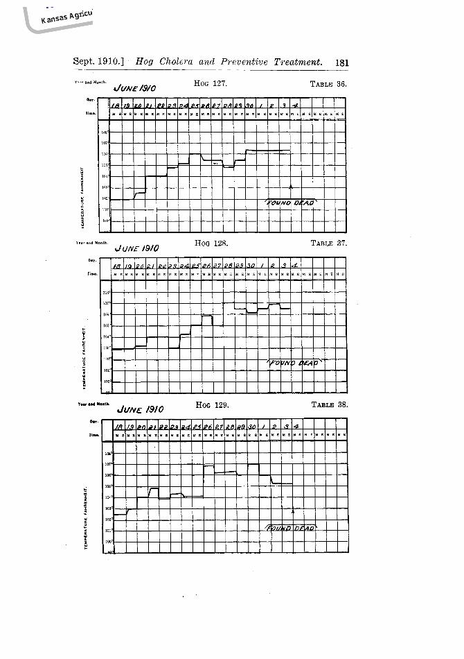
From hog 86 (table 17) 90 cc. of blood was drawn on June 10, sixteen days after the last subcutaneous injection of horse, serum virus. The results from the test of this serum, which are presented in table 53, were similar to those of the pre- ceding test. The temperature curves of the animals used on this test may be found in tables 36, 37, 38 and 39. A glance, a t table 17 will show that hog 86, from which this blood was taken, weighed 130 pounds and received a total of 1105 cc. ofvirus. It is quite possible that a portion of this virus was not absorbed when the animal was bled, sixteen days after it had received the last injection of virus. This view is supported. by the fact that only in the case of two tests, those of the blood of hogs 73 and 86, which received 1000 cc. and 1105 cc. of the virus respectively, were like results obtained.
Experimental hyperimmune hog No. 103 (table 17) was in- jected intravenously and intraperitoneally with 500 cc. of horse serum virus on May 15. On June 8, twenty-four days, after the injection, 200 cc. of blood was drawn from hog 103 and submitted to test, as shown in table 40. The correspond- ing temperature curves are. indicated in tables 41 ,42 and 43.


From a practical point of view the results of this test were satisfactory. Test pig 123, which weighed 120 pounds, and which received 40 cc. of the serum simultaneously with 3 cc. of virus, evidenced a marked thermal reaction a few days after treatment, thus showing absence of natural immunity. The rise in temperature subsided in five days and the animal re- mained normal. Test pig 124, which weighed 100 pounds, and which received 20 cc. of serum, continued on feed for a few days after the control pig, No. 125, showed severe symp- toms. This pig died two days after the death of the control animal. Autopsies of the two animals showed a marked con- trast, as typical lesions of cholera were present in the control
pig, while test pig No. 124 showed only hemorrhagic changes,the lungs, intestines, spleen and lymphatic glands appearing normal. It should be stated that test pig 124 was not in a thrifty condition a t the beginning of the experiment.
The results from the above preliminary experiments rela- tive to the attempted hyperimmunization with horse serum virus make possible the following suggestions:
1. Horse serum and defibrinated horse blood may be in- jected into hogs in relatively large quantities, subcutaneously, intraperitoneally and perhaps intravenously, without danger of loss from hypersusceptibility to the foreign blood.
2. Half-hour horse serum virus, when injected into immune hogs according to the general technique employed in the orig- inal Dorset-Niles method, causes the production of protective substances in the blood of the treated animals.
3. The degree of potency of the hyperimmune serum, pre- pared by the experimental, modified method, may depend upon the amount of horse serum virus used and the method of ap- plication.

PART II.HEMATOLOGICAL STUDIES.
Supplementary to the experiments outlined in part I of this bulletin, studies were made of the blood of two of the experi- mental horses used in the work, and of the blood of normal and diseased hogs. Inasmuch as material for hematological studies was practically always available, this phase of the work was considered of sufficient interest and importance to justify its prosecution, in the hope that the results might contribute, in some small measure, to the knowledge of hog cholera. Studies were made, therefore, (1) to observe the influence exerted by hog cholera virus, injected intravenously, upon the histological structure of horse blood, and (2 ) t o collect additional data relative t o the comparative histological structure of the blood of normal and diseased (hog cholera) swine.
Obsevations relative to the influence exerted by hog cholera virus, in- jected intravenously, upon the histological structure of normal horse blood.
In Bulletin 157 of this Experiment Station, the suggestion was made that the intravenous injection of hog cholera serum into the horse possibly caused some toxic action peculiar t o the virus. As yet, no definite, positive statement can be made re- garding this. It is apparent, however, that the normal horse cannot receive, intravenously, a larger dose than approxi- mately 200 cc. of hog cholera serum without considerable dan- ger of death. While the substitution of normal hog serum for hog cholera serum causes similar symptoms, the results af- forded by limited observations show that the symptoms are much less pronounced in the case of normal hog serum. More- over, as well as can be determined, no distinct hypersensitiza- tion follows the introduction of repeated injections of hog cholera serum into the horse. It has been observed, however, that repeated intravenous injections of hog cholera virus causes a gradually decreasing tolerance on the part of the horse toward the virus. As a possible aid in studying the nature of the action of hog cholera virus upon the horse, a num- ber of observations were made relative to the histological
-4-171

structure of horse blood before and after the intravenous in- jection of the animal with hog cholera serum.
The usual technique was employed in this work. The red and white corpuscles were counted with the Thoma hematocy- tometer, Zappert ruling. For differential counts of the leuco- cytes, Wright’s staining solution was used. The hemoglobin was estimated with Dare’s hemoglobinometer. Hammerschlag’s method was employed in determining the specific gravity, and Wright‘s method was used in estimating the time of coagula- tion.
Table 44, which is a duplicate of table 2, part I, shows at a glance the weight of the animals used, the amount of virus which they received and the time of bleeding. The following appended notes indicate in detail the observations relative to the treatment of the animals with hog cholera serum:
Horse No. 1, bay gelding, 15 years old, weight 1150 pounds. In good condition except lameness in fore legs, due to laminitis. Normal tempera- ture 99.5 degrees, pulse 42, strong and regular, respiration 16.
On December 30, a t 8:15 a.m., was injected intrajugularly with 100 cc. of hog cholera virus. Five minutes after receiving the virus the horse showed evidence of abnormal conditions. Pulse accelerated and respira- tion and temperature increased. Borborygmus pronounced. Pupils di- lated and eyes had staring appearance. Animal became restless and ex- cited. Evacuation one-half hour after injection. During the next half hour had seven evacuations, the feces being quite watery. Abdominal pains were shown, the animal striking and biting at abdomen. A t the end of an hour and a half the animal was apparently normal. On Feb- ruary 12 horse No. 1 was injected with 100 cc. of virus. When 80 cc. had been injected the horse showed signs of uneasiness, which became more marked as he received the other 20 cc. The horse was immediately removed from the stocks. In three minutes violent symptoms appeared. The animal reeled and staggered, almost falling at times. Broke out in profuse perspiration. Pupils became dilated and conjunctiva somewhat

congested. Peristalsis increased. Had passage, normal in consistency, ten minutes after injection. Numerous defecations for an hour. Epis- taxis from both nostrils fifteen minutes after injection. At this time pulse 60 and respiration 30.
One-half hour after injection 1600 cc. of blood was drawn aseptically from jugular vein. The removal of this blood seemed to relieve the horse, and he immediately became more alert.
Injected again April 4 with 120 cc. virus. Twenty minutes after in- jection pulse and respiration accelerated. Pulse 50, respiration 21, eyes dull and staring. Had first evacuation twenty-five minutes after injec- tion. At intervals of two minutes had six evacuations, each evacuation being more watery than the preceding one. Forty minutes after injection pulse 55, respiration 24. Perspired freely at times. One hour after in- jection appeared to be recovered from shock. Pulse action and respira- tion practically normal.
On May 20, 3 p.m., injected with 40 cc. of hog cholera serum. Ten minutes later animal neighed pitifully and seemed to be in a dazed con- dition. Eyes staring and expressionless. Hemorrhage from nose. Had considerable difficulty keeping on feet. Had several watery evacuations. After five minutes these symptoms gradually decreased in intensity. The 40 cc. injected this time produced a more severe reaction than the 100 cc. in former injection.
Horse No. 1 was again injected, on July 11, with 20 cc. virus. The same general symptoms appeared as in former injections, but were less violent and of shorter duration. For an hour after bleeding the horse was quite sick, refusing feed and water and frequently striking a t abdo- men a s if intestinal pain were present. At six o’clock horse appeared to be recovered from the injection, eating and drinking well. Pulse, tempera- ture and respiration normal at this time. The animal received a fifth injection with 20 cc. of virus on July 2. Resulting symptoms practically the same a s those in the preceding injection.
Horse No. 2, gray gelding, broncho stock, weight 800 pounds; thick winded and heavey; otherwise in good condition. Normal pulse 42, rather irregular, respiration 20, temperature 99.5 degrees.
On March 14, 9 a.m., was injected intrajugularly with 100 cc. of hog cholera virus. No symptoms resulted from the injection other than that the heart action and respiratory movement were slightly quickened. Coughed and passed flatus, which was probably caused by the exercise in the lot after injection. No distinct action on intestinal tract, such as was characteristic in horse No. 1.
Horse No. 2 injected again, May 7, at 3:15 p.m., with 140 cc. of virus. No symptoms were apparent for twenty minutes. At this time breathing became difficult, and the animal was somewhat depressed. Perspired slightly, pulse quickened, irregular and weak. Had first defecation in thirty minutes, feces normal. In five minutes more another defecation, more watery than the first. One-half hour after injection 400 cc. of blood was drawn from the jugular vein. One-half hour later 330 cc. more was drawn.
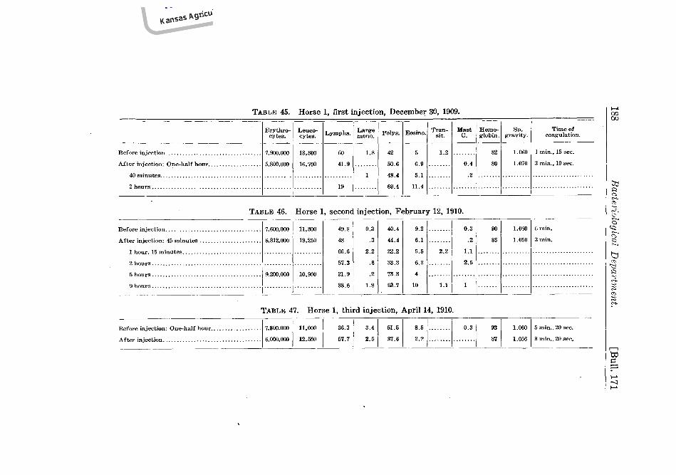
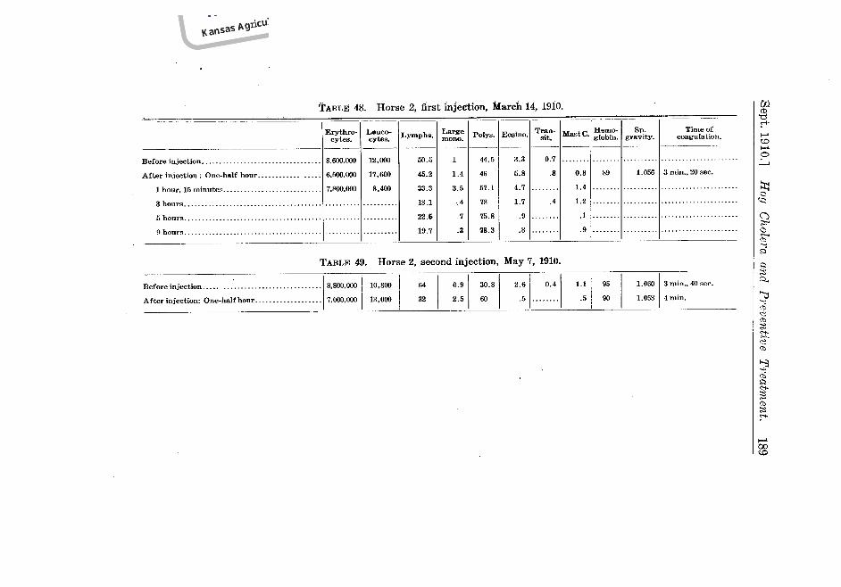


Tables 45, 46, 47, 48 and 49 indicate the results obtained from the study of the blood of the horse before and after the injection with hog cholera virus. In tables 50 to 53below are placed the averages of those results which showed evident changes in the histological structure of the blood following the injection of the virus.
According to the results shown in tables 52 and 53, a period of residence of one-half hour of hog cholera serum in the circu- latory system of the horse causes the following changes in the blood of the horse:
1. A decrease of approximately one and one-half million erythrocytes.
2. Marked leucocytosis, there being an increase of over 4000 leucocytes.
3. A loss of approximately 4 per cent of the hemoglobin. 4. A decrease in the specific gravity and slight decrease in
the time of coagulation.


Compurutire histological structure of t h e blood of normal and diseased (hog cholera) swine.
According to Burnett 6 the literature contains the following summarized data relative t o the structure of the blood of nor- mal swine :
o
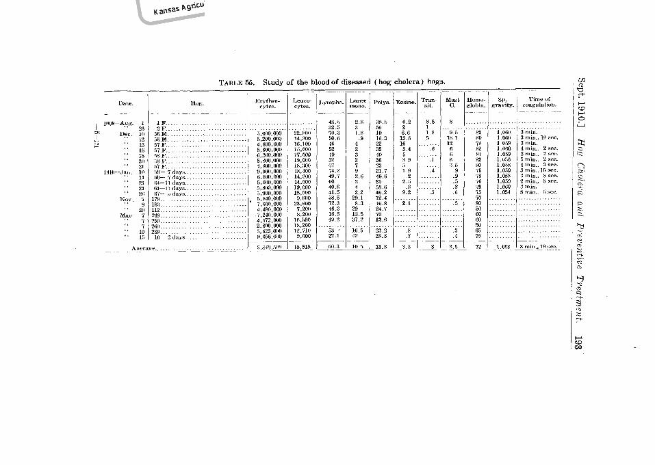
In view of the fact that comparatively little work has been done on the study of the blood of normal hogs, i t appeared necessary that as many specimens of normal blood as possible should be studied, in order to obtain comparative results of any value. Accordingly, a careful study was made of the blood of 43 normal swine. The same technique was used as in the case of the study of the horse blood. The results of this work are shown in table 54, while the results obtained from the study of 22 animals suffering from hog cholera are shown in table 55.
A comparative study of the blood from normal and diseased (hog cholera) swine showed several interesting changes in the case of hog cholera blood. The number of erythrocytes
6. Burnett, textbook, “Clinical Pathology of the Blood of Domesticated Animals.” p. 48.

and the hemoglobin content were decreased, the anemic con- dition increasing according to the progress of the disease.Frequently poikilocytosis was observed in the blood from severe cases of the disease.
Leucopenia was shown in the blood of the diseased hogs, there being an average decrease of nearly 5000 leucocytes per cmm. This depletion in leucocytes, as shown by the average differential counts, involved the decrease of 4 per cent of lymphocytes, 4 per cent of the polynuclears and 0.1 per cent of the eosinophiles. The blood from diseased hogs contained an average increase of 4.8 per cent large mononuclears, 2.8 per cent mast cells and 0.8 per cent transitional forms.
The specific gravity was slightly lowered and the time of coagulation increased one minute.
These results suggest that the changes in the structure of the blood of hogs suffering from hog cholera are analogous to those in cases of typhiod fever in man.


