Intellectual Property Notice: The Biopharma business of GE Healthcare was acquired by Danaher on 31 March 2020 and now operates under the Cytiva™ brand. Certain collateral materials (such as application notes, scientific posters, and white papers) were created prior to the Danaher acquisition and contain various GE owned trademarks and font designs. In order to maintain the familiarity of those materials for long-serving customers and to preserve the integrity of those scientific documents, those GE owned trademarks and font designs remain in place, it being specifically acknowledged by Danaher and the Cytiva business that GE owns such GE trademarks and font designs.
Scalable process for adenovirus productionThis application note describes a process for adenovirus production, from upstream cell culture to downstream purification, using modern tools and technologies. To demonstrate robustness of the upstream process, virus production in suspension adapted cells was performed in both rocking and stirred-tank bioreactors. The downstream process was carefully optimized to meet stringent regulatory demands on product purity and quality. Novel analytical methods were used in parallel with established techniques for comparison and to ensure accurate monitoring of the processed material. The novel process is easily scaled and compatible with both single-use and steamable hard-piped process equipment.
IntroductionHuman embryonic kidney 293 (HEK293) cells are susceptible to a broad range of viruses, and are thus frequently used in production of viral vectors and vaccines. Here, suspension-adapted HEK293 cells were used in production of adenovirus, one of the most frequently used viral vectors in the development of therapeutic vaccines and vaccines against infectious diseases (1). In addition, adenovirus has also been explored as a viral vector for gene therapy and as an oncolytic virus. Because of the wide application range, adenovirus production volumes can vary significantly. Hence, the interest in scalable platforms for industrial adenovirus production is increasing.
Recombinant adenovirus serotype 5 (AdV5) is one of the most studied adenovirus vector, and was therefore selected for this work. The novel process ranges from upstream virus production to a purified sterile-filtrated bulk product (Fig 1). A more detailed description of the process development and optimization work is given in separate application notes (2–7). The novel process was compared in terms of downstream adenovirus recovery and purity with a reference process (8). Outlines for the compared processes are given in Figure 2.
Materials and methodsCell culture
Stirred-tank bioreactor cultureFor the stirred-tank bioreactor process, the Xcellerex XDR-10 bioreactor system was used. HEK293.2sus cells, expanded in shake flask cultures in CDM4HEK293 cell culture medium supplemented with 4 mM L-glutamine, were inoculated at a cell density of approximately 0.3 × 10⁶ cells/mL in 3.5 L prewarmed CDM4HEK293 medium in a 10 L XDA cell culture Pro bag. Culture working volume at start was between 4 and 4.5 L. When viable cell density (VCD) reached 1.6–2.0 × 10⁶ cells/mL, complete CDM4HEK293 medium was added to the culture to reach a VCD at 1 × 10⁶ cells/mL to reach final working volume of 10 L and allow for addition of nutrients and dilution of metabolites before infection.
A more detailed description of the stirred-tank bioreactor process is given in the application note, KA874021017AN (3).
Rocking bioreactor cultureFor the rocking bioreactor process, The ReadyToProcess WAVE 25 bioreactor system was used. A 20 L Cellbag™ bioreactor was inflated with air and 1–1.5 L of CDM4HEK293 medium was added aseptically to the bag. The bioreactor bag was left for equilibration over night at 37ºC, in 5% CO2 and at an agitation of 12 rpm. Before cell inoculation, the agitation was increased to 20 rpm and an offset calibration of pH was performed.
HEK293.2sus cells, expanded in shake flask cultures in CDM4HEK293 cell culture medium supplemented with 4 mM L-glutamine, were inoculated at a cell density of approximately 0.3 × 10⁶ cells/mL. After three days, the culture was diluted in prewarmed CDM4HEK293 medium supplemented with 4 mM L-glutamine to reach a cell density of 0.3 × 10⁶ cells/mL in a total volume of 5–6 L, and agitation was increased to 23 rpm. After three more days and prior to adenovirus infection, the culture was diluted in a similar manner to reach 0.9–1.1 × 10⁶ cells/mL in a total volume of 10 L, and the rocking was increased to 25 rpm.
A more detailed description of the rocking bioreactor process is given in the application KA879160418AN (4).
For virus release, the harvest material was incubated with 0.5% Tween 20 for 4 h at 37°C directly in the bioreactor. DNA fragmentation using 20 U/mL Benzonase™ (Merck) with MgCl2 added to final concentration of 1 mM was performed simultaneously. After incubation, samples were taken for direct analysis of cell viability and remaining intact cells. For analysis of virus protein, infectious virus titer, total protein, and gDNA, samples were centrifuged at 5000 × g for 5 min before supernatants were collected for analysis.
Clarification of the harvest material was performed by normal flow filtration (NFF), using a combination of 2 μm and 0.6 μm GF filters for removal of cell debris and initial impurity reduction. Thereafter, the clarified harvest was frozen (-80°C). After thawing, the clarified harvest was subjected to filtration (NFF) using a 0.6 μm filter before the next process step.
Concentration and buffer exchange was performed by tangential flow filtration (TFF) on a ReadyToProcess hollow fiber filter with a filter area of 1400 cm2 and a NMWC of Mr 300 000 at a shear rate of 3000 sec-1 and a transmembrane pressure (TMP) of 0.3 bar using ÄKTA flux 6 filtration system. The 10-fold ultrafiltration/ 5-fold diafiltration step was conducted for further reduction of impurities and buffer exchange into 20 mM Tris, pH 8.0 + 300 mM NaCl, 2 mM MgCl2 to prepare the adenovirus-containing sample for subsequent downstream purification steps. For maintaining virus stability, MgCl2 was added to all buffers from the first TFF step and onwards.
A more detailed description of the cell lysis and virus filtration steps is given in the application note KA875220218AN (5).
Fig 1. Overview of the novel adenovirus production process.
Fig 2. Process outline for the novel process as well as for the reference process for adenovirus production.
Upstream production
Clarification
Formulation
Cell lysis and DNA fragmentation
Concentration and buffer exchange
Sterile filtration
Reference process Novel process
Capture: Q Sepharose™ XL Capture: Capto Q ImpRes
Polishing: Sepharose 4 Fast Flow
Polishing: Capto Core 700
Process workflow
Virus propagation
HEK293 cells were infected with E1/E3-deleted AdV5, coding for the GFP reporter, at a time of infection (TOI) of 1 × 10⁶ cells/mL and a multiplicity of infection (MOI) of 10. Time of harvest (TOH) was 42 h post infection.
KA877080618AN 3
Chromatography steps
For the novel process, adenovirus capture was performed using Capto Q ImpRes resin packed in an HiScale™ 26 column (column volume [CV] 88 mL) for the 3 L scale and in an HiScale 50 column (CV 294 mL) for the 10 L scale. Columns were operated on an ÄKTA pure 150 system, using the method outlined in Table 1. The subsequent polishing step was conducted using Capto Core 700 resin packed in HiScale 16 (CV 10 mL) for the 3 L scale and in HiScale 16 (CV 29 mL) for the 10 L scale, using the method outlined in Table 2. A more detailed description of the chromatography steps of the novel process is given in the application note KA876080618AN (6).
For the reference process, adenovirus capture was performed using Q Sepharose XL resin packed in HiScale 50 column (CV 249 mL) for a 3 L scale. The column was operated on an ÄKTA pure 150 system, using the method outlined in Table 3. The subsequent polishing step was conducted using Sepharose 4 fast flow (FF) resin packed in HiScale 50 (CV 382 mL) a 3 L scale, using the method outlined in Table 4. For maintaining virus stability, MgCl2 and sucrose were added to the buffers used in reference capture and polishing steps.
Table 1. Method for Capto Q ImpRes capture step
Step Volume Buffer/material RT (min)
Equilibration 3 CV 20 mM Tris, pH 8.0 + 450 mM NaCl, 2 mM MgCl2 6
Sample load ~ 80% of DBC TFF-material conditioned to 450 mM NaCl (from 3.7) 10
Wash 2 CV 20 mM Tris, pH 8.0 + 480 mM NaCl, 2 mM MgCl2 10
Elution 2.5 CV Linear gradient to 20 mM Tris, pH 8.0 + 570 mM NaCl, 2 mM MgCl2 10
Gradient delay 2 CV 20 mM Tris, pH 8.0 + 570 mM NaCl, 2 mM MgCl2 10
CIP 3 CV 1 M NaOH 6
Re-equilibration 5 CV 20 mM Tris, pH 8.0 + 450 mM NaCl, 2 mM MgCl2 6
DBC = dynamic binding capacity
Table 2. Method Capto Core 700 polishing step
Step Volume Buffer/material RT (min)
Equilibration 5 CV 20 mM Tris, pH 8.0 + 500 mM NaCl, 2 mM MgCl2 3
Sample load up to 30 CV Capto Q ImpRes eluate 3
Wash 1.5 CV 20 mM Tris, pH 8.0 + 500 mM NaCl, 2 mM MgCl2 3
CIP 5 CV 1 M NaOH + 27% 1-propanol 6
CIP 30 min pause 30
CIP 5 CV 1 M NaOH + 27% 1-propanol 6
Re-equilibration 5 CV 20 mM Tris, pH 8.0 + 500 mM NaCl, 2 mM MgCl2 3
Table 3. Method for Q Sepharose XL capture step
Step Volume Buffer/material RT (min)
Equilibration 3 CV 20 mM NaP, pH 7.3 + 350 mM NaCl, 2 mM MgCl2, 2% sucrose 10
Sample load ~ 80% of DBC TFF-material conditioned to 350 mM NaCl (from 3.7) 10
Wash 5 CV 20 mM NaP, pH 7.3 + 350 mM NaCl, 2 mM MgCl2, 2% sucrose 10
Elution 3 CV 20 mM NaP, pH 7.3 + 500 mM NaCl, 2 mM MgCl2, 2% sucrose 10
Strip 1 CV 20 mM NaP, pH 7.3 + 1000 mM NaCl, 2 mM MgCl2, 2% sucrose 10
CIP 3 CV 1 M NaOH 10
Re-equilibration 3 CV 20 mM NaP, pH 7.3 + 350 mM NaCl, 2 mM MgCl2, 2% sucrose 10
DBC = dynamic binding capacity
Table 4. Method for Sepharose 4FF polishing step
Step Volume Buffer/material Flow (cm/h)
Equilibration 0.3 CV 20 mM Na-phosphate, pH 7.3 + 200 mM NaCl, 2 mM MgCl2, 2% sucrose 50
Sample load 0.2 CV Eluate from Q Sepharose XL 50
Elution 1.5 CV 20 mM Na-phosphate, pH 7.3 + 200 mM NaCl, 2 mM MgCl2, 2% sucrose 50
4 KA877080618AN
Formulation
Concentration and buffer exchange was performed by TFF on a ReadyToProcess hollow fiber filter with a filter area of 290 cm2 and a NMWC of Mr 300 000 at a shear rate of 3000 sec-1 and a transmembrane pressure (TMP) of 0.3 bar using ÄKTA flux s filtration system. The 5-fold ultrafiltration/ 5-fold diafiltration step was conducted for buffer exchange into a formulation or storage buffer consisting of 20 mM Tris, pH 8 + 25 mM NaCl, 2 mM MgCl2, 2.5% glycerol.
Sterile filtration
Sterile filtration was conducted by NFF on an ULTA SG 0.2 μm filter (5). Process aliquots of 20 mL of the final bulk were filtered with a small-scale syringe filter (PES, 0.2 μm).
Analysis
Trypan blue exclusion method was used for measuring the number of intact cells and cell viability, using Vi-CELL™ cell viability counter.
Determination of the percentage of infected cells was performed by flow cytometry with the BD Accuri™ C6 system (BD Biosciences) using GFP signal (coded by virus).
Infectious virus titer was analyzed using 50% tissue culture infective dose (TCID50) as well as by automated fluorescence microscopy (AFM). For TCID50 assay, serial dilutions of virus were added to plated adherent HEK293 cells and the cytopathic effect was determined after 10 days of incubation. Virus titer was calculated according to the Spearman-Kärber method. The setup of AFM is similar to TCID50 but with the major advance in readout already after 2 days of incubation by automatic counting of infected (viral GFP expression) cells using the IN Cell Analyzer 2200.
Total virus titer was analyzed in triplicate samples by qPCR using PureLink™ Viral RNA/DNA Mini Kit, TaqMan™ Universal PCR Master mix, and forward and reverse primers for hexon DNA and TaqMan MGB 6-FAM probe on the StepOnePlus™ Real-Time PCR System (all Applied Biosystems). Human AdV5 DNA (3.1 × 10⁷ copies/mL) (ViraPur) was used as standard.
Total virus titer was also analyzed using the Biacore T200 surface plasmon resonance (SPR) system. Biacore CAR assay (targeting fiber protein) was performed using recombinant human CAR protein (Abcam) coupled to a Biacore Sensor Chip CM5. AdV5-containing samples were injected at 5 μL/min over 400 s. AdV5 reference material (ATCC) was used as standard. Similarly, the Biacore Factor X assay (targeting hexon protein) was also used for determination of virus concentration. More detailed descriptions of the Biacore assays are given in the application note KA878080618AN (7).
Total and virus protein (hexon) concentrations were analyzed by SDS-PAGE and Western blot. A polyclonal anti-AdV5 primary antibody (Abcam), a Cy™3-labeled secondary antibody, and human AdV5 (ATCC) as standard were used for analysis of virus proteins. For total protein, Cy5-prelabeling was employed, using Amersham QuickStain prelabeling kit. Signal for detection was obtained by the Amersham WB image analysis software.
A HPLC method was used for analysis of intact virus particles, using a 1 mL Tricorn™ 5/50 column packed with Q Sepharose XL. Elution was performed with a gradient of NaCl in 20 mM Tris, pH 7.5 at a flow rate of 1.5 mL/min (9).
Determination of virus purity was performed by analytic size exclusion chromatography (SEC) with Superose™ 6 Increase 10/300 GL coupled to a 1260 HPLC system (Agilent). As running buffer 20 mM tris pH 7.5, 150 mM NaCl was used at a flow rate of 0.5 mL/min.
A BCA assay kit (Thermo Scientific) with an albumin standard was used for analysis of total protein concentration, and total DNA was determined with Quant-iT™ PicoGreen™ dsDNA Reagent (Invitrogen). Analyses were performed in duplicate.
Concentration of host cell DNA (hcDNA) was determined in triplicate samples by qPCR (reagents from Applied Biosystems), using primers for GAPDH (Invitrogen). Samples were prepared using PrepSEQ™ Residual DNA Sample Preparation kit and MagMax™ Express 96 purification instrument (Life Technologies) with purified HEK293 DNA included as standard.
Host cell protein (HCP) content was analyzed using commercially available anti-HEK293 HCP antibodies (Cygnus Technologies Inc.) and Gyrolab™ workstation (Gyros AB).
Residual Benzonase was monitored using a commercially available ELISA (Merck).
Residual Tween 20 was determined by liquid chromatography mass spectrometry (LC-MS) as described previously (3). A UPLC system (Waters) with an Oasis™ MAX 2.1 × 20 mm Online Cartridge Column 30 μm (Waters) was used to separate the Tween 20 peaks that were further analyzed with a Q-TOF MS (Waters). Flow rate was 0.5–1 mL/min and injection volume 5 μL. A gradient of 0.2% formic acid in MQ water and 0.2% formic acid acetonitrile was used.
Analysis of AdV5 samples by transmission electron microscopy (TEM) was made in collaboration with Vironova AB using the MiniTEM™ system.
Nano tracking analysis (NTA) using NanoSight™ (Malvern) was used for particle quantification and particle size distribution on purified samples. NTA was also useful for detection of virus particle aggregation.
ResultsUpstream productionAdenovirus production was performed in HEK293 cells grown in 10 L CDM4HEK293 medium using either the XDR-10 stirred-tank bioreactor system or the ReadyToProcess WAVE 25 rocking bioreactor system. While ReadyToProcess WAVE 25 offers the possibility of shorter seed trains, as cell expansion can be performed in the bioreactor, XDR-10 provides the benefits of scalability, as part of the XDR bioreactor platform.
The almost identical results obtained with the rocking and stirred-tank bioreactor systems not only demonstrate robustness of the process (Fig 3). The similarity in process outcomes also indicates the possibility of process transfer between the bioreactor
1 × 10⁷
1 × 10⁸
1 × 10⁹
1 × 10¹⁰
1 × 10¹¹
1 × 10¹²
1 × 10¹³
IN CellAnalyzer
TCID50 qPCR IN CellAnalyzer
TCID50 qPCR BiacoreCAR assay
BiacoreFX assay
Viru
s tit
er (l
og v
p/m
L)
1 × 10⁵
1 × 10⁶
1 × 10⁷
1 × 10⁸
1 × 10⁹
1 × 10¹⁰
1 × 10¹¹
IVP/mL(IN Cell Analyzer)
IVP/mL(TCID50)
vp/mL(qPCR)
Viru
s tit
er (l
og v
p/m
L) Xcellerex XDR-10ReadyToProcess WAVE 25
0102030405060708090100
0
0.5
1.0
1.5
2.0
2.5
0 1 2 3 4 5 6
Viab
lity
(%)
VCD
(× 1
06 cel
ls/m
L)
Cultivation time (d)
VCD
VCD
ReadyToProcessWAVE 25
Xcellerex XDR-10
Vability
Viability
KA877080618AN 5
formats. Previous work has demonstrated the versatile use of the ReadyToProcess WAVE 25 system in both process development and seed-train applications, when used for inoculum expansion and as scale-down bioreactor together with the XDR-200 stirred-tank bioreactor system in mAb production (10).
The ReadyToProcess WAVE 25 system as well as the XDR bioreactor systems are all well-characterized, and data is available that may be used to facilitate process transfer between the systems (11–16). While ReadyToProcess WAVE 25 covers working volumes up to 25 L, the XDR bioreactor systems are available with maximum working volumes ranging from 10 to 2000 L, from the smallest XDR-10 to the largest XDR-2000 system.
Determination of virus titer
TCID50 is one of the most established methods for determination of infectious adenovirus virus titer. As the TCID50 assay can be perceived as time-consuming, requiring eight to eleven days to perform, and require a sufficient number of replicates to cover for the high variability of the assay, automated fluorescence microscopy using IN Cell Analyzer was performed in parallel (Fig 4). In contrary to the TCID50 assay, which is performed manually, determination of infectious virus titer using the IN Cell Analyzer is performed in an automated manner, several samples can be analyzed simultaneously in a 96-well plate, and the total assay variability is lower. In addition, assay time using the IN Cell Analyzer is only three days. In this work, GFP expression in infected cells was used for determining the virus titer but this assay could also be modified for detection of a viral antigen using a fluorescent antibody.
A comparison of the results from determination of infectious virus titer by TCID50 and automated fluorescence microscopy using the IN Cell Analyzer and for determination of total virus titer by qPCR and by SPR using the Biacore T200 system is shown in Figure 5.
For the two Biacore SPR assays, CAR and FX proteins were immobilized on Biacore sensor chips, and adenovirus particles were allowed to bind either via the fiber protein to CAR (CAR assay) or via the hexon protein to FX (FX assay). Bound particles were detected using the Biacore T200 SPR system. These assays were shown to be sensitive and provided reproducible results, indicating robust performance (8). In comparison with traditionally used assays, such as qPCR, the Biacore assays provided the benefit of reduced assay and hands-on time, enabling their use in determination of virus content in critical steps of an adenovirus production process.
Fig 3. (A) Cell growth, viability, and (B) adenovirus titer in 10 L HEK293 cell cultures conducted using the CDM4HEK293 medium in either the ReadyToProcess WAVE 25 rocking bioreactor system or the XDR-10 stirred-tank bioreactor system.
Fig 4. Determination of infectious virus titer by automated fluorescence microscopy using the IN Cell Analyzer. (A) AdV5-GFP dilutions at 42 h post infection. (B) Automated counting of GFP foci and infectious virus titer (IVP/mL).
Fig 5. Comparison between methods for analysis of virus titer. IN Cell and TCID50 were used for determination of infectious virus titer, and qPCR and Biacore assays were used for analysis of total virus titer. Results from harvest samples are shown in light blue, concentrated purified final bulk in dark blue.
(A)(A) 1:105
GFP Brightfield
1:106 1:107
(B)
(B)
100
80
60
40
A28
0 (m
AU
)
20
00 5 10 15 20 25
Time (min)30 35 40 45 50
Claried harvest material
Final bulk product
500040003000200010000
900
800
700
600
500
400
300
200
100
0
1301201101009080706050403020100
6005004003002001000
1400
1200
1000
800
600
400
200
0
40
35
30
25
20
15
10
5
0
A280
Conductivity
Adenovirus
Adenovirus
Adenovirus
Adenovirus
A280
Conductivity
A280
Conductivity
A280
Conductivity
1600140012001000800
Volume (mL) Volume (mL)
Volume (mL) Volume (mL)
6004002000
3500
3000
2500
2000
A28
0 (m
AU
)A
280 (
mA
U)
Con
duct
ivity
(mS/
cm)
Con
duct
ivity
(mS/
cm)
A28
0 (m
AU
)A
280 (
mA
U)
Con
duct
ivity
(mS/
cm)
Con
duct
ivity
(mS/
cm)
1500
1000
500
0
180
160
140
120
100
80
60
40
20
0
250200150100500
350
300
250
200
150
100
50
0
1201101009080706050403020100
6 KA877080618AN
Downstream purification
As adenovirus particles are negatively charged, an anion exchange resin is an attractive option for the capture step. However, DNA also binds strongly to anion exchangers, and might co-elute with the adenovirus. This could be avoided by careful optimization of elution conditions. In addition to resins, a membrane adsorber was also investigated during development of the capture step (7). Although the membrane was found to have higher binding capacity than the resins and its high flow properties allowed omitting the first TFF step, the sample eluted from the membrane was not as pure as the sample eluted from the Capto Q ImpRes resin. A linear elution gradient clearly improved the removal of DNA. Capto Q ImpRes, not only showed the highest binding capacity among the investigated resins, but was also the only anion exchanger that provided enough DNA clearance in the capture step for Capto Core 700 to be a viable option for the polishing step. The higher binding capacity seen with Capto Q ImpRes resin can be explained by the smaller bead size, offering a larger surface area for virus binding.
While Capto Core 700 is not designed to remove full-length DNA, giving SEC using Sepharose 4 Fast Flow a certain advantage, process economic calculations indicate that polishing with Capto Core 700 has advantages over the SEC approach, mainly due to the possibility of higher sample load volumes (17). By using Capto Q ImpRes in the capture step, the need for a SEC step was omitted.
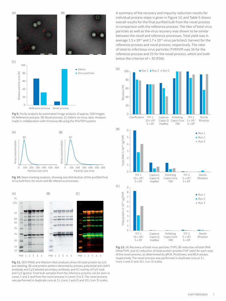
Chromatograms for the Capto Q ImpRes capture and Capto Core 700 polishing steps, together with corresponding chromatograms from a reference process, are shown in Figure 6. Results from analysis by analytical SEC show efficient adenovirus purification and impurity removal using the novel process (Fig 7). TEM analysis shows improved impurity removal, with a sensitivity for small debris that might not be possible using standard impurity analytics (Fig 8). Using the novel adenovirus purification process,
analysis of the TEM images showed relatively lower debris-to-virus particle ratio compared with the reference process (Fig 9). NTA data shows expected particle size and a narrower particle size distribution, suggesting less aggregation or virus-debris complexes with the novel process (Fig 10), and expected viral protein pattern was confirmed by SDS-PAGE analysis (Fig 11).
Fig 6. Chromatogram of adenovirus capture using (A) Capto Q ImpRes or (B) Q Sepharose XL, and polishing using (C) Capto Core 700 or (D) Sepharose Fast Flow. Adenovirus-containing fractions are indicated.
Fig 7. SEC-HPLC analysis of adenovirus purity of the novel process using a Superose 6 Increase column.
Fig 8. TEM images of final bulk samples from (A) reference process and (B) novel process. Analysis made in collaboration with Vironova AB using the MiniTEM system. The size bar corresponds to 500 nm.
(A)
(A)
(C) (D)
(B)
(B)
0
20
40
60
80
100
Clari�cation TFF 110 × UF/5 × DF
CaptureCapto QImpRes
PolishingCapto Core
700
TFF 25 × UF/5 × DF
Sterile�ltration
TFF 110 × UF/5 × DF
CaptureCapto QImpRes
PolishingCapto Core
700
TFF 25 × UF/5 × DF
Sterile�ltration
TFF 110 × UF/5 × DF
CaptureCapto QImpRes
PolishingCapto Core
700
TFF 25 × UF/5 × DF
Sterile�ltration
Run 1 Run 2 Run 3
Run 1Run 2Run 3
Run 1Run 2Run 3
Reco
very
(%)
0
1
2
3
4
5
6
0
1
2
3
4
5
6
7
8
9
Tota
l DN
A (×
10-0
9 ng/
TVP)
Tota
l pro
tein
(× 1
0-09 µ
g/TV
P)
1 2 3 4 5MW 1 2 3 4 5MW 1 2 3 4 5MW
225
97
66
50
35
25
20
14
10
Mr
Viru
s co
ncen
trat
ion
0 600500400300
89
Particle size (nm)200100
Viru
s co
ncen
trat
ion
0 600500400300
87
Particle size (nm)200100
Reference process Novel process
DebrisVirus particles
0
20
40
60
80
100
Rela
tive
part
icle
are
a (%
)
KA877080618AN 7
Fig 9. Purity analysis by automated image analysis of approx. 200 images. (A) Reference process. (B) Novel process. (C) Debris-to-virus ratio. Analysis made in collaboration with Vironova AB using the MiniTEM system.
Fig 10. Nano tracking analysis, showing size distribution of the purified final virus bulk from (A) novel and (B) reference processes.
Fig 11. SDS-PAGE and Western blot analyses show (A) total protein by Cy5 pre-labeling, (B) viral protein pattern detected by primary polyclonal anti-AdV5 antibody and Cy3 labeled secondary antibody, and (C) overlay of Cy5 (red) and Cy3 (green). Final bulk samples from the reference process can be seen in Lanes 1 and 2 and from the novel process in Lanes 3 to 5. The novel process was performed in duplicate runs at 3 L (runs 1 and 2) and 10 L (run 3) scales.
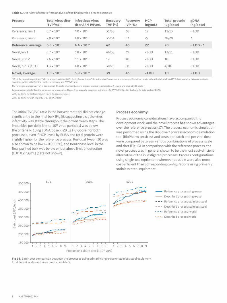
A summary of the recovery and impurity reduction results for individual process steps is given in Figure 12, and Table 5 shows overall results for the final purified bulk from the novel process in comparison with the reference process. The titer of total virus particles as well as the virus recovery was shown to be similar between the novel and reference processes. Total yield was in average 1.5 x 10¹³ and 1.7 x 10¹³ virus particles/L harvest for the reference process and novel process, respectively. The ratio of total-to-infectious virus particles (TVP/IVP) was 16 for the reference process and 25 for the novel process, which are both below the criterion of < 30 (FDA).
Fig 12. (A) Recovery of total virus particles (TVP), (B) reduction of total DNA (DNA/TVP), and (C) reduction of total protein (protein/TVP ratio) for each step of the novel process, as determined by qPCR, PicoGreen, and BCA assays, respectively. The novel process was performed in duplicate runs at 3 L (runs 1 and 2) and 10 L (run 3) scales.
(A)
(A)
(A)
(A)
(C)
(B)
(B)
(B)
(C)
(B)
(C)
150 000
200 000
250 000
300 000
350 000
400 000
450 000
500 000 50 L 200 L 500 L
Batc
h co
st (U
SD/b
atch
)
Production culture titer (× 1013 vp/L)
Reference process single-use
Described process single-use
Reference process stainless steel
Described process stainless steel
Reference process hybrid
Described process hybrid
1 98765432 1 98765432 1 98765432
8 KA877080618AN
The initial TVP/IVP ratio in the harvest material did not change significantly to the final bulk (Fig 5), suggesting that the virus infectivity was stable throughout the downstream steps. The impurities per dose (set to 1011 virus particles) was below the criteria (< 10 ng gDNA/dose, < 20 μg HCP/dose) for both processes, even if HCP levels by ELISA and total protein were slightly higher for the reference process. Residual Tween 20 was also shown to be low (~ 0.0005%), and Benzonase level in the final purified bulk was below or just above limit of detection (LOD 0.2 ng/mL) (data not shown).
Table 5. Overview of results from analysis of the final purified process samples
IVP = infectious virus particles, TVP= total virus particles, LOD= limit of detection, AFM = automated fluorescence microscopy. Disclaimer: analytical methods for IVP and TVP show variation between analysis occasions, which will affect the results for recovery and IVP/TVP ratio.
The reference process was run in duplicate at 3 L scale, whereas the novel process was run in duplicate at 3 L scale and once at 10 L scale.
Two numbers indicate that the same sample was analyzed twice (two separate occasions in triplicate for TVP [qPCR] and in duplicate for total protein [BCA]).
WHO guideline for protein impurity: max. 20 μg protein/dose
WHO guideline for DNA impurity: < 10 ng DNA/dose
Fig 13. Batch cost comparison between the processes using primarily single-use or stainless steel equipment for different scales and virus production titers.
Process economy
Process economic considerations have accompanied the development work, and the novel process has shown advantages over the reference process (17). The process economic simulation was performed using the BioSolve™ process economic simulation tool (BioPharm services), and costs per batch and per viral dose were compared between various combinations of process scale and titer (Fig 13). In comparison with the reference process, the novel process was in general shown to be the most cost-efficient alternative of the investigated processes. Process configurations using single-use equipment wherever possible were also more cost-efficient than corresponding configurations using primarily stainless-steel equipment.
KA877080618AN 9
DiscussionThe novel adenovirus production process ranges from selection of cell culture medium to purified recombinant adenovirus bulk product. The selected CDM4HEK293 medium support high viable HEK293.2sus cell densities and efficient adenovirus production in bioreactor cultures. Rocking bioreactor systems offer the benefits of low minimum working volumes that shorten the seed train prior to inoculation, and agitation can be adjusted to a smooth rocking motion that lowers the shear force for sensitive cells. The ReadyToProcess WAVE 25 bioreactor system is favorable in its simplicity for performing laboratory-scale production cultivations and process intensification in perfusion mode. Stirred-tank XDR bioreactor systems better support scale-up and manufacturing.
Optimization of the downstream purification steps is critical to maximize recovery of each step for an as high overall yield as possible. Factors such as sample load, binding, wash, and elution conditions are important for optimal performance of the capture and polishing steps. For a process with six steps, an average step recovery of 80% will give an overall process recovery of 26%, whereas an average step recovery of 90% will give an overall recovery of 53% for the process. A source of error is the analytic method variation or accuracy depending on sample composition differences (concentrations, impurity, and buffer components). Selection and careful optimization of the analytical methods are therefore also important factors for a successful outcome. The performance of the Biacore and IN Cell Analyzer assays were shown to be good for virus titer determination. These analysis methods were also more convenient to use compared with traditional methods.
Tween 20 showed good performance for the cell lysis step and was used as replacement for the traditionally used Triton™ X-100, now on the REACH list. Tween 20 did not affect the subsequent downstream step performance and was not shown to have a negative impact on virus infectivity (5).
DNA was shown to be the most challenging impurity to remove, and fragmentation with Benzonase needs to be maximized and could also be repeated if shown to be insufficient. However, to keep process costs down, the Benzonase amount used should be kept to a minimum. Some remaining longer DNA fragments are hard to avoid, and might be due to DNA-virus, DNA-protein, or DNA-debris complex formation that protects and makes the DNA less accessible for fragmentation by Benzonase. Careful optimization of the capture step, in this case using high-capacity Capto Q ImpRes resin with gradient elution, was shown to be beneficial for DNA removal.
The use of Capto Core 700 multimodal resin for the polishing step allowed for a 100-fold higher sample load compared with SEC, offering an increased scalability and efficiency.
The combination of steps used in the novel process was beneficial for the purity of the adenovirus in the final bulk. This is clearly demonstrated in the TEM images, showing much lower levels of small debris for the novel process than for the reference process.
The described adenovirus production process is compatible with both single-use as and steamable hard-piped process equipment. For an end-to-end single-use biomanufacturing solution, the ReadyToProcess WAVE 25 and XDR bioreactor systems can be combined with ÄKTA readyflux filtration system and ÄKTA ready chromatography systems, all with disposable flow paths. While ÄKTA readyflux is intended for tangential flow filtration applications in both upstream and downstream workflows, the ÄKTA ready systems operate prepacked and disposable ReadyToProcess columns with inner diameters from 80 to 450 mm for downstream purification of biomolecules from bioreactor culture volumes of 10–2000 L. For larger bioreactor volumes, ÄKTA ready XL can also operate user-packed AxiChrom™ columns with inner diameters of up to 1200 mm.
By omitting the need for cleaning-in-place, steaming-in-place operations, and associated validation procedures, single-use equipment allows more batches to be produced over a certain period of time, and changeover time between production batches or campaigns can be significantly reduced compared with using fixed stainless steel equipment. Single-use equipment also minimizes the cross-contamination risk between different production campaigns.
The modular and electronically controlled single-use FlexFactory™ biomanufacturing platform comprises single-use process equipment from upstream production to downstream purification. The systems are reconfigurable in days to develop and manufacture virtually any biologic-based product from virtually any host cell expression system. FlexFactory Automation monitors and controls the FlexFactory process equipment, ensuring consistency between productions. FlexFactory Automation is based on a combination of Wonderware™ (Schneider Electric) and UNICORN™ software, which can be used in a manner that complies with current good manufacturing practices (cGMP).
ConclusionThis work describes a modern, scalable process for production of adenovirus, with benefits in both virus purity and quality over a reference process. Both novel and established analytical methods were used for virus analysis, and results show that the process outcome meets regulatory requirements with respect to virus purity. The described process is compatible with single-use as well as with steamable hard-piped process equipment. Process economic calculations indicate advantages of the novel process over the reference process, especially for the single-use setup.
10 KA877080618AN
References1. Kallel, H. and Kamen, A.A. Large-scale adenovirus and poxvirus-vectored vaccine
manufacturing to enable clinical trials. Biotechnol J 10, 741–747 (2015).
2. Application note: Evaluation of HEK293 cell growth and adenovirus productivity in HyClone CDM4HEK293 medium. GE Healthcare, 29264715, Edition AA (2017).
3. Application note: Adenovirus production in single-use Xcellerex XDR-10 bioreactor system. GE Healthcare, KA874021017AN (2017).
4. Application note: Adenovirus production in single-use ReadyToProcess WAVE 25 bioreactor system. GE Healthcare, KA879160418AN (2018).
5. Application note: Optimization of midstream cell lysis and virus filtration steps in an adenovirus purification process. GE Healthcare, KA875220218AN (2018).
6. Application note: Downstream process development for efficient purification of adenovirus. GE Healthcare, KA876080618AN (2018).
7. Application note: Determination of adenovirus concentration using Biacore T200. GE Healthcare, KA878080618AN (2018).
8. Reece-Ford et al. Aspects of process development for virus vector production to improve quality and quantity. Pharmaceutical Technology Europe 20, 28–33 (2008).
9. Appaiahgari, M.B. and Vrati, S. Adenoviruses as gene/vaccine delivery vectors: promises and pitfalls. Expert Opin Biol Ther 15, 337-351 (2015).
10. Application note: Efficient, high-titer monoclonal antibody production in a fed-batch process using single-use stirred-tank and rocking bioreactor systems. GE Healthcare, 29119376, Edition AA (2014).
11. Application note: Engineering characterization of ReadyToProcess WAVE 25 bioreactor system with 20 L Cellbag culture chamber. GE Healthcare, 29206229, Edition AA (2016).
12. Application note: Engineering characterization of the single-use Xcellerex XDR-10 stirred-tank bioreactor system. GE Healthcare, 29242383, Edition AB (2017).
13. Application note: Engineering characterization of the single-use Xcellerex XDR-50 stirred-tank bioreactor system. GE Healthcare, 29241667, Edition AA (2017).
14. Application note: Engineering characterization of the single-use Xcellerex XDR-200 stirred-tank bioreactor system. GE Healthcare, 29268546, Edition AA (2017).
15. Application note: Engineering characterization of the single-use Xcellerex XDR-1000 stirred-tank bioreactor system. GE Healthcare, 29242384, Edition AA (2017).
16. Application note: Engineering characterization of the single-use Xcellerex XDR-2000 stirred-tank bioreactor system. GE Healthcare, 29243481, Edition AB (2017).
17. Application note: Process economic simulation for scalable production of adenovirus. GE Healthcare, KA3941080618AN (2018).
gelifesciences.com/bioprocessGE, the GE Monogram, ÄKTA, Amersham, AxiChrom, Biacore, Capto, Cellbag, Cy, CyDye, FlexFactory, HiScale, HyClone, ReadyCircuit, ReadyMate, ReadyToProcess, ReadyToProcess WAVE, Sepharose, Superose, Tricorn, Xcellerex, ULTA, and UNICORN are trademarks of General Electric Company.
Accuri is a trademark and brand of Becton, Dickinson and Company. Benzonase is a trademark of Merck KGaA. BioSolve is a trademark of Biomatrica, Inc. Gyrolab is a trademark of Gyros AB. MagMax and PrepSEQ are trademarks of Applied Biosystems, LLC. Oasis is a trademark of Waters Corp. MiniTEM is a trademark of Vironova AB. NanoSight is a trademark of Malvern. StepOnePlus is a trademark of Applied Biosystems, LLC. PureLink, PicoGreen, and Quant-iT are trademarks of Life Technologies Corp. TaqMan is a trademark of Roche Molecular Systems, Inc. Tween is a trademark of Croda Group of Companies. Triton is a trademark of Union Carbide Chemicals and Plastic Company Inc. Vi-CELL is a trademark of Beckman Coulter, Inc. Wonderware is a trademark of Schneider Electric Software.All other third-party trademarks are the property of their respective owners.
Cy5 is covered under US patent number 6,828,116 and equivalent patents and patent applications in other countries in the name of GE Healthcare UK Limited. The purchase of CyDye products includes a limited license to use the CyDye products for internal research and development but not for any commercial purposes. A license to use the Cy and CyDye trademarks for commercial purposes is subject to a separate license agreement with GE Healthcare. Commercial use shall include:1. Sale, lease, license or other transfer of the material or any material derived or produced from it.2. Sale, lease, license or other grant of rights to use this material or any material derived or produced from it.3. Use of this material to perform services for a fee for third parties, including contract research and drug screening. If you require a commercial license to use the Cy and CyDye trademarks, please contact [email protected].
ReadyMate is covered by US patent number 6,679,529 B2 owned by Johnson & Boley Holdings, LLC and licensed to GE Healthcare companies.
Any use of UNICORN software is subject to GE Healthcare Standard Software End-User License Agreement for Life Sciences Software Products. A copy of this Standard Software End-User License Agreement is available on request.
All goods and services are sold subject to the terms and conditions of sale of the company within GE Healthcare which supplies them. A copy of those terms and conditions is available on request. Contact your local GE Healthcare representative for the most current information.GE Healthcare Bio-Sciences AB, Björkgatan 30, 751 84 Uppsala, SwedenGE Healthcare Bio-Sciences Corp., 100 Results Way, Marlborough, MA 01752, USAGE Healthcare Europe GmbH, Munzinger Strasse 5, D-79111 Freiburg, GermanyGE Healthcare Japan Corp., Sanken Bldg., 3-25-1, Hyakunincho Shinjuku-ku, Tokyo 169-0073, JapanGE Healthcare UK Ltd., Amersham Place, Little Chalfont, Buckinghamshire, HP7 9NA, UKFor local office contact information, visit gelifesciences.com/contact.