Page 1
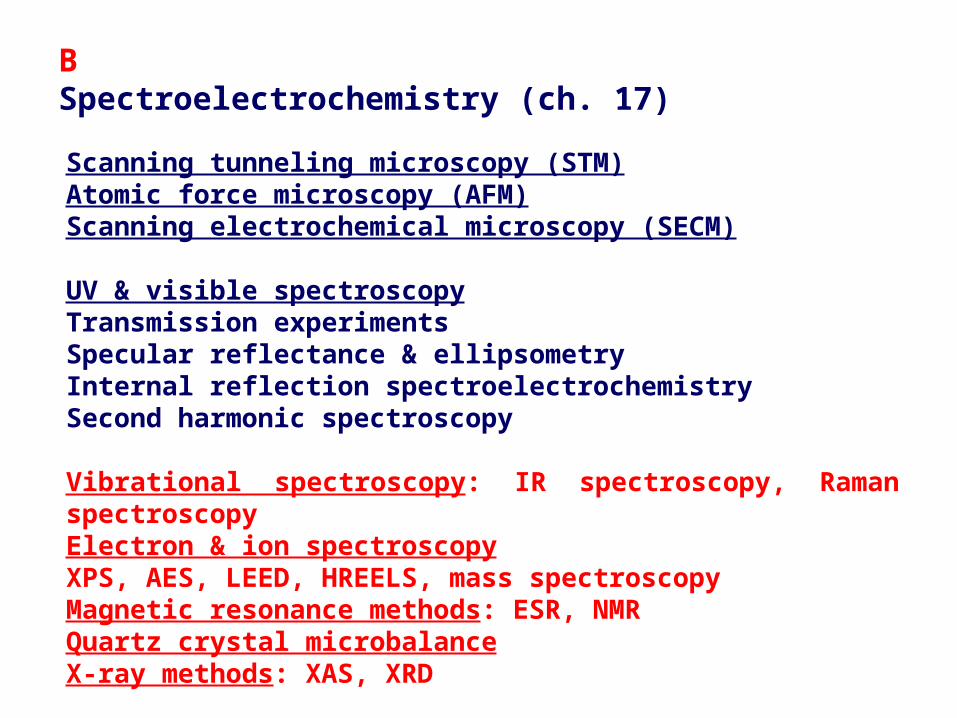
Scanning tunneling microscopy (STM)Atomic force microscopy (AFM)Scanning electrochemical microscopy (SECM)
UV & visible spectroscopyTransmission experimentsSpecular reflectance & ellipsometryInternal reflection spectroelectrochemistrySecond harmonic spectroscopy
Vibrational spectroscopy: IR spectroscopy, Raman spectroscopyElectron & ion spectroscopyXPS, AES, LEED, HREELS, mass spectroscopyMagnetic resonance methods: ESR, NMRQuartz crystal microbalanceX-ray methods: XAS, XRD
BSpectroelectrochemistry (ch. 17)
Page 2
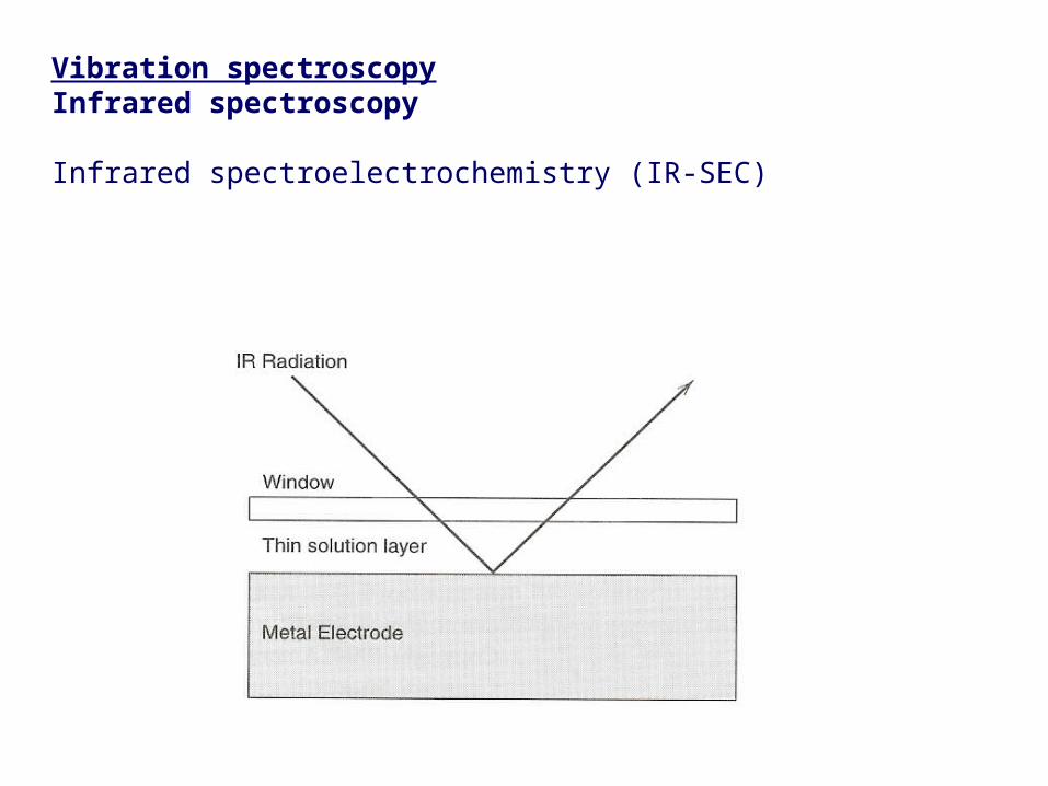
Vibration spectroscopyInfrared spectroscopy
Infrared spectroelectrochemistry (IR-SEC)
Page 4
EMIRS (electrochemically modulated infrared reflectance spectrosocpy)
Potential is modulated between one where the species of interest is absent & one where it is electrochemically generated
Page 5
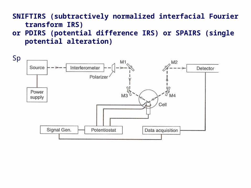
SNIFTIRS (subtractively normalized interfacial Fourier transform IRS)or PDIRS (potential difference IRS) or SPAIRS (single potential alteration)
Spectra obtained separately at two potentials → subtraction
Page 6
IRRAS (IR reflection absorption spectroscopy)
IR absorption at fixed potential
Page 7
SEIRA (surface enhanced IR absorption)
IR to study adsorbed species (reactants, intermediates, products)→ orientation & potential dependence of the adsorbed species
SNIFTIRS
Page 8
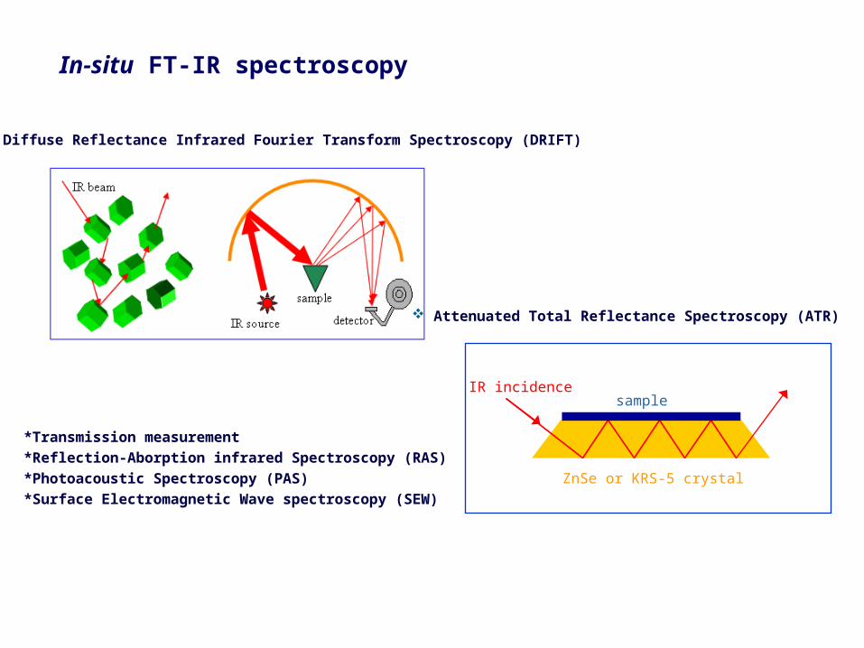
Attenuated Total Reflectance Spectroscopy (ATR)
Diffuse Reflectance Infrared Fourier Transform Spectroscopy (DRIFT)
IR incidencesample
ZnSe or KRS-5 crystal
*Transmission measurement
*Reflection-Aborption infrared Spectroscopy (RAS)
*Photoacoustic Spectroscopy (PAS)
*Surface Electromagnetic Wave spectroscopy (SEW)
In-situ FT-IR spectroscopy
Page 9
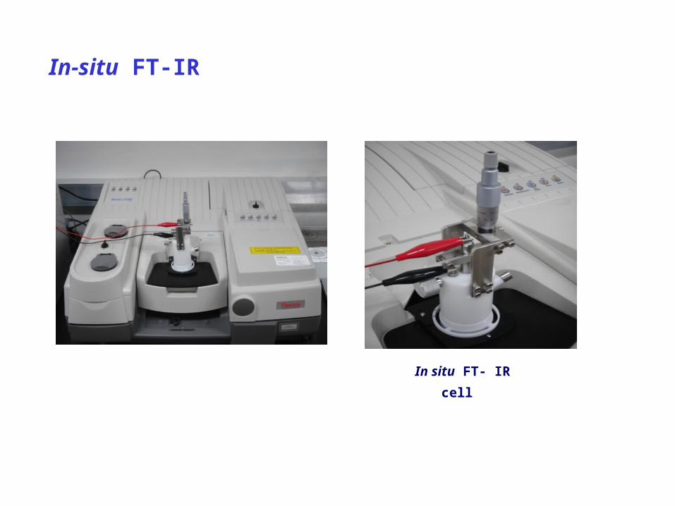
In-situ FT-IR
In situ FT- IR cell
Page 10
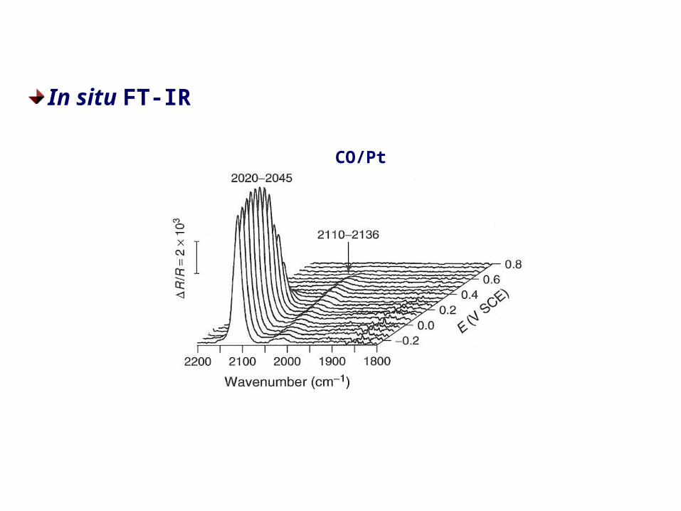
In situ FT-IR
CO/Pt
Page 11
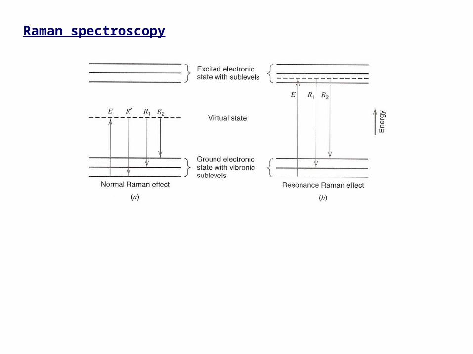
Raman spectroscopy
Page 12
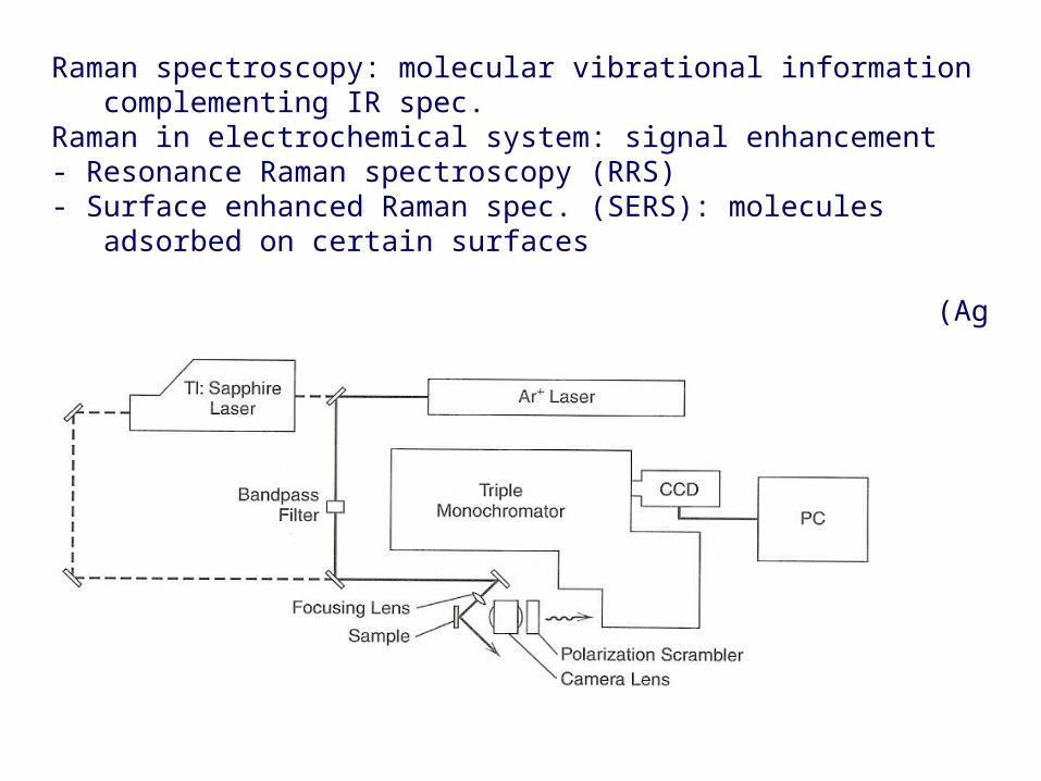
Raman spectroscopy: molecular vibrational information complementing IR spec.Raman in electrochemical system: signal enhancement- Resonance Raman spectroscopy (RRS)- Surface enhanced Raman spec. (SERS): molecules adsorbed on certain surfaces (Ag or Au)
Page 13
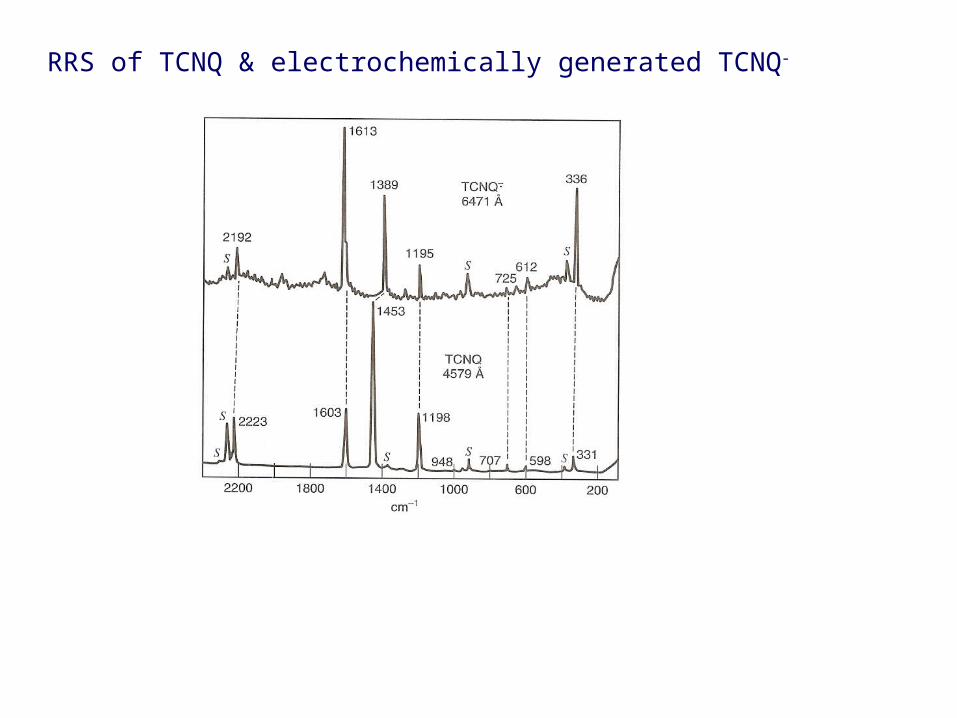
RRS of TCNQ & electrochemically generated TCNQ-
Page 14
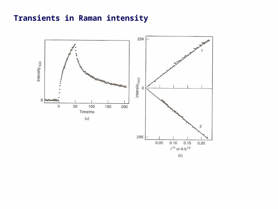
Transients in Raman intensity
Page 15
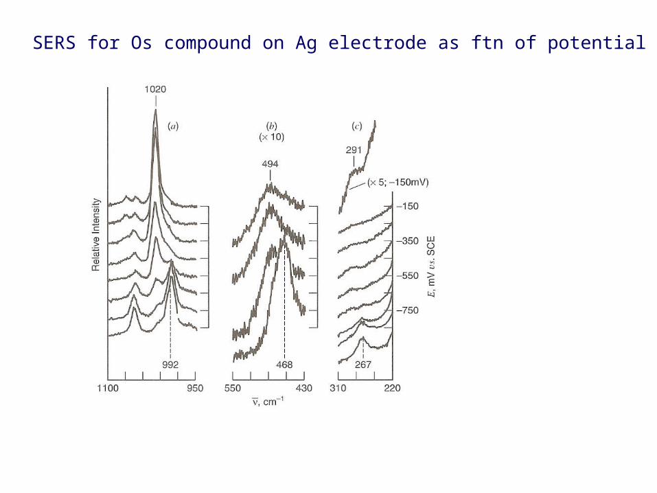
SERS for Os compound on Ag electrode as ftn of potential
Page 16
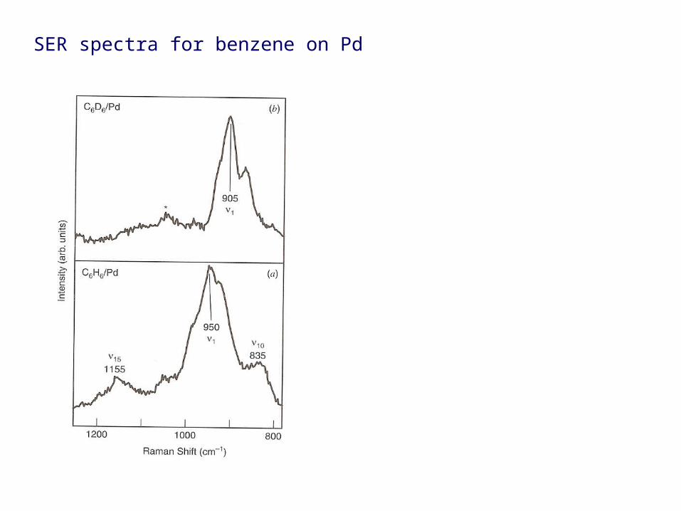
SER spectra for benzene on Pd
Page 17
In-situ Raman Spectroscopy
Page 18
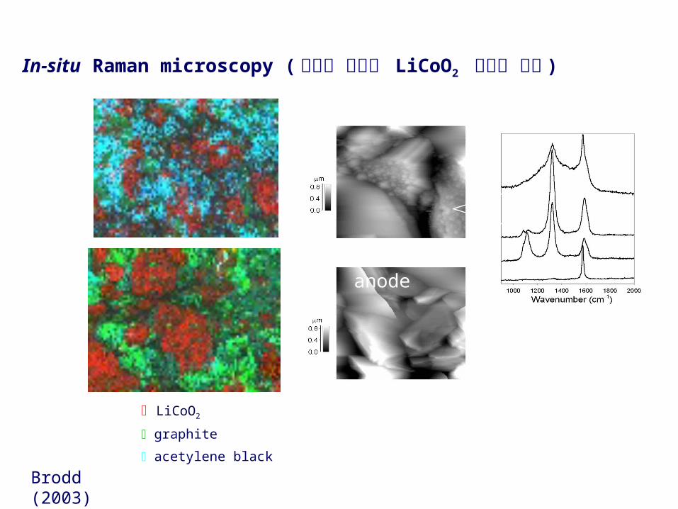
In-situ Raman microscopy ( 탄소재 음극과 LiCoO2 양극재 분석 )
LiCoO2
graphite
acetylene black
Fresh anode
Brodd (2003)
25oC
60oC
Page 19
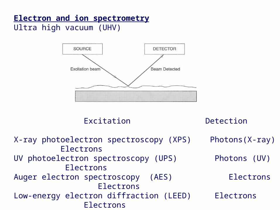
Electron and ion spectrometryUltra high vacuum (UHV)
Excitation Detection
X-ray photoelectron spectroscopy (XPS) Photons(X-ray) ElectronsUV photoelectron spectroscopy (UPS) Photons (UV) ElectronsAuger electron spectroscopy (AES) Electrons ElectronsLow-energy electron diffraction (LEED) Electrons ElectronsHigh resolution e- E loss spec. (HREELS) Electrons ElectronsRutherford backscattering (RBS) H+ or He+ H+ or He+Secondary ion mass spec. (SIMS) Ions IonsLaser desorption mass spec. (LDMS) Photons Ions
Page 20
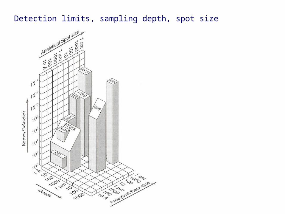
Detection limits, sampling depth, spot size (spatial resolution)
Page 21
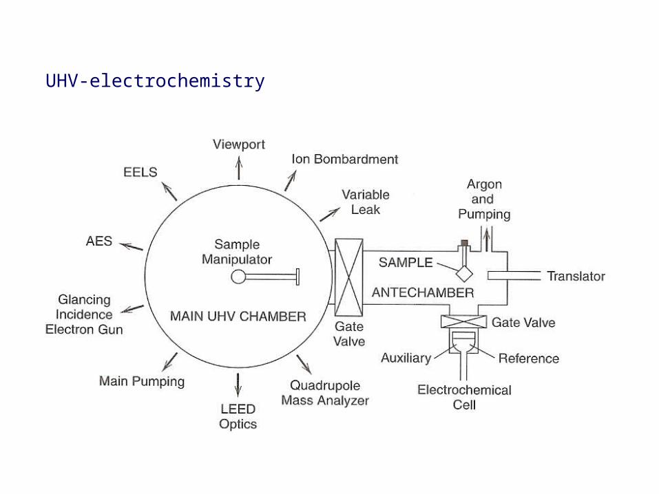
UHV-electrochemistry
Page 22
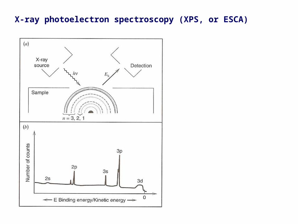
X-ray photoelectron spectroscopy (XPS, or ESCA)
Page 23
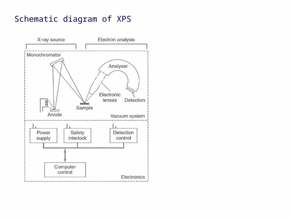
Schematic diagram of XPS
Page 24
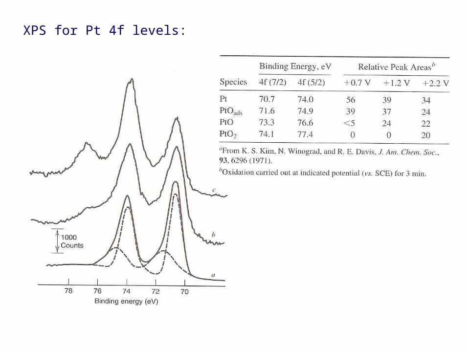
XPS for Pt 4f levels:
Page 25
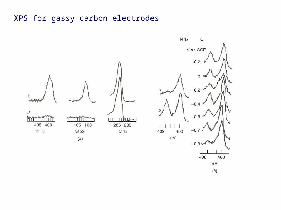
XPS for gassy carbon electrodes
Page 26
XPS for copper electrodeposition(a) Bulk Cu (b) Cu UPD
Page 27
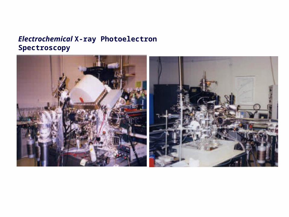
Electrochemical X-ray Photoelectron Spectroscopy
Univ. of Illinois
Page 28
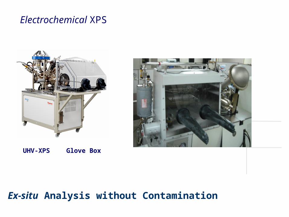
Ex-situ Analysis without Contamination
Glove BoxUHV-XPS
Electrochemical XPS
Page 29
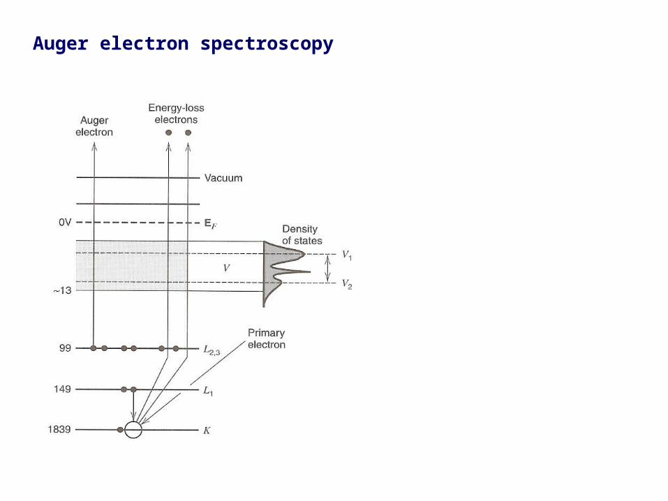
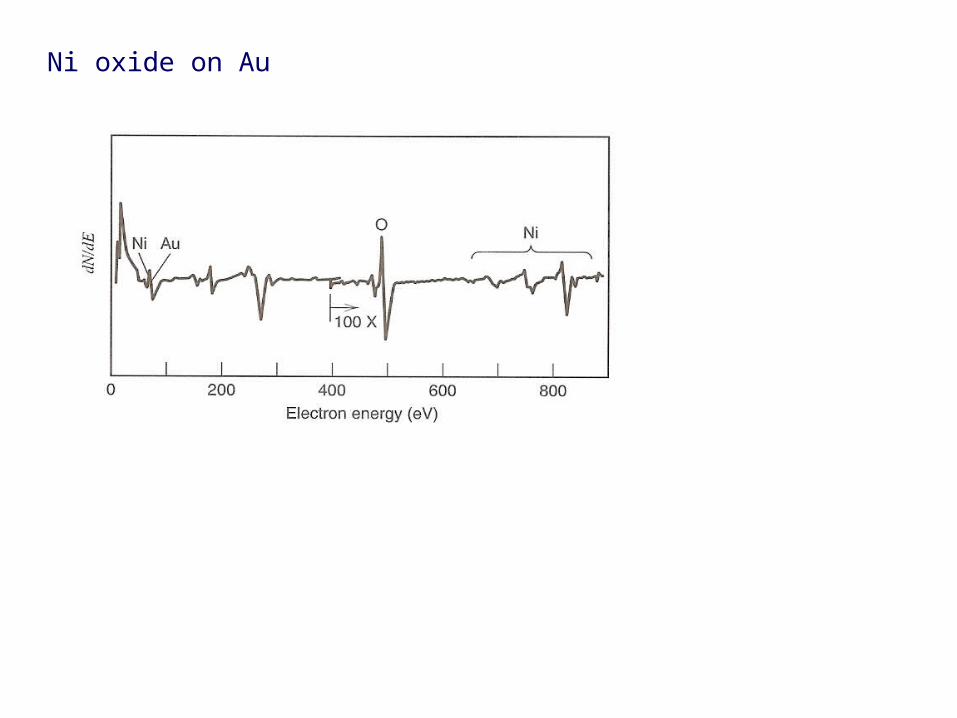
Auger electron spectroscopy
Page 31
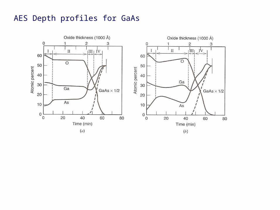
AES Depth profiles for GaAs
Page 32
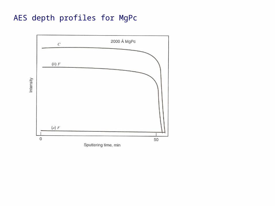
AES depth profiles for MgPc
Page 33
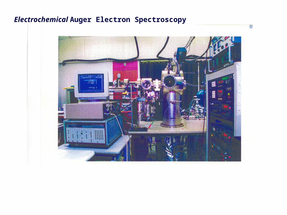
Electrochemical Auger Electron Spectroscopy
Page 34
Low-energy electron diffraction
Page 35
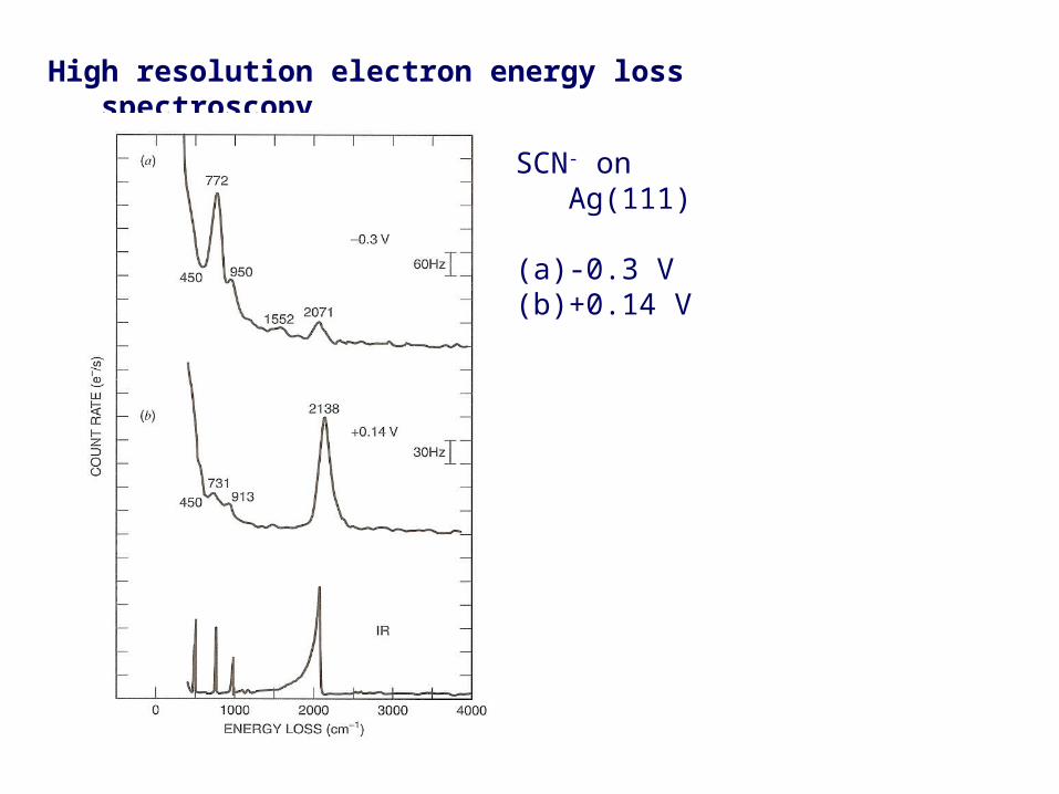
High resolution electron energy loss spectroscopy
SCN- on Ag(111)
(a) -0.3 V(b) +0.14 V
Page 36
Mass spectrometry
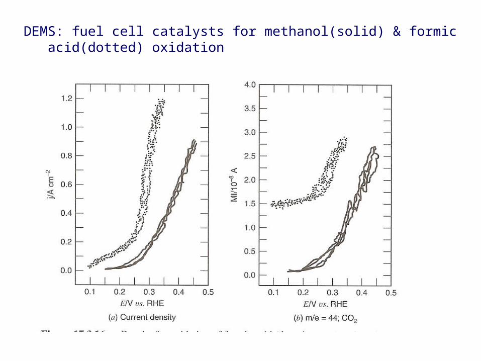
Differential electrochemical mass spectrometry (DEMS)
Page 37
DEMS: fuel cell catalysts for methanol(solid) & formic acid(dotted) oxidation
Page 38
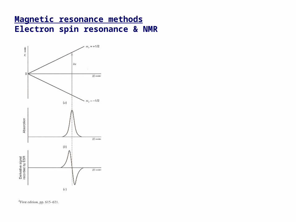
Magnetic resonance methodsElectron spin resonance & NMR
Page 39
Electrochemical ESR
Page 40
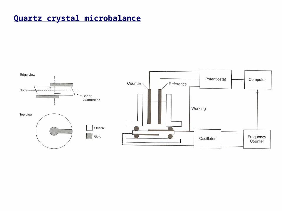
Quartz crystal microbalance
Page 42
X-ray methodsSynchrotron
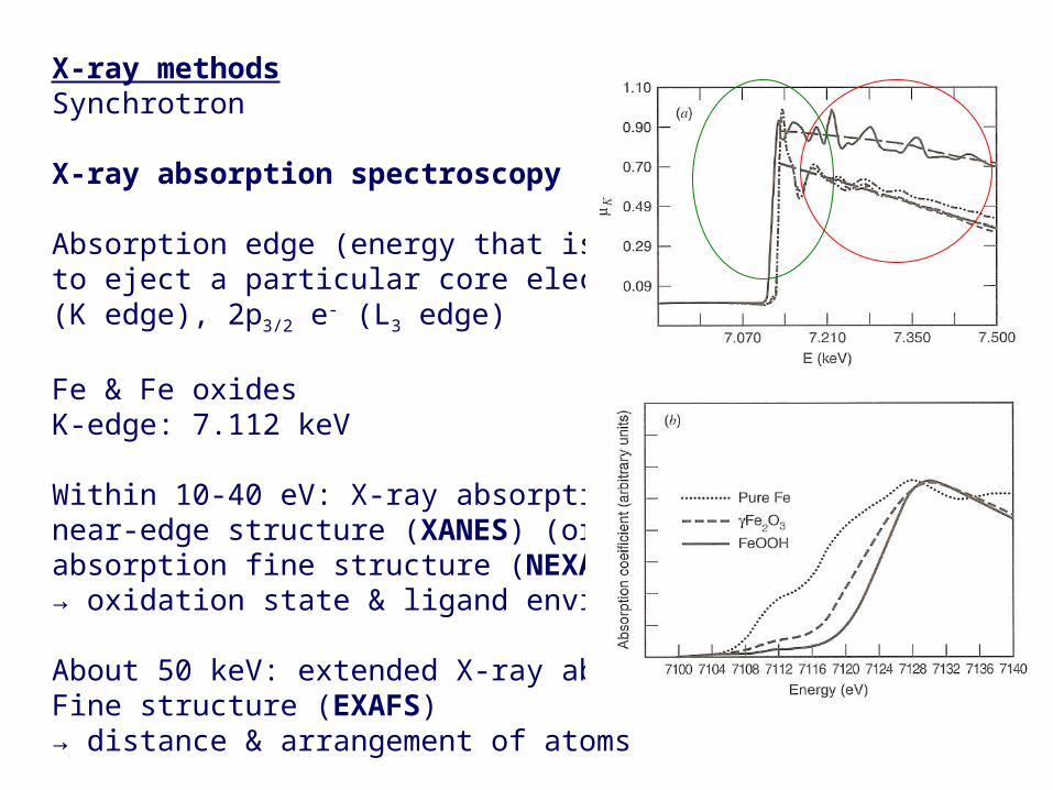
X-ray absorption spectroscopy
Absorption edge (energy that is just needed to eject a particular core electron, e.g., 1s e-
(K edge), 2p3/2 e- (L3 edge)
Fe & Fe oxides K-edge: 7.112 keV
Within 10-40 eV: X-ray absorptionnear-edge structure (XANES) (or near-edgeabsorption fine structure (NEXAFS))→ oxidation state & ligand envirionment
About 50 keV: extended X-ray absorptionFine structure (EXAFS) → distance & arrangement of atoms
Page 43
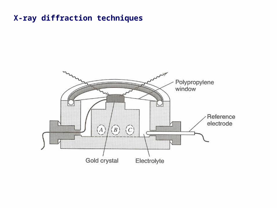
X-ray diffraction techniques
Page 44
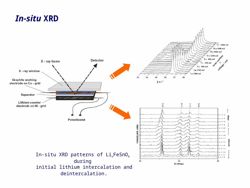
In-situ XRD
In-situ XRD patterns of LixFeSnO4 during
initial lithium intercalation and deintercalation.