Page 1
Dr.saad fawwaz
College of medicine
Al-Anbar University
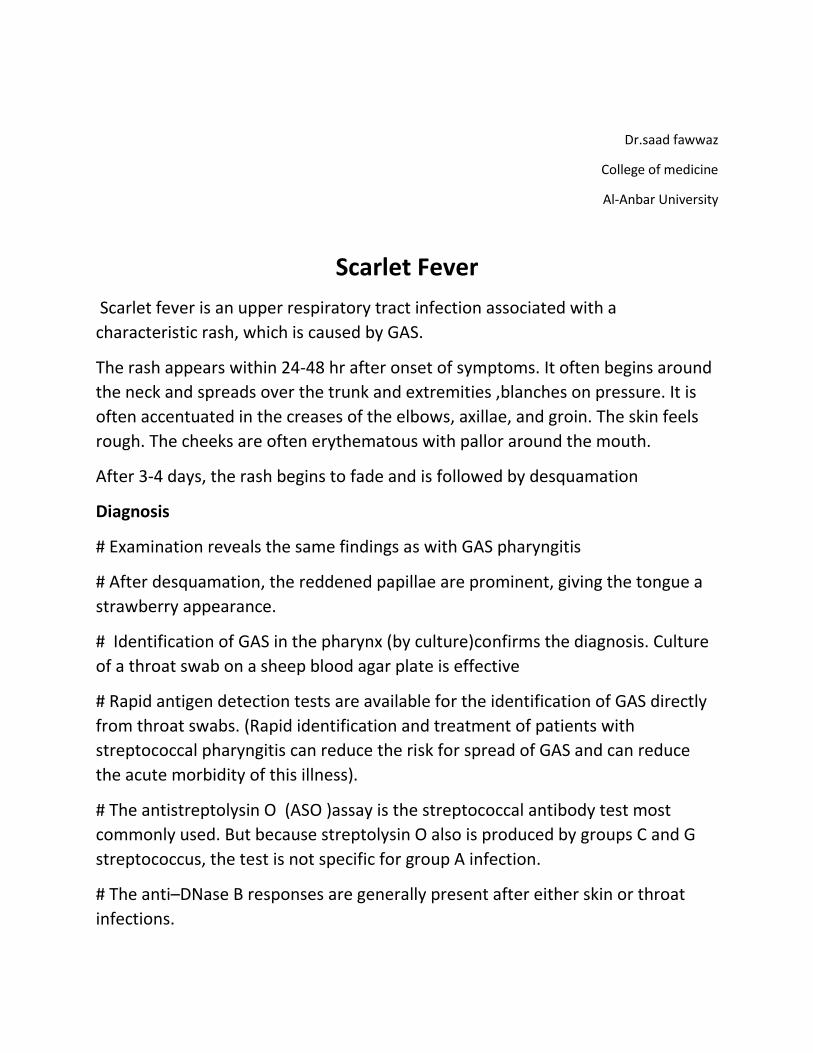
Scarlet Fever
Scarlet fever is an upper respiratory tract infection associated with a
characteristic rash, which is caused by GAS.
The rash appears within 24-48 hr after onset of symptoms. It often begins around
the neck and spreads over the trunk and extremities ,blanches on pressure. It is
often accentuated in the creases of the elbows, axillae, and groin. The skin feels
rough. The cheeks are often erythematous with pallor around the mouth.
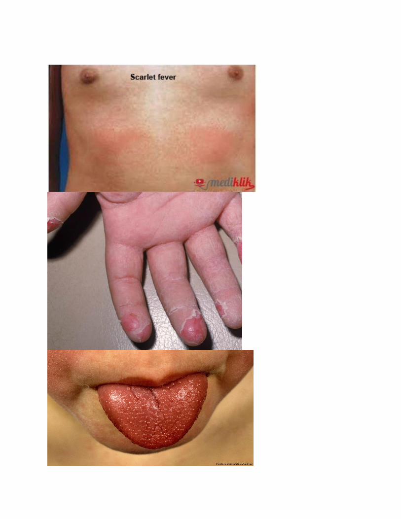
After 3-4 days, the rash begins to fade and is followed by desquamation
Diagnosis
# Examination reveals the same findings as with GAS pharyngitis
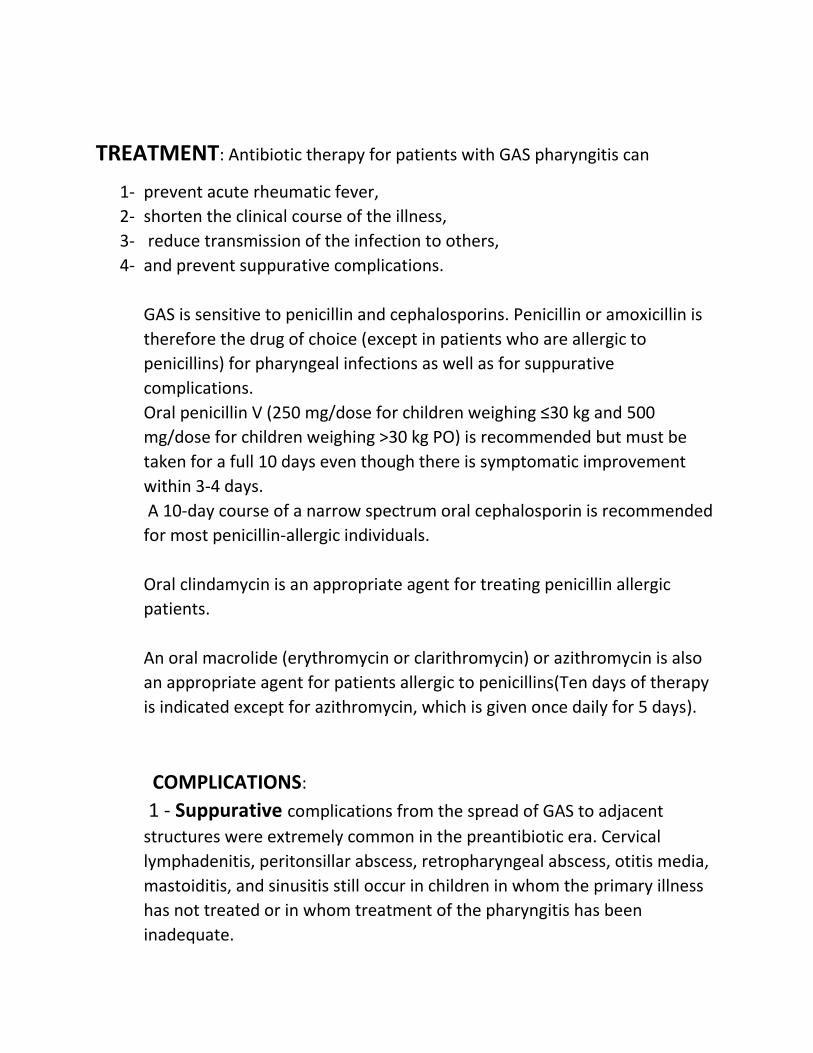
# After desquamation, the reddened papillae are prominent, giving the tongue a
strawberry appearance.
# Identification of GAS in the pharynx (by culture)confirms the diagnosis. Culture
of a throat swab on a sheep blood agar plate is effective
# Rapid antigen detection tests are available for the identification of GAS directly
from throat swabs. (Rapid identification and treatment of patients with
streptococcal pharyngitis can reduce the risk for spread of GAS and can reduce
the acute morbidity of this illness).
# The antistreptolysin O (ASO )assay is the streptococcal antibody test most
commonly used. But because streptolysin O also is produced by groups C and G
streptococcus, the test is not specific for group A infection.
# The anti–DNase B responses are generally present after either skin or throat
infections.
Page 3
TREATMENT: Antibiotic therapy for patients with GAS pharyngitis can
1- prevent acute rheumatic fever,
2- shorten the clinical course of the illness,
3- reduce transmission of the infection to others,
4- and prevent suppurative complications.
GAS is sensitive to penicillin and cephalosporins. Penicillin or amoxicillin is
therefore the drug of choice (except in patients who are allergic to
penicillins) for pharyngeal infections as well as for suppurative
complications.
Oral penicillin V (250 mg/dose for children weighing ≤30 kg and 500
mg/dose for children weighing >30 kg PO) is recommended but must be
taken for a full 10 days even though there is symptomatic improvement
within 3-4 days.
A 10-day course of a narrow spectrum oral cephalosporin is recommended
for most penicillin-allergic individuals.
Oral clindamycin is an appropriate agent for treating penicillin allergic
patients.
An oral macrolide (erythromycin or clarithromycin) or azithromycin is also
an appropriate agent for patients allergic to penicillins(Ten days of therapy
is indicated except for azithromycin, which is given once daily for 5 days).
COMPLICATIONS:
1 - Suppurative complications from the spread of GAS to adjacent
structures were extremely common in the preantibiotic era. Cervical
lymphadenitis, peritonsillar abscess, retropharyngeal abscess, otitis media,
mastoiditis, and sinusitis still occur in children in whom the primary illness
has not treated or in whom treatment of the pharyngitis has been
inadequate.
Page 4
GAS pneumonia can also occur.
2 -Nonsuppurative; Acute rheumatic fever and acute poststreptococcal
glomerulonephritis ((They are both characterized by disease remote from
the site of the primary GAS infection)). Both differ in their clinical
manifestations, epidemiology, and potential morbidity. In addition, ((acute
glomerulonephritis follows a GAS infection of either the upper respiratory
tract or the skin, but acute rheumatic fever only follows an infection of
the upper respiratory tract.))
PROGNOSIS: The prognosis for appropriately treated GAS pharyngitis is
excellent, and complete recovery is the rule. When therapy is instituted
acute rheumatic fever is almost always prevented.
PREVENTION: The only specific indication for long-term use of an antibiotic
to prevent GAS infections is for patients with a history of acute rheumatic
fever and/or rheumatic heart disease. Several vaccines are in
development, including a 30-valent M protein-based recombinant vaccine.
These vaccines are in relatively early stages of development.
Parvoviruses B19
The parvoviruses are small, single-stranded DNA viruses. They are common
infectious agents of a variety of animal species. There are now 4 different types of
parvoviruses known to infect humans. B19 is the most clinically important of the
human parvoviruses and the cause of erythema infectiosum or fifth disease.
Transmission of B19 is by the respiratory route, presumably via nasopharyngeal
viral shedding. Although respiratory spread is the primary mode of transmission,
B19 is also transmissible in blood and blood products.
Page 5
PATHOGENESIS: The primary target of B19 infection is the erythroid cell line,.
Viral infection produces cell lysis, leading a transient arrest of erythropoiesis.
From 7-11 days after inoculation, subjects had viremia and nasopharyngeal viral
shedding with fever, malaise, and rhinorrhea. Reticulocyte counts dropped to
undetectable levels but resulted in only a mild, clinically insignificant fall in serum
hemoglobin. Some manifestations of B19 infection, such as transient aplastic
crisis, appear to be a direct result of viral infection, whereas others, including
the exanthem and arthritis, appear to be postinfectious phenomena related to
the immune response.
Humoral immunity is crucial in controlling infection. Specific immunoglobulin (Ig)
M appears within 1-2 days of infection and is followed by anti-B19 IgG, which
leads to control of the infection, restoration of reticulocytosis, and a rise in serum
hemoglobin. Individuals with impaired humoral immunity are at increased risk
for more serious or persistent infection with B19, which usually manifests as
chronic RBC aplasia
B19 can cross the placenta and cause fetal infection during primary maternal
infection.
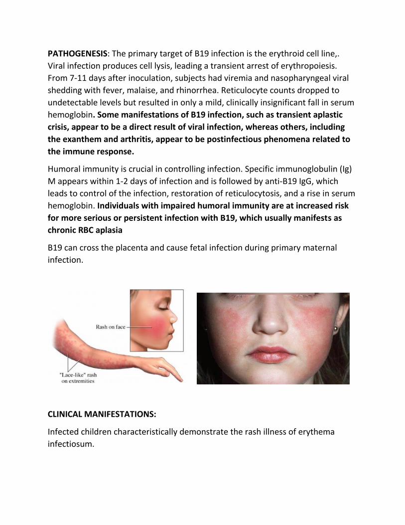
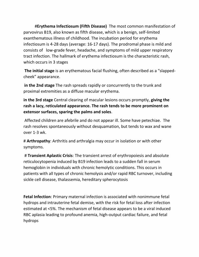
CLINICAL MANIFESTATIONS:
Infected children characteristically demonstrate the rash illness of erythema
infectiosum.
Page 6
#Erythema Infectiosum (Fifth Disease) The most common manifestation of
parvovirus B19, also known as fifth disease, which is a benign, self-limited
exanthematous illness of childhood. The incubation period for erythema
infectiosum is 4-28 days (average: 16-17 days). The prodromal phase is mild and
consists of low-grade fever, headache, and symptoms of mild upper respiratory
tract infection. The hallmark of erythema infectiosum is the characteristic rash,
which occurs in 3 stages
The initial stage is an erythematous facial flushing, often described as a “slapped-
cheek” appearance.
in the 2nd stage The rash spreads rapidly or concurrently to the trunk and
proximal extremities as a diffuse macular erythema.
in the 3rd stage Central clearing of macular lesions occurs promptly, giving the
rash a lacy, reticulated appearance. The rash tends to be more prominent on
extensor surfaces, sparing the palms and soles.
Affected children are afebrile and do not appear ill. Some have petechiae. The
rash resolves spontaneously without desquamation, but tends to wax and wane
over 1-3 wk.
# Arthropathy: Arthritis and arthralgia may occur in isolation or with other
symptoms.
# Transient Aplastic Crisis: The transient arrest of erythropoiesis and absolute
reticulocytopenia induced by B19 infection leads to a sudden fall in serum
hemoglobin in individuals with chronic hemolytic conditions. This occurs in
patients with all types of chronic hemolysis and/or rapid RBC turnover, including
sickle cell disease, thalassemia, hereditary spherocytosis
Fetal Infection: Primary maternal infection is associated with nonimmune fetal
hydrops and intrauterine fetal demise, with the risk for fetal loss after infection
estimated at <5%. The mechanism of fetal disease appears to be a viral induced
RBC aplasia leading to profound anemia, high-output cardiac failure, and fetal
hydrops
Page 7
. Most infants infected in utero are born normally at term, including some who
have had ultrasonographic evidence of hydrops.
DIAGNOSIS: The diagnosis of (erythema infectiosum) and (transient aplastic
crisis in achild with sickle cell disease) is generally made on clinical grounds
without specific virologic testing.
Determination of anti-B19 IgM is the best marker of recent/acute infection .
(Serologic diagnosis is unreliable in immunocompromised persons; diagnosis in
these patients requires methods to detect viral DNA).
TREATMENT: There is no specific antiviral therapy for B19 infection. Commercial
intravenous immunoglobulin (IVIG) have been used with some success to treat
B19-related episodes of anemia and bone marrow failure in immunocompromised
children.
B19-infected fetuses with anemia and hydrops have been managed successfully
with intrauterine RBC transfusions.
COMPLICATIONS: Erythema infectiosum is often accompanied by arthralgias or
arthritis in adolescents and adults that may persist after resolution of the rash.
The incidence of stroke may be increased in children with sickle cell disease
following B19-induced transient aplastic crisis.
Page 8
Diphtheria (Corynebacterium diphtheriae)
Diphtheria is an acute toxic infection caused Corynebacterium diphtheriae.
Although diphtheria was reduced from a major cause of childhood death to a
medical rarity. Recurring cases emphasize the necessity to continue control
principles across the global community.
ETIOLOGY: Corynebacteria are aerobic, nonencapsulated, non–spore-forming,
mostly nonmotile, Gram-positive bacilli .
EPIDEMIOLOGY: Spread is primarily by airborne respiratory droplets, direct
contact with respiratory secretions of symptomatic individuals, or exudate from
infected skin lesions. Skin infection and skin carriage are silent reservoirs of C.
diphtheriae, and organisms can remain viable in dust or on fomites for up to 6
mo. Cutaneous diphtheria: This indolent local infection, compared with mucosal
infection, is associated with more prolonged bacterial shedding, greater
contamination of the environment, and increased transmission to the pharynx
and skin of close contacts.
PATHOGENESIS: The organism usually remains in the superficial layers of skin
lesions or respiratory tract mucosa, inducing local inflammatory reaction, causes
local tissue necrosis. Within the 1st few days of respiratory tractinfection (usually
in the pharynx), a dense necrotic coagulum of organisms, epithelial cells, fibrin,
leukocytes, and erythrocytes forms a gray-brown, leatherlike adherent
pseudomembrane (Diphthera is Greek for leather). Removal is difficult and
reveals a bleeding edematous submucosa. Paralysis of the palate and
hypopharynx is an early local effect of diphtheritic toxin. Toxin absorption can
lead to systemic manifestations: kidney tubule necrosis, thrombocytopenia,
cardiomyopathy, and/or demyelination of nerves( Because the latter 2
complications can occur 2-10 wk after mucocutaneous infection, the
pathophysiology in some cases is suspected to be immunologically mediated)
Page 9
CLINICAL MANIFESTATIONS:
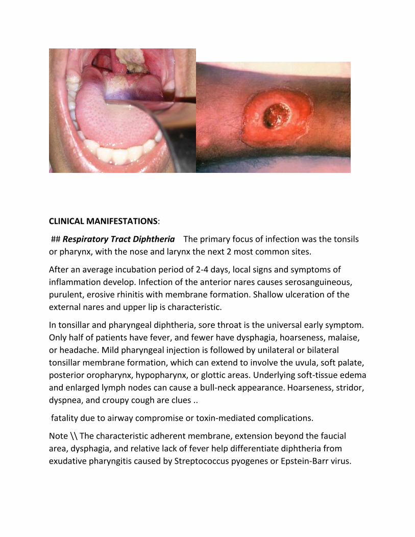
## Respiratory Tract Diphtheria The primary focus of infection was the tonsils
or pharynx, with the nose and larynx the next 2 most common sites.
After an average incubation period of 2-4 days, local signs and symptoms of
inflammation develop. Infection of the anterior nares causes serosanguineous,
purulent, erosive rhinitis with membrane formation. Shallow ulceration of the
external nares and upper lip is characteristic.
In tonsillar and pharyngeal diphtheria, sore throat is the universal early symptom.
Only half of patients have fever, and fewer have dysphagia, hoarseness, malaise,
or headache. Mild pharyngeal injection is followed by unilateral or bilateral
tonsillar membrane formation, which can extend to involve the uvula, soft palate,
posterior oropharynx, hypopharynx, or glottic areas. Underlying soft-tissue edema
and enlarged lymph nodes can cause a bull-neck appearance. Hoarseness, stridor,
dyspnea, and croupy cough are clues ..
fatality due to airway compromise or toxin-mediated complications.
Note \\ The characteristic adherent membrane, extension beyond the faucial
area, dysphagia, and relative lack of fever help differentiate diphtheria from
exudative pharyngitis caused by Streptococcus pyogenes or Epstein-Barr virus.
Page 10
Patients with laryngeal diphtheria are at significant risk for suffocation because
of local soft-tissue edema and airway obstruction by the diphtheritic membrane,.
Establishment of an artificial airway and resection of the pseudomembrane can
be lifesaving.
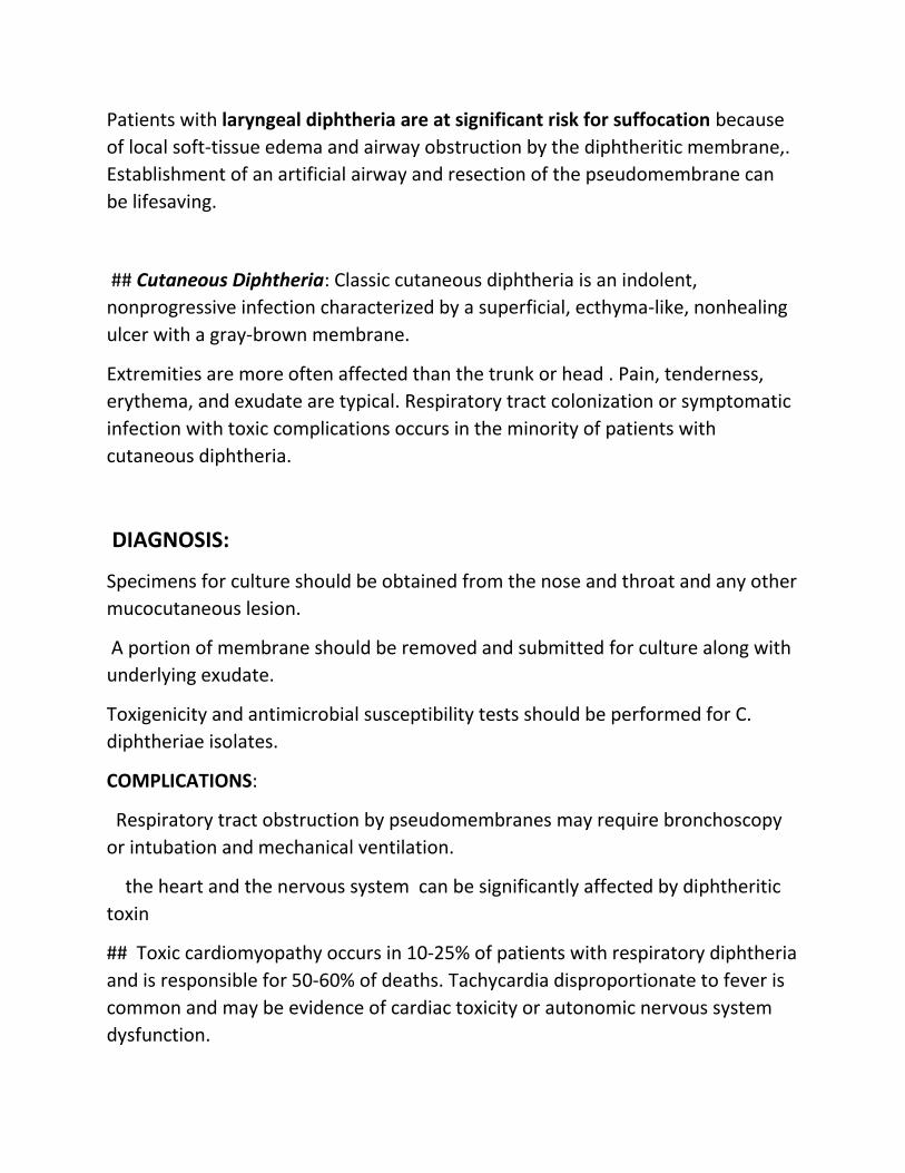
## Cutaneous Diphtheria: Classic cutaneous diphtheria is an indolent,
nonprogressive infection characterized by a superficial, ecthyma-like, nonhealing
ulcer with a gray-brown membrane.
Extremities are more often affected than the trunk or head . Pain, tenderness,
erythema, and exudate are typical. Respiratory tract colonization or symptomatic
infection with toxic complications occurs in the minority of patients with
cutaneous diphtheria.
DIAGNOSIS:
Specimens for culture should be obtained from the nose and throat and any other
mucocutaneous lesion.
A portion of membrane should be removed and submitted for culture along with
underlying exudate.
Toxigenicity and antimicrobial susceptibility tests should be performed for C.
diphtheriae isolates.
COMPLICATIONS:
Respiratory tract obstruction by pseudomembranes may require bronchoscopy
or intubation and mechanical ventilation.
the heart and the nervous system can be significantly affected by diphtheritic
toxin
## Toxic cardiomyopathy occurs in 10-25% of patients with respiratory diphtheria
and is responsible for 50-60% of deaths. Tachycardia disproportionate to fever is
common and may be evidence of cardiac toxicity or autonomic nervous system
dysfunction.
Page 11
## Toxic Neuropathy: Acutely or 2-3 wk after onset of oropharyngeal
inflammation, it is common for local paralysis of the soft palate to occur.
Weakness of the posterior pharyngeal, laryngeal, and facial nerves may follow,
causing a nasal quality in the voice, difficulty in swallowing, and risk for aspiration.
Cranial neuropathies characteristically occur in the 5th wk, leading to oculomotor
and ciliary paralysis, which can cause strabismus, blurred vision, or difficulty with
accommodation.
Symmetric polyneuropathy has its onset 10 days to 3 mo after oropharyngeal
infection and causes principally motor deficits with diminished deep tendon
reflexes. muscle
Recovery from the myocarditis and neuritis is often slow but usually complete.
TREATMENT:
Specific antitoxin is the mainstay of therapy and should be administered on the
basis of clinical diagnosis.
Equine diphtheria antitoxin is available. Antitoxin is administered as a single
empirical dose of 20,000-100,000 units based on the degree of toxicity, site and
size of the membrane, and duration of illness.
# Antitoxin is probably of no value for local manifestations of cutaneous
# diphtheria. Antitoxin is not recommended for asymptomatic carriers.
The role of antimicrobial therapy is to
1-halt toxin production,
2-treat localized infection,
3- prevent transmission of the organism to contacts.
Appropriate therapy is
Page 12
## erythromycin (40-50 mg/kg/day divided every 6 hr by mouth [PO] Or
intravenously [IV]; maximum 2 g/day),
## aqueous crystalline penicillin G (100,000-150,000 units/kg/day divided every 6
hr IV or intramuscularly [IM]), or daily procaine penicillin (300,000 units/day IM
for those <10 kg in weight; 600,000 units/day IM for those >10 kg in weight) for
14 days.
Elimination of the organism should be documented by negative results of at least
2 successive cultures of specimens from the nose and throat (or skin) obtained 24
hr apart after completion of therapy.
Epstein-Barr Virus
Infectious mononucleosis is the best-known clinical syndrome caused by Epstein-
Barr virus (EBV). It is characterized by fatigue, malaise, fever, sore throat, and
generalized lymphadenopathy.
Originally described as glandular fever, it derives its name from the mononuclear
lymphocytosis with atypical-appearing lymphocytes that accompany the illness.
ETIOLOGY: EBV is a double-stranded DNA virus .Two distinct types of EBV, type 1
and type 2. EBV-1 is more prevalent worldwide .
Page 13
CLINICAL MANIFESTATIONS:
# The incubation period of infectious mononucleosis in adolescents is 30-50 days.
In children, it may be shorter.
# In older patients, the onset of illness is usually insidious and vague. Patients may
complain of malaise, fatigue, acute or prolonged (>1 wk) fever, headache, sore
throat, nausea, abdominal pain, and myalgia.
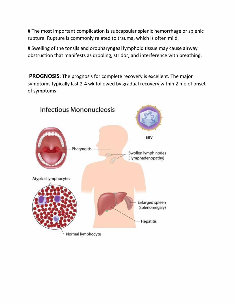
# The classic physical examination findings are generalized lymphadenopathy
(90% of cases), splenomegaly (50% of cases), and hepatomegaly (10% of cases).
# Lymphadenopathy occurs most commonly in the anterior and posterior cervical
nodes and the submandibular lymph nodes and less commonly in the axillary and
inguinal lymph nodes.Epitrochlear lymphadenopathy is particularly suggestive of
infectious mononucleosis.
# Symptomatic hepatitis or jaundice is uncommon, but elevated liver enzymes are
very common.
# Splenomegaly to 2-3 cm below the costal margin is typical (15-65% of cases) and
is seen in most cases by ultrasonography; massive enlargement is uncommon.
# The sore throat is often accompanied by moderate to severe pharyngitis with
marked tonsillar enlargement.
# Palatal petechiae at the junction of the hard and soft palate are frequently seen.
#. Patients with infectious mononucleosis treated with ampicillin or amoxicillin
may experience “ampicillin rash,”. This morbilliform, vasculitic rash is probably
immune mediated and resolves without specific treatment.
DIAGNOSIS: by the presence of typical clinical symptoms with atypical
lymphocytosis in the peripheral blood. The diagnosis is usually confirmed by
serologic testing, either for heterophile antibody or specific EBV antibodies.
Page 14
Laboratory Tests:
#In >90% of cases there is leukocytosis of 10,000-20,000 cells/μL, of which at least
two thirds are lymphocytes
#; atypical lymphocytes usually account for 20-40% of the total number.
# Mild thrombocytopenia to 50,000-200,000 platelets/μL occurs in more than
50% of patients.
#Mild elevation of hepatic transaminases occurs in approximately 50% of
uncomplicated cases, but is usually asymptomatic without jaundice.
# Heterophile Antibody Test: also known as Paul-Bunnell antibodies, are IgM
antibodies. The heterophile antibodies of infectious mononucleosis agglutinate
sheep or, for greater sensitivity, horse red cells. Heterophile antibody tests are
positive in 75% of cases in the 1st wk and 90-95% of cases in the 2nd wk .
TREATMENT:
## There is no specific treatment for infectious mononucleosis.
## The mainstays of management are rest, encouraging adequate fluid and
nutrition intake, and symptomatic treatment with acetaminophen or nonsteroidal
anti inflammatory agents to manage fever, throat discomfort, and malaise.
## Short courses of corticosteroids may be helpful for airway obstruction,
thrombocytopenia with hemorrhaging, autoimmune hemolytic anemia, seizures,
and meningitis.
COMPLICATIONS:
# Very few patients with infectious mononucleosis experience complications.
Page 15
# The most important complication is subcapsular splenic hemorrhage or splenic
rupture. Rupture is commonly related to trauma, which is often mild.
# Swelling of the tonsils and oropharyngeal lymphoid tissue may cause airway
obstruction that manifests as drooling, stridor, and interference with breathing.
PROGNOSIS: The prognosis for complete recovery is excellent. The major
symptoms typically last 2-4 wk followed by gradual recovery within 2 mo of onset
of symptoms