75
| Date post: | 20-Mar-2018 |
| Category: |
Documents |
| Upload: | truongdieu |
| View: | 217 times |
| Download: | 3 times |

Outbreak
2

MICROCEPHALY


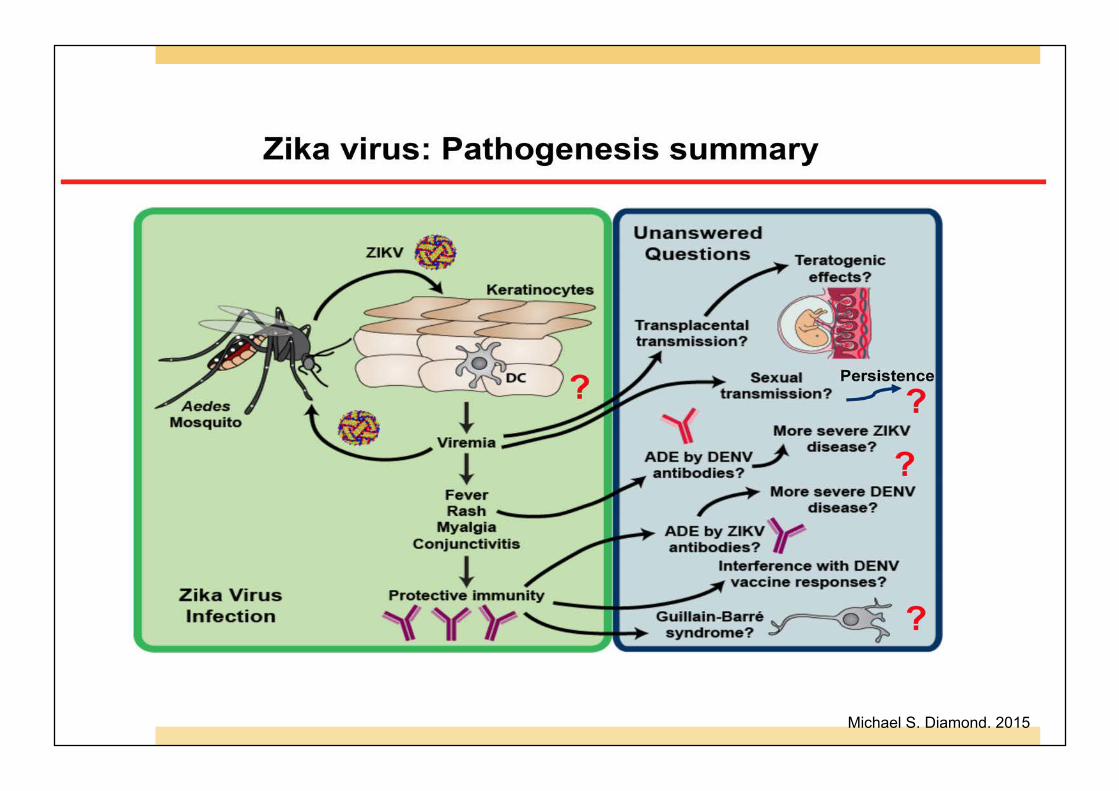
Michael S. Diamond. 2015

Michael S. Diamond. 2015

Michael S. Diamond. 2015

Michael S. Diamond. 2015

Michael S. Diamond. 2015
Michael S. Diamond. 2015

Objectives
1. Explain viral pathogenesis
2. Explain host defense mechanisms to viral infection
3. Explain cellular pathology of viral infection
4. Explain pathology of viral infection
5. Analyze diagnostic techniques for viral infection
6. Illustrate pathogenesis, pathology and diagnostic technique of dengue infection
At the completion of this unit the student will be able to:
12

Outlines
1. General properties and multiplication of virus
2. Viral pathogenesis
3. Host defense mechanisms to viral infection
4. Cellular pathology of viral infection
5. Pathophysiology of viral infection
6. Diagnostic techniques for viral infection
13

Virus
¤ An infectious, obligate intracellular parasite
¤ Package genome in a particle for transmission from host to host
¤ Viral genome contains information to initiate and complete an infectious cycle within a cell
¤ All viral genomes are able to establish themselves in a host population so that viral survival is ensured
14

Viruses are much too small to be seen with any light microscope. The largest smallpox virus is more than 5 times smaller than E. coli and 50 times smaller than a human red blood cell. 15

Viral structure ¤ Genome; DNA or RNA
¤ Capsid; protein shell
¤ Envelope; lipid membrane (presence or absence)
HIV-1
Nucleocapsid
16 Image: US National Institute of Health/Wikipedia.

Viral classification:
Group VII: dsDNA-RT (e.g. Hepadnaviruses)
Baltimore classification system
S.J. Flint et al., 3rd Principles of Virology.ASM Press.2009. 17

Infectious cycle
1. Attachment & Entry into target cell
2. RNA synthesis 3. Replication of DNA viral
genomes 4. Transcription & RNA
processing 5. Reverse transcription &
integretion 6. Translation 7. Assembly
18 Fauci AS. Science.1988; 239: 617.

Viral pathogenesis
19

Viral pathogenesis
¤ Pathogenesis is the process of producing a disease ¤ 2 components of viral diseases:
¤ Effects of viral replication on the host ¤ Effects of host response on virus
¤ Virus infections span the range from benign to lethal ¤ Acute and persistent infections can be quick or amazing
slow – days to year of infection
20

Mechanisms of viruses cause cell injury
Copyright © 2010 by Saunders, an imprint of Elsevier Inc.
Mechanisms of virus: 1. Attachment & entry into
target cell 2. Genome replication and
protein synthesis 3. Assembly
Host defense and virus evasion
21

http://www.virology.ws
Natural barrier: Ø Integrity of skin
and mucosa
Ø Cilliary epithelium
Ø pH & Temperature
Ø Bile
Ø Enzymes
Ø Normal flora
Ø Immune cells
22

Immune response to viral infection
23

Copyright © 2010 by Saunders, an imprint of Elsevier Inc.
IMMUNE EVASION BY VIRUS
24

25

26

27

¤ Cytocidal infection
¤ Steady-state persistent infection
¤ Cellular transformation
Cytopathic effect (CPE) on viral infection è refers to damage to host cells during virus invasion
28

A. Cytocidal Infection
¤ Cell death and histological appearance of characteristic CPE e.g. picornaviruses, herpesviruses
¤ Infected cells usually found cell swelling, shape changes, large nucleus, multinucleated cells, halo, inclusion bodies
29

Tortora GJ, et.al. Pearson Education.2013.
Measles virus 30

Cytomegalovirus infection (CMV)
• Intranuclear basophilic inclusions • spanning half the nuclear diameter
are usually set off from the nuclear membrane by a clear halo.
Copyright © 2010 by Saunders, an imprint of Elsevier Inc. 31

Inclusion bodies (CPE) ¤ Intracellular structures of viral proteins or
Virions
¤ May result from the clustering of subunits or virions within the nucleus or cytoplasm (e.g., the Negri bodies in rabies infections)
¤ May contain cell components such as ribosomes (arenavirus infections) or chromatin (herpesviruses)
¤ Regardless of their composition, these inclusion bodies can directly disrupt cell structure. Negri bodies
Tortora GJ, et.al.Microbiology-An introdution.11th ed Copyright © 2013, 2010, 2007 Pearson Education, Inc.
32

B. Steady-State Persistent Infection
¤ Infected cells: ¤ Produce and release virus but no CPE, may be found syncytia ¤ Can grow and divide but not killed ¤ Little destruction of infected cells
¤ Does not occur with DNA viruses
¤ Occur with several RNA viruses (Lassa virus, Retoviruses, Rubella, some paramyxoviruses)
¤ Virus released by cell budding
33

C. Cellular Transformation
¤ Viruses produce tumors in animals can transform cell culture
¤ Virus DNA integrated into host DNA, alter growth and morphology
¤ Chromosomal abnormalities
¤ New virus Ag and DNA production
¤ e.g. Adenovirus, HPV, HSV, EBV, HBV, Sarcoma virus, HTLV1,2
Patient with EBV Infection
Willey JM, et. al.McGraw-Hill.2008.
34

Brooks GF, et.al.The McGraw-Hill .2010.
Local infection
Systemic infection
SSPE: Subacute sclerosing panencephalitis 35

EXAMPLE OF VIRAL INFECTION THAT CAUSE OF SYSTEMIC PATHOLOGY
36

Dengue infection
Nov 11, 2015
37
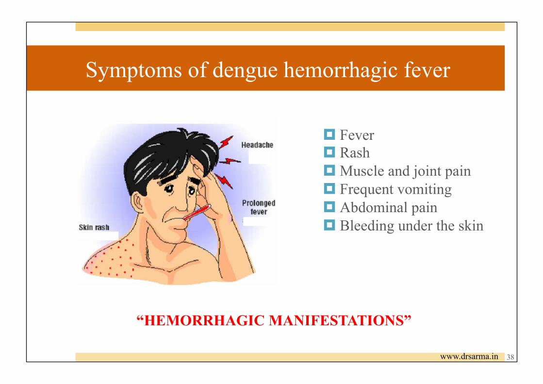
Symptoms of dengue hemorrhagic fever
¤ Fever ¤ Rash ¤ Muscle and joint pain ¤ Frequent vomiting ¤ Abdominal pain ¤ Bleeding under the skin
“HEMORRHAGIC MANIFESTATIONS”
www.drsarma.in 38

¤ Skin hemorrhages: ¤ petechiae (Skin rash), ¤ purpura (Lesions), ¤ ecchymoses
(subcutaneous spot of bleeding)
www.drsarma.in
Hemorrhagic Manifestations
39

Hemorrhagic Manifestations
¤ Skin hemorrhages:
petechiae (Skin rash), purpura (Lesions), ecchymoses (subcutaneous
spot of bleeding)
¤ Epistaxis (Bleeding from the nose, mouth, or gums)
¤ Nausea and/or Hematemesis (vomiting of blood)
¤ Gastro-intestinal bleeding:
hematemesis, melena, hematochezia
¤ Hematuria (Blood in Urine)
40

Seminars in Diagnostic Pathology (2007) 24, 227-236
The pathology of dengue hemorrhagic fever
41

Seminars in Diagnostic Pathology (2007) 24, 227-236
The pathology of dengue hemorrhagic fever Liver biopsy from a 13-year-old Chinese boy from Singapore who presented with dengue hemorrhagic fever and fulminant hepatitis.
42

The pathology of dengue hemorrhagic fever
Seminars in Diagnostic Pathology (2007) 24, 227-236
Avidin–biotin peroxidase staining with antidengue polyclonal abs for 3 serotypes
Peripheral blood-monocytes
Liver-Kupffer cells Spleen-white pulp
Spleen-MΦ in red pulp
43

DENGUE VIRUS INFECTION
¤ Can I get dengue fever after mosquito bites?
¤ How dengue fever become dangerous (DHF)?
¤ What is screening test for dengue infection?
44

DENGUE VIRUS INFECTION
Asymptomatic Symptomatic
Undiffrentiated fever
(viral syndrome)
Dengue fever
(syndrome)
Dengue Haemorrhagic fever
No Shock Dengue Shock
Syndrome (DSS)
(Plasma leakage)
Dengue Fever
Dengue Haemorrhagic fever 45 www.drsarma.in

Pathogenesis of Dengue infection
Navarro-Sa´nchez E, et al.Archi Med Res.2005; 36:425–35. 46

Webster DP, et al. Lancet.2009;9(11):678-87.
Pathogenesis of Dengue infection
47

Tourniquet Test Positive (>20 spots) Negative (≤20 spots)
www.drsarma.in 48

Diagnostic technique for dengue infection
¤ Virus isolation ¤ Specimen: serum or plasma at 2-3 day post-hospitalization ¤ Culture: mosquito, mosquito larve inoculation, 1-2 day-old mice or cell lines
¤ Serology: Four-fold rising titer ¤ Specimen: serum at first collection and 2 wk after 1°collection ¤ Test: Hemagglutination inhibition test, Complement fixaion test,
ELISA
¤ Viral nucleic acid: RT-PCR
49

How to prevent the spread of dengue fever
50 www.drsarma.in

NOW, I know about pathogenesis, pathology and diagnostic technique of dengue infection!
WHAT ABOUT other viral infection?
51 www.google.com

Pathology of Viral infection
¤ Acute (transient) infection Measles, Mumps, Poliovirus, West Nile virus
¤ Chronic (latent) infection (Herpesvirus infection) HSV, VZV, CMV
¤ Chronic productive infection HBV
¤ Transforming infection EBV
52

Case 1 A 32-year-old man presents with high fever, cough, and skin rash of 3 days in duration. His rash began in the form of pink papules behind the ears and spread around his body. Physical examination shows an extensive maculopapular rash over his face, neck, trunk, and limbs. He displays small white spots on buccal surfaces. This patient’s skin rash is most likely caused by infection with which of the following agents?
(A) Candida albicans (B) Epstein-Barr virus (C) Measles virus (D) Mumps virus
NOTE: A maculopapular rash is a type of rash characterized by a flat, red area on the skin that is covered with small confluent bumps.
53

Case 1
Clinical manifestation: Face/trunk - blotchy, reddish brown rash The oral cavity near the opening of Stensen ducts – Koplik spots Lymphoid organs - follicular hyperplasia, large germinal centers, and randomly distributed Warthin-Finkeldey cells
Koplik spots: • Necrosis, • Neutrophilic exudate, and • Neovascularization.
54
Image by Dr. Hani Masaadeh (www.slideplayer.com)

Warthin-Finkeldey cells (grape-like cluster of nuclei)
Case 1
Riede UN&Werner M.Color Atlas of Pathology.Georg Thieme Verlag.1998. Periodic-Acid Schiff (PAS) Special Stain
• Multinucleate giant cells, eosinophilic nuclear and cytoplasmic inclusion bodies. • Found in hyperplastic lymph nodes early in measles and HIV-infection, Kimura disease, and more
rarely in lymphoma and non-neoplastic lymph node disorders
H&E Stain 55

Case 1 - Measles viral infection
56 Image by Dr. Hani Masaadeh (www.slideplayer.com) Riede UN&Werner M.Color Atlas of Pathology.Georg Thieme Verlag.1998.
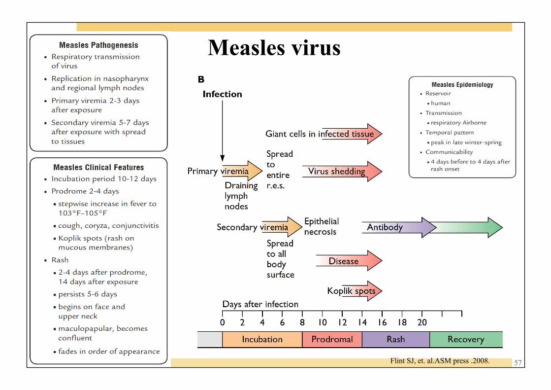
Flint SJ, et. al.ASM press .2008.
Measles virus
57

B Case 1
Case 2 Case 3
58
Image by Dr. Hani Masaadeh (www.slideplayer.com)
www.innovateus.net www.healthline.com

Varicella zoster virus infection
59

Case2 Shingles/Zoster (varicella zoster virus infection)
virus infects keratinocytes and causes vesicular lesions, which, unlike chickenpox, are often associated with intense itching, burning, or sharp pain because of the simultaneous radiculoneuritis.
By WashingtonDeceit 60
www.innovateus.net

Case 3
Chickenpox
Vesicles
By WashingtonDeceit 61
www.healthline.com

Case 3 Case 4
62 www.healthline.com
By WashingtonDeceit

Case 4 Vesicles
63 By WashingtonDeceit

Case 4
64 By WashingtonDeceit

Copyright © 2010 by Saunders, an imprint of Elsevier Inc.
Case 4 INTRANUCLEAR EOSINOPHILIC INCLUSION BODIES (Cowdry bodies)
Multinucleated cells
65

Milikowski C&Berman I.Color Atlas of Basic Histopathology.1997.
Intranuclear inclusions
Multinucleated cells
Pathogen identification: Herpes infected cells have homogeneous cytoplasm resembling ground-glass with homogeneous nucleic inclusion bodies (Cowdry bodies). Immunohistochemical findings include antibodies against HSV-1 and HSV-2.
Herpes Simplex Virus (HSV) Vesicles
66

Herpes Simplex Virus (HSV) Group: I (dsDNA) Order: Herpesvirales Family: Herpesviridae Subfamily: Alphaherpesvirinae Genus: Simplexvirus
Herpes simplex viruses 1 and 2 (HSV-1 and HSV-2) initially infect epithelial cells of the oral or genital mucosa, the skin or the cornea. The virus may enter neurones and may be transported to their nuclei, where they may establish latent infections.
These include: — Ultraviolet radiation; — Febrile infectious factors; — Menstruation; — Emotional irritation; — Immunosuppression.
These factors reactivate replication of the virus, resulting in transport of the virus to the surface of the body and causing recurrence of the rash.
67 Riede UN&Werner M.Color Atlas of Pathology.Georg Thieme Verlag.1998.

Herpes Simplex Virus (HSV) Morphology: Occasionally infected cells will fuse into multinucleated giant cells. The cytopathic effect of the virus disrupts the contact between the cells, causing formation of epidermal vesicles containing infected cells with replicating viruses.
INTRANUCLEAR EOSINOPHILIC INCLUSION BODIES (Cowdry bodies) 68 www.cdc.gov

Diagnostic techniques on viral infection based on Pathology
69

Diagnostic techniques
¤ DIRECT PATHOGEN IMAGING, gross/microscope
¤ Hematoxylin and eosin stain (H&E)
¤ “SPECIAL” (NOT H&E) STAINS, e.g., PAP smear
¤ Cell / Tissue culture, CPE (CytoPathological Effect)
¤ ANTIGENS / ANTIBODIES (SEROLOGY)
¤ PCR / RT-PCR, e.g., viral load
70

Macroscopic analysis
¤ Size/Color
¤ Necrosis
¤ Abscess
¤ Hemorrhage
¤ Rash
http://medicalnotesonline.blogspot.com 71

Microscopic analysis ¤ Normal/Abnormal tissue and cell lining
¤ CPE – inclusion bodies/multinucleated giant cells
¤ Inflammation (Acute-PMNs vs. Chronic-Lymphocytes / plasma cells)
¤ HEMORRHAGE
72 By WashingtonDeceit

73
www.virology.ws

Text books
74

Online search
http://www.ncbi.nlm.nih.gov/pubmed/
75