213
Science and Practice of Pressure Ulcer Management
| Date post: | 21-Aug-2015 |
| Category: |
Health & Medicine |
| Upload: | scu-hospital |
| View: | 42 times |
| Download: | 0 times |

Science and Practice ofPressure Ulcer Management
Editor Marco RomanelliCoeditors Michael Clark, George Cherry,
Denis Colin, and Tom Defloor
Science and Practiceof Pressure UlcerManagement
With 52 Illustrations including 34 Color Plates
Marco Romanelli, MD, PhDDepartment of DermatologyUniversity of Pisa PisaItaly
Michael Clark, PhD George Cherry, D.Phil (Oxon)Wound Healing Research Unit Clinical FacultyUniversity of Wales College of Oxford Medical School
Medicine University of OxfordCardiff, UK Oxford, UK
Denis Colin, MD, PhD Tom Defloor, RN, PhDCentre de l’Arche Nursing ScienceLe Mans Ghent UniversityFrance Ghent
Belgium
British Library Cataloguing in Publication dataA catalogue record for this title is available from the British Library.
Library of Congress Control Number: 2005923439
ISBN 10: 1-85233-839-3 Printed on acid-free paperISBN 13: 978-1-85233-839-8
© Springer-Verlag London Limited 2006
Apart from any fair dealing for the purposes of research or private study, or criticism orreview, as permitted under the Copyright, Designs and Patents Act 1988, this publicationmay only be reproduced, stored or transmitted, in any form or by any means, with theprior permission in writing of the publishers, or in the case of reprographic reproductionin accordance with the terms of licences issued by the Copyright Licensing Agency.Enquiries concerning reproduction outside those terms should be sent to the publishers.
The use of registered names, trademarks, etc. in this publication does not imply, even inthe absence of a specific statement, that such names are exempt from the relevant lawsand regulations and therefore free for general use.
Product liability: The publisher can give no guarantee for information about drug dosageand application thereof contained in this book. In every individual case the respectiveuser must check its accuracy by consulting other pharmaceutical literature.
Printed in the United States of America (BS/MVY)
9 8 7 6 5 4 3 2 1
Springer Science+Business Mediaspringeronline.com

Foreword I
I consider it a great privilege to have been asked to write the forewordfor this book. The European Pressure Ulcer Advisory Panel (EPUAP)is less than 10 years old having been founded in 1997. I had the honourof being the first president of this group and have been amazed anddelighted at the progress and achievements the panel have made sincethat time. The progress is remarkable, not only because it is a trulyEuropean group consisting of a wide range of clinical and academicinterests but also because it has retained its focus on the preventionand treatment of pressure ulcers.
The officers and board should be congratulated in developing arange of educational and research opportunities in this important butoften neglected aspect of clinical practice. Not only have they organ-ised a series of successful annual conferences that have been held in anumber of a European countries but they have developed a number ofother exciting initiatives. These have included setting up workinggroups, developing guidelines, undertaking prevalence studies andresearch projects. The latest addition to these activities is the publica-tion of this book which I am confident will rapidly become the stan-dard textbook for all interested in this subject—not only in Europe buton a global scale.
The editors of this book—who are all internationally known fortheir work in this area—are all key individuals in the success of theEPUAP. They have pulled together a comprehensive review of thissubject written by a range of experts from different professional back-grounds representing many European countries. This is no mean featand they should be congratulated on their vision and determination.
The 22 chapters address key issues in this condition and range fromupdates in research through to epidemiological aspects on to assess-ment of patients and equipment. The book also debates local woundcare either by conservative or surgical methods, complications such asinfection onto issues around developing and implementing guidelinesand the increasingly important subject of litigation in this area. Manyspecial interest groups claim to be working in a ‘Cinderella’ area butfew conditions other than pressure ulceration can really justify thatdescription. In an increasingly diverse world the challenges of provid-ing pressure ulcer care in developing countries are different but no lesschallenging than those of providing care in so called developed or
v
advanced healthcare systems. It is perhaps surprising that in suchadvanced healthcare systems some cancer can be cured, heart diseasecan be prevented and organs can be transplanted but many patients insuch systems can not guarantee that they will receive prompt andappropriate interventions to prevent or treat pressure ulceration. Thechallenge to all caring for such patients is considerable but this bookprovides a reference source for anyone who needs to understand thebasis of many aspects of patient care in this area. In addition, the coloursection provides excellent clinical illustrations that demonstrate anumber of key points in pressure ulceration.
This subject is receiving increasing attention from a number of pro-fessional, governmental and legal directions. The importance, cost andability to use aspects of this clinical problem as an indicator of thequality of health care delivery is to be encouraged but how robust isthe research base, the development of standards of clinical care andconsistency of healthcare practices in pressure ulceration on a localnational and international basis?
This book will not replace all of the work needed to address theseproblems but it will provide a strong foundation from which we canbuild our understanding of this condition for improved standards ofcare to patients in what has been a long standing but neglected clini-cal challenge.
I congratulate the editors, authors and publishers for remainingfocused on their task—to provide the best and most comprehensiveand up to date review of this subject. I commend this book to you asan essential companion to help you improve standards of care for yourpatients.
Keith Harding, MD
vi Foreword I
Foreword II
One of the outcomes of advancing medical technology is that peopleare living longer. As life is extended, the complex issue of managingpersons with chronic diseases becomes increasingly important. Theincreased number of persons with chronic wounds such as pressureulcers is already being realized. The health-care burden of managingthese chronic wounds can only be lessened if effective prevention pro-grams are aggressively implemented and evidence-based managementprotocols are developed and followed.
The information contained in this book provides the critical ele-ments for developing effective, evidence-based protocols for the pre-vention and management of pressure ulcers. What this book cannotprovide is the commitment required to create an environment wherethe development of a pressure ulcer on a person is unacceptable. Pro-tocol development is only one component of a comprehensive programfor prevention and management of pressure ulcers. Everyone involvedin patient care from administration to bedside provider has to makethe commitment that pressure ulcers will not occur in their facility.
This book is a tremendous resource, but it needs to be used effec-tively. In the United States, the government sponsored the developmentof evidence-based guidelines on prevention and management of pres-sure ulcers. These guidelines became available in the early nineties.Since their publication, the prevalence of pressure ulcers in the UnitedStates has not changed at the national level. However, in those facilitiesthat chose to use the guidelines to develop and implement new proto-cols for prevention and management of pressure ulcers, the incidenceof pressure ulcers was reduced to zero or to a very low level.
The information in this book can be used to prevent new pressureulcers from developing, and rapidly healing those that have un-fortunately already developed. The only thing missing is the commit-ment to make change. I hope that everyone who reads this book makesthe personal commitment to prevent pressure ulcers from occurringand to optimize the management of those that occurred at a differentfacility.
George T. Rodeheaver, PhDFounding Member and Past President
National Pressure Ulcer Advisory Panel
vii
Acknowledgments
The European Pressure Ulcer Advisory Panel is grateful to the follow-ing corporate sponsors which have helped make the publication of thisbook possible:
Frontier Therapeutics LtdGaymar IndustriesKCI Europe B.V.Nutricia HealthcareSmith & Nephew
ix
Contents
Contributors . . . . . . . . . . . . . . . . . . . . . . . . . . . . . . . . xiii
1 Pressure Ulcer, the Scale of the ProblemTheo Dassen, Antje Tannen, and Nils Lahmann . . . . . . . 1
2 Pressure Ulcer Patients’ Quality of Life from a Nurse’s PerspectiveHelvi Hietanen . . . . . . . . . . . . . . . . . . . . . . . . . . . . 7
3 Recent Advances in Pressure Ulcer ResearchDan Bader and Cees Oomens . . . . . . . . . . . . . . . . . . . 11
4 Etiology and Risk FactorsMark Collier and Zena Moore . . . . . . . . . . . . . . . . . . 27
5 Pressure Ulcer ClassificationCarol Dealey and Christina Lindholm . . . . . . . . . . . . . 37
6 Risk Assessment Scales for Predicting the Risk ofDeveloping Pressure UlcersJoan-Enric Torra i Bou, Francisco Pedro García-Fernández,Pedro L. Pancorbo-Hidalgo, and Katia Furtado . . . . . . . . 43
7 Equipment SelectionJacqueline Fletcher . . . . . . . . . . . . . . . . . . . . . . . . . . 59
8 Pressure Ulcer Prevention and RepositioningTom Defloor, Katrien Vanderwee, Doris Wilborn, and Theo Dassen . . . . . . . . . . . . . . . . . . . . . . . . . . . . . 67
9 Skin CareSue Bale, Janice Cameron, and Sylvie Meaume . . . . . . . . 75
xi
10 Pressure Ulcers and Nutrition: A New European GuidelineJoseph Schols, Michael Clark, Giuseppe Benati,Pam Jackson, Meike Engfer, Gero Langer, Bernadette Kerry,and Denis Colin . . . . . . . . . . . . . . . . . . . . . . . . . . . 85
11 Clinical and Instrumental Assessment of Pressure UlcersDiego Mastronicola and Marco Romanelli . . . . . . . . . . . 91
12 Pressure Ulcers and Wound Bed PreparationVincent Falanga . . . . . . . . . . . . . . . . . . . . . . . . . . . 99
13 Conservative Management of Pressure UlcersElia Ricci, Andrea Cavicchioli, and Marco Romanelli . . . . 111
14 Surgical Management of Pressure UlcersJens Lykke Sørensen, M.J. Lubbers, and Finn Gottrup . . . . 119
15 Debridement of Pressure UlcersAndrea Bellingeri and Deborah Hofman . . . . . . . . . . . . 129
16 The Role of Bacteria in Pressure UlcersR. Gary Sibbald, Paul Chapman, and Jose Contreras-Ruiz . . . . . . . . . . . . . . . . . . . . . . . . . 139
17 LitigationCourtney H. Lyder . . . . . . . . . . . . . . . . . . . . . . . . . . 163
18 The Development, Dissemination, and Use of Pressure Ulcer GuidelinesR.T. van Zelm, Michael Clark, and Jeen R.E. Haalboom . . . 169
19 Developing a Research AgendaDenis Colin . . . . . . . . . . . . . . . . . . . . . . . . . . . . . . 177
20 The European Pressure Ulcer Advisory Panel: A Means ofIdentifying and Dealing with a Major Health Problem with a European InitiativeGeorge W. Cherry . . . . . . . . . . . . . . . . . . . . . . . . . . 183
21 Pressure Ulcer Prevention and Management in the Developing World: The Developed World Must Provide LeadershipTerence J. Ryan . . . . . . . . . . . . . . . . . . . . . . . . . . . . 189
22 Innovation in Pressure Ulcer Prevention and ManagementKeith G. Harding and Michael Clark . . . . . . . . . . . . . . 197
Index . . . . . . . . . . . . . . . . . . . . . . . . . . . . . . . . . . . . 205
xii Contents
Contributors
xiii
Dan Bader, BSc MSc PhD, MIPEM,DSc
Professor of Medical EngineeringDepartment of EngineeringQueen Mary University of LondonLondon, UK
and
Professor of Soft Tissue RemodellingBiomedical Engineering DepartmentEindhoven University of TechnologyEindhoven, The Netherlands
Sue Bale, PhD, BA, RGN, NDN, RHV,PG Dip, Dip Nursing
ProfessorAssociate Director of NursingGrange HouseLlanfrechfa Grange HospitalCwmbran, UK
Andrea Bellingeri, RNSecretary of Italian Nurse Association
for the Study of WoundItalian Nurse Society on Wound study
(AISLeC)Pavia, Italy
Giuseppe Benati, MDUnita Operativa di Medicina
GeriatricaOspedale Morgagni PierantoniForli, Italy
Cinzia Brilli, RNTissue Viability NurseAzienda Ospedaliera Universitaria
PisanaPisa, Italy
Janice Cameron, MPhil, RGN, ONCClinical Nurse Specialist in Wound
ManagementDepartment of DermatologyOxford Radcliffe Hospitals NHS TrustChurchill HospitalHeadington, Oxford, UK
Andrea Cavicchioli, RNTissue Viability NurseAzienda UnitàSanitaria Locale di Modena-Osp.
EstenseModena, Italy
Paul Chapman, BScPTMedical StudentUniversity of ManitobaWinnipeg, Manitoba, Canada
George W. Cherry, DPhil (Oxon)Secretary Treasurer EPUAPClinical FacultyOxford Medical School University of OxfordOxford, UK
Michael Clark, PhDSenior Research FellowWound Healing Research UnitUniversity of Wales College of
MedicineCardiff, UK
Denis Colin, MD, PhDCentre de l’ArcheLe Mans, France
Mark Collier, BA, RNT, RCNT, ONC, RNLead Nurse/Consultant-Tissue
ViabilityUnited Lincolnshire HospitalsPilgrim HospitalBoston, Lincolnshire, UK
Jose Contreras-Ruiz, MDWound Care FellowDermatology Daycare/Wound Healing
CentreSunnybrook and Women’s College
Health Sciences CentreToronto, Ontario, Canada
and
Dermatologist Hospital General “DrManuel Gea Gonzalez”
Toriello Guerra, Mexico
Theo Dassen, RN, PhDProfessor DrInstitut für Medizin-/Pflegepädagogik
und PflegewissenschaftUniversitätsklinikum CharitéBerlin, Germany
Carol Dealey, PhD, MA, BSc (Hons),RGN, RCNT
Research FellowSchool of Health SciencesUniversity of BirminghamEdgbaston, Birmingham, UK
Tom Defloor, RN, PhDNursing ScienceGhent UniversityGhent, Belgium
Meike Engfer, PhDClinical Nutrition AdviserNumico Clinical NutritionSchiphol, The Netherlands
Vincent Falanga, MD, FACPProfessor of Dermatology and
BiochemistryBoston University School of MedicineChairman, Department of
DermatologyRoger Williams Medical CenterProvidence, Rhode Island, USA
Jacqueline Fletcher, BSc, RGN, PgCert,ILTM
Principal LecturerSchool of Nursing and MidwiferyUniversity of HertfordshireHatfield, Hertfordshire, UK
Katia Furtado, RNCentro de saúde da Penha de FrançaLisbon, Portugal
Francisco Pedro García-Fernández, RNQuality, Research and Formation
ManagerComplejo Hospitalario de JaénJaén, Spain
Finn Gottrup, MD, DMSciProfessorDepartment of Plastic SurgeryOdense University HospitalOdense C, Denmark
Jeen R. E. Haalboom, MD, PhD, EPUAPProfessor of Internal MedicineUniversity Medical CentreUtrecht, The Netherlands
Keith G. Harding, MB ChB, MRCGP,FRCS
ProfessorDepartment of SurgeryWales College of Medicine Cardiff UniversityCardiff, Wales, UK
Helvi Hietanen, RNHead NurseDepartment of Plastic SurgeryTo–o–lo– HospitalHUCH Helsinki University Central
HospitalFinland
Deborah Hofman, BA Hons, RGN, DipNurse
Clinical Nurse SpecialistDepartment of DermatologyChurchill HospitalHeadington, Oxford, UK
xiv Contributors
Contributors xv
Pam Jackson, MPhil, BSc, RGN, RHV,RNT, RCNT, ILT
Senior LecturerUniversity of SouthamptonSouthampton, UK
Bernadette Kerry,RGN,RPN,PGD,Dip Tissue Repair and Wound
ManagementMidland Health BoardTullamore, Co. Offaly, Ireland
Nils Lahmann, RN, BAInstitut für Medizin-/Pflegepädagogik
und PflegewissenschaftUniversitätsklinikum CharitéBerlin, Germany
Gero Langer, MScN (EU), RNCoordinator of the German Centre for
Evidence-based Nursing “sapere aude”Martin Luther UniversityHalle-Wittenberg, Germany
Christina Lindholm, RN, PhDProfessorDepartment of Health SciencesKristianstad UniversitySweden
M.J. LubbersDepartment of SurgeryAMC University HospitalAmsterdam, The Netherlands
Courtney H. Lyder, NDProfessorUniversity of VirginiaMcLeod HallCharlottesville, Virginia, USA
Diego Mastronicola, MDConsultant DermatologistDepartment of DermatologyUniversity of PisaPisa, Italy
Sylvie MeaumeHôpital Charles FoixIvry sur SeineFrance
Zena Moore, RGN, MSc, FFNMRCSILecturerFaculty of Nursing and MidwiferyRoyal College of Surgeons in IrelandDublin 2, Ireland
Cees Oomens, PhDAssociate ProfessorBiological Engineering DepartmentEindhoven University of TechnologyEindhoven, The Netherlands
Pedro L. Pancorbo-Hidalgo, PhD, RNProfessor of Medical-Surgical
NursingSchool of Health SciencesUniversity of JaénLas Lagunillas S/N, Jaén, Spain
Elia RicciConsultant SurgeonWound Healing UnitCasa di Cura San LucaTorino, Italy
Marco Romanelli, MD, PhDDepartment of DermatologyUniversity of PisaPisa, Italy
Terence J. Ryan, BM BCh, DM, MA,FRCP
Emeritus Professor of DermatologyOxford UniversityDepartment of DermatologyChurchill HospitalHeadington, Oxford, UK
Joseph Schols, MD, PhDDepartment TranzoTilburg UniversityThe Netherlands
R. Gary Sibbald, MD, FRCRC, MEdDepartment of MedicineUniversity of TorontoToronto, Ontario, Canada

Jens Lykke Sørensen, PhDClinical DirectorDepartment of Plastic SurgeryOdense University HospitalOdense C, Denmark
Antje Tannen, RN, MAInstitut für Medizin-/Pflegepädagogik
und PflegewissenschaftUniversitätsklinikum CharitéBerlin, Germany
Joan-Enric Torra i Bou, RNClinical ManagerAdvanced Wound Care DivisionSmith and Nephew SpainSant Joan Despi, Barcelona, Spain
Katrien Vanderwee, RN, MAPhD StudentNursing ScienceGhent UniversityGhent, Belgium
R.T. van ZelmAdvisorDutch Institute for Health Care
ImprovementUtrecht, The Netherlands
Doris Wilborn, RN, MANursing ScienceHumboldt-UniversityBerlin, Germany
xvi Contributors

1 Pressure Ulcer, the Scale of the ProblemTheo Dassen, Antje Tannen, and Nils Lahmann
Introduction
The main goal of this chapter is to provide information about the frequency ofpressure ulcers. However, in doing this it becomes evident that the chapter title—the scale of the problem—should really be amended to the problem of the scale.Due to the different rates used (prevalence, incidence), different grades/stages ofpressure ulcers (1, 2, 3, 4), different body sites, different settings (hospital, nursinghome, at home) and different ways of data collection it is almost impossible to findcomparable data about the scale of this phenomenon in human beings. Therefore,this chapter should be regarded more as a guide on how to deal with data on pres-sure ulcers obtained from the literature. First, information is provided about theuse of rates and their application to pressure ulcers. Then some suggestions aregiven about how to interpret the figures from the literature.
Rates
Measures of frequencies in a disease are usually expressed as rates.1 Those ratesare fractions or proportions that consist of three elements: a numerator, a denom-inator, and a time period. In this case the numerator is the number of people suf-fering from pressure ulcers. The denominator is the population that was selectedas the number of possible occurrences (e.g. all patients in a hospital). The timeperiod can be one moment in time or another well-defined period (for instance ayear). In pressure ulcer research it is common practice to express the rate as a per-centage, which means per hundred cases. The numerator is divided by the denom-inator and then multiplied by 100. For example, ten persons out of a thousandsuffer from a pressure ulcer; this is: 10/1000 ¥ 100 = 1%.
The difference between prevalence rates and incidence rates is important. Pres-sure ulcer prevalence refers to the number of people with pressure ulcer as a pro-portion of the total population under investigation. Prevalence rates include all oldand all new cases. If only the new cases are counted this is called the incidence.
So far, it does not appear complicated to provide comparable data about pres-sure ulcer, but the problem is that an exact definition is necessary for both parts,numerator and denominator, to make the calculated rates coherent.2 Every authoruses a definition for both, but there is standardization. This leads to publicationswith rates ranging from 5% to 50% or sometimes even less or more. It is not clear
1

whether there are indeed different rates or if these are the result of differences inthe way the numerator and/or denominator have been defined.
Another aspect is the time period. If a prevalence rate is measured at onemoment in a given period of time it is called a point prevalence. A period preva-lence refers to the condition over a specified period of time. It is obvious that inci-dence rates are always calculated for a period. It is important in both cases that the chosen period of time is the same when comparing rates from different publications.
Numerator Confusion
When looking at a definition of pressure ulcer it becomes obvious that the numer-ator can vary depending on the project. In other chapters of this book this is dis-cussed in more depth. According to the definition of the European Pressure UlcerAdvisory Panel (EPUAP)3 a pressure ulcer can be located anywhere on the skin ofthe body and is a discoloration of the skin (with nonblanchable erythema), but itcan also be an extensive destruction with tissue necrosis, damage to the muscle,bone or supporting structures with or without full-thickness skin loss. This meansthe numerator can include a red, damaged area of skin at the elbow and also a deephole in the skin of the sacrum. Is it sensible to combine all these in a singleclassification? Yes and no! If we know that 10% of all patients in a hospital have apressure ulcer we obtain information about this phenomenon. However, without aclassification of the pressure ulcer according to grades and body sites this infor-mation cannot be used for any kind of policy. For this reason researchers dividethe numerator into grades (or stages) and body sites. Table 1.1 shows an exampleof a division according to body sites, which is derived from a study conducted byEPUAP.4
This table shows that more than 25% of all pressure ulcers are located on thesacrum. This is supported by several other studies.5,6 However, the sites of pressureulcers in children are different. The occipital region of the scalp in infants and tod-dlers and the sacrum in children are prevalent sites of pressure ulcer formation.7
Approximately one third of the pressure ulcers are located on the heel, which isalso supported by the literature.8,9 So far it could be concluded that about half ofthe pressure ulcers are located on either the sacrum or the heel. What about therest? Table 1.1 shows that the division of pressure ulcers at other body sites varies
2 T. Dassen et al.
Table 1.1. Pressure ulcer prevalence rates at different body sites (% of totalprevalence)
Location Belgium Italy Portugal Sweden UK Total
Sacrum 25.6 40.9 26.9 25.3 37.5 532
Heel 34.9 31.9 33.9 30.0 26.2 484
Ischium 12.2 7.6 2.7 11.6 13.7 186
Ankle 3.6 9.1 10.2 24.5 6.4 149
Elbow 14.3 0.0 6.9 3.0 10.3 143
Hip 9.3 10.6 19.3 5.6 5.8 136
Total 301 132 186 233 778 1630
Source: Based on Clark et al.4
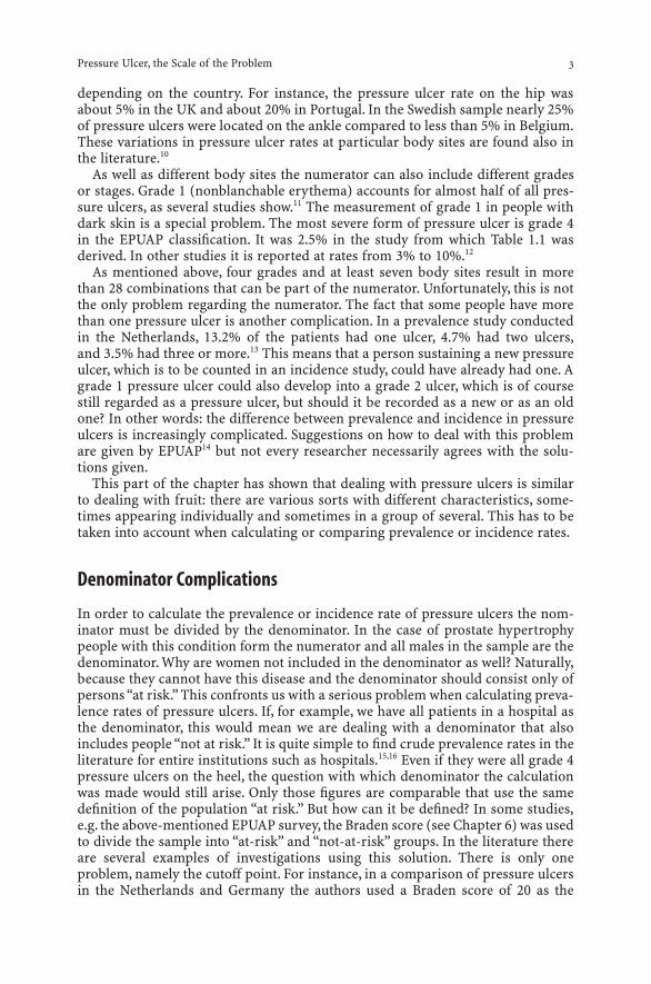
depending on the country. For instance, the pressure ulcer rate on the hip wasabout 5% in the UK and about 20% in Portugal. In the Swedish sample nearly 25%of pressure ulcers were located on the ankle compared to less than 5% in Belgium.These variations in pressure ulcer rates at particular body sites are found also inthe literature.10
As well as different body sites the numerator can also include different gradesor stages. Grade 1 (nonblanchable erythema) accounts for almost half of all pres-sure ulcers, as several studies show.11 The measurement of grade 1 in people withdark skin is a special problem. The most severe form of pressure ulcer is grade 4in the EPUAP classification. It was 2.5% in the study from which Table 1.1 wasderived. In other studies it is reported at rates from 3% to 10%.12
As mentioned above, four grades and at least seven body sites result in morethan 28 combinations that can be part of the numerator. Unfortunately, this is notthe only problem regarding the numerator. The fact that some people have morethan one pressure ulcer is another complication. In a prevalence study conductedin the Netherlands, 13.2% of the patients had one ulcer, 4.7% had two ulcers,and 3.5% had three or more.13 This means that a person sustaining a new pressureulcer, which is to be counted in an incidence study, could have already had one. Agrade 1 pressure ulcer could also develop into a grade 2 ulcer, which is of coursestill regarded as a pressure ulcer, but should it be recorded as a new or as an oldone? In other words: the difference between prevalence and incidence in pressureulcers is increasingly complicated. Suggestions on how to deal with this problemare given by EPUAP14 but not every researcher necessarily agrees with the solu-tions given.
This part of the chapter has shown that dealing with pressure ulcers is similarto dealing with fruit: there are various sorts with different characteristics, some-times appearing individually and sometimes in a group of several. This has to betaken into account when calculating or comparing prevalence or incidence rates.
Denominator Complications
In order to calculate the prevalence or incidence rate of pressure ulcers the nom-inator must be divided by the denominator. In the case of prostate hypertrophypeople with this condition form the numerator and all males in the sample are thedenominator. Why are women not included in the denominator as well? Naturally,because they cannot have this disease and the denominator should consist only ofpersons “at risk.” This confronts us with a serious problem when calculating preva-lence rates of pressure ulcers. If, for example, we have all patients in a hospital asthe denominator, this would mean we are dealing with a denominator that alsoincludes people “not at risk.” It is quite simple to find crude prevalence rates in theliterature for entire institutions such as hospitals.15,16 Even if they were all grade 4pressure ulcers on the heel, the question with which denominator the calculationwas made would still arise. Only those figures are comparable that use the samedefinition of the population “at risk.” But how can it be defined? In some studies,e.g. the above-mentioned EPUAP survey, the Braden score (see Chapter 6) was usedto divide the sample into “at-risk” and “not-at-risk” groups. In the literature thereare several examples of investigations using this solution. There is only oneproblem, namely the cutoff point. For instance, in a comparison of pressure ulcersin the Netherlands and Germany the authors used a Braden score of 20 as the
Pressure Ulcer, the Scale of the Problem 3

cutoff.17 In clinical practice a cutoff point of 16 or 18 is more common. Naturally,the differences in choosing a cutoff point are permitted but they complicate thecomparison of results from the literature with clinical practice.
Due to the different cutoff points the number of at-risk individuals varies con-siderably. The rate increases if the denominator is smaller. This means that a highprevalence rate could be the result of a well-defined risk group. Or, conversely, alow prevalence rate could be the result of a widely defined risk group (e.g. allpatients of the hospital). It should be remembered that the prevalence of prostatehypertrophy would be only half the size if all women were included in the denom-inator as well.
About one third of the hospital population and two thirds of the nursing homepopulation were at risk when using a Braden score of 20 as the cutoff point. In a nationwide study in Germany18 this proportion remained stable over some years. The above-mentioned comparison between Germany and the Netherlandsrevealed a different number of “at-risk” patients, with more than 50% in the Dutchhospitals. The EPUAP survey also showed (cutoff = 16) different proportions “atrisk,” from about 34% in Belgium to 23% in Italy. It is not the intention of thischapter to discuss the risk assessment but to show that calculating prevalence ratesby using total populations of institutions will inevitably lead to figures that are notcomparable. A highly sophisticated solution, called the case mix method, involvescorrection for all kinds of factors that can influence the occurrence of a pressureulcer.19 It is only practicable if all the information is available, which is, however,not always the case.
Apart from the difficulty of defining the risk group the denominator give rise toanother serious problem—the influence of the nonresponse. Researchers have torespect ethical rules when using people’s data for a scientific purpose. This meansthat permission has to be obtained from each patient in the hospital as a basic con-dition for using their data for the calculation of a prevalence or incidence rate. Oneof the side effects is that not every patient agrees to participate in the study.
Prevalence research can be classified as descriptive research. This is research that aims to generalize the results for a whole target group. In this case the targetgroup could comprise all at-risk patients in a hospital or all at-risk residents in anursing home. A high external validity is necessary for this kind of research.20 Itmeans that the sample under investigation should reflect the target group. This willnot be the case in practice. Nonresponse rates can influence the prevalencesignificantly, as Table 1.2 illustrates. A measured prevalence of 19.7% could in factbe lower (15.2%) or higher (38.1%) depending on the nonresponse rate.21 In thisexample the lowest rates were calculated on the assumption that all the at-riskpeople in the nonresponse portion did not have a pressure ulcer. The highest rateswere calculated on the assumption that all the at-risk people in the nonresponseportion had at least one pressure ulcer.
4 T. Dassen et al.
Table 1.2. Example of measured and calculated (lowest and highest) pressure ulcerprevalence rates
Institution Response % Measured % Lowest % Highest %
Nursing home 79.6 12.5 10.0 30.3
Hospital 75.6 24.2 18.3 42.7
Total 76.6 19.7 15.2 38.1
Source: Based on Dassen et al.18

Solution of the Problem
It was established that the calculated rates can differ considerably depending ondifferent definitions of the numerator and/or the denominator. This is a well-known problem that often occurs when using statistics. Tukey expressed it asfollows: “Far better an approximate answer to the right question, which is oftenvague, than an exact answer to the wrong question, which can always be madeprecise.”22 This statement shows us that a question like “what is the pressure ulcerprevalence in this hospital” is wrong. A correct question would be “How manypatients on intensive care wards have a pressure ulcer grade 2, 3, 4 on the sacrum?”An example of the answer then is “58% of all people who had a pressure ulcer.”23
This answer is not precise but it tells us that a pressure ulcer on this part of thebody is not an exception. It can become more informative if the prevalence andthe definition of the risk group are known. In this case, the answer was 21% ofpeople who scored 20 or lower on the Braden scale. This means that about 12% ofthe people “at risk” had a pressure ulcer grade 2, 3, or 4 on the sacrum.
Another example is the number of pressure ulcers in dead bodies that wereinspected prior to cremation. A difference in grade 4 was found in a comparisonbetween Berlin (2.3%) and Hamburg (0.9%).24 Here the population was defined as“all dead bodies that were brought to the crematorium.”Again a specific group wasselected and a clear distinction between grades and body sites was used to presentthe data.
Finally
The problem of prevalence and incidence of pressure ulcers was discussed withoutobtaining a precise answer to the question of the scale of the problem as men-tioned in the title of this chapter. Naturally, it is not intended to evade this ques-tion. The reason quite obviously is that there are almost no comparable data. Aprevalence rate of 10% in one publication can be quite different from 10% inanother publication. Depending on the definition of the numerator and thedenominator a prevalence or incidence rate can include different information. Aclear distinction between prevalence and incidence is nearly impossible owing tofactors such as multiple pressure sores in the same patient and progression tohigher grades in an existing pressure ulcer.
Therefore, information regarding findings from different publications is moreinteresting and safer. Most studies revealed more grade 1 than grade 4 pressureulcers. Several studies mention the sacrum and the heels as those body sites withthe most frequently occurring pressure ulcers. In children other parts of the body(occipital region of the scalp) are more predominant.
Special groups such as intensive care patients or patients on geriatric wards areaffected by pressure ulcers to a larger extent than are hospital patients on otherwards.
Finally, it can be stated that pressure ulcers are found more often in geriatricpatients than in younger patients, more often in intensive care wards than in lowercare wards, and more often on the heel or the sacrum than on other body sites.Furthermore, it is known that a grade 1 pressure ulcer occurs in about 50% ofpatients with pressure ulcers and the higher the grade the lower the proportion of
Pressure Ulcer, the Scale of the Problem 5
all pressure ulcers. Comparable figures concerning the prevalence or incidence ofpressure ulcers in human beings are not known.
References
1. Mulhall A. Epidemiology nursing and healthcare. A new perspective. Basingstoke: Macmillan;1996.
2. Fletcher R, Fletcher S. Clinical epidemiology. The essentials, 4th edn. Baltimore: Williams &Wilkins; 2005.
3. EPUAP. Pressure ulcer treatment guidelines. http://www.epuap.org.4. Clark M, Bours G, Defloor T. Summary report on the prevalence of pressure ulcers. EPUAP Review
2002; 4:49–56.5. Ash D. An exploration of the occurrence of pressure ulcers in a British spinal injuries unit. J Clin
Nurs 2002; 11:470–478.6. Eriksson E, Hietanen H, Asko-Seljavaara S. Prevalence and characteristics of pressure ulcers. A
one-day patient population in a Finnish city. Clin Nurse Spec 2000; 14:119–125.7. Curley M, Razmus I, Roberts K, Wypij D. Predicting pressure ulcer risk in pediatric patients: the
Braden Q Scale. Nurs Res 2003; 52:22–33.8. Schue R, Langemo D. Prevalence, incidence, and prediction of pressure ulcers on a rehabilitation
unit. J Wound Ostomy Continence Nurs 1999; 26:121–129.9. Levett D, Smith S. Survey of pressure ulcer prevalence in nursing homes. Elder Care 2000; 12:12–16.
10. Williams D, Stotts N, Nelson K. Patients with existing pressure ulcers admitted to acute care. JWound Ostomy Continence Nurs 2000; 27:216–226.
11. Halfens R, Bours G, Ast W van. Relevance of the diagnosis “Stage 1 pressure ulcer”: an empiricalstudy of the clinical course of stage 1 ulcers in acute care and long-term hospital populations. JClin Nurs 2001; 10:748–757.
12. O’Brien S, Wind S, Rijswijk L van, Kerstein M. Sequential biannual prevalence studies of pressureulcers at Allegheny-Hahnemann University Hospital. Ostomy Wound Manage 1998; 44:78–89.
13. Bours G, Halfens R, Wansink S. Landelijk prevalentie onderzoek Decubitus, Resultaten zesde jaar-lijkse meting 2003. Universiteit Maastricht, 2003.
14. Defloor T, Bours G, Schoonhoven L, Clark M. Draft EPUAP statement on prevalence and incidencemonitoring. EPUAP Review 2002; 4:13–15.
15. Thoroddsen A. Pressure sore prevalence: a national survey. J Clin Nurs 1999; 8:170–179.16. Pearson A, Francis K, Hodgkinson B, Curry G. Prevalence and treatment of pressure ulcers in
northern New South Wales. Aust J Rural Health 2000; 8:103–110.17. Tannen A, Dassen T, Bours G, Halfens R. A comparison of pressure ulcer prevalence: concerted
data collection in The Netherlands and Germany. Int J Nurs Stud 2004; 41(6):607–612.18. Dassen T, et al. Pflegeabhängigkeit, Sturzereignisse, Inkontinenz, Dekubitus, Erhebung 2003.
Humboldt-Universität zu Berlin, 2003.19. Bours G, Halfens J, Berger P. Development of a model for case-mix adjustment of pressure ulcer
prevalence rates. Med Care 2003; 41:45–55.20. Polit D, Beck Ch. Nursing research, principles and methods. Philadelphia: Lippincott; 2004.21. Lahmann N, Halfens R, Dassen T. Prevalence of ulcers in Germany, submitted for publication.22. Tukey J. Cited in: Silverman W, Where’s the evidence, Debates in modern medicine. (Chapter 2:
Does a difference make a difference.) Oxford: Oxford Medical Publications, 1998.23. Heinrichs P, Dassen T. Zahlen zur Prävalenz des Dekubitusgeschwürs in der Intensivpflege. Crit
Care 2004; 115–119.24. Troike W, Schneider V. Zur Prävalenz von Decubitalulcera, Ergebnisse einer Stichprobe bei der
zweiten Leichenschau in drei Berliner Krematorien. Berl Ärzte 2000; 12:28–30.
6 T. Dassen et al.
2 Pressure Ulcer Patients’ Quality of Life from a Nurse’s PerspectiveHelvi Hietanen
Part of a nurse’s work is to assist patients with their physical, spiritual, and socialneeds if patients are unable to satisfy these needs on their own. Hygiene and skincondition, including nutritional balance, are significant factors in preventing pres-sure ulcers. The occurrence of pressure ulcers has an important influence on thepatient’s quality of life in many ways. According to the literature,1–5 factorsinfluencing the pressure ulcer patient’s quality of life, and which can be influencedby nursing, include skin condition, cost-effective wound care, comfortableness ofthe mattress, quality of sleep, high-quality auxiliary devices, and treatment of painincluding appropriate care practices.6,7 In addition, the nursing staff ’s motivation,competence, and skills in effective methods8–10 influence the success of preventivemeasures.
The patient’s wellbeing, feeling of comfort in bed, and quality of sleep can bepromoted by selecting an appropriate mattress for the patient, taking the knownrisk factors of ulceration into account. Experience has shown that even though thepatient is informed about the beneficial effects of an alternating pressure mattress,the patient may not be willing to test such a mattress. Reasons for this decisionmay be the patient’s previous negative experiences or beliefs. For some patients,even the most silent machinery is experienced as annoying and affecting thequality of sleep. On the other hand, the spasticity of a patient with a spinal cordinjury may be activated, a very skinny and small patient may feel as though theyare “drowning” in the mattress, and an extremely obese or tall and large patientmight experience the dimensions of the mattress as uncomfortable. Consequently,the patient’s own wishes and experiences of special mattresses must always betaken into account. Sometimes, the best solution is to allow patients to bring theirown special mattress for the hospital stay.
In some cases, the patient’s quality of life and motivation improve if the patientbecomes aware of the costs arising from pressure ulcers and the effects of theseulcers.11–13 Regrettably, young patients especially often only understand the actualrisk of having a pressure ulcer when the first ulcer occurs. In the research data ofthe Helsinki University Hospital14 over half of the patients with pressure ulcerswere patients with spinal cord injury. Thus, in particular young patients with aspinal cord injury should have peer support and practical examples in their ownlanguage. The care staff should create ways, together with the patients, by whichthe best possible preventive methods for pressure ulcers can be offered.15,16 Thisrequires personnel who have appropriate education, competence, and motivation
7

for high-quality nursing.17 In her doctoral dissertation “Pressure Ulcer Risk Assess-ment in Long-term Care. Developing an Instrument,” Lepistö18 concludes that staffare aware of the need to prevent pressure ulcers in high-risk patients, for examplebedridden patients, but that prevention of other patients’ pressure ulcers is moredifficult.
However, not all pressure ulcers can be prevented. Treatment of pressure ulcers,preventing infections, and preventing an infection from spreading are a veryimportant part of nursing. Pressure ulcers are usually located in difficult places,which is unpleasant for the patients, and it is impossible for them to treat theseulcers themselves. The patients might easily feel like “prisoners” of the ulcers andisolate themselves, being anxious about the bandages becoming soaking wet orodors coming through.9 Nurses are required to have expertise in selecting the mosteconomical bandages that will also have a positive effect on patients’ quality of life,allowing patients to lead as normal a life as possible. In western countries, thereare hundreds of products from which to choose. However, the problem is that theproducts are usually very expensive and knowledge of their effects is based mainlyon recommendations generated through experience and information given by themanufacturers. Whenever possible, the most economical treatment should beselected if its effect is as good as the more expensive alternative. In treatment ofchronic wounds, no differences have been observed in healing of the wounds whenthe use of sterile and factory clean techniques, including sterile wound cleaning,and the use of drinking water have been compared.19 However, using drinkingwater is significantly cheaper.A pressure ulcer in itself causes significant additionalcosts for the patient in addition to human suffering.
A Practical Example of the Methods Used for Prevention of a PlasticSurgery Patient’s Pressure Ulcers
The patient’s risk of having a pressure ulcer is individually evaluated. There is norisk evaluation indicator in regular use but the risk evaluation is based on experi-ence, research, and the most recent available knowledge including following up ofthe incidence of pressure ulcers and common agreements. For example, the Euro-pean Pressure Ulcer Advisory Panel (EPUAP) prevention and treatment guidelineshave been utilized in teaching.
All patients coming for corrective surgery of pressure ulcers or patients whoalready have a pressure ulcer when they are hospitalized, including all immobilepatients, will have an alternating pressure mattress preoperatively at the hospital.If the number of mattresses is not sufficient on the ward, it is possible to rent themand they are available within a few hours. The patient’s nutritional imbalance isprimarily treated with dietary supplements. Those patients who are not allowed tochange their position freely in bed postoperatively will have a mattress of this kindat latest in the recovery room. The nurse receiving the patient evaluates his or herneed of special mattresses and other auxiliary devices when the patient enters thehospital. In addition to written instructions, regular training is organized in pre-vention of pressure ulcers, for example use of auxiliary devices and correct liftingtechniques. It has also been commonly agreed that a physiotherapist and severalnurses participate for the first few times in moving those patients who need a lotof help. The physiotherapist guides the patient but also shows the nursing staff howto use the best methods. Following up the incidence of pressure ulcers is an issue
8 H. Hietanen

of utmost importance. If a pressure ulcer occurs during the patient’s stay on theward, the reasons why it may have occurred are examined together with the patientat the earliest possible opportunity.An open discussion on the ward which includesthe nursing staff, the physicians, and the surgery personnel has decreased the inci-dence of pressure ulcers. On the other hand, when the issue has become public soto say, it seems to have improved the nursing personnel’s motivation to implementhigh-quality nursing.
References
1. Grindley A, Acres J. Alternating pressure mattresses: comfort and quality of sleep. Br J Nurs 1996;5(21):1303–1310.
2. Ballard K. Pressure-relief mattresses and patient comfort. Prof Nurse 1997; 13(1):27–32.3. Buckle P, Fernandes A. Mattress evaluation—assessment of contact pressure, comfort and dis-
comfort. Appl Ergon 1998; 29(1):35–39.4. Bader GG, Engdal S. The influence of bed firmness on sleep quality. Appl Ergon 2000;
31(5):487–497.5. Kaufman MW. The WOC nurse: economic, quality of life, and legal benefits. Dermatol Nurs 2001;
13(3):215–219, 222.6. Eriksson E, Hietanen H, Asko-Seljavaara S. Prevalence and characteristics of pressure ulcers. A
one-day patient population in a Finnish city. Clin Nurse Spec 2000; 143(3):119–125.7. Meaume S, Gemmen E. Cost-effectiveness of wound management in France: pressure ulcers and
venous leg ulcers. J Wound Care 2002; 11(6):219–224.8. Yang KP. Relationships between nurse staffing and patient outcomes. J Nurs Res 2003;
11(3):149–158.9. Gunningberg L, Lindholm C, Carlsson M, Sjoden PO. Reduced incidence of pressure ulcers in
patients with hip fractures: a 2-year follow-up of quality indicators. Int J Qual Health Care 2001;13(5):399–407.
10. Lepistö M, Erksson E, Hietanen H, Asko-Seljavaara S. Patients with pressure ulcers in Finnish hos-pitals. Int J Nurs Pract 2001; 7(4):280–287.
11. Harding K, Cutting K, Price P. The cost-effectiveness of wound management protocols of care. BrJ Nurs 2000; 9(19 Suppl):S6, S8, S10 passim.
12. Hirshberg J, Rees RS, Marchant B, Dean S. Osteomyelitis related to pressure ulcers: the cost ofneglect. Adv Skin Wound Care 2000; 13(1):25–29.
13. Kaufman MW. The WOC nurse: economic, quality of life, and legal benefits. Dermatol Nurs 2001;13(3):215–219, 222.
14. Juutilainen V, et al. 2004. In: Haava, WSDY. Helsinki. Article Painchaava, p. 186–187.15. Baier RR, Gifford DR, Lyder CH, et al. Quality improvement for pressure ulcer care in the nursing
home setting: the Northeast Pressure Ulcer Project. J Am Med Dir Assoc 2003; 4(6):291–301.16. Dukich J, O’Connor D. Impact of practice guidelines on support surface selection, incidence of
pressure ulcers, and fiscal dollars. Ostomy Wound Manage 2001; 47(3):44–53.17. Langemo DK, Melland H, Hanson D, et al. The lived experience of having a pressure ulcer: a qual-
itative analysis. Adv Skin Wound Care 2000; 13(5):225–235.18. Lepistö M. Pressure Ulcer Risk Assessment in Long-term Care. Developing an Instrument. Turun
yliopisto. Hoitotieteen laitos; 2004.19. Stotts NA, Barbour S, Griggs K, et al. Sterile versus clean technique in postoperative wound care
of patients with open surgical wounds: a pilot study. J Wound Ostomy Continence Nurs 1997;24(1):10–18.
Pressure Ulcer Patients’ Quality of Life 9

3 Recent Advances in Pressure Ulcer ResearchDan Bader and Cees Oomens
Introduction
The concept of scientific research aimed at both the prevention and treatment of pressure ulcers has been evident in the literature for at least four decades.Indeed in 1975 a seminal conference entitled Bed Sore Biomechanics was organized at Strathclyde University, the proceedings of which were published in abook, Bed Sore Biomechanics,1 which included an impressive list of contributionsfrom a variety of scientific and medical disciplines. The book contained a number of critical messages concerning the factors associated with the absolutelevels of prolonged pressure at the patient–support interface that can cause tissue breakdown. In particular, the time of prolonged pressure2 and the pre-sence of shear forces3,4 were both clearly established as important factors. In addition, the effects of a number of external mechanical stimuli on tissue usinganimal models were described, the damage being assessed using histologicalmethods.
Given this knowledge base it may be worth asking what has been achieved inthe last 25 years as prevalence rates have remained unacceptably high as describedin other chapters. This is, at least, partly due to the limited fundamental knowl-edge related to the etiology of the clinical condition. Thus, the design and appli-cation of preventive aids and risk assessment techniques are still dominated bysubjective measures or, at best, based on a relatively small amount of data focus-ing on skin, which are largely outdated or misinterpreted.
A striking example is the traditionally quoted value for capillary closure pres-sure of 32 mmHg (4.3 kPa) that is still frequently used as a threshold for tissuedamage. This value was based on the measured pressure in the skin capillarieswithin the nail folds5 and thus represents a measure of localized interstitial pres-sure not relevant to areas at risk of pressure-induced damage. Its use is totally inap-propriate as a threshold value for interface pressures at load-bearing sites. Interfacepressures at the contact area between skin and supporting surfaces in excess ofthis value are assumed to produce a degree of ischemia that, if applied for asufficient period of time, may lead to tissue breakdown.6,7 Ignoring factors otherthan pressure-induced ischemia for tissue breakdown in pressure ulcers, capillaryclosure depends on local pressure gradients across the vessel wall and not just on
11
interface pressures at skin level. Hence interface pressures well above capillarypressures can be supported by the soft tissues before blood flow is seriouslyimpaired.8 An interesting observation reported by Husain9 was that localized inter-face pressures obliterated more vessels in the skin and subcutaneous tissue thanin the muscle, while the latter was severely damaged and the skin and subcutiswere not. Later studies also demonstrated that muscle tissue is more susceptibleto mechanical loading than skin.6,10
In order to be able to reduce the prevalence of pressure ulcers it is essential toimprove and expand understanding of the etiology in terms of both basic scienceand clinical experience. A more rigorous analysis of existing data is postulated followed by a hierarchical research approach in which the effects of mechanicalloading on the different functional units of soft tissue are studied. This chapterevaluates the current research achievements and proposes new avenues which canprovide the necessary scientific evidence to enable the development of successfulprevention strategies.
Interface Pressure Measurements
It has long been recognized that the field of bioengineering can play a major rolein the research activity. Perhaps its most established activity in pressure soreresearch has involved the development of a range of pressure monitoring systems,to supersede the previous gold standard, the Talley-Schimedics single cell system(described by Reswick and Rogers2). One such advance was the Oxford Mk I/II,later the Talley Pressure Monitoring system, employing an array of 96 sensors.11
This system has been replaced by other more numerous sensor arrays with asso-ciated elegant software to display pressure profiles, produced by companies suchas Tekscan, FSA, and Novel. Such monitoring systems are clearly valuable in bothresearch and clinical settings, either in assessing the performance of one product(often new) against its competitors or in the comparison of a range of supportproducts with an individual patient. However, it is well recognized that pressuremeasurements alone are not able to either alert the clinician to areas of tissue thatare particularly vulnerable to the initiation of ulcers or provide insight into manyfundamental aspects of the clinical problem, such as etiology or identification ofsusceptible subjects.
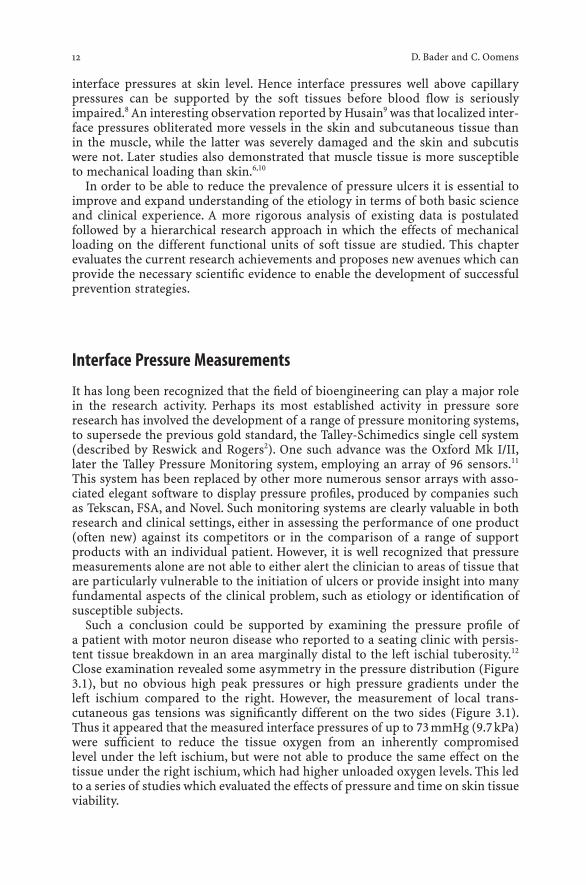
Such a conclusion could be supported by examining the pressure profile ofa patient with motor neuron disease who reported to a seating clinic with persis-tent tissue breakdown in an area marginally distal to the left ischial tuberosity.12
Close examination revealed some asymmetry in the pressure distribution (Figure3.1), but no obvious high peak pressures or high pressure gradients under the left ischium compared to the right. However, the measurement of local trans-cutaneous gas tensions was significantly different on the two sides (Figure 3.1).Thus it appeared that the measured interface pressures of up to 73 mmHg (9.7 kPa)were sufficient to reduce the tissue oxygen from an inherently compromised level under the left ischium, but were not able to produce the same effect on thetissue under the right ischium, which had higher unloaded oxygen levels. This ledto a series of studies which evaluated the effects of pressure and time on skin tissueviability.
12 D. Bader and C. Oomens

Evaluation of Tissue Status under External Loading
Over the last two decades, a number of techniques have been proposed to indicatethe viability, or status, of soft tissues subjected to periods of loading. These tech-niques have been, to date, largely restricted to examining the response of skinlayers to mechanical loading, and include measurements of blood flow in the skinusing laser Doppler fluxmetry13 and reflective spectrophotometry.14,15 Advances inthe latter technique have enabled distinct absorption spectra to be identified foroxygenated and deoxygenated blood in skin. The authors claimed that a numberof other skin biomolecules, such as melanin and collagen, can also be distin-guished.15 However, the most common technique employed to measure skin via-bility involves transcutaneous gas tensions (TcPO2 and TcPCO2), which to ensuremaximum vasodilation have to be measured at elevated skin temperatures.8,16–19
One such study examined the effects of cyclic loading on the tissue viability ofhealthy and debilitated subjects.8 Two distinct responses were observed as shownin Figure 3.2. The normal response yielded a rapid and complete tissue recoveryto unloaded TcPO2 levels and the apparent effect of the applied load diminishedwith successive cycles. By contrast, in some cases recovery was not fully achievedwithin a prescribed period and subsequent loading had a cumulative effect on thediminution of TcPO2 levels. It is this latter group who must be considered to be atparticular risk of developing pressure ulcers.
The technique has also been employed specifically to investigate patients both in the acute phase20 and in the subacute phase of spinal cord injury.21 Thelatter study employed an assessment criterion for tissue viability based on the
Recent Advances in Pressure Ulcer Research 13
Interface pressures (mmHg)
Local transcutaneous oxygen tension (mmHg)
L
31
33
49
15
25
42
39
73
39
41
62
71
31
56
67
29
34
39
39
39
33
45 63
31
22
14
R
Figure 3.1 Interface pressure profile under theischia of a patient with recurrent tissue break-down under the left ischium.(Based on Bader andChase.12)

percentage time at which the TcPO2 and TcPCO2 values were within acceptablelevels when subjects were seated on prescribed support cushions. Clear relation-ships were indicated between depressed levels of TcPO2 and elevated levels ofTcPCO2, at associated high values of interface pressure.21 In addition, it wasreported that changes in tissue viability do occur during a 12-month period,although a subpopulation, involving paraplegic subjects with flaccid paralysis,remain highly susceptible to the development of pressure ulcers.
This research activity spawned the routine use of these objectives measures toassess all patients with spinal cord injury at a specialized seating clinic.18 Thisrecent paper questioned the efficiency of short-term pressure lifts in restoring thetissue oxygen levels following prolonged seated periods. Indeed the authors rec-ommend the use of alternative pressure relief strategies tailored to individualpatients. Although yielding solid practical education for both patients and carers,these and related studies have still yielded no clear guidelines as to the precise rela-tionship between compromised tissue gas levels for a set time period and the onsetof progressive tissue breakdown that will ultimately result in a pressure ulcer.
Tissue Biochemistry
An alternative biochemical approach to assessing tissue status is to examine themetabolite levels in localized soft tissue areas subjected to pressure ischemia andsubsequent reperfusion. These metabolites can be transferred via the sweat glands,which are simple tubular glands, and can be collected at the skin surface. Sweat isa hypotonic solution of sodium and chloride ions in water, together with other con-stituents including lactate, urea, and potassium, these metabolites accounting forabout 95% of the osmotically active substances in sweat.23
In one of the few relevant studies, Hagisawa and colleagues24 used a bulky systemto chemically induce sweat production. By contrast, a series of studies by the authorand colleagues25–28 collected thermally induced sweat by absorption on thin pads,made from filter paper, attached to the skin surface. This collection system
14 D. Bader and C. Oomens
Impaired
Tissueviability
Loadon
Normal
Impaired
Time
Figure 3.2 A schematic rep-resentation of two distinctresponses with respect to theviability of soft tissues sub-jected to repeated loading.Arrows up (down) representstart of applied loading(recovery) period. (Based onBader and Chase.12)
provided minimal distortion and proved ideal for use at a loaded tissue supportinterface. One such study compared sweat collected during periods of loading atthe ischium and sacrum with sweat collected during unloaded periods at adjacenttissue sites.25 The study revealed that tissues subjected to partial ischemia,specifically produced by a uniaxial indenter system, yielded a general increase inconcentrations of sweat lactate, chloride, urea, and urate associated with adecreased sweat rate. Following the removal of loading, the levels of both sweatmetabolites tended to be restored to basal levels.
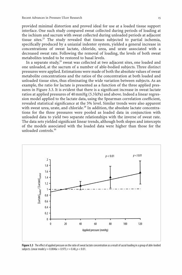
In a separate study,29 sweat was collected at two adjacent sites, one loaded andone unloaded, at the sacrum of a number of able-bodied subjects. Three distinctpressures were applied. Estimations were made of both the absolute values of sweatmetabolite concentrations and the ratios of the concentration at both loaded andunloaded tissue sites, thus eliminating the wide variation between subjects. As anexample, the ratio for lactate is presented as a function of the three applied pres-sures in Figure 3.3. It is evident that there is a significant increase in sweat lactateratios at applied pressures of 40 mmHg (5.3 kPa) and above. Indeed a linear regres-sion model applied to the lactate data, using the Spearman correlation coefficient,revealed statistical significance at the 5% level. Similar trends were also apparentwith sweat urea, urate, and chloride.28 In addition, the absolute lactate concentra-tions for the three pressures were pooled as loaded data in conjunction withunloaded data to yield two separate relationships with the inverse of sweat rate.The data sets yielded significant linear trends, although both slopes and interceptsof the models associated with the loaded data were higher than those for theunloaded controls.30
Recent Advances in Pressure Ulcer Research 15
p < 0.01p < 0.01
Applied pressure (mmHg)
[Lac
tate
] load
ed /
[Lac
tate
] cont
rol
0
0 20 40 60 80 100 120
0.2
0.4
0.6
0.8
1
1.2
1.4
1.6
1.8
2
Figure 3.3 The effect of applied pressure on the ratio of sweat lactate concentration as a result of sacral loading in a group of able-bodiedsubjects. Linear model y = 0.0046x + 0.975; r = 0.48, p < 0.01.

The study was extended by employing two independent techniques in combi-nation to assess the soft tissue response to applied pressure in a group of able-bodied subjects, to establish baseline data.29 The methods involved thesimultaneous measurement of the local tensions of oxygen and carbon dioxide(TcPO2 and TcPCO2) and the collection and subsequent analysis of metabolite con-centrations of sweat samples. Adjacent loaded and unloaded sites on the sacrumwere tested to allow for between-subject variation. Several parameters wereselected from each of the techniques and their interrelationships were examined.Results indicated that oxygen levels (TcPO2) were lowered in soft tissues subjectedto applied pressures of between 40 mmHg (5.3 kPa) and 120 mmHg (16.0 kPa).29 Atthe higher pressure levels, this decrease was generally associated with an increasein carbon dioxide levels well above the normal basal levels of 45 mmHg (6 kPa). Bycomparing selected parameters, a threshold value for loaded TcPO2 could beidentified, representing a reduction of approximately 60% from unloaded values,as indicated in Figure 3.4a. Above this threshold level there was a significant rela-tionship between this parameter and the loaded/unloaded concentration ratios forboth sweat lactate and urea.29 Given that tissue oxygen and sweat lactate reflect dif-ferent aspects of tissue ischemia, this degree of reduction (60% in median oxygentension) may represent a critical level for the development of tissue damage. Thestudy also related the lactate ratio to the percentage time at which TcPCO2 exceeded50%. Figure 3.4b indicates the presence of two distinct clusters of data. Forexample, when the carbon dioxide parameter exceeded 37%, the lactate ratios werewell in excess of unity. Differences could be attributed to the degree of pressure-induced tissue ischemia. Thus under conditions of mild ischemia elevated levelsof tissue carbon dioxide may be released from loaded areas in a normal manner,resulting in TcPCO2 values below 50 mmHg, whereas in severe conditions, bothsweat lactate and TcPCO2 will be elevated (Figure 3.4b).
Sweat lactate is generally thought to be derived from the sweat gland itself.23,31
During normal metabolism, oxidative phosphorylation is believed to be the mainmetabolic pathway of the eccrine sweat gland.32 However, under conditions ofischemia and/or in anaerobic conditions, glycolysis becomes the main metabolicpathway resulting in the formation of lactate. This explains the elevated lactate con-centrations observed in the sweat collected from the loaded experimental site andsuggests that a sufficient degree of ischemia was induced in the sacral tissue duringthe two loading periods.
Sweat urea is believed to be derived mainly from serum urea by the passive dif-fusion across the glandular wall and cell membrane, although it is still unknownwhether it is also produced by the sweat gland.32 Urea is the main product ofprotein metabolism and can thus be an indicator of tissue damage if elevated levelsare found in bodily fluids, such as urine or blood. Prolonged periods of ischemiacan lead to muscle damage, resulting in an increased serum urea level which, inturn, can result in enhanced concentrations of sweat urea.32 These findings as evi-denced in the published study29 suggest that the tissue was compromised duringthe loading period. It was strongly proposed by the authors that such an approach,using a series of parameters, might prove useful in identifying those subjectswhose soft tissue may be compromised during periods of pressure ischemia.
Current work by the authors suggests that monitoring sweat lactate and ureaalone is not sufficient to give a full indication of the tissue status, particularlyduring reperfusion.30 Sweat purines, specifically uric acid, xanthine, and hypoxan-thine, are undoubtedly useful markers or “finger prints,” as they provide an
16 D. Bader and C. Oomens

Recent Advances in Pressure Ulcer Research 17
2.5
2.0
1.5
1.0
0.5
0.00 20 40 60 80 100
Lact
ate r
atio
(loa
ded/
unlo
aded
)
Percentage reduction in median TcPO2a
b
2.5
2.0
1.5
1.0
0.5
0.00 20 40 60 80 100
Lact
ate r
atio
(loa
ded/
unlo
aded
)
Percentage loading time TcPO2 > 50 mmHg
Figure 3.4 Relationship between ratio of sweat lactate concentration and (a) percentage reduction in transcutaneous gas tension (TcPO2)and (b) percentage of time for which transcutaneous carbon dioxide tension (TcPCO2) exceeded 50 mmHg, as a result of sacral loading onindividual subjects. (Based on Knight et al.29)

indication of the metabolic status of the tissue during both ischemia, when thereis energy depletion, and reperfusion and, as such, may be of significant potentialuse to identify patients at risk of developing pressure ulcers. It is clear that the useof a combination of biochemical markers is required to monitor the status of softtissues.
Internal Mechanical Environment
Although it is well acknowledged that pressure sores are primarily caused by sus-tained mechanical loading of the soft tissues of the body, prevention of the soresby reducing the degree of loading alone remains difficult. This is mainly due to thefact that the underlying pathways whereby mechanical loading leads to tissuebreakdown are poorly understood. It is not clear how global, external loading con-ditions are transferred to local stresses and strains inside the tissues and how theseinternal conditions may ultimately lead to tissue breakdown.
As mentioned in the introduction, surface or interface pressures are not repre-sentative of the internal mechanical conditions inside the tissue,which are most rel-evant for tissue breakdown. This is especially the case when tissue geometry andcomposition are complex and surface pressures result in highly inhomogeneousinternal mechanical conditions, as is the case adjacent to bony prominences.Nonetheless, in order to study the response of various tissue layers to mechanicalloading the local mechanical environment within these layers needs to be known.There are options available to measure the internal mechanical state, although theyinevitably involve invasive techniques such as a wick catheter.33,34 Sangeorzan et al.34
reported that the values for interface and intersitial pressures were not equivalentand were highly dependent on the nature of the intervening soft tissues. Thus thethickness, tone, and mechanical integrity of subcutaneous tissues, and the proxim-ity of bony prominences will influence this relationship. A more recent investiga-tion of elderly subjects during a single surgical procedure, namely the fixation of afractured neck of femur, examined the response of tissues adjacent to the lateralaspect of the proximal thigh. Results indicated that skin interface pressures weredissipated within the depth of the tissues resulting in reduced internal stresses.35
Indeed linear models of the data suggested interstitial stresses ranging between 29%and 40% of the applied interface pressures, as illustrated in Figure 3.5. This high-lights the protective nature of tissues to attenuate the effects of sustained pressure.
An alternative approach to investigate the transition from global external loadsto local internal stresses and strains involves the use of computer models, in par-ticular using finite element analysis (FEA).36–39 This approach, which models thecomplex geometries and material behavior of the human buttocks, is often unfa-miliar to experimentalists and clinical and nursing staff. In the study by Todd andTacker,37 the seated positions were simulated, thereby manipulating boundary con-ditions of the model. These authors concluded that there was no clear correlationbetween interface pressures and the local mechanical conditions. Oomens and co-workers40 created a finite element model of a human subject sitting on a cushion,which incorporated three different tissues, overlaying the human ischial tuberosi-ties, simulated by an undeformable bony indenter. The soft tissues, namely themuscle, fat, and skin, were modeled as nonlinear viscoelastic materials. Figure 3.6clearly shows the inhomogeneous mechanical condition of the various tissue layersand areas of high internal stresses in the deeper fat and muscle layers.
18 D. Bader and C. Oomens

However, any extrapolation of results from these computer analyses to the clin-ical setting must be undertaken with extreme caution. Specifically these modelsare dependent on the lack of reliable material properties for soft tissues, which canbe influenced by many systemic and local factors, such as temperature and nutri-tional status. Thus, although several studies have examined uniaxial and biaxialproperties of skin parallel to its surface, there are few reported studies examiningthe compressive properties of the soft tissue composite. Such studies have been
Recent Advances in Pressure Ulcer Research 19
80
70
60
50
40
30
20
10
00 20 40 60 80 100 120 140
Inte
rstit
ial p
ress
ure (
mm
Hg)
Interface pressure (mmHg)
Figure 3.5 The relationshipbetween interface pressuresand interstitial pressureswithin the soft tissues adja-cent to the greater trochanterof two surgical patientsundergoing hip screw fixa-tion of an intertrochantericfemoral fracture. Slopes oftwo linear models are 0.28and 0.41, r > 0.96 in bothcases. (Based on Bader andWhite.35)
Cushion
Skin
Buttockmodel
BoneMuscle
Fat2.0 [MPa]1.61.20.80.40.0
Figure 3.6 Simplified com-puter model of axisymmetricdeformed buttock (top right)demonstrating the differen-tial response of the separatesoft tissue layers (left) duringsitting of an 80 kg malesubject on a foam cushion.Values indicate Von Misesstresses, representing distor-tional energy. Note the areasof high stress in the subcuta-neous fat and muscle layers(arrows). (Based on Oomenset al.40)
hampered by the lack of appropriate non-invasive techniques that can character-ize material properties of tissue under load. For example, ultrasound has offeredmuch potential for many years but has, as yet, not proved reliable, although moresophisticated systems involving elastography in association with ultrasoundimaging might prove successful in the future. Other imaging technologies involving infrared spectroscopy and magnetic resonance imaging/spectroscopy(MRI/MRS) may also provide valuable data under loading conditions for bothhealthy tissues and where tissue status is compromised. Indeed in recent studiesGefen and colleagues41,42 have determined mechanical stiffness of soft tissuesunder load, using routine MRI scans. An increased mechanical stiffness was alsoreported corresponding to mixed tissue specimens around human ulcers com-pared to control values.43
Mechanisms of Pressure Ulcer Development
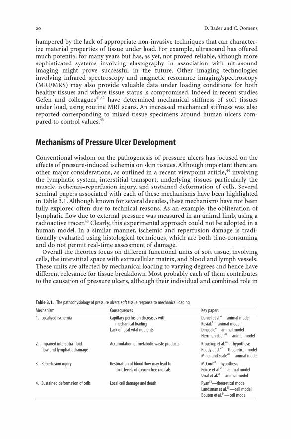
Conventional wisdom on the pathogenesis of pressure ulcers has focused on theeffects of pressure-induced ischemia on skin tissues. Although important there areother major considerations, as outlined in a recent viewpoint article,44 involvingthe lymphatic system, interstitial transport, underlying tissues particularly themuscle, ischemia–reperfusion injury, and sustained deformation of cells. Severalseminal papers associated with each of these mechanisms have been highlightedin Table 3.1. Although known for several decades, these mechanisms have not beenfully explored often due to technical reasons. As an example, the obliteration oflymphatic flow due to external pressure was measured in an animal limb, using aradioactive tracer.48 Clearly, this experimental approach could not be adopted in ahuman model. In a similar manner, ischemic and reperfusion damage is tradi-tionally evaluated using histological techniques, which are both time-consumingand do not permit real-time assessment of damage.
Overall the theories focus on different functional units of soft tissue, involvingcells, the interstitial space with extracellular matrix, and blood and lymph vessels.These units are affected by mechanical loading to varying degrees and hence havedifferent relevance for tissue breakdown. Most probably each of them contributesto the causation of pressure ulcers, although their individual and combined role in
20 D. Bader and C. Oomens
Table 3.1. The pathophysiology of pressure ulcers: soft tissue response to mechanical loading
Mechanism Consequences Key papers
1. Localized ischemia Capillary perfusion decreases with Daniel et al.6—animal model mechanical loading Kosiak7—animal model
Lack of local vital nutrients Dinsdale4—animal model Herrman et al.45—animal model
2. Impaired interstitial fluid Accumulation of metabolic waste products Krouskop et al.46—hypothesis flow and lymphatic drainage Reddy et al.47—theoretical model
Miller and Seale48—animal model
3. Reperfusion injury Restoration of blood flow may lead to McCord49—hypothesis toxic levels of oxygen free radicals Peirce et al.50—animal model
Unal et al.51—animal model
4. Sustained deformation of cells Local cell damage and death Ryan52—theoretical modelLandsman et al.53—cell modelBouten et al.54—cell model
tissue breakdown will undoubtedly vary depending on the nature of the mechan-ical insult and patient characteristics such as illness or age,55 which affect softtissue properties and hence the liability to tissue breakdown.
Hierarchical Approach
A hierarchical approach has recently proposed44 in which the effects of loading arestudied using different, yet complementary, model systems with increasing com-plexity and length scale and incorporating one or more functional tissue units.Thus, in vitro models, ranging from the single cell (mm scale) to cell-matrix con-structs (mm scale) and individual tissue layers (mm–cm scale), might be used tostudy the relationship between cell deformation and cell damage as well as theinfluence of the surrounding extracellular matrix and three-dimensional tissuearchitecture on this relationship. The role of tissue (re)perfusion and lymph flowas well as the interaction between tissue layers in bulk tissue might further beassessed using in vivo studies with animal models or human subjects.
The different length scales of these models can be coupled to multiscale computer calculations that enable the prediction of the internal microscopicmechanical environment within a given model from global, macroscopic loadingconditions, such as interface pressures (and vice versa). In this way relationshipsbetween, for instance, cell deformation and cell damage54 can be extrapolated tothe level of bulk tissue to give clinically relevant predictions on tissue breakdown.
Recent Focus on Pressure-Induced Muscle Damage
Muscle tissue is particularly susceptible to sustained compression. Compression-induced muscle breakdown predominantly occurs in muscle layers associated withbony prominences, eventually leading to gross tissue degeneration in the form ofdeep pressure ulcers.8,21,57–59 This breakdown starts at the cellular level with nuclearpyknosis and disintegration of the contractile proteins and the cell membrane, fol-lowed by inflammatory reactions.6,7,10,60,61 Although it is clear that both the magni-tude and the duration of compression affect the cellular breakdown, the underlyingpathways whereby tissue compression leads to injury of the cell remain poorlyunderstood. Moreover, most of the mechanisms detailed in Table 3.1 ignore the direct effects of cellular deformation due to prolonged tissue compression,which have recently been suggested as an important trigger for pressure ulcerdevelopment.52–54
The earlier study52 was extended to study cellular breakdown in response to sus-tained cell deformation, independently of other factors, such as blood perfusion.It utilized a three-dimensional in vitro system, incorporating cultured muscle cellsseeded in an agarose gel construct. The feasibility of this system to induce pro-longed cell deformation during gross construct compression was recently demon-strated by the authors.54 Strain applied to the translucent agarose gel results indeformation of the muscle cells to an elliptical form, which can be quantified usingconfocal laser scanning microscopy. Identical cylindrical cores cut from theagarose/cell suspension were subjected to two separate compressive strains, 10%and 20%. The strain was applied for time periods ranging from 0.5 to 12 hours,using a specially designed loading apparatus.62 After each compression period,
Recent Advances in Pressure Ulcer Research 21

sections taken from the central horizontal plane of the individual constructs werestained using both histological and fluorescent probes, to assess the proportion ofdamage. It was found that constructs subjected to the higher strain values demon-strated significantly higher values of nonviable cells for equivalent time pointscompared to the unstrained constructs, as illustrated in Figure 3.7. These findingsimply a relationship between the duration of applied compression and damage tomuscle cells seeded in the gel. Such an approach might be useful in establishingdamage threshold levels at a cellular level. The model was extended further bydeveloping a more physiological tissue equivalent muscle,63 by suspending pre-mature muscle cells in a collagen scaffold. The muscle cells fused into a branchednetwork of multinucleated, contractile myofibers by the application of appropri-ate biochemical and mechanical cues. Results indicated that cell death was evidentwithin 1–2 hours at clinically relevant straining percentages.
22 D. Bader and C. Oomens
100
10% strain
20% strain
90
80
70
60
50
40
30
20
10
00 2 4 6
Time of compression/h
Perc
enta
ge
diff
eren
ces
in c
ell v
iab
ility
dta
co
mp
ress
ion
8 10 12
Figure 3.7 THE effects of prolonged static compres-sion at two applied strains onthe viability of muscle cellsseeded in agarose constructs,as indicated by histologicalassessment.

In addition, the uniform distribution of dead cells throughout the muscle con-structs suggested that sustained deformation was the principal cause of cell death.A hybrid approach was then adopted by the authors in which these experimentaldata were used in the derivation of a damage law.64 In particular, the evolution ofdamage was predicted in a single microstructural unit, which could be extrapo-lated to the macroscopic scale. A damage evolution parameter, D, was defined,which accumulates with time when the dimensionless strain energy density para-meter, U, in a cell is higher than a cell tolerance parameter, a. The authors pro-posed a damage evolution equation:
where both a and b are material parameters that can be determined from the invitro experiment. Although limited at the present time to qualitative insight intotissue damage, this multilevel finite element approach has future potential as aquantitative predictor of damage in patient-related simulations.
The advent of new technologies that are sensitive to changes throughout the soft tissue composite provide new opportunities for the examination of animalmodels,50,51,60 despite their limitations associated with intrinsic biological variation,ethical issues, and inadequate experimental controls. As an example, Bosboom etal.60 examined the ability of MRI to assess local muscle damage after prolongedtransverse loading. The tibialis anterior muscle (TA) and overlying skin of a ratwere compressed between an indenter and tibia. A very large pressure, equivalentto 1875 mmHg (250 kPa), was applied for 2 hours. Histological examination, usinga semi-automated image-processing program, and in vivo T2-weighted MRI wereperformed 24 hours after the completion of the loading session. Figure 3.8 (seecolor section) illustrates the damage in transverse histological slices (below) andthe associated MR images for three sets of experiments. In each case, the locationof damage coincided well in the two assessment techniques. However, the inter-animal variability in damage is evident. Current work has involved a modified MR-compatible loading apparatus to produce more reproducible tissue damage andlearn more about the influence of deformation of the tissue and the influence ofreperfusion.65
A large variety of imaging techniques have been developed that can be appliedto assess structure, function, and metabolism of skeletal muscle. These includetagging MRI and perfusion MRI, which can be used to measure local tissue defor-mation and tissue perfusion, respectively. In addition, MR spectroscopy could beapplied to examine the biochemical status of the tissue.
Final Comments
After a stagnant period of research on pressure ulcers and their etiology, there isnow real hope of a resurgence of progress, largely associated with the applicabil-ity of new technology allied to the well-established financial implications of thecosts of the clinical problem to the health of individual nations. This can only beachieved by research teams, medical doctors, carers, and organizations such as theEuropean Pressure Ulcer Advisory Panel (EPUAP) lobbying the appropriate agen-cies to release valuable research funds.66
D U dtt
= -( )Úb a ,0
Recent Advances in Pressure Ulcer Research 23
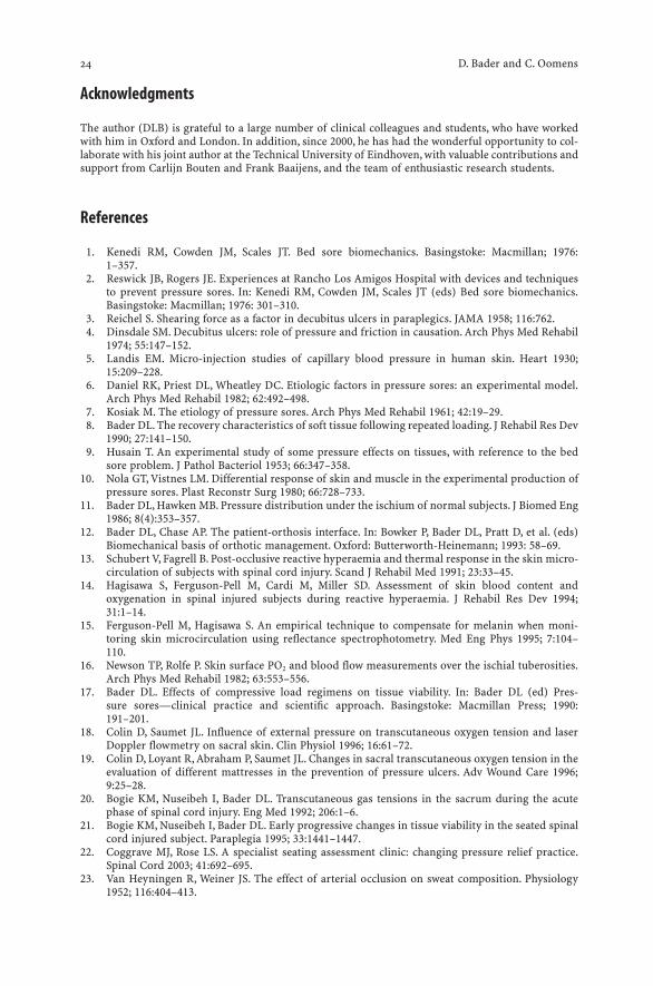
Acknowledgments
The author (DLB) is grateful to a large number of clinical colleagues and students, who have workedwith him in Oxford and London. In addition, since 2000, he has had the wonderful opportunity to col-laborate with his joint author at the Technical University of Eindhoven, with valuable contributions andsupport from Carlijn Bouten and Frank Baaijens, and the team of enthusiastic research students.
References
1. Kenedi RM, Cowden JM, Scales JT. Bed sore biomechanics. Basingstoke: Macmillan; 1976:1–357.
2. Reswick JB, Rogers JE. Experiences at Rancho Los Amigos Hospital with devices and techniquesto prevent pressure sores. In: Kenedi RM, Cowden JM, Scales JT (eds) Bed sore biomechanics.Basingstoke: Macmillan; 1976: 301–310.
3. Reichel S. Shearing force as a factor in decubitus ulcers in paraplegics. JAMA 1958; 116:762.4. Dinsdale SM. Decubitus ulcers: role of pressure and friction in causation. Arch Phys Med Rehabil
1974; 55:147–152.5. Landis EM. Micro-injection studies of capillary blood pressure in human skin. Heart 1930;
15:209–228.6. Daniel RK, Priest DL, Wheatley DC. Etiologic factors in pressure sores: an experimental model.
Arch Phys Med Rehabil 1982; 62:492–498.7. Kosiak M. The etiology of pressure sores. Arch Phys Med Rehabil 1961; 42:19–29.8. Bader DL. The recovery characteristics of soft tissue following repeated loading. J Rehabil Res Dev
1990; 27:141–150.9. Husain T. An experimental study of some pressure effects on tissues, with reference to the bed
sore problem. J Pathol Bacteriol 1953; 66:347–358.10. Nola GT, Vistnes LM. Differential response of skin and muscle in the experimental production of
pressure sores. Plast Reconstr Surg 1980; 66:728–733.11. Bader DL, Hawken MB. Pressure distribution under the ischium of normal subjects. J Biomed Eng
1986; 8(4):353–357.12. Bader DL, Chase AP. The patient-orthosis interface. In: Bowker P, Bader DL, Pratt D, et al. (eds)
Biomechanical basis of orthotic management. Oxford: Butterworth-Heinemann; 1993: 58–69.13. Schubert V, Fagrell B. Post-occlusive reactive hyperaemia and thermal response in the skin micro-
circulation of subjects with spinal cord injury. Scand J Rehabil Med 1991; 23:33–45.14. Hagisawa S, Ferguson-Pell M, Cardi M, Miller SD. Assessment of skin blood content and
oxygenation in spinal injured subjects during reactive hyperaemia. J Rehabil Res Dev 1994;31:1–14.
15. Ferguson-Pell M, Hagisawa S. An empirical technique to compensate for melanin when moni-toring skin microcirculation using reflectance spectrophotometry. Med Eng Phys 1995; 7:104–110.
16. Newson TP, Rolfe P. Skin surface PO2 and blood flow measurements over the ischial tuberosities.Arch Phys Med Rehabil 1982; 63:553–556.
17. Bader DL. Effects of compressive load regimens on tissue viability. In: Bader DL (ed) Pres-sure sores—clinical practice and scientific approach. Basingstoke: Macmillan Press; 1990:191–201.
18. Colin D, Saumet JL. Influence of external pressure on transcutaneous oxygen tension and laserDoppler flowmetry on sacral skin. Clin Physiol 1996; 16:61–72.
19. Colin D, Loyant R, Abraham P, Saumet JL. Changes in sacral transcutaneous oxygen tension in theevaluation of different mattresses in the prevention of pressure ulcers. Adv Wound Care 1996;9:25–28.
20. Bogie KM, Nuseibeh I, Bader DL. Transcutaneous gas tensions in the sacrum during the acutephase of spinal cord injury. Eng Med 1992; 206:1–6.
21. Bogie KM, Nuseibeh I, Bader DL. Early progressive changes in tissue viability in the seated spinalcord injured subject. Paraplegia 1995; 33:1441–1447.
22. Coggrave MJ, Rose LS. A specialist seating assessment clinic: changing pressure relief practice.Spinal Cord 2003; 41:692–695.
23. Van Heyningen R, Weiner JS. The effect of arterial occlusion on sweat composition. Physiology1952; 116:404–413.
24 D. Bader and C. Oomens
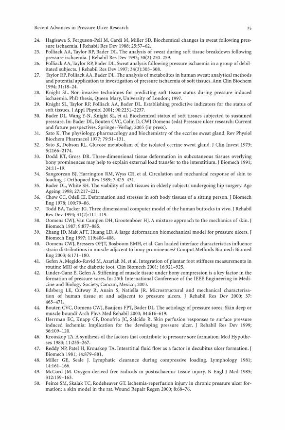
24. Hagisawa S, Ferguson-Pell M, Cardi M, Miller SD. Biochemical changes in sweat following pres-sure ischaemia. J Rehabil Res Dev 1988; 25:57–62.
25. Polliack AA, Taylor RP, Bader DL. The analysis of sweat during soft tissue breakdown followingpressure ischaemia. J Rehabil Res Dev 1993; 30(2):250–259.
26. Polliack AA, Taylor RP, Bader DL. Sweat analysis following pressure ischaemia in a group of debil-itated subjects. J Rehabil Res Dev 1997; 34(3):303–308.
27. Taylor RP, Polliack AA, Bader DL. The analysis of metabolites in human sweat: analytical methodsand potential application to investigation of pressure ischaemia of soft tissues. Ann Clin Biochem1994; 31:18–24.
28. Knight SL. Non-invasive techniques for predicting soft tissue status during pressure inducedischaemia. PhD thesis, Queen Mary, University of London; 1997.
29. Knight SL, Taylor RP, Polliack AA, Bader DL. Establishing predictive indicators for the status ofsoft tissues. J Appl Physiol 2001; 90:2231–2237.
30. Bader DL, Wang Y-N, Knight SL, et al. Biochemical status of soft tissues subjected to sustainedpressure. In: Bader DL, Bouten CVC, Colin D, CWJ Oomens (eds) Pressure ulcer research: Currentand future perspectives. Springer-Verlag; 2005 (in press).
31. Sato K. The physiology, pharmacology and biochemistry of the eccrine sweat gland. Rev PhysiolBiochem Pharmacol 1977; 79:51–131.
32. Sato K, Dobson RL. Glucose metabolism of the isolated eccrine sweat gland. J Clin Invest 1973;5:2166–2174.
33. Dodd KT, Gross DR. Three-dimensional tissue deformation in subcutaneous tissues overlyingbony prominences may help to explain external load transfer to the interstitium. J Biomech 1991;24:11–19.
34. Sangeorzan BJ, Harrington RM, Wyss CR, et al. Circulation and mechanical response of skin toloading. J Orthopaed Res 1989; 7:425–431.
35. Bader DL, White SH. The viability of soft tissues in elderly subjects undergoing hip surgery. AgeAgeing 1998; 27:217–221.
36. Chow CC, Odell EI. Deformation and stresses in soft body tissues of a sitting person. J BiomechEng 1978; 100:79–86.
37. Todd BA, Tacker JG. Three dimensional computer model of the human buttocks in vivo. J RehabilRes Dev 1994; 31(2):111–119.
38. Oomens CWJ, Van Campen DH, Grootenboer HJ. A mixture approach to the mechanics of skin. JBiomech 1987; 9:877–885.
39. Zhang JD, Mak AFT, Huang LD. A large deformation biomechanical model for pressure ulcers. JBiomech Eng 1997; 119:406–408.
40. Oomens CWJ, Bressers OFJT, Bosboom EMH, et al. Can loaded interface characteristics influencestrain distributions in muscle adjacent to bony prominences? Comput Methods Biomech BiomedEng 2003; 6:171–180.
41. Gefen A, Megido-Ravid M, Azariah M, et al. Integration of plantar foot stiffness measurements inroutine MRI of the diabetic foot. Clin Biomech 2001; 16:921–925.
42. Linder-Ganz E, Gefen A. Stiffening of muscle tissue under bony compression is a key factor in theformation of pressure sores. In: 25th International Conference of the IEEE Engineering in Medi-cine and Biology Society, Cancun, Mexico; 2003.
43. Edsberg LE, Cutway R, Anain S, Natiella JR. Microstructural and mechanical characterisa-tion of human tissue at and adjacent to pressure ulcers. J Rehabil Res Dev 2000; 37:463–471.
44. Bouten CVC, Oomens CWJ, Baaijens FPT, Bader DL. The aetiology of pressure sores: Skin deep ormuscle bound? Arch Phys Med Rehabil 2003; 84:616–619.
45. Herrman EC, Knapp CF, Donofrio JC, Salcido R. Skin perfusion responses to surface pressureinduced ischemia: Implication for the developing pressure ulcer. J Rehabil Res Dev 1999;36:109–120.
46. Krouskop TA. A synthesis of the factors that contribute to pressure sore formation. Med Hypothe-ses 1983; 11:255–267.
47. Reddy NP, Patel H, Krouskop TA. Interstitial fluid flow as a factor in decubitus ulcer formation. JBiomech 1981; 14:879–881.
48. Miller GE, Seale J. Lymphatic clearance during compressive loading. Lymphology 1981;14:161–166.
49. McCord JM. Oxygen-derived free radicals in postischaemic tissue injury. N Engl J Med 1985;312:159–163.
50. Peirce SM, Skalak TC, Rodeheaver GT. Ischemia-reperfusion injury in chronic pressure ulcer for-mation: a skin model in the rat. Wound Repair Regen 2000; 8:68–76.
Recent Advances in Pressure Ulcer Research 25
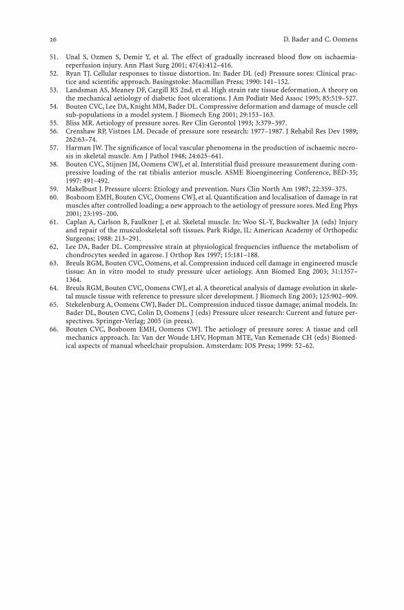
51. Unal S, Ozmen S, Demir Y, et al. The effect of gradually increased blood flow on ischaemia-reperfusion injury. Ann Plast Surg 2001; 47(4):412–416.
52. Ryan TJ. Cellular responses to tissue distortion. In: Bader DL (ed) Pressure sores: Clinical prac-tice and scientific approach. Basingstoke: Macmillan Press; 1990: 141–152.
53. Landsman AS, Meaney DF, Cargill RS 2nd, et al. High strain rate tissue deformation. A theory onthe mechanical aetiology of diabetic foot ulcerations. J Am Podiatr Med Assoc 1995; 85:519–527.
54. Bouten CVC, Lee DA, Knight MM, Bader DL. Compressive deformation and damage of muscle cellsub-populations in a model system. J Biomech Eng 2001; 29:153–163.
55. Bliss MR. Aetiology of pressure sores. Rev Clin Gerontol 1993; 3:379–397.56. Crenshaw RP, Vistnes LM. Decade of pressure sore research: 1977–1987. J Rehabil Res Dev 1989;
262:63–74.57. Harman JW. The significance of local vascular phenomena in the production of ischaemic necro-
sis in skeletal muscle. Am J Pathol 1948; 24:625–641.58. Bouten CVC, Stijnen JM, Oomens CWJ, et al. Interstitial fluid pressure measurement during com-
pressive loading of the rat tibialis anterior muscle. ASME Bioengineering Conference, BED-35;1997: 491–492.
59. Makelbust J. Pressure ulcers: Etiology and prevention. Nurs Clin North Am 1987; 22:359–375.60. Bosboom EMH, Bouten CVC, Oomens CWJ, et al. Quantification and localisation of damage in rat
muscles after controlled loading; a new approach to the aetiology of pressure sores. Med Eng Phys2001; 23:195–200.
61. Caplan A, Carlson B, Faulkner J, et al. Skeletal muscle. In: Woo SL-Y, Buckwalter JA (eds) Injuryand repair of the musculoskeletal soft tissues. Park Ridge, IL: American Academy of OrthopedicSurgeons; 1988: 213–291.
62. Lee DA, Bader DL. Compressive strain at physiological frequencies influence the metabolism ofchondrocytes seeded in agarose. J Orthop Res 1997; 15:181–188.
63. Breuls RGM, Bouten CVC, Oomens, et al. Compression induced cell damage in engineered muscletissue: An in vitro model to study pressure ulcer aetiology. Ann Biomed Eng 2003; 31:1357–1364.
64. Breuls RGM, Bouten CVC, Oomens CWJ, et al. A theoretical analysis of damage evolution in skele-tal muscle tissue with reference to pressure ulcer development. J Biomech Eng 2003; 125:902–909.
65. Stekelenburg A, Oomens CWJ, Bader DL. Compression induced tissue damage; animal models. In:Bader DL, Bouten CVC, Colin D, Oomens J (eds) Pressure ulcer research: Current and future per-spectives. Springer-Verlag; 2005 (in press).
66. Bouten CVC, Bosboom EMH, Oomens CWJ. The aetiology of pressure sores: A tissue and cellmechanics approach. In: Van der Woude LHV, Hopman MTE, Van Kemenade CH (eds) Biomed-ical aspects of manual wheelchair propulsion. Amsterdam: IOS Press; 1999: 52–62.
26 D. Bader and C. Oomens
4 Etiology and Risk FactorsMark Collier and Zena Moore
Introduction
Despite an increased number of publications being dedicated to issues relevant to the etiology, prevention, and development of pressure ulceration—especiallyduring the past decade—recent prevalence studies undertaken throughoutEurope1 indicate that there is still much work to be undertaken and that pressureulceration is a real problem both for patients and for the healthcare systems inwhich those patients are being cared for.
This chapter seeks to explore the etiology of pressure ulceration, relating thesame to known pathophysiological effects likely to be experienced by the patient,and also highlights some of the known risk factors that may predispose an individual to pressure ulcer development—as supported by available current literature.
While it is acknowledged that there is still much to discover about the etiologyof pressure ulceration, most of the evidence to date focuses on the relationshipbetween external pressures applied to a patient’s skin and tissues not adapted tothese pressures, as well as the effects of the same on the local microcirculation.2,3
Pressure Defined
Bennett and Lee defined pressure as a perpendicular load or force exerted on aunit of area such as the sacrum.4 This gravitational force is also often referred toas compression. The average pressure exerted on the skin can be calculated usingthe following formula:
Pressure = Body weight/Skin contact area
or by the use of pressure-sensitive equipment.5
In addition to the overall concept of pressure, other differing forms of pressurehave also been highlighted—those of shear and friction.
Shear (a Stretching Force)
Shear is a mechanical stress that is parallel to a plane of interest.4 When a high levelof shear is present, then the amount of external pressure necessary to produce vas-cular occlusion is only about half the amount when shear is not present.6 When
27
trying to describe various clinical examples of shear, many authors have noted thatwhen the head of the bed is elevated there is automatically a greater compressiveforce placed on the sacral tissues than when the bed is in the flat position.7–9 It isthought that the shear ulcer may typically develop as a result of the patient’s sacralskin adhering to the bed linen (in the sitting position); the deep fascia moves in adownward direction with the skeletal structure as a result of gravitational forces,while at the same time the sacral fascia remains attached to the sacral dermis. Thiseffect can be minimized if the patient support surface is covered with a vapor-permeable two-way stretch cover that helps to reduce moisture build-up at theinterface.10 However, if the effects of shear are prolonged or exacerbated by the pres-ence of moisture, regional stretching of the microcirculation of the skin may occur.If this is left unchecked it can lead to the avulsion of local capillaries and arterioles,increasing the possibility of the development of some localized tissue necrosis.
Although shear can be differentiated from pressure (compression), it has beenpreviously highlighted that it is difficult to create pressure without shear and shearwithout pressure.11
Friction
Friction occurs when two surfaces move across one another,12 for example when apatient undertakes a sliding transfer from a bed to a wheelchair. Friction itself isnot thought to be a primary factor in the development of pressure ulcers. However,it can exacerbate the stripping of broken epidermis or be the cause of an initialbreak in the skin, which may then be compounded by the effects of pressure andshear forces. If the surface on which the patient is being supported is moist, it hasbeen shown that the friction coefficient will rise and if great enough will actuallylead to adherence of the patient’s skin to the damp surface,13 thereby resulting inan increase in any associated shearing effects.
In summary, then, the physical parameters that must be considered when think-ing of the etiology of pressure ulcers are:
• pressure/compression,• shear,• friction,• humidity of the patient’s skin (may increase risk of adherence as previously
described).
Transmission of Pressure
Any external pressure measured at an interface will be transmitted from the bodysurface (the skin) to the underlying skeletal anatomy (the bone), compressing allof the intermediate tissues. The resultant pressure gradient has been described asthe McClemont “cone of pressure,”14 in which external pressures can increase bythree to five times at the point of greatest pressure experienced, such as at a bonysurface. For example, an external interface pressure of 50 mmHg could rise to asmuch as 200 mmHg at a bony prominence such as an ischial tuberosity.
With pressure being distributed in this way it should become apparent that anyexternal skin blemishes, however minor, identified as a result of the use of a
28 M. Collier and Z. Moore
pressure ulcer/wound classification tool such as those of Torrance15 and Collier16
amongst others,17 may be indicating that necrosis of the underlying tissue isalready becoming established. It is therefore important that all practitioners areable to distinguish between a normal and abnormal physiological response and ifthe latter is suspected that they initiate the further assessment of the same withavailable technology such.18,19
The Normal Physiological Response to Pressure
The previous information should be considered in the light of research20 that hasshown that the pressure in the capillary bed in healthy medical student volunteersranges between 12 and 32 mmHg (Figure 4.1—see color section). Landis in 1930suggested that a value of 32 mmHg was the mean capillary pressure at its arterialinflow—using a micro-injection technique—and other studies suggested that ifthis pressure is exceeded then capillary occlusion occurs—predisposing to tissuedamage.21,22 However, in 1941 Landis revised his work—using an amended tech-nique—identifying that a more realistic figure to be considered as the capillaryclosing pressure should be between 45 and 50 mmHg, over which thresholddamage was likely to ensue. It should be remembered, though, that any pressuresmeasured may have different effects on different parts of the body depending onthe local bone, muscle, and skin structure.
This information becomes clinically relevant when interface pressures betweenthe skin and the standard National Health Service contract mattress have beenreported as between 70 and 100 mmHg over the main bony prominences and the interface pressure between the skin and a commercially available pressure-reducing replacement mattress5 has been shown to be between 30 and 40 mmHgwhen measured on an “average” individual lying in the supine position.23
The capillary loops in the skin run vertically to the surface and are coiled at their bases, thereby limiting the risk of occlusion as a result of direct pres-sure. However, in the subcutaneous tissue, the blood vessels lie mainly in the parallel planes of the deep fascia and follow the paths of ligaments and nerves.This renders them very vulnerable to distortion and occlusion as a result ofpressure from both external sources and the underlying bony structures.24
Prolonged pressure may cause ischemic changes at and around the point ofocclusion. If this occlusion is prolonged, the result is both anoxia and a build-upof circulating metabolites. A release of pressure, however, produces a large andsudden increase in blood flow, as the anoxia and metabolites act on structureswithin the circulatory system, such as precapillary sphincters. This increase inblood flow may be as much as 30 times the resting value and the bright red flush,which is often noted, is referred to as reactive or blanching hyperemia,25 a normalresponse! As little as 5 seconds of external pressure can provoke a physiologicalreaction that may last between one third and three quarters of the period ofischemia.26 If the lymphatic vessels of the dependent tissue remain intact andexcess interstitial fluid is removed, then it is said that permanent tissue changeswill not progress.12 Blanching hyperemia has been described as the distinct erythema caused by reactive hyperemia which when light finger pressure is applied will blanch (change color—whiten), indicating that the patient’s microcir-culation is generally intact. Nonblanching hyperemia—an abnormal physiologicalresponse—is detected when the color of the erythema remains when light finger
Etiology and Risk Factors 29
pressure is applied, indicating a degree of microcirculatory disruption often asso-ciated with other clinical signs such as blistering, induration (alteration in textureof the skin), and edema.15
Note: The vessels in the subcutaneous tissues also give rise to the perforatorsthat also supply the skin, and so deep vessel obstruction is likely to result in bothcutaneous and subcutaneous ischemia if the period of occlusion is sustained. Theresults of transcutaneous oxygen assessments have suggested that perfusion of theskin is affected more greatly by subcutaneous pressure than by external interfacepressures only.27
In order to accurately recognize both blanching and nonblanching hyperemia,it is important for the assessing practitioner not only to fully understand thedefinition of a pressure ulcer, but also to understand the pathophysiology of reac-tive hyperemia as has been reported elsewhere.28
Although the fragile nature of the microcirculation has been acknowledged, thisdoes not take account of the protective function of collagen. It appears that if tissuecollagen levels are not depleted, this helps to prevent disruption to the microcir-culation by buffering the interstitial fluid from external pressures, thereby main-taining the optimum hydrostatic pressure.
Risk Factors
In order to identify which individuals are at risk of pressure ulcer development, itis first necessary to understand what is meant by risk. Risk has been defined as theprobability of an individual developing a specific problem, such as a pressureulcer.29 Interventions employed to combat risk are often expensive and healthcareresources are not infinite; therefore, it is important for all practitioners to accu-rately identify those patients who need prevention strategies.
Many authors have attempted to identify the factors that influence the develop-ment of pressure ulcers and have summarized these factors into risk assessmenttools for use in clinical practice.30–33 This has proved a difficult task, as it is knownthat there are a vast number of potential risk factors. Indeed this is borne out in areview of 100 pressure ulcer articles by Gosnell,34 where a possible 126 risk factorswere identified.
Despite the apparent lack of clarity regarding what precisely predisposes anindividual to risk, what appears to be central is that pressure ulcers will onlydevelop if the individual cannot withstand the adverse effects of pressure, shear,and friction35 as previously discussed. This ability had been defined by Braden andBergstrom33 as the person’s “tissue tolerance,” which they suggest is affected byboth intrinsic and extrinsic factors. Whereas it is acknowledged that there arenumerous potential risk factors it has been postulated that some specific factorsplay a key role in the development of pressure ulcers, namely mobility, age, nutri-tion, skin condition, and perfusion.36,37 Mobility, age, and nutrition will form thebasis of discussion in the remainder of this chapter.
Mobility
The role of mobility/immobility in pressure ulcer development has been an impor-tant area of interest to those involved in pressure ulcer prevention for many years.This is brought to mind when one considers that much of the expense related to
30 M. Collier and Z. Moore
this area of patient care revolves around the use of equipment based upon remov-ing or reducing interface pressures caused by prolonged periods of immobility.
Healthy individuals regularly change their position while seated or recumbent.Indeed, Keane38 suggested that the minimum physiological mobility requirement(MPMR) to maintain healthy tissue, while lying on a soft mattress, is one gross postural change every 11.6 minutes. This MPMR is based on observations of aver-age individuals’ repositioning frequencies during sleep. Allman39 agrees that theassociation between limited activity and mobility remains an important con-sideration as highlighted in the seminal work of Exton-Smith and Sherwin.40 In thisstudy, the authors found that the amount of spontaneous nocturnal movement ofelderly individuals was positively related to the development of pressure ulcers.Furthermore, as the number of movements increased the number of pressure ulcers decreased.Patients who made 50 or more movements had no pressure ulcers,whereas 90% of patients who made 20 or fewer movements developed ulcers.39
Pressure, from lying or sitting on a particular part of the body, results in oxygendeprivation to the affected area.38 There is a responding painful stimulus that moti-vates the individual to move if this feedback mechanism has not been impaired asa result of previous injury for example; failure to reposition will result in ongoingoxygen deprivation and inevitable tissue damage.35 The amount of damage thatensues is partly influenced by the individual’s level of adipose tissue and the typeof surface they are lying on.38 Importantly, the duration of pressure sustained isalso affected by a number of factors. The primary concern is the individual’s abilityto feel pain and the secondary concern is the individual’s actual physical ability tomove or reposition themselves.35
Using regression techniques, Papanikolaou et al.41 estimated the probability ofpressure ulcer occurrence in patients with reduced mobility, compared with thosewithout reduced mobility. The odds ratio (OR) was identified as 5.41 (p = 0.001, CI2.00–14.63). Odds ratio is a way of comparing whether the probability of a certainevent is the same for two groups. In this case, because the odds ratio is greater thanone, this would suggest that reduced mobility increases the likelihood of pressureulcer development.29
This study is supported by the earlier work of Mino et al.42 who found a four-fold greater relative risk (RR) for the development of pressure ulcers in patientswho are unable to turn over in bed (RR 4.09). Relative risk is calculated by divid-ing the risk of an event in one group (pressure ulcers, in those incapable of turningin bed) by the risk of the event in the other group (pressure ulcers, in those capableof turning in bed).29
The relationship between pressure ulcer development and immobility has alsobeen noted by Berlowitz et al.43 (OR 1.1) and Lindgren et al.44 (OR 0.53, p = 0.011).Although these studies have been conducted on different groups of patients, in dif-ferent healthcare settings, they do suggest that prolonged periods of immobilitywill increase an individual’s risk of developing pressure ulcers. Therefore, the levelsof activity and mobility appear to be important factors to consider in assessing anindividual’s risk of pressure ulcer development.
Age
The association between age and pressure ulcer development is of value to explorein today’s healthcare climate. Demographic forecasts suggest that in 50 years there
Etiology and Risk Factors 31
will be three times more elderly people living in the world.45 Indeed, by the year2050, it is estimated that the elderly will comprise almost 17% of the global popu-lation compared to 7% in 2002.45 The older population appear to be at greater riskof pressure ulcer development due to the likelihood of underlying neurological andcardiovascular problems.46 Furthermore,as a consequence of aging, the skin under-goes a number of pathological changes.37 These changes alter the elastin and colla-gen content of the skin, reducing its elasticity and resilience, which in turn lowersthe skin’s protective mechanism against the adverse effects of shear and friction.35
The precise association between age and pressure ulcer development has beenexplored by Margolis et al.47 In this UK study the authors identified an incidenceof 0.57–0.60 g/l per 100 person-years, over a 3–9 month period among elderlypatients (>65 years) attending general medical practice services.47 Pressure ulcersof stage 2 or more were included in the data, as defined by Margolis.48 Increasingage was noted to heighten the likelihood of pressure ulcer development and thiswas found to be statistically significant (p < 0.001).
A relationship between age and pressure ulcer development was also found inan incidence study conducted in 116 acute care facilities in the USA.49 In this studythe incidence of pressure ulcers was noted to be 7%. Grade 1–4 pressure ulcerswere included as per the NPUAP grading50 and most pressure ulcers were observedto be of grade 1 or grade 2 damage (91%). Seventy-three percent of ulcers devel-oped in those over 65 years of age, with the most common anatomical sites affectedbeing the sacrum/coccyx and the heels.
Other authors have noted the association between increasing age and pressureulcers; for example, Halfens et al.51 identified an odds ratio of 2.68, and found thisto be statistically significant (p < 0.001). Furthermore, Casimiro et al.,52 Young etal.,53 and Baumgarten et al.54 found odds ratios of 1.03, 1.3, and 6.0 respectively,linking age with pressure ulcer development. These findings have been confirmedby Bergstrom et al.,55 who identified an odds ratio of 0.91 using logistic regression,and this again was noted to be statistically significant (p < 0.001).
Therefore, it is reasonable to assume that the older the individual the greater therisk of pressure ulcer development. However, this information should not be inter-preted blindly as any individual of any age can develop a pressure ulcer if theircondition is sufficiently poor.46 Therefore, although the older population are ahigh-risk group, one should also be alert for other vulnerable individuals.
Nutrition
The precise role of nutrition in the development of pressure ulcers remains asubject of debate.56 However, despite this uncertainty, there remains a great interest in this area and thus it is of value to explore the subject further. It appears that, primarily, poor nutrition leads to increased muscle wasting and soft tissue loss, increasing the prominence of bony points.57 This in turn com-pounds the adverse effects of prolonged immobility. Furthermore, collagen pro-duction is influenced by nutritional status and adequate synthesis and depositionis needed for tissue strength.37 Adequate tissue strength is required in order toprotect the individual from the negative effects of pressure, shear, and frictionforces.35
Anthony et al.58 suggest that the serum albumin levels of individual patients havebeen traditionally the focus of wide research, including its potential role in pres-
32 M. Collier and Z. Moore

sure ulcer development. Working on the basis that serum albumin is the mostcommon method of assessing nutritional status, Anthony et al.58 set out to exploreits relevance as a predictor of pressure ulcer risk. Serum albumin levels wererecorded for 773 patients, over the age of 65 years, admitted without a pressureulcer to an acute hospital setting. The patients were expected to have a hospitalstay of greater than 7 days and all had Waterlow pressure ulcer risk scores recorded.A statistically significant difference (p < 0.001) was noted regarding the serumalbumin levels of those patients who went on to develop a pressure ulcer whencompared to those who did not. The authors conducted further statistical analy-sis, using logistic regression, and serum albumin levels remained a statisticallysignificant consideration (p = 0.009). The odds ratio was calculated at 0.9465(adjusted), suggesting that reducing the serum albumin level by 10 would increasethe individual’s risk of pressure ulcer development by two thirds.58 Despite the limitations of the study, such as the restriction of the population to only the elderly and the purposive method of sampling, the authors do demonstrate a linkbetween albumin and pressure ulcer risk. Furthermore this link has also beennoted by Mino et al.,42 who identified that the relative risk for the development ofpressure ulcers in patients with hypoalbuminemia was 5.9 and this was found tobe statistically significant (p < 0.001).
In a study by Margolis et al.47 that looked at pressure ulcer risk in communitypatients, 0.4% of the population were noted to be suffering with malnutrition.When pressure ulcer rates for those with malnutrition were compared with ratesfor those without malnutrition, the authors noted that the relative risk was 3.06.The role of malnutrition in pressure ulcer development has also been explored byBaumgarten et al.54 In a sample of 9400 elderly patients with hip fractures, 6% werenoted to be suffering from cachexia or malnutrition. Of those who were poorlynourished, 19.6% developed a pressure ulcer, compared to 8.1% in the group whowere nutritionally stable. The odds ratio for pressure ulcer development in thecachexia or malnourished group was 1.1 (adjusted).
Although there is a body of evidence suggesting that there is an associationbetween pressure ulcer development and nutritional status, what remains unclearis the precise mechanism by which malnutrition affects this development.35 Oneneeds to bear in mind factors such as general wellness, ability to eat, quality andavailability of food, and psychosocial factors, all which influence nutritional intake.As nutrition may impact on the individual’s ability to withstand the adverse effectsof pressure shear and friction, an emphasis on improving the intake of food andfluids is essential.56
Conclusion
Issues relevant to pressure ulcers remain a major challenge in today’s healthcaresettings. Knowledge of both the etiology and risk factors associated with pressureulcer development is the key to successful prevention strategies. Although thereare a vast number of potential risk factors there are a few (Table 4.1) that have beenreinforced in the literature as being of considerable importance. Mobility, age, andnutrition have been discussed in this chapter and have been found to be positivelyassociated with the development of pressure ulcers. It is therefore important thatdue consideration be given in particular to these risk factors when planning pres-sure ulcer prevention strategies/interventions.36 It is also important, however, to
Etiology and Risk Factors 33
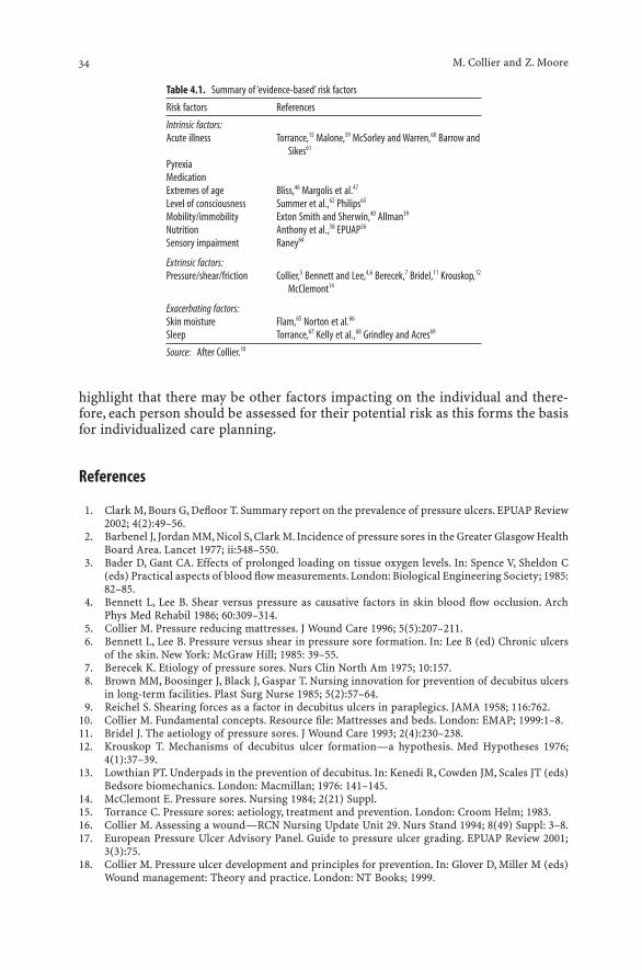
highlight that there may be other factors impacting on the individual and there-fore, each person should be assessed for their potential risk as this forms the basisfor individualized care planning.
References
1. Clark M, Bours G, Defloor T. Summary report on the prevalence of pressure ulcers. EPUAP Review2002; 4(2):49–56.
2. Barbenel J, Jordan MM, Nicol S, Clark M. Incidence of pressure sores in the Greater Glasgow HealthBoard Area. Lancet 1977; ii:548–550.
3. Bader D, Gant CA. Effects of prolonged loading on tissue oxygen levels. In: Spence V, Sheldon C(eds) Practical aspects of blood flow measurements. London: Biological Engineering Society; 1985:82–85.
4. Bennett L, Lee B. Shear versus pressure as causative factors in skin blood flow occlusion. ArchPhys Med Rehabil 1986; 60:309–314.
5. Collier M. Pressure reducing mattresses. J Wound Care 1996; 5(5):207–211.6. Bennett L, Lee B. Pressure versus shear in pressure sore formation. In: Lee B (ed) Chronic ulcers
of the skin. New York: McGraw Hill; 1985: 39–55.7. Berecek K. Etiology of pressure sores. Nurs Clin North Am 1975; 10:157.8. Brown MM, Boosinger J, Black J, Gaspar T. Nursing innovation for prevention of decubitus ulcers
in long-term facilities. Plast Surg Nurse 1985; 5(2):57–64.9. Reichel S. Shearing forces as a factor in decubitus ulcers in paraplegics. JAMA 1958; 116:762.
10. Collier M. Fundamental concepts. Resource file: Mattresses and beds. London: EMAP; 1999:1–8.11. Bridel J. The aetiology of pressure sores. J Wound Care 1993; 2(4):230–238.12. Krouskop T. Mechanisms of decubitus ulcer formation—a hypothesis. Med Hypotheses 1976;
4(1):37–39.13. Lowthian PT. Underpads in the prevention of decubitus. In: Kenedi R, Cowden JM, Scales JT (eds)
Bedsore biomechanics. London: Macmillan; 1976: 141–145.14. McClemont E. Pressure sores. Nursing 1984; 2(21) Suppl.15. Torrance C. Pressure sores: aetiology, treatment and prevention. London: Croom Helm; 1983.16. Collier M. Assessing a wound—RCN Nursing Update Unit 29. Nurs Stand 1994; 8(49) Suppl: 3–8.17. European Pressure Ulcer Advisory Panel. Guide to pressure ulcer grading. EPUAP Review 2001;
3(3):75.18. Collier M. Pressure ulcer development and principles for prevention. In: Glover D, Miller M (eds)
Wound management: Theory and practice. London: NT Books; 1999.
34 M. Collier and Z. Moore
Table 4.1. Summary of ‘evidence-based’ risk factors
Risk factors References
Intrinsic factors:Acute illness Torrance,15 Malone,59 McSorley and Warren,60 Barrow and
Sikes61
PyrexiaMedicationExtremes of age Bliss,46 Margolis et al.47
Level of consciousness Summer et al.,62 Philips63
Mobility/immobility Exton Smith and Sherwin,40 Allman39
Nutrition Anthony et al.,58 EPUAP56
Sensory impairment Raney64
Extrinsic factors:Pressure/shear/friction Collier,5 Bennett and Lee,4,6 Berecek,7 Bridel,11 Krouskop,12
McClemont14
Exacerbating factors:Skin moisture Flam,65 Norton et al.66
Sleep Torrance,67 Kelly et al.,68 Grindley and Acres69
Source: After Collier.10
19. Longport Incorporated. Applications of ultrasound biomicroscopy in wound care; 2003(www.longportinc.com).
20. Landis E. Microcirculation studies of capillary blood pressure in human skin. Heart 1930; 15:209–228.
21. Barton A, Barton M. The management and prevention of pressure sores. London: Faber; 1981.22. Daniel R, Priest D, Wheatley D. Etiologic factors in pressure sores: an experimental model. Arch
Phys Med Rehabil 1981; 62:492–498.23. Scales J, Lowthian P, Poole A, Ludman W. Vaperm patient support system: a new general-purpose
hospital mattress. Lancet 1982; ii(8308):1150–1152.24. Bliss M. Aetiology of pressure sores. Clin Gerontol 1993; 3:379–397.25. Lamb J, Ingram C, Johnson T, Pitman R. Essentials of physiology. London: Blackwell Scientific;
1980.26. Lewis T, Grant R. Observations upon reactive hyperaemia in man. Heart 1925; 4(1):37–39.27. Sangeorzan B, Harrington R, Wyss C, et al. Circulatory response of skin to loading. J Orthop Res
1989; 7:425–431.28. Collier M. Blanching and non-blanching hyperaemia. J Wound Care 1999; 8(2):63–64.29. Deeks J, Higgins J, Riis J, Silagy C. Module 11: Summary statistics for dichotomous outcomes data.
In: Alderson P, Green S (eds) Cochrane Collaboration open learning material for reviewers.Version1.1. Chichester: John Wiley; 2002: 87–102.
30. Gosnell DJ. An assessment tool to identify pressure sores. Nurs Res 1973; 22:55–59.31. Lowthian P. Pressure sore prevalence. Nurs Times 1979; 75:358–360.32. Waterlow J. A risk assessment card. Nurs Times 1985; 81(48):49–56.33. Braden B, Bergstrom N. A conceptual schema for the study of the etiology of pressure sores.
Rehabil Nurs 1987; 12(1):8–16.34. Gosnell DJ. Pressure sore risk assessment part 2: analysis of risk factors. Decubitus 1988; 2(3):
40–43.35. Defloor T. The risk of pressure sore: a conceptual scheme. J Clin Nurs 1999; 8:206–216.36. National Institute for Clinical Excellence. Pressure ulcer prevention: Clinical Guideline No. 7.
London: NICE; 2003.37. Nixon J. Pressure sores. In: Morison MJ, Ovington LG, Wilkie K (eds) Chronic wound care, a
problem-based learning approach. London: Mosby; 2004: 227–245.38. Keane FX. The minimum physiological mobility requirement for man supported on a soft surface.
Paraplegic 1978; 16:383–389.39. Allman RM. Pressure ulcer prevalence, incidence, risk factors and impact. Clin Geriatr Med 1997;
13(3):421–436.40. Exton-Smith AN, Sherwin RW. The prevention of pressure sores: significance of spontaneous
bodily movements. Lancet 1961; 2(7212):1124–1126.41. Papanikolaou P, Lyne PA, Lycett EJ. Pressure ulcer risk assessment: application of logistic
analysis. J Adv Nurs 2003; 44(2):128–136.42. Mino Y, Morimoto S, Okaishi K, et al. Risk factors for pressure ulcers in bedridden elderly
subjects: Importance of turning over in bed and serum albumin level. Geriatr Gerontol Int 2001; 1:38–44.
43. Berlowitz DR, Brandeis GH, Morris JN, et al. Developing a risk-adjustment model for pressure ulcer development using the minimum data set. J Am Geriatr Soc 2001; 49(7):866–871.
44. Lindgren M, Unosson M, Fredrikson M, Ek AC. Immobility—a major risk factor for the develop-ment of pressure ulcers among hospitalised patients: a prospective study. Scand J Caring Sci 2004;18:57–64.
45. US Census Bureau. International population reports WP/02, global population profile, 2002.Washington DC: US Government Printing Office; 2004.
46. Bliss M. Geriatric medicine. In: Bader DL (ed) Pressure sores: Clinical practice and scientificapproach. London: Macmillan; 1990: 65–80.
47. Margolis DJ, Bilker W, Knauss J, et al. The incidence and prevalence of pressure ulcers amongelderly patients in general medical practice. Ann Epidemiol 2002; 12:321–325.
48. Margolis DJ. Definition of a pressure ulcer. Adv Wound Care 1995; 8:8–10.49. Whittington K, Patrick M, Roberts J. A national study of pressure ulcer prevalence and incidence
in acute care hospitals. J Wound Care Nurs 2000; 24(4):209–215.50. National Pressure Ulcer Advisory Panel. Pressure ulcer prevalence, cost and risk assessment:
consensus development conference statement. Decubitus 1989; 2:24–28.51. Halfens RJ, van Achterberg T, Bal RM.Validity and reliability of the Braden scale and the influence
of other risk factors: a multi-centre prospective study. Int J Nurs Stud 2000; 37:313–319.
Etiology and Risk Factors 35

52. Casimiro C, Garcia-de-Lorenzo A, Usan L. Prevalence of decubitus ulcer and associated risk factorsin an institutionalised Spanish elderly population. Nutrition 2002; 18:408–414.
53. Young J, Nikoletti S, McCaul K, et al. Risk factors associated with pressure ulcer development at amajor Western Australian teaching hospital from 1998 to 2000; Secondary data analysis. J WoundCare Nurs 2002; 29:234–241.
54. Baumgarten M, Margolis D, Berlin JA, et al. Risk factors for pressure ulcers among elderly hip fracture patients. Wound Repair Regen 2003; 11:96–103.
55. Bergstrom N, Braden B, Kemp M, et al. Multi-site study of incidence of pressure ulcers and the relationship between risk level, demographic characteristics, diagnoses and prescription ofpreventive interventions. J Am Geriatr Soc 1996; 44(1):22–30.
56. European Pressure Ulcer Advisory Panel. Report from the guideline development group. EPUAPReview 2003; 5(3):80–82.
57. Eachempati S, Hydo LJ, Barie PS. Factors influencing the development of decubitus ulcers in critically ill surgical patients. Crit Care Med 2001; 29(9):1678–1682.
58. Anthony D, Reynolds T, Russell L. An investigation into the use of serum albumin in pressure soreprediction. J Adv Nurs 2000; 32(2):359–365.
59. Malone C. Intensive pressures. Nurs Times 1992; 88(Suppl):3–8.60. McSorley P, Warren D. The effects of propranolol and metoprolol on the peripheral circulation.
BMJ 1978; ii:1598–1600.61. Barrow T, Sikes C. Decubitus ulcers in rheumatic fever treated with cortisone. JAMA 1951; 147:
41–42.62. Summer W, Curry P, Haponikm E, et al. Continuous mechanical turning of intensive care
unit patients shortens length of stay in some diagnostic related groups. J Crit Care 1989;4:45–53.
63. Philips P. Obesity and weight reduction programmes. Geriatr Med 1981; 11(6):53–57.64. Raney J. A comparison of the prevalence of pressure sores in hospitalised ALS and MS patients.
Decubitus 1989; 2(2):48–49.65. Flam E. Skin maintenance in the bedridden patient. Ostomy Wound Manage 1990; May/June:
48–54.66. Norton D, McLaren R, Exton-Smith A. An investigation of geriatric nursing problems in hospital.
Edinburgh: Churchill Livingstone; 1975.67. Torrance C. Sleep and wound healing. Surg Nurse 1990; 3(3):16–20.68. Kelly M, Coverdale S, Williams S, et al. Easing the pressure. Nurs Times 1995; 91(22):72–76.69. Grindley A, Acres J. Alternating pressure mattresses: comfort and quality of sleep. Br J Nurs 1996;
5:1303–1310.
36 M. Collier and Z. Moore

5 Pressure Ulcer ClassificationCarol Dealey and Christina Lindholm
Introduction
Pressure ulcer classification is a method of determining the severity of a pressureulcer. A classification system describes a series of numbered stages or grades eachdetermining a different degree of tissue damage. The deeper the ulcer and the moreextensive the tissue damage the higher the grade number, as illustrated in Table5.1. Pressure ulcer classification is a valuable tool for prevalence and incidencesurveys as well as clinical practice and research.
The first author to publish a pressure ulcer classification system was Shea.1 Sincethen, numerous systems have been developed with varying numbers for gradesranging from a 0–5 grade classification to a 1–7 grade classification. The mostcomplex system is the Stirling Grading System, which has 0–4 grades with up tofour subscales within some of the grades; thus a deep necrotic infected ulcer wouldbe labeled as 4.131.2 A review by Hitch3 identified ten different classificationsystems and a later review by Haalboom et al.4 found a further four systems. Prob-ably the most widely used classification is that developed by the National PressureUlcer Advisory Panel (NPUAP)5 and later adopted by the European Pressure UlcerAdvisory Panel (EPUAP) with some minor textual changes (e.g. NPUAP refers tostages and EPUAP to grades; see Table 5.1).6 Figures 5.1–5.4 (see color section)show examples of each of the EPUAP grades with line drawings to show diagram-matically the degree of tissue damage.
The major weakness of all classification systems is the lack of evidence tosupport their use, the most important factor being inter-rater reliability. Healey7
studied inter-rater reliability amongst 109 nurses when using three classificationsystems (Stirling,2 Torrance,8 and Surrey9) and found that although none of thesystems showed a high level of reliability, it was significantly lower in the mostcomplex scoring system (Stirling). Healey also found that there was greater reliability in reporting the grades of severe ulcers compared with the less severegrades. Russell and Reynolds compared the reliability of the Stirling and EPUAPclassification systems when used by 200 specialist and nonspecialist nurses andagain found that the Stirling classification system was less reliable than the simplerEPUAP system.10 Russell and Reynolds conclude that classification of pressureulcers is not easy. Sharp concurs and suggests that such is the complexity of someclassification systems that they require a level of expertise beyond the capabilityof general nurses.11 Certainly, education is essential to ensure high levels of inter-rater reliability.10
37

Defloor and Schoonhooven describe the validation process for an educationaltool using the EPUAP classification to grade photographs of pressure ulcers.12 Inthe first stage of the process nine specialists were asked to review the clarity of 67photographs and grade the ulcers. Eleven unclear photographs were eliminated. Inthe second phase 44 experts were asked to grade the pressure ulcers in the 56remaining photographs and their findings were compared with the original nineexperts. A high level of agreement was found between all the experts. However, theauthors consider that it is likely that there would be less agreement amongst thosewith little experience.
Controversies in Pressure Ulcer Classification
There are a number of controversies relating to pressure ulcer classification thatmay well be linked to reliability. They are listed below and each will be discussedin turn.
• Grade 1 ulcers• Assessing dark skin• Reverse grading• Identifying incontinence lesions
Grade 1 Ulcers
Russell has stressed the difficulties in defining early skin damage.13 A reaction totemporary closure of the dermal capillaries is called reactive hyperemia, clinicallyseen as a bright flush or reddened area that blanches under light pressure. It isthought to last from 30 minutes to 48 hours.14,15 At this stage, damage to the under-lying tissues has not yet occurred.
Shea provided the definition of a grade 1 ulcer as a persistent reddened area thatdoes not blanch.1 This definition was later supported by Versluysen and Yarkonyet al.14,15 Dinsdale put forward an alternative definition of a grade 1 pressure ulceras persistent redness for more than a 24-hour period.16 Lyder states that blanchingerythema indicates that tissue damage has not yet occurred.17 Hence, this mustprecede pressure ulcer development and thus nonblanching erythema should betaken as a true presentation of a grade 1 ulcer. Lyder also set up criteria for assess-ing a grade 1 pressure ulcer. Hitch considered that there is consensus on Lyder’scriteria for a grade 1 ulcer.3 Lyder’s criteria have subsequently been adapted byRussell13 and are shown in Table 5.2.
38 C. Dealey and C. Lindholm
Table 5.1. EPUAP pressure ulcer classification system
Grade Definition
1 Nonblanchable erythema of intact skin. This may be difficult to identify in darkly pigmented skins
2 Partial thickness skin loss involving epidermis and/or dermis: the pressure ulcer is superficial and presents clinically as an abrasion, blister or shallow crater
3 Full-thickness skin loss involving damage or necrosis of subcutaneous tissue that may extend down to, but not through,underlying fascia: the pressure ulcer presents clinically as a deep crater with or without undermining of adjacent tissue
4 Extensive destruction tissue necrosis, or damage to muscle, bone or supporting structures with or without full-thickness skin loss

Assessing Dark Skin
Darkly pigmented skin creates problems because early skin changes are difficultto see.13 Meehan demonstrated that patients with dark skin had a larger per-centage of high-grade ulcers and the least number of stage 1 ulcers.18 Since nonblanchable erythema is difficult to detect in the darkly pigmented skin,palpation has been recommended.19 Observation of localized heat has also beensuggested.13
The NPUAP convened a task force to review the definition of stage 1 pressureulcer and determine the adequacy of this definition in assessing individuals withdarkly pigmented skin. Following a comprehensive review of the literature andpeer review by attendees at the Fifth NPUAP Conference in February 1997, theNational Task Force on Darkly Pigmented Skin and Stage 1 Pressure Ulcers draftedthe following new definition for stage 1 pressure ulcers, which was approved by theNPUAP Board of Directors in 1998:
A Stage I pressure ulcer is an observable pressure related alteration of intact skinwhose indicators as compared to the adjacent or opposite area on the body mayinclude changes in one or more of the following:
skin temperature (warmth or coolness), tissue consistency (firm or boggy feel)and/or sensation (pain, itching).
The ulcer appears as a defined area of persistent redness in lightly pigmented skin,whereas in darker skin tones, the ulcer may appear with persistent red, blue, or purplehues.20
Reverse Grading
As the use of pressure ulcer classification systems became established in clinicalpractice a number of misconceptions also crept in. One was the assumption thatpressure ulcers will first present as a grade 1 and then naturally progress throughto grade 4 without preventative measures, even though there is no evidence tosupport this belief. The other was the practice of reverse grading or describing ahealing ulcer as progressing from a grade 4 to a grade 3 and so on back to grade1. There is no logic in this practice as the tissues in a healing wound do not equateto the tissues as they were before pressure damage occurred. Thus a healing grade4 pressure ulcer, which initially penetrated through to muscle, does not first replace
Pressure Ulcer Classification 39
Table 5.2. Criteria for grade 1 pressure ulcers
Skin area that ranges from pale pink to bright red in color
Skin area that is nonblanching (blanchable erythema being a precursor to a grade 1 pressure ulcer)
Skin area that is warmer or cooler to touch
Skin area with erythema that does not resolve within 2 hours
Skin area that possibly has edema or induration that is ill defined when palpated
Skin area with epidermis intact
Source: Russell.13 Reproduced by kind permission of MA Healthcare Ltd.
the muscle tissue and then the dermis but gradually fills with granulation tissue.This topic was the subject of considerable debate in the USA21,22 and led to theNPUAP making a position statement on the subject.23 Within the position state-ment the NPUAP state that:
Pressure ulcer staging is only appropriate for defining the maximum anatomicaldepth of tissue damage.
We support that viewpoint. If a pressure ulcer presents as a grade 3 it must alwaysbe described as such. However, it is also important to monitor healing and describeit clearly. Dealey described different ways in which wound healing could be evaluated.24
Incontinence Lesions
Persistent incontinence can cause erythema, maceration, and excoriation of theskin, which can be mistaken for a pressure ulcer, as can be seen in Figure 5.5 (seecolor section). Until recently there has been little discussion of this problem in theliterature.25 Schnelle et al. monitored the impact of incontinence on the skin andfound a high incidence of blanchable erythema, particularly in the perinealregion.26 They consider it to be a marker for increased risk of pressure ulcers andother skin disorder, but it seems reasonable to postulate that an unskilled observermay consider such incontinence lesions to be grade 1 pressure ulcers. Incontinencelesions can be identified in the following ways:
• They are unlikely to occur over bony prominences.• They may be more purple than red in appearance.• The skin may be swollen or edematous.• The skin may also be macerated and/or excoriated.• The patient is incontinent or suffers from diarrhea.
Education is essential to alert staff to the possibility of incontinence lesions andlearn how to differentiate them from superficial pressure ulcers.
Conclusions and Recommendations
Pressure ulcer grading is a useful tool for defining the severity of pressure ulcers.However, it is obvious that education is essential in order to ensure that grades are correctly identified and that incontinence lesions are not mistaken for pres-sure ulcers. The EPUAP provides access to a very useful educational program viaits website (www.epuap.org.uk). The PUCLAS program was developed at the Uni-versity of Ghent, Belgium. It provides both educational material and a self-assess-ment quiz. It is currently available in nine languages—English, Dutch, Finnish,French, German, Italian, Portuguese, Spanish, and Swedish—and may be freelyused for personal or educational purposes. This is a great opportunity for individuals to improve their own assessment skills. Wide use of such educationaltools could substantially improve the accuracy of pressure ulcer grading in the clinical area.
40 C. Dealey and C. Lindholm

References
1. Shea JD. Pressure sores, classification and management. Clin Orthop 1975; 112:89–100.2. Reid J, Morison M. Classification of pressure sore severity. Nurs Times 1994; 90(20):46–50.3. Hitch S. NHS Executive Nursing Directorate—Strategy for major clinical guidelines—prevention
and management of pressure sores, a literature review. J Tissue Viability 1995; 5(1):3–24.4. Haalboom JRE, van Everdingen JJE, Cullum N. Incidence, prevalence and classification. In: Parish
LC, Witkowski JA, Crissey JT (eds) The decubitus ulcer in clinical practice. London: Springer;1997.
5. Agency for Health Care Policy and Research. Pressure ulcers in adults: prediction and prevention.Rockville, MD: AHCPR; 1992.
6. EPUAP. Guidelines on the treatment of pressure ulcers. EPUAP Review 1999; 2:31–33.7. Healey F. The reliability and utility of pressure sore grading scales. J Tissue Viability 1995; 5(4):
111–114.8. Torrance C. Pressure sores: aetiology, treatment and prevention. Beckenham: Croom Helm; 1983.9. David J, Chapman RG, Chapman EJ. An investigation of the current methods used in nursing
for the care of patients with established pressure sores. Harrow: Nursing Practice Research Unit;1983.
10. Russell L, Reynolds T. How accurate are pressure ulcer grades? An image-based survey of nurseperformance. J Tissue Viability 2001; 11(2):67–75.
11. Sharp A. Pressure ulcer grading tools: how reliable are they? J Wound Care 2004; 13(2):75–77.12. Defloor T, Schoonhoven L. Inter-rater reliability of the EPUAP pressure ulcer classification system
using photographs. J Clin Nurs 2004; 13(8):952–959.13. Russell L. Pressure ulcer classification: defining early skin damage. Br J Nurs 2002; (Suppl)
11(16):33–41.14. Versluysen M. Pressure sores: causes and prevention. Nursing 1986; 5(3):216–218.15. Yarkony GM, Kirk PM, Carlsson C, et al. Classification of pressure ulcers. Arch Dermatol 1990;
126(9):1218–1219.16. Dinsdale SM. Decubitus ulcers: Role of pressure and friction in causation. Arch Phys Med Rehabil
1974; 62:492–498.17. Lyder CH. Conceptualization of the stage 1 pressure ulcer. J ET Nursing 1991; 18(5):162–165.18. Meehan M. National pressure ulcer prevalence survey. Adv Wound Care 1994; 7(3):27–38.19. Young T. Classification of pressure sores: 1. Br J Nurs 1996; 5(7):438–446.20. NPUAP. Stage I assessment in darkly pigmented skin. NPUAP, 1998 (http://www.npuap.ord/
positn4.htm).21. Maklebust J. Policy implications of using reverse staging to monitor pressure ulcer status. Adv
Wound Care 1997; 10(5):32–35.22. Xakellis G, Frantz R. Pressure ulcer healing: what is it? What influences it? How is it measured?
Adv Wound Care 1997; 10(5):20–26.23. National Pressure Ulcer Advisory Panel. The facts about reverse staging in 2000. The NPUAP
Position Statement, 2000 (www.npuap.org/positn5.htm).24. Dealey C. Care of Wounds 3rd ed. Oxford: Blackwell Publishing; 2005.25. Defloor T. Drukreductie en wisselhouding in de preventie van decubitus. [Pressure reduction and
turning in the prevention of pressure ulcers]. PhD thesis, Ghent University, 2000.26. Schnelle JF, Adamson GM, Cruise PA, et al. Skin disorders and moisture in incontinent nursing
home residents: intervention implications. J Am Geriatr Soc 1997; 45(10):1182–1188.
Pressure Ulcer Classification 41

6 Risk Assessment Scales for Predicting the Risk ofDeveloping Pressure UlcersJoan-Enric Torra i Bou, Francisco Pedro García-Fernández, Pedro L. Pancorbo-Hidalgo,and Katia Furtado
Introduction
Currently, pressure ulcers are an important health problem, both for people withpressure ulcers and their caregivers and for health institutions and professionals.Therefore it is evident, with the knowledge acquired, that the best strategy to copewith this problem is to prevent it, since the majority of pressure ulcers (up to 95%according to some authors1) can be avoided, if the appropriate preventive measuresare applied with adequate resources and within the correct context (such as clinical practice guidelines).
Nevertheless, pressure ulcer prevention is not costless, because it implies highexpenditure, in equipment and human resources.2,3 Thus there is a need to findassessment tools that can determine which patients require preventive measuresand to what extent, and which patients can be spared these measures. It is feasiblefor experienced nurses to do the selection and apply preventive measures, accord-ing to their own clinical judgment. The issue is whether there are risk assessmenttools that could be used (especially by less trained nurses or those who lack experience in managing these patients) that have the same success (or more) indetecting risk as expert clinical judgment. This is the main reason why differentrisk assessment scales have been proposed as tools to assess patients’ risk ofdeveloping pressure ulcers.
We could define a pressure ulcer risk assessment scale (PURAS) as a tool thatestablishes a point scale according to a group of parameters regarded as risk factorsfor the development of pressure ulcers.
PURAS could be beneficial for patients. For example, Hodge et al.4 and Bale etal. 19955 demonstrated that patients assessed using the Norton scale, received 76%more preventive measures than a control group not systematically assessed, andmoreover that the allocation of pressure-relieving surfaces was optimized accord-ing to risk factors, thus reducing incidence. However, there is not a general use ofassessment scales nationally or internationally. In the first national pressure ulcerprevalence study in Spain,6 with professionals who answered a questionnaire aboutepidemiological data and preventive measures, a systematic use of PURAS wasfound in 72.8% of cases in hospital care, 60.31% in residential settings, and 59.5%in primary healthcare.
It is necessary to be aware of the usefulness of PURAS and what they are beingused for. Simply using a PURAS without introducing the appropriate protocol on
43
prevention, which would support the necessary preventive measures according totheir risk staging, has no effect on the reduction of pressure ulcer incidence. Theuse of scales must be followed by other preventive methods which would be muchmore effective if adequately prescribed according to the patient’s risk. Therefore,PURAS must be settled in a protocol context for healthcare, extracted from evidence-supported procedures as developed by clinical practice guidelines,which in turn are a result of the best possible evidence. These should emanate from different worldwide investigation projects, not only in English, which wouldguarantee, when implementation is compulsory, that all professionals have the best knowledge, abilities, time, and resources for implementation in a scenario ofcontinuous evaluation for quality in assistance.7–9
Hence, when a pressure ulcer prevention program is to be designed, one of thefirst steps, and therefore one of the most important, should be the selection of aPURAS.
Since Doreen Norton10 first published her scale in 1962, more than 30 otherscales have appeared in the scientific literature, plus a large number ofmodifications to some of them.11–12
Criteria for selection and implementation of any PURAS must have scientificallybased arguments. In this chapter, we shall present the tools, and the scientific evidence for the use of the PURAS, and analyze in detail the most important scales, as well as the evidence and support behind them.
Scientific Evidence for the Use of Risk Assessment Scales for Pressure Ulcers
As mentioned above, in several clinical practice guidelines (CPG), we find recom-mendations for the use of PURAS based on the best scientific evidence available.Nevertheless, taking into account the lack, so far, of studies that compare clinicaljudgment with the use of scales, we have encountered some uncertainty as to therequirement for adopting a risk assessment scale rather than relying on the clini-cal judgment of individual nurses.
As basic methodological support, almost all CPGs are based on the systematicreviews made by Cullum et al. in 199512 and McGough in 1999.13
These reviews, and subsequent publications, consider that there is not enoughevidence to demonstrate the effectiveness of risk evaluation scales in reducing thedevelopment of pressure ulcers, even though there is some evidence that supportsthe use of PURAS over and above clinical judgment on it own. We should take intoaccount the risk of a possible publication bias in these publications and CPGs, sincethe sources on which they are based were written in English, and any other inves-tigations published in other languages that could reinforce or refute some of theseresults have not been included or considered.
In a recent systematic review of papers published in four languages (English,Spanish, French, and Portuguese) made by Pancorbo et al.,14 well-known state-ments have been confirmed, but it seems that some of the knowledge disseminatedin the most recent publications on the issue are still not included in clinical prac-tice guidelines.
From the first reviews (Cullum et al.12 and McGough13) we show in Tables 6.1and 6.2 the most important risk assessment advice for the CPGs and in Table 6.3the latest review done by Pancorbo et al. concerning works on this subject.
44 J.-E. Torra i Bou et al.
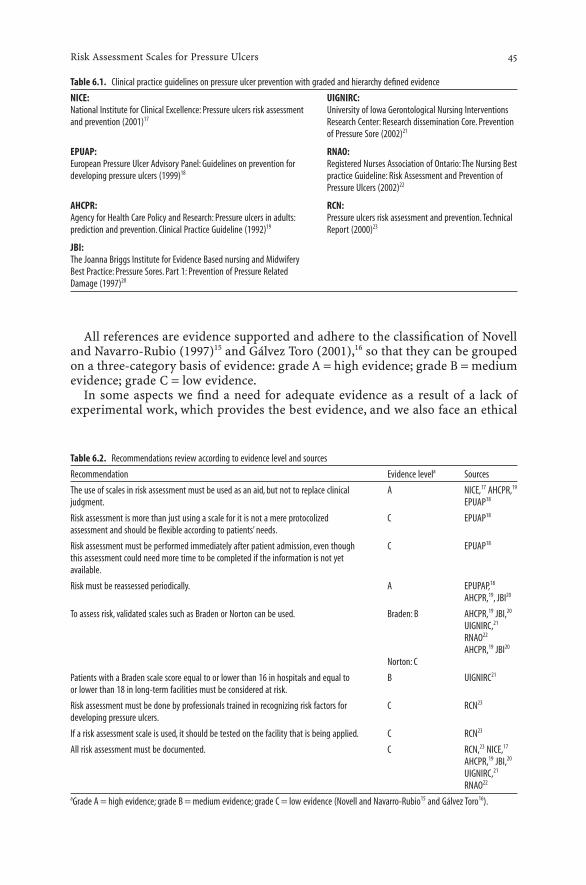
Risk Assessment Scales for Pressure Ulcers 45
Table 6.1. Clinical practice guidelines on pressure ulcer prevention with graded and hierarchy defined evidence
NICE: UIGNIRC:National Institute for Clinical Excellence: Pressure ulcers risk assessment University of Iowa Gerontological Nursing Interventionsand prevention (2001)17 Research Center: Research dissemination Core. Prevention
of Pressure Sore (2002)21
EPUAP: RNAO:European Pressure Ulcer Advisory Panel: Guidelines on prevention for Registered Nurses Association of Ontario: The Nursing Best developing pressure ulcers (1999)18 practice Guideline: Risk Assessment and Prevention of
Pressure Ulcers (2002)22
AHCPR: RCN:Agency for Health Care Policy and Research: Pressure ulcers in adults: Pressure ulcers risk assessment and prevention. Technical prediction and prevention. Clinical Practice Guideline (1992)19 Report (2000)23
JBI:The Joanna Briggs Institute for Evidence Based nursing and Midwifery Best Practice: Pressure Sores. Part 1: Prevention of Pressure RelatedDamage (1997)20
Table 6.2. Recommendations review according to evidence level and sources
Recommendation Evidence levela Sources
The use of scales in risk assessment must be used as an aid, but not to replace clinical A NICE,17 AHCPR,19
judgment. EPUAP18
Risk assessment is more than just using a scale for it is not a mere protocolized C EPUAP18
assessment and should be flexible according to patients’ needs.
Risk assessment must be performed immediately after patient admission, even though C EPUAP18
this assessment could need more time to be completed if the information is not yet available.
Risk must be reassessed periodically. A EPUPAP,18
AHCPR,19, JBI20
To assess risk, validated scales such as Braden or Norton can be used. Braden: B AHCPR,19 JBI,20
UIGNIRC,21
RNAO22
AHCPR,19 JBI20
Norton: C
Patients with a Braden scale score equal to or lower than 16 in hospitals and equal to B UIGNIRC21
or lower than 18 in long-term facilities must be considered at risk.
Risk assessment must be done by professionals trained in recognizing risk factors for C RCN23
developing pressure ulcers.
If a risk assessment scale is used, it should be tested on the facility that is being applied. C RCN23
All risk assessment must be documented. C RCN,23 NICE,17
AHCPR,19 JBI,20
UIGNIRC,21
RNAO22
aGrade A = high evidence; grade B = medium evidence; grade C = low evidence (Novell and Navarro-Rubio15 and Gálvez Toro16).
All references are evidence supported and adhere to the classification of Novelland Navarro-Rubio (1997)15 and Gálvez Toro (2001),16 so that they can be groupedon a three-category basis of evidence: grade A = high evidence; grade B = mediumevidence; grade C = low evidence.
In some aspects we find a need for adequate evidence as a result of a lack ofexperimental work, which provides the best evidence, and we also face an ethical
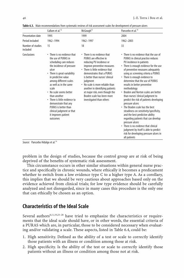
problem in the design of studies, because the control group are at risk of beingdeprived of the benefits of systematic risk assessment.
This circumstance occurs in other similar situations within general nurse prac-tice and specifically in chronic wounds, where ethically it becomes a predicamentwhether to switch from a low evidence type C to a higher type A. As a corollary,this implies that we should be very cautious about approaches based only on theevidence achieved from clinical trials; for low type evidence should be carefullyanalyzed and not disregarded, since in many cases this procedure is the only onethat can ethically be chosen as an option.
Characteristics of the Ideal Scale
Several authors8,11,19,23–28 have tried to emphasize the characteristics or require-ments that the ideal scale should have, or in other words, the essential criteria ofa PURAS which are, in particular, those to be considered necessary when evaluat-ing and/or validating a scale. These aspects, listed in Table 6.4, could be:
1. High sensitivity. Defined as the ability of a test or scale to correctly identifythose patients with an illness or condition among those at risk.
2. High specificity. Is the ability of the test or scale to correctly identify thosepatients without an illness or condition among those not at risk.
46 J.-E. Torra i Bou et al.
Table 6.3. Main recommendations from systematic reviews of risk assessment scales for development of pressure ulcers
Cullum et al.12 McGough13 Pancorbo et al.14
Presentation date 1995 1999 2004
Period included 1962–1994 1962–1997 1962–2003
Number of studies 15 18 33included
Conclusions • There is no evidence that • There is no evidence that • There is no evidence that the use ofthe use of PURAS in PURAS are effective in PURAS in clinical practice reduces scheduling care reduces reducing PU incidence or PU incidence in patientsthe incidence of pressure improve preventive measures • There is enough evidence for the use ulcer • There is little evidence that of preventive measures adequately
• There is great variability demonstrates that a PURAS using as screening criteria a PURASin predictive value is better than nurses’ clinical • There is enough evidence to among different scales judgment determine that the use of PURAS as well as in the same • No scale is more reliable than results in better preventive scale another in identifying patients methodology
• No scale seems better at major risk, even though the • Braden and Norton scales are better than another Braden scale has been more than nurses’ clinical judgment to
• There is little evidence to investigated than others predict the risk of patients developing demonstrate that any pressure ulcersPURAS is better than • The Braden scale has the best clinical judgment or that steadiness on sensitivity/specificity,it improves patient and the best predictive abilityoutcomes regarding patients that can develop
pressure ulcers• There is no evidence that clinical
judgment by itself is able to predict risk for developing pressure ulcers inall patients
Source: Pancorbo Hidalgo et al.14
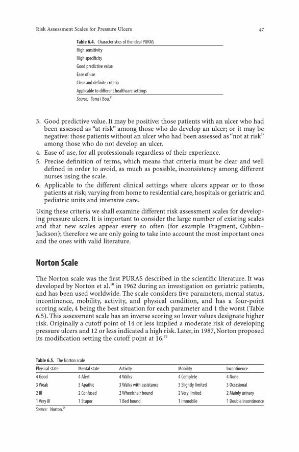
3. Good predictive value. It may be positive: those patients with an ulcer who hadbeen assessed as “at risk” among those who do develop an ulcer; or it may benegative: those patients without an ulcer who had been assessed as “not at risk”among those who do not develop an ulcer.
4. Ease of use, for all professionals regardless of their experience.5. Precise definition of terms, which means that criteria must be clear and well
defined in order to avoid, as much as possible, inconsistency among differentnurses using the scale.
6. Applicable to the different clinical settings where ulcers appear or to thosepatients at risk; varying from home to residential care, hospitals or geriatric andpediatric units and intensive care.
Using these criteria we shall examine different risk assessment scales for develop-ing pressure ulcers. It is important to consider the large number of existing scalesand that new scales appear every so often (for example Fragment, Cubbin–Jackson); therefore we are only going to take into account the most important onesand the ones with valid literature.
Norton Scale
The Norton scale was the first PURAS described in the scientific literature. It wasdeveloped by Norton et al.10 in 1962 during an investigation on geriatric patients,and has been used worldwide. The scale considers five parameters, mental status,incontinence, mobility, activity, and physical condition, and has a four-pointscoring scale, 4 being the best situation for each parameter and 1 the worst (Table6.5). This assessment scale has an inverse scoring so lower values designate higherrisk. Originally a cutoff point of 14 or less implied a moderate risk of developingpressure ulcers and 12 or less indicated a high risk. Later, in 1987, Norton proposedits modification setting the cutoff point at 16.29
Risk Assessment Scales for Pressure Ulcers 47
Table 6.4. Characteristics of the ideal PURAS
High sensitivity
High specificity
Good predictive value
Ease of use
Clear and definite criteria
Applicable to different healthcare settings
Source: Torra i Bou.11
Table 6.5. The Norton scale
Physical state Mental state Activity Mobility Incontinence
4 Good 4 Alert 4 Walks 4 Complete 4 None
3 Weak 3 Apathic 3 Walks with assistance 3 Slightly limited 3 Occasional
2 Ill 2 Confused 2 Wheelchair bound 2 Very limited 2 Mainly urinary
1 Very ill 1 Stupor 1 Bed bound 1 Immobile 1 Double incontinence
Source: Norton.29

The Norton scale is quite easy to use24 and has been widely validated9,30–40 (Table6.6). Mean values are:
• sensitivity 66% (range 0–92%);• specificity 65% (range 31–94%);• predictive positive value 27% (range 7–53%);• predictive negative value 93% (range 80–99%).
As such, it displays some inconveniences that may limit its clinical effectiveness.The main deficiencies are:
1. It does not have a functional definition of the applied parameters.2. It does not consider nutritional factors.3. It does not take into account frictional forces on the skin surface.
Many scales have been derived from the Norton scale, adding other parameters tothe five original ones. Among them are the following:
• The Gosnell scale (1973) includes five parameters: mental status, incontinence,activity, mobility, and nutrition (which tends to substitute the general state condition of the original scale drawn up by Doreen Norton), plus three furtherparameters without point scales: vital signs, skin appearance, and medication.Scoring is also inversely depicted and similar to the Norton scale.41
• The Ek scale (1987), or modified Norton scale, has seven elements, the basicNorton scale plus two nutritional parameters: food and liquid ingestion. It hasbeen used in Scandinavia and submitted to several studies.42
• In Spain there are several modifications of the Norton scale, for example theNova-4 scale43,44 created by a group of nurses from the Institut Català de la Salut
48 J.-E. Torra i Bou et al.
Table 6.6. Norton scale validation studies
Positive Negative Authors and Sample predictive predictivepublication year Type of facility size Sensitivity Specificity value value
Norton et al., 196210 Geriatric center 250 63% 70% 39% 86%
Roberts and Goldstone, Hospital 64 92% 61% 37% 96%197930
Newman and West, 198130 88 83% 63% 14% 98%
Goldstone and Roberts, Hospital 64 92% 61% — —198032
Goldstone and Goldstone, Hospital (traumatology) 40 89% 36% 53% 80%198233
Lincoln et al. 198634 Hospital 50 0% 94% — —
Smith, 198935 Hospital (traumatology) 101 60% 31% — —
Stotts, 198836 Hospital (cardiovascular 387 16% 94% — —surgery and neurosurgery)
Wai-Han et al., 199737 Geriatric center 185 75% 67% 9% 98%
Pang and Wong, 199838 Hospital (rehabilitation) 138 81% 59% 33% 93%
García et al., 199939 Hospital 3030 89% 81% 21% 99%
Schoonhoven et al., 200240 Hospital 1229 46% 60% 7% 94%
Source: Created by the authors from data in Torra i Bou,11 Cullum et al.,12 and McGough.13
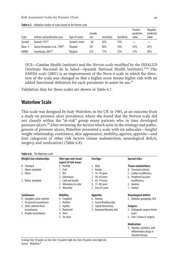
(ICS—Catalan Health Institute) and the Norton scale modified by the INSALUD(Instituto Nacional de la Salud—Spanish National Health Institute).45,46 TheEMINA scale (2001) is an improvement of the Nova-4 scale in which the direc-tion of the scale was changed so that a higher score means higher risk with anadded functional definition for each parameter to assist its use.47
Validation data for these scales are shown in Table 6.7.
Waterlow Scale
This scale was designed by Judy Waterlow, in the UK in 1985, as an outcome froma study on pressure ulcer prevalence, where she found that the Norton scale didnot classify within the “at-risk” group many patients who in time developed pressure ulcers.48 After reviewing the factors which arise in the etiology and patho-genesis of pressure ulcers, Waterlow presented a scale with six subscales—height/weight relationship, continence, skin appearance, mobility, age/sex, appetite—andfour categories of other risk factors (tissue malnutrition, neurological deficit,surgery, and medication) (Table 6.8).
Risk Assessment Scales for Pressure Ulcers 49
Table 6.7. Validation studies of scales based on the Norton scale
Positive Negative Sample predictive predictive
Scale Authors and publication year Type of center size Sensitivity Specificity value value
Gosnell Gosnell, 197341 Geriatric center 30 50% 73% — —
Nova- 4 García Fernández et al., 199943 Hospital 187 84% 54% 43% 67%
EMINA Fuentelsalz, 200148 Hospital 673 77% 72% 17% 98%
Table 6.8. The Waterlow scale
Weight/size relationship: Skin type and visual Sex/Age: Special risks:aspect of risk areas:
0. Standard 0. Healthy 1. Male Tissue malnutrition:1. Above standards 1. Frail 2. female 8. Terminal/cachexia2. Obese 1. Dry 1. 14–49 years 5. Cardiac insufficiency
2. Edematous 2. 50–64 years 6. Peripheral vascular3. Below standards 1. Cold and humid 3. 65–74 years insufficiency
2. Alterations in color 4. 75–80 years 2. Anemia3. Wounded 5. Over 81 years 1. Smoker
Continence: Mobility: Appetite: Neurological deficit:0. Complete, urine catheter 0. Complete 0. Normal 5. Diabetes, paraplegic, ACV1. Occasional incontinence 1. Restless 1. Scarce/feeding tube
Surgery:2. Urine catheter/fecal 2. Apathy 2. Liquid intravenous5. Orthopedic surgery belowincontinence 3. Restricted 3. Anorexia/Absolute diet
waist3. Double incontinence 4. Inert5. Over 2 hours in surgery5. On chair
Medication:4. Steroids, cytotoxics, anti-
inflammatory drugs inelevated dosage
Scoring: Over 10 points: at risk. Over 16 points: high risk. Over 20 points: very high risk.Source: Waterlow.50
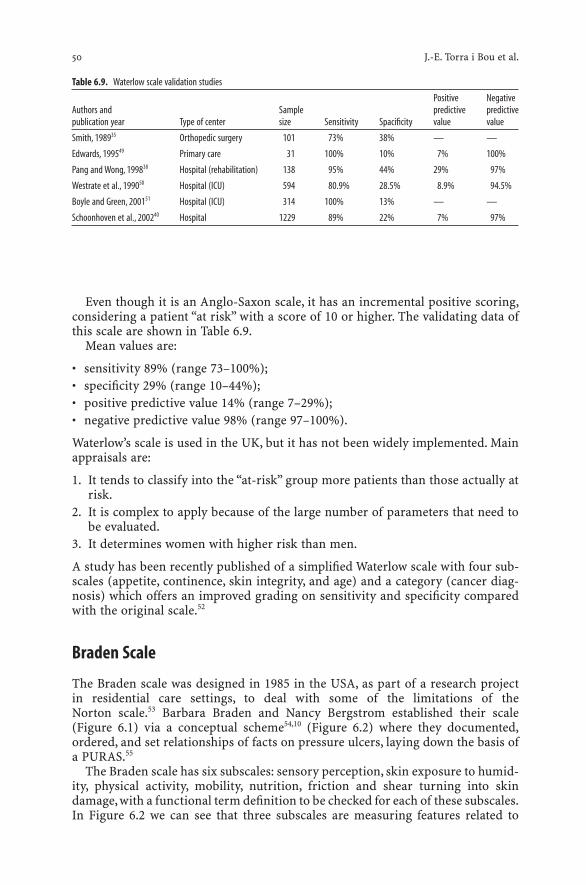
Even though it is an Anglo-Saxon scale, it has an incremental positive scoring,considering a patient “at risk” with a score of 10 or higher. The validating data ofthis scale are shown in Table 6.9.
Mean values are:
• sensitivity 89% (range 73–100%);• specificity 29% (range 10–44%);• positive predictive value 14% (range 7–29%);• negative predictive value 98% (range 97–100%).
Waterlow’s scale is used in the UK, but it has not been widely implemented. Mainappraisals are:
1. It tends to classify into the “at-risk” group more patients than those actually atrisk.
2. It is complex to apply because of the large number of parameters that need tobe evaluated.
3. It determines women with higher risk than men.
A study has been recently published of a simplified Waterlow scale with four sub-scales (appetite, continence, skin integrity, and age) and a category (cancer diag-nosis) which offers an improved grading on sensitivity and specificity comparedwith the original scale.52
Braden Scale
The Braden scale was designed in 1985 in the USA, as part of a research project in residential care settings, to deal with some of the limitations of the Norton scale.53 Barbara Braden and Nancy Bergstrom established their scale(Figure 6.1) via a conceptual scheme54,10 (Figure 6.2) where they documented,ordered, and set relationships of facts on pressure ulcers, laying down the basis ofa PURAS.55
The Braden scale has six subscales: sensory perception, skin exposure to humid-ity, physical activity, mobility, nutrition, friction and shear turning into skindamage, with a functional term definition to be checked for each of these subscales.In Figure 6.2 we can see that three subscales are measuring features related to
50 J.-E. Torra i Bou et al.
Table 6.9. Waterlow scale validation studies
Positive NegativeAuthors and Sample predictive predictivepublication year Type of center size Sensitivity Spacificity value value
Smith, 198935 Orthopedic surgery 101 73% 38% — —
Edwards, 199549 Primary care 31 100% 10% 7% 100%
Pang and Wong, 199838 Hospital (rehabilitation) 138 95% 44% 29% 97%
Westrate et al., 199050 Hospital (ICU) 594 80.9% 28.5% 8.9% 94.5%
Boyle and Green, 200151 Hospital (ICU) 314 100% 13% — —
Schoonhoven et al., 200240 Hospital 1229 89% 22% 7% 97%

Patient’s Name_____________________________ Evaluator’s Name_____________________________
SENSORY PERCEPTION
1. Completely LimitedUnresponsive (does not moan, flinch, or grasp) to painful stimuli, due to diminished level ofconsciousness or sedation ORlimited ability to feelpain over most of body.
2. Very Limited Responds only to painfulstimuli. Cannot communicatediscomfort except by moaning or restlessness ORhas a sensory impairment which limits the ability to feel pain or discomfort over ½ of body.
3. Slightly LimitedResponds to verbal com-mands, but cannot alwayscommunicate discomfort or the need to be turned ORhas some sensory impairment which limits ability to feel painor discomfort in 1 or 2 extremities.
4. No ImpairmentResponds to verbalcommands. Has no sensory deficit which would limit ability to feel or voice pain or discomfort
1. Constantly MoistSkin is kept moist almostconstantly by perspiration, urine, etc. Dampness is detected every time patient is moved or turned.
2 .Very MoistSkin is often, but not alwaysmoist. Linen must be changed at least once a shift.
3. Occasionally Moist:Skin is occasionally moist, requiring an extra linen change approximatelyonce a day.
4. Rarely MoistSkin is usually dry, linen only requires changing at routine intervals.
ability to respond meaning-fully to pressure-relateddiscomfort
MOISTURE
degree to which skin isexposed to moisture
1. Completely ImmobileDoes not make even slightchanges in body or extremity position without assistance.
2 .Very LimitedMakes occasional slight changes in body or extremity position but unable to make frequent or significant changes independently.
3. Slightly LimitedMakes frequent though slightchanges in body or extremityposition independently.
4. No LimitationMakes major and frequent changes in position withoutassistance.
MOBILITY
ability to change and control body position
1. Very PoorNever eats a complete meal. Rarely eats more than½ of any food offered. Eats 2 servings or less of protein (meat or dairy products) per day. Takes fluids poorly. Does not take a liquid dietary supplement ORis NPO and/or maintained on clear liquids or IV’s for more than 5 days
2. Probably InadequateRarely eats a complete meal and generally eats only about ½ of any food offered. Protein intake incudes only 3 servings of meat or dairy products per day. Occasionally will take a dietary supplement ORreceives less than optimum amount of liquid diet or tube feeding.
3. AdequateEats over half of most meals. Eats a total of 4 servings of protein (meat, dairy products) per day. Occasionally will refuse a meal, but will usually take a supplement whenoffered ORis on a tube feeding or TPNregimen which probably meets most of nutritional needs.
4. ExcellentEats most of every meal. Never refuses a meal. Usually eats a total of 4 or more servings of meat anddairy products.Occasionally eats between meals. Does not requiresupplementation.
NUTRITION
usual food intake pattern
1. ProblemRequires moderate to maximum assistance in moving. Complete lifting without sliding againstsheets is impossible. Frequently slides down in bed or chair, requiring frequent repositioningwith maximum assistance.Spasticity, contractures oragitation leads to almostconstant friction.
2. Potential ProblemMoves feebly or requires minimum assistance. During a move skin probably slides to some extent against sheets, chair, restraints orother devices. Maintains relatively good position in chair or bed most of the time but occasionally slidesdown.
3. No Apparent ProblemMoves in bed and in chairindependently and has sufficient muscle strength to lift up completely during move. Maintainsgood position in bed or chair.
FRICTION & SHEAR
” Copyright Barbara Braden and Nancy Bergstrom, 1988 All rights reserved Total Score
1. BedfastConfined to bed.
2 . ChairfastAbility to walk severely limited or non-existent. Cannot bear own weight and/or must be assisted intochair or wheelchair.
3. Walks OccasionallyWalks occasionally during day, but for very short distances, with or without assistance. Spends majority of each shift in bed or chair.
4. Walks FrequentlyWalks outside room at least twice a day and inside room at least once every two hours during waking hours.
ACTIVITY
degree of physical activity
Date of Assessment
3
Figure 6.1 The Braden scale for predicting pressure sore risk.
strong and prolonged exposure to pressure, while the others are related to tissuetolerance.
The Braden scale is an inverse scoring tool, which means that the lower scoreimplies major risk, with a range varying from 5 to 23 points. Patients “at risk” arethose with scores equal to or below 16 points on this scale; 15–16 is “low risk,”13–14“moderate risk,” and between 5 and 12 “high risk.” Table 6.10 shows the results ofmore than a dozen works for validation of the Braden scale in different care settings,varying from hospitals for acute patients to long-term facilities, including intensivecare, nursing homes for the elderly, and home care.56–67 According to these studies,mean sensitivity is 74% (range 27–100%); specificity is 69% (19–95%); positive pre-dictive value is 43% (8–77%); and negative predictive value is 90% (71–100%).
As can be seen, this scale is the most validated by scientific literature, having the best evidence for to its usefulness, being very sensitive and specific. The mainproblem is its difficulty of use, for it requires more training than the Norton scale.
Risk Assessment Scales for Pressure Ulcers 51

52 J.-E. Torra i Bou et al.
Decreasedmobility
Decreasedactivity
Decreasedsensorial
perception
Extrinsic factors(humidity, friction,
shear forces)
Intrinsic factors(nutrition, age,
arteriolar pressure andothers)
Pressure
Tissuetolerance
Developmentof pressure
ulcers
Figure 6.2 Concept diagram for the development of pressure ulcers. (From Braden and Bergstrom.54 Reprinted from Rehabilitation Nursing12: 9, with permission of the Association of Rehabilitation Nurses, 4700 W. Lake Avenue, Glenview, IL 60025-1485. Copyright © 1987.)
Table 6.10. Validation of Braden scale studies
Positive Negative Authors and Sample predictive predictivepublication year Type of center size Sensitivity Specificity value value
Bergstrom, Braden et al., 198755 Hospital 99 100% 90% 70% 97%
Bergstrom, Braden et al., 198755 Long-term hospital 100 100% 64% 25% 100%
Bergstrom et al., 198756 Intensive care 60 83% 64% 61% 85%
Langemo et al., 1991 57 Hospital 190 64% 87% 47% 93%
Bergstrom and Braden, 199258 Nursing home 200 97% 19% 77% 71%
Salvadalena et al., 199259 Hospital 99 40% 70% 23% 79%
Barnes and Payton, 199360 Hospital 361 73% 91% 33% 91%
Braden and Bergstrom, 199461 Nursing home 102 79% 74% 54% 90%
Ramundo, 199562 Home care 48 100% 34% 21% 100%
Capobianco and McDonald, 199663 Hospital 50 71% 83% 63% 88%
Halfens, 199764 Hospital 320 74% 70% 30% 94%
Pang and Wong, 199838 Hospital (rehabilitation) 138 91% 62% 37% 96%
Vap and Donahue, 200065 Long-term hospital 555 27% 95% 53% 74%
Schoonhoven et al., 200240 Hospital 1229 43% 68% 8% 95%
Seongsook et al., 200466 Hospital (ICU) 125 97% 26% 37% 95%
Source: Created by the authors using data from Torra i Bou,11 Cullum et al.,12 and McGough.13

PURAS in Intensive Care Patients
The constant increase in knowledge about PURAS has led to deep and inter-esting debates about the need for specific tools for complex and special clinical situations, as in the case of patients in intensive care units (ICU) or pediatricpatients.
For intensive care units there is wide disagreement in the literature about theuse of general scales. Thus, some works stress the use of PURAS such as the Bradenscale in ICU neurological patients67,68 and cardiological surgery patients,69,70 otherspropose a modification of the cutoff point for ICU traumatology patients,71 andstill others emphasize the value of the Waterlow scale,51 while there is anothergroup that is quite unconvinced of it.52
There are also PURAS specifically designed for ICU patients such as theCubbin–Jackson scale,51,72 which appeared in 1991 as a modification of the Nortonscale; the Cornell Ulcer Risk Score,73 the 1995 Sunderland scale,74,75 and the BirtyPressure Risk Assessment scale.76 Nevertheless none has validation and they arescarcely mentioned in the literature. There have been attempts to validate somesuch as the Decubitus Ulcer Potential Analyzer (DUPA), a modified version of theGosnell, Norton, and Braden scales, which sensitivity wise, shows lower scores thanthe Braden scale on the same patients.71
As a further addition of specific factors on existing scales, Halfens et al.77
underline that including variables such as blood circulation to the Braden scale does not improve its sensitivity or specificity, while Séller et al.78 concludethat there are no specific risk factors for ICU patients that would justify the design of a PURAS exclusively for them. Therefore, many ICU professionals lookat tissue damage rather than using different scales when deciding on preventivemeasures.79
PURAS in Pediatric Patients
Pressure ulcers in pediatric patients are gaining significance. Baldwin,80 in a reviewon pressure ulcers in pediatric patients, found that there were 22 works publishedin English since 1972. Waterlow,81 in her 1997 study of children at risk of develop-ing pressure ulcers, considered that available PURAS were not appropriate forpediatric patients, especially babies. In 1998 Cocket published the Pediatric Pres-sure Sore Risk Assessment,82 a PURAS for pediatric patients that has not yet beenvalidated. Willock et al. later wrote an interesting review83 about the inadequacy ofusing PURAS intended for adults in children, mentioning the Cocket scale andthree others (Bedi A (1993), Olding L (1998) and Pickersgill J (1997)), so far notvalidated or at least with no published validation.
In 1996 Quigley and Curley published the Braden Q scale, which is a revised version of the Braden scale for pediatric use.84 Recently Curley et al.85
published a paper where they validate a modified Braden Q scale that consists of three subscales for the Braden Q scale, mobility, sensory percep-tion, and perfusion/tissue oxygenation, with a cutoff point of 7; sensitivity was 92% and specificity 59%, values quite similar to those on the Braden scale foradults.
Risk Assessment Scales for Pressure Ulcers 53
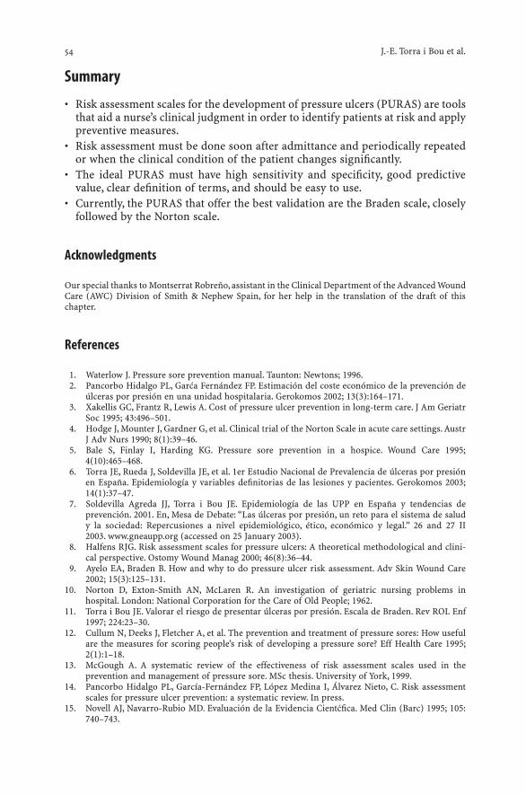
Summary
• Risk assessment scales for the development of pressure ulcers (PURAS) are toolsthat aid a nurse’s clinical judgment in order to identify patients at risk and applypreventive measures.
• Risk assessment must be done soon after admittance and periodically repeatedor when the clinical condition of the patient changes significantly.
• The ideal PURAS must have high sensitivity and specificity, good predictivevalue, clear definition of terms, and should be easy to use.
• Currently, the PURAS that offer the best validation are the Braden scale, closelyfollowed by the Norton scale.
Acknowledgments
Our special thanks to Montserrat Robreño, assistant in the Clinical Department of the Advanced WoundCare (AWC) Division of Smith & Nephew Spain, for her help in the translation of the draft of thischapter.
References
1. Waterlow J. Pressure sore prevention manual. Taunton: Newtons; 1996.2. Pancorbo Hidalgo PL, Garca Fernández FP. Estimación del coste económico de la prevención de
úlceras por presión en una unidad hospitalaria. Gerokomos 2002; 13(3):164–171.3. Xakellis GC, Frantz R, Lewis A. Cost of pressure ulcer prevention in long-term care. J Am Geriatr
Soc 1995; 43:496–501.4. Hodge J, Mounter J, Gardner G, et al. Clinical trial of the Norton Scale in acute care settings. Austr
J Adv Nurs 1990; 8(1):39–46.5. Bale S, Finlay I, Harding KG. Pressure sore prevention in a hospice. Wound Care 1995;
4(10):465–468.6. Torra JE, Rueda J, Soldevilla JE, et al. 1er Estudio Nacional de Prevalencia de úlceras por presión
en España. Epidemiología y variables definitorias de las lesiones y pacientes. Gerokomos 2003;14(1):37–47.
7. Soldevilla Agreda JJ, Torra i Bou JE. Epidemiología de las UPP en España y tendencias de prevención. 2001. En, Mesa de Debate: “Las úlceras por presión, un reto para el sistema de saludy la sociedad: Repercusiones a nivel epidemiológico, ético, económico y legal.” 26 and 27 II 2003. www.gneaupp.org (accessed on 25 January 2003).
8. Halfens RJG. Risk assessment scales for pressure ulcers: A theoretical methodological and clini-cal perspective. Ostomy Wound Manag 2000; 46(8):36–44.
9. Ayelo EA, Braden B. How and why to do pressure ulcer risk assessment. Adv Skin Wound Care2002; 15(3):125–131.
10. Norton D, Exton-Smith AN, McLaren R. An investigation of geriatric nursing problems in hospital. London: National Corporation for the Care of Old People; 1962.
11. Torra i Bou JE. Valorar el riesgo de presentar úlceras por presión. Escala de Braden. Rev ROL Enf1997; 224:23–30.
12. Cullum N, Deeks J, Fletcher A, et al. The prevention and treatment of pressure sores: How usefulare the measures for scoring people’s risk of developing a pressure sore? Eff Health Care 1995;2(1):1–18.
13. McGough A. A systematic review of the effectiveness of risk assessment scales used in the prevention and management of pressure sore. MSc thesis. University of York, 1999.
14. Pancorbo Hidalgo PL, García-Fernández FP, López Medina I, Álvarez Nieto, C. Risk assessmentscales for pressure ulcer prevention: a systematic review. In press.
15. Novell AJ, Navarro-Rubio MD. Evaluación de la Evidencia Cientcfica. Med Clin (Barc) 1995; 105:740–743.
54 J.-E. Torra i Bou et al.
16. Gálvez Toro A. Enfermería Basada en la Evidencia. Granada: Fundación Index; 2001.17. National Institute for Clinical Excellence. Pressure ulcer risk assessment and prevention.
Inherited Clinical Guideline B. London: NICE; 2001.18. European Pressure Ulcer Advisory Panel: Directrices sobre la prevención de úlceras por presión
del Grupo Europeo de Úlceras por presión. Gerokomos 1999; 10(1):30–33.19. Bergstrom N, Allman RM, Carlson CE, et al. Pressure ulcers in adults: prediction and prevention.
Clinical Practice Guideline number 3. Rockville, MD: US Department of Health and Human Services. Public Health Service. Agency for Health Care Policy and Research; 1992.
20. Best Practice. Pressure sores. Part 1: Prevention of pressure related damage. The Joanna BriggsInstitute for Evidence Based Nursing and Midwifery. 1997; 1(1):1–6.
21. Research Dissemination Core. Prevention of pressure sore. Iowa City: University of Iowa Gerontological Nursing Interventions Research Center; May 2002.
22. Registered Nurses Association of Ontario. The nursing best practice guideline. Risk assessmentand prevention of pressure ulcers. www.rnao.org (accessed on 30 March 2003).
23. Rycroft-Malone J, MacInnes E. Pressure ulcer risk assessment and prevention. Technical Report.London: Royal College of Nursing: 2000.
24. Smith LN, Booth N, Douglas D, et al. A critique of ‘at risk’ pressure sore assessment tools. J ClinNurs 1995; 4:153–159.
25. Deeks JJ. Pressure sore prediction: using and evaluating risk assessment tools. Br J Nurs 1996;5(5):313–320.
26. Bridel J. Assessing the risk pressure sores. Nurs Stand 1993; 7(25):32–35.27. MacDonald K. The reliability of pressure sore risk assessment tools. Prof Nurse 1995; 2(3):169–171.28. Scott EM. The prevention of pressure ulcers through risk assessment. J Wound Care 2000;
9(2):69–70.29. Norton D. Norton revised risk scores. Nurs Times 1987; 83(41):6.30. Roberts BV, Goldstone LA. A survey of pressure sores in the over sixties on two orthopedic wards.
Int J Nurs Stud 1979; 16(4):335–364.31. Newman P, West J. Pressure sores 2—the value of Norton score. Nurs Times 1981; 29(21):
15–21.32. Goldstone LA, Roberts BV. A preliminary discriminant function analysis of elderly orthopedic
patients who will or will not contract a pressure sore. Int J Nurs Stud 1980; 17(1):17–23.33. Goldstone LA, Goldstone J. The Norton score: an early warning of pressure sores? J Adv Nurs 1982;
7:419–426.34. Lincoln R, Roberts R, Maddox A, et al. Use of Norton pressure sore risk assessment scoring system
with elderly patients in acute care. J ET Nurs 1986; 13:132–138.35. Smith I. Waterlow/Norton scoring system: a ward view. Care Sci Pract 1989; 7:93–95.36. Stotts NA. Predicting pressure ulcer development in surgical patients. Heart Lung 1988; 17:
641–647.37. Wai-Han C, Kit-Wai C, French P, et al. Which pressure sore risk calculator? A study of the
effectiveness of the Norton scale in Hong Kong. Int J Nurs Stud 1997; 34(2):165–169.38. Pang SM, Wong TK. Predicting pressure sore risk with the Norton, Braden and Waterlow scales in
a Hong Kong rehabilitation hospital. Nurs Res 1998; 47(3):147–153.39. Garcia AM, Rosa G de la, Garrido G, Rodriguez P. Escala de Norton: ¿es válida como método de
predicción del desarrollo de úlceras por presión? Medicina Preventiva 1999; V(3):24–27.40. Schoonhoven L, Hallboom JRE, Bousema MT, et al. Prospective cohort study of routine use of risk
assessment scales for prediction of pressure ulcers. BMJ 2002; 235:797–800.41. Gosnell DJ. An assessment tool to identify pressure sores. Nurs Res 1973; 22:55–59.42. Berglund B, Nordström G. The use of the modified Norton scale in nursing-home patients. Scand
J Caring Sci 1995; 9:165–169.43. Garcia Fernandez FP, Bermejo J, Pérez MJ, et al. Validación de dos escalas de valoración del riesgo
de úlceras por presión: Gosnell y Nova-4. Rev ROL Enf 1999; 22(10):685–687.44. Aguado H, Aguilar M, Casado A. Protocol de prevenció i tratacment de les ulceres per pressió.
Institut Catalá de la Salut. Ciutat Sanitaria i Universitaria de Bellvitge, 1994.45. Bermejo CJ, Beamud M, Puerta M de la, et al. Fiabilidad interobservadores de dos escalas de
detección del riesgo de formación de úlceras por presión en enfermos de 65 o más años. EnfClínica 1998; 8(6):242–247.
46. Grupo de Enfermerca del Institut Català de la Salut. Ulceras por presión: método de consensocomo estrategia de mejora de la calidad asistencial. Enf Clínica 1998; 8(3):110–115.
47. Fuentelsalz C. Validación de la escala EMINA ©: un instrumento de valoración del riesgo de desarrollar úlceras por presión en pacientes hospitalizados. Enf Clcnica 2001; 11(3):97–103.
48. Waterlow J. A risk assessment card. Nurs Times 1985; 81(49):5155.
Risk Assessment Scales for Pressure Ulcers 55
49. Edwards M. The levels of reliability and validity of the Waterlow pressure sore risk calculator. JWound Care 1995; 4(8):373–378.
50. Weststrate JT, Hop WC, Aalbers AG. The clinical relevance of the Waterlow pressure sore risk scalein the ICU. Intensive Care Med 1998; 24(8):815–820.
51. Boyle M, Green M. Pressure sores in intensive care: defining their incidence and associated factorsand assessing the utility of two pressure sore risk assessment tools. Aust Crit Care 2001; 14(1):24–30.
52. Papanikolau P, Clark M, Lyne PA. Improving the accuracy of pressure ulcer risk calculators: somepreliminary evidence. Int J Nurs Stud 2002; 39:187–194.
53. Bergstrom N, Braden B, Laguzza A, Holman V. The Braden Scale for predicting pressure sore risk:reliability studies. Nurs Res 1985; 34(6):383.
54. Braden B, Bergstrom N. A conceptual schema for the study of the aetiology of pressure sores.Rehabil Nurs 1987; 12(1):8–16.
55. Bergstrom N, Braden B, Laguzza A, Holman V. The Braden Scale for predicting pressure sore risk:reliability studies. Nurs Res 1987; 36(4):205–210.
56. Bergstrom N, Demuth PJ, Braden BJ. A clinical trial of the Braden scale for predicting pressuresore risk. Nurs Clin North Am 1987; 22(2):417–428.
57. Langemo DK, Olson B, Hunter S, et al. Incidence and prediction of pressure ulcers in five patientcare settings. Decubitus 1991; 4(3):25–36.
58. Bergstrom N, Braden BJ. Prospective study of pressure risk among institutionalised elderly. J AmGeriatr Soc 1992; 40(8):747–758.
59. Salvadalena G, Snyder ML, Brogdon KE. Clinical trial of the Braden scale on an acute care medicalunit. J ET Nurs 1992; 19:160–165.
60. Barnes D, Payton RG. Clinical application of the Braden scale in the acute-care setting. DermatolNurs 1993; 5(5):386–388.
61. Braden BJ, Bergstrom N. Predictive validity of the Braden scale for pressure sore risk in a nursinghome population. Res Nurs Health 1994; 17:459–470.
62. Ramundo J. Reliability and validity of the Braden scale in the home care setting. J Wound OstomyContinence Nurs 1995; 22(3):128–134.
63. Capobianco ML, McDonald DD. Factors affecting the predictive validity of the Braden Scale. AdvWound Care 1996; 9(6):32–36.
64. Halfens RJ. The reliability and validity of the Braden scale. In: Harding KG, Leaper DJ, Turner TD(eds) Proceedings of the 7th European conference on advances in wound management. London:Macmillan; 1997.
65. Vap P, Donahue T. Pressure risk assessment in long-term care nursing. J Gerontol Nurs 2000;26(6):37–45.
66. Seongsook J, Ihnsook J, Younghee L. Validity of pressure ulcer risk assessment scales;Cubbin–Jackson, Braden, and Douglas scale. Int J Nurs Stud 2004; 41(2):199–204.
67. Anonymus. Low Braden scale scores predicted the development of pressure ulcers in neurologicintensive and intermediate care units. ACP J Club 2001; 135(2):76.
68. Fife C, Otto G, Capsuto EG, et al. Incidence of pressure ulcers in a neurologic intensive care unit.Crit Care Med 2001; 29(2):283–290.
69. Lewicki LJ, Mion L, Splane KG, et al. Patient risk factors for pressure ulcers during cardiac surgery.AORN J 1997; 65(5):933–942.
70. Stordeurs S, Laurent S, d’Hoore W. The importance of repeated risk assessment of pressure soresin cardiovascular surgery. J Cardiovasc Surg (Torino) 1998; 39(3):343–349.
71. Jiricka MK, Ryan P, Carvalho MA, et al. Pressure ulcer risk factors in ICU population. Am J CritCare 1995; 4(5):361–367.
72. Hunt J. Application of a pressure area risk calculator in an intensive care unit. Intensive Crit CareNurs 1993; 9(4):226–231.
73. Eachempati SR, Hydo LJ, Barie PS. Factors influencing the development of decubitus ulcers in critically ill surgical patients. Crit Care Med 2001; 29(9):1678–1682.
74. Sollars A. Pressure area risk assessment in intensive care. Nurs Crit Care 1998; 3(6):267–273.75. Lowery MT. A pressure sore risk calculator for intensive care patients. The Sunderland experience.
Intensive Crit Care Nurs 1995; 11(6):344–35376. Birtwhistle J. Pressure sore formation and risk assessment in intensive care. Care Crit Ill 1995; 11:
121–125.77. Halfens RJ,Van Achterberg T, Bal RM.Validity and reliability of the Braden Scale and the influence
of other risk factors: a multicentre prospective study. Int J Nurs Stud 2000; 37(4):313–319.78. Séller BP, Wille J, van Ramsshort B, et al. Pressure ulcers in intensive care patients: a review of
risks and prevention. Intensive Care Med 2002; 28(10):1379–1388.
56 J.-E. Torra i Bou et al.

79. Weststrate JT, Bruinining HA. Pressure sores in an intensive care unit and related variables: adescriptive study. Intensive Crit Care Nurs 1996; 12(5):280–284.
80. Baldwin KM. Incidence and prevalence of pressure ulcers in children. Adv Skin Wound Care 2002;15(3):121–124.
81. Waterlow JA. Pressure sore risk assessment in children. Paediatr Nurs 1997; 9(6):21–24.82. Cocket A. Paediatric pressure sore risk assessment. J Tissue Viability 1998; 8(1):30.83. Willock J, Hugues J, Tickle S, et al. Pressure sores in children—the acute hospital perspective.
J Tissue Viability 2000; 10(2):59–62.84. Quigley SM, Curley MAQ. Skin integrity in the pediatric population: preventing and managing
pressure ulcers. J Soc Pediatr Nurs 1996; 1:7–18.85. Curley M, Razmus IS, Roberts KE, Wypij D. Predicting pressure ulcer risk in paediatric patients:
the Braden Q Scale. Nurs Res 2003; 52(1):22–33.
Risk Assessment Scales for Pressure Ulcers 57
7 Equipment SelectionJacqueline Fletcher
Background Information
The European Pressure Ulcer Advisory Panel (EPUAP) guidelines for preventionof pressure ulcers1 state that most pressure ulcers can be prevented and it is impor-tant to have prevention strategies in place that are based on the best available evi-dence. These strategies encompass a wide range of actions but will include the useof specialized equipment such as overlays, mattresses, and specialist beds tomanage the load on the tissues.2 Selection of the appropriate piece of equipmentfor any one individual is complicated as there is little good quality evidence tosupport the efficacy of any individual piece of equipment and indeed the tests usedto determine the efficacy of these systems have been inconsistent in design andmeasurement techniques.3 There is also a lack of consensus regarding the termi-nology used to describe the mode of action of many pieces of equipment.2,4 Mostguidelines on the prevention of pressure ulcers2,4–7 discuss the use of equipmentbut are only able to give broad statements which unfortunately do not address therequirements of individual patients.
Efficacy of Equipment
There have been several published reviews of the efficacy of support surfaces8,9
with detailed methodologies also published for clinicians wishing to replicate thesereviews.9,10
Efficacy of individual support systems may be measured in a variety of waysincluding: clinical outcomes, interface pressure measurement,measurement of trans-cutaneous oxygen, and other measures of microcirculation. The most commonmethod, however, appears to be the measurement of interface pressure. In order toimprove comparability of published research a EPUAP working group proposedrecommendations for a standardized protocol for laboratory evaluation of supportsurfaces.3 It is recommended that any future research on support surfaces shouldfollow these recommendations where possible.
Mechanisms of Action/Types of Equipment
Mattresses are defined as a piece of equipment placed directly onto the bed frame;an overlay is used in addition to the mattress and should not be placed directlyonto the bed frame.
59
Broadly speaking equipment is described11 as providing:
• pressure reduction—the equipment maximizes the area of skin in contact withthe surface thereby reducing the pressure at an individual point; or
• pressure relief—the equipment removes pressure from a localized skin area ineither a static or cyclical mechanism.
Other guidelines9 use alternative terminology such as low-tech devices (those thatuse a conforming support surface to distribute body weight over a large surfacearea) and high-tech devices (use of alternating support surfaces where inflatablecells alternately inflate and deflate).
Pressure Reduction
Equipment that falls within this category includes static systems such as foam mat-tresses and overlays, fiber-filled overlays, air-filled overlays, gel-filled mattressesand overlays, and powered systems such as low-air-loss overlays and mattresses.As well as the material the mattress itself is made from, consideration must alsobe given to the covers of the mattress, which must be able to conform with theinternal content; otherwise a hammocking effect occurs, negating the pressure-reducing properties of the mattress/overlay.11 Consideration should also be givento the impact of the bed frame on the mattress. Solid bed frames do not allow themattress to breathe and where condensation occurs it is possible for mold to growbetween the mattress and bed frame (Figure 7.1—see color section). Mechanicalbed frames that contour to improve the patient’s position and reduce shearingforces may also affect the pressure-reducing properties of the mattress and careshould be taken to ensure that the mattress is able to follow the contours of theframe without causing damage to the foam/gel and generating high points of pres-sure where the foam creases.
Pressure-Relieving Devices
Pressure-relieving devices are usually powered devices and would include, forexample, alternating pressure mattresses and overlays and air fluidized systems.
Alternating systems operate by the cyclical inflation and deflation of sections ofthe mattress (usually known as cells). This cycle varies between products in termsof both the time taken to complete the cycle and the number of cells involved inthe cycle (usually two or three). Where the cell is inflated the body is in contactand is subject to high interface pressures. However, this is for a fixed time span andas the cycle progresses the pressure is partially or totally removed and transferredto different parts of the body as the cells inflate and deflate. These devices fre-quently incorporate a pressure sensor and may regulate the pressure within thecells in response to the patient’s weight and the distribution of the weight. Forexample, if the patient is in the semi-recumbent position their weight is primarilysupported along the legs and buttocks as opposed to lying where the weight is dis-tributed also along the trunk.
Air fluidized systems constantly change the points of the body supporting theweight. They operate by continually circulating warm air through fine ceramic
60 J. Fletcher
beads covered by a permeable sheet. It is debatable whether this type of systemworks by redistributing the pressure through extremely close contact, or by reduc-ing the pressure as the movement of the beads is constant and therefore the contactpoints are constantly changing.
Selecting Equipment
As there is such a plethora of equipment available a set of criteria need to be fol-lowed to assist in appropriate selection. These criteria usually relate to the patient’srisk of developing a pressure ulcer or the grade of pressure ulcer already present,with differing criteria being available for prevention and management. It is widelyacknowledged that there are limitations within the current risk assessment tools12
and the NICE guideline9 avoids this situation by referring to patients who are “vul-nerable to” or “at elevated risk of” pressure ulcers. Their recommendations statethat although there is very little evidence to suggest the high-tech devices are moreeffective than the low-tech equipment, professional consensus recommends use ofalternating pressure or other high-tech devices:
• as a first-line prevention strategy for people at elevated risk following holisticassessment;
• when the patient has a history of previous ulceration or a clinical conditionwhich suggests they are best cared for on a high-tech device; or
• when a low-tech device has failed.
The United Kingdom pressure ulcer benchmark statement regarding provision ofequipment also clearly states that the patients should be comfortable on the equip-ment.13 This is particularly important because if patients are not comfortable it islikely that they will be reluctant to use the equipment and therefore howeverefficacious the equipment may be they will not benefit from its allocation.
Selecting Equipment for Individual Patients
Selecting equipment for individual patients presents additional practical factorswhich must be considered alongside the effectiveness of the equipment. These willalso determine the suitability of the equipment for use in the particular settingwhether home care, hospital care, or an intermediate care setting.
These more practical considerations require in-depth knowledge of the patientand the setting in which care will be delivered. Although these are less evidence-based considerations, it is the complex interplay between these variables that mayultimately be the deciding factors in the provision of appropriate equipment forindividual patients. These factors may be considered as clinical, practical, andfinancial.
Clinical Factors
These will include not only the current risk level but also the likely prognosis. Con-sideration should be given to the likelihood of the patient’s condition improving
Equipment Selection 61

or deteriorating and the level of equipment supplied should reflect this or at leastplans should be made to replace the equipment as the patient’s condition changes.
The overall objectives of care must also be addressed. Therefore considerationshould be given to requirements for appropriate moving and handling, otherwound-related factors such as management of high levels of exudate, and generalclinical requirements such as control of temperature. Any factors that may con-traindicate the use of a particular piece of equipment must also be considered. Forexample, patients with unstable spinal fractures should not be managed on alter-nating systems and patients with unstable cardiac conditions may also be advisedto avoid this type of equipment, although a small study in the Netherlands sug-gests the gradual movement of these systems did not affect the patients’ generalmedical condition.14 Patients with bilateral amputation may also find it difficult tomaintain their balance of either very soft or moving surfaces, and this will have anadverse effect on their independence. Some patients may find it difficult to sleep14
or feel nauseated on a surface they perceive to be moving; frequently they will quickly become acclimatized to this but for some patients the movement isintolerable.
In critical care settings, where there is a higher risk of cardiac arrest, specificattention must be paid to what happens in relation to resuscitation. The surfacemust be suitable to resuscitate the patient on but at the same time maintain appro-priate pressure redistribution while the patient has very compromised circulation.Powered systems often have a rapid deflate mechanism. However, it must be con-sidered what happens when the mattress deflates; frequently the mattress deflatesunder the patient but the air remains in the edges. This results in the patientbecoming enveloped in the mattress and if clinicians kneel on the edge of the mat-tress this can result in the patient being jerked around on the bed. When the mat-tress is fully deflated the patient must still be receiving some form of pressurereduction so equipment should not deflate fully onto the bed frame. In someinstances a hyper-inflate facility may be preferable.
Practical Factors
Practical factors to be considered may relate to the patient, the surface, or both.Most commonly these relate to the environment in which the patient is being caredfor. Patients cared for in the home setting present a much more complex scenariothan those cared for in institutional settings. The shape, size, and weight of theequipment needs to be considered in terms of access to the home and sometimesinto older hospital settings. Some equipment such as air fluidized beds areextremely heavy and need to be positioned carefully in any setting to avoid struc-tural damage. The size of a mattress or overlay must be compatible with the bedframe on which it is to be used. This is a particular problem in the home, espe-cially if the patient wishes to continue sharing a double bed with a partner (orindeed if there is nowhere else for the partner to sleep). Most standard mattressand overlays are more than half of a double bed. However, some manufacturers dosupply specialist sizes designed for use on double beds which allow the partnerspace (Figure 7.2). There are psychological benefits to both the patient and thecarer of remaining in close proximity.
Where overlays are being used, consideration must be given to the effect on theheight of the mattress. Increasing the height may reduce the patient’s level of inde-
62 J. Fletcher
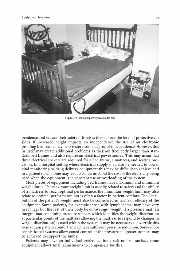
pendence and reduce their safety if it raises them above the level of protective cotsides. If increased height impacts on independence the use of an electronicprofiling bed frame may help restore some degree of independence. However, thisin itself may create additional problems as they are frequently larger than stan-dard bed frames and also require an electrical power source. This may mean thatthree electrical sockets are required for a bed frame, a mattress, and seating pro-vision. In a hospital setting where electrical supply may also be needed to powervital monitoring or drug delivery equipment this may be difficult to achieve andin a patient’s own home may lead to concerns about the cost of the electricity beingused when the equipment is in constant use or overloading of the system.
Most pieces of equipment including bed frames have maximum and minimumweight limits. The maximum weight limit is usually related to safety and the abilityof a mattress to reach optimal performance; the minimum weight limit may alsorelate to optimal performance but is often a factor in patient comfort. The distri-bution of the patient’s weight must also be considered in terms of efficacy of theequipment. Some patients, for example those with lymphedema, may have veryheavy legs but the rest of their body be of “average” weight; if a pressure mat (anintegral mat containing pressure sensors which identifies the weight distributionat particular points of the mattress allowing the mattress to respond to changes inweight distribution) is used within the system it may be necessary to override thisto maintain patient comfort and achieve sufficient pressure reduction. Some moresophisticated systems allow zoned control of the pressure so greater support maybe achieved to support the limbs.
Patients may have an individual preference for a soft or firm surface; someequipment allows small adjustments to compensate for this.
Equipment Selection 63
Figure 7.2 Alternating overlay on a double bed.
Alarm systems and the ability to rectify faults easily is a particular considera-tion in the home; carers must be advised what to do in the event of an alarm inthe home setting. In the hospital setting the alarm needs to be sufficiently stridentto be noticed above the general noise levels but not so loud that it disturbs otherpatients at night. Most electrical systems now have indicator panels which high-light the most common problems such as detachment of the power source.
In the home situation, particularly if the patient requires long-term care, theaesthetics of the equipment may be important. Many companies now provide elec-tronic bed frames and other equipment designed to look as much like standardfurniture as possible. This may allow the individual to feel less of a patient andmore of a person.
Most equipment will need to be cleaned or even decontaminated and this needsto be considered within the selection process. If it is likely that the cover may beregularly or heavily contaminated then it may be beneficial to consider a piece ofequipment with replaceable covers. Laundering of equipment such as fiber over-lays may also be a factor to consider if specialist facilities are not available. Mostequipment can be cleaned on site. However, some equipment, such as air fluidizedbeds, needs to be taken to specialist decontamination centers.
If selecting equipment for use within a healthcare setting where it may not bein permanent use, appropriate storage and transportation to and from the storagearea needs to be identified.
Financial Considerations
While unit cost of equipment must always be a factor in the decision process othercosts must also be taken into account. The costs of treating a patient who devel-ops a pressure ulcer far outweigh the costs of prevention.
In terms of actual costs, differing prices are usually charged for purchase, rentalor leasing of equipment and discounts are frequently available for bulk purchasesor purchases of complete systems/packages such as a bed frame, mattress, andseating. More hidden costs relate to:
• the training of staff to use the equipment,• cleaning/decontamination,• running of the equipment, and• service and maintenance.
Conclusion
Equipment selection is complex whether for an individual patient or for a wholeservice. The research and clinical evidence to support individual pieces of equip-ment must always be reviewed but in many instances other practical factors carryequal weight in the decision-making process. As so many types of equipment arenow available many national and local guidelines7,9 present flow charts to assist inselection. These usually commence with the patient’s level of risk and then proceedthrough a series of clinical considerations. These are helpful in selection for themajority of patients but a thorough holistic assessment of every patient mustunderpin the selection process. Where patients state a dislike for a particular piece
64 J. Fletcher
of equipment great care should be taken to determine what it is they actuallydislike about it; if patients are uncomfortable on a piece of equipment it is verylikely that they will not use it.
New equipment appears on the market on a regular basis. In order to make areasoned selection the practitioner must understand the principles by which theequipment works and the clinical and practical considerations in managingpatients. Care must be taken to review both the clinical effectiveness and the prac-tical considerations of equipment usage.
References
1. EPUAP. Pressure ulcer prevention guidelines. Oxford: EPUAP; 1998.2. EPUAP. Pressure ulcer treatment guidelines. Oxford: EPUAP; 1999.3. Support Services Working Group. Draft guidelines for the laboratory evaluation of pressure redis-
tributing support surfaces. EPUAP Review 2002; 4:8–12.4. AISLeC. Council Development of consensus upon the description of patient support surfaces
within Italy. EPUAP Review 2003; 5:101–102.5. Defloor T, Vanden Bossche K, Derre B, et al. Recommandations pour la prévention des escarres.
Ghent: University of Ghent; 2003.6. CBO. Conceptrichtlijn t.b.v. richtlijnbijeenkomst Decubitus (tweede herziening). Utrecht: CBO;
2002.7. Clinical Resource Efficiency Support Team. Guidelines for the prevention and management of
pressure sores. CREST, 1998.8. Cullum N, Nelson EA, Fleming K, Sheldon T. Systematic reviews of wound care management: (5)
beds; (6) compression; (7) laser therapy, therapeutic ultrasound, electrotherapy and electromag-netic therapy. Health Technol Assess 2001; 5(9):1–221.
9. National Institute for Clinical Excellence. Clinical Guideline 7: Pressure ulcer risk assessment andprevention, including the use of pressure-relieving devices (beds, mattresses and overlays) for theprevention of pressure ulcers in primary and secondary care. London: NICE; 2003.
10. EPUAP. EPUAP and the evaluation of support surfaces. EPUAP Review 2000; 2:7–9.11. Kenney L, Rithalia S. Technical aspects of support surfaces. J Wound Care 1999; Resource File—
Mattresses and Beds Part 2:1–8.12. Balzer K, Schrniedl C, Dassen T. Norton, Waterlow, Braden and the Care Dependency Scale: com-
paring their validity predicting patients’ pressure sore risk. Le Mans: EPUAP 5th Open Meeting,2001.
13. UK Department of Health. Essence of care. London: London Department of Health; 2001.14. Grindley A, Acres J. Alternating pressure mattresses: comfort and quality of sleep. Br J Nurs 1996;
5:1303–1310.
Equipment Selection 65

8 Pressure Ulcer Prevention and RepositioningTom Defloor, Katrien Vanderwee, Doris Wilborn, and Theo Dassen
Introduction
Preventing pressure ulcers is important, but not always easy to achieve. The mosteffective measures decrease the level and/or the duration of the pressure and shear-ing force. The pressure level is reduced by means of (for example) viscoelastic mattresses or low-air-loss systems. Repositioning and (for example) alternatingmattresses, by contrast, are oriented towards decreasing the duration of pressureand shearing force.
Repositioning
Repositioning is generally regarded as one of the most important and most effec-tive measures for preventing pressure ulcers.1 By regularly positioning patients ina different position, one modifies “the pressure points,” the points on which thebody is supported. If the position is modified frequently enough and the oxygenshortage in the tissues does not last too long, the chance of developing pressureulcers is limited. Salisbury2 demonstrated that transcutaneous oxygen levels at thelevel of the pressure points declined quickly in the first minutes after assuming anew body position on a standard hospital mattress. He therefore concludes thateven schemes of repositioning every 2 hours will not be enough to always preventpressure ulcers in all patients.
History
It has long been known that changing position is important in the prevention ofpressure ulcers. Robert Graves (1796–1853) wrote in 1848 in his Clinical Lectureson the Practice of Medicine that pressure ulcers could be prevented through regularchanges in position.3
Already in 1955 Guttman4 recommended repositioning every 2 hours for para-plegic patients. Nevertheless, the first studies on the effect of the duration andintensity of pressure on the development of pressure ulcers date from 1961,5 andthe first pressure measurements were performed in 1965.6
67
Traditional Repositioning Frequency
The frequency of changing position determines whether this preventive measureis effective, and thus leads to a decrease in the incidence of pressure ulcers. Tradi-tionally, repositioning every 2 hours7 or every 3 hours8 has been recommended.
Internationally one often hears the story that the choice of a 2-hour frequencycan be traced back to a nursing unit for war victims during the Second World War.9
In this unit, two soldiers were given the task of turning all of the patients. As soonas they had finished turning them all, they immediately had to start all over again.It took 2 hours to turn all of the patients. Whether this legend has any basis in factis uncertain, but Xakellis et al.10 calculated that it takes an average of 3.5 minutesto change a patient’s position. Thus, turning all of the patients in a 32-bed wardwould take 2 hours.
Very little research has been done to determine the necessary repositioning fre-quency. The first study on repositioning dates from 1962. In this research Nortonet al.11 compared the pressure ulcer incidence between two groups of elderly hos-pitalized women. One group had their lying positions changed, while the othergroup did not. The pressure ulcer incidence in the repositioning group amountedto 9%, while in the other group 26% of the patients developed pressure ulcers.However, on precisely what basis the nurses decided whose position would be reg-ularly changed, at which time(s) during their hospitalization, and how frequently,is unclear.
A PubMed search using the keywords “decubitus ulcer(s),” “pressure ulcer(s),”or “pressure sore(s)” in combination with “turning” or “repositioning” and “RCT”came up with eight references. In only two of these studies was the effect ofrepositioning on the development of pressure ulcers examined. Knox et al.12 com-pared the effect of repositioning after 1, 11/2, and 2 hours. Sixteen healthy elderlypeople, five whom had a dark skin type, were placed in one position for 2 hours,then in another position for 11/2 hours, and finally for 1 hour in yet another posi-tion. The skin temperature had increased more after 2 hours of immobilizationthan after 1 or 11/2 hours. No significant differences were found with respect tocontact pressure and color. The test subjects found that lying in the same positionfor 2 hours was more uncomfortable than for 1 or 11/2 hours. Due to the limitednumber of test subjects, the difficulty of detecting skin color changes in personswith a dark skin type, and the brief period of the study, the results are difficult togeneralize.
A randomized clinical experiment with 838 geriatric patients showed that thenumber of pressure ulcer injuries (pressure ulcer grade 2 and higher13) could bereduced by changing the lying position every 2 hours and to an even greater degreeby repositioning every 4 hours on a viscoelastic mattress in combination with pres-sure-reducing positions and seat cushions.1,14 Changing the lying position every 3hours did not appear to be sufficient to prevent pressure ulcers. These results havemajor consequences for nursing care. If patients lying on a non-pressure-reducingmattress do not have their position changed every 2 hours—both day and night,7 days a week—it makes little sense to opt for repositioning as a preventivemeasure. In that case it is better to choose other measures. Changing position every4 hours instead of every 2 hours is less labor-intensive and thus in practice muchmore feasible. It requires less effort on the part of the nurses and patients are lessdisturbed during their night’s sleep. After all, changing position every 2 hours canbe experienced as an unwanted intrusion by some patients.
68 T. Defloor et al.

The labor-intensive character of repositioning means that this effective preven-tion method is not often applied in practice, even among high-risk patients. TheEPUAP prevalence study performed among 5947 patients from five Europeancountries demonstrated that only 38.2% of the at-risk patients were having theirpositions changed.15
Combining Repositioning with Pressure-Reducing Measures
In order to make repositioning a more feasible method, its frequency can bereduced. This can be done only if repositioning is combined with pressure-reduc-ing mattresses and cushions and adapted body positions; otherwise the preventiveeffect disappears, because pressure ulcers are a function of the duration and levelof the pressure and shearing force. Increasing the duration can only be offset bydecreasing the size of the tissue deformation.
The pressure level is determined inter alia by the position of a patient and bythe hardness of the underlying layer. The contact surface is much greater in somebody positions than it is in others. The greater the contact surface, the more widelythe pressure can be distributed and the lower the pressure. The thickness and com-pressibility of the tissue on which the body is supported also differs greatly fromposition to position. The body position thus substantially defines the degree towhich the tissue is deformed, and therefore the degree to which the oxygen supplyof the tissue is impeded.
Supine Lying Position
In the flat supine lying position16,17 and in a semi-Fowler’s position of 30°,18,19 thepressure would be lowest and thus the risk of pressure ulcers smallest. In the 30° semi-Fowler’s position, the head end and the feet end are raised around 30°(Figure 8.1).
Pressure Ulcer Prevention and Repositioning 69
Prone Position
Supine Position Semi-Fowler 30° - 30°
30° 30°
30°
Lateral 30°
Figure 8.1 Lying positions.
Lateral Lying Position
The lowest pressure in the lateral lying position is measured in a 30° position.20,21
In this case, the contact surface at the level of the pelvis is greater than in the classiclateral position of 90°. The tissue mass at the level of the contact surface is thicker,and so the pressure can be better absorbed and distributed. In a lateral lying posi-tion of 30° the patient is turned at a 30° angle to the mattress and is supported inthe back with a cushion that has a 30° angle (Figure 8.1). The lower of the two legsis minimally bent at the level of the hip and the knee, while the upper leg is laidbehind the lower one with a bend of 30° at the level of the hip and 35° at the levelof the knee.22
Restricting Sitting Upright in Bed
The more the head end is raised, the smaller the contact surface and the more the pressure increases.23 The pressure is greatest in a 90° upright-sitting position,because the compression area is then the smallest, which results in a high pressureand thus a greater chance of the development of pressure ulcers.
Prone Lying Position, Sometimes an Alternative
The prone lying position is sometimes used as alternative lying position (Figure8.1). The pressure in this position is very low and roughly comparable to the pres-sure in the semi-Fowler’s position.18 Comfort is sometimes a problem, certainly ona harder mattress.
The prone lying position can be combined with a ventral-lateral form of the 30°lateral position. A small cushion is placed under the rib cage. The hip crest thencomes to lie in a pressure-free position.
Adapted Repositioning Scheme
In a repositioning scheme the patient is placed as frequently as possible in the posi-tions with the lowest pressure (in this case the supine lying position). In the laterallying position the pressure is greater and the risk of pressure ulcers is also greater.A scheme which takes this into account would be: supine lying position—laterallying position on the left—supine lying position—lateral lying position on theright.
Repositioning and Changing Sitting Position
Chair-bound patients develop pressure ulcers more frequently than bedriddenpatients with the same degree of helplessness.24,25 This is because the pressure inthe sitting position is much higher than in the lying position.26 Moreover, patientsoften sit up for a long period.
70 T. Defloor et al.
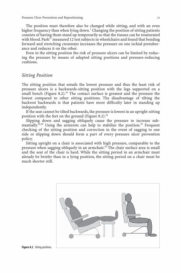
The position must therefore also be changed while sitting, and with an evenhigher frequency than when lying down.7 Changing the position of sitting patientsconsists of having them stand up temporarily so that the tissues can be resaturatedwith blood. Park27 measured 12 test subjects in wheelchairs and found that bendingforward and stretching crossways increases the pressure on one ischial protuber-ance and reduces it on the other.
Even in the sitting position the risk of pressure ulcers can be limited by reduc-ing the pressure by means of adapted sitting positions and pressure-reducing cushions.
Sitting Position
The sitting position that entails the lowest pressure and thus the least risk ofpressure ulcers is a backwards-sitting position with the legs supported on a small bench (Figure 8.2).26 The contact surface is greatest and the pressure thelowest compared to other sitting positions. The disadvantage of tilting the backrest backwards is that patients have more difficulty later in standing up independently.
If the seat cannot be tilted backwards, the pressure is lowest in an upright-sittingposition with the feet on the ground (Figure 8.2).26
Slipping down and sagging obliquely cause the pressure to increase sub-stantially.26,28 Using the armrests can help to stabilize the position.29 Frequentchecking of the sitting position and correction in the event of sagging to one side or slipping down should form a part of every pressure ulcer prevention policy.
Sitting upright on a chair is associated with high pressure, comparable to thepressure when sagging obliquely in an armchair.26 The chair surface area is smalland the seat of the chair is hard. While the sitting period in an armchair mustalready be briefer than in a lying position, the sitting period on a chair must bemuch shorter still.
Pressure Ulcer Prevention and Repositioning 71
Figure 8.2 Sitting positions.

Conclusion
Repositioning is an effective way to prevent pressure ulcers, but it must be com-bined with sitting and lying positions in which the pressure is as low as possible.There are many opinions but little actual research on the frequency of positionchanges.
References
1. Defloor T. Drukreductie en wisselhouding in de preventie van decubitus [Pressure reduction andturning in the prevention of pressure ulcers]. PhD thesis, Ghent University, 2000.
2. Salisbury RE. Transcutaneous PO2 monitoring in bedridden burn patients: a physiological analy-sis of four methods to prevent pressure sores. In: Lee BY (ed) Chronic ulcers of the skin. New York:McGraw-Hill; 1985;189–195.
3. Sebastian A. Robert Graves (1796–1853). In: Sebastian A (ed) A dictionary of the history ofmedicine. New York: Partenon Publishing Group; 2000.
4. Guttman L. The problem of treatment of pressure sores in spinal paraplegics. Br J Plast Surg 1955;8:196–213.
5. Kosiak M. Etiology of decubitus ulcers. Arch Phys Med Rehabil 1961; 42:19–29.6. Lindan O, Greenway R. Pressure distribution on the surface of the human body. Arch Phys Med
Rehabil 1965; 46:378–385.7. Panel for the Prediction and Prevention of Pressure Ulcers in Adults. Pressure ulcers in adults:
prediction and prevention. Clinical practice guideline number 3. Rockville: Agency for Health CarePolicy and Research, Public Health Service, US Department of Health and Human Services,AHCPR Publication No. 92–0047, 1992.
8. Bakker H. Herziening consensus decubitus [Revision of pressure ulcer consensus]. Utrecht: CBO;1992.
9. Dealey C. Managing pressure sore prevention. Dinton: Mark Allen; 1997.10. Xakellis GC, Frantz R, Lewis A. Cost of pressure ulcer prevention in long-term care. J Am Geriatr
Soc 1995; 43:496–501.11. Norton D, McLaren R, Exton-Smith AN. An investigation of geriatric nursing problems in hospi-
tal. New York: Churchill Livingstone; 1975.12. Knox DM, Anderson TM, Anderson PS. Effects of different turn intervals on skin of healthy older
adults. Adv Wound Care 1994; 7:48–52, 54.13. EPUAP. Guidelines on treatment of pressure ulcers. EPUAP Review 1999; 2:31–33.14. Defloor T.Wisselhouding, minder frequent en toch minder decubitus [Less frequent turning inter-
vals and yet less pressure ulcers]. Tijdschr Gerontol Geriatr 2001; 32:174–177.15. Clark M, Bours G, Defloor T. Summary report on the prevalence of pressure ulcers. EPUAP Review
2002; 4:49–57.16. Jeneid P. Static and dynamic support systems-pressure differences on the body. In: Kenedi RM,
Cowden JM, Scales JT (eds) Bedsore biomechanics. London: Macmillan; 1976: 287–299.17. Barnett RI, Shelton FE. Measurement of support surface efficacy: pressure. Adv Wound Care 1997;
10:21–29.18. Defloor T. The effect of position and mattress on interface pressure. Appl Nurs Res 2000; 13:
2–11.19. Rondorf Klym LM, Langemo D. Relationship between body weight, body position, support surface,
and tissue interface pressure at the sacrum. Decubitus 1993; 6:22–30.20. Seiler WO, Allen S, Stahelin HB. Influence of the 30 degrees laterally inclined position and the
“super-soft” 3-piece mattress on skin oxygen tension on areas of maximum pressure—implica-tions for pressure sore prevention. Gerontology 1986; 32:158–166.
21. Colin D, Abraham P, Preault L, et al. Comparison of 90 degrees and 30 degrees laterally inclinedpositions in the prevention of pressure ulcers using transcutaneous oxygen and carbon dioxidepressures. Adv Wound Care 1996; 9:35–38.
22. Garber SL, Campion LJ, Krouskop TA. Trochanteric pressure in spinal cord injury. Arch Phys MedRehabil 1982; 63:549–552.
23. Sideranko S, Quinn A, Burns K, Froman RD. Effects of position and mattress overlay on sacral andheel pressures in a clinical population. Res Nurs Health 1992; 15:245–251.
72 T. Defloor et al.

24. Barbenel JC, Jordan MM, Nicol SM, Clark MO. Incidence of pressure-sores in the Greater GlasgowHealth Board area. Lancet 1977; ii:548–550.
25. Gebhardt K, Bliss MR. Preventing pressure sores in orthopaedic patients—is prolonged chairnursing detrimental? J Tissue Viability 1994; 4:51–54.
26. Defloor T, Grypdonck MHF. Sitting posture and prevention of pressure ulcers.Appl Nurs Res 1999;12:136–142.
27. Park CA. Activity positioning and ischial tuberosity pressure: a pilot study. Am J Occup Ther 1992;46:904–909.
28. Koo TK, Mak AF, Lee YL. Posture effect on seating interface biomechanics: comparison betweentwo seating cushions. Arch Phys Med Rehabil 1996; 77:40–47.
29. Gilsdorf P, Patterson R, Fisher S. Thirty-minute continuous sitting force measurements with dif-ferent support surfaces in the spinal cord injured and able-bodied. J Rehabil Res Dev 1991; 28:33–38.
Pressure Ulcer Prevention and Repositioning 73

9 Skin CareSue Bale, Janice Cameron, and Sylvie Meaume
Introduction
The skin is the largest organ in the body and it is essential that clinicians involvedin pressure area care are cognizant of the need to maintain or improve its condi-tion. Healthy, normal skin is the first and best line of defense against the invasionof microorganisms, chemicals, and trauma. The skin is constantly exposed topotential irritants and chemicals, any of which may cause damage.1 In addition,mechanical forces, allergy, inflammation, systemic disease, and burns also impairskin integrity, producing a range of responses. These include erosion, pressureulceration and ulceration, erythema, papules, and vesicles.2
With regard to pressure ulcer prevention and management of the older person,skin care is a particular challenge, as people live for longer and are continuallyraising their expectations of healthcare. In addition, the developed world is expe-riencing increased numbers of older people within their populations. Davies3
reports that despite differing welfare systems country policies for older people arebroadly consistent in their targets. The aims of such policies are to maintain olderpeople in their chosen environment, whilst promoting autonomy and a meaning-ful life.4–6 Nolan7 describes global initiatives that aim to prevent or delay ill health,where nurses are encouraged to be proactive in improving the health of olderpeople, especially in the community setting.
In the UK an Audit Commission review8 recommended that increased attentionbe paid to the problems of incontinence in patients cared for in the community.Incontinence has been identified as a factor that precedes skin damage and itwould seem appropriate that preventing and managing incontinence should be animportant aim of nursing care. As people become older, protection of the skinagainst the effects of incontinence is of particular importance, and as recom-mended by Le Lievre,9 the development of cost-effective, evidenced-based man-agement strategies should be a priority.
Normal Skin
Originating from the embryonic ectoderm and mesoderm, the skin is the largestorgan in the human body, making up approximately 16% of total body weight(about 9 kg) and covering a surface area of 1.8 m2.10 The skin has four main func-tions: protection, sensation, vitamin D manufacture, and thermal regulation.
75

Additional functions include acting as an energy and water reserve, excreting ureaand salts in sweating.
Protection
The skin provides protection against loss of water and electrolytes, chemical andmechanical assaults, bacterial and pathogenic invasion, and ultraviolet radiation.In essence, the skin maintains a homeostatic environment and acts as a barrier.
Sensation
This is part of the body’s ability to protect itself from the surrounding environ-ment. Normal skin is sensitive to pain, touch, temperature, and pressure, throughits network of nerve endings or receptors. When stimulated these receptors trans-mit impulses or signals to the cerebral cortex where they are interpreted.
Manufacture of Vitamin D
Vitamin D is synthesized in the presence of daylight. Epidermal cells synthesize 7-dehydrocholesterol, converting it to cholecalciferol or vitamin D when exposed toultraviolet rays.10
Thermal Regulation
In health normal core body temperature is around 37°C. The skin controls thisoptimum temperature by the two mechanisms of sweating and blood circulation.When the body is too warm vasodilation occurs, which draws blood to the surfaceof the skin so cooling it down. In addition, sweat coats the skin’s surface and as itevaporates, also cools the skin. When too cold vasoconstriction of blood vesselsredirects heat to the body core and internal organs so preserving heat. At the sametime shivering results from the arrector pili muscles attached to hair follicles con-tracting. This has the effect of causing the hairs to stand erect so preserving heatby forming an insular layer of air between the hair and skin.
The epidermis and the dermis are the two layers of which the skin is comprised.Supporting these main layers is a layer of subcutaneous fat as insulation and pro-tection from physical forces, although some parts of the body including the heels,elbows, and shins do not have this protective fatty layer.
The Epidermis
The epidermis is the avascular, outer layer of the skin nourished by the diffusionof nutrients, and is thickest on the soles of the feet and palms of the hands.11 Start-ing from the outside it comprises six layers:
• Horny layer—stratum corneum. The horny outer layer consisting of cells thatare dead and desquamating. These cells are thin and flat and keratinized.
76 S. Bale et al.

• Clear cell layer—stratum lucidum. The translucent layer, just below the stratumcorneum and only present where the skin is thickened. The cells comprising thislayer contain large amounts of keratin, and this layer is commonly found wheretrauma and friction are evident, leading to the development of calluses andcorns.
• Granular layer—stratum granulosum. The precursor of keratin, keratohyalin agranular substance is found in this layer. Keratohyalin gradually replaces thecytoplasm of the cells in this layer.
• Prickle cell layer—stratum spinosum The cells that comprise this layer are char-acterized by the fine cusps or processes that resemble prickles. These hold thecells together and protect against the physical forces of shear and friction.
• Basal layer—stratum basale. The layer joins the dermis and contains cells thatare rapidly dividing. Over a period of 21 to 28 days these cells migrate up to theouter layer of the epidermis.
• The epidermal/dermal junction. Here there is an undulation of dips and peaks,the rete malpighii pegs that help to give strength to the skin, protecting fromphysical forces such as shear and friction.
The Dermis
This vascular layer is derived from the embryonic mesoderm and is approximately0.5 to 3 mm in thickness.Well supplied by blood vessels and nerves, it contains hairfollicles, blood capillaries, sebaceous glands, and sweat glands. These structuresare contained by a matrix of collagen and elastin and form support for underly-ing structures. Between 40 and 80% of total body water is held in the dermis.10 Thedermis includes the following structures and cells:
• Ground substance. The gel-like material in which connective tissue cells andfibers are embedded. It provides an emergency water store.
• Tissue mast cells. These cells are closely approximated to hair follicles and bloodvessels, producing heparin and histamine, as part of tissue repair when injuryto the skin occurs.
• Tissue macrophages. These cells are able to engulf and digest foreign bodies suchas debris and bacteria and are especially active when tissues are injured.Macrophages also play a key role in regulating the healing process.
• Collagen fibers. Collagen is the major structural protein and is secreted bydermal fibroblasts as tropocollagen. Normal human dermis mainly consists oftype I collagen, a fiber-forming collagen. Type I collagen accounts for between77 and 85% of collagen.12 Collagen is the protein that gives skin its tensilestrength.
• Elastin fibers. Another dermal protein that provides skin with its elastic recoilproperties. Elastin prevents skin from being permanently misshapen and thesefibers form spirals or coils that allow for distraction and return to normalconfiguration. It contains high amounts of proline and glycine, though itaccounts for less than 2% of the skin’s dry weight.12
• Lymph vessels. Drain excess fluid and plasma proteins from the dermis10 andconnect with the body’s lymphatic system.
Skin Care 77

• Nerve endings. Sensory or afferent nerves that carry information about theoutside world to the brain and spinal cord, and continually monitor the envi-ronment of individuals. The sensory nerves convey the sensations of heat, cold,touch, and pain. Specialized sensory receptors are found in the dermis (and alsoin the basal layer of the epidermis).
• Sweat glands—sudoriferous glands. Spiral structures composed of epithelialtissue that emerge from the dermis or subcutaneous tissue, opening by a ductonto the surface of the skin. They secrete a mixture of water, sodium chloride,and small amounts of urea, lactic acid, and potassium ions. In extreme temper-atures as much as 3.5 kg of body weight can be lost in a day.
• Sebaceous glands. There are thousands of these minute holocrine glands on the skin that secrete an oily, colorless, odorless fluid, sebum, through the hair follicles. Sebum is a moisturizing substance that forms a waterproofcovering.
How Age Damages Skin
Aging is a normal process where humans gradually experience a degeneration ofbody tissues and functions. In old age the epidermis and dermis of skin graduallybecomes thinner and the underlying structural support, collagen, is reported byHunter13 to diminish at a rate of 1% per annum.
Thinning and Loss of Elasticity
It is estimated that the paper-thin appearance of the skin in older people is due toa 20% reduction in dermal skin thickness compared to youth.1 In the normal agingprocess, the epidermal junctions become flattened and dermal papillae and epi-dermal rete ridges or pegs are destroyed, rendering the skin vulnerable to physi-cal damage as the epidermal layers can more easily separate from each other.1 Skinalso loses elasticity as the fibroblasts responsible for elastin and collagen synthe-sis decline in number, elastic fibers thicken, and the ability for elastic recoil is lost,so causing creases and wrinkles.
Reduction of Fatty Layers and Drying
At the same time the amount of subcutaneous or adipose fat lessens, so providingless of a cushion for underlying bone. This occurs primarily in areas such as theface, shins, hands, and feet. Additionally, natural moisture from sebum secretionreduces in old age, as these sweat glands become smaller, leading to increaseddryness of the skin. Overall, the aging process adversely affects skin quality causingdry, thin, inelastic skin that is susceptible to damage. Potential sources of skindamage include pressure, friction, and shear, either individually or in combination.In addition, damp skin caused by exposure to excessive moisture is more vulner-able to shearing forces and at risk from loss of barrier function. Incontinence inold age renders the skin vulnerable to damage when excess or caustic moisturefrom urine, stool, or frequent washing reduces skin tolerance.
78 S. Bale et al.

How Incontinence Damages Skin
It is an expected norm that adults are in control of bladder and bowel functions,incontinence only being tolerated in babies and the very young. Indeed, much value is often placed on children’s achieving continence in the western world. However,epidemiological research reports that the number of people experiencing incontinencefar exceeds the number that seek help and advice from healthcare professionals.14 Nota disease in its own right, incontinence is a symptom of a broad range of underlyingconditions. Incontinence is most common in older women, affecting 11.6% of allwomen included in a postal survey of 22,430 people.15 In this survey stress inconti-nence and urge incontinence were significantly increased in parous women comparedto nulliparous women, particularly in those who had borne four or more children.
There is some evidence that attitudes towards incontinence are improving.Willis16 reports the value of a national awareness campaign by the Department ofHealth and the Continence Foundation in directing people to the appropriate pro-fessional services. In addition, the Royal College of Physicians report that thenumber of people seeking healthcare advice is increasing.17
Swaffield14 highlights the extent of the problem of incontinence in healthcareinstitutions and social services facilities. She reports that many surveys havedemonstrated high rates of incontinence in these care settings and argues that thisis due to inappropriate assessment and intervention on the part of healthcare pro-fessionals. Swaffield also suggests that there is a need not only to correctly iden-tify patients who could be treated but also to improve public and professionalunderstanding, assessment, treatment, and management of incontinence. A recentcensus of nursing care and care homes18 highlighted incontinence as beingextremely prevalent, where caring for patients’ incontinence problems accountedfor the greatest input in nursing time.
Urinary Incontinence
The most common types of urinary incontinence are stress incontinence, urgeincontinence, and overflow incontinence:
• Stress incontinence is a failure of the urethral sphincter that results from a weak-ness in the pelvic floor, which allows the urethra to descend and the sphincterto open. This type of incontinence commonly occurs with sudden abdominalpressure on the bladder, usually on coughing, laughing, or sneezing.
• Urge incontinence is caused either by an overactive detrusor function (motorurgency) or by hypersensitivity (sensory urgency). This type of incontinence iscaused by contraction of the detrusor muscle of the bladder leading to the urgeto void even though only a small amount of urine has collected.
• Overflow incontinence is caused by urinary retention that arises due to anobstruction (feces or tumor), an underactive detrusor muscle or failure of theurethra to open.
Fecal Incontinence
This is far less common than urinary incontinence. Johanson and Lafferty19
report fecal incontinence as being especially prevalent in older people and those
Skin Care 79

requiring long-term care. This type of incontinence is typically caused by con-stipation or fecal impaction, and also by damage to the pelvic floor and anal sphincter.
Maceration and Incontinence Dermatitis
Maceration occurs a result of prolonged exposure of the skin to excessive mois-ture from profuse sweating, urinary incontinence, and wound exudate. Cutting20
describes macerated skin as a frequent result of urinary incontinence. He cites literature from 1974 onwards and reports a strong relationship between excessiveskin moisture and the development of pressure ulcers. Hampton and Collins21
highlight the problem of maceration and associated excoriation as increasing therisk of damage to the skin from friction.
As discussed above, patients generally experience urinary incontinence morefrequently than fecal incontinence (Figure 9.1—see color section). Fiers22 high-lights the harmful effects of urinary incontinence on the skin where bacteria andammonia cause undesirable alkaline skin conditions and destructive enzymaticactivity is also increased. However, Leyden et al.,23 Berg,24 and Kemp25 suggest thata combination of urinary and fecal incontinence is most harmful to skin. Urineand feces together raise the pH of skin and thus increase the harmful activity ofproteases and lipases. Andersen et al.26 described this when reporting the resultsof a study that included healthy human volunteers. These researchers observed that when applied directly onto healthy skin, the digestive enzymes found in feces caused severe skin irritation. Exposure to excessive moisture increases the permeability of the skin and leads to a reduction of the skin barrier func-tion. Patients in whom the skin barrier function has been disturbed in this way are at risk from developing contact dermatitis, an exogenous eczema, caused byexternal factors that have either irritated the skin or caused an allergic reaction.27
Incontinence dermatitis is an irritant dermatitis, which occurs as a result ofhigh moisture exposure, friction, bacteria, and enzymatic activity. Nursing assess-ment tools and clinical guidelines designed to identify patients at particular riskof skin damage highlight both urinary and fecal incontinence as contributoryfactors.28
The Evidence that Rejects the Use of Soap and Water
When patients experience episodes of incontinence they are washed to remove theharmful chemicals contained in urine and/or feces and also to eliminate malodorand promote patient comfort. When patients are frequently incontinent it followsthat they are washed frequently. If soap and water is used the pH of skin alters,becoming alkaline instead of acidic, thus adversely affecting its protective func-tion.29 The pH of normal skin is about 5.5, which is referred to as the “acid mantle”because this pH prevents bacterial growth and inhibits the action of digestiveenzymes.1 As the skin becomes more alkaline, it increases its permeability to water-soluble irritants,30 thus rendering it more vulnerable to tissue breakdown. Soapconsists of fatty acids or triglycerides and has been used as a cleansing agent forthousands of years. In general use soap is beneficial. Kirsner and Froelich31 reportthe benefits of using soap in healthcare for infection control to cleanse skin and
80 S. Bale et al.
prevent disease, but this is not the case for patients who are experiencing inconti-nence. Alkaline soap reduces the thickness and number of the layers of cells in thestratum corneum and emulsifies and removes the protective lipid coating of theskin.12 She reports that it takes 45 minutes to restore normal skin pH followingwashing with soap, and that prolonged exposure may need 19 hours. In addition,washing macerated, excoriated skin with soap and water will lead to dryness of theskin from a decrease in skin surface lipids.
The Evidence that Supports the Use of Specialized Skin-Care Products
Skin care of the incontinent patient consists of a regimen of skin cleansing andskin protection with a barrier preparation. Lutz and White32 report the benefits ofusing specialized skin moisturizers when caring for patients with incontinence asit relieves dryness and protects against excessive moisture and irritants. Theseresearchers report that specialized skin protectants provided better protectionagainst washing than other protectants. Other research has demonstrated the effectiveness of implementing skin-care protocols for patients with incontin-ence. Lewis-Byers et al.33 report the results of a small randomized controlled trial in which it was found that the use of soap and water together with a mois-turizer was less effective and more time-consuming than using a no-rinse cleanserand a durable barrier cream. Bale et al.30 report similar results in a study thatexplored the benefits of implementing a new skin-care protocol that included theintroduction of specialized skin-care products. These researchers report a statis-tically significant reduction in the incidence of incontinence dermatitis and grade1 pressure ulcers in combination with significant savings in staff time and productcosts.
Elements of Skin Care
The US Agency for Health Care Policy and Research (ACHPR) guidelines for man-aging patients with urinary incontinence28 recommend that: skin is inspected regularly, gently cleansed with a mild cleansing agent immediately after soiling,absorptive pads are used, and topical barriers are used to protect the skin frommoisture.
• Skin inspections. Skin condition should be assessed regularly. For the olderperson with incontinence this may be daily or more frequently.
• Assess level of continence and treat incontinence appropriately. This may involveadapting patients’ physical environment to include providing clothing that canbe easily removed, physiotherapy, improving access to toilets, providing walkingaids and assistance to access toilets, regular toileting or provision of commode,and regular cleansing and changing of soiled incontinence aids.
• Skin care. The aim here is to keep the skin clean, dry, and well moisturized tomaintain the best barrier possible against skin damage. The use of specialized,pH-balanced skin cleansers, the avoidance of damaging soaps, and protectingskin with skin barriers appropriate to individual patient needs are importantelements.
Skin Care 81
How Excessive Wound Exudate Damages Skin
Wound fluid has a beneficial role to play in wound repair in a normal healing acutewound. It has been shown that in the normal healing process high levels of enzymeactivity, responsible for clearing the debris from the wound, decrease as the woundheals. However, research studies suggest that exudate from chronic ulcerationappears to have a damaging effect on normal wound healing due to continuedraised levels of tissue destructive enzymes.34–36
Normal skin barrier function has been shown to be compromised in peri-woundskin compared to normal skin.37 Excessive exudate can damage the vulnerableperi-wound skin through enzymatic activity and by causing physical damage tothe structure of skin. Cutting and White38 argue that when patients have existingpressure ulcers, the exudate that drains can cause skin damage by irritating thesurrounding skin. In chronic wounds, proteases (present in the exudate), particu-larly matrix metalloproteases, are thought to actively damage healthy skin throughtheir enzymatic action.36
Excessive wound exudate can cause physical damage to the structure of the skin.Cutting20 describes how the stratum corneum initially absorbs fluid, causingswelling. Further saturation reduces barrier function, leading to skin breakdown.As with urinary incontinence the peri-wound skin can become macerated fromprolonged contact with the wound exudate.
Protection of the Peri-Wound Skin from Wound Exudate
The aim of exudate management is to achieve an optimal moisture balance withinthe wound environment and prevent damage to the surrounding skin. Dressingchoice and peri-wound protection plays a large part in patient comfort. It is impor-tant to understand how the different dressings handle moisture and thus their suit-ability for the wound and the expected wear time. Dressings with adhesive bordersshould be avoided on patients with edematous tissue, fragile skin, wet skin, orwhere there is localized inflammation present around the wound.
Prolonged exposure to wound exudate on previously healthy skin may result inmaceration and further loss of epithelium (Figure 9.2—see color section). Themacerated skin may appear white, thickened, and hard. The use of a suitable skinprotectant applied to the peri-wound skin will prevent skin damage from woundexudate and reduce the risk of further loss of epithelium. Where maceration andinflammation are present, the skin will appear erythematous and may be moist orweeping.39 The patient may complain of burning, stinging, and itching of theaffected area. Treatment of erythematous maceration may require the applicationof a topical corticosteroid preparation to reduce the local inflammation prior tothe use of a barrier preparation. Creams are easier to apply to wet skin than oint-ments. A potent topical steroid should be used for 1 to 2 days only and graduallyreduced over the next few days. A barrier preparation can then be applied to theperi-wound area as a skin protectant. Various skin barrier preparations are avail-able including ointments, creams, and a barrier film that leaves a protective filmon the skin surface. The barrier film comes as a spray and also in an impregnatedfoam on a stick. It can be applied to vulnerable skin under adhesive dressings toaid adhesion and prevent trauma on removal.
82 S. Bale et al.

References
1. Baranoski S, Ayello EA (eds) Wound care essentials. Springhouse, PA: Lippincott, Williams &Wilkins; 2004.
2. Bryant R. Skin pathology and types of damage. In: Bryant RA (ed) Acute and chronic wounds:Nursing management. St Louis: Mosby; 2000.
3. Davies B. The reform of community and long-term care of elderly persons: an international per-spective. In: Scharf T, Wenger GC (eds) International perspectives on community care for olderpeople. Aldershot: Avebury; 1995.
4. International Association of Gerontology. Adelaide Declaration on Aging. Australas J Aging 1998;17(1):3–4.
5. Department of Health. Modernizing social services: Promoting independence, improving protec-tion, reviewing standards. London: The Stationery Office; 1998.
6. Hanford L, Easterbrook L, Stevenson J. Rehabilitation for older people: The emerging policyagenda. London: King’s Fund; 1999.
7. Nolan J. Improving the health of older people: what do we do? Br J Nurs 2001; 10(8):524–528.8. Audit Commission. First assessment: A review of district nursing services in England and Wales.
London: Audit Commission; 1999.9. Le Lievre S. The management and prevention of incontinence dermatitis. Br J Nurs 2001;
6:(4)180–185.10. Docherty C, Hodgson R. Skin disorders. In: Alexander MF, Fawcett JN, Runciman PJ (eds) Nursing
practice: Hospital and home, the adult. Edinburgh: Churchill Livingstone; 2000.11. Bale S, Harding KG. Chronic wounds 3: Pressure ulcers. In: Bale S, Harding K, Leaper D (eds) An
introduction to wounds. London: Emap Healthcare; 2000.12. Wysocki AB.Anatomy and physiology of skin and soft tissue. In: Bryant RA (ed) Acute and chronic
wounds: Nursing management. St Louis: Mosby; 2000.13. Hunter JAA. Clinical dermatology, 2nd edn. Oxford: Blackwell Science; 1995.14. Swaffield J. Continence. In: Alexander MF, Fawcett JN, Runciman PJ (eds) Nursing practice:
Hospital and home, the adult. Edinburgh: Churchill Livingstone; 2000.15. Haggar V. Strong developments. Nurs Times 2000; 91:33.16. Willis J. Outreach for prevention. Nurs Times 1996; 92.17. Royal College of Physicians. Incontinence: causes, management and provision. A report from the
Royal College of Physicians. London: RCP; 1995.18. Donald I, Cope B, Roberts S. Nursing care and care homes—a census view. J Community Nurs
2002; 16(8):14–15.19. Johanson JF, Lafferty J. Epidemiology of faecal incontinence. The silent affliction. Am J Gastroen-
terol 1996; 91(1):33–36.20. Cutting KF. The causes and prevention of maceration of the skin. J Wound Care 1999; 8(4):
200–201.21. Hampton S, Collins F. SuperSkin: the management of skin susceptible to breakdown. Br J Nurs
2001; 10(11):742–746.22. Fiers SA. Breaking the cycle: the etiology of incontinence dermatitis and evaluating and using skin
care products. Ostomy/Wound Manage 1996; 2(3):33–43.23. Leyden JJ, Katz S, Stewart R, Klingman AM. Urinary ammonia and ammonia producing micro-
organisms in infants with and without diaper dermatitis. Arch Dermatol 1997; 113(12):1678–1680.
24. Berg RW. Aetiology and pathophysiology of diaper dermatitis. Adv Dermatol 1986; 3:75–98.25. Kemp MG. Protecting the skin from moisture and associated irritants. J Gerontol Nurs 1994;
20(9):8–14.26. Andersen PH, Bucher AP, Saeed I, et al. Faecal enzymes: in vivo human skin irritant. Contact
Dermatitis 1994; 30:152–158.27. Cameron J, Powell S. Contact dermatitis: its importance in leg ulcer patients. Wound Manage 1992;
2(3):12–13.28. Agency for Health Care Policy and Research. Urinary incontinence in adults: Acute and chronic
management. Clinical Practice Guideline Number 2 (1996 Update). AHCPR Publication No.96–0682: March 1996.
29. Gfatter R, Hackl P, Braun F. Effects of soap and detergents on skin surface pH, stratum corneumhydration and fat content in animals. Dermatology 1997; 195:258–262.
30. Bale S, Tebble N, Jones VJ, Price PE. The benefits of introducing a new Cavilon skin care protocolin patients cared for in nursing homes. J Tissue Viability 2004; 15:3.
Skin Care 83
31. Kirsner RS, Froelich CW. Soaps and detergents: understanding their composition and effect.Ostomy Wound Manage 1998; 44 (3A Suppl):62S–69S.
32. Janning B, Lutz JB. Measuring skin barrier washing-off resistance. Proceedings of the 7th European Conference on Advances in Wound Management 1997 London. EMAP Healthcare Ltd.
33. Lewis-Byers K, Thayer D, Kahl A. An evaluation of two incontinence skin care protocols in a long-term care setting. Ostomy Wound Management 2000; 48:1244–51.
34. Drinkwater SL, Smith A, Sawyer BM, Barnard KG. Effect of venous ulcer exudates on angiogene-sis in vitro. Br J Surg 2002; 89(6):709–713.
35. Wysocki AB, Staiano-Coico L, Grinnell F. Wound fluid from chronic leg ulcers contains elevatedlevels of metalloproteinases MMP-2 and MMP-9. J Invest Dermatol 1993; 101:64–68.
36. Trengrove N, Langton SR, Stacey MC. Biochemical analysis of wound fluid from non-healing andhealing chronic leg ulcers. Wound Repair Regen 1996; 4:234–239.
37. Bishop SM, Walker M, Rogers AA, Chen WYJ. Importance of moisture balance at the wound-dressing interface. J Wound Care 2003; 12(4):125–128.
38. Cutting KF, White RJ. Maceration of the skin and wound bed 1: its nature and causes. J WoundCare 2002; 11(7):275–278.
39. Newton H, Cameron J. Skin care in wound management. A clinical education in wound manage-ment booklet. Medical Communications UK; 2004.
Additional Reading
Bergstrom N, Bennett MA, Carlson CE, et al. Treatment of pressure ulcers. Clinical practice guideline,No. 15. Rockville MD: US Department of Health and Human Services. Public Health Service, Agencyfor Health Care Policy and Research. AHCPR Publication No 95–0652. December, 1994.
Jeter KF, Lutz JB. Skin care in the frail, elderly, dependent, incontinent patient. Adv Wound Care 1996;9(1):29–34.
Korting HC, Kober M, Mueller et al. Influence of repeated washings with soap and synthetic detergentson PM and resident flora on the skin of forehead and forearm. Acta Derm Venereol 1987; 67:41–47.
Rottman WL, Grove G, Lutz JB, et al. Scientific basis of protecting peri-wound skin. Proceedings 3rdEuropean Conference in Advances in Wound Management. London: Macmillan; 1994: 38–40.
84 S. Bale et al.

10 Pressure Ulcers and Nutrition:A New European GuidelineJoseph Schols, Michael Clark, Giuseppe Benati, Pam Jackson, Meike Engfer, Gero Langer,Bernadette Kerry, and Denis Colin
Introduction
Given that the occurrence of pressure ulcers is increasingly viewed as one indica-tor of the quality of care delivered to patients, the development, dissemination, andimplementation of appropriate guidelines and policies covering aspects of pres-sure ulcer prevention and treatment have been of growing interest across allhealthcare sectors.1 Despite this focused attention upon pressure ulceration thesewounds remain common, with almost 20% of hospital inpatients exhibiting someform of pressure-induced damage.2 The development of pressure ulcers is admit-tedly complex, depending upon a wide variety of extrinsic and intrinsic riskfactors. Extrinsic risk factors such as mechanical loads on the skin and soft tissueshave been frequently discussed in the literature3 while intrinsic factors haverecently been explored in a number of studies that have used logistic regressiontechniques to identify independent risk factors4–7 with the following factors asso-ciated with the presence of pressure ulcers—age, sex, limitation in activity, needfor assistance with the activities of daily living, bowel and/or bladder incontinence,elevated Braden scale score, anemia, infection, and nutritional status. However, therelative influence and importance of each of these factors remains unclear.
Interventions to correct many of the intrinsic risk factors are difficult. However,nutritional status is a factor that can be readily influenced by patients, their carers,and health professionals. Previous studies have indicated that poor nutritionalstatus, a low body weight, and poor oral food intake are all independent risk factorsfor pressure ulcer development.8–15 However, the exact causal relationships betweennutrition and pressure ulceration remain unclear while there is also confusionregarding the precise role of various macronutrients and micronutrients in pres-sure ulcer prevention and healing.10,11 Regardless of this uncertainty it is widelyassumed that an adequate nutritional intake may help protect vulnerable patientsfrom developing pressure ulcers.16,17 Moreover, intake of oral supplements or tubefeeding with a high content of protein may also improve the rate of woundhealing.18 More recently, a positive effect on wound healing was found followingprotein and energy supplementation, together with the use of arginine, trace ele-ments, and vitamins with antioxidant effects.19
Such assumptions need to be treated with some caution for they are based onrelatively small studies, typically heterogeneous with regard to type of participantsand intervention.20
85

Regardless of the ambiguous nature of the evidence base implicating poor nutrition and pressure ulcers, the majority of healthcare professionals regard malnutrition as one of the main causal factors contributing to pressure ulcer devel-opment and delayed healing.21 However, despite this conviction not only do mostpatients receive only limited nutritional attention; but even where this is givenintervention is often started too late.22 Perhaps one reason for the lack of inter-vention, or its delay, might be the relative lack of focus on nutrition within currentclinical practice guidelines devoted to pressure ulcer prevention and management.A recent study on the treatment of nutrition within pressure ulcer guidelines devel-oped across 13 countries identified a wide variation in their content related tonutrition and pressure ulcers.23 If nutrition was mentioned at all, then the major-ity of guidelines focused on the need to prevent malnutrition, but were ratherunspecific regarding how this was to be achieved. Most guidelines did not coverthe full nutritional cycle from nutritional assessment, through nutritional inter-vention, to evaluation and follow-up of nutritional status; and it was also surpris-ing how seldom referral to a dietician was recommended. Furthermore, most ofthe reviewed guidelines paid little attention to the possibility of providing eithernutritional supplements or tube feeding, which is surprising because, in practice,many patients with pressure ulcers or at elevated vulnerability may have difficultyin obtaining sufficient nutrients entirely from their normal food intake.
In 2002 the European Pressure Ulcer Advisory Panel (EPUAP) formed a workinggroup to develop specific clinical guidance on the role of nutrition in pressure ulcerprevention and management. This project was initially led by Professor GerryBennett who sadly died in 2003. The entire working group would like to pay tributeto Gerry for his enthusiasm for and support of this EPUAP initiative. The objec-tive of the guideline development group was to prepare a clinical guideline thatelaborated upon the comments about nutrition and pressure ulcers within theEPUAP’s existing guidelines on pressure ulcer prevention (1998) and treatment(1999). This chapter describes the process of constructing the guideline, its contentstressing the importance of nutritional assessment and intervention, and subse-quent actions to enhance the practical implementation of the guideline.
Guideline Development Process
A multidisciplinary working group, with relevant healthcare professionals from sixdifferent countries, was established by Professor Bennett and subsequently facili-tated by Dr Clark. This working group met in Amsterdam to agree their objectives,after which all contact was by email and telephone. Previous literature that linkednutrition and pressure ulcers was identified through a search of Medline withhand-searching of relevant conference proceedings; this identified over 400 publi-cations. The abstracts were circulated to the working group and key papersidentified for further review. In addition, the conclusions of, and studies reviewedwithin, a recent Cochrane Review on nutrition and pressure ulcer prevention andmanagement were reviewed.20
Several drafts of the guideline were discussed within the working group with anadvanced draft presented to delegates who attended the annual conference of theEPUAP held in Tampere, Finland in September 2003. Concurrently the draft waspublished within the EPUAP Review. Both the presentation and publication gen-erated comments which were used to guide the final version of the text. The final
86 J. Schols et al.
text was published in the EPUAP Review early in 200324 and the full guideline waslaunched at the 2nd World Union of Wound Healing Societies Conference held inParis in July 2004; at the time of the launch the guideline had been translated intoeight languages.
The Guideline
The essentials of the guideline are summarized below. To assist the implementa-tion of this new guideline additional work has been undertaken to draw out theessential elements of the guideline within a decision tree to aid practitioner deci-sion-making (Figure 10.1). The view of the EPUAP is that all people vulnerable todeveloping pressure ulcers or with established ulcers should undergo nutritionalscreening and that this screening should take place within the context of all otherappropriate interventions and assessments relevant for the overall management ofpressure ulceration. Where the nutritional screening (which may include theoutcome from a validated nutritional assessment tool) indicates that the individ-ual may be malnourished then a comprehensive nutritional assessment should beperformed by a dietician or member of a local nutritional team. Where patientsare not considered to be vulnerable to malnutrition at the initial screening theyshould be monitored regularly, to identify any change in nutritional behavior.Where assessment of nutritional status indicates that malnutrition may be present,nutritional interventions need to be initiated, taking into account patient choiceand the expected outcome of treatment. Nutritional intervention should be imple-mented in combination with all other appropriate interventions including loadmanagement.
The primary goal of nutritional intervention is to correct protein-energy mal-nutrition, ideally through oral feeding. If enhanced oral feeding is not possible,protein-energy rich oral supplements should be considered14,15,17,19,25 and in thosecases where both normal feeding and oral supplementation fail to resolve appar-ent malnutrition then tube feeding may be undertaken although the potential risksassociated with this intervention should be considered.
Where patients already have established pressure ulcers their nutritionaldemands may be greater. There are a number of observational studies whichsuggest that protein and calorie supplementation, along with the use of arginine,vitamins and trace elements with antioxidant effects, have a positive effect on pres-sure ulcer healing.14,15,17,19,25
The nutritional plan of care, as well as the criteria for monitoring its successshould be clear to the patient, caregivers, and to the healthcare professionalsregardless of the care setting.26 How is success to be monitored? The outcomes ofany nutritional intervention should be reviewed within ongoing regular nutritionalassessments and may be indicated by such outcome criteria as increased weight,improved functional ability, and/or enhanced health-related quality of life. Suc-cessful nutritional intervention may also be measured by a reduced incidenceand/or the improved healing of established pressure ulcers, although the directattribution of these changes in the status of pressure ulcers to the nutritional inter-vention alone may be problematic given the concurrent deployment of other inter-ventions such as load management.
Where the nutritional interventions fail to meet the goals set with the pa-tient, further diagnostic tests may be required and/or the level of nutritional
Pressure Ulcers and Nutrition: A New European Guideline 87

88 J. Schols et al.
Patient at risk of developing PU or with PU
NUTRITIONAL SCREENING• clinical judgment• weight (kg), height (m), BMI (kg/m2)• screening tool (e.g. MUST)
NUTRITIONAL ASSESSMENTthis should be done preferably by a qualified member of a nutritional team
NUTRITIONAL INTERVENTION
Is it possible to achieve adequate oral intake with normal diet?
Can it be achieved using (specific) supplements in addition to normaldiet?
Can adequate intake be achieved by partial or total enteral feeding?
Did this plan meet the goals set with the patient?
Refer to the nutrition team or reset the goals with the patient.
Plan includes patient’s choice and expected outcome
NO
STEPS
FIRST
SECOND
THIRD
YES
NO YES
YES
NO YES
In association with all other appropriateinterventions according to guidelines
REGULARNUTRITIONALFOLLOW-UP
NOT AT RISKOFMALNUTRITION
BMI (kg/m2) < 20% weight loss > 10% in 6 months or > 5% in 1 monthhigh risk score from screening tool
AT RISK OF OR WITH MALNUTRITION
Figure 10.1 Decision tree on nutrition in pressure ulcer (PU) prevention and treatment.

intervention may need to be increased. In these cases the goals of the interventionsmay need to be reset with the patient.
Discussion
The nutritional guideline presented in this chapter extends the existing EPUAPguidelines on pressure ulcer prevention and treatment. The purpose of the guide-line is to provide clinicians with specific guidance on nutritional screening, assess-ment, appropriate nutritional intervention, and follow-up within the context ofpressure ulcer prevention and treatment.
The EPUAP believes that the new guideline is generally appropriate across allhealthcare settings although logistical issues may prevent full compliance with allaspects of the guideline (for example access to weighing scales and dieticians maybe limited). EPUAP recognizes that other clinical guidelines on nutrition exist andthat the specific guidance EPUAP offers on nutrition and pressure ulcers shouldbe considered within the context of general guidelines on nutritional manage-ment.24 Moreover, it is also clear that the evidence base that underpins nutritionalsupport in pressure ulcer prevention and management must be strengthened andthat this process will lead to greater understanding of the relationship between one important intrinsic risk factor and pressure ulceration and so ultimatelystrengthen this new guideline.
Having produced a guideline there is no guarantee that its recommendationswill be implemented—EPUAP now needs to consider how best all those involvedin pressure area care can be made aware of such issues as the performance of nutri-tional screening and assessment, the preparation, presentation and delivery ofattractive and appetizing meals, and the use of appropriate nutritional supple-ments or tube feeding, if required. Such education and training will be an impor-tant step towards establishing a nutritional culture within all care settings.
Acknowledgments
This guideline and the subsequent development of the decision tree were made possible by an unre-stricted educational grant from Nutricia. The full text of the guideline can be downloaded fromwww.epuap.org
References
1. Clark M. Barriers to the implementation of clinical guidelines. J Tissue Viability 2003; 13(2):62–68.2. Clark M, Defloor T, Bours G. A pilot study of the prevalence of pressure ulcers in European hos-
pitals. In: Clark M (ed) Pressure ulcers; Recent advances in tissue viability. Salisbury: Quay Books;2004: 8–22.
3. Bouten CV, Oomens CW, Baaijens FP, Bader DL. The etiology of pressure ulcers: skin deep ormuscle bound? Arch Phys Med Rehabil 2003; 84(4):616–619.
4. Bours GJ, De Laat E, Halfens RJ, Lubbers M. Prevalence, risk factors and prevention of pressureulcers in Dutch intensive care units. Results of a cross-sectional survey. Intensive Care Med 2001;27(10):1599–1605.
5. Bergquist S, Frantz R. Pressure ulcers in community-based older adults receiving home healthcare. Prevalence, incidence and associated risk factors. Adv Wound Care 1999; 12(7):339–351.
6. Breslow R. Nutritional status and dietary intake of patients with pressure ulcers: review of researchliterature 1943–1989. Decubitus 1991; 4(1):16–21.
Pressure Ulcers and Nutrition: A New European Guideline 89

7. Guenter P, Malyszek R, Bliss DZ, et al. Survey of nutritional status in newly hospitalized patientswith stage III or stage IV pressure ulcers. Adv Skin Wound Care 2000; 13(4 Pt 1):164–168.
8. Ek AC, Unosson M, Larsson J, et al. The development and healing of pressure ulcers related to thenutritional state. Clin Nutr 1991; 10:245–250.
9. Green CJ. Existence, causes and consequences of disease-related malnutrition in the hospital andthe community and clinical and financial benefits of nutritional intervention. Clin Nutr 1999;18(Suppl 2):3–28.
10. Pinchcofsky-Devin GD, Kaminski MV. Correlation of pressure sores and nutritional status. J AmGeriatr Soc 1986; 34:435–440.
11. Thomas DR. The role of nutrition in prevention and healing of pressure ulcers. Clin Geriatr Med1997; 13:497–511.
12. Berlowitz DR, Wilking SVB. Risk factors for pressure sores. A comparison of cross-sectional andcohort-derived data. J Am Geriatr Soc 1989; 37:1043–1050.
13. Green SM, Winterberg H, Franks PJ, et al. Nutritional intake in community patients with pressureulcers. J Wound Care 1999; 8:325–330.
14. Delmi M, Rapin CH, Bengoa JM, et al. Dietary supplementation in elderly patients with fracturedneck of the femur. A randomised controlled trial. Lancet 1990; 335:1013–1016.
15. Bourdel-Marchasson I, Barateau M, Rondeau V, et al. A multicenter trial of the effects of oral nutri-tional supplementation in critically ill older inpatients. Nutrition 2000; 16:1–5.
16. Allman RM, Walker JM, Hart MK, et al. Air-fluidized beds or conventional therapy for pressuresores. A randomized trial. Ann Intern Med 1987; 107:641–648.
17. Chernoff RS, Milton KY, Lipschitz DA. The effect of a high protein formula (Replete) on decubi-tus ulcer healing in long-term tube fed institutionalized patients. J Am Diet Assoc 1990; 90:A130.
18. Breslow RA, Halfrisch J, Goldberg AP. Malnutrition in tube fed nursing home patients with pres-sure sores. J Parenter Enteral Nutr 1992; 15:663–668.
19. Benati G, Delvecchio S, Cilla D, Pedone V. Impact on pressure ulcer healing of an arginine-enrichednutritional solution in patients with severe cognitive impairment. Arch Gerontol Geriatr 2001; 33(Suppl 7):43–47.
20. Langer G, Schloemer G, Knerr A, et al. Nutritional interventions for preventing and treating pres-sure ulcers (Cochrane Review). In: The Cochrane Library, Issue 4, 2003. Chichester, UK: JohnWiley; 2003.
21. Schols JMGA, Kleijer CN. Nutrition in nursing home patients with pressure ulcers; knowing is notyet doing. Tijdschr Verpleeghuisgeneeskd 2000; 24(1):9–12. (Dutch).
22. Kerstetter JE, Holthausen BA, Fitz PA. Malnutrition in the institutionalized older adult. J Am DietAssoc 1992; 92:1109–1116.
23. Schols JMGA, de Jager-van den Ende MA. Nutritional intervention in pressure ulcer guidelines;an inventory. Nutrition 2004; 20(6):548–553.
24. Clark M (on behalf of EPUAP guideline group on nutrition). Guideline on nutrition in pressureulcer prevention and treatment. EPUAP Review 2003; 5(3):80–82.
25. Breslow RA, Hallfrisch J, Guy DG, et al. The importance of dietary protein in healing pressureulcers. J Am Geriatr Soc 1993; 41(4):357–362.
26. Stratton RJ, Green CJ, Elia M. Disease-related malnutrition: an evidence-based approach to treat-ment. Wallingford, UK: CAB International; 2003.
90 J. Schols et al.

11 Clinical and Instrumental Assessment of Pressure UlcersDiego Mastronicola and Marco Romanelli
Clinical and instrumental wound assessment are essential elements in acute andchronic wound management. The main objectives of wound assessment are theidentification of a proper plan of care, the quantification and monitoring of theeffectiveness of several treatment modalities in an objective and reproducible way,and accurate prediction of wound healing rate. The parameters that have beenincluded as part of general wound assessment are not only qualitative but alsoquantitative; they include measurements such as length, width, depth, area, volume,healing rate, as well as other wound aspects such as assessment of location, appear-ance of the wound bed, evaluation of wound edges, amount and type of exudates,wound odor, and monitoring of surrounding skin.
Over the past decade there has been increasing interest in the use of non-invasive assessment tools in the field of wound healing, thanks to the increasingnumber of measurement techniques available. The evaluation of normal woundhealing processes has until now mainly been based on clinical observation.
Today non-invasive measurement techniques are able to define the stages oflesions and their evolution. The results of instrumental measurement are moresensitive, objective, reproducible, and comparable than clinical evaluation on itsown. These non-invasive wound evaluations, in real time, allow significant differ-entiation between mild, moderate, and severe levels of tissue damage.
Clinical Assessment
The management of chronic wounds such as pressure ulcers requires an overallassessment of the general health status of the patient, together with a focus on thewound history and its characteristics. The assessment should start from a baselinerecording of location, size, depth, and condition of the wound bed. These clinicallyassessed parameters represent a picture of the wound and serve as an evaluationtool for healing. Anatomical location is important for the definition of the healingpotential of the wound and must always be recorded in the patient file.
Another essential aspect of clinical assessment is the determination of thenature of tissue involvement. In the case of pressure ulcers, a four-stage classifica-tion is used to evaluate the extent of tissue damage.
Stage 1 pressure ulcers appear as a defined area of persistent redness, which doesnot disappear after finger compression. Stage 2 is a superficial ulcer and clinicallypresents as an abrasion, blister (Figure 11.1—see color section), or shallow craterinvolving epidermis, dermis, or both. A stage 3 pressure ulcer presents clinically as
91
a deeper crater involving damage or necrosis of subcutaneous tissue, with orwithout undermining of surrounding tissue. Stage 4 pressure ulcer involves exten-sive destruction of muscle, bone, joint capsule, or tendon.1
The amount, color, odor, and consistency of exudate should be assessed in orderto exclude the presence of infection or edema. Exudate may be serous or san-guineous, reflecting a normal inflammatory process or damage to blood vessels.The presence of purulent exudate accompanied by foul odor may suggest the onsetof bacterial contamination and proliferation, with progression to clinical infec-tion.2 Assessment of wound odor is important in the evaluation of wound para-meters, being related to certain microbial species frequently found in pressureulcers. Changes in wound condition may be suspected if there are changes in theamount and quality of odor. Contamination of the wound with specific organisms(such as Pseudomonas aeruginosa) or anaerobes can be detected by their charac-teristic odor.3
The amount of viable or nonviable tissue should be recorded; this procedure iscommonly made by assessing the color of the wound base as a percentage of black,yellow, and red (Figure 11.2—see color section). A black wound bed reflects thepresence of necrotic tissue or eschar due to local alteration of tissue perfusion orischemia. The clinical appearance of necrotic tissue, such as color, consistency, andadherence, should be noted in order to determine the quantity and type of tissuedevitalized. A yellowness in the wound bed indicates the presence of slough orfibrinous tissue. A red wound bed indicates the presence of granulation tissue, butattention must be paid to appearance and shade of red: dark red may indicateinfection, while pale red with spontaneous bleeding could be a sign of ischemia orinfection. Areas of hypergranulation tissue may reflect an excess of moisture in thewound bed4 and also a malignant degeneration of the tissue into epithelial cancer,such as basal cell or squamous cell carcinoma.5
The presence of undermining tissue is common in pressure ulcers, reflecting anecrosis of the subcutaneous fat tissue; it is directly correlated to the severity ofdestruction. A careful evaluation of the location and extent of sinus tracts orundermining must be performed in full-thickness wounds that are complicated byshear forces, such as pressure ulcer.6 A simple cotton-tipped applicator may beuseful in assessing the undermining of wound edges or for documentation of theextent of sinus tracts.
Assessment of the surrounding skin and wound edges may be a source of addi-tional information for the diagnosis and treatment of the wound. The edge shouldbe assessed for the presence of new epithelial tissue, while surrounding skin maybe characterized by the occurrence of discoloration, maceration, erythema, pale-ness, or erosion. Palpation of the skin may indicate the presence of an induratedarea such as in lipodermatosclerosis or in stage 1 pressure ulcers.
Maceration of the wound margin may suggest an excess of exudates, possiblydue to an inadequate choice of dressing or uncontrolled edema, or may be an earlysign of local infection. Pressure ulcers are commonly colonized by multiple organ-isms even in the absence of clinical signs of infection.7 If infection is clinically sus-pected, the significance of quantitative laboratory tests must be critically evaluatedand drug susceptibility tests must be considered, according to the clinician’s expe-rience and the evidence from the literature.8
The final outcome of infected pressure ulcers depends on the balance betweenfactors that promote further complications and those that lead to their resolution.9
The host defense mechanisms are particularly relevant in infected pressure ulcers,
92 D. Mastronicola and M. Romanelli

because in these cases patients are generally critically ill and have already receivedseveral courses of antibiotic therapy, in addition to wound management. Becausethere are four different stages in pressure ulcers, according to the EPUAP scoringsystem,10 it is essential to further differentiate the level of infection into superficialand deep tissue, considering also that systemic involvement may be reached afterrapid progression. Superficial infection mainly affects stage 2 pressure ulcers andis characterized by the classic signs and symptoms of infection: delayed healing,change in color of the wound bed, abnormal odor, increased exudate and pain, andfriable granulation tissue. In this case the use of topical antiseptics has been foundto be of great benefit in controlling the bacterial burden, while at the same timeavoiding systemic complications.11 Deep infection is a frequent complication ofstage 3 and stage 4 pressure ulcers and is characterized by an increase in warmth,tenderness, and pain. There may also be extended erythema, reaching to the bone,and new areas of breakdown. Osteomyelitis is a common complication in infectedpressure ulcers, with Staphylococcus aureus the cause of approximately 60% of allcases. The diagnosis of osteomyelitis is obtained by blood culture and bone biopsy;a prolonged parenteral therapeutic regimen of 4 to 6 weeks is often required.12
Wound Area and Volume
Many characteristics of the wound are one-dimensional parameters. These includediameter, width, length, and circumference and can be measured by ruler devices;this procedure may, however, be problematic in terms of recognition of the woundedge or in the exact definition of perimeter in irregularly shaped wounds. Anothercommonly used parameter, especially for stage 3 and 4 pressure ulcers, is depth.Measurement of wound depth is done with a ruler or cotton-tipped applicator,which is inserted into the wound and marked at the deepest level. Inaccuracy inthis procedure may arise if the wound bed is not uniform, and the level of inac-curacy particularly increases when measurement is performed by different people.These methods are, however, convenient, inexpensive, and very easy to perform.13
The area of the wound is a two-dimensional parameter and represents the bestmeasure of wound size. Techniques include the use of rulers, acetate tracing plusmanual square counting using a metric grid,14 photogrammetry,15 stereopho-togrammetry or stereophotography,16 image-processing methods, or the comput-erized planimetry of wound tracing (Figure 11.3—see color section), which isconsidered the gold standard.
The most popular and cost-effective indirect measurement method for woundarea is sheet tracing. This method provides an inexpensive and convenient graphicreproduction of the wound shape. Furthermore, with this technique it is also pos-sible to compare and quantify wound perimeter and area of the same lesion overtime,17 although some errors may arise in the tracing procedure itself due to prob-lems in boundary recognition.
Another popular and inexpensive method is the electronic or computerizedplanimetry device, which replaces time-consuming manual counting. Digital pho-tography and computerized planimetry techniques are very accurate and usefulmethods for the inspection and measurement of wound surface. Many of the pho-tographic techniques are non-contact, non-invasive methods. They eliminate therisk of contamination of the lesion or damage to wound bed and/or surroundingskin; however, they involve expensive equipment and require proper training.
Clinical and Instrumental Assessment of Pressure Ulcers 93

Some of these techniques can be used for wound area measurement together withcolor analysis.18
Other instruments measure wound area or volume using two digital cameras tocreate a three-dimensional image.14 Pressure ulcers has also been assessed using acombined technique with full-scale photography plus transparency tracings tomeasure wound area.19
The wound volume measurement seems to be more satisfactory than two-dimensional measurement for deeper wounds with wide-scale tissue loss. Thereare clinical situations such as deep wounds where it is very useful to measure andassess the amount and progress of granulation tissue. Several dental impressionmaterials, such as alginate hydrocolloid compound20 or normal saline solution,21
have been used to fill the ulcer and to provide an indirect estimation of tissue lossvolume. These methods are very easy to perform and cost-effective, but are alsotime-consuming, with possible risk of infection and sensitization.
A more accurate non-invasive volume evaluation comes from the utilization ofphotographic methods with image processing such as structured-light technique,stereophotogrammetry, or stereophotography, but costs must be considered.
Characterization of the shape of small wounds can be achieved by using three-dimensional scanners based on active optical approaches.22,23 Some ofthese systems also support the integrated acquisition of the color of the scanned region, and color plays a very important role in the analysis of the statusof a skin lesion. The quality of current three-dimensional scanning devices allowsaccurate geometric and chromatic characterizations of the skin lesion to beachieved.
A new integrated tool has been developed at the University of Pisa to measureand assess the evolution of skin lesions over time. A laser triangulation scanner isused to acquire the wound geometry with high precision and to capture an RGB(red-green-blue) image aligned to the geometry, in order to obtain a color-basedcharacterization of the skin lesion status. The system provides a single anduniform interface with which to manage patient data, to support three-dimensional scanning of the lesion region and to perform different kinds of geo-metric (on the three-dimensional model) and colorimetric (on the RGB image)measurements and relative comparisons. All acquired data (three-dimensionalgeometries and images), as well as the measures calculated, are stored in a data-base for monitoring the evolution of the skin lesion over time.24
Tissue Density
The assessment of tissue density in pressure ulcers is important in obtaining awound assessment, due to the multiple anatomical structures frequently involvedin patients with pressure ulcers. High frequency ultrasound imaging is often usedfor the assessment of inflammatory reaction and for the measurement of theechostructure, thickness, and water content of the whole dermis; for this, digitalimage analysis is used. Non-invasive assessment of skin structure with this tech-nique gathers further information for the understanding of fundamental patho-genic factors in wound healing.25 This method has been shown to be an objective,valid, and reproducible instrument for the assessment of the healing process untilscar formation.26
94 D. Mastronicola and M. Romanelli
Using high resolution 20 MHz B-mode ultrasonography, it is possible to obtainan image of the skin and to identify physiological and pathological skin structures.This method allows the assessment of epidermal atrophy and dermal changes, pro-viding objective quantification and the standardization of such changes.
High frequency ultrasonography, with an ultrasound velocity of about 1580 m/s,has been used for the determination of ultrastructure in chronic ulcers, hyper-trophic scars, keloids, and normal surrounding skin.27 The parameters analyzedwere the depth between skin surface and the inner limit of the dermis, and thetissue density. The depth measurement, expressed in mm, can give an estimationof wound and scar thickness. The values of echogenicity are the expression oftissue density and are characterized by high echogenicity of dermis in contrast toa relative hypoechogenicity of the subcutaneous fat. The technique revealed areduction in chronic ulcer thickness and a relatively equal echogenicity comparedto normal skin. A significant increase in hypertrophic tissue thickness and aninsignificant difference in echogenicity were also found. Moreover, a significantcorrelation between echogenicity and the duration of scars has been proven. Withthis technique it is also possible to make an accurate evaluation and quantificationof the amount of granulation tissue, sloughy tissue, and necrotic tissue present,together with measurement of the length and width of the wound.28
High frequency ultrasound represents a safe, objective, non-invasive and pain-less method for the evaluation of the wound healing process, allowing an accurateestimation of re-epithelialization, formation of granulation tissue, and contractionof ulcers.
Tissue Perfusion
Adequate skin blood flow is fundamental to the maintenance of the normal struc-ture and function of the skin. Different layers of local skin microcirculation canbe directly detected by laser Doppler flowmetry and laser Doppler perfusionimaging. Laser Doppler techniques are non-invasive medical devices based on theDoppler effect and laser light. The movement of blood cells leads to a scattering ofthe laser light, inducing a Doppler shift.
Laser Doppler flowmetry is widely used because it is a non-invasive, simple,objective and fast instrumental measurement which quantifies cutaneous blood flow 1–2 mm under the skin surface and provides a continuous or near-continuous record.
The backscattered signal containing data on flux, cell concentration, and cellvelocity is displayed on screens and the data may be recorded by a computer.29
Capillaries and dermal vessels are usually present at a depth of 1 mm and canbe easily evaluated with this technique. In the healing process this measurementis able to monitor perfusion in the wound bed, adjacent normal skin, and scars. Ithas been shown that blood flow in all types of chronic ulcers is 170% higher thanin normal skin and that a potential healing index of less than 100% is not a goodprognosis. Blood flow in hypertrophic scars and keloids increases by 180% whencompared to normal skin.30 However, there are some limitations with this tech-nique, such as the necessity for contact with the skin area evaluated, the potentialfor pain or sepsis when applying the probe to the skin surface, and poor accuracyin the determination of tissue volume.
Clinical and Instrumental Assessment of Pressure Ulcers 95
Laser Doppler imaging combines laser Doppler and scanning techniques andovercomes the above limitations.31 The instrument is equipped with a movingmirror and light collection system, instead of optical fibers. This technique dis-plays on a computer screen a two-dimensional color-coded image of the local flux,in which each color corresponds to a different level of perfusion. It can thereforebe used for evaluating tissue viability and ischemic areas32 in patients at risk ofpressure ulcer development.
With regard to colors, blue-violet is an expression of poor flux, whereas green,yellow, and red correspond to areas with higher flux. Gray areas represent regionswhere no flux can be detected.
These techniques have been used in the evaluation of wound healing and fordefinition of ischemia, inflammation, and reperfusion. They could be useful indelimiting areas that need debridement. The advantage of these methods is thatthey can visualize subclinical reactions through blood flow changes, at a time whenclinical assessment cannot detect any modification of blood flow.33
Laser Doppler flowmetry is useful in the evaluation of wound healing, microan-giopathy in diabetic patients, and burn depth; it has also been used to monitorflaps and replants.34
Laser Doppler flowmetry has been used in stage 2 and 3 pressure ulcers for thecontinuous evaluation of local skin microcirculation and it has been shown thatthe local blood flow increases at the ulcer edge at rest and after heat stress at 44°C,when compared to surrounding skin.35
This non-invasive instrument is not directly applicable to clinical practice, butis reliable in several fields of dermatological research, providing excellent moni-toring of cutaneous microcirculation.
Wound pH
pH is defined as the negative logarithm of the activity of hydrogen ions in anaqueous solution, and is used to express the acidity and alkalinity on a scale of 0to 14.
Normal values of pH in intact skin range from 4.8 to 6.0 due to the presence ofthe acid mantle, while the interstitial fluid is characterized by neutral values.36 Theacid mantle appears to play a central role as a regulating factor in stratum corneumhomeostasis. Alteration in the skin pH seems to play a role in pathogenesis, pre-vention, and healing in several cutaneous diseases, such as irritant contact der-matitis, atopic dermatitis, ichthyosis, and also in wound healing.
Two major methods are used for measuring cutaneous pH: the colorimetrictechnique and the glass electrode potentiometric measurement.
The most common pH-measuring instrument, in use since 1972, is a flat glasselectrode, which is connected to a meter and applied on the skin, interposing oneor two drops of bi-distilled water between the electrode and the skin.37 The mea-surement is non-invasive and the electrical current is low, constant and causes noskin damage.
In contrast, the colorimetric procedure with dye pH indicators is less accurate,owing to the interference of several factors. The electrode is attached to the skinfor an interval of 10 seconds until stabilization of the reading. Measurements areperformed at room temperature below 23°C and relative humidity less than 65%
96 D. Mastronicola and M. Romanelli

because sweat can influence the results. Readings should be taken at least 12 hoursafter the application of detergents or creams to the skin.
A new instrument for pH reading makes use of pH transistor technology, inwhich the sensor is an ion-sensitive field effect transistor.38 This non-invasive tech-nique for the measurement of skin surface pH has been used in the past to assessthe barrier properties of the stratum corneum and also to evaluate the relation-ship between changes in superficial skin microflora and the development of skinirritation. In fact, it has already been established that there is a relationshipbetween the acidity of the skin surface and its antimicrobial activity.
There are many reports concerning the relationship between skin pH and theincidence of cutaneous diseases. Glibbery and Mani39 used a glass electrode for themeasurement of skin surface pH on ulcers and control sites, showing an associa-tion between acid medium and healing. Wound bed pH has been proven to be offundamental importance during the healing of chronic wounds, since a prolongedacidification of the wound bed enhances the healing rate of chronic leg ulcers,while the pH of nonhealing chronic venous leg ulcers and pressure ulcers wasshown to be alkaline or neutral when compared to normal perilesional skin. Thesame authors described a significant difference between wound pH in differentstages of pressure ulcers with a progressively increasing alkalinity in the moreadvanced stages.
Conclusion
Wound assessment represents an essential step in wound management. The tech-niques involved play an important role in correct diagnosis and proper treatmentof chronic, invalidating lesions such as pressure ulcers. However, what is requiredis a uniform, standardized, and well-established approach to wound assessment,so that non-invasive measurements may be used to identify a management strat-egy, determine proper standards of treatment, and appropriately reassess progressto healing together with specific modifications of intervention.
References
1. National Pressure Ulcer Advisory Panel. National Consensus Conference. Washington, DC:NPUAP: 1998.
2. Ovington LG. Dealing with drainage: the what, why and how of wound exudate. Home HealthcareNurse 2002; 20:368–374.
3. Van Rijswijk L. Wound assessment and documentation. In: Krasner DL, Rodeheaver GT, SibbaldRG (eds) Chronic wound care: a clinical source book for healthcare professionals, 3rd edn. Wayne,PA: HMP Communications; 2001: 101–115.
4. Flanagan M. A practical framework for wound assessment 2: methods. Br J Nurs 1997; 6:6–11.5. Harris B, Eaglstein WH, Falanga V. Basal cell carcinoma arising in venous ulcers and mimicking
granulation tissue. J Dermatol Surg Oncol 1993; 19:150–152.6. Bates-Jensen BM. Indices to include in wound healing assessment. Adv Wound Care 1995; 8(4):28.7. Tammelin A, Lindholm C, Hambraeus A. Chronic ulcers and antibiotic treatment. J Wound Care
1998; 7(9):435–437.8. Cooper R, Lawrence J. The isolation and identification of bacteria from wounds. J Wound Care
1996; 5(7):335–340.9. Haalboom JR. Pressure ulcers (Letter). Lancet 1998; 352(9127):581.
10. EPUAP. Guidelines on the treatment of pressure ulcers. EPUAP Review 1999; 2:31–33.
Clinical and Instrumental Assessment of Pressure Ulcers 97
11. Dow G, Browne A, Sibbald RG. Infection in chronic wounds: controversies in diagnosis and treat-ment. Ostomy/Wound Manage 1999; 45(8):23–40.
12. Darouiche RO, Landon GC, Klima M, et al. Osteomyelitis associated with pressure sores. ArchIntern Med 1994; 154:753–758.
13. Krasner D. Wound measurements: some tools of the trade. Am J Nurs 1992; 92:89–90.14. Thawer HA, Houghton PE, Woodbury G, et al. A comparison of computer-assisted and manual
wound size measurement. Ostomy Wound Manage 2002; 48:46–53.15. Anthony D, Barnes E. Measuring pressure sores accurately. Nurs Times 1984; 80:33–35.16. Frantz RA, Johnson DA. Stereophotography and computerized image analysis: a three-
dimensional method of measuring wound healing. Wounds 1992; 4:58–64.17. Thomas AC, Wysocki AB. The healing wound: a comparison of three clinically useful methods of
measurement. Decubitus 1990; 3:18–25.18. Williams C. The Verge Videometer wound measurement package. Br J Nurs 2000; 9:237–239.19. Lucas C, Classen J, Harrison D, De Haan RJ. Pressure ulcer surface area measurement using instant
full-scale photography and transparency tracings. Adv Skin Wound Care 2002; 15:17–23.20. Resch CS, Kerner E, Robson MC, et al. Pressure sore volume measurement: a technique to docu-
ment and record wound healing. J Am Geriatr Soc 1988; 36:444–446.21. Berg W, Traneroth C, Gunnarsson A, Lossing C. A method for measuring pressure sores. Lancet
1990; 335:1445–1446.22. Chen F, Brown GM, Song M. Overview of three-dimensional shape measurement using optical
methods. Optical Engineering 39(10):2000.23. Bernardini F, Rushmeier HE. The 3D model acquisition pipeline. Computer Graphics Forum 2002;
21:149.24. Romanelli M, Gaggio G, Collugia M, et al. Technological advances in wound bed measurements.
Wounds 2002; 14:58–66.25. Whiston RJ, Melhuish J, Harding KG. High resolution ultrasound imaging in wound healing.
Wounds 1993; 5:116.26. Katz SM, Frank DH, Leopold GR, Wachtel TL. Objective measurement of hypertrophic burn scar.
A preliminary study of tonometry and ultrasonography. Ann Plast Surg 1985; 14:121–127.27. Van Den Kerckhove E, Staes F, Flour M, et al. Reproducibility of repeated measurements on post-
burn scars with Dermascan C. Skin Res Technol 2003; 9:81–84.28. Dyson M, Moodley S,Verjee L, et al.Wound healing assessment using 20 MHz ultrasound and pho-
tography. Skin Res Technol 2003; 9:116–121.29. Nilsson GE, Tenland T, Oberg PA. Evaluation of a laser Doppler flowmeter for measurement of
tissue blood flow, IEEE Trans Biomed Eng 1980; 27:597.30. Timar-Banu O, Beauregard H, Tousignant J, et al. Development of noninvasive and quantitative
methodologies for the assessment of chronic ulcers and scars in humans. Wound Repair Regen2001; 9:123–132.
31. Wardell K, Jakobsson A, Nilsson GE. Laser Doppler perfusion imaging by dynamic light scatter-ing, IEEE Trans Biomed Eng 1993; 40:309.
32. Gschwandtner ME,Ambrozy E, Schneider B, et al. Laser Doppler imaging and capillary microscopyin ischemic ulcers. Atherosclerosis 1999; 142:225–232.
33. Wahlberg JE. Skin irritancy evaluated by laser Doppler flowmetry. Acta Pharm Nord 1992; 4:126.34. Olavi A, Kolari PJ, Esa A. Edema and lower leg perfusion in patients with post traumatic dys-
function. Acupunct Electrother Res 1991; 16:11.35. Gschwandtner ME, Ambrozy E, Fasching S, et al. Microcirculation in venous ulcers and the sur-
rounding skin: findings with capillary microscopy and a laser Doppler imager. Eur J Clin Invest1999; 29:708–716.
36. Dikstein S, Zlotogorski A. Skin surface hydrogen ion concentration (pH). In: Levegue JL (ed) Cuta-neous investigation in health and disease: Noninvasive methods and instrumentation. NewYork/Basel: Marcel Dekker; 1988: 59–78.
37. Peker J, Wahlbas W. Zur Methodic der pH-Messung der Hautoberflache. Dermatol Wochenschr1972; 158:572.
38. Von Kaden H, Oelssner W, Kaden A, Schirmer E. Die Bestimmung des pH-Wertes in vivo mitIonensensitiven Feldeffecttransistoren. Z Med Lab Diagn 1991; 32:114.
39. Glibbery AB, Mani R. pH in leg ulcers. Int J Microcirc Clin Exp 1992; 2:109.
98 D. Mastronicola and M. Romanelli
12 Pressure Ulcers and Wound Bed PreparationVincent Falanga
Introduction
Over the last several years, considerable progress has been made in the field ofwound healing. Clear examples are the cloning and testing of growth factors,1,2 theevolution of better techniques for growing primary human cells in vitro,3,4 and thedevelopment of sophisticated skin substitutes.5,6 The process of fibrosis, too, hasreceived great attention, and we now have a better understanding of the mecha-nisms that might be involved in the downregulation of scarring.7–10 Undoubtedly,progress in our understanding of the normal wound healing process has been facil-itated by increasingly complex ways of evaluating the effect of single genes in vivo,as with the use of transgenic and knockout animal models.11
Yet, in spite of these advances in the scientific basis for tissue repair and in thedevelopment of new and advanced therapeutic products, the improvement in ourcare of chronic wounds has not been as dramatic as was initially predicted. Thereare many reasons for this, but the most important may be our inability to properlycorrect the fundamental pathophysiological abnormalities present in such chronicwounds as venous, diabetic, and pressure ulcers. Recently, a new paradigm for“preparing” chronic wounds to accelerate their healing and to improve the efficacyof advanced therapeutic products has emerged. This paradigm, termed “woundbed preparation,” is becoming widely accepted as a way to manage difficult-to-healwounds. Here we will discuss the fundamental aspects of wound bed preparationin the context of pressure ulcers.
Pressure Ulcers: Basic Principles
The subject of pressure ulcers has been reviewed from the clinical standpoint indetail elsewhere.12–19 Here we will focus on the main points as they apply to ourdiscussion about wound bed preparation. Pressure ulcers, also called decubitusulcers, represent the most common type of chronic wound in the western world.Figures vary as to the frequency of pressure ulcers, but they have been said to occurin up to 10% of patients in the acute care setting. The prevalence of pressure ulcersincreases dramatically when patients have major predisposing factors, such as cardiovascular disease, neurological dysfunction, and orthopedic injury. The trueprevalence of pressure ulcers in chronic care facilities is unknown, but a figure ofapproximately 20% is likely.
99
An important feature of pressure ulcers, one with great clinical implications,is that their apparent surface area does not correlate well with the full extent and severity of the problem. The ulcer area can appear to be quite small, but theremay be extensively undermined edges and tunneling to deep structures. The classical explanation proposed for this phenomenon has been that the skin maybe more resistant to pressure than subcutaneous tissue and muscle. As a result ofthis, the shape of the wound in pressure ulcers resembles a conical defect, with the base of the cone away from the skin. Undermining of the pressure ulcer’s edges is important, because this space may provide a protected environment forbacterial overgrowth. The location of the direct pressure is also critical. Forexample, the amount of pressure registered over bony prominences can be as highas 2000 mmHg.
Ultimately, the actual development of ulceration is due to ischemia from pres-sure applied to the blood vessels. However, a number of cofactors play a funda-mental role in the development, persistence, and recurrence of pressure ulcers.Malnutrition and inability to move, shearing forces, the local environment createdby urinary and fecal incontinence are all important in the pathogenesis.
Full understanding of pressure ulcers has to take into account various otherfactors besides the direct forces of pressure alone. A number of predictors for thedevelopment of such ulcers have been proposed. Hypoalbuminemia, bone frac-tures, and incontinence are stated to be important, but few studies have been doneto confirm these as independent predictors.
Impaired Healing and Wound Bed Preparation
It may be preferable to talk about “impaired healing” when addressing chronicwounds, although the tendency has been to use the term “failure to heal.”20 Thereality is that, with ulcers due to pressure, healing should occur almost unimpededonce the pressure is removed. We will now discuss some of the pathophysiologicalcomponents that lead to impaired healing, and then place them in the context ofwound bed preparation. Some factors are common to all chronic wounds, whileothers may be more specific for pressure ulcers. Later, we will describe some of theadvanced solutions for healing pressure ulcers and other types of chronic wounds.These advanced therapies rely very heavily on appropriate wound bed preparation.
Bacterial Burden and Biofilms
Colonization with bacterial and, less commonly, fungal organisms is a feature ofchronic wounds. Pathophysiological factors leading to sustained bacterial colo-nization include absent epithelium and thus lack of barrier function, exudate conducive to bacterial growth, and poor blood flow and hypoxia.21,22 The term “bac-terial burden” has become widely used when the describing the level of bacterialcolonization. Because of the lack of well-defined human experimental data, ques-tions remain as to what constitutes an unacceptable bacterial burden that inter-feres with wound closure. There is evidence that, regardless of the type of bacteriapresent, a level greater than or equal to 106 organisms per gram of tissue is associated with serious healing impairment.23–27 For pressure ulcers, govern-mental guidelines in the United States indicate that quantitative bacteriology,
100 V. Falanga
requiring a wound biopsy, may be needed in the context of continued healingimpairment.
The level of bacterial burden may not be the entire story, as the configurationof bacterial growth may play a pathogenic role too. For example, there is nowincreasing interest in the role bacterial biofilms may play in chronic wounds, bothin impaired healing as well as in ulcer recurrence. Biofilms represent bacterialcolonies surrounded by a protective coat of polysaccharides; such colonies becomemore easily resistant to the action of antimicrobials.28–31 Together with researchaimed at a better understanding of the role of biofilms in chronic wounds, thereare efforts to address therapeutic approaches, since present therapeutic measures,including antiseptic agents, do not seem to be effective. It might be that surgicaldebridement is important in the elimination of biofilms.
Growth Factor “Trapping”
The concept that chronic leakage of macromolecules into the wound might impairhealing by “trapping” cytokines and growth factors was first developed in thecontext of venous ulcers, but it has applicability to a variety of chronic wounds.The idea of trapping is that, although the levels of critical cytokines might be adequate or even increased, the polypeptides are bound and unavailable to thehealing process.32 There is indirect evidence for trapping by macromolecules, andthere is no question that such pathogenic events would lead to disruption ofthe critical processes involved in appropriate wound matrix formation and re-epithelialization. Common macromolecules that might be involved in trappinginclude albumin, fibrinogen, and a-2-macroglobulin.32,33 The latter is particularlyimportant because it is an established scavenger for growth factors. Fibrinogen canbind to fibronectin, providing a mechanism for the trapping of transforminggrowth factor-b1 (TGF-b1). Indeed, there is evidence that TGF-b1 may be trappedand bound in the fibrin deposited in chronic wounds.34
Wound Fluid and Metalloproteinases
The benefits of maintaining a moist wound environment are well established.Although the notion of preventing wound desiccation is not new, it was Winter in1962 who first proved that experimental animal wounds re-epithelialize fasterwhen kept moist. This finding is true of human acute wounds as well.35,36 Theseand other studies in acute wounds have led to the development of moisture-retentive dressings, of which there are now a very large variety.37,38 From our per-spective of wound bed preparation in pressure ulcers, we need to consider whethermoist wound healing is indeed applicable. The problem has been that the best evi-dence for the use of moist wound healing is in acute wounds, not in chronicwounds. However, it is likely that moist wound healing does help chronic woundsin terms of the formation of granulation tissue, pain control, and debridement.Moreover, fears that moisture-retentive dressings may increase the incidence ofinfection are unfounded.39–41
The mechanisms of action by which moist wound healing contributes favorablyto wound bed preparation are still unclear. The proposed mechanisms by whichmoisture-retentive dressings help wound healing (keeping cytokines within
Pressure Ulcers and Wound Bed Preparation 101

wound, facilitating keratinocyte migration, preventing bacterial contamination,favorable electrical gradients) remain theoretical, especially in chronic wounds.Moreover, while acute wound fluid stimulates the in vitro proliferation offibroblasts, keratinocytes, and endothelial cells,42–44 fluid and exudate from chronic wounds appears to have a decidedly adverse effect on cellular pro-liferation.45,43 Importantly, chronic wound exudate contains excessive amounts ofmatrix metalloproteinases (MMPs),46,47 which can break down wound bed proteinsthat are essential to keratinocyte migration, such as fibronectin and vitronectin.48
The data regarding MMPs are mixed and, of course, they are important to thehealing process.49 Interstitial collagenase (MMP-1) is critical for keratinocytemigration.50 However, other enzymes (MMP-2, MMP-9) may adversely affecthealing.51,52
The above observations bring to the forefront what is sure to become a muchdebated issue regarding the role of exudate in chronic wounds. On one hand, theexperimental evidence in acute wounds and a large clinical experience suggest thatmoist wound healing is clearly beneficial. On the other hand, the sheer amount ofexudate in chronic wounds, particularly when they are heavily colonized with bac-terial organisms and inflamed, suggests that one would not want to keep all of thewound fluid in constant contact with the wound. Therefore, modifications areneeded in the way we think of moist wound healing and how much exudate shouldbe tolerated in chronic wounds. One of the central components of wound bedpreparation is to avoid excessive wound exudate, which can break down tissue,growth factors, and even bioengineered skin products. That much seems to beclear. However, we are still not certain how much and what type of exudate is tol-erable. Present and evolving methods for removing fluid by vacuum-assisteddevices may prove useful, but more studies are needed.53–55
Impaired Blood Flow and Hypoxia
Ultimately, the pathogenic step leading from pressure to tissue breakdown andnecrosis is ischemia, with other contributing factors (malnutrition, bacterial colo-nization and infection, concomitant medical illnesses) playing an important rolein impairing healing. An interesting issue is the role of low oxygen tension inwounds. There is very little question that long-term hypoxia is detrimental to thehealing process. For example, and this is most evident with diabetic ulcers, lowlevels of transcutaneous oxygen tension (TcPO2) correlate with inability to heal.56–58
However, recent laboratory data suggest a possible role for periods of hypoxia instimulating wound cells. This is true for fibroblast proliferation, fibroblast clonalgrowth, and the synthesis of certain growth factors, such as TGF-b, platelet-derivedgrowth factor (PDGF), endothelin, and vascular endothelial growth factor(VEGF).59–63 One might hypothesize that hypoxia may play an initial stimulatoryrole in the healing process. It is when the hypoxia is prolonged that healing isimpaired.64
Phenotypic Alteration of Wound Cells
Wound bed preparation takes us beyond the clinical appearance of the wound. Weneed to be concerned about the cellular make-up of the wound, and what conse-
102 V. Falanga

quences that might have on impaired healing. Recently, a number of clinical andlaboratory observations indicate that chronic wounds, including pressure ulcers,may be stuck in one of the phases of the repair process. Although this seems to bebetter worked out with pressure ulcers due to diabetic neuropathy, it is likely thatsimilar abnormalities may be present with other types of pressure ulcers.65 Thereis mounting evidence that the resident cells of chronic wounds have undergonephenotypic changes that interfere with their response to endogenous and exoge-nous stimuli. This might affect cellular proliferation, locomotion, and the overallcapacity to heal.66,67 Cellular senescence may also be involved in these pathogeniccellular abnormalities, and this in turn may affect the ability of cells to respond togrowth factors.68–70 Unresponsiveness to the action of TGF-b71 and PDGF72 has beenfound. Also, the signaling mechanisms, which are so critical to the action ofcytokines, may be impaired.73
Advanced Approaches to Impaired Healing
As we approach impaired healing of pressure ulcers with a more open mind,we find opportunities in existing as well as new approaches. For example,we have now come to realize that surgical debridement may remove not only the necrotic tissue but also the excessive bacterial burden and the “cellularburden” of phenotypically abnormal cells discussed above. The concept ofwound bed preparation has become a way to think more globally about chronicwounds.
Wound Bed Preparation: A New Way of Approaching Chronic Wounds
The concept of wound bed preparation is quickly gaining acceptance as a way tothink about chronic wounds in a global fashion, relying not only on clinical appear-ance, but also on possible pathological abnormalities that need to be addressed.74
This approach, born out of the realization that advanced therapeutic advanceswould not be effective unless appropriate steps are taken to maximize the statusof the wound, promotes the endogenous process of wound healing. When thewound still fails to heal with standard approaches, advanced therapies can be used.Overall, there are both basic and more advanced approaches to wound bed prepa-ration. The more basic aspects of wound bed preparation emphasize the impor-tant components we have been discussing, including debridement, decreasing thebacterial burden, and surgical correction of underlying defect when required.75,76
There are also more advanced approaches, which may include bioengineered skin,growth factors, and other emerging therapeutic modalities, such as the use of genetherapy and stem cells.
Recently, an international advisory panel has proposed the use of the TIMEconcept to bring together many of the aspects of wound bed preparation. TIME isan acronym for correcting the following: T = inadequate tissue within the woundbed; I = the presence of infection and/or inflammation; M = excessive moisturecontrol; E = lack of epithelialization. This concept can be used to further advanceour discussion of what can be done to provide accelerated healing of pressureulcers.
Pressure Ulcers and Wound Bed Preparation 103

Growth Factors
In recent years, a number of purified recombinant growth factors have been usedto accelerate the healing of impaired wounds, including venous, diabetic, and pres-sure ulcers. Results have been promising with such growth factors as epidermalgrowth factor (EGF)77 and keratinocyte growth factor-278 for venous ulcers, fibrob-last growth factor (FGF)79 and platelet-derived growth factor (PDGF)80,81 for pres-sure ulcers. However, at this time and based on randomized controlled clinicaltrials, the only commercially available growth factor in the United States isPDGF.82–84 Some promising results have been obtained with the treatment of pres-sure ulcers with nerve growth factor85,86 and basic fibroblast growth factor.87 Itremains unclear whether sequential treatment with growth factors would be moreeffective.87 As mentioned at the beginning of our discussion, the use of topicallyapplied growth factors has not resulted in dramatic outcomes for chronic wounds.Several possible explanations are applicable, including faulty dosage and mode ofdelivery87–89 and, very pertinent to our discussion, inadequate preparation of thewound bed.74 The latter may be a critical reason for the less than exciting responseto growth factors. For example, a more aggressive debridement approach workedsynergistically with the application of PDGF.83
Bioengineered Skin
While growth factors were being developed, the approach of cell and matrixtherapy, mainly with the use of bioengineered skin, was being investigated in burnsas well as acute and chronic wounds. As a result of numerous clinical trials, severalbioengineered skin products or skin equivalents have become available. At first, anexciting development was the use of keratinocyte sheets.4,90,91 Later, more complexconstructs were developed and tested. These constructs may contain living cells,such as fibroblasts or keratinocytes or both,6,92–94 or matrix material alone with orwithout other cellular components.95,96 The results have been quite good for someallogeneic constructs consisting of living cells derived from neonatal foreskin,although the data have been best for venous and diabetic pressure ulcers.97,98 Again,wound bed preparation has been critical to the success of these products.Bioengineered skin products may also be useful in the treatment of pressure (decubitus) ulcers.99,100
The mechanisms of action by which bioengineered skin works remainunknown. It has been stated that the delivery of living cells is associated with therelease of growth factors and cytokines.101,102 The available evidence indicates thatthe cells from these allogeneic constructs do not remain in chronic wounds.103
Gene Therapy
Some of the drawbacks associated with the use of topically applied recombinantgrowth factors (i.e. protein delivery) could possibly be corrected by the use of genetherapy methods. This often involves either the use of naked plasmid DNA or theintroduction of certain growth factor-encoding genes by gene gun or biological
104 V. Falanga
vectors, including viruses. Therefore, a number of approaches have evolved. In exvivo approaches cells may be manipulated before reintroduction into the wound.More direct in vivo techniques, as stated, rely on simple injection or the use of thegene gun.104–106 This is a very active field of research.107 It is important to realizethat stable transduction may not be necessary, and that transient expression myeventually prove to be adequate and perhaps safer.105 In addition to experimentalanimal wounds,108 gene therapy has been used with promising results in humanchronic wounds. This includes the use of naked plasmid DNA for such desperatesituations as inoperable arterial insufficiency.109
Stem Cell Therapy
Cell therapy is not restricted to the use of bioengineered skin products. Indeed, itmay be that the cells making up well-defined skin equivalents are simply too dif-ferentiated to greatly stimulate a nonhealing chronic wound. As a result, we arelikely to see greater emphasis being placed on the use of stem or progenitor cells.These cells do not need to be embryonal, but may be derived from adult tissue.110
Although controversy remains about the pluripotential nature (plasticity) of stemcells from different adult organs, the early results appear promising. A recentuncontrolled report suggests that direct application of autologous bone marrowand its cultured cells may accelerate the healing of nonhealing chronic wounds.111
Since that early report, our group has worked on ways to deliver bone-marrow-derived cells using more effective delivery methods. The use of fibrin, delivered as a spray in which cells are incorporated, appears to be ideal (unpublished, V.Falanga).
Summary
Ultimately, the approach to nonhealing pressure ulcers will require a greaterunderstanding of the clinical factors involved and the pathogenic factors leadingto impaired healing. Wound bed preparation, with the TIME concept representinga crystallized form of the approach, offers hope that the endogenous process ofwound healing can be accelerated. Moreover, attention to wound bed preparationcan lead to greater effectiveness of advanced therapeutic products, includinggrowth factors, gene therapy, bioengineered skin products, and stem cells.
Acknowledgment
Work supported by NIH grants AR42936 and AR46557.
References
1. Harding KG, Morris HL, Patel GK. Science, medicine and the future: healing chronic wounds.BMJ 2002; 324(7330):160–163.
Pressure Ulcers and Wound Bed Preparation 105
2. Singer AJ, Clark RA. Cutaneous wound healing. N Engl J Med 1999; 341(10):738–746.3. Navsaria HA, Myers SR, Leigh IM, McKay IA. Culturing skin in vitro for wound therapy. Trends
Biotechnol 1995; 13(3):91–100.4. Leigh IM, Navsaria H, Purkis PE, McKay I. Clinical practice and biological effects of keratinocyte
grafting. Ann Acad Med Singapore 1991; 20(4):549–555.5. Bell E, Ehrlich HP, Buttle DJ, Nakatsuji T. Living tissue formed in vitro and accepted as skin-
equivalent tissue of full thickness. Science 1981; 211(4486):1052–1054.6. Boyce ST. Design principles for composition and performance of cultured skin substitutes. Burns
2001; 27(5):523–533.7. Longaker MT, Chiu ES, Adzick NS, et al. Studies in fetal wound healing. V. A prolonged presence
of hyaluronic acid characterizes fetal wound fluid. Ann Surg 1991; 213(4):292–296.8. Longaker MT, Whitby DJ, Ferguson MW, et al. Adult skin wounds in the fetal environment heal
with scar formation. Ann Surg 1994; 219(1):65–72.9. Mackool RJ, Gittes GK, Longaker MT. Scarless healing. The fetal wound. Clin Plast Surg 1998;
25(3):357–365.10. Mast BA, Diegelmann RF, Krummel TM, Cohen IK. Scarless wound healing in the mammalian
fetus. Surg Gynecol Obstet 1992; 174(5):441–451.11. Martin P. Wound healing–aiming for perfect skin regeneration. Science 1997; 276(5309):75–81.12. Fox GN. Management of pressure ulcers. JAMA 2003; 289(17):2210; author reply.13. Kanj LF, Wilking SV, Phillips TJ. Pressure ulcers. J Am Acad Dermatol 1998; 38(4):517–536; quiz
537–538.14. Kaufman JL. Management of pressure ulcers. JAMA 2003; 289(17):2210; author reply.15. Kumar P, Bhaskara KG, Bharadwaj S. Management of pressure ulcers. Plast Reconstr Surg 2003;
111(7):2480–2481.16. Nelson EA, Nixon J, Mason S, et al. A nurse-led randomised trial of pressure-relieving support
surfaces. Prof Nurse 2003; 18(9):513–516.17. Phillips L. Cost-effective strategy for managing pressure ulcers in critical care: a prospective,
non-randomised, cohort study. J Tissue Viability 2000; 10(3 Suppl):2–6.18. Sims A, McDonald R. An overview of paediatric pressure care. J Tissue Viability 2003;
13(4):144–146, 1488.19. Summers JB, Kaminski JM. Management of pressure ulcers. JAMA 2003; 289(17):2210; author
reply.20. Falanga V, Grinnell F, Gilchrest B, et al.Workshop on the pathogenesis of chronic wounds. J Invest
Dermatol 1994; 102(1):125–127.21. Hunt TK, Hopf HW. Wound healing and wound infection. What surgeons and anesthesiologists
can do. Surg Clin North Am 1997; 77(3):587–606.22. Gottrup F. Prevention of surgical-wound infections. N Engl J Med 2000; 342(3):202–204.23. Supp DM, Wilson-Landy K, Boyce ST. Human dermal microvascular endothelial cells form
vascular analogs in cultured skin substitutes after grafting to athymic mice. Faseb J 2002;16(8):797–804.
24. Robson MC. Wound infection. A failure of wound healing caused by an imbalance of bacteria.Surg Clin North Am 1997; 77(3):637–650.
25. Robson MC, Heggers JP. Bacterial quantification of open wounds. Mil Med 1969; 134(1):19–24.26. Robson MC, Stenberg BD, Heggers JP. Wound healing alterations caused by infection. Clin Plast
Surg 1990; 17(3):485–492.27. Cooper R, Lawrence JC. Micro-organisms and wounds. J Wound Care 1996; 5(5):233–236.28. Edwards R, Harding KG. Bacteria and wound healing. Curr Opin Infect Dis 2004; 17(2):91–
96.29. Siroky MB. Pathogenesis of bacteriuria and infection in the spinal cord injured patient. Am J
Med 2002; 113(Suppl 1A):67S-79S.30. Wysocki AB. Evaluating and managing open skin wounds: colonization versus infection. AACN
Clin Issues 2002; 13(3):382–397.31. Zegans ME, Becker HI, Budzik J, O’Toole G. The role of bacterial biofilms in ocular infections.
DNA Cell Biol 2002; 21(5–6):415–420.32. Falanga V, Eaglstein WH. The “trap” hypothesis of venous ulceration. Lancet 1993;
341(8851):1006–1008.33. Falanga V. Chronic wounds: pathophysiologic and experimental considerations. J Invest Derma-
tol 1993; 100(5):721–725.34. Higley HR, Ksander GA, Gerhardt CO, Falanga V. Extravasation of macromolecules and pos-
sible trapping of transforming growth factor-beta in venous ulceration. Br J Dermatol 1995;132(1):79–85.
106 V. Falanga

35. Winter G. Formation of scab and the rate of epithelialization of superficial wounds in the skinof the young domestic pig. Nature 1962; 193:293–294.
36. Hinman CMH. Effect of air exposure and occlusion on experimental human skin wounds. Nature1963; 200:377–378.
37. Helfman T, Ovington L, Falanga V. Occlusive dressings and wound healing. Clin Dermatol 1994;12(1):121–127.
38. Ovington LG. Wound care products: how to choose. Adv Skin Wound Care 2001; 14(5):259–264;quiz 265–266.
39. Hutchinson JJ. Infection under occlusion. Ostomy Wound Manage 1994; 40(3):28–30, 32–33.40. Smith DJ, Jr, Thomson PD, Bolton LL, Hutchinson JJ. Microbiology and healing of the occluded
skin-graft donor site. Plast Reconstr Surg 1993; 91(6):1094–1097.41. Hutchinson JJ, Lawrence JC. Wound infection under occlusive dressings. J Hosp Infect 1991;
17(2):83–94.42. Katz MH, Alvarez AF, Kirsner RS, et al. Human wound fluid from acute wounds stimulates
fibroblast and endothelial cell growth. J Am Acad Dermatol 1991; 25(6 Pt 1):1054–1058.43. Drinkwater SL, Smith A, Sawyer BM, Burnand KG. Effect of venous ulcer exudates on angiogen-
esis in vitro. Br J Surg 2002; 89(6):709–713.44. Schaffer MR, Tantry U, Ahrendt GM, et al. Stimulation of fibroblast proliferation and matrix
contraction by wound fluid. Int J Biochem Cell Biol 1997; 29(1):231–239.45. Bucalo B, Falanga V. Inhibition of cell proliferation by chronic wound fluid. Wound Repair Regen
1993; 1:181–186.46. Wysocki AB, Staiano-Coico L, Grinnell F. Wound fluid from chronic leg ulcers contains elevated
levels of metalloproteinases MMP-2 and MMP-9. J Invest Dermatol 1993; 101(1):64–68.47. Trengove NJ, Stacey MC, MacAuley S, et al. Analysis of the acute and chronic wound environ-
ments: the role of proteases and their inhibitors. Wound Repair Regen 1999; 7(6):442–452.48. Grinnell F, Ho CH,Wysocki A. Degradation of fibronectin and vitronectin in chronic wound fluid:
analysis by cell blotting, immunoblotting, and cell adhesion assays. J Invest Dermatol 1992;98(4):410–416.
49. Madlener M, Parks WC, Werner S. Matrix metalloproteinases (MMPs) and their physiologicalinhibitors (TIMPs) are differentially expressed during excisional skin wound repair. Exp Cell Res1998; 242(1):201–210.
50. Pilcher BK, Dumin JA, Sudbeck BD, et al. The activity of collagenase-1 is required for keratinocytemigration on a type I collagen matrix. J Cell Biol 1997; 137(6):1445–1457.
51. Weckroth M,Vaheri A, Lauharanta J, et al. Matrix metalloproteinases, gelatinase and collagenase,in chronic leg ulcers. J Invest Dermatol 1996; 106(5):1119–1124.
52. Yager DR, Zhang LY, Liang HX, et al. Wound fluids from human pressure ulcers contain elevatedmatrix metalloproteinase levels and activity compared to surgical wound fluids. J Invest Dermatol 1996; 107(5):743–748.
53. Loree S, Dompmartin A, Penven K, et al. Is vacuum assisted closure a valid technique for debrid-ing chronic leg ulcers? J Wound Care 2004; 13(6):249–252.
54. Eldad A, Tzur T. [Vacuum—a novel method for treating chronic wounds]. Harefuah 2003;142(12):834–836, 878, 877.
55. Ford CN, Reinhard ER,Yeh D, et al. Interim analysis of a prospective, randomized trial of vacuum-assisted closure versus the healthpoint system in the management of pressure ulcers. Ann PlastSurg 2002; 49(1):55–61; discussion.
56. McMahon JH, Grigg MJ. Predicting healing of lower limb ulcers. Aust N Z J Surg 1995;65(3):173–176.
57. Fife CE, Buyukcakir C, Otto GH, et al. The predictive value of transcutaneous oxygen tensionmeasurement in diabetic lower extremity ulcers treated with hyperbaric oxygen therapy: a ret-rospective analysis of 1,144 patients. Wound Repair Regen 2002; 10(4):198–207.
58. Kalani M, Brismar K, Fagrell B, et al. Transcutaneous oxygen tension and toe blood pressure aspredictors for outcome of diabetic foot ulcers. Diabetes Care 1999; 22(1):147–151.
59. Kourembanas S, Hannan RL, Faller DV. Oxygen tension regulates the expression of the platelet-derived growth factor-B chain gene in human endothelial cells. J Clin Invest 1990; 86(2):670–674.
60. Kourembanas S, Marsden PA, McQuillan LP, Faller DV. Hypoxia induces endothelin gene expres-sion and secretion in cultured human endothelium. J Clin Invest 1991; 88(3):1054–1057.
61. Kourembanas S. Hypoxia and carbon monoxide in the vasculature. Antioxid Redox Signal 2002;4(2):291–299.
62. Falanga V, Qian SW, Danielpour D, et al. Hypoxia upregulates the synthesis of TGF-beta 1 byhuman dermal fibroblasts. J Invest Dermatol 1991; 97(4):634–637.
Pressure Ulcers and Wound Bed Preparation 107
63. Sheikh AY, Gibson JJ, Rollins MD, et al. Effect of hyperoxia on vascular endothelial growth factorlevels in a wound model. Arch Surg 2000; 135(11):1293–1297.
64. Falanga V, Zhou L, Yufit T. Low oxygen tension stimulates collagen synthesis and COL1A1 tran-scription through the action of TGF-beta1. J Cell Physiol 2002; 191(1):42–50.
65. Loots MA, Lamme EN, Zeegelaar J, et al. Differences in cellular infiltrate and extracellular matrixof chronic diabetic and venous ulcers versus acute wounds. J Invest Dermatol 1998; 111(5):850–857.
66. Loot MA, Kenter SB, Au FL, et al. Fibroblasts derived from chronic diabetic ulcers differ in theirresponse to stimulation with EGF, IGF-I, bFGF and PDGF-AB compared to controls. Eur J CellBiol 2002; 81(3):153–160.
67. Loots MA, Lamme EN, Mekkes JR, et al. Cultured fibroblasts from chronic diabetic wounds onthe lower extremity (non-insulin-dependent diabetes mellitus) show disturbed proliferation.Arch Dermatol Res 1999; 291(2–3):93–99.
68. Bruce SA, Deamond SF. Longitudinal study of in vivo wound repair and in vitro cellular senes-cence of dermal fibroblasts. Exp Gerontol 1991; 26(1):17–27.
69. Hehenberger K, Heilborn JD, Brismar K, Hansson A. Inhibited proliferation of fibroblasts derivedfrom chronic diabetic wounds and normal dermal fibroblasts treated with high glucose is asso-ciated with increased formation of l-lactate. Wound Repair Regen 1998; 6(2):135–141.
70. Stanley A, Osler T. Senescence and the healing rates of venous ulcers. J Vasc Surg 2001; 33(6):1206–1211.
71. Hasan A, Murata H, Falabella A, et al. Dermal fibroblasts from venous ulcers are unresponsive tothe action of transforming growth factor-beta 1. J Dermatol Sci 1997; 16(1):59–66.
72. Agren MS, Steenfos HH, Dabelsteen S, et al. Proliferation and mitogenic response to PDGF-BBof fibroblasts isolated from chronic venous leg ulcers is ulcer-age dependent. J Invest Dermatol1999; 112(4):463–469.
73. Kim BC, Kim HT, Park SH, et al. Fibroblasts from chronic wounds show altered TGF-beta-signaling and decreased TGF-beta type II receptor expression. J Cell Physiol 2003; 195(3):331–336.
74. Falanga V. Classifications for wound bed preparation and stimulation of chronic wounds. WoundRepair Regen 2000; 8(5):347–352.
75. Barwell JR, Taylor M, Deacon J, et al. Surgical correction of isolated superficial venous refluxreduces long-term recurrence rate in chronic venous leg ulcers. Eur J Vasc Endovasc Surg 2000;20(4):363–368.
76. Gloviczki P, Bergan JJ, Menawat SS, et al. Safety, feasibility, and early efficacy of subfascial endo-scopic perforator surgery: a preliminary report from the North American registry. J Vasc Surg1997; 25(1):94–105.
77. Falanga V, Eaglstein WH, Bucalo B, et al. Topical use of human recombinant epidermal growthfactor (h-EGF) in venous ulcers. J Dermatol Surg Oncol 1992; 18(7):604–606.
78. Robson MC, Phillips TJ, Falanga V, et al. Randomized trial of topically applied repifermin (recom-binant human keratinocyte growth factor-2) to accelerate wound healing in venous ulcers.Wound Repair Regen 2001; 9(5):347–352.
79. Robson MC, Phillips LG, Lawrence WT, et al. The safety and effect of topically applied recombi-nant basic fibroblast growth factor on the healing of chronic pressure sores. Ann Surg 1992;216(4):401–406; discussion 406–408.
80. Robson MC, Phillips LG, Thomason A, et al. Platelet-derived growth factor BB for the treatmentof chronic pressure ulcers. Lancet 1992; 339(8784):23–25.
81. Pierce GF, Tarpley JE, Allman RM, et al. Tissue repair processes in healing chronic pressure ulcers treated with recombinant platelet-derived growth factor BB. Am J Pathol 1994; 145(6):1399–1410.
82. Steed DL. Clinical evaluation of recombinant human platelet-derived growth factor for the treatment of lower extremity diabetic ulcers. Diabetic Ulcer Study Group. J Vasc Surg 1995;21(1):71–78; discussion 79–81.
83. Steed DL, Donohoe D, Webster MW, Lindsley L. Effect of extensive debridement and treatmenton the healing of diabetic foot ulcers. Diabetic Ulcer Study Group. J Am Coll Surg 1996;183(1):61–64.
84. Smiell JM, Wieman TJ, Steed DL, et al. Efficacy and safety of becaplermin (recombinant humanplatelet-derived growth factor-BB) in patients with nonhealing, lower extremity diabetic ulcers:a combined analysis of four randomized studies. Wound Repair Regen 1999; 7(5):335–346.
85. Landi F, Aloe L, Russo A, et al. Topical treatment of pressure ulcers with nerve growth factor: arandomized clinical trial. Ann Intern Med 2003; 139(8):635–641.
86. Thomas DR. The promise of topical growth factors in healing pressure ulcers. Ann Intern Med2003; 139(8):694–695.
108 V. Falanga

87. Robson MC, Hill DP, Smith PD, et al. Sequential cytokine therapy for pressure ulcers: clinical andmechanistic response. Ann Surg 2000; 231(4):600–611.
88. Robson MC. Growth factors as wound healing agents. Curr Opin Biotechnol 1991; 2(6):863–867.89. Cross SE, Roberts MS. Defining a model to predict the distribution of topically applied growth
factors and other solutes in excisional full-thickness wounds. J Invest Dermatol 1999; 112(1):36–41.
90. Gallico GG, 3rd. Biologic skin substitutes. Clin Plast Surg 1990; 17(3):519–526.91. Phillips TJ, Gilchrest BA. Clinical applications of cultured epithelium. Epithelial Cell Biol 1992;
1(1):39–46.92. Sabolinski ML, Alvarez O, Auletta M, et al. Cultured skin as a ‘smart material’ for healing wounds:
experience in venous ulcers. Biomaterials 1996; 17(3):311–320.93. Hansbrough JF, Dore C, Hansbrough WB. Clinical trials of a living dermal tissue replacement
placed beneath meshed, split-thickness skin grafts on excised burn wounds. J Burn Care Rehabil1992; 13(5):519–529.
94. Hansbrough JF, Mozingo DW, Kealey GP, et al. Clinical trials of a biosynthetic temporary skinreplacement, Dermagraft-Transitional Covering, compared with cryopreserved human cadaverskin for temporary coverage of excised burn wounds. J Burn Care Rehabil 1997; 18(1 Pt 1):43–51.
95. Margolis DJ, Lewis VL. A literature assessment of the use of miscellaneous topical agents, growthfactors, and skin equivalents for the treatment of pressure ulcers. Dermatol Surg 1995; 21(2):145–148.
96. Phillips TJ. Biologic skin substitutes. J Dermatol Surg Oncol 1993; 19(8):794–800.97. Gentzkow GD, Iwasaki SD, Hershon KS, et al. Use of dermagraft, a cultured human dermis, to
treat diabetic foot ulcers. Diabetes Care 1996; 19(4):350–354.98. Veves A, Falanga V, Armstrong DG, Sabolinski ML. Graftskin, a human skin equivalent, is effec-
tive in the management of noninfected neuropathic diabetic foot ulcers: a prospective random-ized multicenter clinical trial. Diabetes Care 2001; 24(2):290–295.
99. Brem H, Lyder C. Protocol for the successful treatment of pressure ulcers.Am J Surg 2004; 188(1ASuppl):9–17.
100. Brem H, Tomic-Canic M, Tarnovskaya A, et al. Healing of elderly patients with diabetic footulcers, venous stasis ulcers, and pressure ulcers. Surg Technol Int 2003; 11:161–167.
101. Mansbridge J, Liu K, Patch R, et al. Three-dimensional fibroblast culture implant for the treat-ment of diabetic foot ulcers: metabolic activity and therapeutic range. Tissue Eng 1998; 4(4):403–414.
102. Falanga V, Isaacs C, Paquette D, et al. Wounding of bioengineered skin: cellular and molecularaspects after injury. J Invest Dermatol 2002; 119(3):653–660.
103. Phillips TJ, Manzoor J, Rojas A, et al. The longevity of a bilayered skin substitute after applica-tion to venous ulcers. Arch Dermatol 2002; 138(8):1079–1081.
104. Slama J DJ, Eriksson E. Gene therapy of wounds. London: Martin Dunitz; 2001.105. Badiavas EV, Falanga V. Gene therapy. J Dermatol 2001; 28(4):175–192.106. Eming SA, Medalie DA, Tompkins RG, et al. Genetically modified human keratinocytes overex-
pressing PDGF-A enhance the performance of a composite skin graft. Hum Gene Ther 1998;9(4):529–539.
107. Human gene marker/therapy clinical protocols. Hum Gene Ther 1999; 10:2037–2088.108. Yao F, Eriksson E. Gene therapy in wound repair and regeneration. Wound Repair Regen 2000;
8(6):443–451.109. Isner JM, Baumgartner I, Rauh G, et al. Treatment of thromboangiitis obliterans (Buerger’s
disease) by intramuscular gene transfer of vascular endothelial growth factor: preliminary clinical results. J Vasc Surg 1998; 28(6):964–973; discussion 73–75.
110. Quesenberry PJ, Colvin GA, Lambert JF, et al. The new stem cell biology. Trans Am Clin Climatol Assoc 2002; 113:182–206.
111. Badiavas E, Falanga V. Treatment of chronic wounds with bone-marrow derived cells. Arch Dermatol 2003; 139(4):510–516.
Pressure Ulcers and Wound Bed Preparation 109
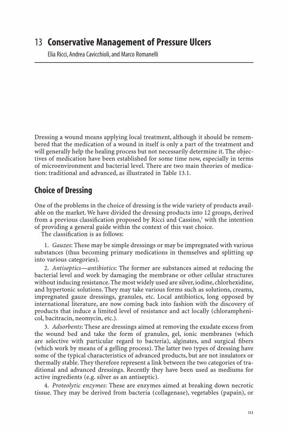
13 Conservative Management of Pressure UlcersElia Ricci, Andrea Cavicchioli, and Marco Romanelli
Dressing a wound means applying local treatment, although it should be remem-bered that the medication of a wound in itself is only a part of the treatment andwill generally help the healing process but not necessarily determine it. The objec-tives of medication have been established for some time now, especially in termsof microenvironment and bacterial level. There are two main theories of medica-tion: traditional and advanced, as illustrated in Table 13.1.
Choice of Dressing
One of the problems in the choice of dressing is the wide variety of products avail-able on the market. We have divided the dressing products into 12 groups, derivedfrom a previous classification proposed by Ricci and Cassino,1 with the intentionof providing a general guide within the context of this vast choice.
The classification is as follows:
1. Gauzes: These may be simple dressings or may be impregnated with varioussubstances (thus becoming primary medications in themselves and splitting upinto various categories).
2. Antiseptics—antibiotics: The former are substances aimed at reducing thebacterial level and work by damaging the membrane or other cellular structureswithout inducing resistance. The most widely used are silver, iodine, chlorhexidine,and hypertonic solutions. They may take various forms such as solutions, creams,impregnated gauze dressings, granules, etc. Local antibiotics, long opposed byinternational literature, are now coming back into fashion with the discovery ofproducts that induce a limited level of resistance and act locally (chlorampheni-col, bacitracin, neomycin, etc.).
3. Adsorbents: These are dressings aimed at removing the exudate excess fromthe wound bed and take the form of granules, gel, ionic membranes (which are selective with particular regard to bacteria), alginates, and surgical fibers(which work by means of a gelling process). The latter two types of dressing havesome of the typical characteristics of advanced products, but are not insulators orthermally stable. They therefore represent a link between the two categories of tra-ditional and advanced dressings. Recently they have been used as mediums foractive ingredients (e.g. silver as an antiseptic).
4. Proteolytic enzymes: These are enzymes aimed at breaking down necrotictissue. They may be derived from bacteria (collagenase), vegetables (papain), or
111
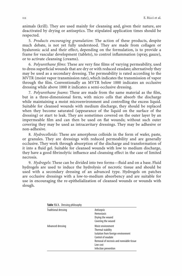
animals (krill). They are used mainly for cleansing and, given their nature, aredeactivated by drying or antiseptics. The stipulated application times should berespected.
5. Products encouraging granulation: The action of these products, despitemuch debate, is not yet fully understood. They are made from collagen orhyaluronic acid and their effect, depending on the formulation, is to provide aframe for vascular development (tablets), to control inflammation (spray, gauze),or to activate cleansing (creams).
6. Polyurethane films: These are very fine films of varying permeability, usedto dress superficial wounds that are dry or with reduced exudate; alternatively theymay be used as a secondary dressing. The permeability is rated according to theMVTR (moist vapor transmission rate), which indicates the transmission of vaporthrough the film. Conventionally an MVTR below 1000 indicates an occlusivedressing while above 1000 it indicates a semi-occlusive dressing.
7. Polyurethane foams: These are made from the same material as the film,but in a three-dimensional form, with micro cells that absorb the discharge while maintaining a moist microenvironment and controlling the excess liquid.Suitable for cleansed wounds with medium discharge, they should be replacedwhen they become saturated (appearance of the liquid on the surface of the dressing) or start to leak. They are sometimes covered on the outer layer by animpermeable film and can then be used on flat wounds; without such outer covering they may be used as intracavitary dressings. They may be adhesive ornon-adhesive.
8. Hydrocolloids: These are amorphous colloids in the form of wafer, paste,or granules. They are dressings with reduced permeability and are generally occlusive. They work through absorption of the discharge and transformation ofit into a fluid gel. Suitable for cleansed wounds with low to medium discharge,they have a good fibrinolytic influence and cleansing effect in the case of limitednecrosis.
9. Hydrogels: These can be divided into two forms—fluid and on a base. Fluidhydrogels are used to induce the hydrolysis of necrotic tissue and should be used with a secondary dressing of an advanced type. Hydrogels on patches are occlusive dressings with a low-to-medium absorbency and are suitable for use in encouraging the re-epithelialization of cleansed wounds or wounds withslough.
112 E. Ricci et al.
Table 13.1. Dressing philosophy
Traditional dressing AntisepsisHemostasisDrying the woundCovering the wound
Advanced dressing Moist environmentThermal stabilityIsolation from foreign environmentControl of exudateRemoval of necrosis and nonviable tissueLow costInfection prevention

10. Interactive dressings: These include the group of biodressings, skin deriva-tives, metalloprotease inhibitors, engineered tissue, and growth factors. These tech-niques are for use by specialists only and their use is described in the section oninteractive dressings below.
11. Technical devices: These are an evolution of dressing/debridement tech-niques, which combine machine-controlled functions with the action of the dressing. Although only used to a limited extent as yet, they are acquiring greaterimportance in daily practice. The most widespread product at the moment is the VAC system described in the section on topical negative pressure therapy below.
12. Non-allopathic dressings: A wide range of products held to be effective inthe treatment of cutaneous lesions, often without clinical studies to support them.Although frequently based on traditions and customs that are centuries old (e.g.aloe, honey), their usage should be postponed until the effects have beenscientifically proven.
With regard to guidelines for use, in a text aiming to provide quick consultationwe will limit ourselves to giving some general indications. Dressing a wound meanschoosing the right product for the wound at its specific stage and deciding on thelength of application.2 Generally speaking we can say that the clinical status ofthe wound and the tissues present determine the choice of product to be used: thequantity of exudate will determine the wear time of the dressing in inverse pro-portion.3 Table 13.2 shows the various clinical stages of wounds and the productssuitable for dressing them.
Interactive Dressings
The next frontier in the treatment of cutaneous lesions is represented by dressingsand products created by means of engineering tissues; these enable those operat-ing in the sector to intervene directly in the healing process (Figures 13.1 and13.2—see color section). We may say that traditional medication, based mainly on
Conservative Management of Pressure Ulcers 113
Table 13.2. Dressings and guidelines for their use, based on clinical examination ofthe wound
Wound Dressing
Colonized/infected AntisepticsLocal antibioticsIonic adsorbentsHypertonic dressings
Necrotic Lytic enzymesHydrogel
Sloughy Lytic enzymesHydrogelHydrocolloidsHypertonic dressings
Cleansed Products encouraging granulationPolyurethane film and foamHydrocolloidsAdsorbents

antisepsis, has turned its attention towards the outside environment and its inter-action with the wound. Advanced dressings attempt to create an environment thatencourages spontaneous repair.4 With the progressive gains in scientific knowl-edge, determined in part by increased attention to the problem, and expressed andorganized according to the theories of wound bed preparation (WBP) and con-centrating in particular on the identification of corrupt and functionally limitedcellular matrices in chronic cutaneous lesions, the need to interact with the healingprocess directly has become pressing.
We may begin by dividing these products according to method, drafting aclassification as shown in Table 13.3.
This is a field that needs further exploration; not all the mechanisms behind the effects have been fully understood and the timing of their use needs to be defined; however, the initial results are promising (Figures 13.3 and 13.4—see color section). The use of single growth factors needs developing, espe-cially considering the specific language involved, which is still far from beingunderstood.
The basic objective (which also requires developing) and the hope for thisapproach to dressing is to change the wound bed from a chronic to an acute state.
Therapeutic Devices
Pressure ulcers represent a major health problem, causing a considerable amountof suffering for patients and a high financial burden for healthcare systems. Thepercentage of the population that is geriatric, and therefore with an increased riskof chronic wound development, is rising constantly. Evidence clearly indicates thatpreventive measures are essential to reduce the prevalence rates of pressure ulcers;therefore healthcare professionals must be able to identify the appropriate strate-gies to adopt, in order to meet the individual patient’s requirements.
The past decade has seen a rise in the number of therapeutic options availablefor the management of acute and chronic wounds. The introduction of advancedmedical devices and new concepts of systemic treatment have led to a better under-standing of the mechanism of tissue repair in chronic wounds, which has beensupported by the development of standardized guidelines for prevention and treatment.
Topical Negative Pressure Therapy
Wound management with negative pressure represents a non-invasive mechanicalwound care treatment, using negative pressure to facilitate wound healing.5 VAC®(Vacuum Assisted Closure) therapy is used to reduce wound fluid, stimulate granulation tissue formation, and reduce bacterial colonization. Negative pressurewound therapy acts by localized and controlled negative pressure, which is appliedin continuous or intermittent cycles.6,7 The equal distribution of negative pressureto every surface of the wound is ensured by a polyurethane open-cell foam dress-ing. The foam is trimmed to fit the entire surface of the lesion, placed in the wound bed, and sealed with an adhesive drape (Figure 13.5—see color section).Negative pressure is applied via an evacuation tube by means of a computerized,
114 E. Ricci et al.

Conservative Management of Pressure Ulcers 115
Table 13.3. Interactive dressings
Group Action Name
SurgicalSurgical procedures Removal of degraded cells from the wound bed. This is an invasive, painful technique Debridement
and is expensive.Wet to dry Progressive removal of the bottom layer of the wound by tearing, using gauzes made Wet to dry
to stick to the wound bed. Extremely painful.Grafting Removal of the wound bed until healthy tissue is reached, after which the wound is Grafting
covered with skin taken from elsewhere on the patient. If the skin takes,immediate healing is achieved; if not, there may in any event be a rapid resumption of the repair process and re-epithelialization.
Skin substitutesGlycerol skin This is skin taken from organ donors and treated with glycerol, which makes it Skin bank
non-vital and suitable for preserving. It was originally intended for use as a biological covering for patients with severe burns and has been progressively extended to chronic wounds. It is not clear how it works and a thoroughly cleansed base is required.
Cryopreserved skin Skin taken from organ donors and preserved by freezing. Used as a biological covering, Skin bankit may work by releasing growth factors.
Culture-grown skin After a skin biopsy, the cells are placed on special mediums which make them larger Apligraf®and more concentrated. This is an autograft, which is capable of activating the repair process.
Heterologous culture- Skin substitute derived from the fetal prepuce. This is a heterologous graft that is Dermagraft®grown skin capable of reactivating repair processes (growth factors?) and of taking in some
cases that are not clearly defined as yet.
Metalloprotease inhibitorsMetalloprotease Metalloprotease, elastase and plasmin are enzymes derived from cellular decay, Promogran®
inhibitors which are capable of halting the healing process when present in large quantities.These products, made from oxidized and regenerated cellulose, appear to be able to deactivate the excess of such enzymes and to bind them.
Growth factorsGrowth factor Proteins capable of mediating cellular functions through membrane receptors.
Produced by a secretory mechanism within the body itself, they are currently being studied and are produced industrially by means of recombinant genes. The principal ones involved in the healing process are: platelet-derived growth factor (PDGF), transforming growth factor beta (TGFb), TGFa, tumor necrosis factor (TNF), insulin-like growth factor (IGF), interleukin enhancer binding factor (ILF),interleukin-1 (IL-1), IL-2, nerve growth factor (NGF), vascular endothelial growth factor (VEGF), beta fibroblast growth factor (FGF), keratocyte growth factor (KGF).
Platelet gel Prepared on the spot from the patient’s or donor’s serum (from a blood bank) or Regranex®prepared industrially, these act by releasing large quantities of growth factors,mainly PDGF.
Cultured cellCulture-grown Once a sample of skin has been taken, the fibroblasts are isolated and grown so as to Hyalograft 3D®
fibroblasts create a sort of “three-dimensional dressing” on a hyaluronic acid base. This is a self-graft of fibroblasts, the cells responsible for healing processes and growth factors.
Culture-grown As with the fibroblasts, a sample of skin is taken and once the keratinocytes have Laser Skin®keratinocyte been isolated they are cultivated on a transparent sheet of hyaluronic acid. Hyalograft 3D KC®
This is a very delicate product and its established use for treating acute wounds has yet to be discussed for chronic wounds.
OtherEngineered derma Three-dimensional structure made from cellulose and collagen or hyaluronic acid Integra®
capable of providing a base mainly for neovascularization, which is considerably Hyalomatrix®increased. It is covered with a coating which must be removed after 1–3 weeks.
programmable pump. The target pressure for wound therapies varies from 50 mmHg to 200 mmHg, based on the characteristics of the individual wound. Inpressure ulcers, a negative pressure of 125 mmHg is used in a continuous cycle of48 hours. Negative pressure therapy provides a moist wound healing environment,assists in uniformly drawing the wound border, enhances epithelial migration,reduces bacterial colonization, and reduces localized edema by increasing localblood perfusion and accelerating the rate of granulation tissue formation.8
Despite the successful treatment of different wound types, some limitations mayoccur when attempting to treat certain areas of the body that involve irregular sur-faces surrounding the wounds, such as the perineum.9 VAC® therapy is indicatedin acute and traumatic wounds, dehisced incisions, neuropathic ulcers, stage 3 and4 pressure ulcers, vascular wounds, and chronic debilitating wounds. Split-thickness mesh skin grafts also benefit from VAC® therapy.
Contraindications for negative pressure treatment include cutaneous malignantlesions, untreated osteomyelitis, necrotic tissue within the wound bed and fistuladirectly communicating with organs and cavities. Caution should be used whenthere is active bleeding, unstable local hemostasis, use of anticoagulants, or distaldiabetic foot lesions.10 The nutritional status of the patient should be stable andthe patient should be continuously monitored by nurses and should be positionedon a support surface so as to redistribute his or her weight over a large area andreduce pressure. An adequate amount of intact peri-wound skin for adherent dressing should be available and the ulcer should be free of necrotic tissue orosteomyelitis.
The dressing is changed every 48 h or every 12 h if infection is present. Thewound bed is cleansed per routine, the sponge is placed in the wound, and theevacuating tube is laid on top of the foam, linked to a collection chamber locatedon the pump. An adhesive clear dressing is placed over the foam and the tube.Duration of the therapy varies from 4 to 6 weeks, with continuous or intermittentcycles of treatment.11 Continuous therapy facilitates removal of wound fluids andreduction of edema, while intermittent therapy acts as a mechanical stretch andresults in the repeated release of biochemical messengers. Wound measurement,tissue and fluid characterization, odor, and surrounding skin should be monitoredat each dressing change.
Negative pressure therapy should be used in pressure ulcers to achieve completehealing or to prepare the wound bed for surgical closure, especially in chronic non-healing wounds of considerable depth, rather than the traditional saline wet-to-moist dressings.
Hydrotherapy
Hydrotherapy is another treatment that is frequently used in many countries, prin-cipally in patients with leg ulcers.12 The patient is immersed in special pools wherethe water, containing antiseptics, is shaken with artificial movement so as to makethe removal of dead tissue easier. Hydrotherapy provides cleansing pressure-irrigation and hydromassage. The cleansing is useful either to remove secretionsand bacteria or to soften necrotic wound material. This procedure is generally per-formed with a saline solution only or with an antiseptic solution or with othertypes of detergent solutions—used according to wound conditions and dressingcompatibility. Pressure-irrigation uses water pressure to remove the necrotic mate-
116 E. Ricci et al.
rial from the wound surface.13 However, if the pressure used to deliver irrigationsolution is too low (below 4 psi), the lavage will not clean effectively; therefore,pressure irrigation has to be in a range from 4 to 15 psi.
A simpler method uses a 35 ml syringe with a 19-gauge needle producing an 8psi pressure action, which is able to clean a wound without causing damage to newgranulation tissue.
Another method makes use of hydromassage, that is, water pressure jets gener-ated in a whirlpool. In this case the tissue becomes soft in the whirlpool and isremoved by the pressure of the jets. The massage reduces edema and inflamma-tion and improves circulation in ischemic legs. A further refinement of this tech-nique consists of immersing the patient for 20–30 minutes after a 30-secondtreatment at maximum pressure. The sessions are twice-weekly. This method isindicated in very exudative wounds, necrotic wounds, and eschars, but is con-traindicated in clean and granulation wounds.
Moreover, there are now special devices available that can direct a high-pressurewater jet onto lesions and then, with suction, remove the devitalized tissue beforeit dissolves. Among such instruments, one that deserves particular attention is adevice developed in Switzerland, based on high-pressure microjet technology. Thedevice consists of a liquid pump driven by compressed air, which generates highhydraulic pressure in a liquid directed through a nozzle installed in a hand-piece.The major advantage of this debridement technique is the reduction of the dura-tion of treatment. The aim is to provoke a decisive healing impulse in a stagnantwound. Through the nozzle, Ringer solution, NaCl or aqua ad injectabilia areinjected onto the surface of the wound in the form of a very fine jet under pre-cisely controlled pressure. The duration of each intervention depends on theimportance and degree of necrosis of the wound and varies from 10 to 30 minutes,usually at weekly intervals. Advanced dressings sustain the treatment. The threemajor results of this application are: shortening of the wound healing process,reduction of the scar tissue, and low stress effects for the patients, because the treat-ment is relatively painless. These treatments are contraindicated in patients suf-fering from anticoagulation, tumors, and unprotected or open blood vessels.
Warming Therapy
Most chronic wounds become hypothermic and hypothermia has been shown toimpair the healing process by reducing the normal function of the immune systemand promoting wound infection. A recent technology utilizing radiant heat wasintroduced for the treatment of chronic wounds. This warming treatment is ableto maintain 100% relative humidity at the interface between dressing and woundbed and to restore both peri-wound and wound bed temperature toward nor-mothermia. The system is provided with a temperature control unit (TCU), whichoperates either from an AC outlet or with a rechargeable battery pack for porta-bility. A disposable wound cover made of a polyurethane foam provides a non-contact surface that will not disrupt the wound, will absorb the exudate, and has a transparent window which provides easy viewing for monitoring woundprogress. An infrared warming card, which is connected to the TCU, slides into asleeve in the cover and warms to a temperature of 38°C. Using warming therapy,the skin and subcutaneous tissue are returned to a temperature that is closer tonormal. Warming encourages blood vessels to dilate, which increases blood flow
Conservative Management of Pressure Ulcers 117
to the wound and peri-wound area. Greater blood flow delivers more oxygen, nutri-ents, and growth factor to the wound. The use of warmth on fibroblasts andendothelial cells in vitro was found to reduce the inhibitory effect of chronicwound fluid upon neonatal fibroblasts.14 In a recent study the warming therapywas compared to standard treatment in patients with stage 3 to 4 pressure ulcers.15
Results showed a statistically significant accelerated rate of healing for patientsreceiving heat therapy compared to standard treatment.
References
1. Ricci E, Cassino R. Piaghe da decubito. Turin: Minerva Medica; 2004.2. Bennet G, Moody M. Wound care for health care professionals. London: Chapman & Hall; 1995.3. Dealey C. The care of wounds. Oxford: Blackwell; 1999.4. Hess CT. Guida clinica alla cura delle lesioni cutanee. Milan: Masson; 1999.5. Argenta LC, Morykwas MJ. Vacuum-assisted closure: A new method for wound control and treat-
ment: Clinical experience. Ann Plast Surg 1997; 38(6):563–576.6. Morykwas MJ,Argenta LC, Shelton-Brown EI, McGuirt W.Vacuum-assisted closure: A new method
for wound control and treatment: Animal studies and basic foundation. Ann Plast Surg 1997;38(6):553–562.
7. Deva AK, Buckland GH, Fisher E, et al. Topical negative pressure in wound management. Med JAust 2000; 173(3):128–131.
8. Baynham SA, Kohlman P, Katner HP. Treating stage IV pressure ulcers with negative pressuretherapy: a case report. Ostomy Wound Manage 1999; 45(4):28–32, 34–35.
9. Greer SE, Duthie E, Cartolano B, et al. Techniques for applying subatmospheric pressure to woundsin difficult regions of anatomy. J Wound Ostomy Continence Nurs 1999; 26(5):250–253.
10. Evans D, Land L. Topical negative pressure for treating chronic wounds: a systematic review. Br JPlast Surg 2001; 54:238–242.
11. Joseph E, Hamori CA, Bergman S, et al.A prospective randomized trial of vacuum-assisted closureversus standard therapy of chronic non healing wounds. Wounds 2000; 12(3):60–67.
12. Waspe J. Treating leg ulcers with high pressure irrigation devices. Nurs Stand 1996; 11(6):53–54.13. Ho C, Burke DT, Kim HJ. Healing with hydrotherapy. Adv Directors Rehabil 1998; 7(5):45–49.14. Park HY, Shon K, Phillips T. The effect of heat on the inhibitory effects of chronic wound fluid on
fibroblasts in vitro. Wounds 1998; 10(6):189–192.15. Price P, Bale S, Crook H, et al. The effect of a radiant heat dressing on pressure ulcers. J Wound
Care 2000; 9(4):201–205.
118 E. Ricci et al.
14 Surgical Management of Pressure UlcersJens Lykke Sørensen, M.J. Lubbers, and Finn Gottrup
Introduction
Most pressure ulcers do not need surgical intervention.Candidates for surgery are a selected group of patients where debridement and
conservative measures are not enough to ensure healing of a sufficient quality orspeed, and where the patients will benefit from surgical intervention. In general,these patients will have grade 3 and 4 pressure ulcers.
The cornerstones of successful surgical treatment of pressure ulcers are a com-petent staff, correct selection of patients, correct and meticulous surgical method,and sufficient postoperative support.
No unambiguous criteria for selection of patients or methods exist, and an indi-vidual assessment is a necessity. However, guidelines are a useful tool, particularlyat the initial assessment.1–3
The Staff
The surgeon is a link in a chain. Success in pressure ulcer treatment is dependenton a well-educated and committed multidisciplinary staff.4
Preceding surgery is a period of observation of the patient, either on an out-patient basis or as an inpatient, to evaluate the optimal treatment. During hospi-talization the nursing staff are the most important group, since they can observeand cooperate with the patient around the clock.
Concomitant diseases must be controlled by relevant specialists preoperatively,as should the dietary intake, and pressure relief is secured as soon as possible.
Postoperatively the patient must be helped to follow restrictions to ensure totalpressure relief of the operated region, and at a later stage the occupational thera-pist and physiotherapist participate in rehabilitating and mobilizing the patient.The hospital pharmacist is often included early in the team to prepare the dis-charge of the patient.
Selection of Patients
It is important to look at the whole patient and not only the ulcer. All patients with deep pressure ulcers (grade 3 or 4) should be evaluated for surgical treatment.Pressure ulcer patients, however, tend to have other diseases as well as the
119
pressure ulcer, and this must be taken into account before deciding whether tooperate. As mentioned, concurrent diseases must be dealt with preoperatively. Thepatient’s ability to cooperate and to tolerate operation and the postoperativeregimes must be evaluated. The patient’s activities of daily living should be com-patible with postoperative pressure relief and general care, and education of thepatient is necessary.
Debilitated patients without the capacity for cooperation or patients expectedto make a full recovery might be sufficiently treated by revision alone. Terminalpatients are not candidates for reconstructive procedures. Spinal cord injuredpatients usually are. Special attention must be paid to patients with advanced disseminated sclerosis and other diseases influencing their intellect, since thesepatients will often have difficulties in cooperating because of the impact of thedisease on their behavior.
In patients likely to make a total recovery, such as some multitrauma patients,even large pressure ulcers may eventually heal and these could be left for sponta-neous healing. However, other factors such as time and the restrictions caused bythe pressure ulcer can be an indication for surgery.
Surgical Methods
The initial step in surgical treatment of pressure ulcer is always a thoroughdebridement including removal of not only necrotic tissue but also inferior,scarified tissue, extra-osseous calcifications, and infected bone. This is dealt within Chapter 15. Denuded bone should always be smoothed out to create an evenpressure-distributing surface beneath the soft tissue used in the reconstruction.2
When the pressure ulcer cavity is clean and vital, the decision is whether to leavethe cavity to a time-consuming spontaneous healing or to perform a fast but moreor less complicated reconstruction.
Small and superficial pressure ulcers should be left for secondary healing.Correct wound care and sufficient pressure relief are prerequisites for success.Large superficial pressure ulcers should usually be operated on. Small but deeppressure ulcers will often benefit from reconstructive procedures.
One-stage procedures with revision and immediate reconstruction are usuallyrecommended.1,5,6 A number of reconstructive alternatives exist, and the leastdemanding method suitable for the purpose should be chosen.
Split-Thickness Skin Grafts
From a surgical point of view split-thickness skin grafting is a simple and fast pro-cedure. Donor sites are abundant in pressure ulcer patients. The recipient site mustbe clean and well vascularized. The skin graft consists of the epidermis and a frac-tion of the dermis. Split-thickness skin grafts are harvested with a dermatomeadjusted to a thickness of about 0.3–0.4 mm. The graft is meshed to allow fluid toescape since this will hamper healing if it is trapped beneath the graft. The graftsticks to the graft bed, where granulation tissue will turn into scar tissue afterhealing, covering the area with a thin non-pliable surface making the split skingraft prone to erosion.7 The graft is covered with a bandage, which allows diffu-
120 J.L. Sørensen et al.

sion of superfluous liquid away from the graft without desiccation. One of the fewindications for use of local antibiotics is in the dressing covering the graft toprevent infection. If a tie-over dressing or an ordinary bandage is not suitable, aVAC® (Vacuum Assisted Closure) sponge can secure the graft.8 The bandage isremoved after 2–6 days depending on the method, the risk of complications, andthe surgeon’s preference. Healing is completed in about 10 days, but the skin graftshould not be submitted to mechanical loading for about 3 weeks. The donor siteis covered with a bandage for moist healing. After 7–10 days the donor site shouldbe healed and the bandage removed.9
Split-thickness skin grafting is indicated for well-granulating, large, shallowulcers, where future mechanical loading will be limited. The sacral area and the back are the typical locations where split-thickness skin grafting may be indicated.
Full-Thickness Skin Grafts
Full-thickness skin grafts contain all the dermis. This makes the full-thicknessgraft thicker and more resistant to mechanical wear and tear, but the demand forvascularization of the recipient bed is bigger. When a full-thickness skin graft isconsidered, a skin flap will often be indicated as well. Full-thickness skin grafts canusually only be harvested when skin is loose enough to allow direct suture of thedonor site, since no epithelial elements are left to allow healing from the donorbed. The recipient bed must have a good blood supply, and the graft must be firmlyimmobilized. Full-thickness skin grafts are not meshed.
Full-thickness skin grafts are indicated for small superficial pressure ulcers withwell-granulating, even surfaces on locations where friction is to be expected or cos-metic outcome is to be taken into consideration. Grafting does not alter the char-acter of the skin.9 The heel, the plantar, and the head can sometimes be treatedwith a full-thickness skin graft.
Direct Closure
Direct closure is the simplest surgical method for eliminating a defect, but in pres-sure ulcer surgery it is only exceptionally indicated. If it is indicated, closure is per-formed in layers over suction drainage. An underlying cavity must be avoided. Apressure ulcer develops when tissue is too scanty.With direct suture even less tissueis available, increasing the risk for recurrence of the pressure ulcer.5,10
Skin Flaps
Skin flaps, or cutaneous flaps if no subcutaneous tissue is incorporated in the flap,can literally be raised anywhere on the body. As with other types of flaps, theirblood supply can be random, or an axial vessel can be incorporated in the flap. Anaxial vessel makes the planning more pliable, since the blood supply to the flap isindependent of the supply from the base of the flap. Skin flaps are elevated bycutting the skin and subcutaneous tissue to the desired depth according to thedrawing on the patients from the preoperative planning. The underside of the flap
Surgical Management of Pressure Ulcers 121
is cut to its base, the surroundings are mobilized as necessary, the flap is moved tothe defect, and the donor site and the flap are sutured in layers. Suction drainageis compulsory.
In pressure ulcer surgery, no tightness of the flaps or the surroundings can beaccepted, reducing the availability of these flaps. The donor site must be withoutscarification or other consequences of being close to a pressure ulcer.
Cutaneous flaps are indicated anywhere where a skin graft is insufficient and amyocutaneous flap is too much. Pressure ulcers of moderate size, not too deep, andwithout underlying bony disorders or exposure are potential candidates for cuta-neous flaps.2,9,11
Fasciocutaneous Flaps
Adding an underlying fascia to a skin flap improves the blood supply. The extrapadding probably adds little to the pressure-distributing abilities of the flap.
Fasciocutaneous flaps are managed in the same way as skin flaps but with incor-poration of the underlying fascia in the flap. The fascia in the donor site can usuallynot be sutured.
Fasciocutaneous flaps are indicated in reconstruction of pressure ulcer defectswithout too deep a tissue defect, preferably without a history of osteomyelitis, andwithout a big demand for pressure distribution in the region.12,13
The scrotal flap9 is a well-known fasciocutaneous flap. This flap is not often usedby the authors. The distal part of the tensor fasciae latae flap (the central part isactually a myocutaneous flap) is a more recommendable type of fasciocutaneousflap.5,9,14
Myocutaneous Flaps
Myocutaneous flaps are axial flaps. They possess muscle tissue with an excellentblood supply and provide full-thickness skin coverage; a large amount of tissuecan be incorporated in the flap to supply it with sufficient bulk to fill even largedefects. The excellent blood supply makes these flaps well suited for fighting infec-tion and promoting healing. The intact skin and subcutaneous tissue have thepotential for effective pressure and shear distribution, and the bulk adds to theability to distribute pressure and fill a large defect. These physiological andmechanical properties make myocutaneous flaps the treatment of choice for deeppressure ulcers.1,10,15
In general the pressure points on the human body are not padded with muscle.Yet, experiments indicate that muscle tissue beneath pressure-loaded skin is advantageous.16
Some muscles are indispensable for normal muscle function, for examplegluteus maximus in normal gait. This fact must be considered when the selectionof a myocutaneous flap is carried out in an ambulatory patient. In spinal cordinjured patients, a group constituting the majority of patients with myocutaneousflap surgery, these considerations are usually irrelevant.
The myocutaneous flap is advanced into the defect by cutting its cutaneous,subcutaneous, and fascial borders according to a drawing from the planning on
122 J.L. Sørensen et al.
the patient. The muscles are then freed from their bony connections or tendons,and the areolar tissue around the vessels is loosened enough to mobilize the flapinto the defect. Usually the vessels do not need to be totally freed, not with theprimary use of the flap, anyhow. The whole mass of tissue is then advanced intothe defect, and over suction drainage in both the donor and the recipient site theflap and donor site are sutured in layers. The drains in the pressure ulcer cavitycan be left for a couple of weeks, if formation of a cavity beneath the flap is feared.5
Myocutaneous flaps are indicated anywhere on the body where deep pressureulcers are treated for osteomyelitis or optimal pressure-distributing abilities arerequested. The pelvic region is the region most often treated with myocutaneousflaps, since many of the pressure ulcers needing a myocutaneous flap are situatedhere.5,17
In the pelvic region the ischial region is most often the site of pressure ulcers.Several flaps are available for the repair.1,2,9,18–20 The primary choice of the authorsis a flap based on the hamstrings (Figure 14.1—see color section). This is a versatile and safe flap, which can be readvanced in case of recurrence18—if it israised in its full length. The muscle content of the flap can be varied, making itusable for both spinal cord injured and ambulatory patients. The gluteus maximusor the tensor fasciae latae are alternative solutions.2,9
The sacral pressure ulcer is often treated using a flap based on the gluteusmaximus muscle.5,13,21 Several variations in creating myocutaneous flaps based onthe muscle exist, making the flap very versatile.5,9,21 By splitting the muscle, whichhas a dual blood supply, ambulatory patients can benefit from this flap too.9
A trochanteric pressure ulcer invites the use of a tensor fasciae latae flap, whichis just at the edge of the ulcer (Figure 14.2—see color section). The flap is safe, andthe muscle is expendable. Often the donor site can be closed primarily; otherwisea split skin graft can be used for the donor defect.10,14,22,23 Alternatives are the vastuslateralis muscle, the rectus femoris muscle, or the inferiorly based gluteus maximusmuscle.2,5,9
Flaps Without Skin Coverage
In selected cases—or merely by need—a muscle flap can be indicated. If too muchbulk from subcutaneous tissue is to be avoided (not a frequent problem in pres-sure ulcer surgery!) or if the survival of the cutaneous part of a myocutaneous flapis hampered, a muscle flap can be used to supply bulk and blood supply, and theflap can be covered by a split-thickness skin graft. If possible a myocutaneous flapshould be preferred.
In flaps without skin coverage the tissue is isolated and transposed to the recip-ient site using the same dissection technique as in raising other flaps.
Isolated fascial flaps are hardly ever indicated in pressure ulcer surgery exceptif they are the only way to get a vital ulcer bed.
Special Types of Flaps or Surgical Procedures
Sensate myocutaneous flaps in some spinal cord injured patients can be elevatedto transfer sensate skin to an insensate region with a pressure ulcer.15,24,25 Recur-
Surgical Management of Pressure Ulcers 123
rence rates might be reduced by making the skin sensitive to pain from pressureand ischemia, but the new sensation might be unpleasant, making the patient movesupport to an insensitive area, where a new pressure ulcer can develop.10,24
Complicated procedures to gain sensitivity in pressure ulcer risk areas exist, butthe more simple tensor fasciae latae flap is probably the most popular with a lesionbelow L3.15,24
Free flaps are an option in selected cases of pressure ulcers.26,27 The proceduresare demanding and time-consuming and are infrequently indicated. An exampleis a free latissimus dorsi myocutaneous flap to a pelvic pressure ulcer. Temporarycasting or external fixation of the region might be indicated to avoid lesion of themicrovascular anastomosis.
Muscle-sparing perforator flaps are attracting increasing interest in pressureulcer surgery.12,28
Tissue expansion has been used in both skin and other tissue to gain coverageof pressure ulcer defects.10,29,30 Implantation of foreign material to reinforce tissuecovering former or threatening pressure ulcers31 has not been widely reported. Theauthors have no experience with the latter two methods.
Multiple or Recurrent Pressure Ulcers
Extensive or multiple pressure ulcers should be treated in as few sessions as pos-sible to reduce time, the use of resources, and, probably, the risk of cross-infectionfrom an untreated to a treated pressure ulcer.5,17 When reconstruction becomes anoption, the extensive ulcers are situated in the pelvic region. A total thigh flap givesgood soft tissue covering even for huge pressure ulcers on the ipsilateral pelvis.5,32
A smaller amount of tissue can be provided by a rectus abdominis myocutaneousflap.33
Recurrence is unfortunately a quite common problem in pressure ulcersurgery.5,15,17,21,34 There is no major difference in treating primary or recurrent pres-sure ulcers. The same reconstructive methods are used. In the primary planningit is important to design the reconstruction so that the future use of flaps is nothampered. The flaps should, if possible, be designed large enough for reuse in caseof recurrence. With an intelligent design it is usually possible to close the donorsite without grafting.5
Postoperative Care
Postoperatively the pressure ulcer is dealt with, but the risk factors might still bepresent and will need continuous monitoring. The patient will still need optimalpressure relief, diet, and treatment of concomitant diseases. If this is forgotten,recurrence is almost inevitable.
Postoperative Pressure Ulcer Prevention
Immediately after the operation impairment to the circulation in the region mustbe avoided, otherwise healing might be hampered. Skin grafts tolerate some pres-sure. Actually a certain amount of pressure is an advantage to immobilize the graft
124 J.L. Sørensen et al.
to the recipient bed and to avoid oozing of liquid beneath the graft. Flaps must notbe loaded in the initial postoperative period.
The operated region can be pressure relieved in two ways: The patient can bepositioned to totally relieve the region; for example, a reconstruction on the backof the pelvis can be relieved by the prone position. Some patients, such astetraplegics, may be unable to stay in the prone position, since their respirationmight be obstructed. Other complications can also occur as a result of lying in theprone position for long periods.10 An alternative to positioning the patient is apressure-relieving mattress. Turning regimes are necessary with some types ofmattresses. Loading or straining the flap during the turning must absolutely beavoided. Social isolation of the patient during the postoperative restrictions shouldalso be avoided.5
A number of mattresses and special beds are available (see Chapter 7). Air-fluidized beds and some of the low-air-loss beds and mattresses are recommendedfor relieving flaps.5,15,35 The recommended period for postoperative pressure reliefvaries. Usually 2–3 weeks are recommended.5,10
After total pressure relief the flap is conditioned to loading5 In ambulant patientsnormal behavior can be resumed. In high-risk patients restrictions on loading therisk areas should continue, since the risk factors are permanent. As a general prin-ciple, risk areas should not be loaded for more than 2 hours.10 The authors advo-cate fixed planes for mobilization and regular postoperative control, naturally withindividually based variations.
Postoperative Patient Care
The operation field is observed according to normal surgical procedures. Fluidaccumulation, infection, and tissue necrosis are particularly observed for, andintervention should generally be swift.5
In the postoperative period the protein and caloric intake should be increasedafter estimation of need by a dietician.
The operation wound, as well as being protected from load and strain, shouldbe kept clean. Indwelling catheters, fluid or low fiber diet for a few days precededby preoperative enema and constipating medicine can be used.10,15 Medication totreat spasm is sometimes necessary.36
Physical training for all patients is a prerequisite. Mobilization is performed toa degree compatible with the individual patient’s ability. All of the staff are respon-sible for participating in teaching patients to take responsibility for their owntraining and pressure sore prevention. Prevention and educational programs areused to help with training and reduce recurrence.4,10,37,38
Pressure prevention is modified according to alterations in the patient’s condi-tion. If the pressure ulcer risk is permanent, the prevention measures should bepermanent.
References
1. Foster RD, Anthony JP, Mathes SJ, Hoffman WY. Ischial pressure sore coverage: a rationale for flapselection. Br J Plast Surg 1997; 50:374–379.
2. Linder RM, Morris D. The surgical management of pressure ulcers: a systematic approach basedon staging. Decubitus 1990; 81:32–38.
Surgical Management of Pressure Ulcers 125
3. Pressure Ulcer Advisory Panel. Pressure ulcer treatment. Am Fam Physician 1995; 51:1207–1222.4. Sinclair L, Berwiczonek H, Thurston N, et al. Evaluation of an evidence-based education program
for pressure ulcer prevention. J Wound Ostomy Continence Nurs 2004; 31:43–50.5. Sørensen JL, Jørgensen B, Gottrup F. Wound management: Surgical intervention. In: Morison MJ
(eds) The prevention and treatment of pressure ulcers. London: Mosby; 2001: 155–175.6. Rubayi S, Burnett CC. The efficacy of a single-stage surgical management of multiple pressure
sores in spinal cord injured patients. Ann Plast Surg 1999; 42:533–539.7. Place MJ, Herber SC, Hardesty RA. Basic technique and principles in plastic surgery. In: Aston SJ,
Beasley RW, Thorne CNM (eds) Grabb and Smith’s Plastic surgery, 4th edn. London: Little, Brownand Company; 1997: 13–26.
8. Genecov DG, Schneider AM, Morykwas MJ, et al. A controlled subatmospheric pressure dressingincreases the rate of skin graft donor site reepithelialization. Ann Plast Surg 1998; 40:219–225.
9. Bergström N, Bennett MA, Carlson CE, et al. Treatment of pressure ulcers. Clinical Practice Guide-line no. 15. Rockville, MD: US Department of Health and Human Services, Public Health Service,Agency for Health Care Policy and Research; December 1994.
10. Strauch B,Vasconez LO, Hall-Findlay EJ (eds) Grabb’s encyclopedia of flaps. London: Little, Brownand Company; 1990: 1529–1800.
11. Mithat Akan I, Sungur N, Ozdemir R, et al.“Pac Man” flap for closure of pressure sores. Ann PlastSurg 2001; 46:421–425.
12. Yu P, Sanger JR, Matloub HS, et al.Anterolateral thigh fasciocutaneous island flap in perineoscrotalreconstruction. Plast Reconstr Surg 2002; 109:610–616.
13. Borman H, Maral T. The gluteus fasciocutaneous rotation-advancement flap with V-Y closure inthe management of sacral pressure sores. Plast Reconstr Surg 2002; 109:2325–2329.
14. Dermiseren ME, Gökrem S, Özedemir OM, et al. Hatchet-shaped tensor fascia lata musculocuta-neous flap for coverage of trochanteric pressure sores: A new modification. Ann Plast Surg 2003;51:419–422.
15. Niazi ZBM, Salzberg CA. Surgical treatment of pressure ulcers. Ostomy/Wound Manage 1997; 43:44–52.
16. Naito M, Ogata K. Vulnerability of the skin circulation without underlying muscle. An experi-mental study. 33rd Annual Meeting of the ISOP, Kobe, 1994; MT V-1.
17. Schryvers OI, Stranc MF, Nance PW. Surgical treatment of pressure ulcers: 20-years experience.Arch Phys Med Rehabil 2000; 81:1556–1562.
18. Kroll SS, Hamilton S. Multiple and repetitive use of the extended hamstring V-Y myocutaneousflap. Plast Reconstr Surg 1989; 84:296–302.
19. Angrigiani C, Grill D, Siebert J, Thorne C. A new musculocutaneous island flap from the distalthigh for recurrent ischial and perineal pressure sores. Plast Reconstr Surg 1995; 96:935–940.
20. Josvay J, Donath A. Modified hamstring musculocutaneous flap for coverage of ischial pressuresores. Plast Reconstr Surg 1999; 103:1715–1718.
21. Aggrawal A, Sangwan SS, Siwach RC, Batra KM. Gluteus island flap for repair of sacral pressureulcers. Spinal Cord 1996; 34:346–350.
22. Siddique A, Wiedrich T, Lewis VL. Tensor fascia latae V-Y retroposition myocutaneous flap: clin-ical experience. Ann Plast Surg 1993; 31:313–317.
23. Ercöen AR, Apaydin I, Emiroglu M, et al. Island V-Y tensor fasciae latae fasciocutaneous flap forcoverage of trochanteric pressure sores. Plast Reconstr Surg 1998; 102:1524–1530.
24. Kuhn BA, Lüscher NJ, de Roche R, et al. The neurosensory musculocutaneous tensor fasciae lataeflap: long term results. Paraplegia 1992; 30:396–400.
25. Thomson HG, Azhar AM, Healy H. The recurrent neurotrophic buttock ulcer in themyelomeningocele paraplegic: a sensate flap solution. Plast Reconstr Surg 2001; 108:1192–1196.
26. Park S, Koh KS. Superior gluteal vessel as a recipient for free flap reconstruction of lumbosacraldefects. Plast Reconstr Surg 1998; 101:1842–1849.
27. Hung SJ, Chen HC, Wei FC. Free flap for reconstruction of the lower back and sacral area. Micro-surgery 2000; 20:72–76.
28. Blondeel PN, Van Landuyt K, Hamdi M, Monstrey SJ. Soft tissue reconstruction with the superiorgluteal artery perforator flap. Clin Plast Surg 2003; 30:371–382.
29. Espositio GE, Ziccardi P, Di Caprio G, Scuderi N. Reconstruction of ischial pressure ulcers by skinexpansion. Scand J Plast Reconstr Surg 1993; 27:133–136.
30. Gray BC, Salzberg CA, Petro JA, Salisbury RE. The expanded myocutaneous flap for reconstruc-tion of the difficult pressure sore. Decubitus 1990; 3:17–20.
31. Minns RM, Sutton RA. Carbon fibre pad insertion as a method of achieving soft tissue augmen-tation in order to reduce the liability to pressure sore development in the spinal injury patient. BrJ Plast Surg 1991; 44:615–618.
126 J.L. Sørensen et al.

32. Peters JW, Johnson GE. Proximal femurectomi for decubitus ulceration in the spinal cord injurypatient. Paraplegia 1990; 28:55–61.
33. Kierney PC, Cardenas DD, Engrav LH, et al. Limb-salvage in reconstruction of recalcitrant pres-sure sores using the inferiorly based rectus abdominis myocutaneous flap. Plast Reconstr Surg1998; 102:111–116.
34. Evans GRD, Dufresne CR, Manson PN. Surgical correction of pressure ulcers in an urban centre:is it efficacious? Adv Wound Care 1994; 7:40–45.
35. Allen V, Ryan DW, Murray A. Air-fluidized beds and their ability to distribute interface pressuresgenerated between the subject and the bed surface. Physiol Measure 1993; 14:359–364.
36. Mess S-A, Kim S, Davison S, Heckler F. Implantable Baclofen pump adjuvant in treatment of pres-sure sores. Ann Plast Surg 2003; 51:465–467.
37. US Department of Health and Human Services. Preventing pressure ulcers. A patient’s guide.Decubitus 1992; 5:34–40.
38. Maklebust HA, Magnan MA. Approaches to patient and family education for pressure ulcer man-agement. Decubitus 1988; 5:18–28.
Surgical Management of Pressure Ulcers 127
15 Debridement of Pressure UlcersAndrea Bellingeri and Deborah Hofman
Debridement is an accepted principle of good wound care, especially when debris isacting as a focus for infection. (NICE Guidelines, UK, 2001)
Introduction
The term debridement was first used by a French surgeon in the eighteenth century to describe surgical removal of debris from open wounds.1 Debris may consist of foreign bodies in a wound, for example following trauma, but inchronic wounds such as pressure ulcers it is more likely to be devitalized tissue.This may manifest itself as: slough (soft and ill-defined yellow-brownish hydratedtissue), necrotic tissue (black or brown), or eschar (well-circumscribed blackadherent slough).2 Such devitalized tissue may be distributed over the entiresurface of the wound, separated from the wound edges, or patchy over the woundsurface. Chronic wounds and in particular pressure ulcers are particularly proneto accumulate devitalized necrotic tissue, which reduces the possibility of nutri-ents reaching the wound and damages new epithelial and granulation cells. Theremoval of this type of tissue or debridement is therefore essential to facilitatehealing.
Why Necrotic Tissue Is Present in Chronic Wounds
Tissue ischemia resulting from poor circulation, from unrelieved pressure or froma combination of both of these factors deprives the tissue of oxygen and causestissue death. Devitalized tissue deprived of blood tends to become dehydrated andto contract, forming an eschar. Classically the eschar is dry, black, and rigid andwith the passage of time tends to separate at the margins from the surroundingtissue. When the tissue becomes hydrated either as a result of occlusive dressings,edema, or exudate the eschar softens and the color changes, becoming first brownand then yellow. In the final stages of degeneration the eschar becomes slough, ayellowish fibrous tissue which adheres to the wound bed. This is part of the naturalhealing process (autolysis) in which the endogenous proteolytic enzymes digestthe devitalized tissue.
129

Reasons for Debridement
Removal of dead tissue from the wound is necessary to promote the proliferationof new cells. The presence of necrotic tissue in the wound bed will result in:
• a heightened risk of infection (since devitalized tissue is a medium for bacter-ial growth);
• increased odor;• cellular dysfunction—necrotic tissue gives a persistent pro-inflammatory stim-
ulus resulting in impaired cell migration and connective tissue deposition andinhibition of growth factors;3
• inability of the wound to contract because the eschar forms a “plug” within thewound.
The presence of necrotic tissue will also prevent assessment of the depth and extentof the wound.
As in many other aspects of wound management, debridement must be carriedout following a detailed clinical and nursing assessment taking into account thepatient’s objectives and expectations as well as those of the clinicians. There aremany criteria which should be taken into account before deciding whether or notto debride. Sibbald et al.4 suggested that the choice of method should depend on:
• the speed of debridement required;• how selective it needs to be;• level of wound-related pain;• the presence or absence of infection;• cost.
In addition, the site and extent of the lesion, the general condition of the patient,availability of resources and the environment in which debridement is to takeplace, for example community or hospital, should be factors influencing choice.Selection of the debridement procedure should be made after discussion with thepatient.
When Not to Debride
It is often the nurse who is attending to the patient on a daily basis who makes thedecision as to whether the wound should be debrided and by what means. Some-times it is in the patient’s best interests for the wound(s) to be left with the necrotictissue in place.
1. A patient who is terminally ill should have as few interventions as possible,and unless the necrosis is causing unacceptable odor, debridement should not beundertaken. An eschar is at least a covering requiring infrequent dressing com-pared to a wound from which the eschar has been removed.
2. There is debate as to whether black heels should be debrided. The EuropeanPressure Ulcer Advisory Panel’s guidelines5 recommend that black eschar shouldbe left until it shows signs of separation.
3. It is rarely beneficial to perform limited debridement in the presence ofobliterative arterial disease as amputation through vital tissue is preferred. It is
130 A. Bellingeri and D. Hofman
generally advisable not to attempt any form of debridement on an ischemic limband to keep the necrosed area as dry as possible, avoiding the use of moist occlu-sive dressings.
4. There are certain conditions, such as pyoderma gangrenosum, where removalof necrotic tissue is contraindicated in the acute phase of the disease as there is arisk that debridement will extend the wound necrosis.6
It should always be borne in mind that the presence of devitalized tissue in awound bed is due to poor local perfusion. Unless the blood flow is improved,necrotic tissue will rapidly reappear following its removal. If there is no possibil-ity of improving local perfusion debridement should only be carried out to reducebacterial burden and odor.
Nurses attempting wound debridement should have an adequate knowledge oflocal anatomy and be able to distinguish between different abnormal wound cov-erings and tissue within the wound, for example yellow slough, fibrin, tendon, lig-ament, cartilage, and fatty tissue (Figure 15.1—see color section).
Black necrotic tissue may also pose some problems in correct identification. Forexample, it may be confused with heavy anaerobic contamination which is besttreated with appropriate antibacterial therapy, or with dried blood where a productto dissolve the blood such as a dilute hydrogen peroxide preparation is the mosteffective tool. On close examination the differences become apparent. Contamina-tion with anaerobic bacteria gives rise to a slimy appearance to the wound, whereaseschar is black and dry. Dried blood in a wound will have a reddish hue.
Debridement is complete when 100% of the wound bed consists of healthy gran-ulation tissue.7 To achieve complete clearance of devitalized tissue consecutivetreatments and use of a combination of methods may be necessary.
In discussing the various methods of wound debridement it should be remem-bered that there are limitations in availability among different countries. Forexample, enzymatic debridement is not available in the UK, apart from a strep-tokinase preparation that is now rarely used. Larval debridement is not yet avail-able in some European countries. Some countries allow nurse practitioners toperform sharp debridement whereas others do not.
Practitioners should be aware of the limits of their expertise and be able todecline intervention if they feel unsure of their competence. This is of course ofparticular importance when undertaking sharp debridement.8
Sharp Debridement
There is often confusion between the terms sharp and surgical debridement. Sur-gical debridement involves wide excision of necrotic tissue often removing viabletissue from the wound margins. This procedure is normally carried out by asurgeon in theater. Sharp debridement can be defined as the removal of loosenecrotic tissue or dead material to just above the level of viable tissue. However,podiatrists and surgeons will often sharp debride to bleeding tissue. The proce-dure is carried out with the assistance of instruments such as scalpel or scissors.If nurses are to undertake sharp debridement they must do so in line with hospi-tal policy and only accept the responsibility if they are confident that the appro-priate level of knowledge and understanding of the procedure has been achieved.They should be aware of the underlying structures likely to be encountered during
Debridement of Pressure Ulcers 131
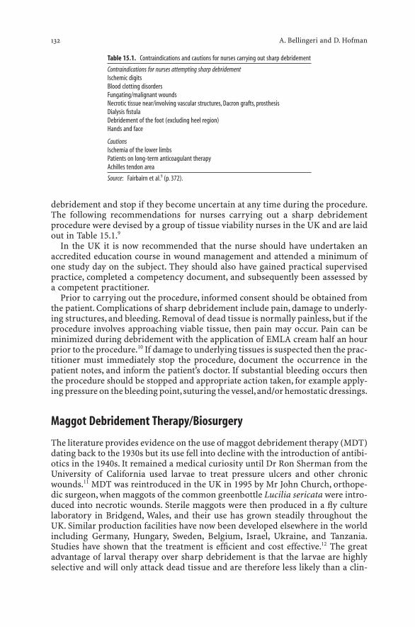
debridement and stop if they become uncertain at any time during the procedure.The following recommendations for nurses carrying out a sharp debridement procedure were devised by a group of tissue viability nurses in the UK and are laidout in Table 15.1.9
In the UK it is now recommended that the nurse should have undertaken anaccredited education course in wound management and attended a minimum ofone study day on the subject. They should also have gained practical supervisedpractice, completed a competency document, and subsequently been assessed bya competent practitioner.
Prior to carrying out the procedure, informed consent should be obtained fromthe patient. Complications of sharp debridement include pain, damage to underly-ing structures, and bleeding. Removal of dead tissue is normally painless, but if theprocedure involves approaching viable tissue, then pain may occur. Pain can beminimized during debridement with the application of EMLA cream half an hourprior to the procedure.10 If damage to underlying tissues is suspected then the prac-titioner must immediately stop the procedure, document the occurrence in thepatient notes, and inform the patient’s doctor. If substantial bleeding occurs thenthe procedure should be stopped and appropriate action taken, for example apply-ing pressure on the bleeding point,suturing the vessel,and/or hemostatic dressings.
Maggot Debridement Therapy/Biosurgery
The literature provides evidence on the use of maggot debridement therapy (MDT)dating back to the 1930s but its use fell into decline with the introduction of antibi-otics in the 1940s. It remained a medical curiosity until Dr Ron Sherman from theUniversity of California used larvae to treat pressure ulcers and other chronicwounds.11 MDT was reintroduced in the UK in 1995 by Mr John Church, orthope-dic surgeon, when maggots of the common greenbottle Lucilia sericata were intro-duced into necrotic wounds. Sterile maggots were then produced in a fly culturelaboratory in Bridgend, Wales, and their use has grown steadily throughout theUK. Similar production facilities have now been developed elsewhere in the worldincluding Germany, Hungary, Sweden, Belgium, Israel, Ukraine, and Tanzania.Studies have shown that the treatment is efficient and cost effective.12 The greatadvantage of larval therapy over sharp debridement is that the larvae are highlyselective and will only attack dead tissue and are therefore less likely than a clin-
132 A. Bellingeri and D. Hofman
Table 15.1. Contraindications and cautions for nurses carrying out sharp debridement
Contraindications for nurses attempting sharp debridementIschemic digitsBlood clotting disordersFungating/malignant woundsNecrotic tissue near/involving vascular structures, Dacron grafts, prosthesisDialysis fistulaDebridement of the foot (excluding heel region)Hands and face
CautionsIschemia of the lower limbsPatients on long-term anticoagulant therapyAchilles tendon area
Source: Fairbairn et al.9 (p. 372).

ical practitioner to damage healthy tissue during the debridement procedure. Ithas even been observed that they appear to leave small capillary vessels intact whileconsuming adjacent necrotic tissue. They are also able to access sinuses and cavi-ties which would not be possible without laying the wound open with extensivesurgery. They are, however, air breathing and this limits the depth to which theycan penetrate wounds.
Maggots, when introduced into a wound containing devitalized tissue, producesecretions containing proteolytic enzymes, which break down necrotic tissue intoa semiliquid form that they subsequently ingest. In addition, research indicates thatlarvae have an antibacterial effect.13 They ingest and destroy bacteria includingmethicillin-resistant Staphylococcus aureus. Further, maggot secretions have beenshown to stimulate the growth of fibroblast cells, which may explain the regularlyobserved finding that granulation tissue formation is enhanced after successfulMDT. When using MDT it is important to ensure that any secondary dressingspermit the ingress of oxygen and free drainage of excess fluid. Although the devel-opment of maggots is unaffected by commonly prescribed antibiotics, their devel-opment can be adversely affected by the presence of residues of hydrogel dressingscontaining propylene glycol.14 All traces of such dressings should therefore be thor-oughly removed before the application.
Figures 15.2 to 15.5 (see color section) show a pressure ulcer on the calf beingtreated with maggot debridement.
There are no reported significant adverse reactions to maggot therapy. As withany debriding agent, care must be taken to protect the surrounding skin fromsecretions. Some patients, especially those with ischemic or vasculitic ulcers, reportincreased levels of pain during treatment. However, in general, patient acceptanceof the technique is very high. Recently larvae bags have been introduced whichcontain the larvae and yet allow them to feed through the pores of the bag. Suchbags make the treatment much more acceptable for patient and practitioner, butmay be less effective at cleansing wound sinuses and wound crevices.
Enzymatic Debridement
In many cases when autolytic debridement is not sufficiently rapid and invasivemethods such as surgical or sharp debridement need to be avoided, enzymaticdebridement is the treatment of choice. There are several different pharmaceuti-cal enzyme preparations. One of the most widely used is collagenase, which is apurified product derived from the bacterium Clostridium histolyticum and actsbest with a pH of between 6 and 8,15 which is the pH of normal skin. It is a hydro-soluble proteinase favoring the removal of the necrotic “plug.” It is sensitive to temperature16 and is naturally found in wounds as a metalloprotease matrix.4 Incultivated cells collagenase accelerates keratinocyte migration threefold and theindividual cellular mobility tenfold.15 Antiseptics with metallic ions (e.g. silver ormercury) will inactivate the product. It is therefore necessary to avoid using prod-ucts containing silver concurrently with collagenase.15 Some patients suffer skinirritation when the collagenase cream comes into contact with the skin surround-ing the wound.4,15 Collagenase acts most effectively on fibers of collagen and elastinfibers in the center of the eschar, so that its activity is at the base of the woundrather than on the surface.18–22 Clinically collagenase may seem the slowest of theenzymatic debriding agents since its activity is at the base of the wound where it
Debridement of Pressure Ulcers 133
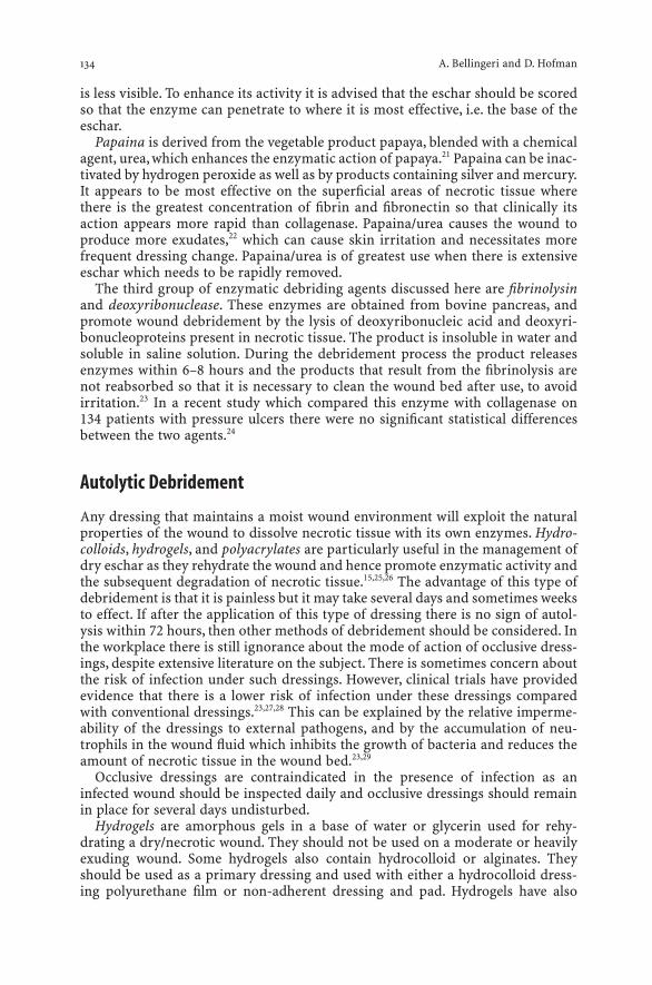
is less visible. To enhance its activity it is advised that the eschar should be scoredso that the enzyme can penetrate to where it is most effective, i.e. the base of theeschar.
Papaina is derived from the vegetable product papaya, blended with a chemicalagent, urea, which enhances the enzymatic action of papaya.21 Papaina can be inac-tivated by hydrogen peroxide as well as by products containing silver and mercury.It appears to be most effective on the superficial areas of necrotic tissue wherethere is the greatest concentration of fibrin and fibronectin so that clinically itsaction appears more rapid than collagenase. Papaina/urea causes the wound toproduce more exudates,22 which can cause skin irritation and necessitates morefrequent dressing change. Papaina/urea is of greatest use when there is extensiveeschar which needs to be rapidly removed.
The third group of enzymatic debriding agents discussed here are fibrinolysinand deoxyribonuclease. These enzymes are obtained from bovine pancreas, andpromote wound debridement by the lysis of deoxyribonucleic acid and deoxyri-bonucleoproteins present in necrotic tissue. The product is insoluble in water andsoluble in saline solution. During the debridement process the product releasesenzymes within 6–8 hours and the products that result from the fibrinolysis arenot reabsorbed so that it is necessary to clean the wound bed after use, to avoidirritation.23 In a recent study which compared this enzyme with collagenase on 134 patients with pressure ulcers there were no significant statistical differencesbetween the two agents.24
Autolytic Debridement
Any dressing that maintains a moist wound environment will exploit the naturalproperties of the wound to dissolve necrotic tissue with its own enzymes. Hydro-colloids, hydrogels, and polyacrylates are particularly useful in the management ofdry eschar as they rehydrate the wound and hence promote enzymatic activity andthe subsequent degradation of necrotic tissue.15,25,26 The advantage of this type ofdebridement is that it is painless but it may take several days and sometimes weeksto effect. If after the application of this type of dressing there is no sign of autol-ysis within 72 hours, then other methods of debridement should be considered. Inthe workplace there is still ignorance about the mode of action of occlusive dress-ings, despite extensive literature on the subject. There is sometimes concern aboutthe risk of infection under such dressings. However, clinical trials have providedevidence that there is a lower risk of infection under these dressings comparedwith conventional dressings.23,27,28 This can be explained by the relative imperme-ability of the dressings to external pathogens, and by the accumulation of neu-trophils in the wound fluid which inhibits the growth of bacteria and reduces theamount of necrotic tissue in the wound bed.23,29
Occlusive dressings are contraindicated in the presence of infection as aninfected wound should be inspected daily and occlusive dressings should remainin place for several days undisturbed.
Hydrogels are amorphous gels in a base of water or glycerin used for rehy-drating a dry/necrotic wound. They should not be used on a moderate or heavilyexuding wound. Some hydrogels also contain hydrocolloid or alginates. Theyshould be used as a primary dressing and used with either a hydrocolloid dress-ing polyurethane film or non-adherent dressing and pad. Hydrogels have also
134 A. Bellingeri and D. Hofman
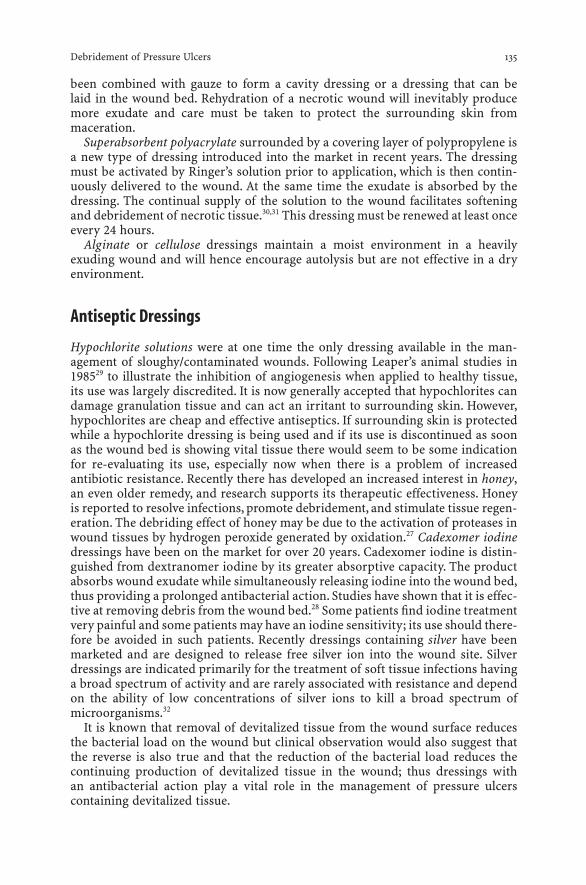
been combined with gauze to form a cavity dressing or a dressing that can be laid in the wound bed. Rehydration of a necrotic wound will inevitably producemore exudate and care must be taken to protect the surrounding skin from maceration.
Superabsorbent polyacrylate surrounded by a covering layer of polypropylene isa new type of dressing introduced into the market in recent years. The dressingmust be activated by Ringer’s solution prior to application, which is then contin-uously delivered to the wound. At the same time the exudate is absorbed by thedressing. The continual supply of the solution to the wound facilitates softeningand debridement of necrotic tissue.30,31 This dressing must be renewed at least onceevery 24 hours.
Alginate or cellulose dressings maintain a moist environment in a heavilyexuding wound and will hence encourage autolysis but are not effective in a dryenvironment.
Antiseptic Dressings
Hypochlorite solutions were at one time the only dressing available in the man-agement of sloughy/contaminated wounds. Following Leaper’s animal studies in198529 to illustrate the inhibition of angiogenesis when applied to healthy tissue,its use was largely discredited. It is now generally accepted that hypochlorites candamage granulation tissue and can act an irritant to surrounding skin. However,hypochlorites are cheap and effective antiseptics. If surrounding skin is protectedwhile a hypochlorite dressing is being used and if its use is discontinued as soonas the wound bed is showing vital tissue there would seem to be some indicationfor re-evaluating its use, especially now when there is a problem of increasedantibiotic resistance. Recently there has developed an increased interest in honey,an even older remedy, and research supports its therapeutic effectiveness. Honeyis reported to resolve infections, promote debridement, and stimulate tissue regen-eration. The debriding effect of honey may be due to the activation of proteases inwound tissues by hydrogen peroxide generated by oxidation.27 Cadexomer iodinedressings have been on the market for over 20 years. Cadexomer iodine is distin-guished from dextranomer iodine by its greater absorptive capacity. The productabsorbs wound exudate while simultaneously releasing iodine into the wound bed,thus providing a prolonged antibacterial action. Studies have shown that it is effec-tive at removing debris from the wound bed.28 Some patients find iodine treatmentvery painful and some patients may have an iodine sensitivity; its use should there-fore be avoided in such patients. Recently dressings containing silver have beenmarketed and are designed to release free silver ion into the wound site. Silverdressings are indicated primarily for the treatment of soft tissue infections havinga broad spectrum of activity and are rarely associated with resistance and dependon the ability of low concentrations of silver ions to kill a broad spectrum ofmicroorganisms.32
It is known that removal of devitalized tissue from the wound surface reducesthe bacterial load on the wound but clinical observation would also suggest thatthe reverse is also true and that the reduction of the bacterial load reduces the continuing production of devitalized tissue in the wound; thus dressings with an antibacterial action play a vital role in the management of pressure ulcers containing devitalized tissue.
Debridement of Pressure Ulcers 135

Conclusion
“Devitalized material is an integral part of pressure ulcer pathology and is a majorbarrier to healing.”2 The problem for the practitioner when healing is the desiredoutcome is how best to achieve debridement. The Cochrane report on debridingagents concluded that there were no trials which suggested that any dressing wasmore effective than any other in the removal of devitalized tissue from a wound.33
Clinical experience is necessary in making the correct choice for each individualpatient.
References
1. Dolynchuk K. Debridement. In: Krasner D, Rodeheaver GT, Sibbald G (eds) Chronic wound care.Wayne, PA: HMP Communications; 2001; 385–389.
2. Romanelli M, Mastronicola D. The role of wound bed preparation in managing chronic pressureulcers. J Wound Care 2002; 11(8):305–310.
3. Mast BA, Schultz GS. Interactions of cytokines, growth factors and proteases in acute wounds.Wound Repair Regen 1996; 4:411–420.
4. Sibbald RG, Williamson D, Orsted HL, et al. Preparing the wound bed: debridement, bacterialbalance and moisture balance. Ostomy Wound Manage 2000; 46(11):14–35.
5. European Pressure Ulcer Advisory Panel. Guidelines on treatment of pressure ulcers. EPUAPReview 1999; 1(2):31–33.
6. Coady K. The diagnosis and treatment of pyoderma gangrenosum. J Wound Care 2000; 9:282–285.7. Vowden K, Vowden P. Wound debridement: 2 Sharp techniques. J Wound Care 1999; 8(6):291–294.8. Ashworth J, Chivers, M. Conservative sharp debridement: the professional and legal issues. Prof
Nurse 2002; 17(10):585–588.9. Fairbairn K, Gier J, Hunter C, Preece J. A sharp debridement procedure devised by specialist
nurses. J Wound Care 2002; 11(10):371–375.10. Hansson C, Holm J, Lillieborg S, Syren A. Repeated treatment with lidocaine/prilocaine cream
(EMLA) as a topical anaesthesic for the cleansing of venous leg ulcers. Acta Derm Venereal(Stockh) 1993; 73:231–233.
11. Sherman RS, Wyle F, Vulpe M. Maggot therapy for treating pressure ulcers in spinal cord injurypatients. J Spinal Cord Med 1995; 18(2):71–74.
12. Thomas S, Jones M, Wynn K, Fowler T. The current status of maggot therapy in wound healing.Br J Nurs 2001; 10(22 Suppl):5–8.
13. Thomas S, Andrews A, Nigel P, et al. The antimicrobial activity of maggot secretions, a result of aprevious study. J Tissue Viability 1999; 9:127–133.
14. Thomas S, Andrews A. The effect of hydrogel dressings on maggot development. J Wound Care1999; 8(2):75–77.
15. Ayello EA, Cuddingan JE. Debridement: controlling the necrotic/cellular burden. Adv Skin WoundCare 2004; 17(2):66–75.
16. Dolynchuk K, Keast D, Campbell K, et al. Best practices for the prevention and treatment of pres-sure ulcers. Ostomy Wound Manage 2000; 46(11):38–52.
17. Rao CN, Ladin DA, Liu YY, et al. Alpha 1 antitrypsin is degraded and non-functional in chronicwounds but intact and functional in acute wounds: the inhibitor protects fibronectin from degra-dation by chronic wound fluid enzymes. J Invest Dermatol 1995; 105:572–578.
18. Herman I. Stimulation of human keratinocyte migration and proliferation in vitro: insights intothe cellular responses to injury and wound healing. Wounds 1996; 8(2):33–41.
19. Herman I. Extracellular matrix–cytoskeletal interactions in vascular cells. Tissue Cell 1987;19(1):1–19.
20. Kreig T. Collagen in the healing wound. Wounds 1995; 7(Suppl):5A–12A.21. Hebda PA, Lo C. The effects of active ingredients of standards debriding agents—papain and col-
lagenase—on digestion of native and denatured collagenous substrates, fibrin and elastin.Wounds2001; 13(5):190–194.
22. Hebda PA, Flynn KJ, Dohar JE. Evaluation of efficacy of enzymatic debriding agents for removalof necrotic tissue and promotion of healing in porcine skin wounds. Wounds 1998; 10:83–86.
23. Falabella A. Debridement of wounds. Wounds 1998; 10(Suppl C):1C–9C.
136 A. Bellingeri and D. Hofman

24. Pullen R, Popp R, Volkers P, et al. Prospective randomized double-blind study of the wound-debriding effects of collagenase and fibrinolysin/deoxyribonuclease in pressure ulcers.Age Ageing2002; 31:126–130.
25. O’Brien M. Method of debridement and patient focused care. JCN online 2003; 17, 11.26. Zacus H, Kirsner S. Debridement: rationale and therapeutic options. Wounds 2002; 14(7,
Suppl):2S–6S.27. Molan PC. The role of honey in the management of wounds. J Wound Care 1999; 8(8):415–418.28. Hillstrom L. Iodosorb compared to standard treatment in chronic venous leg ulcers—a multi-
center study. Acta Chir Scand Suppl 1988; 544:53–56.29. Cameron S, Leaper D. Antiseptic toxicity in open wounds. Nurs Times 1988; 25:77–78.30. Scholz S, Rompel R, Petres J. A new approach to wet therapy of chronic leg ulcers. Arzt & Praxis
1999; 816:517–522.31. Mosti G, Mattaliano V, Iabichella ML, Picerni P. Uso del tenderwet nella detersione delle ulcere
degli arti inferiori ad eziologia vascolare. Acta Vulnologia (in press).32. Lansdown ABG. Silver 1: Its antibacterial properties and mechanism of action. J Wound Care 2002;
11(4):125–130.33. Bradley M, Cullum N, Sheldon T. The debridement of chronic wounds: a systematic review. Health
Technol Assess 1999; 3:(17 Pt 1).
Debridement of Pressure Ulcers 137
16 The Role of Bacteria in Pressure UlcersR. Gary Sibbald, Paul Chapman, and Jose Contreras-Ruiz
Introduction
The approach to a person with a pressure ulcer must start with treating the causeand patient-centered concerns before the bacteria–host relationship can be ade-quately assessed and treated (Table 16.1). The role of bacteria in pressure ulcers iscomplex. There is no longer a straightforward clinical distinction between conta-mination, colonization, and infection. A number of factors must be examined todetermine the effect of bacterial burden on chronic pressure ulcer healing. Forhealing to occur the patient must be assessed holistically with the cause treatedand the local wound care optimized. An ulcer that is not healing at the expectedrate may be the result of a nonhealable or uncorrected cause. Alternatively, bacte-ria can damage local tissue and delay healing (critical colonization) with orwithout the classical features of infection.
The bacterial burden can be assessed in pressure ulcers using a compartmentalmodel that delineates multiple levels of bacterial involvement (above the wound,surface and deep wound compartments, surrounding skin, and systemic sepsis). Aclinical approach to management and assessment of bacterial burden can then beoutlined depending on the level of invasion. Most bacterial contamination origi-nates from external sources requiring infection control measures. The level ofdamage caused by bacteria will then determine the choice of topical and/or sys-temic treatment.
The effect of bacteria on pressure ulcer healing is dependent on organismnumbers and virulence, but the most important factor is host resistance. The rela-tionship can be expressed as:
Infection = (Organism number ¥ Virulence)/(Host resistance)
The approach to infection in a person with pressure ulcers is illustrated in Figure 16.1.1 We must look at the whole patient before treating the hole in thepatient.
139

Table 16.1. The approach to a person with pressure ulcers and increased bacterial burden / infection
Patient as a whole1. Assess the cause of the ulcer and determine the ability for healing (healability).2. The patient and the caregivers should be part of the decision-making process to improve adherence to treatment plans and to
optimize pain control and quality of life.
Regional treatment3. The diagnosis of infection is made clinically with the choice of topical and/or systemic antimicrobial therapy guided by investigative
testing (bacterial swab, X-rays, and laboratory tests including CRP and ESR).Do not use swab cultures to diagnose infection.
4. Diagnose and treat deep infection with appropriate systemic antimicrobial agents (deep compartment, surrounding skin, systemicinfection). Determine clinical and investigative parameters to monitor treatment and wound healing.
Local wound care5. Cleanse or compress the local wound with saline or water.6. Topical antiseptics may be appropriate in patients with nonhealable ulcers or where the reduction of bacterial burden is more
important than toxicity to granulation tissue.(a) In general, choose agents with broad spectrum antimicrobial activity and low tissue toxicity.
7. Debride devitalized tissue to decrease bacterial burden and control pro-inflammatory stimulus.(a) Choose most appropriate method (surgical, mechanical, biotherapy, enzymatic, autolytic).(b) Use systemic antibiotics prior to deep surgical debridement procedures, especially in the compromised host.(c) In wounds without the ability to heal, aggressive sharp surgical debridement to bleeding base is contraindicated.
8. Consider a 2-week trial of topical antimicrobials if wound is not healing despite optimal care (increased bacterial burden or covertinfection suspected).(a) Use agents with low tissue toxicity that can also promote moisture balance.(b) Avoid agents that can cause allergic sensitization or frequent bacterial resistance.
9. Perform semiquantitative bacterial cultures and re-evaluate for infection or osteomyelitis if wound fails to improve.10. Reassess outcomes of therapy at appropriate time intervals by monitoring clinical, laboratory, and investigative parameters.
Determineability to heal
PERSON WITH APRESSURE
ULCER
NONHEALABLE
PHOTENTIALTO HEAL
TREAT THECAUSE
TISSUEDEBRIDEMENT
INFECTION /INFLAMMATION
NOT HEALINGAT EXPECTED
RATE
PATIENT-CENTEREDCONCERNS
MOISTUREBALANCE
LOCALWOUND CARE
•Topical antiseptics•Supportive care
•Biologicals
•Adjunctive •Electrical stimulation •Ultrasound •VAC
Figure 16.1 Holistic approach to pressure ulcer management. VAC, Vacuum Assisted Closure.

Can this individual make new granulation tissue and heal? Is healing the ulcer partof the patient- or caregiver-centered plan? How much more should be done to treator heal the pressure ulcers?
Many factors are involved in this assessment. The patient and caregiver must beinvolved in the decisions. The general health and fitness of the patient must bedetermined including: ability to perfuse tissue, vital organ function (brain, heart,lungs, kidney, liver), and drugs that will inhibit healing (e.g. chemotherapeuticagents, corticosteroids).
Although the diagnosis of pressure ulcers is usually obvious, there are somecases where other diagnoses are confused with pressure ulcers. An early stage (1or 2) pressure ulcer in the gluteal fold region of the buttocks can be confused withcandidial intertrigo or a contact dermatitis. Deeper lesions mistakenly labeled aspressure ulcers may in fact be perianal bacterial abscesses or ruptured pilonidalsinuses. Once the diagnosis is established, specific pressure ulcer contributingfactors must also be corrected (e.g. nutritional deficiencies, friction and shear,incontinence) and patient/caregiver cooperation with the treatment plan is vital.2
In some settings availability of resources can be a limiting factor. All these factorsmust be considered as part of the holistic assessment of the patient’s healing potential.
Patient/Caregiver-Centered Concerns
Patient-centered concerns often revolve around issues of quality of life and pain.Ulcer healing may be less important than other issues facing a patient includingthe control of odor, sleep, and pain.3 Particularly in persons with nonhealable pres-sure ulcers, sepsis prevention and minimizing dressing changes become veryimportant considerations of the care plan. It is senseless to concentrate only onhealing an ulcer and not take into account previous social activities. It is verydemoralizing to put a person with a pressure ulcer to bed for long periods of timefor maximum healing only for them to resume normal activities and have a recurrence.4
The Role of Bacteria in Pressure Ulcers 141
Assessment and Diagnosis of Patients with Pressure Ulcers
Case one scenario
Ms S is a 57-year-old woman with a nontreatable brain tumor (anaplastic astrocytoma). Shehas been semi-comatose for the past month and developed four pressure ulcers located in theocciput, sacrum, and both heels. The family advocated for palliative care and you were calledby the treating physician to address the nonhealing skin ulcers because the odor is disturb-ing both the family and other patients.
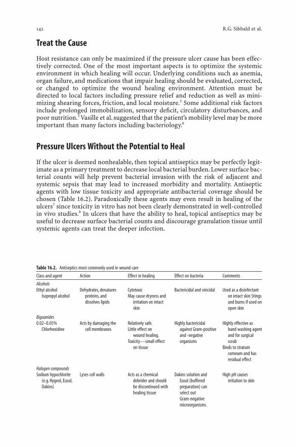
Treat the Cause
Host resistance can only be maximized if the pressure ulcer cause has been effec-tively corrected. One of the most important aspects is to optimize the systemicenvironment in which healing will occur. Underlying conditions such as anemia,organ failure, and medications that impair healing should be evaluated, corrected,or changed to optimize the wound healing environment. Attention must bedirected to local factors including pressure relief and reduction as well as mini-mizing shearing forces, friction, and local moisture.5 Some additional risk factorsinclude prolonged immobilization, sensory deficit, circulatory disturbances, andpoor nutrition.5 Vasille et al. suggested that the patient’s mobility level may be moreimportant than many factors including bacteriology.6
Pressure Ulcers Without the Potential to Heal
If the ulcer is deemed nonhealable, then topical antiseptics may be perfectly legit-imate as a primary treatment to decrease local bacterial burden. Lower surface bac-terial counts will help prevent bacterial invasion with the risk of adjacent andsystemic sepsis that may lead to increased morbidity and mortality. Antisepticagents with low tissue toxicity and appropriate antibacterial coverage should bechosen (Table 16.2). Paradoxically these agents may even result in healing of theulcers7 since toxicity in vitro has not been clearly demonstrated in well-controlledin vivo studies.8 In ulcers that have the ability to heal, topical antiseptics may beuseful to decrease surface bacterial counts and discourage granulation tissue untilsystemic agents can treat the deeper infection.
142 R.G. Sibbald et al.
Table 16.2. Antiseptics most commonly used in wound care
Class and agent Action Effect in healing Effect on bacteria Comments
AlcoholsEthyl alcohol Dehydrates, denatures Cytotoxic Bactericidal and viricidal Used as a disinfectant
Isopropyl alcohol proteins, and May cause dryness and on intact skin Stingsdissolves lipids irritation on intact and burns if used on
skin open skin
Biguanides0.02–0.05% Acts by damaging the Relatively safe. Highly bactericidal Highly effective as
Chlorhexidine cell membranes Little effect on against Gram-positive hand washing agentwound healing. and -negative and for surgical
Toxicity—small effect organisms scrubon tissue Binds to stratum
corneum and hasresidual effect
Halogen compoundsSodium hypochlorite Lyses cell walls Acts as a chemical Dakins solution and High pH causes
(e.g. Hygeol, Eusol, debrider and should Eusol (buffered irritation to skinDakins) be discontinued with preparation) can
healing tissue select out Gram-negativemicroorganisms.
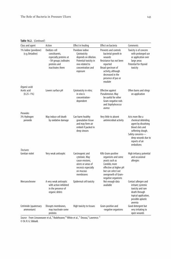
The Role of Bacteria in Pressure Ulcers 143
Table 16.2. (Continued )
Class and agent Action Effect in healing Effect on bacteria Comments
1% Iodine (povidone) Oxidizes cell Povidone iodine Prevents and controls Toxicity is of concern (e.g. Betadine) constituents, Cytotoxicity bacterial growth in with prolonged use
especially proteins at depends on dilution. wounds or application over–SH groups; iodinates Potential toxicity in Resistance has not been large areasproteins and vivo related to reported Potential for thyroidinactivates them concentration and Broad spectrum of toxicity
exposure activity, althoughdecreased in thepresence of pus orexudate
Organic acidsAcetic acid Lowers surface pH Cytotoxicity in vitro; Effective against Often burns and stings
(0.25–1%) in vivo is Pseudomonas. May on applicationconcentration- be useful for otherdependent Gram-negative rods
and Staphylococcusaureus
Peroxides3% Hydrogen May induce cell death Can harm healthy Very little to absent Acts more like a
peroxide by oxidative damage granulation tissue antimicrobial activity chemical debridingand may form air agent by dissolvingemboli if packed in blood clots anddeep sinuses softening slough.
Safety concerns—deep wounds due toreports of airembolisms
TincturesGentian violet Very weak antiseptic Carcinogenic and Kills Gram-positive High irritancy potential
cytotoxic. May organisms and some and occasional cause erosions, yeasts such as allergiesulcers or areas of Candida, morenecrosis especially effective at higher pHon mucous but can select outmembranes overgrowth of Gram-
negative organismsMercurochrome A very weak antiseptic Epidermal cell toxicity Not enough data Contact allergen and
with action inhibited available irritant; systemic in the presence of toxicity and rare organic debris death through
topical application,possible aplasticanemia
Cetrimide (quaternary Disrupts membranes, High toxicity to tissues Gram-positive and Good detergent butammonium) may inactivate some -negative organisms very irritating to
proteins open wounds
Source: From Lineaweaver et al.,9 Rodeheaver,10 White et al.,11 Drosou,8 Lawrence.12
© Dr. R. G. Sibbald.

Pressure Ulcers with Potential to Heal
Local Wound Care
Local wound care includes three components: tissue debridement, control ofincreased bacterial burden or prolonged inflammation (infection/inflammation),and moisture balance. If these three components are controlled and the wound isstill not healing, we need to reassess the cause, patient-centered concerns, and thelocal wound care to be sure that treatment has been optimized. If the wound is nothealing at the expected rate, the edge effect of the TIME (Tissue debridement,Infection/Inflammation, Moisture Balance, Edge Effect) paradigm13,14 reminds usof the appropriate use of advanced therapies in our toolkit: biological agents(growth factors, living skin equivalents), adjunctive therapies (ultrasound, electri-cal stimulation, negative pressure therapy), and skin grafts or flaps.Advanced ther-apies are expensive and will only work when patient care has been optimized asoutlined in Figure 16.1. These advanced therapies are not a substitute for bestwound care practices.
Debridement
Debridement is necessary to remove devitalized tissue and to reduce bacterial con-tamination. There are five methods to consider for debridement: sharp surgical,autolytic, enzymatic, biological, and mechanical.13 Devitalized tissue serves as agood culture medium for bacteria to grow within a chronic wound. This dead tissue
144 R.G. Sibbald et al.
Case One Completion
Returning to our patient with the nontreatable brain tumor, the cause cannot be treated andthe patient’s family is advocating palliative treatment.There was no deep infection requiringsystemic agents. The patient had excellent pain control, with long-acting narcotic agents fornormal nerve function pain,a short-acting narcotic for breakthrough,and a tricyclic agent highin norepinephrine (noradrenaline) for the neuropathic component of chronic wound pain.Theodor was derived from nonviable slough and Pseudomonas.The debris was removed with sur-gical debridement and acetic acid compresses to acidify the local wounds and discouragePseudomonas growth. A new modern dressing combining ionized silver with calcium alginatewas then applied every second day for bacterial balance and moisture balance and autolyticdebridement.The wounds were monitored every second day for deep infection and size. Non-viable slough that accumulated on the wound surface was removed with dressing changes.Thepatient died 3 weeks later with no further extension of the wounds, no significant local paineven at dressing changes (short-acting breakthrough narcotic was administered 30 minutesprior to the procedure).
also acts as a foreign body that may induce a pro-inflammatory response. Persis-tent inflammation as well as infection can delay healing (Figure 16.2—see colorsection). The presence of foreign material such as necrotic debris, retained packingmaterials, or small fragments of gauze dressing will significantly decrease hostresistance and lower the number of bacteria necessary to cause wound infectionor damage to the granulation tissue in the ulcer base. Universal precautions arenecessary to prevent wound contamination. The surface compartment may havean increased bacterial burden and this area of the wound can be treated topicallywith newer antibacterial dressings that will also control moisture balance and onoccasion have additional autolytic debridement functions. The deep compartmentof the wound requires systemic antimicrobial therapy and is defined as the areathat is not accessible by topical treatments. This compartment can have bacterialinvasion of the underlying bone or localized infections in the form of an abscess.In individuals with pressure ulcers, abscesses below the surface must be soughtand this deep compartment infection drained (Figure 16.3—see color section).15
The choice of debridement depends on the availability of healthcare providerswith skill in performing sharp surgical debridement or local preference forautolytic methods with dressings (calcium alginates, hydrogels, or hydrocolloids),topical enzymatic agents, biological methods with maggots, or mechanical irriga-tion with saline wet to dry dressings.
Deep surgical debridement has been associated with transient bacteremia inpatients with pressure ulcers.16 Positive blood cultures were obtained during fiveout of eight active surgical debridement procedures in a study by Glenchur et al.16
A majority of these included anaerobes so the recommendation is that thesignificant incidence of bacteremia occurring during surgical debridement indi-cates the need for broad spectrum antibiotic coverage during and after deep sur-gical procedures.17 This is important when large and/or deep ulcers are debrided,especially in the compromised host.16
Bacteremia is a common complication of pressure ulcers and may lead to mor-bidity and mortality. Galpin et al.18 documented bacteremia in 16/21 patients withpressure ulcers. The most commonly isolated bacteria were anaerobes. In patientswith spinal cord injury, a mortality of 7–8% is frequently associated with sec-ondary wound infection.19 In a study of 21 patients with systemic bacteremia (fre-quently polymicrobial) and sepsis, 76% of the isolated bacteria originated from apressure ulcer. Overall, mortality was 48% and all patients over 60 died despiteempiric antibiotic treatment. Five patients had bacteremia that persisted withantibiotic treatment and resolved only after local debridement. The mortality rate among patients with pressure ulcers and bacteremia is close to 50%.18 Anothergrave and often fatal complication of pressure ulcers is necrotizing soft tissue infection.20,21
Bacterial Balance/Chronic Inflammation
Assessment of Infection
The diagnosis of infection is made based on clinical criteria, with bacterial swabsor deep cultures, laboratory and radiological tests used as adjuncts for diagnosisand treatment. Pressure ulcers are prone to infection.13 All wounds contain
The Role of Bacteria in Pressure Ulcers 145

bacteria at levels ranging from contamination, through colonization, critical colo-nization (also known as increased bacterial burden, occult or covert infection) toinfection. The increased bacterial burden may be confined to the superficial woundbed or may be present in the deep compartment and surrounding tissue of thewound margin. Therefore, it becomes very important to diagnose both the bacte-rial imbalance and level of invasion in order to diagnose and treat infection prop-erly. Increased bacterial burden in pressure ulcers has been demonstrated to delayhealing in patients with chronic ulceration.22
Clinical Assessment
In this discussion, we identify contamination as the presence of bacteria on thewound surface. Colonization is the presence of replicating bacteria attached to thewound tissue but not causing injury to the host. Infection is the presence of repli-cating microorganisms in a wound with associated host injury. The bordersbetween these concepts are not clearly established. The clinician must assess thepatient’s symptoms and signs present in the wound to distinguish contamination,colonization, and healing from critical colonized or infected wounds that are nothealing, or even endangering the life of the patient. Critical colonization occurswhen bacteria delays or stops healing of the wound without the classical symp-toms and signs of infection being present. The wound care specialist needs to care-fully identify the clinical signs and symptoms of infection to make an accuratediagnosis. In patients with pressure ulcers some of these signs may be obscured23
by factors such as malnutrition, anemia, drugs, or immunosuppression includingchronic illness such as diabetes.
The classical signs of infection are: pain, erythema, edema, purulent discharge,and increased warmth. These are related to the inflammatory process occurring inthe wound. Increased blood flow produces a rise in temperature, and fluid leakingfrom intravascular spaces accumulates in the tissue, causing visible swelling.Vasoactive mediators such as histamine produce the characteristic erythema, andpain is caused through activation of biochemical mediators secreted near unmyeli-nated nerve fiber endings.
In chronic wounds other signs should be added: delayed healing or new areasof breakdown, increased discharge (often it is initially serous or clear and waterybefore it becomes pustular), bright red discoloration of granulation tissue, friableand exuberant granulation, new areas of slough on the wound surface, undermin-ing, and a foul odor (Figure 16.4—see color section).24 Serous exudate may beincreased in a chronic wound with increased bacterial burden before purulence isnoted with the clinical signs usually recognized in infections. It has been suggestedthat chronic wounds should show some evidence of healing within 4 weeks toprogress to healing by week 12. If this time limit is exceeded then increased bac-terial burden or infection should be suspected as one of the causes of delayedhealing.25
Discoloration of granulation tissue arises from loose, poorly formed granula-tion tissue, while friable granulation tissue that bleeds easily occurs from exces-sive angiogenesis stimulated by bacterial pathogens (Figure 16.5—see colorsection). Healthy granulation tissue is pink-red and firm with a moist translucentappearance. When infected, it will appear dull and may have patches of greenishor yellow discoloration. Certain anaerobic species such as Bacteroides fragilis and
146 R.G. Sibbald et al.

streptococci can produce a dullish, dark-red hue, while Pseudomonas will producegreen or blue patches which may fluoresce at 365 nm (Wood’s) light. Underminingresults from atrophic granulation tissue inhibited or digested by bacteria. Foulodor is usually produced by Gram-negative bacilli especially Pseudomonas speciesor anaerobes digesting granulation tissue.26
Deep infection will often cause erythema and warmth extending 2 cm or morebeyond the wound margin when the surrounding skin becomes involved (Figure16.6—see color section). This bacterially stimulated increased inflammatoryresponse is painful and will cause the wound to increase in size or lead to satelliteareas of tissue breakdown resulting in adjacent tissue ulceration. Deep infections,especially in ulcers of long duration, can often lead to underlying osteomyelitis.Probing to bone is a simple clinical test that may indicate osteomyelitis, especiallyin patients with neuropathic foot ulcers often associated with diabetes.27
These symptoms and signs of wound infection are summarized in Table 16.3.Gardner et al.29,30 examined the reliability and validity of clinical signs of infec-
tion in two recent papers. These studies identified various symptoms and signs of infection and compared diagnoses made using these signs with the results ofquantitative cultures from tissue biopsies to correlate each sign or symptom withthe stated criteria of infection. Increasing pain, friable granulation tissue, foul odor,and wound breakdown all demonstrated validity for a diagnosis of infection basedon discriminatory power and positive predictive values. A checklist was then con-structed to test the ability of different observers to distinguish these signs (relia-bility). There was a very high level of agreement between observers but it is alsoimportant to assess the discriminatory power of the sign or symptom when infec-tion is present compared to when it is absent. A kappa test was performed to quan-tify the usefulness of each criterion. High kappa values (above 0.7) indicate a highreliability associated with a diagnosis of infection and the values for the symptomsand signs were:
• increasing pain (1.0),• edema (0.93),• wound breakdown (0.89),• delayed healing (0.87),• friable granulation (0.8),• purulent exudate (0.78),• serous exudate (0.74).
The Role of Bacteria in Pressure Ulcers 147
Table 16.3. Clinical signs of wound infection
Superficial increased bacterial burden Deep or surrounding wound infection Systemic infection
Nonhealing Pain Fever
Bright red granulation tissue Swelling, induration Rigors
Friable and exuberant granulation Erythema Chills
New areas of breakdown or necrosis on the wound Increased temperature Hypotensionsurface (yellow, brown, or black slough) Wound breakdown Multiple organ failure
Increased exudate that may be translucent or clear Increased size or satellite areasbefore becoming purulent Undermining
Foul odor Probing to bone
Source: Adapted from Sibbald et al.28

The sample size was small with only 31 patients and there were five different types of wounds including pressure ulcers. Although these observations must beconsidered preliminary, these characteristics (especially when more than one ispresent) will assist clinicians to more accurately identify bacterial damage andinfection in chronic wounds. These studies need to be expanded to larger numbersof patients and separate analysis of each wound type including pressure ulcersmust be determined.
The classical signs of overt infection are generally easy to identify but it is moredifficult to make a judgment about wounds that display abnormal or persistentinflammation. Wounds may display signs of covert infection, where the host isharmed enough to impede healing but not enough to cause typical inflammatorysymptoms. Covert infection is difficult to diagnose as many of the signs listedabove may be absent. The most obvious sign is the failure of the pressure ulcer toheal or initial progress to healing is stalled. Disorganization (hypertrophy oratrophy) of previously healthy granulation tissue, discoloration of granulationtissue to pale gray or deep red, and increased friability and bleeding are also likelyto be detectable. The exudate may increase in quantity and can be serous or wateryin consistency. Some clinicians utilize infrared thermometry as an aid in the diag-nosis of infection or inflammation, as validated by Armstrong and colleagues forthe active inflammatory process associated with a Charcot foot in people with dia-betes.31,32 This technique has not been properly validated in pressure ulcer patientsbut it may be a useful tool to discriminate the increase in temperature in the sur-rounding skin compared to mirror image locations. In general, the clinician shouldassess for swelling, warmth, tenderness, pain, and erythema extending more than2 cm beyond the ulcer margin to diagnose surrounding skin cellulitis and proba-ble deep tissue compartment infection in the ulcer base.
Bacterial Tests
Bacterial swabs and cultures are not used to diagnose infection but to guideantimicrobial therapy choices and screen patients for multiresistant bacterialorganisms such as methicillin-resistant Staphylococcus aureus (MRSA). There is noneed to culture a pressure ulcer that is healing at an expected rate and does notdisplay any signs or symptoms of infection. As all wounds are contaminated andpotentially colonized, a culture will simply confirm that microorganisms arepresent without providing any information as to whether they are having a detri-mental effect on the host.
Bacterial swabs can provide information on the predominant flora on thesurface of a nonprogressing, deteriorating, or heavily exudating wound. Micro-biological tests can also screen for multiresistant bacteria such as MRSA and van-comycin-resistant enterococcus (VRE). The degree of the inflammatory responseis measured by the presence and number of neutrophils per high power field inthe Gram stain. The relationship between increased local infiltration of neutrophilsand chronicity of wounds is extremely important. A granulation tissue biopsystudy of pressure ulcers by Diegelmann33 demonstrated delayed healing when thebiopsies included an increased number of neutrophils. The numbers of neutrophilscorrelated well with an increase in myeloperoxidase activity and metallopro-teinases. The elevated number of neutrophils and their destructive enzymes maybe the cause of the matrix dysfunction and perpetuation of the ulcer. Sibbald
148 R.G. Sibbald et al.
et al.34 demonstrated an association between increased bacterial burden and anincreased number of neutrophils in nonhealing venous stasis ulcers. Both the bacteria and neutrophilic infiltrate delayed healing. Once bacterial load reaches 106 colony-forming units (CFU) per gram of tissue, wound healing is usuallyimpaired.35 In 1964 Bendy et al.36 reported that healing in pressure ulcers wasinhibited if the bacterial load was greater than 1 ¥ 106 CFU/ml of wound fluid.Superficial wound swabs were used in this study, but other studies using the goldstandard (tissue biopsy specimens) reported similar results for pressure ulcers andsurgical wounds.37–41 In quantitative biopsies from 17 patients Vande Berg et al.42
determined that fibroblast growth inhibition was not related to the type of bacte-ria but to the bacterial load (greater than 1 ¥ 105 CFU/g).
Quantitative microbiology has a role to play in predicting the risk of infectionas many studies have shown that bacterial load correlates with risk.43 Neverthe-less, these findings need to be viewed in perspective. At least 20% of wounds col-onized with more than 105 CFU/g of bacteria will still heal.44 If host resistance ishigh, normal skin flora present in very high quantities have the potential in someclinical situations to enhance wound healing.45 On the other hand, reduced hostresistance, or the presence of foreign objects in the wound, can significantly reducethe bacterial load that is required to trigger infection. Some microorganisms suchas streptococcus may cause tissue damage at very low concentrations.15,46 Thusquantitative microbiology does not necessarily provide an unambiguous diagno-sis of infection. Under certain circumstances, quantitative biopsies may also havepoor sensitivity and low reliability. Woolfrey et al.47 showed that there was a 25%chance of missing an organism using biopsy probably due to uneven distributionof organisms within the wound bed and the techniques used to clean the speci-men. Results varied by 2 logarithms (logs) in 27% of paired isolates. Ehrenkranzet al.48 demonstrated that an irrigation-aspiration technique could produce similarresults to qualitative biopsy in pressure ulcers.49 It is not always necessary in every-day clinical practice to quantify the bacterial load through tissue biopsy or alter-native invasive techniques that require expertise and timely processing in themicrobiology laboratory.
Evidence for the Clinical Use of the Bacterial Swab
Evidence from comparative studies confirms that microbiology obtained by a swabmay adequately correlate with qualitative findings obtained through tissuebiopsy.50,51 When plated in the laboratory bacteria are streaked in four quadrantson blood agar in a Petri dish. Growth in the fourth quadrant (the most dilute bac-terial swab specimen streaking of the Petri dish) corresponds to a growth of105 CFU/g of tissue as determined by quantitative biopsy.46,50 In most cases the colonizing bacteria come from exogenous sources, and would be present in the superficial compartment before reaching the deep tissues.15 Sapico et al.26 com-pared pressure ulcer bacterial swab results with quantitative biopsy cultures anddemonstrated a 75% concordance. In a similar comparison of diabetic foot ulcerinfections Wheat et al.52 obtained comparable bacterial culture results with swabsand tissue biopsy. There was a fairly high rate of false positive and negative resultsusing the swab but most of the false positives were commensal organisms that didnot require antimicrobial therapy. The authors concluded that 92% of antibiotictherapy choices would have been adequate based on the swab alone. Rudensky
The Role of Bacteria in Pressure Ulcers 149
et al.,49 on the other hand, concluded that blood samples or deep tissue biopsiesare more clinically significant than bacterial swabs due to the high number of falsepositive isolations in the latter.
Taking a Bacterial Swab
The appropriate use of bacterial swabs is for antibacterial therapy selection and toidentify the specific organisms in a chronic wound such as multiresistant bacteria.
A bacterial swab result is only as good as the technique used to obtain the spec-imen and the processing in the laboratory. There is much discussion about the typeof swab to use and the procedure for taking a specimen. Some clinicians have rec-ommended alginate or rayon-tipped swabs in the belief that the fatty acids con-tained in cotton swabs might inhibit growth in certain bacteria. However, theorganisms commonly encountered in infection are likely to withstand the envi-ronment of a cotton swab. Pre-moistening a swab in the transport media is usefulif the surface of the wound is dry as it can improve the yield. This is not necessaryif the wound is already very moist. There are two swab culture techniques com-monly used in clinical practice. In the first technique, the tip of the swab shouldbe rolled on its side for one full rotation over the part of the wound granulationtissue with the most obvious signs of infection, avoiding slough and surface puru-lent discharge. A zigzag pattern can be used for sample collection on the swabsurface for wounds larger than 5 cm2. This technique is likely to increase the yieldof nonsignificant colonizers and it may be preferable to take more than oneregional swab from the upper and lower areas of the wound. An alternative tech-nique involves pressing the swab on the surface at a single point of the granula-tion to express wound fluid and then rotating the swab 360° to obtain the bacterialspecimen. The wound bed must first be cleaned with saline or water andsuperficially debrided so that the cultures from the surface of the wound moreclosely resemble those in the tissue. There will undoubtedly be more colonizingorganisms than pathogens on the surface of the wound but there is a correlationbetween the pathogens found on the surface and in the deep compartments.Culture results by themselves, even results of bone culture or culture of other deep-tissue biopsy specimens, should not be used as the sole criteria for infectionwithout clinical or histopathological evidence of infection.
Infected Wounds: Causative Species
The microbial flora in a chronic wound changes over time in a predictablefashion.46 Clinicians often need to treat infected wounds before the results of bac-terial cultures are available. In wounds of less than one month’s duration there isusually a high percentage of Gram-positive organisms. In wounds of longer thanone month’s duration, the wound is likely to acquire multiple organisms includingGram-negatives and anaerobes in addition to the Gram-positive bacterial flora.46
In combination with clinical signs it may be possible to attempt identification ofthe invading pathogens while waiting for culture results (Table 16.4).
It is widely believed that aerobic or facultative pathogens such as Staphylococ-cus aureus, Pseudomonas aeruginosa, and the beta-hemolytic streptococci are pri-marily responsible for delayed healing and infection in all types of wounds, butthis has largely been based on studies in which the culture and isolation of anaer-obic bacteria was minimal or omitted.43 However, anaerobes can be highly viru-
150 R.G. Sibbald et al.
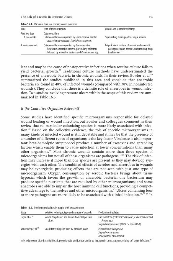
lent and may be the cause of postoperative infections when routine culture fails toyield bacterial growth.53 Traditional culture methods have underestimated thepresence of anaerobic bacteria in chronic wounds. In their review, Bowler et al.43
summarized the studies published in this area and conclude that anaerobic bacteria are found in 48% of infected wounds (compared with 38% in noninfectedwounds). They conclude that there is a definite role of anaerobes in wound infec-tion. Two studies involving pressure ulcers within the scope of this review are sum-marized in Table 16.5.
Is the Causative Organism Relevant?
Some studies have identified specific microorganisms responsible for delayedwound healing or wound infection, but Bowler and colleagues comment in theirreview that no particular colonizing species is more likely associated with infec-tion.43 Based on the collective evidence, the role of specific microorganisms inmany kinds of infected wound is still debatable and it may be that the presence ofa number of different types of organisms is the key factor.Virulence is also impor-tant: beta-hemolytic streptococci produce a number of exotoxins and spreadingfactors which enable them to cause infection at lower concentrations than manyother organisms.46 Most chronic wounds contain more than three species ofmicroorganisms but not all of these organisms are pathogens.15,56 The risk of infec-tion may increase if more than one species are present as they may develop syn-ergies with each other. The combined effects of aerobes and anaerobes in woundsmay be synergistic, producing effects that are not seen with just one type ofmicroorganism. Oxygen consumption by aerobic bacteria brings about tissuehypoxia, which favors the growth of anaerobic bacteria; one bacterium mayproduce specific nutrients that are required by other microorganisms; and someanaerobes are able to impair the host immune cell functions, providing a compet-itive advantage to themselves and other microorganisms.43 Ulcers containing fouror more pathogens are more likely to be associated with clinical infection.26,57–60 In
The Role of Bacteria in Pressure Ulcers 151
Table 16.4. Microbial flora in a chronic wound over time
Time Type of microorganism Clinical and laboratory findings
First few days Cutaneous flora1 to 4 weeks Cutaneous flora accompanied by Gram-positive aerobic Suppurating, Gram-positive, single species
cocci, often streptococci, Staphylococcus aureus
4 weeks onwards Cutaneous flora accompanied by Gram-negative Polymicrobial mixture of aerobic and anaerobic facultative anaerobic bacteria, particularly coliforms pathogens, tissue necrosis, undermining, deepfollowed by anaerobic bacteria and Pseudomonas spp. involvement
Table 16.5. Predominant isolates in people with pressure ulcers
Study Isolation technique, type and number of wounds Predominant isolates
Heym et al.54 Swabs, deep tissue. and liquids from 101 pressure Enterobacteria (Enterococcus faecalis, Escherichia coli andulcers Proteus sp.)
Staphylococcus aureus (MRSA > non-MRSA)
Vande Berg et al.42 Quantitative biopsies from 17 pressure ulcers Pseudomonas aeruginosaStaphylococcus aureusAcinetobacter calcoaceticus
Infected pressure ulcer bacterial flora is polymicrobial and is often similar to that seen in some acute necrotizing soft tissue infections.55

a study of chronic leg ulcers, Trengove et al.61 reported that no single microorgan-ism or group of bacteria were more detrimental to healing than any other but thatthere was a significantly lower probability of healing if there were four or morebacterial groups present in the ulcer. Bowler and Davies62 also reported that therewere more species isolated in infected than in noninfected leg ulcers. A similartrend may be found in pressure ulcers but the data have not been analyzed to date.In wounds that are infected with a number of species it is often impossible to detectthe specific causative role of each organism.
In conclusion, bacterial swabs or wound cultures do not diagnose infection butthey can be used as guidance for antimicrobial therapy: The diagnosis of infectionis based on clinical symptoms and signs.
Osteomyelitis
Deep infection, especially in ulcers of long duration, can often be complicated by underlying osteomyelitis. Osteomyelitis is caused by an infecting organism cre-ating an inflammatory process and resulting in bone destruction.63–68 The diagno-sis of osteomyelitis under pressure ulcers is important, but is also challengingbecause deep signs of infection can be obscured in patients with pressure ulcers.A study of clinical signs of a single criterion for the diagnosis of osteomyelitisfound a 53% accuracy rate, with sensitivity of 33% and specificity of 60%.69 Theuse of a sterile probe to determine if any sinuses or deeper pocketing or under-mining is present can often alert clinicians to potential deeper tissue and bonyinvolvement. The diagnosis of bone infection in pressure ulcers should be consid-ered in deep ulcers whenever the ulcer fails to improve with proper conventionallocal treatment or after removal of pressure.69–71 Sugarman et al.72 discoveredosteomyelitis in 32% of pressure ulcers that did not respond promptly to localtherapy (as early as 2 weeks). These ulcers often had surrounding erythema anddrainage. Three studies detected osteomyelitis in 17 to 32% of patients with long-standing pressure ulcers,69,70,72 with one study noting a less frequent occurrence.73
The presence of a nonhealing wound or exposed bone did not always indicateosteomyelitis;69 nevertheless probing to bone is a simple clinical test that may indi-cate osteomyelitis, especially in those with diabetic foot ulcers (Figure 16.7—seecolor section).27
The role of surface bacterial swabs to help in antibiotic selection is controver-sial. Some authors15,46,50 support the idea that bacteria on the surface may eventu-ally penetrate the deeper tissue and potentially infect bone; therefore, a swab mayisolate the responsible bacteria. Other authors believe that organisms in bonecannot be accurately detected with a swab. This belief stems from evidence thatsinus tract cultures do not usually correlate with those cultures obtained from thebone biopsy, except when Staphylococcus aureus is isolated from the sinus (it islikely to be the cause of the underlying osteomyelitis).17,54,74 These authors concludethat bone biopsies are superior to swabs at picking infecting organisms from thebone rather than surface colonizers.
Bone biopsies remain the gold standard for diagnosing osteomyelitis in patientswith pressure ulcers. If a bone biopsy is performed, one sample should be sent forculture and another for histological examination. Cultures should be performedfor both aerobes and anaerobes. The most common organism isolated in any typeof osteomyelitis is Staphylococcus aureus. Other microorganisms associated with
152 R.G. Sibbald et al.
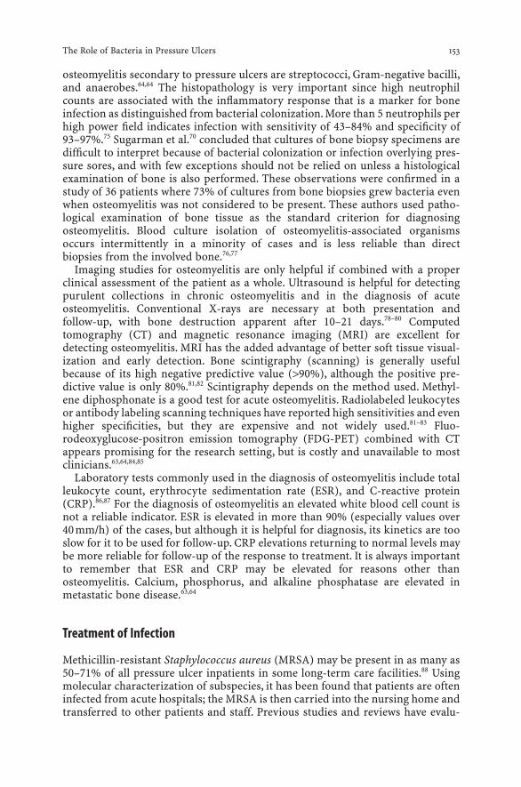
osteomyelitis secondary to pressure ulcers are streptococci, Gram-negative bacilli,and anaerobes.64,64 The histopathology is very important since high neutrophilcounts are associated with the inflammatory response that is a marker for boneinfection as distinguished from bacterial colonization. More than 5 neutrophils perhigh power field indicates infection with sensitivity of 43–84% and specificity of93–97%.75 Sugarman et al.70 concluded that cultures of bone biopsy specimens aredifficult to interpret because of bacterial colonization or infection overlying pres-sure sores, and with few exceptions should not be relied on unless a histologicalexamination of bone is also performed. These observations were confirmed in astudy of 36 patients where 73% of cultures from bone biopsies grew bacteria evenwhen osteomyelitis was not considered to be present. These authors used patho-logical examination of bone tissue as the standard criterion for diagnosingosteomyelitis. Blood culture isolation of osteomyelitis-associated organismsoccurs intermittently in a minority of cases and is less reliable than direct biopsies from the involved bone.76,77
Imaging studies for osteomyelitis are only helpful if combined with a properclinical assessment of the patient as a whole. Ultrasound is helpful for detectingpurulent collections in chronic osteomyelitis and in the diagnosis of acuteosteomyelitis. Conventional X-rays are necessary at both presentation and follow-up, with bone destruction apparent after 10–21 days.78–80 Computed tomography (CT) and magnetic resonance imaging (MRI) are excellent for detecting osteomyelitis. MRI has the added advantage of better soft tissue visual-ization and early detection. Bone scintigraphy (scanning) is generally usefulbecause of its high negative predictive value (>90%), although the positive pre-dictive value is only 80%.81,82 Scintigraphy depends on the method used. Methyl-ene diphosphonate is a good test for acute osteomyelitis. Radiolabeled leukocytesor antibody labeling scanning techniques have reported high sensitivities and evenhigher specificities, but they are expensive and not widely used.81–83 Fluo-rodeoxyglucose-positron emission tomography (FDG-PET) combined with CTappears promising for the research setting, but is costly and unavailable to mostclinicians.63,64,84,85
Laboratory tests commonly used in the diagnosis of osteomyelitis include totalleukocyte count, erythrocyte sedimentation rate (ESR), and C-reactive protein(CRP).86,87 For the diagnosis of osteomyelitis an elevated white blood cell count isnot a reliable indicator. ESR is elevated in more than 90% (especially values over40 mm/h) of the cases, but although it is helpful for diagnosis, its kinetics are tooslow for it to be used for follow-up. CRP elevations returning to normal levels maybe more reliable for follow-up of the response to treatment. It is always importantto remember that ESR and CRP may be elevated for reasons other thanosteomyelitis. Calcium, phosphorus, and alkaline phosphatase are elevated inmetastatic bone disease.63,64
Treatment of Infection
Methicillin-resistant Staphylococcus aureus (MRSA) may be present in as many as50–71% of all pressure ulcer inpatients in some long-term care facilities.88 Usingmolecular characterization of subspecies, it has been found that patients are ofteninfected from acute hospitals; the MRSA is then carried into the nursing home andtransferred to other patients and staff. Previous studies and reviews have evalu-
The Role of Bacteria in Pressure Ulcers 153
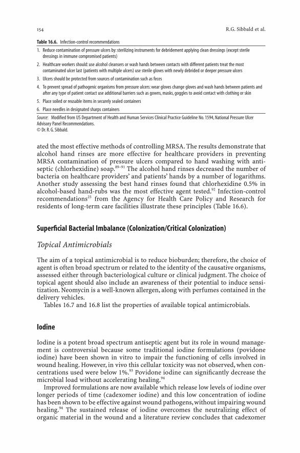
ated the most effective methods of controlling MRSA. The results demonstrate thatalcohol hand rinses are more effective for healthcare providers in preventingMRSA contamination of pressure ulcers compared to hand washing with anti-septic (chlorhexidine) soap.89–91 The alcohol hand rinses decreased the number ofbacteria on healthcare providers’ and patients’ hands by a number of logarithms.Another study assessing the best hand rinses found that chlorhexidine 0.5% inalcohol-based hand-rubs was the most effective agent tested.92 Infection-controlrecommendations25 from the Agency for Health Care Policy and Research for residents of long-term care facilities illustrate these principles (Table 16.6).
Superficial Bacterial Imbalance (Colonization/Critical Colonization)
Topical Antimicrobials
The aim of a topical antimicrobial is to reduce bioburden; therefore, the choice ofagent is often broad spectrum or related to the identity of the causative organisms,assessed either through bacteriological culture or clinical judgment. The choice oftopical agent should also include an awareness of their potential to induce sensi-tization. Neomycin is a well-known allergen, along with perfumes contained in thedelivery vehicles.
Tables 16.7 and 16.8 list the properties of available topical antimicrobials.
Iodine
Iodine is a potent broad spectrum antiseptic agent but its role in wound manage-ment is controversial because some traditional iodine formulations (povidoneiodine) have been shown in vitro to impair the functioning of cells involved inwound healing. However, in vivo this cellular toxicity was not observed, when con-centrations used were below 1%.93 Povidone iodine can significantly decrease themicrobial load without accelerating healing.94
Improved formulations are now available which release low levels of iodine overlonger periods of time (cadexomer iodine) and this low concentration of iodinehas been shown to be effective against wound pathogens, without impairing woundhealing.94 The sustained release of iodine overcomes the neutralizing effect oforganic material in the wound and a literature review concludes that cadexomer
154 R.G. Sibbald et al.
Table 16.6. Infection-control recommendations
1. Reduce contamination of pressure ulcers by: sterilizing instruments for debridement applying clean dressings (except steriledressings in immune compromised patients)
2. Healthcare workers should: use alcohol cleansers or wash hands between contacts with different patients treat the mostcontaminated ulcer last (patients with multiple ulcers) use sterile gloves with newly debrided or deeper pressure ulcers
3. Ulcers should be protected from sources of contamination such as feces
4. To prevent spread of pathogenic organisms from pressure ulcers: wear gloves change gloves and wash hands between patients andafter any type of patient contact use additional barriers such as gowns, masks, goggles to avoid contact with clothing or skin
5. Place soiled or reusable items in securely sealed containers
6. Place needles in designated sharps containers
Source: Modified from US Department of Health and Human Services Clinical Practice Guideline No. 1594, National Pressure UlcerAdvisory Panel Recommendations.© Dr. R. G. Sibbald.
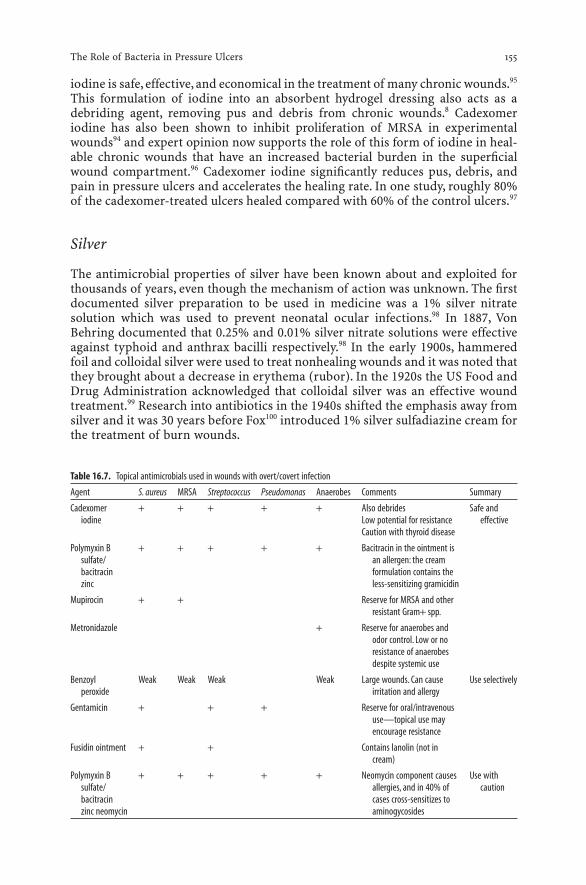
iodine is safe, effective, and economical in the treatment of many chronic wounds.95
This formulation of iodine into an absorbent hydrogel dressing also acts as adebriding agent, removing pus and debris from chronic wounds.8 Cadexomeriodine has also been shown to inhibit proliferation of MRSA in experimentalwounds94 and expert opinion now supports the role of this form of iodine in heal-able chronic wounds that have an increased bacterial burden in the superficialwound compartment.96 Cadexomer iodine significantly reduces pus, debris, andpain in pressure ulcers and accelerates the healing rate. In one study, roughly 80%of the cadexomer-treated ulcers healed compared with 60% of the control ulcers.97
Silver
The antimicrobial properties of silver have been known about and exploited forthousands of years, even though the mechanism of action was unknown. The firstdocumented silver preparation to be used in medicine was a 1% silver nitrate solution which was used to prevent neonatal ocular infections.98 In 1887, VonBehring documented that 0.25% and 0.01% silver nitrate solutions were effectiveagainst typhoid and anthrax bacilli respectively.98 In the early 1900s, hammeredfoil and colloidal silver were used to treat nonhealing wounds and it was noted thatthey brought about a decrease in erythema (rubor). In the 1920s the US Food andDrug Administration acknowledged that colloidal silver was an effective woundtreatment.99 Research into antibiotics in the 1940s shifted the emphasis away fromsilver and it was 30 years before Fox100 introduced 1% silver sulfadiazine cream forthe treatment of burn wounds.
The Role of Bacteria in Pressure Ulcers 155
Table 16.7. Topical antimicrobials used in wounds with overt/covert infection
Agent S. aureus MRSA Streptococcus Pseudomonas Anaerobes Comments Summary
Cadexomer + + + + + Also debrides Safe and iodine Low potential for resistance effective
Caution with thyroid disease
Polymyxin B + + + + + Bacitracin in the ointment is sulfate/ an allergen: the cream bacitracin formulation contains the zinc less-sensitizing gramicidin
Mupirocin + + Reserve for MRSA and other resistant Gram+ spp.
Metronidazole + Reserve for anaerobes and odor control. Low or noresistance of anaerobes despite systemic use
Benzoyl Weak Weak Weak Weak Large wounds. Can cause Use selectivelyperoxide irritation and allergy
Gentamicin + + + Reserve for oral/intravenous use—topical use may encourage resistance
Fusidin ointment + + Contains lanolin (not in cream)
Polymyxin B + + + + + Neomycin component causes Use with sulfate/ allergies, and in 40% of cautionbacitracin cases cross-sensitizes to zinc neomycin aminogycosides
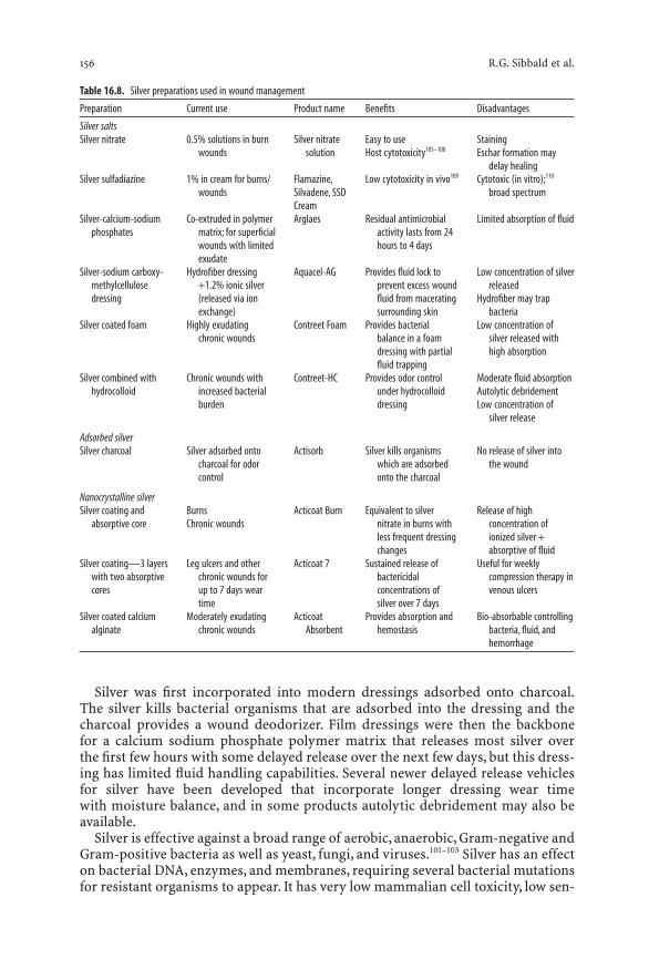
Silver was first incorporated into modern dressings adsorbed onto charcoal.The silver kills bacterial organisms that are adsorbed into the dressing and thecharcoal provides a wound deodorizer. Film dressings were then the backbone for a calcium sodium phosphate polymer matrix that releases most silver over the first few hours with some delayed release over the next few days, but this dress-ing has limited fluid handling capabilities. Several newer delayed release vehiclesfor silver have been developed that incorporate longer dressing wear time with moisture balance, and in some products autolytic debridement may also beavailable.
Silver is effective against a broad range of aerobic, anaerobic, Gram-negative andGram-positive bacteria as well as yeast, fungi, and viruses.101–103 Silver has an effecton bacterial DNA, enzymes, and membranes, requiring several bacterial mutationsfor resistant organisms to appear. It has very low mammalian cell toxicity, low sen-
156 R.G. Sibbald et al.
Table 16.8. Silver preparations used in wound management
Preparation Current use Product name Benefits Disadvantages
Silver saltsSilver nitrate 0.5% solutions in burn Silver nitrate Easy to use Staining
wounds solution Host cytotoxicity105–108 Eschar formation may delay healing
Silver sulfadiazine 1% in cream for burns/ Flamazine, Low cytotoxicity in vivo109 Cytotoxic (in vitro);110
wounds Silvadene, SSD broad spectrumCream
Silver-calcium-sodium Co-extruded in polymer Arglaes Residual antimicrobial Limited absorption of fluidphosphates matrix; for superficial activity lasts from 24
wounds with limited hours to 4 daysexudate
Silver-sodium carboxy- Hydrofiber dressing Aquacel-AG Provides fluid lock to Low concentration of silvermethylcellulose +1.2% ionic silver prevent excess wound releaseddressing (released via ion fluid from macerating Hydrofiber may trap
exchange) surrounding skin bacteriaSilver coated foam Highly exudating Contreet Foam Provides bacterial Low concentration of
chronic wounds balance in a foam silver released with dressing with partial high absorptionfluid trapping
Silver combined with Chronic wounds with Contreet-HC Provides odor control Moderate fluid absorptionhydrocolloid increased bacterial under hydrocolloid Autolytic debridement
burden dressing Low concentration ofsilver release
Adsorbed silverSilver charcoal Silver adsorbed onto Actisorb Silver kills organisms No release of silver into
charcoal for odor which are adsorbed the woundcontrol onto the charcoal
Nanocrystalline silverSilver coating and Burns Acticoat Burn Equivalent to silver Release of high
absorptive core Chronic wounds nitrate in burns with concentration of less frequent dressing ionized silver +changes absorptive of fluid
Silver coating—3 layers Leg ulcers and other Acticoat 7 Sustained release of Useful for weekly with two absorptive chronic wounds for bactericidal compression therapy in cores up to 7 days wear concentrations of venous ulcers
time silver over 7 daysSilver coated calcium Moderately exudating Acticoat Provides absorption and Bio-absorbable controlling
alginate chronic wounds Absorbent hemostasis bacteria, fluid, and hemorrhage
sitization potential and is not used systemically so that it is an ideal agent forsuperficial compartment infection treatment.
In vitro concentrations of silver as low as 10 mg/l can control bacteria. Higherconcentrations are delivered in some topical wound dressings. The minimuminhibitory concentration (MIC) in wounds in one study was estimated to bebetween 20 and 40 mg/l.104 A study on common wound pathogens using a complexorganic growth medium found that the MIC in vitro ranged between 5 and 12.5 mg/ml.103
In a study of 29 chronic wound patients not healing at the expected rate, Sibbaldet al.28 applied nanocrystalline silver dressings after baseline superficial bacterialswabs and quantitative biopsies. Improved healing was related to improvement inthe semiquantitative surface swabs but the deep quantitative bacterial biopsieswere often unchanged. If the deep compartment was out of bacterial balance andthis was delaying healing, topical silver dressings did not reverse the impairedhealing response or the increased bacteria in the deep compartment requiring systemic therapy.
Uncommon or rare silver allergic sensitization has been reported but there havebeen no other significant adverse effects despite the large amounts of silver used inburn wound treatment.On the other hand, the nitrate molecule in silver nitrate maybe pro-inflammatory while the cream base in silver sulfadiazine reacts with seriousexudate to produce a pseudo-eschar which must be removed before reapplica-tion.105 In both of these preparations, a large excess of silver has to be supplied tothe wound to compensate for inactivation; new technologies have therefore beendeveloped to improve the controlled release of silver ions. The silver ions canprovide antibacterial and anti-inflammatory properties topically as well as provid-ing moisture balance with absorptive dressing cores as outlined in Table 16.8.
Nanocrystalline Silver
Nanocrystalline silver is composed of very small crystals of less than 20 nm wherethe silver may exist in a new lattice solid state.111 In amorphous matter, atoms andmolecules interact only with their nearest neighbor, whereas in crystals each com-ponent interacts with immediate and distant neighbors through the crystal lattice.During dissolution, the silver reaches a steady state where the concentration insolution is between 70 and 100 mg/ml and antimicrobial levels can be maintainedin the dressing for at least 7 days. Nanocrystalline silver is effective against a broadrange of bacteria, including MRSA and VRE.28
For over a century the anti-inflammatory effects of silver have been observedand documented but the mechanism by which silver exerted this effect was notunderstood. The anti-inflammatory effect was largely masked or even counteredby the silver preparations available in the twentieth century (silver nitrate, silversulfadiazine) whereas the development of nanocrystalline silver may shed somelight on this mechanism.Wright et al.112 studied matrix metalloproteinases (MMP),cell apoptosis, and healing in a porcine wound model where wounds were dressedwith nanocrystalline silver, silver nitrate, and saline soaks. They suggest that thenanocrystalline silver may modulate the actions of the MMPs. Another pilot studyexamined the wound fluid of ten patients treated with either a nanocrystallinedressing or a control. Those patients with the active dressing had lower MMP-9and tumor necrosis factor a (TNFa) levels relative to the controls.112 Newer topical
The Role of Bacteria in Pressure Ulcers 157

silver products offer variable levels of silver release with moisture balance and mayhave autolytic debridement properties for optimal wound bed preparation.
The European Pressure Ulcer Advisory Panel recommends that systemic antibi-otics are not required for pressure ulcers that have clinical signs of local infection(or increased bacterial burden) only.113 A period of 2 weeks would be a reasonabletrial with these agents before considering systemic treatments or re-examining thetreatment of the cause or the ability of the ulcer to heal. This paradigm in Figure16.1 will now be used to illustrate the steps in the diagnosis and treatment of therole of bacteria in chronic pressure ulcers.
158 R.G. Sibbald et al.
Case Two
Mr CP, a 45-year-old man, has been paraplegic since a motor vehicle accident 18 years ago. Hedeveloped a deep pressure ulcer over the buttocks 18 months ago and a satellite area openedup 12 cm from the original opening (Figure 16.3—see color section).
Patient as a whole:
1. Assess case. The patient’s ulcer is caused by sliding from the bed to the wheelchair withfriction and shearing during transfers. The problem can be corrected.
2. The patient and caregivers decided to install an automatic ceiling lift for self-transfersfrom the bed and to use a transfer board at other times.
Regional treatment:
3. Clinically there were increased exudates and bright red granulation tissue that wasfriable, with undermining and a communication between the two ulcer beds. A bacter-ial swab grew S. aureus (not MRSA) and Pseudomonas.The ESR was elevated at 45 (Nl.20)and CRP high at 25.0.An X-ray of the pelvis was normal with no evidence of osteomyelitis.
4. The deep infection was treated with oral ciprofloxacin 500 mg twice daily and clin-damycin 300 mg four times daily by mouth for 6 weeks.
5. The wounds were cleansed with saline.
6. Topical antiseptic treatment with Betadine-soaked gauze ribbon was changed daily untilsurgical debridement was scheduled.
7. Surgical debridement was performed to remove the bridge of tissue over the underminedwound edge.
8. Topical ionized silver impregnated foam was applied to the wound to perform autolyticdebridement, surface bacterial balance, and moisture balance three times per week.
9. The wound was 25% of its initial size at week 12. Swabs showed scant S. aureus and ESRwas 15; C-reactive protein was normal at 7.
10. The wound completely healed at 20 weeks and Mr CP adhered to his new transferringtechnique.

References
1. Schultz GS, Sibbald RG, Falanga V, et al. Wound bed preparation: a systematic approach to woundmanagement. Wound Repair Regen 2003; 11(Suppl 1):S1–S28.
2. Von Renteln-Kruse W, Krause T, Anders J, et al. [High-grade pressure sores in frail older high-risk persons.A retrospective postmortem case-control-study]. Z Gerontol Geriatr 2004; 37:81–85.
3. Reddy M, Keast D, Fowler E, Sibbald RG. Pain in pressure ulcers. Ostomy Wound Manage 2003;49:30–35.
4. Norton L, Sibbald RG. Is bed rest an effective treatment modality for pressure ulcers? OstomyWound Manage 2004; 50(10):44–52.
5. Kanj LF, Wilking SV, Phillips TJ. Pressure ulcers. J Am Acad Dermatol 1998; 38:517–536.6. Vasile J, Chaitin H. Prognostic factors in decubitus ulcers of the aged. Geriatrics 1972; 27:126–129.7. Lee BY, Trainor FS, Thoden WR. Topical application of povidone-iodine in the management of
decubitus and stasis ulcers. J Am Geriatr Soc 1979; 27:302–306.8. Drosou A, Falabella A, Kirsner RS. Antiseptics on wounds: an area of controversy. Wounds 2003;
15(5):149–166.9. Lineaweaver W, Howard R, Soucy D, et al. Topical antimicrobial toxicity. Arch Surg 1985; 120:
267–270.10. Rodeheaver G. Wound cleansing, wound irrigation, wound disinfection. In: Krasner DL, Rode-
heaver GT, Sibbald RG (eds) Chronic wound care: a clinical source book for healthcare profes-sionals, 3rd edn. Wayne, PA: HMP Communications; 2001: 369–383.
11. White RJ, Cooper R, Kingsley A. Wound colonization and infection: the role of topical antimi-crobials. Br J Nurs 2001; 10:563–578.
12. Lawrence JC. The use of iodine as an antiseptic agent. J Wound Care 1998; 7:421–425.13. Ayello EA, Dowsett C, Schultz GS, et al. TIME heals all wounds. Nursing. 2004; 34:36–41.14. Sibbald RG, Orsted H, Schultz GS, et al. Preparing the wound bed 2003: focus on infection and
inflammation. Ostomy Wound Manage 2003; 49:23–51.15. Bowler PG. The 10(5) bacterial growth guideline: reassessing its clinical relevance in wound
healing. Ostomy Wound Manage 2003; 49:44–53.16. Glenchur H, Patel BS, Pathmarajah C. Transient bacteremia associated with debridement of decu-
bitus ulcers. Mil Med 1981; 146:432–433.17. Brem H, Lyder C. Protocol for the successful treatment of pressure ulcers. Am J Surg 2004;
188:9–17.18. Galpin JE, Chow AW, Bayer AS, Guze LB. Sepsis associated with decubitus ulcers. Am J Med 1976;
61:346–350.19. Reuler JB, Cooney TG. The pressure sore: pathophysiology and principles of management. Ann
Intern Med 1981; 94:661–666.20. Cunningham SC, Napolitano LM. Necrotizing soft tissue infection from decubitus ulcer after
spinal cord injury. Spine 2004; 29:E172–E174.21. Chan JW, Virgo KS, Johnson FE. Hemipelvectomy for severe decubitus ulcers in patients with
previous spinal cord injury. Am J Surg 2003; 185:69–73.22. Heggers JP. Defining infection in chronic wounds: does it matter? J Wound Care 1998; 7:389–392.23. Parish LC, Witkowski JA. The infected decubitus ulcer. Int J Dermatol 1989; 28:643–647.24. Cutting KF, Harding KG. Criteria for identifying wound infection. J Wound Care 1994; 5(4):
198–201.25. Bergstrom N, Bennett MA, Carlson CE, et al. Clinical practice guideline number 15: Treatment
of pressure ulcers.AHCPR Publication 95–0652. Rockville, MD: Agency for Healthcare Policy andResearch (AHCPR); 1994.
26. Sapico FL, Ginunas VJ, Thornhill-Joynes M, et al. Quantitative microbiology of pressure sores indifferent stages of healing. Diagn Microbiol Infect Dis 1986; 5:31–38.
27. Grayson ML, Gibbons GW, Balogh K, et al. Probing to bone in infected pedal ulcers. A clinicalsign of underlying osteomyelitis in diabetic patients. JAMA 1995; 273:721–723.
28. Sibbald RG, Browne AC, Coutts P, Queen D. Screening evaluation of an ionized nanocrystallinesilver dressing in chronic wound care. Ostomy Wound Manage 2001; 47:38–43.
29. Gardner SE, Frantz RA, Doebbeling BN. The validity of the clinical signs and symptoms used toidentify localized chronic wound infection. Wound Repair Regen 2001; 9:178–186.
30. Gardner SE, Frantz RA, Troia C, et al. A tool to assess clinical signs and symptoms of localizedinfection in chronic wounds: development and reliability. Ostomy Wound Manage 2001; 47:40–47.
31. Armstrong DG. Infrared dermal thermometry: the foot and ankle stethoscope. J Foot Ankle Surg1998; 37:75–76.
The Role of Bacteria in Pressure Ulcers 159
32. Murff RT, Armstrong DG, Lanctot D, et al. How effective is manual palpation in detecting subtletemperature differences? Clin Podiatr Med Surg 1998; 15:151–154.
33. Diegelmann RF. Excessive neutrophils characterize chronic pressure ulcers.Wound Repair Regen2003; 11:490–495.
34. Sibbald RG, Coutts P, Freiheller M, et al. Use of nanocrystalline silver dressing in the treatmentof non-healing venous ulcers. In preparation. 2004.
35. Dow, G. Infection in chronic wounds. In: Krasner DL, Rodeheaver GT, Sibbald RG (eds) Chronicwound care: A clinical source book for healthcare professionals, 3rd edn. Wayne, PA: HMP Com-munications; 2001: 343–356.
36. Bendy RH, Jr, Nuccio PA, Wolfe E, et al. Relationship of quantitative wound bacterial counts tohealing of decubiti: effect of topical gentamicin.Antimicrob Agents Chemother 1964; 10:147–155.
37. Robson MC, Lea CE, Dalton JB, Heggers JP. Quantitative bacteriology and delayed wound closure.Surg Forum 1968; 19:501–502.
38. Robson MC, Heggers JP. Bacterial quantification of open wounds. Mil Med 1969; 134:19–24.39. Robson MC, Heggers JP. Delayed wound closure based on bacterial counts. J Surg Oncol 1970;
2:379–383.40. Vande Berg JS, Rudolph R. Cultured myofibroblasts: a useful model to study wound contraction
and pathological contracture. Ann Plast Surg 1985; 14:111–120.41. Vande Berg JS, Rudolph R. Pressure (decubitus) ulcer: variation in histopathology—a light and
electron microscope study. Hum Pathol 1995; 26:195–200.42. Vande Berg JS, Rudolph R, Hollan C, Haywood-Reid PL. Fibroblast senescence in pressure ulcers.
Wound Repair Regen 1998; 6:38–49.43. Bowler PG, Duerden BI, Armstrong DG. Wound microbiology and associated approaches to
wound management. Clin Microbiol Rev 2001; 14:244–269.44. Robson MC, Duke WF, Krizek TJ. Rapid bacterial screening in the treatment of civilian wounds.
J Surg Res 1973; 14:426–430.45. Tenorio A, Jindrak K, Weiner M, et al. Accelerated healing in infected wounds. Surg Gynecol
Obstet 1976; 142:537–543.46. Dow G, Browne A, Sibbald RG. Infection in chronic wounds: controversies in diagnosis and treat-
ment. Ostomy Wound Manage 1999; 45:23–40.47. Woolfrey BF, Fox JM, Quall CO. An evaluation of burn wound quantitative microbiology. I. Quan-
titative eschar cultures. Am J Clin Pathol 1981; 75:532–537.48. Ehrenkranz NJ, Alfonso B, Nerenberg D. Irrigation-aspiration for culturing draining decubitus
ulcers: correlation of bacteriological findings with a clinical inflammatory scoring index. J ClinMicrobiol 1990; 28:2389–2393.
49. Rudensky B, Lipschits M, Isaacsohn M, Sonnenblick M. Infected pressure sores: comparison ofmethods for bacterial identification. South Med J 1992; 85:901–903.
50. Dow G. Bacterial swabs and the chronic wound: when, how, and what do they mean? OstomyWound Manage 2003; 49:8–13.
51. Hill KE, Davies CE, Wilson MJ, et al. Molecular analysis of the microflora in chronic venous legulceration. J Med Microbiol 2003; 52:365–369.
52. Wheat LJ, Allen SD, Henry M, et al. Diabetic foot infections. Bacteriologic analysis. Arch InternMed 1986; 146:1935–1940.
53. Nichols RL, Smith JW. Anaerobes from a surgical perspective. Clin Infect Dis 1994; 18(Suppl 4):S280–S286.
54. Heym B, Rimareix F, Lortat-Jacob A, Nicolas-Chanoine MH. Bacteriological investigation ofinfected pressure ulcers in spinal cord-injured patients and impact on antibiotic therapy. SpinalCord 2004; 42:230–234.
55. Kingston D, Seal DV. Current hypotheses on synergistic microbial gangrene. Br J Surg 1990;77:260–264.
56. Bucknall TE. The effect of local infection upon wound healing: an experimental study. Br J Surg1980; 67:851–855.
57. Eriksson G, Eklund AE, Kallings LO. The clinical significance of bacterial growth in venous legulcers. Scand J Infect Dis 1984; 16:175–180.
58. Gilchrist B, Reed C. The bacteriology of chronic venous ulcers treated with occlusive hydrocol-loid dressings. Br J Dermatol 1989; 121:337–344.
59. Annoni F, Rosina M, Chiurazzi D, Ceva M. The effects of a hydrocolloid dressing on bacterialgrowth and the healing process of leg ulcers. Int Angiol 1989; 8:224–228.
60. Handfield-Jones SE, Grattan CE, Simpson RA, Kennedy CT. Comparison of a hydrocolloid dressing and paraffin gauze in the treatment of venous ulcers. Br J Dermatol 1988; 118:425–427.
160 R.G. Sibbald et al.
61. Trengove NJ, Stacey MC, McGechie DF, Mata S. Qualitative bacteriology and leg ulcer healing. JWound Care 1996; 5:277–280.
62. Bowler PG, Davies BJ. The microbiology of infected and noninfected leg ulcers. Int J Dermatol1999; 38:573–578.
63. Lew DP, Waldvogel FA. Osteomyelitis. N Engl J Med. 1997; 336:999–1007.64. Lew DP, Waldvogel FA. Osteomyelitis. Lancet 2004; 364:369–379.65. Waldvogel FA, Medoff G, Swartz MN. Treatment of osteomyelitis. N Engl J Med 1970; 283:
822.66. Waldvogel FA, Medoff G, Swartz MN. Osteomyelitis: a review of clinical features, therapeutic con-
siderations and unusual aspects. N Engl J Med 1970; 282:198–206.67. Waldvogel FA, Medoff G, Swartz MN. Osteomyelitis: a review of clinical features, therapeutic con-
siderations and unusual aspects. 3. Osteomyelitis associated with vascular insufficiency. N EnglJ Med 1970; 282:316–322.
68. Waldvogel FA, Papageorgiou PS. Osteomyelitis: the past decade. N Engl J Med 1980; 303:360–370.69. Darouiche RO, Landon GC, Klima M, et al. Osteomyelitis associated with pressure sores. Arch
Intern Med 1994; 154:753–758.70. Sugarman B. Pressure sores and underlying bone infection. Arch Intern Med 1987; 147:553–555.71. Thornhill-Joynes M, Gonzales F, Stewart CA, et al. Osteomyelitis associated with pressure ulcers.
Arch Phys Med Rehabil 1986; 67:314–318.72. Sugarman B, Hawes S, Musher DM, et al. Osteomyelitis beneath pressure sores. Arch Intern Med
1983; 143:683–688.73. Brandeis GH, Morris JN, Nash DJ, Lipsitz LA. The epidemiology and natural history of pressure
ulcers in elderly nursing home residents. JAMA 1990; 264:2905–2909.74. Mackowiak PA, Jones SR, Smith JW. Diagnostic value of sinus-tract cultures in chronic
osteomyelitis. JAMA 1978; 239:2772–2775.75. Abdul-Karim FW, McGinnis MG, Kraay M, et al. Frozen section biopsy assessment for the pres-
ence of polymorphonuclear leukocytes in patients undergoing revision of arthroplasties. ModPathol 1998; 11:427–431.
76. Howard CB, Einhorn M, Dagan R, et al. Fine-needle bone biopsy to diagnose osteomyelitis. J BoneJoint Surg Br 1994; 76:311–314.
77. Jacobson IV, Sieling WL. Microbiology of secondary osteomyelitis. Value of bone biopsy. S AfrMed J 1987; 72:476–477.
78. Gold RH, Hawkins RA, Katz RD. Bacterial osteomyelitis: findings on plain radiography, CT, MR,and scintigraphy. AJR Am J Roentgenol 1991; 157:365–370.
79. Kaim AH, Gross T, von Schulthess GK. Imaging of chronic posttraumatic osteomyelitis. EurRadiol 2002; 12:1193–1202.
80. Santiago RC, Gimenez CR, McCarthy K. Imaging of osteomyelitis and musculoskeletal soft tissueinfections: current concepts. Rheum Dis Clin North Am 2003; 29:89–109.
81. Peters AM. The use of nuclear medicine in infections. Br J Radiol 1998; 71:252–261.82. Tumeh SS, Tohmeh AG. Nuclear medicine techniques in septic arthritis and osteomyelitis. Rheum
Dis Clin North Am 1991; 17:559–583.83. Oyen WJ, van H, Jr, Claessens RA, Slooff TJ, et al. Diagnosis of bone, joint, and joint prosthesis
infections with In-111-labeled nonspecific human immunoglobulin G scintigraphy. Radiology1992; 182:195–199.
84. Robiller FC, Stumpe KD, Kossmann T, et al. Chronic osteomyelitis of the femur: value of PETimaging. Eur Radiol 2000; 10:855–858.
85. Schmitz A, Kalicke T, Willkomm P, et al. Use of fluorine-18 fluoro-2-deoxy-D-glucose positronemission tomography in assessing the process of tuberculous spondylitis. J Spinal Disord 2000;13:541–544.
86. Hovi I, Valtonen M, Korhola O, Hekali P. Low-field MR imaging for the assessment of therapyresponse in musculoskeletal infections. Acta Radiol 1995; 36:220–227.
87. Lewis VL, Jr, Bailey MH, Pulawski G, et al. The diagnosis of osteomyelitis in patients with pres-sure sores. Plast Reconstr Surg 1988; 81:229–232.
88. Capitano B, Leshem OA, Nightingale CH, Nicolau DP. Cost effect of managing methicillin-resistant Staphylococcus aureus in a long-term care facility. J Am Geriatr Soc 2003; 51:10–16.
89. Girou E, Loyeau S, Legrand P, et al. Efficacy of handrubbing with alcohol based solution versus standard handwashing with antiseptic soap: randomised clinical trial. BMJ 2002; 325:362.
90. Girard R, Amazian K, Fabry J. Better compliance and better tolerance in relation to a well-conducted introduction to rub-in hand disinfection. J Hosp Infect 2001; 47:131–137.
The Role of Bacteria in Pressure Ulcers 161
91. Pittet D, Hugonnet S, Harbarth S, et al. Effectiveness of a hospital-wide programme to improvecompliance with hand hygiene. Infection Control Programme. Lancet 2000; 356:1307–1312.
92. Rochon-Edouard S, Pons JL,Veber B, et al. Comparative in vitro and in vivo study of nine alcohol-based handrubs. Am J Infect Control 2004; 32:200–204.
93. Burks RI. Povidone-iodine solution in wound treatment. Phys Ther 1998; 78:212–218.94. Mertz PM, Oliveira-Gandia MF, Davis SC. The evaluation of a cadexomer iodine wound dressing
on methicillin resistant Staphylococcus aureus (MRSA) in acute wounds. Dermatol Surg 1999;25:89–93.
95. Sundberg J, Meller R. A retrospective review of the use of cadexomer iodine in the treatment ofchronic wounds. Wounds 1997; 9:68–86.
96. Gilchrist B. Should iodine be reconsidered in wound management? European Tissue RepairSociety. J Wound Care 1997; 6:148–150.
97. Moberg S, Hoffman L, Grennert ML, Holst A. A randomized trial of cadexomer iodine in decu-bitus ulcers. J Am Geriatr Soc 1983; 31:462–465.
98. Grier N. Silver and its compounds. In: Block SS (ed) Disinfection, sterilization and preservation,3rd edn. Philadelphia, PA: Lea & Febiger; 1983.
99. Hollinger MA. Toxicological aspects of topical silver pharmaceuticals. Crit Rev Toxicol 1996;26:255–260.
100. Fox CL, Jr. Silver sulfadiazine—a new topical therapy for Pseudomonas in burns. Therapy ofPseudomonas infection in burns. Arch Surg 1968; 96:184–188.
101. Wright JB, Lam K, Burrell RE. Wound management in an era of increasing bacterial antibioticresistance: a role for topical silver treatment. Am J Infect Control 1998; 26:572–577.
102. Wright JB, Lam K, Hansen D, Burrell RE. Efficacy of topical silver against fungal burn woundpathogens. Am J Infect Control 1999; 27:344–350.
103. Yin HQ, Langford R, Burrell RE. Comparative evaluation of the antimicrobial activity of ACTI-COAT antimicrobial barrier dressing. J Burn Care Rehabil 1999; 20:195–200.
104. Ricketts CR, Lowbury EJ, Lawrence JC, et al. Mechanism of prophylaxis by silver compoundsagainst infection of burns. BMJ 1970; i:444–446.
105. Demling RH, De Santi L. Effects of silver on wound management. Wounds 2001; 13(Suppl A):4.106. Bader KF. Organ deposition of silver following silver nitrate therapy of burns. Plast Reconstr Surg
1966; 37:550–551.107. Coombs CJ, Wan AT, Masterton JP, et al. Do burn patients have a silver lining? Burns 1992;
18:179–184.108. Hall RE, Bender G, Marquis RE. In vitro effects of low intensity direct current generated silver
on eukaryotic cells. J Oral Maxillofac Surg 1988; 46:128–133.109. Paddock HN, Schultz GS, Perrin KJ, et al. Clinical assessment of silver coated antimicrobial dress-
ing on MMPs and cytokine levels in non-healing wounds.Annual Meeting of the Pressure WoundHealing Society, 28 May 2002.
110. McCauley RL, Linares HA, Pelligrini V, et al. In vitro toxicity of topical antimicrobial agents tohuman fibroblasts. J Surg Res 1989; 46:267–274.
111. Birringer R. Nanocrystalline materials. Mat Sci Eng 1989; A117:33–43.112. Wright JB, Lam K, Buret AG, et al. Early healing events in a porcine model of contaminated
wounds: effects of nanocrystalline silver on matrix metalloproteinases, cell apoptosis, andhealing. Wound Repair Regen 2002; 10:141–151.
113. European Pressure Ulcer Advisory Panel. Guidelines on treatment of pressure ulcers. EPUAPReview 1999; 1:31–33.
162 R.G. Sibbald et al.
17 LitigationCourtney H. Lyder
Introduction
The development of pressure ulcers can often be viewed by the legal communityas a violation in quality of care. Patients and/or family members may perceive their development as a failure in the healthcare system. When there is a lack ofexplanation as to its development and/or unavoidability patients and/or familymembers may seek legal remedies.
Many pressure ulcer cases are often settled through an inquiry by a health trust.However, when families are not satisfied, families may seek financial remedies.Coupled with this is the growing number of pressure ulcer cases (usually the worstcases) being publicized in local or national media as evidence of poor healthcareor a failing healthcare system.
Explanations in the media concerning their development may range frompatients being exposed to untrained healthcare professionals to lack of appropriatenursing to patient–staff ratios. Often the media may give the lay community theperception that all pressure ulcers are avoidable. Thus, when pressure ulcers dodevelop, patients and/or families may have more incentive to pursue legal recourse.
This chapter will review several key factors that may place healthcare providersand healthcare systems at risk for litigation. It should be noted that the legalsystems in various European countries may vary. However, the concept of provingnegligence remains a universal principle. This chapter will also highlight essentialdocumentation that could decrease healthcare provider exposure to litigation.
Litigation
A growing number of health professionals view the development of pressure ulcersas negligent care by a healthcare provider or healthcare system. One German studyinvestigating 10,222 corpses found a pressure ulcer prevalence rate of 11.2%.1
Although the majority of corpses were elderly, the investigators concluded that themajority of physicians did not correlate fatality (e.g. sepsis) with pressure ulcerdevelopment. These investigators further concluded that pressure ulcer prevalencerates are an excellent indicator to determine quality of nursing and medical care.
The increasing use of pressure ulcers as a quality indicator on nursing andmedical care has led to increased litigation. Healthcare systems continue to placethemselves unduly at risk of litigation due to lack of proactive pressure ulcer
163

prevention. One retrospective study investigating hospital litigation in the UnitedKingdom found that there was a lack of comprehensive preventive measures imple-mented in UK hospitals. Because of the dearth of comprehensive pressure ulcerprevention strategies being used, amounts of £3500 to £12,500 were usuallyawarded, with a few cases receiving damages in excess of £100,000.2
Although Europe continues to have increasing litigation, it appears that the USAleads in awarding damages. Presently, throughout the USA plaintiff attorneys placeadvertisements on billboards, newspapers, and television seeking pressure ulcercases. A retrospective study investigating typical pressure ulcer awards in the USAfound that awards ranged from $5000 to $82,000,000, with a median award ofapproximately $250,000 reported.3
The following case study highlights elements of how healthcare providers andhealthcare institutions can be easily exposed to litigation.
164 C.H. Lyder
An 87-year-old woman was admitted to hospital with history of a closed stage 4 pressureulcer on right hip, peripheral vascular disease, non-insulin dependent diabetes mellitus, leftcerebral vascular accident,hypertension,and urinary incontinence.A pressure ulcer risk assess-ment scale was completed indicating that the patient was at mild risk for pressure ulcers.Thepatient was placed on a standard mattress, turned every two hours while in bed and chair. Onday 2 of hospital admission, a nurse indicated an “erythematic” area on the right hip and heel.By day 5, a stage 2 pressure ulcer was noted on the right hip and heel. A hydrocolloid dress-ing was placed on the right hip and nothing was ordered for the heel. The charts noted thata tissue viability nurse would be consulted.
This case highlights some common errors made by the hospital staff. First, thepatient was at high risk for pressure ulcers since she had multiple health condi-tions that placed her at risk (closed stage 4 pressure ulcer on right hip, peripheralvascular disease, non-insulin-dependent diabetes mellitus, left cerebral vascularaccident, hypertension, and urinary incontinence). Moreover, the risk assessmenttool placed her at mild risk. This is an important factor, indicating that the toolmay have been completed incorrectly. It also highlights an important fact that nopressure ulcer risk assessment tool has 100% sensitivity and specificity.4 Thus,independent of the risk assessment tool used, a patient may be at risk for pressureulcers, so continuous assessments must be conducted. The patient was placed onlyon a standard mattress. Given the patient’s high risk level, a dynamic surface (alter-nating air mattress or low-air-loss mattress) might have been preferable. Ofconcern, although erythematic areas were identified on the patient, no interven-tions were undertaken until the development of the stage 2 pressure ulcers. Clearly,at this point in time, the tissue was breaking down. The lack of intervention forboth stage 1 pressure ulcers clearly placed the patient at greater risk for furtherdevelopment. Finally, of concern was the lack of treatment noted for the stage 2pressure ulcer on her left heel. Although the heels can be difficult to manage,no off-loading was used to salvage the heel. In this case study, it was obvious that

additional preventive measures were not instituted, nor was aggressive treatmentused in a timely manner; thus these pressure ulcers might have been avoided. Giventhe paucity of information documented, both the hospital and healthcare providersare exposed to litigation.
Negligence
The above case study could occur anywhere in the world. Thus, any healthcareprovider could be exposed to litigation when caring for a patient with a pressureulcer. In the above case study, the plaintiff attorney may be able to demonstratethat the hospital was negligent in providing care to the patient. This is based ontheir lack of ability to complete an appropriate risk assessment and to follow itwith appropriate preventive strategies and timely pressure ulcer interventions.Because of a dearth of documentation, the plaintiff attorney is left with many con-jectures on the quality of care provided. A plaintiff attorney has to show that threemajor factors were met to prove negligence. These three factors are accountability,causation, and breach of standard of care.5 When all three factors have been met,the verdict will usually be for the plaintiff.
Accountability
A breach of accountability must be proved in any negligence case. Thus, the plain-tiff was owed a duty of care, and this duty of care was breached. Moreover,the breach of care resulted in permanent damage or injury, and the plaintiff is owed compensation due to the injury. This factor is easily acknowledged since any patient who enters a hospital, nursing home, or home care setting is owed acertain level of care by healthcare providers. If there is a violation of the health-care system’s policies and procedures or inconsistency in providing care not consistent with level of education of the healthcare provider, these may all be indi-cations of breaching accountability. Further, since pressure ulcers can developwhen preventive measures are not implemented or if an existing pressure ulcerexists but the medical team does not treat it adequately, it is very easy for the plain-tiff to meet this standard.
Causation
Causation examines whether the harm suffered by the patient was a reasonable,foreseeable consequence of the breach of the duty of care. Most pressure ulcers donot result in death of the patient. In fact, it has been noted that only approximately5% of pressure ulcers lead to osteomyelitis; however, they may expose the patientto cellulitis or pain.6 Because there are numerous factors associated with pressureulcer development, failure to recognize a risk factor (e.g. poor nutrition, immobil-ity, etc.) and provide immediate remedies may make it possible for a plaintiff attor-ney to associate the failure with causation. Furthermore, when there is a dearth ofdocumentation demonstrating interventions for prevention and/or treatment pro-vided, this can make proving causation easy.
Litigation 165

Standard of Care
The last key factor to prove in negligence is a breach of the standard of care bynursing and/or medical staff. It is important to note that the standard of care isnot at the level of an expert, but rather that of an average healthcare professional.Most often, nursing and medical experts are used to determine the expected skillmix of the average healthcare provider related to wound care. Which healthcareexperts are used to evaluate a medical record will be dictated by which disciplineis implicated in the plaintiff case. Hence physician experts would comment onmedical practice, whereas a nurse expert would appraise the nursing care provided.If national pressure ulcer prevention and treatment guidelines exist in a particu-lar European country, often they will guide the healthcare expert/consultant opin-ions. A US retrospective study investigating the impact of implementing andcomplying with pressure ulcer practice guidelines in 49 plaintiff cases with com-pensations worth $14,418,770 estimated that if guidelines had been used, thesecould have saved the defendants $11,389,989 in litigation.7
Although the development and use of pressure ulcer guidelines has been prolific,their implementation can be costly. One British study found that costs for imple-menting support surface replacements could range from £100 for some foam over-lays to over £30,000 for some bed replacements.8 These costs do not take intoaccount the continued maintenance of such support surfaces. This is especiallychallenging when the daily cost of managing a pressure ulcer ranges from £38 to£196.9 Thus, the challenge for healthcare systems of allocating funds for preven-tion and/or treatment may strain a healthcare system. However, the alternative forhealthcare systems would be greater exposure to pressure ulcer litigation.
Documentation to Reduce Litigation Exposure
Good and thoughtful documentation remains the single best measure to decreasea healthcare provider’s exposure to litigation.Although it does not guarantee 100%that the healthcare provider will be litigation free, it does ensure that the health-care provider will be better positioned to defend their practice. It is also impor-tant to note that most cases are not brought to trial for several years, so there is anincreasing dependence on the medical record to reconstruct the care that was pro-vided. Essential documentation should include the following, independent ofhealthcare setting.10
Prevention of Pressure Ulcers
1. Risk assessment tool (e.g. Waterlow, Norton, Braden scales)2. Daily skin assessment3. Repositioning (off-loading) and turning schedules4. Use of support surfaces to address pressure redistribution (both bed and chair)5. Moisture control from perspiration, and urinary and fecal incontinence6. Nutritional assessment and supplementation when appropriate7. Education of patient and/or family
166 C.H. Lyder
Treatment of Pressure Ulcers
1. Regular assessment/reassessment of the wound (daily, weekly, etc.)2. Characteristics of the ulcer
(a) length(b) width(c) depth(d) exudate amount(e) tissue type(f) pain
3. Local wound care(a) wound bed preparation
4. Repositioning (off-loading) and turning schedules5. Use of support surfaces to address pressure redistribution (both bed and chair)6. Moisture control from perspiration, and urinary and fecal incontinence7. Nutritional assessment and supplementation when appropriate8. Use of adjunctive therapies (e.g. negative pressure wound therapy, electrical
stimulation, etc.)9. Education of patient and/or family
Conclusion
As healthcare consumers become more educated about pressure ulcers, healthcareproviders will become increasingly exposed to litigation. Thus, it is vitally impor-tant for healthcare providers to document the quality of care that is delivered. Moreimportantly, it is critical for healthcare providers to increase communication withpatients and/or families to discuss the issue of avoidable and unavoidable pressureulcers, which may temper the expectations of both patients and their families.
References
1. Tsokos M, Heinemann A, Puschel K. Pressure sores: epidemiology, medico-legal implications andforensic argumentation concerning causality. Int J Legal Med 2000; 113:283–287.
2. Franks PJ. Health economics: The cost to nations. In: Morrison MJ (ed) The prevention and treat-ment of pressure ulcers. Edinburgh: Mosby; 2001: 52–53.
3. Bennett RG, O’Sullivan J, DeVito EM, Remsberg R. The increasing medical malpractice risk relatedto pressure ulcers in the United States. J Am Geriatr Soc 2000; 48(1):73–81.
4. Lyder C. Exploring pressure ulcer prevention and management. JAMA 2003; 289:223–226.5. Dimond B. Pressure ulcers and litigation. Nurs Times 2003; 99:61–63.6. Lyder C. Pressure ulcers. In: Geriatric review syllabus: A core curriculum in geriatric medicine,
5th edn. New York: American Geriatrics Society; 2002: 202–209.7. Goebel RH, Goebel MR. Clinical practice guidelines for pressure ulcer prevention can prevent mal-
practice lawsuits in older patients. J Wound Ostomy Continence Nurs 1999; 26:175–184.8. NHS. 2002 (www.guideline.gov/summary).9. Bennett G, Dealey C, Posnett J. The cost of pressure ulcers in the UK. Age Ageing 2004; 33:230–235.
10. Lyder C. Regulation and wound care. In: Baranoski S, Ayello E (eds) Wound care essentials: Prac-tice principles. Springhouse, PA: Lippincott Williams & Wilkins; 2003: 35–46.
Litigation 167
18 The Development, Dissemination, and Use of PressureUlcer GuidelinesR.T. van Zelm, Michael Clark, and Jeen R.E. Haalboom
Introduction
Clinical guidelines have been defined as “systematically developed statement(s) toassist practitioner and patient decisions about appropriate health care for specificclinical circumstances.”1 In this definition the use of “assist” clearly indicates thata successful guideline does not seek to compel practitioners to practice in a rigid,inflexible manner but rather that evidence-based or evidence-linked recommen-dations are offered to help reduce inequities in healthcare provision. While clini-cal guidelines are a relatively recent phenomenon, there are now a wide range ofnational and international clinical guidelines that address pressure ulcer preven-tion and/or management2 beginning with the consensus guidelines developed inthe Netherlands in 1985,3 through the US Agency for Health Care Policy andResearch guidance issued in the early 1990s (on prevention4 and treatment5) toEuropean guidelines developed by the European Pressure Ulcer Advisory Panel(EPUAP).6,7 Recently the wheel has turned full circle with the development of newnational guidelines in both the Netherlands and the UK under the respective aus-pices of the Dutch Institute for Healthcare Improvement (CBO),8 and the NationalInstitute for Clinical Excellence (NICE).9 The CBO is an independent, not-for-profitorganization advising on clinical guideline development across the whole spec-trum of healthcare, while NICE was established as a Special Health Authoritywithin England and Wales in April 1999. Working within the UK National HealthService, NICE seeks to deliver “authoritative, robust and reliable guidance”(www.nice.org.uk) regarding what constitutes best practice, with this informationavailable to all consumers, be they patients, the public, or the health professionals.
The wealth of pressure ulcer guidelines has been developed using a variety ofmethods that seek to synthesize the available scientific and clinical knowledgeavailable during each guideline’s development. Early national guidelines, forexample the Dutch guidelines of 1985 (prevention) and 1986 (treatment), weredeveloped using informal consensus techniques. Later guidelines such as those ofthe US Agency for Health Care Policy and Research (AHCPR) were based on formalconsensus techniques, with more recent guidelines seeking to be based solely uponthe best practices of evidence-based medicine.
This chapter discusses the evolution of pressure ulcer guideline developmentusing the new Dutch guidelines as examples of evidence-based national guidelines.Beyond guideline evolution aspects of their dissemination, implementation, andappraisal will also be considered.
169
The Evolution of Pressure Ulcer Guidelines
Consensus guideline development processes, using either informal or formal tech-niques, has until recently been the foundation of many pressure ulcer guidelines.Informal consensus methods such as those employed during the development ofthe EPUAP pressure ulcer prevention and treatment guidelines in the late 1990srely upon the collective experience and knowledge of the guideline panel. Thereare no attempts to ensure that all the relevant literature is searched or appraisedwhen deriving practice recommendations, leaving the guideline vulnerable tobeing driven by panel members with the greatest authority rather than the rec-ommendations flowing from scientific evidence. To overcome this problem, formalconsensus strategies have been used since the early 1990s with these introducingliterature searches, the use of Delphi methods, and weighed consensus techniques.Usually, both informal and formal consensus guidelines are discussed in draftformat within open public meetings with a panel established to sift the evidenceand opinions presented and then derive the final recommendations.
Evidence-Based Pressure Ulcer Guidelines—the Development Process
In contrast to consensus driven guidelines, those that claim to be evidence-basedexplicitly seek to integrate the best available research evidence with both clinicalexpertise and patient preferences and values. These evidence-based guidelines,almost regardless of their country of origin, are typically developed following aclearly defined process that includes seven key stages:
1. Development of the guideline scope and purpose2. Development of the draft guideline3. External review of the draft4. Endorsement of the revised guideline5. Publication and dissemination6. Implementation7. Evaluation and updating
In the first step, the topic of the guideline is selected and defined; within NICE thisprocess involves the creation of a scope document which sets out the limits of theguideline. For example, the recent NICE guideline on pressure-relieving supportsurfaces was preceded by a scope document that set out the technologies to beincluded in the review, the care settings to be considered, and the patient popula-tions that the guideline was intended to cover. This scope document is circulatedto registered stakeholders including professional bodies, patient groups, and therelevant healthcare manufacturers. Once a scope document is agreed, this limitsthe work of the guideline development group (GDG), who cannot stray outside theagreed limits of the guideline. Having established the scope of the guideline, thena GDG is formed and a chair identified. This group includes representatives fromall parties likely to be affected by the guideline (excluding manufacturers) alongwith subject experts and technical support (literature reviewers, economists, forexample), the key questions to be addressed by the GDG being already identified
170 R.T. van Zelm et al.
through the scope document. The definition of the guideline topic and the subse-quent steps in its development are illustrated in the following text boxes using thenew Dutch guideline as a specific example (Box 18.1).
The second stage is the creation of the draft guideline by the GDG with the rec-ommendations based upon a systematic search of the literature and preexistingguidelines (Box 18.2). Technical reports will be produced to summarize the bodyof relevant research evidence and the draft recommendations based on this review.At this stage the recommendations will be weighted to reflect the strength of theevidence base that underpins each recommendation. The draft guideline will typ-ically be circulated to all stakeholders for their comments.
The third step finalizes the external review by stakeholders (Box 18.3); all com-ments passed on the draft guideline will be considered and the text of the guide-line revised if required. At this stage the revised guideline will be circulated onceagain to stakeholders to gain their acceptance of the revised document.While NICEtends to refine draft guidelines through written submissions from registered
The Development, Dissemination, and Use of Pressure Ulcer Guidelines 171
Box 18.1 Preparation of the scope
The development, or rather the revision, of the Dutch guideline started with a project aimedat developing clinical indicators for several topics, including pressure ulcers. A committee ofpressure ulcer researchers was formed to develop clinical indicators for professionals; initiallythese were to be drawn from the CBO guideline on pressure ulcers—first revision (1992).During this process the committee came to view this guideline as being outdated given thatthe available scientific evidence on pressure ulcers was believed to have grown significantlysince 1992. The original committee was then extended with clinical experts and started thedevelopment of the new guideline. The key questions were based on the previous guideline.
The questions were:
• What is the most effective method for risk assessment?
• Which are effective preventive measures?
• What are the relevant aspects in diagnosing and staging of pressure ulcers?
• What are effective treatments?
Box 18.2 Development of draft guideline
This phase started with a search for existing guidelines and for scientific literature in severaldatabases including Cochrane, Medline, Embase, and Cinahl from 1992 through to July 2000.The literature was then appraised and graded, using a Cochrane-like grading system.
Based on the literature, recommendations were formulated to answer the key questions.The guideline committee met several times to discuss the conclusions and to finalize the draftguideline.
stakeholders, this review process can also be undertaken through open (national)meetings. In some cases a pilot implementation phase may be adopted to explorethe use of the draft guideline in clinical practice. Once external review is complete,then the final version of the guideline will be formally endorsed by the organiza-tion(s) responsible for creating the guideline; this endorsement may be explicit orimplied through the publication of the guideline by organizations such as NICE.
The final version of the guideline is then widely disseminated to all groups thatmay implement the guideline (Box 18.4). This dissemination may take many forms,with the guideline often available in versions for clinicians and patients and as afull technical report including information about the details of the methods usedto identify and appraise evidence. Increasingly dissemination of clinical guidelinesoccurs through the availability of PDF versions of the documents available overthe internet.
Once a guideline is disseminated the next stage lies with achieving its imple-mentation into practice (Box 18.5). Although this step has traditionally been seenas the responsibility of health professionals and their workplace, there has been agrowing trend towards guideline developers suggesting appropriate interventionstrategies and tactics.
The final step completes the development cycle by evaluating the impact of theguideline in clinical practice and using the fruit of this evaluation to refine thescope and content of the guideline as new evidence emerges (Box 18.6). Theseprocesses involve monitoring compliance with the recommendations of the guide-
172 R.T. van Zelm et al.
Box 18.3 External review/endorsement
The draft guideline has not been tested in practice but a consensus of experts was achievedduring a national meeting in which over 500 professionals participated. Each participantreceived the draft guideline and a set of forms to give their written comments before thenational meeting. Some of these comments were discussed during the meeting; the rest ofthe comments were addressed by the committee in two meetings after the national meeting.Finally, based on the discussions during the national meeting and on the written comments,the final version of the guideline was drawn up.
Box 18.4 Publication and dissemination
The guideline has been published as a book and on the internet (www.cbo.nl) in Portable Doc-ument Format (PDF). Furthermore, three copies of the guideline have been sent to all Dutchacute care hospitals. A number of articles summarizing the guideline have been published.Sending copies of clinical guidelines to acute care hospitals is a standard mode of guidelinediffusion used by the CBO. However, many other relevant care settings (e.g. nursing homes)did not receive the guideline, leaving staff in these sectors having to search for the guidelineon the internet or to order it directly from the CBO.

line, evaluating the change in the processes and outcomes of care that flow fromguideline implementation, and planning scheduled reviews of the evidence base—in this last case many guidelines have scheduled review dates. However, how manyof these are actually reviewed at the “due date” following changes in heath priori-ties and policies is unclear.
The above description of the processes involved in developing, disseminating,and implementing evidence-based guidelines in pressure ulcer prevention andmanagement highlights that this is not a simple process and requires the coordi-nated effort of many individuals and professional groups. Traveling from definingthe scope of the guideline to its evaluation in practice may take several years andthis exacting process highlights the extent of the evolution of pressure ulcer guide-lines from the early informal consensus documents to today’s evidence-basedguideline industry.
Removing Barriers to the Dissemination, Implementation, andMonitoring of Pressure Ulcer Guidelines
Given that the dissemination of clinical guidelines forms part of their developmentprocess, what about implementation? For if this step is not undertaken then regard-less of the rigor of the development process the guideline will effectively sit on ashelf, and not contribute to improving the care of patients.
There are many steps towards guideline implementation and one of these lies inthe recognition of barriers to guideline use. Clark10 reported the five key barriers to
The Development, Dissemination, and Use of Pressure Ulcer Guidelines 173
Box 18.5 Implementation
To facilitate implementation,two related products were developed.In the first a summary cardshowing the key recommendations for risk assessment, prevention, classification, and treat-ment was developed.The second project developed a set of clinical indicators. For each activ-ity (assessment, prevention, classification, and treatment) process and outcome indicatorswere identified. For example, when considering risk assessment, the process indicator is thepercentage of patients assessed using a formal risk assessment. The outcome measure is theincidence of patients developing pressure ulcers despite the formal risk assessment havingbeen performed.There has been pilot implementation of these indicators in nine institutes.12
Box 18.6 Evaluation and updating
There has not yet been an evaluation of the guideline although indicators are available forthis evaluation to be performed.The guideline proposed that revision of its recommendationsmay be required by a “due date” in 2007. If revision is considered necessary in 2007 a newguideline development group will be formed to undertake the revision.
wound care guideline use in the United Kingdom—lack of resources, lack of aware-ness of the guideline content, a lack of acceptance of the recommendations, uncer-tainty as to how to monitor successful implementation, and the perceived failure ofthe guideline to identify best practice. There are many tactics available to help over-come some of these barriers—in particular, the use of implementation aids, clini-cal indicators, and protocols or integrated care pathways deserves mention.Implementation aids are intended to help professionals to easily access and actupon the guideline and can take the form of summary cards or quick referencesheets, decision algorithms or electronic devices such as clinical support systems orhandheld computer (PDA/palmtop) versions of the guideline. Clinical indicatorsare used to measure the degree of compliance to the guideline based on the guide-line recommendations and can be used to give feedback to the professionals ontheir performance in a specific care process. For example, one guideline recom-mendation may be to use a visual analog scale (VAS) to assess a patient’s level ofpain associated with their pressure ulcer(s).The clinical indicator in this case wouldbe the percentage of patients whose pain level was assessed using a VAS.
Guidelines while often developed at the international or national level need tobe implemented locally. Clark10 reported that 42% (n = 200) of respondents to aquestionnaire on wound care guideline development used locally derived versionsof national or international guidelines. These translations from the macro to themicro level help to associate evidence-based guidelines with local resources; forexample, a guideline recommendation on prevention of hip fractures might involvethe use of hip protectors. The (local) protocol based on this guideline will have tospecify the brand, type, or name of the hip protector that is used in the specificcare facility.
International Developments
Recently there has been increasing interest in forging international collaborationsand consensus upon guideline development and implementation. Two interna-tional initiatives in this regard deserve mention; firstly the AGREE instrument(Appraisal of Guidelines for Research and Evaluation).11 This tool was developedby an international group of researchers to assess the quality of clinical guidelinesand was field-tested using 100 guidelines developed across 11 countries. The finalversion of the AGREE instrument consists of 23 items divided into six domains:
1. Scope and purpose—the overall aim of the guideline, the specific clinical ques-tions considered, the care settings, and the patient populations reflected in theguideline.
2. Stakeholder involvement—the extent to which the guideline represents theviews of its intended users.
3. Rigor of development—the process used to gather and synthesize the evidence,the methods to formulate the recommendations and to update them.
4. Clarity and presentation—the language and format of the guideline.5. Applicability—the (potential) organizational, behavioral, and cost implications
of applying the guideline.6. Editorial independence—the extent to which the guideline committee were
independent of pressures that might bias the guideline recommendations.
174 R.T. van Zelm et al.
The criteria mainly concern the methods used for developing the guideline andthe quality of the reporting rather than the clinical content of the guideline rec-ommendations. Currently, many guideline developers across the world use theAGREE instrument as a checklist in the development of new guidelines.
The second recent development is the foundation of the Guidelines InternationalNetwork (GIN) in 2002. GIN is an international not-for-profit association of orga-nizations and individuals involved in clinical practice guidelines. GIN has nowgrown to more than 50 member organizations from 26 countries.The network seeksto improve the quality of healthcare by promoting systematic development of clin-ical practice guidelines and their application into practice, through supportinginternational collaboration (www.g-i-n.net). Their website contains a guidelinelibrary, which provides the ability to search and review the guideline programs ofall member organizations. The library also includes development tools andresources about techniques and instruments for developing evidence-based guide-lines, training materials on producing and using clinical practice guidelines, andpatient/consumer resources from GIN members. The AGREE Instrument and theestablishment of GIN could be considered as the result of a growing consensusabout the methodology of evidence-based guideline development.While this focusupon guideline development is to be welcomed, there remains much to do toimprove the utilization of pressure ulcer and other clinical guidelines in practice.
References
1. Effective Health Care. Implementing clinical practice guidelines. Can guidelines be used toimprove clinical practice? University of Leeds, 1994.
2. Clark M. Developing Guidelines for Pressure Ulcer Prevention and Management. J Wound Care1999; 8(7):357–359.
3. Dutch Health Care Improvement Institute. The Dutch Consensus Prevention of Bedulcers. CBO,Utrecht, 1985.
4. Panel for the Prediction and Prevention of Pressure Ulcers in Adults. Pressure ulcers in adults:prediction and prevention. Clinical practice guideline number 3. Rockville, MD: Agency for HealthCare Policy and Research, Public Health Service, US Department of Health and Human Services,AHCPR Publication No. 92–0047, 1992.
5. Bergstrom N, Bennett MA, Carlson CE, et al. Treatment of pressure ulcers. Clinical practice guide-line, number 15. Rockville, MD: US Department of Health and Human Services. Public HealthService, Agency for Health Care Policy and Research. AHCPR Publication No 95–0652,1994.
6. European Pressure Ulcer Advisory Panel. Pressure ulcer prevention guidelines. Br J Nurs 1998;7(15):888–889.
7. European Pressure Ulcer Advisory Panel. Guidelines on treatment of pressure ulcers. EPUAPReview 1999; 1(2):31–33.
8. Kwaliteitsinstituut voor de Gezondheidszorg CBO. Richtlijn decubitus—tweede herziening(Guideline on pressure ulcers—second revision). Utrecht: CBO; 2002.
9. National Institute for Clinical Excellence. Pressure ulcer risk management and prevention (Inher-ited Guideline B). National Institute for Clinical Excellence, 2001.
10. Clark M. Barriers to the implementation of clinical guidelines. J Tissue Viability 2003; 13(2):62–64,66, 68 passim.
11. The AGREE Collaboration. Development and validation of an international appraisal instrumentfor assessing the quality of clinical practice guidelines: the AGREE project. Qual Safety HealthCare 2003; 12:18–23.
12. Van Zelm RT. Improving quality of pressure ulcer care using an evidence based guideline and clin-ical indicators. Second International Conference on Best Practice Guidelines, Toronto, Canada, 5–6June 2003.
The Development, Dissemination, and Use of Pressure Ulcer Guidelines 175

19 Developing a Research AgendaDenis Colin
In recent years much has been written and done throughout the world concern-ing the prevention and treatment of pressure ulcers, particularly in terms of edu-cation. However, it is widely recognized that the level of evidence underpinningguidelines or good practice recommendations is fairly poor. The majority of pres-sure ulcer prevention and care is derived from expert opinion rather than empir-ical evidence. The scientific basis in this area must certainly be improved and theEuropean Pressure Ulcer Advisory Panel (EPUAP) is convinced that this is a worth-while challenge. All research programs are expected to use a rigorous and efficientdesign including methodology that maximizes their validity, reliability, repro-ducibility, and usefulness in clinical situations. Research needs to be grounded inthe realities of clinical practice and address caregiver needs; it also needs to havea clear theoretical framework and to analyze pertinent literature data. A multi-disciplinary research approach also needs to be developed since it is widely recognized that it is a realistic approach to pressure ulcer management. We shouldinclude appropriate outcome measures such as quality of life, satisfaction withcare, and resource use (e.g. institutional care, length of stay, equipment or drugcosts). We certainly need fundamental research but this research has to be directlylinked to clinical research in order to improve our daily practice; this research musthelp us to find new ways of prevention in terms of techniques, materials, and drugs.Lastly we have also to consider all the means allowing us to translate theoreticalconcepts into practice.
A research agenda must be focused on key areas of uncertainty, which arenumerous. Considering that it would probably be unrealistic to treat all aspects ofpressure ulcers in a short period of time, we have to select some of the priorities.For this reason a pragmatic approach is adopted. A research agenda must be followed by concrete decisions and lead to real improvements in clinical results.Several priorities have been selected and will be scheduled and later debated overthe next five years.
Epidemiology: Prevalence, Incidence, Mortality
There is an absolute necessity to standardize an accurate way of measuring pres-sure ulcer prevalence and incidence. This is the only way of taking into accountthe importance of pressure ulcers in terms of health priority. It is generallyacknowledged that incidence provides information about the factors contributingto the development of pressure ulcers and leads to the interventions of prevention.
177
Prevalence does not provide this information but prevalence studies contribute toour knowledge of the problem of pressure ulcers. Prevalence is of value when thedetermination of the magnitude of a problem (in this case pressure ulcers) is concerned.1–3 Prevalence studies have shown that pressure ulcers occur more oftenthan was assumed. The value of prevalence may be considered as limited. Forinsight into the factors influencing the development of pressure ulcers the bestmethod is continuous registration, that is the use of incidence.4,5 However, thereare not enough validated incidence methods known. The development of sufficientincidence methods for use in all types of institutions is needed. Incidence shouldbe an integral part of the regular quality reports to the health authorities.
Moreover there is another key issue: the number of deaths arising from pres-sure ulcers is unknown.6,7 We certainly need to emphasize this aspect and developthe principle of evaluating precisely mortality in the European countries.
How Much Do Pressure Ulcers Cost?
The reported costs of pressure ulcers vary widely from study to study.8–11 There isno real consensus in pressure ulcer cost evaluation although this topic seems fun-damental. Existing information is rather poor and sometimes controversial. Amethodology allowing a pragmatic and precise evaluation of the economicalimpact of pressure ulcers has obvious advantages:
• giving relevant information to the health authorities;• better allocation of resources inside hospitals or community;• proving (if necessary) that prevention is less expensive than treatment.
It seems essential to find a reproducible methodology that could be used by healthauthorities.
Do We Know All the Mechanisms of Pressure Ulcer Occurrence?
Despite considerable input from many scientific organizations and educational ini-tiatives in numerous countries, pressure ulcer incidence remains at an unaccept-ably high level. This observation may partly be due to limited knowledge about theprecise mechanisms of pressure ulcer occurrence.Although it is generally assumedthat pressure and shear are major components of tissue breakdown,12 our under-standing of the basic pathways whereby mechanical loading leads to soft tissuebreakdown is less clear. The relationships between external pressure and/or shearapplied to tissues and their impact on microcirculation,13 soft tissue deformation,cell damage and dysfunction are not clearly understood.14,15 We certainly need abetter understanding of the physiological impact of these mechanical factors. Inpractice, identification and prevention of pressure ulcers focus mainly on skintissue, even though the underlying muscle tissue may be more susceptible tomechanical loading.
A research program could investigate the relationships between
• the global mechanical stress at skin level;• the resulting mechanical conditions within the soft tissue extending from skin
to muscle and bone;
178 D. Colin
• the pathophysiological response to loading of all these tissues including tissuedeformation and tissue biological consequences.
This research could lead to objective ways of assessing all the tissue units involvedin soft tissue breakdown: the cells, the interstitial space, blood and lymph vessels.This research should provide fundamental knowledge about the etiology of pres-sure ulcers which could result in effective prevention and early identification ofpressure ulcers.
Pressure-Relieving Devices16–19
Evaluation of support surfaces needs to be based on a scientific approach. We donot know for certain how to assess technically and clinically low pressure devices.There is no strong evidence that an alternative mode is more efficient than a staticmode in air devices. There is also no strong documented evidence of low-air lossprinciple interest, even if pathophysiological theories are often used as clinicaldemonstrations. Investigation is obviously needed in terms of quality of life ofindividuals and caregivers. Prospective evaluation of the impact and effectivenessof support surfaces in specific areas such as acute care settings, community orelderly groups should be done. Comparisons of support surface efficiency includ-ing ethical aspects may be proposed.
Diagnosing Early Pressure Ulcers; Imaging Early Tissue Damage
Evaluation, in clinical wards, of the early damage to soft tissues is difficult. Thereis certainly still a need for early diagnosis of pressure ulcers occurring in softtissues. Is there a place for techniques such as nuclear magnetic resonance (NMR)or ultrasonography? Could they provide the potential for non-invasive examina-tion of these underlying tissues? We know that parameters relevant to pressureulcers, such as ischemia, edema, and inflammatory responses, have previously beenexamined for other purposes using NMR. Some authors have examined inflamma-tory responses to encapsulated foreign bodies using NMR, and found that someNMR parameters were correlated strongly with blood activation studies and histology.20 Others21 have evaluated the viability of skin flaps using magnetic resonance spectroscopy. This process could generate a normative database anddocument the natural history of the pressure ulcer from an entirely unseen per-spective. It seems essential to continue this research activity in order to identifynew strategies for prediction, early identification, and prevention of pressureulcers.
Living with a Pressure Ulcer
What is the lived experience of patients with pressure ulcers? Little is known of theimpact of pressure ulcers on an individual’s quality of life. In one qualitativestudy,22 the authors reported that pressure ulcers had a profound impact on thelives of sufferers. They suggested that a larger study was required to obtain agreater understanding of the patient’s experience of living with a pressure ulcer.
Developing a Research Agenda 179
Other authors23 studied the experience of five patients who had had grade 4 pres-sure ulcers that were healed or nearly healed. The patients in this study were rel-atively young with an age range of 30 to 64 years. This is by no means representativeof patients with pressure ulcers as a whole as the majority have been found to beover 65 years of age in a number of surveys. For example, Whittington et al.24 sur-veyed 17,560 acute patients and found an incidence rate of 7%; of these 73% wereaged over 65 years. Also the majority of patients in Langemo et al.’s study22 hadspinal cord injury and it was difficult to separate the impact of this injury fromthe impact of the pressure ulcer.
It is proposed that the EPUAP builds on current research by undertaking afurther qualitative study in a range of countries across Europe, collecting data froma variety of people of different age groups and with different underlying patholo-gies. It is considered that the evidence obtained from such a study will providevaluable supporting information regarding the impact of pressure ulcers on peopleliving in Europe. This evidence could be used when seeking grants from the Euro-pean Commission to support the work of EPUAP.
Assessment of Pressure Ulcer Healing
Several methods of wound healing assessment are described and used.25 However,there is no real consensus and there is often a lack of accuracy. An importantresearch objective would be to find methods for accurate and repeatable evalua-tion of wound healing.
Despite the considerable improvement of medical knowledge and its impact onthe domain of health in the past 30 years, pressure ulcers remain a major health-care preoccupation. Efficient research programs closely linked to clinical practiceare probably the most effective way of reducing the incidence of pressure ulcers.Numerous gaps remain in our understanding of effective pressure ulcer preven-tion and treatment. Moreover, the majority of pressure ulcer management isderived from expert opinion rather than scientific evidence. Thus further researchis vital in order to reduce the incidence of this severe disease and improve qualityof life for our patients. We also need broad financial support for research; it is ourresponsibility to promote pressure ulcer as a major health problem. The financialinvestment in this area is actually very low. Beyond our technical involvement,which is our responsibility, there is also a political challenge, which depends onour credibility with politicians and health authorities.
References
1. Barrois B,Allaert FA, Colin D.A survey of pressure sore prevalence in hospitals in the greater Parisregion. J Wound Care 1995; 4(5):234–236.
2. Bours G, Halfens RJ, Abu-Saad, Grol R. Prevalence, prevention and treatment of pressure ulcers;descriptive study in 89 institutions in the Netherlands. Res Nurs Health 2002; 25(2):99–110.
3. Clark M, Defloor T, Bours G. A pilot study of the prevalence of pressure ulcers in European hos-pitals. In: Clark M (ed) Pressure ulcers; Recent advances in tissue viability. Salisbury: Quay Books;2004; 8–22.
4. Robinson C, Gloekner M, Bush S, et al. Determining the efficacy of a pressure ulcer preventionprogram by collecting prevalence and incidence data: a unit-based effort. Ostomy Wound Manage2003; 49(5):44–46, 48–51.
180 D. Colin

5. Benbow M. Pressure ulcer incidence reporting. Nurs Stand 2004; 18(32):57–60, 62, 64.6. Kiely DK, Flacker JM. Resident characteristics associated with mortality in long-term care nursing
homes: is there a gender difference? J Am Med Dir Assoc 2000; 1(1):8–13.7. Baudoin C, Fardellone P, Bean K, et al. Clinical outcomes and mortality after hip fracture: a 2-year
follow-up study. Bone 1996; 18:149S–157S.8. Gebhardt KS. Cost-effective management of pressure relieving equipment in a large teaching trust.
J Tissue Viability 2003; 13(2):74–77.9. Cho SH, Ketefian S, Barkauskas VH, Smith DG. The effects of nurse staffing on adverse events,
morbidity, mortality, and medical costs. Nurs Res 2003; 52(2):71–79.10. Harding K, Cutting K, Price P. The cost-effectiveness of wound management protocols of care. Br
J Nurs 2000; 9(19 suppl):S6, S8, S10.11. Lyder C. Cost-effectiveness of wound management in long term care. Director 2002; 10(3):100–102.12. Nixon J. The pathophysiology and aetiology of pressure ulcers. In: Morrison MJ (ed) The pre-
vention and treatment of pressure ulcers. Edinburgh: Mosby; 2001: 55–74.13. Knight S, Taylor R, Polliack A, Bader DL. Establishing predictive indicators for the status of loaded
tissues. J Appl Physiol 2001; 90:2231–2237.14. Bouten CV, Oomens CW, Baaijens FP, Bader DL. The etiology of pressure ulcers: skin deep or
muscle bound? Arch Phys Med Rehabil 2003; 84(4):616–619.15. Houwing R, Overgoor M, Kon M, et al. Pressure-induced skin lesions in pigs: reperfusion injury
and the effects of vitamin E. J Wound Care 2000; 9:36–40.16. Rithalia SV. Evaluation of alternating pressure air mattresses: one laboratory-based strategy. J
Tissue Viability 2004; 14(2):51–58.17. Collins F. Russka pressure-relieving low air-loss mattress system. Br J Nurs 2004; 13(6 Suppl):
S50–S54.18. Shelton F, Lott JW. Conducting and interpreting interface pressure evaluations of clinical support
surfaces. Geriatr Nurs 2003; 24(4):222–227.19. Wolsley CJ, Hill PD. Review of interface pressure measurement to establish a protocol for their use
in the assessment of patient support surfaces. J Tissue Viability 2000; 10(2):53–57.20. Alikacem N, Stroman PW, Marois Y, et al. Non-invasive follow-up of tissue encapsulation of foreign
materials. A magnetic resonance imaging and spectroscopy breakthrough. ASAIO J 1995; 41(3):M617–M624.
21. Klein HW, Gourley IM. Use of magnetic resonance spectroscopy in the evaluation of skin flaps.Ann Plast Surg 1988; 20(6):547–551.
22. Langemo DK, Melland H, Hanson D, et al. The lived experience of having a pressure ulcer: a qual-itative analysis. Adv Skin Wound Care 2000; 13(5):225–235.
23. Fox C. Living with a pressure ulcer: a descriptive study of patients’ experiences. J Wound Care2002; 11(6):10–22.
24. Whittington K, Patrick M, Roberts JL. A national study of pressure ulcer prevalence and incidencein acute care hospitals. J Wound Ostomy Continence Nurs 2000: 27(4):209–215.
25. Bolton L, McNees P, van Rijswijk L, et al; Wound Outcomes Study Group.Wound-healing outcomesusing standardized assessment and care in clinical practice. J Wound Ostomy Continence Nurs2004; 31(2):65–71.
Developing a Research Agenda 181
20 The European Pressure Ulcer Advisory Panel:A Means of Identifying and Dealing with a Major Health Problem with a European InitiativeGeorge W. Cherry
Introduction: The Problem
Pressure ulcers are a major health problem worldwide. Historically and even todaythey have not had a high profile when compared to other medical conditions suchas cardiovascular disease and cancer. However, awareness of the extent and cost ofthe problem is growing. Factors contributing to this are an increase in the agingpopulation in developed countries as well as in developing countries, immuno-compromising diseases such as HIV and AIDS, and trauma such as burns leadingto an increased susceptibility of pressure-related wounds.1 Figure 20.1 (see colorsection) illustrates how pressure ulcers particularly affect the elderly population.
The financial burden on healthcare systems is also increasing due to treatmentcosts, up to £1.4–2.1 billion (4% annual NHS expenditure) per year in the NationalHealth Service in the United Kingdom, and in the USA it has been estimated to beeven higher, particularly with the indirect effect of litigation.2
Pressure ulcers have been a major medical problem since the beginning of civ-ilization. In the nineteenth century.3 William Heberdeen in a presentation to theCollege of Physicians in London in 1815 gave a description of a bed frame thatwould aid in the treatment of these wounds (Figure 20.2). He summarized his pre-sentation by stating:
As the ultimate object of the medical art is the removal or alleviation of those evilsto which the human body is exposed, I make no scruple of laying before the Collegeof Physicians some account of a contrivance from which I have lately experiencedgreat benefit; though strictly speaking the calamity be no disease and the remedy nomedicine. There is no-one in the habit of attending the sick but must have had reasonto deplore the wretched condition of those who, being bedridden through accidentor infirmity, have contracted sores of a very painful and dangerous kind by long pres-sure. Especially if the patient lie in the wet and filth of his own body which he isunable to restrain.
Development of the European Pressure Ulcer Advisory Panel (EPUAP)
The organization of the EPUAP began in Amsterdam at the 6th European Con-ference of the Wound Management Association in October 1996 when a few ofthe participants were approached by Dr Willi Jung of Germany to meet with
183
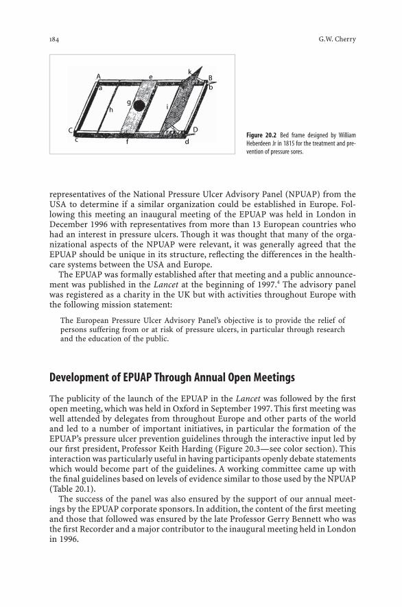
representatives of the National Pressure Ulcer Advisory Panel (NPUAP) from theUSA to determine if a similar organization could be established in Europe. Fol-lowing this meeting an inaugural meeting of the EPUAP was held in London inDecember 1996 with representatives from more than 13 European countries whohad an interest in pressure ulcers. Though it was thought that many of the orga-nizational aspects of the NPUAP were relevant, it was generally agreed that theEPUAP should be unique in its structure, reflecting the differences in the health-care systems between the USA and Europe.
The EPUAP was formally established after that meeting and a public announce-ment was published in the Lancet at the beginning of 1997.4 The advisory panelwas registered as a charity in the UK but with activities throughout Europe withthe following mission statement:
The European Pressure Ulcer Advisory Panel’s objective is to provide the relief ofpersons suffering from or at risk of pressure ulcers, in particular through researchand the education of the public.
Development of EPUAP Through Annual Open Meetings
The publicity of the launch of the EPUAP in the Lancet was followed by the firstopen meeting, which was held in Oxford in September 1997. This first meeting waswell attended by delegates from throughout Europe and other parts of the worldand led to a number of important initiatives, in particular the formation of theEPUAP’s pressure ulcer prevention guidelines through the interactive input led byour first president, Professor Keith Harding (Figure 20.3—see color section). Thisinteraction was particularly useful in having participants openly debate statementswhich would become part of the guidelines. A working committee came up withthe final guidelines based on levels of evidence similar to those used by the NPUAP(Table 20.1).
The success of the panel was also ensured by the support of our annual meet-ings by the EPUAP corporate sponsors. In addition, the content of the first meetingand those that followed was ensured by the late Professor Gerry Bennett who wasthe first Recorder and a major contributor to the inaugural meeting held in Londonin 1996.
184 G.W. Cherry
cC
f d
D
bB
keA
a
hg
i
Figure 20.2 Bed frame designed by WilliamHeberdeen Jr in 1815 for the treatment and pre-vention of pressure sores.
The second annual open meeting was held in Oxford with its theme “Learningfrom each other—the European experience.” Again this was well attended and amajor outcome was the publication of the EPUAP pressure ulcer treatment guide-lines (Figure 20.3—see color section). Ownership of both of the guideline docu-ments was established with the input from the delegates attending these meetings.
Recently nutritional guidelines for pressure ulcer prevention and treatment werepublished and presented at the 2nd World Union of Wound Healing SocietiesMeeting in Paris in July 2004. Again the formation of these guidelines followed thesame protocols as the prevention and treatment guidelines in their development(Figure 20.4—see color section).
The EPUAP guidelines have been translated into a number of European languages and more than 100,000 individual brochures have been distributed to clinicians throughout Europe. They are also available on the EPUAP website(www.epuap.org).
An early achievement of the EPUAP led by Professor Jeen Haalboom, our second president, was highlighted in another article published in the Lancetin 1998 on the need for establishing a uniform method in Europe to deal with the prevention and treatment of pressure ulcers.5 From the answers to question-naires sent to EPUAP representatives in different countries, he was able to high-light how information concerning such items as basic registration of pressureulcers and related aspects of management was lacking (Figure 20.5a and b—seecolor section).
Further annual open meetings were held in Amsterdam (the Netherlands), Pisa(Italy), Le Mans (France), Budapest (Hungary), Tampere (Finland), and Aberdeen(Scotland), each with a specific theme ranging from “Technology in the new mil-lennium” to “Pressure ulcers; back to basics—the fundamental principles.” Theannual meetings are a major educational opportunity and the Recorder and thescientific committee play an important role in the success of these events. DrMichael Clark has been Recorder since the Budapest meeting in 2002, where thetheme was “Pressure ulcers—a quality of care indicator.” At this meeting a majorfocus of the program was dedicated to work that the EPUAP itself had carried out,with the results of the Prevalence of Pressure Ulcers in Europe study being animportant aspect of the Budapest program.
Additional Education Activities
The EPUAP has been producing its own journal, the EPUAP Review, three timesper year, under the editorship of Dr Michael Clark published through the EPUAPBusiness Office (Figure 20.6—see color section). The Review has served as a forum
The European Pressure Ulcer Advisory Panel 185
Table 20.1. Levels of evidence
A Results of two or more randomized, controlled trials in pressure ulcers in humans
B Results of two or more controlled clinical trials in humans or, where appropriate, results in two or more controlled trials in ananimal model provide indirect support
C This rating requires one or more of the following:(1) results of one controlled trial(2) results of at least two case series/descriptive studies on pressure ulcers in humans, or(3) expert opinion
for debates on issues about pressure ulcers. One item that stimulated considerablediscussion was an article written by Professor Joe Barbenel and S. Hagisawa: “Thelimits of pressure sore prevention.”6 The Review is also published on our websitewww.epuap.org, which was established shortly after the Panel was established.
A major recent educational project has been carried out under the leadership ofProfessor Tom Defloor of the University of Ghent, Belgium, who produced thecomputer CD PUCLAS (pressure ulcer classification system), which is available innine languages (Figure 20.7a, b, and c—see color section).
Educational grants have been set up through contributions from industry, whichhave led to a number of studies including the major European pressure ulcer preva-lence study as well as the development of the nutritional guidelines.
In addition to ascertaining the extent of the problem of pressure ulcers through-out Europe, we thought that it would be important to determine the effectivenessand impact of our guidelines and a baseline of the problem was necessary. Theimportance of having such information has recently been emphasized in the workof the NPUAP in the USA, where shortly after the panel was established in 1989they set as a national goal to reduce the incidence of pressure ulcers by 50% by theyear 2000.7 This organization has recently published its progress in reaching thatgoal in “Pressure ulcers in America: Prevalence, incidence and implications for thefuture,” where they state that the major problem in assessing this goal has beeninconsistency in the initial figures presented on incidence and inconsistencies inmethodology. The EPUAP prevalence study was designed to reduce or eliminatethese obstacles in order to assess the problem as well as benefits from interven-tions that arise from the work of the EPUAP.
Education of the Public and Government
Education is a major part of our mission statement and educating the public as tothe extent of pressure ulcers and their cost to health budgets is extremely impor-tant. The role of litigation following pressure ulcers, although not as extensive asin the USA, is beginning to be publicized in European countries. This awarenesshas been further highlighted in popular television medical dramas where thedevelopment of pressure ulcers and their consequences have been emphasized.
The EPUAP has made a major effort in pointing out the problem of pressureulcers to government, though not to the same successful extent as has been donein the Netherlands where economic studies revealed that the costs of preventionand treatment of pressure ulcers approach those of cardiovascular disease andcancer.5 To make governments aware of the problem the EPUAP has written toMembers of Parliament in the UK as well as to the European Parliament empha-sizing the extent and cost of pressure ulcers.
Summary
The European Pressure Ulcer Advisory Panel (EPUAP) is an example of how a ded-icated organization from a number of countries can approach a major chronicwound healing problem such as pressure ulcers through education, research, anddissemination through a cooperative program.
186 G.W. Cherry

References
1. Romanelli M. Pressure ulcer prevention and management in Italy. In: Cherry GW (ed) Concepts inpressure relief – importance in the prevention and treatment of pressure ulcers. Oxford: PositifPress; 2004: 18–19.
2. Bennett G, Dealey C, Posnett J. The cost of pressure ulcers in the UK. Age Ageing 2004; 33(3):230–235.
3. Bedsores over the centuries. In: Parish LC, Witkowski JA, Crissey JT (eds) The decubitus ulcer inclinical practice. Berlin, Heidelberg: Springer-Verlag; 1997: 3–8.
4. EPUAP. European Pressure Ulcer Panel launched. Lancet 1997; 349:262.5. Haalboom JRS. Pressure ulcers. Lancet 1998; 352(15 August): 581.6. Barbenel J, Hagisawa S. The limits of pressure sore prevention. J R Soc Med 1999; 92:576–578;
reviewed in the EPUAP Review 2000; 2(2):43–44.7. National Pressure Ulcer Panel. Cuddingan J,Ayello EA, Sussman C (eds) Pressure ulcers in America:
Prevalence, incidence and implications for the future. Reston, VA: NPUAP; 2001.8. Clark M, Bours G, Defloor T. The prevalence of pressure ulcers. in Europe. Hospital Decisions
2003/2004; Winter: 123–129.
The European Pressure Ulcer Advisory Panel 187
21 Pressure Ulcer Prevention and Management in the Developing World: The Developed World Must Provide LeadershipTerence J. Ryan
This is not a review of pressure ulcers in the developing world because there areinsufficient data to write one. The pressure ulcers that I have seen there, in the ter-minally ill and in the paraplegic, mostly come for treatment after great delay andthe patients are very sick. There is a case of need and the solutions must come inpart from the developed world and none better than the US National PressureUlcer Advisory Panel and the European Pressure Ulcer Advisory Panel.
The pathogenesis of the pressure ulcer is no different in the developing worldfrom that in the developed world. It is due to pressure and occurs in the sick. Thereare two major subgroups: those who are systemically sick who depend on carersto manage their predisposition to develop pressure sores and to heal them, and asecond group such as the paraplegic who with self-help can both prevent and healtheir pressure sores.
It was Ludwig Guttman who with passion persuaded the world that the para-plegic need not develop pressure sores. He came to Oxford during the SecondWorld War as a refugee and, in a third world setting of the country wartime hos-pital the Radcliffe Infirmary at a time of great deprivation, he worked on severelyinjured soldiers and civilians. Nearby in the equally third world conditions of awonderfully antique Radcliffe Observatory, the Spanish surgeon Joseph Truetaworked on crush injury. Two decades later I collaborated with Trueta on an exhi-bition on blood supply of skin and bone and I was a registrar at Stoke Mandevillewhere Ludwig Guttman proved that pressures sores were preventable andrepairable. This experience did not employ high technology. It was very basic.
The prevention of pressure sores begins with detailed attention to preventingthe impairment of blood supply due to pressure and includes maneuvers at thescene of an accident such as removing solid objects like car keys from a hip pocketupon which the patient is lying and, on the ward, nurses turning the patient. AtStoke Mandeville we healed large pressure sores with low-cost wet dressings andfrequent turns. We had none of the water or air-filled beds now used in the devel-oped world and our usual wetting agent was the now much maligned Eusol andparaffin.
One of the most celebrated sufferers from paraplegia of recent times wasChristopher Reeve, or one time “Superman.” He did much for the advocacy of “bestpractice.” Interestingly he was especially an advocate for research into stem cellswith which to repair the spinal cord injury.1 This is high technology and it is both
189

costly and questionable whether it will ever be affordable in the developing world.It illustrates a dilemma. How much attention should be given to costly researchwhich may benefit only an elite and wealthy few? Where safety is unproven howmuch should the advice of the scientist or the marketing needs of the pharma-ceutical and devices industry be allowed to throw doubt on cheap solutions? Thereare many examples in the field of pressure sore management and they includeEusol, gentian violet, honey, and maggot therapy, none of which would be avail-able if their critics had been listened to.
The European Pressure Ulcer Advisory Panel (EPUAP)
The story since 1995 of the progress of the EPUAP is described2 as first asking thequestion “what are we talking about?” It led to the discussions on definitions andclassifications. Then there were the discussions on the size of the problem and on“what does it cost?” A great service was done by sorting out incidence from preva-lence. The next stage was the registration of all known preventive and therapeuticinterventions as well as the level of evidence for their effectiveness. The Panel thenapplied “Marks of Quality” to devices.3 The final steps were the production of edu-cational material and the identification of the army of workers that should readthem.
Under the heading of Health Services Research and terms such as “access” onecan debate whether investment in increasing the knowledge of the carer in thedeveloping world should be at the level of postgraduate training of doctors or ofallied health professionals or at the level of family members of the patient. Myexperience has been that the greatest disappointments come from training doctorsin the developing world, especially if they are male. They emigrate and they takeup full-time private practice, where there is no public health ethos. If while over-seas they are any good at research they do not return to their country of origin.
There is thus the need for a further stage of development for organizations suchas the EPUAP and that is applying “Quality Marks” to the carer. It should be basedon availability, commitment, cost, and their belief in the possibility of preventionand healing of pressure ulcers. Barbenel and Hagisawa4 alarmed the EPUAP bysubstituting realism for a range of views from pessimism to optimism. They sug-gested that not all ulcers are preventable. What has not been measured is whethermore ulcers are healed when the carer expects them to heal.
Aging
It is often said that our increasing life expectancy will create an aged populationwithout the young carers to look after them. By the year 2030 half the populationof Europe will be over 30 years of age and will expect to live a further 40 years. Inthe UK there will be approaching a quarter of a million people over 100 years ofage. At the same time declining fertility will reduce the young population.
Recently it has been argued5 that this aged population is not only fitter but ismore capable of doing a carer’s job perhaps even into their nineties. Issues of accessrequire that this population is helped to be more effective and to be more knowl-edgeable. Early retirement at around age 50 should be followed by encouragementto take on community projects for at least 20 years.
190 T.J. Ryan
In the meantime the AIDS epidemic in the developing world is reducing lifeexpectancy and increasing the number of chronic sick with heightened vulnera-bility to pressure ulcers.
The Family
There is some debate whether in the twenty-first century the family is more dys-functional than before. Especially in urban developments the older members ofthe family cannot expect their young to give up their employment to look afterthem unpaid. Others5 argue that there is preservation of and maybe an increase ina sense of responsibility for care of elderly relatives. Increasingly the bond withthe elderly is strengthened by the greater role they play in managing the dailydemands of a family out at work when the children are out of school. It is povertythat is the main concern because the elderly need financial support for themselvesand it is common for all their dwindling resources to be spent on the needs of anextended family, perhaps orphaned as a result of AIDS.
A threat to rational management is the discovery by families and their lawyersthat there is money to be made by suing hospitals for allowing pressure ulcers tohappen in one of their relatives. A complaint that enriches a lawyer is far less desir-able than one that improves the health service. Scott6 stated that some lessons aretoo big to learn. It takes a Ludwig Guttman or perhaps an EPUAP to give the masterclass that breaks bad habits, and also demonstrates that errors are a consequenceof problems not their cause.
Information Technology
Information technology is one way forward but writing in the sand may requireless backup.
During the past decade I have visited on several occasions villages nearPondicherry in southern India, where there is great poverty and illiteracy. Anexperiment led by the Swaminathan Foundation supported by the Canadian gov-ernment has provided internet to these villages. Information comes to them intheir own language and important messages are vocalized.A majority of the villageinhabitants become computer literate and many of the women retrieve healthinformation. Such experiments are being undertaken in many other parts of thedeveloping world.
At conferences devoted to pressure ulcers no one doubts that knowledge existsbut a familiar cry is that it is not getting to where it is needed. Sharing knowledgehas never been easier. The expert patient is now a potential replacement for theapathetic one. One area of knowledge that has not been widely disseminated is thatof nutrition, which is well described in the literature on pressure ulcers in thedeveloped world. This needs rewriting for the developing world, where even in hos-pital the patient’s food is provided unsupervised by the family away from thecheaper resources of their home support. This requires someone to understand thefamily background before giving advice. Thus for the developing world “povertyalleviation” has to be to the fore. Often the visitor to healthcare projects in thedeveloping world will be first shown the private hospitals of a not insubstantialmiddle class. But even their best practice and gold standards are replaced by silver
Pressure Ulcer Prevention and Management in the Developing World 191
plate. It must be the role of leaders such as the EPUAP to become partners withthe poor. “Macroeconomics and Health”7 demand that such an organization rep-resenting best practice in the developed world should set aside time to boost theirown reputation and finance by providing good advice to donors and governanceglobally and not just to their own parish. It will be advice in a language of goldstandards, “evidence based medicine,” “low technology transfer,” and “cost effec-tiveness” for much needed National Commissions. It must make sure that thesupport given to the middle class raises the standard of care given to the poor andat the same time the poor are real beneficiaries of their intervention.
Rewards and Awards
Where the patient is too sick to play a leading role in self-help there is a greatproblem of the need for and provision of a devoted carer. Such people need respiteand rewards. A fraction of the costs of wound healing research directed at exam-ining ways to encourage care would go a long way to improving the wellbeing ofthe chronically sick.
At the top of the pyramid, a National Commission will focus on the leadingmedical and nursing schools in tertiary hospitals. The focus should not be onpathogenesis alone, but should have education of lower tiers in the pyramid as amain objective. At the lowest level encouragement of the carer and of self-helprequires more than knowledge of wound healing. It requires especially experiencein leadership and rewards and awards. These will be influenced by communityneeds, poverty, and cultural differences.
The developing world is most unlikely to generate the knowledge it needs byitself. The remarkable achievements of the EPUAP in its first five years could nothave happened in the developing world but they could now be copied there; butthe copy will need to be culturally sensitive.
Skin Assessment
Pressure ulcers are a skin problem and skin observation and assessment is centralto diagnosis and intervention. Ulcers are a huge economic burden that shouldjustify the focus of the custodians of the skin namely the dermatologist. Duringthe last few decades dermatology departments have created wound healing units,such as that from which this chapter is written, journals, and societies. The major-ity of dermatologists have, however, steered clear of being expert in the manage-ment of pressure ulcers. Hopefully the concept of the wound healing team willallow the dermatologist to select a field of interest and share, most likely with askin-care nursing profession, areas that are a weighty burden. So far the leadingpolicy-makers such as the World Health Organization or major funding bodiessuch as the Melinda and Bill Gates Foundation have not perceived skin care as apriority. Sadly the majority of dermatologists see no role for themselves in thisrespect. At a recent international workshop on the Intensified Control of NeglectedDiseases,8 “skin failure” was not on the agenda. In due course maybe the impor-tance of absent skin and ulceration will be recognized. Furthermore “the woundhealers” will have promoted models of the prevention of non-communicabledisease that demonstrate the importance of examining the skin. They will also have
192 T.J. Ryan

promoted models that organize the patient within the context of a team thatincludes the community. This is something demanding leadership and the kind oforganization demonstrated by the European Pressure Ulcer Advisory Panel. Thisis knowledge that has to be taken into the “general health services” and not just toa few leading tertiary hospitals in the developing world. It is appropriate for it tobe transported there with other knowledge of the skin such as common infections,wounds and burns, leprosy, lymphatic filariasis, and several other conditions allrequiring the same basic interventions of skin care when first seen in a rural healthcenter.
Looking at the skin and seeing the subtleties of what is there needs a new cur-riculum that is less dermatology orientated. What is required is less naming of pat-terns with Latin names but a greater skill at recognizing poor health and early skinfailure and potential vulnerability. We must teach the examination of the skin forloss of barrier function, failure of reflex hyperemia, or early sensory loss, so earlysigns of skin failure are picked up as well as most physicians recognize heartfailure.
The largest healthcare profession is nursing and pressure ulcers are given promi-nence in their practice. The professions involved in early diagnosis of skin failurein disorders such as leprosy, the diabetic foot, or lymphedema are not small. Untilrecently dermatologists have not been invited to be writers of their guidelines,because they have not been interested. Fortunately this is now changing. No onecaring for the skin can ignore the developing knowledge base of dermatology. For-tunately the concept of team management and collaboration is encouraging thisknowledge base to be a contributor to all aspects of the management of skin failure.The development of a skin-care nursing profession points the way to a betterfuture.
The International Foundation for Dermatology
Directing attention from the range of interests usually shown by the urban-basedprivate practitioner to a broader curriculum that includes skin care where it isneeded in the general health services has been the focus of the International Foun-dation for Dermatology. It has made a point of including wounds and burns in itsprogram because these are skin problems commonly presenting at a health center.This kind of dermatology also embraces sexually transmitted disease. The empiresbuilt for dermatology on syphilis in the nineteenth century are now potentiallybeing re-established mostly by other branches of medicine, but with a focus onHIV/AIDS. The recognition of physical signs in the skin embracing the early signsof AIDS and much else besides, such as tropical diseases like leprosy, leishmania-sis, onchocerciasis, the lymphedema of lymphatic filariasis, and of course woundsand burns. In taking the management of these into the general health services oneis aiming to teach skin signs to the allied health professionals that manage healthcenters. It is appropriate that in looking at the skin they should recognize the signspresaging pressure ulcers. Furthermore if they are to manage ulcers due to venousdisease or diabetes or leprosy it is appropriate that they should also manage pres-sure ulcers.
Increasingly the AIDS epidemic is a prevailing influence. It provides the sick but also inhibits healing. In one study9 of skin grafting in burns, healing in HIVpatients was 22% compared to 69% in non-HIV patients. In the developing world
Pressure Ulcer Prevention and Management in the Developing World 193
effective management is so costly that those affected return to traditional healthsystems and mostly these do not fail to give welcome support and a degree of effec-tiveness unrealized in the developed world. Of course there is quackery and thereare roadside sellers with no expertise. There are also traditional healers or practi-tioners of Chinese medicine or Indian systems of medicine who are expert. Breath-ing, posture or movement, so effective for lymphedema, are better understood byAsian systems of medicine than by practitioners of biomedicine. One should notforget when examining the prescription of herbal medicines that plants developedbefore the animal kingdom, and many have antiseptic, antioxidant, and antihista-mine properties, and much else besides.
It is in terminal illness that the management of pressure ulcers poses difficultquestions, since in a dying patient with falling blood perfusion pressure, death oftissue may be unavoidable. Expectations of healing may be irrelevant and woundbed preparation unacceptably invasive. Pain relief becomes a priority. As withanyone so sick, issues of hydration and nutrition require fine judgment.
Getting Management of Pressure Ulcers into the Curriculum
At all levels of the health service the curriculum is overloaded. The medical studentwho must become a safe doctor and the nurse who may have to survive overnightsolely responsible for an intensive care unit have to know an enormous amount.They are also increasingly under observation in case mistakes made are worthy of litigation. Every branch of medicine will have advocates for including their interests in the curriculum. How to decide on priorities for each discipline isincreasingly difficult, as more and more knowledge becomes available to us.The prevalence and cost of pressure sores is such that a strong case can be madefor ensuring that it is understood by every doctor and nurse and indeed that it becomes public knowledge under the heading of public health.
Fifteen years ago there were 25 countries in Africa without anyone to advise gov-ernment or universities about skin priorities. Since then at the Regional Derma-tology Training Centre in Tanzania 120 allied health professionals have undergone2 years of additional training about the skin. With a university qualification theyhave enlarged the knowledge base of 14 countries. Recently in Mali, and previouslyin Guatemala, the International Foundation for Dermatology focused on one-daycourses for nurses. The curriculum was determined by the case of need. Taking thecommonest diseases and observing how misdiagnosis would lead to wrong,ineffective, and costly prescribing, it was not difficult to establish priorities. It wasimportant to eliminate the roadside seller but it was equally important to examinehigh utilization of traditional medicine. Such utilization is of public healthsignificance and should not be ignored in training a doctor or nurse. It must bemade safe, but its efficacy, sustainability, local availability, and low cost must notbe undervalued. Wherever there is a break in the surface continuity of skin, tradi-tional therapy will have a role to play and agents such as honey or larvae therapymust not be downplayed.
Recently in India I have helped to initiate three programs. India is a nationwithout a substantial body of nurses interested in the skin. In January 2004 a new“Skin-Care Nursing Group” was inaugurated at the annual Indian DermatologyCongress in Bombay. There have been a number of wound healing programs alsoinaugurated in India and in neighboring Sri Lanka. So far these have been mostly
194 T.J. Ryan

for the benefit of a substantial but minority middle class and their private hospi-tals. It has to be said that if best practice exists anywhere, it may raise standardsand filter through to the majority.
India is a nation with large government-supported “Indian Systems of Medi-cine.” They are of especial importance for the rural poor. Taking lymphatic filari-asis as a condition highly prevalent in rural areas, I have initiated in Kerala aprogram of management supported by the Cochrane body in the UK and by inter-national ethics foundations.
Recently it has become evident that China has developed its cities at the expenseof the rural peasant. A plan to retrain 5 million “village doctors” is being exam-ined. Their curriculum must be worked on. Maybe here too the EPUAP could giveadvice. It is my hope that a younger generation of dermatologists will perceive theirrole as part of a team sharing responsibility for skin failure and that their orien-tation will include poverty alleviation. Such a focus will identify pressure ulcers asa high priority.
References
1. Cherry GW. Letter from the editor. Eur Tissue Repair Soc Bull 2004; 11:48.2. Haalboom JRE. Pressure ulcer management in Europe. The Oxford wound healing course hand-
book. Oxford Wound Healing Institute; 2002.3. Haalboom JRE. Quality marks, a European dream. EPUAP Review 2002; 4:6–7.4. Barbenel JC, Hagisawa S. The limits of pressure sore prevention. J R Soc Med 1999; 92:576–578.5. Harper S. Ageing society. Oxford Today 2004; 16–18.6. Scott H. Education-sharing our experiences. Can complaint change culture? EPUAP Review 2001;
3:90–92.7. Morrow RH. Macroeconomics and health. BMJ 2002; 325:53–55.8. International control of neglected diseases. Report of an International Workshop, Berlin, Decem-
ber 2003 (further information from: [email protected]).9. Mzezewa S. Burns in Zimbabwe. Epidemiology, immunosuppression, infection and surgical man-
agement. Doctoral dissertation. Department of Plastic Surgery, Malmo University Hospital,Sweden, 2003.
10. Ryan TJ. Pressure sores: prevention management and future research–-a medical perspective.Palliat Med 1989; 3:249–525.
Pressure Ulcer Prevention and Management in the Developing World 195

22 Innovation in Pressure Ulcer Prevention and ManagementKeith G. Harding and Michael Clark
Introduction
For many practitioners achieving successful pressure ulcer prevention and man-agement has long been regarded as a straightforward task albeit one that is oftennot achieved. This view was succinctly described in the first paper presented at thefirst UK pressure ulcer conference back in 1975 when Roaf1 commented that “weknow how to avoid bed sores and tissue necrosis—maintain the circulation, avoidlong continued pressure, abrasions, extremes of heat and cold, maintain afavourable micro-climate, avoid irritating fluids and infection. The problem is thelogistics of this programme.” So now, thirty years after this seminal meeting onpressure ulcers, does such a statement still hold true and have we really achievedsignificant innovations in our research and practice which have helped resolve thelogistical challenges in service delivery? There are four key dimensions where pres-sure ulcer innovations might be encountered—in clinical practice, research, theorganization and logistics of service delivery, and finally in society’s views on thesignificance of pressure ulceration. Each dimension clearly overlaps with its neigh-bors but will be treated separately in this chapter both to tease out advances andto identify the challenges that remain.
Innovations in Clinical Practice?
In the early 1980s pressure ulcer management differed significantly from today.There were no specialist nursing positions, with the first wave of UK tissue viabil-ity nurses appointed in 1986–1987; by 1992, 23 tissue viability nurse specialistswere in post;2 today the precise number is unclear but is likely to be over 400. Thisexplosion in the number of specialist nursing posts has created a demand for tar-geted education in tissue viability to enable the new cadres to be competent prac-titioners with the specific competencies having been defined in recent years.3 Suchcourses extend from informal company-delivered seminars through to formal aca-demic training such as the MSc in Wound Healing and Tissue Repair organized by the Wound Healing Research Unit. For medical colleagues, there remains ascarcity of education (and interest) in pressure ulcer prevention and treatment.Bennett4 surveyed the wound care training delivered by the 27 medical schools inthe UK; of the 19 respondents only 13 delivered any formal teaching on pressure
197

ulcer management; and of these schools pressure ulcer preventive care was coveredby 10, the use of wound dressings by 10, and pressure-redistributing devices by 8, with on average 6 hours of content delivered (range 0 to 35.5 hours). If such a survey were to be repeated today across Europe would training of medical practitioners in pressure ulcer prevention and management be commonplace orwould this remain a topic that relatively few medical schools would consider to berelevant?
Not only have the numbers and training of health professionals changed overthe past twenty years, the use of resources has evolved significantly. For example,in 1983 David and colleagues5 surveyed the care received by 961 patients with pres-sure ulcers across 132 hospitals in England and Wales. The vast majority (n = 599;62.3%) were nursed upon standard hospital mattresses. Since 1983 the diffusion ofpressure-redistributing mattresses within healthcare has significantly expanded—in 2001 the European Pressure Ulcer Advisory Panel (EPUAP) completed a pilotsurvey of pressure ulcer prevalence across 26 hospitals located in five Europeancountries.6 Of the 5947 patients included in the pilot survey; 52.1% (n = 3099) hadbeen allocated a pressure-redistributing mattress. The expanded use of pressure-redistributing beds and mattresses was particularly marked within some coun-tries; for example, in the UK only 53 patients considered to be vulnerable topressure ulcer development were nursed on standard mattresses, with the use of such devices extended into the patient population considered to be at minimalrisk of pressure ulcers (1234 (84.3%) of 1464 patients not at risk were allocated apressure-redistributing mattress).
These trends towards enhanced diffusion of interventions have recently beenaugmented by the introduction of partnerships between equipment suppliers andthe health service leading to the initiation of total bed management (TBM) wherea supplier is selected to supply all pressure-redistributing surfaces for a healthcareprovider usually within a limited defined budget. These schemes offer an exampleof true partnership between the commercial sector and the health service and itis probable that one future trend will be the spread of such schemes across theexpanded European Union. However, there remains a lack of evidence that any ofthese structural and process changes in the prevention and management of pres-sure ulcers have helped to reduce the size of the problem. This point will be con-sidered in depth when reviewing innovation at the organizational and societallevels.
What about Innovation in Research?
It is without question that pressure ulcer research lies at the unglamorous end ofthe research spectrum—attracting little attention from funding bodies and careerscientists alike. Much of the available research on pressure ulcers has benefitedfrom commercial support and without this source of income pressure ulcer studieswould be relatively scarce. While commercial support has been important and willprobably continue to be the largest single sponsor of pressure ulcer studies, theneed to obtain data in a timescale attractive to industrial sponsors has limitedmany studies—studies are frequently underpowered with little long-term follow-up of subjects. These weaknesses have been compounded by the common beliefthat it is somehow impossible to conduct blinded pressure ulcer interventionstudies. This belief often relates to the investigation of devices such as beds and
198 K.G. Harding and M. Clark
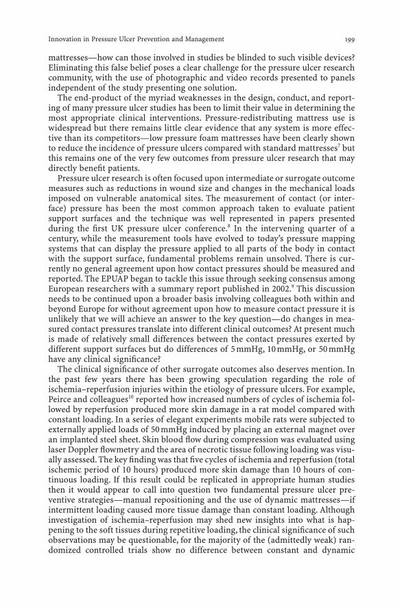
mattresses—how can those involved in studies be blinded to such visible devices?Eliminating this false belief poses a clear challenge for the pressure ulcer researchcommunity, with the use of photographic and video records presented to panelsindependent of the study presenting one solution.
The end-product of the myriad weaknesses in the design, conduct, and report-ing of many pressure ulcer studies has been to limit their value in determining themost appropriate clinical interventions. Pressure-redistributing mattress use iswidespread but there remains little clear evidence that any system is more effec-tive than its competitors—low pressure foam mattresses have been clearly shownto reduce the incidence of pressure ulcers compared with standard mattresses7 butthis remains one of the very few outcomes from pressure ulcer research that maydirectly benefit patients.
Pressure ulcer research is often focused upon intermediate or surrogate outcomemeasures such as reductions in wound size and changes in the mechanical loadsimposed on vulnerable anatomical sites. The measurement of contact (or inter-face) pressure has been the most common approach taken to evaluate patientsupport surfaces and the technique was well represented in papers presentedduring the first UK pressure ulcer conference.8 In the intervening quarter of acentury, while the measurement tools have evolved to today’s pressure mappingsystems that can display the pressure applied to all parts of the body in contactwith the support surface, fundamental problems remain unsolved. There is cur-rently no general agreement upon how contact pressures should be measured andreported. The EPUAP began to tackle this issue through seeking consensus amongEuropean researchers with a summary report published in 2002.9 This discussionneeds to be continued upon a broader basis involving colleagues both within andbeyond Europe for without agreement upon how to measure contact pressure it isunlikely that we will achieve an answer to the key question—do changes in mea-sured contact pressures translate into different clinical outcomes? At present muchis made of relatively small differences between the contact pressures exerted bydifferent support surfaces but do differences of 5 mmHg, 10 mmHg, or 50 mmHghave any clinical significance?
The clinical significance of other surrogate outcomes also deserves mention. Inthe past few years there has been growing speculation regarding the role ofischemia–reperfusion injuries within the etiology of pressure ulcers. For example,Peirce and colleagues10 reported how increased numbers of cycles of ischemia fol-lowed by reperfusion produced more skin damage in a rat model compared withconstant loading. In a series of elegant experiments mobile rats were subjected toexternally applied loads of 50 mmHg induced by placing an external magnet overan implanted steel sheet. Skin blood flow during compression was evaluated usinglaser Doppler flowmetry and the area of necrotic tissue following loading was visu-ally assessed. The key finding was that five cycles of ischemia and reperfusion (totalischemic period of 10 hours) produced more skin damage than 10 hours of con-tinuous loading. If this result could be replicated in appropriate human studiesthen it would appear to call into question two fundamental pressure ulcer pre-ventive strategies—manual repositioning and the use of dynamic mattresses—ifintermittent loading caused more tissue damage than constant loading. Althoughinvestigation of ischemia–reperfusion may shed new insights into what is hap-pening to the soft tissues during repetitive loading, the clinical significance of suchobservations may be questionable, for the majority of the (admittedly weak) ran-domized controlled trials show no difference between constant and dynamic
Innovation in Pressure Ulcer Prevention and Management 199
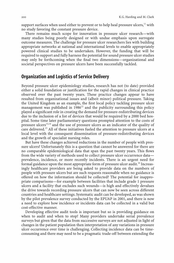
support surfaces when used either to prevent or to help heal pressure ulcers,11 withno study favoring the constant pressure device.
There remains much scope for innovation in pressure ulcer research—withmany studies being poorly designed or with undue emphasis upon surrogateoutcome measures. The challenge for pressure ulcer researchers lies with buildingappropriate networks at national and international levels to enable appropriatelypowered clinical studies to be undertaken. However, the funding that will berequired to support and fully harness the potential for sound pressure ulcer studiesmay only be forthcoming when the final two dimensions—organizational andsocietal perspectives on pressure ulcers have been successfully tackled.
Organization and Logistics of Service Delivery
Beyond pressure ulcer epidemiology studies, research has not (to date) providedeither a solid foundation or justification for the rapid changes in clinical practiceobserved over the past twenty years. These practice changes appear to haveresulted from organizational issues and (albeit minor) political pressure. Takingthe United Kingdom as an example, the first local policy tackling pressure ulcermanagement was published in 198612 and the publicity surrounding this policyplayed a significant role in creating the demand for pressure-redistributing devicesdue to the inclusion of a list of devices that would be required by a 2000 bed hos-pital. Some time later parliamentary questions prompted attention to the costs ofpressure ulcers13,14 and the use of pressure ulcers as an indicator of the quality ofcare delivered.15 All of these initiatives fueled the attention to pressure ulcers at alocal level with the consequent dissemination of pressure-redistributing devicesand the growth of specialist nursing roles.
But have these changes achieved reductions in the number of people with pres-sure ulcers? Unfortunately this is a question that cannot be answered for there areno comparable epidemiological data that span the past twenty years. This flowsfrom the wide variety of methods used to collect pressure ulcer occurrence data—prevalence, incidence, or more recently incidents. There is an urgent need forformal guidance upon the most appropriate form of pressure ulcer audit.16 Increas-ingly healthcare providers are being asked to provide data on the numbers ofpeople with pressure ulcers but are such requests reasonable when no guidance isoffered on how the information should be collected? The potential for inappro-priate comparisons—for example between facilities that include grade 1 pressureulcers and a facility that excludes such wounds—is high and effectively devaluesthe drive towards recording pressure ulcers that can now be seen across differentcountries and healthcare settings. Systematic audit can be developed, as was shownby the pilot prevalence survey conducted by the EPUAP in 2001, and there is nowa need to explore how incidence or incidents data can be collected in a valid butcost-effective manner.
Developing effective audit tools is important but so is providing guidance onwhen to audit and when to stop! Many providers undertake serial prevalencesurveys but given that the data from successive surveys are not adjusted in light ofchanges in the patient population then interpretation of any variations in pressureulcer occurrence over time is challenging. Collecting incidence data can be time-consuming and there may need to be a pragmatic trade-off between extending the
200 K.G. Harding and M. Clark
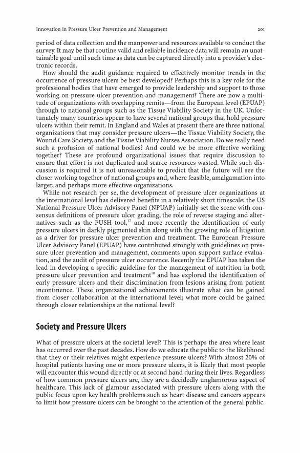
period of data collection and the manpower and resources available to conduct thesurvey. It may be that routine valid and reliable incidence data will remain an unat-tainable goal until such time as data can be captured directly into a provider’s elec-tronic records.
How should the audit guidance required to effectively monitor trends in theoccurrence of pressure ulcers be best developed? Perhaps this is a key role for theprofessional bodies that have emerged to provide leadership and support to thoseworking on pressure ulcer prevention and management? There are now a multi-tude of organizations with overlapping remits—from the European level (EPUAP)through to national groups such as the Tissue Viability Society in the UK. Unfor-tunately many countries appear to have several national groups that hold pressureulcers within their remit. In England and Wales at present there are three nationalorganizations that may consider pressure ulcers—the Tissue Viability Society, theWound Care Society, and the Tissue Viability Nurses Association. Do we really needsuch a profusion of national bodies? And could we be more effective workingtogether? These are profound organizational issues that require discussion toensure that effort is not duplicated and scarce resources wasted. While such dis-cussion is required it is not unreasonable to predict that the future will see thecloser working together of national groups and, where feasible, amalgamation intolarger, and perhaps more effective organizations.
While not research per se, the development of pressure ulcer organizations atthe international level has delivered benefits in a relatively short timescale; the USNational Pressure Ulcer Advisory Panel (NPUAP) initially set the scene with con-sensus definitions of pressure ulcer grading, the role of reverse staging and alter-natives such as the PUSH tool,17 and more recently the identification of earlypressure ulcers in darkly pigmented skin along with the growing role of litigationas a driver for pressure ulcer prevention and treatment. The European PressureUlcer Advisory Panel (EPUAP) have contributed strongly with guidelines on pres-sure ulcer prevention and management, comments upon support surface evalua-tion, and the audit of pressure ulcer occurrence. Recently the EPUAP has taken thelead in developing a specific guideline for the management of nutrition in bothpressure ulcer prevention and treatment18 and has explored the identification ofearly pressure ulcers and their discrimination from lesions arising from patientincontinence. These organizational achievements illustrate what can be gainedfrom closer collaboration at the international level; what more could be gainedthrough closer relationships at the national level?
Society and Pressure Ulcers
What of pressure ulcers at the societal level? This is perhaps the area where leasthas occurred over the past decades. How do we educate the public to the likelihoodthat they or their relatives might experience pressure ulcers? With almost 20% ofhospital patients having one or more pressure ulcers, it is likely that most peoplewill encounter this wound directly or at second hand during their lives. Regardlessof how common pressure ulcers are, they are a decidedly unglamorous aspect ofhealthcare. This lack of glamour associated with pressure ulcers along with thepublic focus upon key health problems such as heart disease and cancers appearsto limit how pressure ulcers can be brought to the attention of the general public.
Innovation in Pressure Ulcer Prevention and Management 201
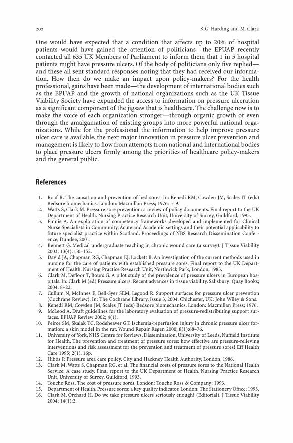
One would have expected that a condition that affects up to 20% of hospitalpatients would have gained the attention of politicians—the EPUAP recently contacted all 635 UK Members of Parliament to inform them that 1 in 5 hospitalpatients might have pressure ulcers. Of the body of politicians only five replied—and these all sent standard responses noting that they had received our informa-tion. How then do we make an impact upon policy-makers? For the healthprofessional, gains have been made—the development of international bodies suchas the EPUAP and the growth of national organizations such as the UK Tissue Viability Society have expanded the access to information on pressure ulcerationas a significant component of the jigsaw that is healthcare. The challenge now is tomake the voice of each organization stronger—through organic growth or eventhrough the amalgamation of existing groups into more powerful national orga-nizations. While for the professional the information to help improve pressureulcer care is available, the next major innovation in pressure ulcer prevention andmanagement is likely to flow from attempts from national and international bodiesto place pressure ulcers firmly among the priorities of healthcare policy-makersand the general public.
References
1. Roaf R. The causation and prevention of bed sores. In: Kenedi RM, Cowden JM, Scales JT (eds)Bedsore biomechanics. London: Macmillan Press; 1976: 5–9.
2. Watts S, Clark M. Pressure sore prevention: a review of policy documents. Final report to the UKDepartment of Health. Nursing Practice Research Unit, University of Surrey, Guildford, 1993.
3. Finnie A. An exploration of competency frameworks developed and implemented for ClinicalNurse Specialists in Community, Acute and Academic settings and their potential applicability tofuture specialist practice within Scotland. Proceedings of NBS Research Dissemination Confer-ence, Dundee, 2001.
4. Bennett G. Medical undergraduate teaching in chronic wound care (a survey). J Tissue Viability2003; 13(4):150–152.
5. David JA, Chapman RG, Chapman EJ, Lockett B. An investigation of the current methods used innursing for the care of patients with established pressure sores. Final report to the UK Depart-ment of Health. Nursing Practice Research Unit, Northwick Park, London, 1983.
6. Clark M, Defloor T, Bours G. A pilot study of the prevalence of pressure ulcers in European hos-pitals. In: Clark M (ed) Pressure ulcers: Recent advances in tissue viability. Salisbury: Quay Books;2004: 8–22.
7. Cullum N, McInnes E, Bell-Syer SEM, Legood R. Support surfaces for pressure ulcer prevention(Cochrane Review). In: The Cochrane Library, Issue 3, 2004. Chichester, UK: John Wiley & Sons.
8. Kenedi RM, Cowden JM, Scales JT (eds) Bedsore biomechanics. London: Macmillan Press; 1976.9. McLeod A. Draft guidelines for the laboratory evaluation of pressure-redistributing support sur-
faces. EPUAP Review 2002; 4(1).10. Peirce SM, Skalak TC, Rodeheaver GT. Ischemia-reperfusion injury in chronic pressure ulcer for-
mation: a skin model in the rat. Wound Repair Regen 2000; 8(1):68–76.11. University of York, NHS Centre for Reviews, Dissemination, University of Leeds, Nuffield Institute
for Health. The prevention and treatment of pressure sores: how effective are pressure-relievinginterventions and risk assessment for the prevention and treatment of pressure sores? Eff HealthCare 1995; 2(1). 16p.
12. Hibbs P. Pressure area care policy. City and Hackney Health Authority, London, 1986.13. Clark M, Watts S, Chapman RG, et al. The financial costs of pressure sores to the National Health
Service: A case study. Final report to the UK Department of Health. Nursing Practice ResearchUnit, University of Surrey, Guildford, 1993.
14. Touche Ross. The cost of pressure sores. London: Touche Ross & Company; 1993.15. Department of Health. Pressure sores: a key quality indicator. London: The Stationery Office; 1993.16. Clark M, Orchard H. Do we take pressure ulcers seriously enough? (Editorial). J Tissue Viability
2004; 14(1):2.
202 K.G. Harding and M. Clark

17. Stotts NA, Rodeheaver GT, Thomas DR, et al. An instrument to measure healing in pressure ulcers:development and validation of the pressure ulcer scale for healing (PUSH). J Gerontol A Biol SciMed Sci 2001; 56(12):N797–N799.
18. Clark M, Schols JMGA, Benati G, et al. Pressure ulcers and nutrition: a new European guideline.J Wound Care 2004; 13(7):267–274.
Innovation in Pressure Ulcer Prevention and Management 203

Acetic acid, 143Acid mantle, 96Acinetobacter calcoaceticus, 151Acticoat, 156Actisorb, 156Africa, 194Agency for Health Care Policy and Research,
USA (AHCPR), 169Aging, 190–191
of skin, 78AGREE. See Appraisal of Guidelines for Research
and EvaluationAHCPR. See Agency for Health Care Policy and
Research, USAAIDS, 191, 193Albumin, 32–33, 101Alcohol hand rinses, 154Alginate, 135Allogeneic constructs. See Bioengineered skinAlternating pressure mattress, 7, 8Antibiotics, 111Antibody labeling scanning, 153Antimicrobials, 155Antioxidant vitamins, 85, 87Antiseptics, 142–143
alcohols and, 142biguanides and, 142dressings and, 135for enzymatic debridement, 134halogen compounds and, 142iodine and, 143organic acids and, 143peroxides and, 143tinctures and, 143
Apligraf®, 115Appraisal of Guidelines for Research and
Evaluation (AGREE), 174Aquacel-AG, 156Arginine, 85, 87Arglaes, 156Autolytic debridement, 134–135
alginate dressings and, 135cellulose dressings and, 135hydrocolloids and, 134hydrogels and, 134occlusive dressings and, 135
polyacrylates and, 135Ringer’s solution and, 135
Bacitracin zinc, 155Bacteremia, 145Bacteria. See InfectionBenzoyl peroxide, 155Betadine, 143Bioengineered skin, 104
keratinocyte sheets and, 104neonatal foreskin cells and, 104
Biopsyof bone, 153of tissue, 149
Birty Pressure Risk Assessment scale, 53Blanching hyperemia, 29Bone scintigraphy, 153Braden Q scale, 53Braden scale
parameters of, 50–51validation studies on, 51–52
Braden score, 3, 45
Cadexomer iodine, 135, 154–155Capillary closure pressure, 11, 29Case mix method, 4CBO. See Dutch Institute for Healthcare
ImprovementCellular deformation
in muscle tissue, 21, 23pathogenesis and, 20
Cellular senescence, 103Cellulose, 135Cetrimide, 143Charcot foot, 148China, 195Chlorhexidine, 142Classification, 37–40
controversies in, 38dark skin assessment for, 39EPUAP systems for, 3, 37–38grade 1 ulcers and, 38–39
criteria for, 39definitions of, 38erythema and, 38reactive hyperemia and, 38
Index
205

Classification (continued)incontinence lesion identification for, 40inter-rater reliability of, 37NPUAP system for, 37PUCLAS and, 40reverse grading and, 39–40Stirling Grading System for, 37
Clinical practice guidelines (CPG), 44–45, 86,169–175
barriers to, 173–174clinical indicators and, 174implementation aids and, 174
consensus-based, 170Delphi methods for, 170literature searches for, 170weighed consensus techniques for, 170
evidence-based, 170–173draft of, 171evaluation of, 172–173external review of, 171–172implementation of, 172–173publication and dissemination of, 172scope of, 170–171
international development ofAGREE and, 174GIN and, 175
national development ofAHCPR and, 169CBO and, 169EPUAP and, 169NICE and, 169
on nutrition, 86Collagen, 22, 30, 32, 77Collagenase, 133Compression. See PressureComputed tomography, 153Computerized planimetry, 93Confocal laser scanning microscopy, 21Contreet Foam, 156Contreet -HC, 156Cornell Ulcer Risk Score, 53Costs, 183, 186C-reactive protein, 153Cryopreserved skin, 115Cubbin-Jackson scale, 53Culture-grown fibroblasts, 115Culture-grown keratinocyte, 115Culture-grown skin, 115Cutaneous flaps, 121Cytokines, 101
Dakins, 142Damage law, 23Data collection, 1Debridement, 129–136, 144–145
antiseptic dressings for, 135cadexomer iodine for, 135honey for, 135hypochlorite solutions for, 135silver for, 135
autolytic method of, 134–135
206 Index
alginate dressings and, 135cellulose dressings and, 135hydrocolloids and, 134hydrogels and, 134occlusive dressings and, 135polyacrylates and, 135Ringer’s solution and, 135
bacteremia and, 145contraindications for, 130–131
black heels and, 130obliterative arterial disease, 130pyoderma gangrenosum and, 131terminally ill patients and, 130
enzymatic method of, 133–134antiseptics and, 134collagenase use for, 133deoxyribonuclease use for, 134fibrinolysin use for, 134papaina use for, 134
eschar and, 129maggot method of, 132–133
antibacterial effects of, 133propylene glycol and, 133proteolytic enzymes and, 133
necrotic tissue and, 129perfusion and, 131sharp method of, 131–132
complications with, 132contraindications for, 132
slough and, 129TIME concept and, 103–104, 140, 144wound bed preparation and, 103
Decubitus Ulcer Potential Analyzer, 53Dental impression materials, 94Deoxyribonuclease, 134Dermagraft®, 115Dermatitis, 80Dermatology, 192–194Dermis. See SkinDeveloping world, 189–195
access of, 190Africa and, 194aging and, 190–191China and, 195dermatology in, 192–194
signs of early skin failure and, 193encouraging care in, 192EPUAP and, 189–190family and, 191HIV/AIDS in, 191, 193India and, 194–195inexpensive treatment solutions for, 190information technology in, 191–192International Foundation for Dermatology
and, 193litigation and, 191medical curricula in, 194NPUAP and, 189poverty and, 191skin assessment in, 192
Devitalized tissue. See Debridement

Index 207
Dressings, 111–114. See also Debridementadsorbents for, 111antibiotics for, 111antiseptics for, 111, 135gauzes for, 111granulation encouraging products for, 112growth factor group of, 115guidelines for, 113hydrocolloids for, 112hydrogels for, 112interactive methods for, 113, 114
cultured cell group of, 115metalloprotease inhibitor group of, 115skin substitute group of, 115surgical group of, 115
non-allopathic types of, 113philosophy of, 112polyurethane films for, 112polyurethane foams for, 112proteolytic enzymes for, 111–112technical devices for, 113
hydrotherapy related, 116–117topical negative pressure therapy related,
114, 116warming therapy related, 117, 118
Dutch Institute for Healthcare Improvement(CBO), 169
EGF. See Epidermal growth factorEk scale, 48–49Elastography, 20Engineered derma, 115Enzymatic debridement, 133–134
antiseptics and, 134collagenase use for, 133deoxyribonuclease use for, 134fibrinolysin use for, 134papaina use for, 134
Epidermal growth factor (EGF), 104Epidermis. See SkinEpithelial cancer, 92EPUAP Review, 86–87, 185–186Equipment, 59–65
efficacy of, 59pressure reduction with, 60
powered systems for, 60static systems for, 60
pressure relief withair fluidized systems for, 60–61alternating pressure mattresses for, 60
selection of, 61aesthetics and, 64alarm systems and, 64cleaning and, 64contraindicated conditions and, 62electrical power sources and, 63exudate levels and, 62financial considerations for, 64handling requirements and, 62home care and, 62–64mattress height and, 62
resuscitation and, 62storage and transportation for, 64temperature control and, 62weight limits and, 63
Erythema, 38, 146–147Erythrocyte sedimentation rate, 153Eschar, 129Ethyl alcohol, 142Etiology
capillary closure pressure and, 11interface pressures and, 11–12pressure and, 27–30
blanching hyperemia and, 29at capillary closure, 29capillary occlusion and, 29formula for calculation of, 27friction and, 28in healthy capillary bed, 29humidity and, 28nonblanching hyperemia and, 29perfusion and, 30shear and, 27–28tissue collagen levels and, 30transmission of, 28–29
prolonged pressure and, 11shear forces and, 11
European Pressure Ulcer Advisory Panel(EPUAP), 183–186, 201
classification systems by, 3, 37–38CPGs and, 169definition of pressure ulcers by, 2developing world and, 189–190development of, 183–184educational initiatives of
EPUAP Review and, 185–186PUCLAS and, 40, 186
guidelines bynutritional, 185prevention, 8, 184treatment, 8, 185
Marks of Quality and, 190NPUAP and, 184open meetings of, 184–185survey by, 2–3website of, 185
Eusol, 142, 189–190Exudate
enzyme levels in, 82maceration from, 82peri-wound skin protection from
barrier preparations for, 82corticosteroids for, 82dressings for, 82
wound bed preparation and, 102
Fasciocutaneous flaps, 122FEA. See Finite element analysisFGF. See Fibroblast growth factorFibrin, 105Fibrinogen, 101Fibrinolysin, 134
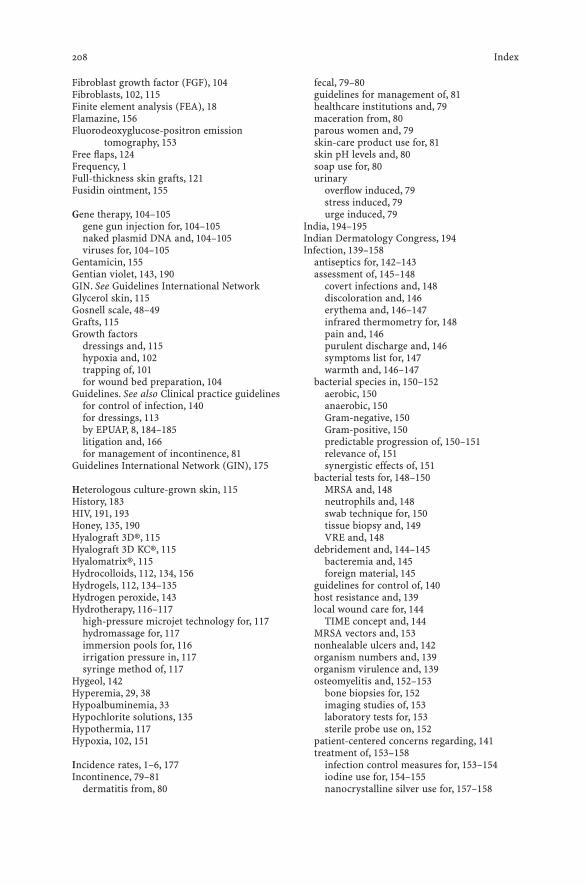
Fibroblast growth factor (FGF), 104Fibroblasts, 102, 115Finite element analysis (FEA), 18Flamazine, 156Fluorodeoxyglucose-positron emission
tomography, 153Free flaps, 124Frequency, 1Full-thickness skin grafts, 121Fusidin ointment, 155
Gene therapy, 104–105gene gun injection for, 104–105naked plasmid DNA and, 104–105viruses for, 104–105
Gentamicin, 155Gentian violet, 143, 190GIN. See Guidelines International NetworkGlycerol skin, 115Gosnell scale, 48–49Grafts, 115Growth factors
dressings and, 115hypoxia and, 102trapping of, 101for wound bed preparation, 104
Guidelines. See also Clinical practice guidelinesfor control of infection, 140for dressings, 113by EPUAP, 8, 184–185litigation and, 166for management of incontinence, 81
Guidelines International Network (GIN), 175
Heterologous culture-grown skin, 115History, 183HIV, 191, 193Honey, 135, 190Hyalograft 3D®, 115Hyalograft 3D KC®, 115Hyalomatrix®, 115Hydrocolloids, 112, 134, 156Hydrogels, 112, 134–135Hydrogen peroxide, 143Hydrotherapy, 116–117
high-pressure microjet technology for, 117hydromassage for, 117immersion pools for, 116irrigation pressure in, 117syringe method of, 117
Hygeol, 142Hyperemia, 29, 38Hypoalbuminemia, 33Hypochlorite solutions, 135Hypothermia, 117Hypoxia, 102, 151
Incidence rates, 1–6, 177Incontinence, 79–81
dermatitis from, 80
208 Index
fecal, 79–80guidelines for management of, 81healthcare institutions and, 79maceration from, 80parous women and, 79skin-care product use for, 81skin pH levels and, 80soap use for, 80urinary
overflow induced, 79stress induced, 79urge induced, 79
India, 194–195Indian Dermatology Congress, 194Infection, 139–158
antiseptics for, 142–143assessment of, 145–148
covert infections and, 148discoloration and, 146erythema and, 146–147infrared thermometry for, 148pain and, 146purulent discharge and, 146symptoms list for, 147warmth and, 146–147
bacterial species in, 150–152aerobic, 150anaerobic, 150Gram-negative, 150Gram-positive, 150predictable progression of, 150–151relevance of, 151synergistic effects of, 151
bacterial tests for, 148–150MRSA and, 148neutrophils and, 148swab technique for, 150tissue biopsy and, 149VRE and, 148
debridement and, 144–145bacteremia and, 145foreign material, 145
guidelines for control of, 140host resistance and, 139local wound care for, 144
TIME concept and, 144MRSA vectors and, 153nonhealable ulcers and, 142organism numbers and, 139organism virulence and, 139osteomyelitis and, 152–153
bone biopsies for, 152imaging studies of, 153laboratory tests for, 153sterile probe use on, 152
patient-centered concerns regarding, 141treatment of, 153–158
infection control measures for, 153–154iodine use for, 154–155nanocrystalline silver use for, 157–158
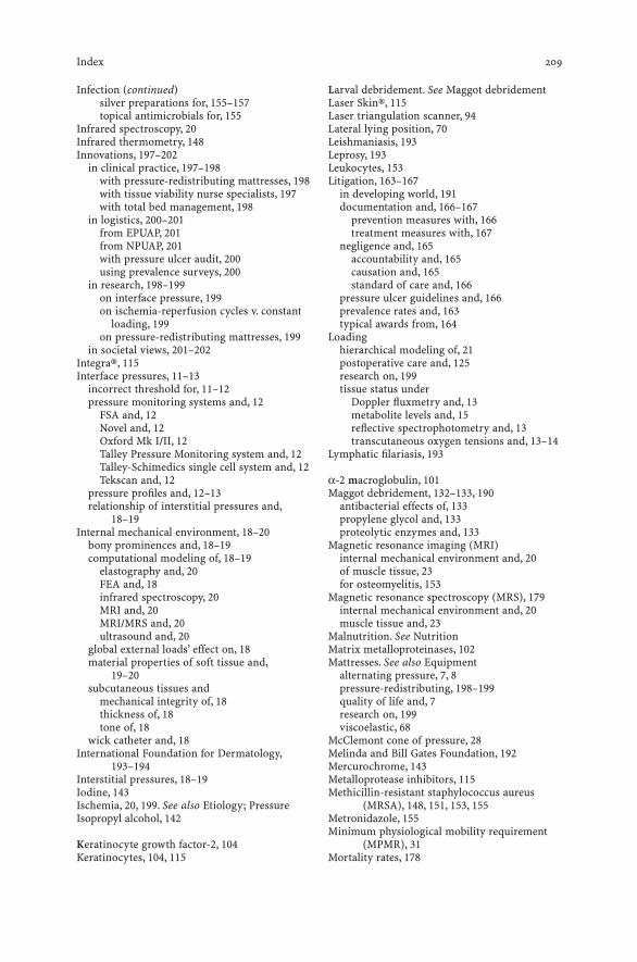
Index 209
Infection (continued)silver preparations for, 155–157topical antimicrobials for, 155
Infrared spectroscopy, 20Infrared thermometry, 148Innovations, 197–202
in clinical practice, 197–198with pressure-redistributing mattresses, 198with tissue viability nurse specialists, 197with total bed management, 198
in logistics, 200–201from EPUAP, 201from NPUAP, 201with pressure ulcer audit, 200using prevalence surveys, 200
in research, 198–199on interface pressure, 199on ischemia-reperfusion cycles v. constant
loading, 199on pressure-redistributing mattresses, 199
in societal views, 201–202Integra®, 115Interface pressures, 11–13
incorrect threshold for, 11–12pressure monitoring systems and, 12
FSA and, 12Novel and, 12Oxford Mk I/II, 12Talley Pressure Monitoring system and, 12Talley-Schimedics single cell system and, 12Tekscan and, 12
pressure profiles and, 12–13relationship of interstitial pressures and,
18–19Internal mechanical environment, 18–20
bony prominences and, 18–19computational modeling of, 18–19
elastography and, 20FEA and, 18infrared spectroscopy, 20MRI and, 20MRI/MRS and, 20ultrasound and, 20
global external loads’ effect on, 18material properties of soft tissue and,
19–20subcutaneous tissues and
mechanical integrity of, 18thickness of, 18tone of, 18
wick catheter and, 18International Foundation for Dermatology,
193–194Interstitial pressures, 18–19Iodine, 143Ischemia, 20, 199. See also Etiology; PressureIsopropyl alcohol, 142
Keratinocyte growth factor-2, 104Keratinocytes, 104, 115
Larval debridement. See Maggot debridementLaser Skin®, 115Laser triangulation scanner, 94Lateral lying position, 70Leishmaniasis, 193Leprosy, 193Leukocytes, 153Litigation, 163–167
in developing world, 191documentation and, 166–167
prevention measures with, 166treatment measures with, 167
negligence and, 165accountability and, 165causation and, 165standard of care and, 166
pressure ulcer guidelines and, 166prevalence rates and, 163typical awards from, 164
Loadinghierarchical modeling of, 21postoperative care and, 125research on, 199tissue status under
Doppler fluxmetry and, 13metabolite levels and, 15reflective spectrophotometry and, 13transcutaneous oxygen tensions and, 13–14
Lymphatic filariasis, 193
a-2 macroglobulin, 101Maggot debridement, 132–133, 190
antibacterial effects of, 133propylene glycol and, 133proteolytic enzymes and, 133
Magnetic resonance imaging (MRI)internal mechanical environment and, 20of muscle tissue, 23for osteomyelitis, 153
Magnetic resonance spectroscopy (MRS), 179internal mechanical environment and, 20muscle tissue and, 23
Malnutrition. See NutritionMatrix metalloproteinases, 102Mattresses. See also Equipment
alternating pressure, 7, 8pressure-redistributing, 198–199quality of life and, 7research on, 199viscoelastic, 68
McClemont cone of pressure, 28Melinda and Bill Gates Foundation, 192Mercurochrome, 143Metalloprotease inhibitors, 115Methicillin-resistant staphylococcus aureus
(MRSA), 148, 151, 153, 155Metronidazole, 155Minimum physiological mobility requirement
(MPMR), 31Mortality rates, 178
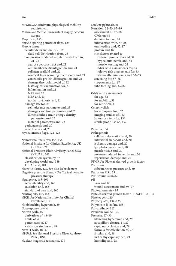
MPMR. See Minimum physiological mobilityrequirement
MRSA. See Methicillin-resistant staphylococcusaureus
Mupirocin, 155Muscle-sparing perforator flaps, 124Muscle tissue
cellular deformation in, 21, 23dead cell distribution from, 23
compression-induced cellular breakdown in,21–23
agarose gel construct and, 21cell membrane disintegration and, 21collagen scaffold and, 22confocal laser scanning microscopy and, 21contractile protein disintegration and, 21damage threshold model of, 22histological examination for, 23inflammation and, 21MRI and, 23MRS and, 23nuclear pyknosis and, 21
damage law for, 23cell tolerance parameter and, 23damage evolution parameter and, 23dimensionless strain energy density
parameter and, 23material parameters and, 23
pathogenesis and, 20reperfusion and, 23
Myocutaneous flaps, 122–123
Nanocrystalline silver, 156–158National Institute for Clinical Excellence, UK
(NICE), 169National Pressure Ulcer Advisory Panel, USA
(NPUAP), 201classification system by, 37developing world and, 189EPUAP and, 184
Necrotic tissue, 129. See also DebridementNegative pressure therapy. See Topical negative
pressure therapyNegligence, 165–166
accountability and, 165causation and, 165standard of care and, 166
Neutrophils, 148, 153NICE. See National Institute for Clinical
Excellence, UKNonblanching hyperemia, 29Nonresponse rate, 4Norton scale, 45
derivatives of, 48–49limits of, 48parameters of, 47validation studies on, 48
Nova-4 scale, 48–49NPUAP. See National Pressure Ulcer Advisory
Panel, USANuclear magnetic resonance, 179
210 Index
Nuclear pyknosis, 21Nutrition, 32–33, 85–89
assessment of, 87–88CPGs on, 86decision tree on, 88intervention with, 87–88oral feeding and, 85, 87protein and, 87risk factors related to
collagen production and, 32hypoalbuminemia and, 33muscle wasting and, 32odds ratio assessments for, 33relative risk assessments for, 33serum albumin levels and, 32–33
screening for, 87–88supplements for, 87tube feeding and, 85, 87
Odds ratio assessmentsfor age, 32for mobility, 31for nutrition, 33
Osteomyelitisbone biopsies for, 152imaging studies of, 153laboratory tests for, 153sterile probe use on, 152
Papaina, 134Pathogenesis
cellular deformation and, 20interstitial transport and, 20ischemic damage and, 20lymphatic system and, 20muscle tissue and, 20pressure-induced ischemia and, 20reperfusion damage and, 20
PDGF. See Platelet-derived growth factorPerfusion
subcutaneous pressure and, 30Perfusion MRI, 23Peri-wound skin, 82pH
skin and, 80wound assessment and, 96–97
Photogrammetry, 93Platelet-derived growth factor (PDGF), 102, 104Platelet gels, 115Polyacrylates, 134–135Polymyxin B sulfate, 155Polyurethane, 112Povidone iodine, 154Pressure, 27–30
blanching hyperemia and, 29at capillary closure, 11, 29capillary occlusion and, 29formula for calculation of, 27friction and, 28in healthy capillary bed, 29humidity and, 28
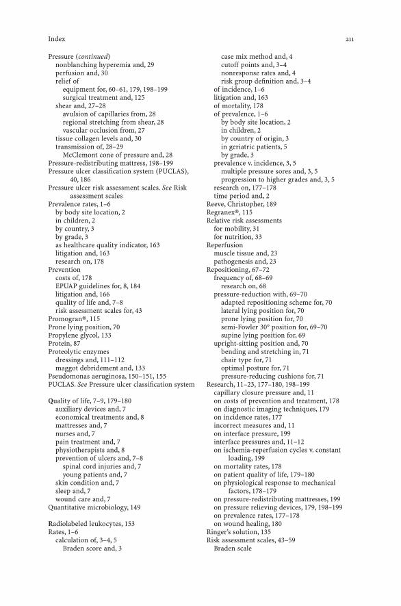
Index 211
Pressure (continued)nonblanching hyperemia and, 29perfusion and, 30relief of
equipment for, 60–61, 179, 198–199surgical treatment and, 125
shear and, 27–28avulsion of capillaries from, 28regional stretching from shear, 28vascular occlusion from, 27
tissue collagen levels and, 30transmission of, 28–29
McClemont cone of pressure and, 28Pressure-redistributing mattress, 198–199Pressure ulcer classification system (PUCLAS),
40, 186Pressure ulcer risk assessment scales. See Risk
assessment scalesPrevalence rates, 1–6
by body site location, 2in children, 2by country, 3by grade, 3as healthcare quality indicator, 163litigation and, 163research on, 178
Preventioncosts of, 178EPUAP guidelines for, 8, 184litigation and, 166quality of life and, 7–8risk assessment scales for, 43
Promogran®, 115Prone lying position, 70Propylene glycol, 133Protein, 87Proteolytic enzymes
dressings and, 111–112maggot debridement and, 133
Pseudomonas aeruginosa, 150–151, 155PUCLAS. See Pressure ulcer classification system
Quality of life, 7–9, 179–180auxiliary devices and, 7economical treatments and, 8mattresses and, 7nurses and, 7pain treatment and, 7physiotherapists and, 8prevention of ulcers and, 7–8
spinal cord injuries and, 7young patients and, 7
skin condition and, 7sleep and, 7wound care and, 7
Quantitative microbiology, 149
Radiolabeled leukocytes, 153Rates, 1–6
calculation of, 3–4, 5Braden score and, 3
case mix method and, 4cutoff points and, 3–4nonresponse rates and, 4risk group definition and, 3–4
of incidence, 1–6litigation and, 163of mortality, 178of prevalence, 1–6
by body site location, 2in children, 2by country of origin, 3in geriatric patients, 5by grade, 3
prevalence v. incidence, 3, 5multiple pressure sores and, 3, 5progression to higher grades and, 3, 5
research on, 177–178time period and, 2
Reeve, Christopher, 189Regranex®, 115Relative risk assessments
for mobility, 31for nutrition, 33
Reperfusionmuscle tissue and, 23pathogenesis and, 23
Repositioning, 67–72frequency of, 68–69
research on, 68pressure-reduction with, 69–70
adapted repositioning scheme for, 70lateral lying position for, 70prone lying position for, 70semi-Fowler 30° position for, 69–70supine lying position for, 69
upright-sitting position and, 70bending and stretching in, 71chair type for, 71optimal posture for, 71pressure-reducing cushions for, 71
Research, 11–23, 177–180, 198–199capillary closure pressure and, 11on costs of prevention and treatment, 178on diagnostic imaging techniques, 179on incidence rates, 177incorrect measures and, 11on interface pressure, 199interface pressures and, 11–12on ischemia-reperfusion cycles v. constant
loading, 199on mortality rates, 178on patient quality of life, 179–180on physiological response to mechanical
factors, 178–179on pressure-redistributing mattresses, 199on pressure relieving devices, 179, 198–199on prevalence rates, 177–178on wound healing, 180
Ringer’s solution, 135Risk assessment scales, 43–59
Braden scale

Risk assessment scales (continued)parameters of, 50–51validation studies on, 51–52
characteristics of, 46–47applicability and, 47definite criteria and, 47ease of use and, 47predictive value and, 47sensitivity and, 46specificity and, 46
CPG recommendations about, 44–45for intensive care patients, 53Norton scale, 45
derivatives of, 48–49limits of, 48parameters of, 47validation studies on, 48
for pediatric patients, 53preventative measures based on, 43systematic review of, 46Waterlow scale
appraisals of, 50parameters of, 49validation studies on, 50
Risk factors, 30–34age and, 31–32
odds ratio assessments for, 32pathological skin changes and, 32
mobility and, 30–31MPMR and, 31odds ratio assessments for, 31relative risk assessments for, 31spontaneous nocturnal movement and,
31nutrition and, 32–33
collagen production and, 32hypoalbuminemia and, 33muscle wasting and, 32odds ratio assessments for, 33relative risk assessments for, 33serum albumin levels and, 32–33
reference table of, 34tissue tolerance and, 30
Scale. See Risk assessment scalesSemi-Fowler 30° position, 69–70Sensate myocutaneous flaps, 123–124Sharp debridement, 131–132
complications with, 132contraindications for, 132
Shear, 11avulsion of capillaries from, 28regional stretching from shear, 28vascular occlusion from, 27
Silvadene, 156Silver, 133, 135, 155–158
adsorbed, 156allergic sensitization to, 156minimum inhibitory concentrations of, 157nanocrystalline, 157
212 Index
preparations of, 156salts, 156
Silver-calcium-sodium phosphates, 156Silver charcoal, 156Silver coated foam, 156Silver combined with hydrocolloid, 156Silver nitrate, 156Silver-sodium carboxymethylcellulose, 156Silver sulfadiazine, 156Sinus tracts, 92Skin, 75–82
acid mantle, 80aging of
drying during, 78elasticity loss during, 78fatty layer reduction during, 78thinning during, 78
bioengineered, 104dark colored, 39dermal layer of
collagen fibers in, 77elastin fibers in, 77ground substance of, 77lymph vessels in, 77nerve endings in, 78sebaceous glands in, 78sweat glands in, 78tissue mast cells in, 77tissue microphages in, 77
epidermal layer ofbasal layer of, 77clear cell layer of, 77dermal junction with, 77granular layer of, 77horny layer of, 76prickle cell layer of, 77
failure of, 193functions of, 75–76
protection, 76sensation, 76thermal regulation, 76vitamin D production, 76
grafts of, 120–123incontinence and, 75, 80–81peri-wound, 82pH levels of, 80quality of life and, 7wound assessment and, 92
Skin bank, 115Skin flaps, 121Slough, 129Sodium hypochlorite, 142Spectrophotometry, 13Split-thickness skin grafts, 120–121SSD Cream, 156Staphylococcus aureus, 150–151, 152, 155Stem cell therapy, 105
fibrin use for, 105pluripotentiality of, 105
Stereophotogrammetry, 93–94

Index 213
Stereophotography, 93–94Stirling Grading System, 37Stratum basale, 77Stratum corneum, 76, 81, 82, 96Stratum granulosum, 77Stratum lucidum, 77Stratum spinosum, 77Streptococci, 150, 153, 155Sunderland scale, 53Supine lying position, 69Surgical treatment, 119–125
debridement during, 120patient selection for, 119–120postoperative care after, 124–125
diet during, 125load conditioning during, 125physical training during, 125pressure relief during, 125
reconstruction withdirect closure for, 121fasciocutaneous flaps for, 122flaps without skin coverage for, 123free flaps for, 124full-thickness skin grafts for, 121muscle-sparing perforator flaps for, 124myocutaneous flaps for, 122–123sensate myocutaneous flaps for, 123–124skin flaps for, 121split-thickness skin grafts for, 120–121tissue expansion for, 124
staff for, 119Swaminathan Foundation, 191Sweat, 78. See also Tissue biochemistry
T2-weighted MRI, 23Tagging MRI, 23TGF-b1. See Transforming growth factor-b1TIME. See Tissue debridement,
infection/inflammation, moisturebalance, edge effect
Tissue biochemistry, 14–18carbon dioxide levels and, 16–17metabolite levels under loading and, 14–15oxygen levels under, 16–17sweat lactate levels and, 15, 16–17sweat metabolite constituents and, 14sweat purines and, 16sweat urea levels and, 16
Tissue debridement, infection/inflammation,moisture balance, edge effect (TIME),103–104, 140, 144
Topical negative pressure therapy, 114, 116contraindications for, 116duration of, 116indications for, 116target pressure of, 116Vacuum Assisted Closure device for, 114, 116
Transcutaneous oxygen tension, 13–14, 102Transforming growth factor-b1 (TGF-b1), 101,
102
Ultrasound, 20, 153, 179Upright-sitting position, 70–71
Vacuum Assisted Closure, 114, 116Vancomycin-resistant enterococcus (VRE),
148Vascular endothelial growth factor (VEGF),
102VEGF. See Vascular endothelial growth factorViscoelastic mattresses, 67, 68Vitamins, 76, 85, 87VRE. See Vancomycin-resistant enterococcus
Warming therapy, 117–118blood flow and, 117hypothermia and, 117
Waterlow pressure ulcer risk scores, 33Waterlow scale
appraisals of, 50parameters of, 49validation studies on, 50
World Health Organization, 192Wound assessment, 91–97
area and volume measurements in, 91, 93–94computerized planimetry for, 93dental impression materials for, 94image processing for, 93laser triangulation scanner use for, 94metric grid use for, 93photogrammetry use for, 93sheet tracing for, 93stereophotogrammetry use for, 93–94stereophotography use for, 93–94three-dimensional scanner use for, 94
exudate and, 92infection and, 92–93odor and, 92pH measurements in, 96–97
colorimetric technique for, 96flat glass electrode use for, 96pH transistor technology for, 97
surrounding skin and, 92tissue density measurements in, 94–95
echogenicity values and, 95ultrasonography use for, 94–95
tissue perfusion measurements in, 95–96laser Doppler flowmetry use for, 95laser Doppler imaging use for, 96
undermining tissue and, 92viable tissue quantity and, 92wound edges and, 92
Wound bed preparation, 99–105bacterial burdens and, 100bioengineered skin and, 105biofilms and, 101debridement and, 103gene therapy for, 104–105growth factors for, 104
PDGF and, 104sequential treatment with, 104

Wound bed preparation (continued)growth factor trapping and, 101hypoxia and, 102
fibroblast proliferation and, 102growth factors and, 102
impaired healing and, 100phenotypic wound cell alteration and,
102–103cellular senescence and, 103
214 Index
healing capacity and, 103stem cell therapy for, 105TIME concept for, 103–104wound fluid and, 101–102
exudate levels and, 102matrix metalloproteinases and,
102
X-rays, 153


