1 4/6/22 Name Student number http://www.eurekalert.org/pub_releases/2015-11/udg-hdt111115.php How depleting the gut microbiota protects from obesity Microbiota has also an impact on the way calories are absorbed and how fat cells develop In the past few years, research on gut microbiota (that is, all microorganisms, mainly bacteria, inhabiting our gut) has started to unravel its tremendous role in our body, and how it symbiotically affects the functioning of our organs. In particular, microbiota has also an impact on the way calories are absorbed and how fat cells develop. By studying mice without microbiota, scientists from the University of Geneva (UNIGE) Faculty of Medicine, Switzerland, were able to demonstrate how the absence of microbiota has a remarkable effect against obesity. Indeed, it triggers a surprising metabolic mechanism: white fat cells - which in excess cause obesity and insulin resistance - are transformed into cells similar to brown fat (they are called "beige fat"), that protects the body against excess weight and its damaging consequences. This discovery, published in Nature Medicine, could open the door to completely new anti-obesity treatments. Mammals have two types of fat: brown fat, whose primary function is to burn calories to produce heat; and white fat, which is used as energy storage. In healthy humans, white adipose tissue constitutes about 25% of the body mass. However, when in excess, white fat contributes to insulin resistance and diabetes. Conversely, brown fat improves insulin sensitivity and is reversely correlated to obesity. In response to cold or exercise, cells similar to brown fat - the beige fat - can appear within the white fat, a phenomenon known as "browning". Although the origin of these beige cells seems alike to that of the white fat, their function differs: the more beige fat appear within the white adipose tissue, the more calories are burned. This suggests that stimulating beige fat growth could be a way to reduce obesity and limit insulin resistance. The unexpected role of gut microbiota Only recently, scientists have started to grasp the extent of the relationships between the gut microbiota and its human host. An increasing number of studies are now highlighting its impact on the regulation of multiple metabolic pathways, thus interconnecting the gastrointestinal tract, skin, liver, brain and many other organs. Today, researchers from UNIGE Faculty of Medicine demonstrate that it also has a direct impact on obesity: the microbiota of obese people has a specific composition, different from the microbiota of lean people. Indeed, germ-free mice (born and kept in sterile conditions, i.e. without microbiota), which receive gut microbiota transplant from obese people, tend to develop obesity and insulin resistance. "Having observed that microbiota can affect the obesity onset, we suspected that microbiota depletion can change the insulin sensitivity by modifying the amount and balance of these various types of fat", explains Mirko Trajkovski, lead author of the study and Professor at the Faculty of Medicine Department of Cell Physiology and Metabolism. To confirm their hypothesis, the researchers fed three groups of mice with a high-
Transcript
1 5/20/23 Name Student number http://www.eurekalert.org/pub_releases/2015-11/udg-hdt111115.phpHow depleting the gut microbiota protects from obesity
Microbiota has also an impact on the way calories are absorbed and how fat cells develop
In the past few years, research on gut microbiota (that is, all microorganisms, mainly bacteria, inhabiting our gut) has started to unravel its tremendous role in our body, and how it symbiotically affects the functioning of our organs. In particular, microbiota has also an impact on the way calories are absorbed and how fat cells develop. By studying mice without microbiota, scientists from the University of Geneva (UNIGE) Faculty of Medicine, Switzerland, were able to demonstrate how the absence of microbiota has a remarkable effect against obesity. Indeed, it triggers a surprising metabolic mechanism: white fat cells - which in excess cause obesity and insulin resistance - are transformed into cells similar to brown fat (they are called "beige fat"), that protects the body against excess weight and its damaging consequences. This discovery, published in Nature Medicine, could open the door to completely new anti-obesity treatments.Mammals have two types of fat: brown fat, whose primary function is to burn calories to produce heat; and white fat, which is used as energy storage. In healthy humans, white adipose tissue constitutes about 25% of the body mass. However, when in excess, white fat contributes to insulin resistance and diabetes. Conversely, brown fat improves insulin sensitivity and is reversely correlated to obesity.In response to cold or exercise, cells similar to brown fat - the beige fat - can appear within the white fat, a phenomenon known as "browning". Although the origin of these beige cells seems alike to that of the white fat, their function differs: the more beige fat appear within the white adipose tissue, the more calories are burned. This suggests that stimulating beige fat growth could be a way to reduce obesity and limit insulin resistance.The unexpected role of gut microbiotaOnly recently, scientists have started to grasp the extent of the relationships between the gut microbiota and its human host. An increasing number of studies are now highlighting its impact on the regulation of multiple metabolic pathways, thus interconnecting the gastrointestinal tract, skin, liver, brain and many other organs.Today, researchers from UNIGE Faculty of Medicine demonstrate that it also has a direct impact on obesity: the microbiota of obese people has a specific composition, different from the microbiota of lean people. Indeed, germ-free mice (born and kept in sterile conditions, i.e. without microbiota), which receive gut microbiota transplant from obese people, tend to develop obesity and insulin
resistance. "Having observed that microbiota can affect the obesity onset, we suspected that microbiota depletion can change the insulin sensitivity by modifying the amount and balance of these various types of fat", explains Mirko Trajkovski, lead author of the study and Professor at the Faculty of Medicine Department of Cell Physiology and Metabolism. To confirm their hypothesis, the researchers fed three groups of mice with a high-calorie diet: germ-free mice, standard mice and mice previously treated with high doses of antibiotics that have the effect of totally depleting their microbiota. While normal mice exposed to a high-calorie diet did develop obesity and insulin resistance, the two other groups remained lean, had an improved sensitivity to insulin and tolerated glucose better. Importantly, their amount of white fat decreased, and this was accompanied with increased levels of brown fat markers.Reducing obesity by creating additional beige fatThe scientists observed that depleting microbiota - either through antibiotics or in germ-free mice - stimulated the development of functional beige fat within the white fat, in the same way as when exposed to cold or exercise. But how does this work? It all has to do with a specific cell type, called macrophages. Macrophages are an essential component of the immune system and fulfill various metabolic functions, including tissue remodelling. They express different functional programmes in response to micro-environmental signals, a process called "polarization". Polarized macrophages can be broadly classified in two main groups: M1 and M2, the latter being able to act on the adipose and increase the production of beige fat. When the microbiota is depleted, the number of specific cells, called eosinophils, increases in white fat, which secretes small signalling proteins ("type 2 cytokines") that act on macrophages polarization. Thanks to these proteins, M1 macrophages turn into M2 macrophages, which activate the browning of white fat and reduce obesity."In mice, the effect of the antibiotics lasts for a couple of weeks after the treatment", stress Nicolas Suarez-Zamorano and Salvatore Fabbiano, the first co-authors of this study. "Although treating obesity with high doses of antibiotics is unrealistic - mainly due to the risk of antibiotic resistance - we want to explore alternative ways of suppressing or modifying the microbiota, and to identify the exact bacterial genes responsible for this phenomenon. We would then target only those, without having to deplete the entire microbiota", explains Mirko Trajkovski. To search for effective clinical treatments of obesity, the scientists will use particular antibiotics, as well as bacterial phages, a kind of virus that kills only specific bacterial strains. The possibility of microbiota transplant from a lean to an obese person whose microbiota would have been previously depleted will also be studied.
2 5/20/23 Name Student number http://www.eurekalert.org/pub_releases/2015-11/htcs-mcd111315.php
Moderate coffee drinking may lower risk of premature deathDrinkers of both caffeinated and decaffeinated coffee saw benefits
Boston, MA - People who drink about three to five cups of coffee a day may be less likely to die prematurely from some illnesses than those who don't drink or drink less coffee, according to a new study by Harvard T.H. Chan School of Public Health researchers and colleagues. Drinkers of both caffeinated and decaffeinated coffee saw benefits, including a lower risk of death from cardiovascular disease, neurological diseases, type 2 diabetes, and suicide."Bioactive compounds in coffee reduce insulin resistance and systematic inflammation," said first author Ming Ding, a doctoral student in the Department of Nutrition. "That could explain some of our findings. However, more studies are needed to investigate the biological mechanisms producing these effects."The study will appear online in Circulation on November 16, 2015.Researchers analyzed health data gathered from participants in three large ongoing studies: 74,890 women in the Nurses' Health Study; 93,054 women in the Nurses' Health Study 2; and 40,557 men in the Health Professionals Follow-up Study. Coffee drinking was assessed using validated food questionnaires every four years over about 30 years. During the study period, 19,524 women and 12,432 men died from a range of causes.In the whole study population, moderate coffee consumption was associated with reduced risk of death from cardiovascular disease, diabetes, neurological diseases such as Parkinson's disease, and suicide. Coffee consumption was not associated with cancer deaths. The analyses took into consideration potential confounding factors such as smoking, body mass index, physical activity, alcohol consumption, and other dietary factors."This study provides further evidence that moderate consumption of coffee may confer health benefits in terms of reducing premature death due to several diseases," said senior author Frank Hu, professor of nutrition and epidemiology. "These data support the 2015 Dietary Guidelines Advisory Report that concluded that 'moderate coffee consumption can be incorporated into a healthy dietary pattern.'"Other Harvard Chan School authors include Ambika Satija, Shilpa Bhupathiraju, Yang Hu, Qi Sun, Jiali Han, Esther Lopez-Garcia, Walter Willett, and Rob van Dam.This study was supported by National Institutes of Health research grants UM1 CA186107, UM1 CA176726, UM1 CA167552, P01 CA87969, P01 CA055075, R01 HL034594, HL088521, HL35464, and HL60712."Association of Coffee Consumption with Total and Cause-specific Mortality in Three Large Prospective Cohorts," Ming Ding, Ambika Satija, Shilpa N. Bhupathiraju, Yang Hu, Qi Sun, Walter Willett, Rob M. van Dam, Frank B. Hu, Circulation, online November 16, 2015.
http://www.eurekalert.org/pub_releases/2015-11/uol-doh111615.phpDiscovery of hidden earthquake presents challenge to earthquake
early-warning systemsSeismologists at the University of Liverpool studying the 2011 Chile earthquake have discovered a previously undetected earthquake which took place seconds
after the initial rupture.This newly discovered phenomena which they called a `closely-spaced doublet' presents a challenge to earthquake and tsunami early warning systems as it increases the risk of larger-than-expected tsunamis in the aftermath of a typical subduction earthquake.In a study published in Nature Geoscience, University researchers analysed in detail the seismic wave recordings from 2 January 2011 when an earthquake of magnitude 7 occurred in Chile along the plate boundary separating the subducting Nazca plate from the South American continent.They discovered that just 12 seconds later and 30 km further offshore, a second rupture of a similar size, which was un-detected by national and global earthquake monitoring centres, occurred along an extensional (pull-apart) fault in the middle of the South American plate beneath the Pacific Ocean.Liverpool seismologist, Professor Andreas Rietbrock, said: "Real-time global seismic monitoring and early warning events have come a long way and it is possible for a magnitude 5 or greater earthquake to be detected within a matter of minutes. Therefore, it is striking that an earthquake with magnitude close to 7 was effectively hidden from our standard monitoring systems.""Previous doublet events have been documented in subduction zones before, but such instantaneous triggering of large ruptures at close distances has no known precedent. Such triggered events dramatically complicate potential earthquake impact assessments and tsunami early warning systems as the risk of a larger than expected tsunami is higher following a typical subduction earthquake."Dr Stephen Hicks, who was part of the research team, said: "We believe that seismic waves travelling outward from the first rupture immediately shook up and weakened the shallower second fault, causing the hidden rupture. Scientists believe that the overlying plate at collisional plate boundaries is broken up on a large scale and contains networks of faults. It is plausible that similar closely-spaced doublets may occur elsewhere around the Pacific Ring of Fire. "Professor Rietbrock added: "This work challenges the commonly-held notion that slip during large earthquakes may only occur along a single fault. The result was surprising as there was no indication of such a complicated rupture from global earthquake monitoring systems. "
3 5/20/23 Name Student number "Our findings present a concern for tsunami early warning systems. Without real-time monitoring of seismometers located close to the fault, it is possible that tsunami and shaking hazard from future subduction earthquakes may be underestimated."As part of the University's Liverpool Earth Observatory, seismologists are installing a seismic network in Southern Peru in close collaboration with the Geophysical Institute of Peru. This area along the South American continental margin has the potential for a large magnitude 8+ earthquake and it is important to understand the associated seismic and tsunami hazard.The research is published in Nature Geoscience. http://dx.doi.org/10.1038/ngeo258.
http://www.eurekalert.org/pub_releases/2015-11/bu-sec111615.phpStudy: Earth's climate more sensitive to CO2 than previously
thoughtReturn to hothouse climate may take less carbon dioxide than expected
BINGHAMTON, NY – Ancient climates on Earth may have been more sensitive to carbon dioxide than was previously thought, according to new research from Binghamton University.A team of Binghamton University researchers including geology PhD student Elliot A. Jagniecki and professors Tim Lowenstein, David Jenkins and Robert Demicco examined nahcolite crystals found in Colorado's Green River Formation, formed 50 million years old during a hothouse climate. They found that CO2 levels during this time may have been as low as 680 parts per million (ppm), nearly half the 1,125 ppm predicted by previous experiments. The new data suggests that past predictions significantly underestimate the impact of greenhouse warming and that Earth's climate may be more sensitive to increased carbon dioxide than was once thought, said Lowenstein."The significance of this is that CO2 50 million years ago may not have been as high as we once thought it was, but the climate back then was significantly warmer than it is today," said Lowenstein."CO2 levels in the atmosphere today have reached 400 ppm. According to current projections, doubling the CO2 will result in a rise in the global average temperature of 3 degrees Centigrade. This new research suggests that the effects of CO2 on global warming may be underestimated."Take notice that carbon dioxide 50 million years ago may not have been as high as we once thought it was. We may reach that level in the next century, and so the climate change from that increase could be pretty severe, pretty dramatic. CO2 and other climate forcings may be more important for global warming than we realized."
The only direct measurement of carbon dioxide is from ice cores, which only go back less than 1 million years. Lowenstein and his team are trying to develop ways to estimate ancient carbon dioxide in the atmosphere using indirect proxies. He said that their approach is different than any ever undertaken."These are direct chemical measurements that are based on equilibrium thermodynamics," he said. "These are direct laboratory experiments, so I think they're really reliable.Lowenstein wants to look at nahcolite deposits in China to confirm the results found in Colorado.The study, "Eocene atmospheric CO2 from the nahcolite proxy," was published Oct. 23 in Geology.
http://www.eurekalert.org/pub_releases/2015-11/nlmc-san111615.phpSurgeons at NYU Langone Medical Center perform the most
extensive face transplant to datePatient is country's first 'first responder' to undergo the procedure to repair
injuries suffered in the line of dutyNYU Langone Medical Center announced today the successful completion of the most extensive face transplant to date, setting new standards of care in this emerging field. Equally important, for the first time a face transplant has been performed on a first responder - a volunteer firefighter who suffered a full face and scalp burn in the line of duty.The surgery -- the first of its kind performed in New York State -- began the morning of August 14, 2015 and concluded 26 hours later on the morning of August 15. It involved a team of more than 100 physicians, nurses, technical and support staff, led by Eduardo D. Rodriguez, MD, DDS, the Helen L. Kimmel Professor of Reconstructive Plastic Surgery and chair of the Hansjörg Wyss Department of Plastic Surgery. The team worked in two adjoining operating rooms -- in one room, the donor's face was procured (along with other donated organs), and in the other, the recipient's face and scalp burn was removed and the transplant took place.The recipient, volunteer firefighter Patrick Hardison, 41, of Senatobia, MS, was injured in September 2001 - ironically just days before the 9/11 attacks. Patrick entered a burning home on a rescue search, when its roof collapsed on him, leaving him with disfiguring burns across his entire face, head, neck and upper torso. He lost his eyelids, ears, lips, most of his nose as well as his hair including his eyebrows. After enduring more than 70 surgeries in Mississippi and elsewhere, Patrick was still unable to return to a normal life. He was brought to Dr. Rodriguez's attention by a member of his church and fellow firefighter, who wrote to the doctor describing Patrick's situation.
4 5/20/23 Name Student number Highlights from the SurgeryDr. Rodriguez -- who previously performed a complex face transplant in 2012 -- and his NYU Langone team transplanted not only the face, but also the entire scalp. Patrick's surgery was pivotal in that the donor's eyelids and the muscles that control blinking were transplanted--a significant milestone and a procedure that had not been previously performed on a seeing patient.Among other milestones achieved in Patrick's surgery: Transplantation of the ears and ear canals Transplantation of selective bony structures from the donor, including portions of the chin, cheeks and the entire nose Advanced use of three-dimensional modeling, computerized modeling and 3D printed patient-specific cutting guides designed from the recipient's and the donor's CT scans to provide the most precise "snap-fit" of the skeleton Precise placement of patient-specific metal plates and screws to ensure the proper contour and symmetry of the transplanted faceThe transplantation of the donor's eyelids and blinking mechanisms was particularly important to the surgery's success, as Patrick was in danger of losing his sight and had been unable to perform independent daily tasks such as driving. Blinking enables the body to appropriately hydrate and clean the eyes to prevent infection and preserve vision. Earlier this year, Dr. Rodriguez and others published a study in the peer-reviewed journal, Plastic and Reconstructive Surgery, in which they detailed the importance of eyelid preservation and enhancement in facial transplantation.Patrick's RecoveryWithin the final hours of surgery, signs of success were evident. Patrick's new face, particularly his new lips and ears, were robust with color, indicating circulation had been restored. The hair on his scalp, as well as the beard on his face, began growing back immediately. He was able to use his new eyelids and blink on the third day of recovery, after the swelling began to diminish. He was sitting up in a chair within a week. And now, just three months removed from surgery, swelling has greatly subsided and he is quickly returning to the routines of daily life independently.As part of his recovery, Patrick continues to go through extensive rehabilitative therapy, including: Physical therapy to build his strength and stamina Speech and swallowing therapy to further restore and enhance his ability to speak correctly using his new lips and to regain normal eating and swallowing abilities Occupational therapy to re-learn daily tasks, such as shaving again for the first time in 14 years
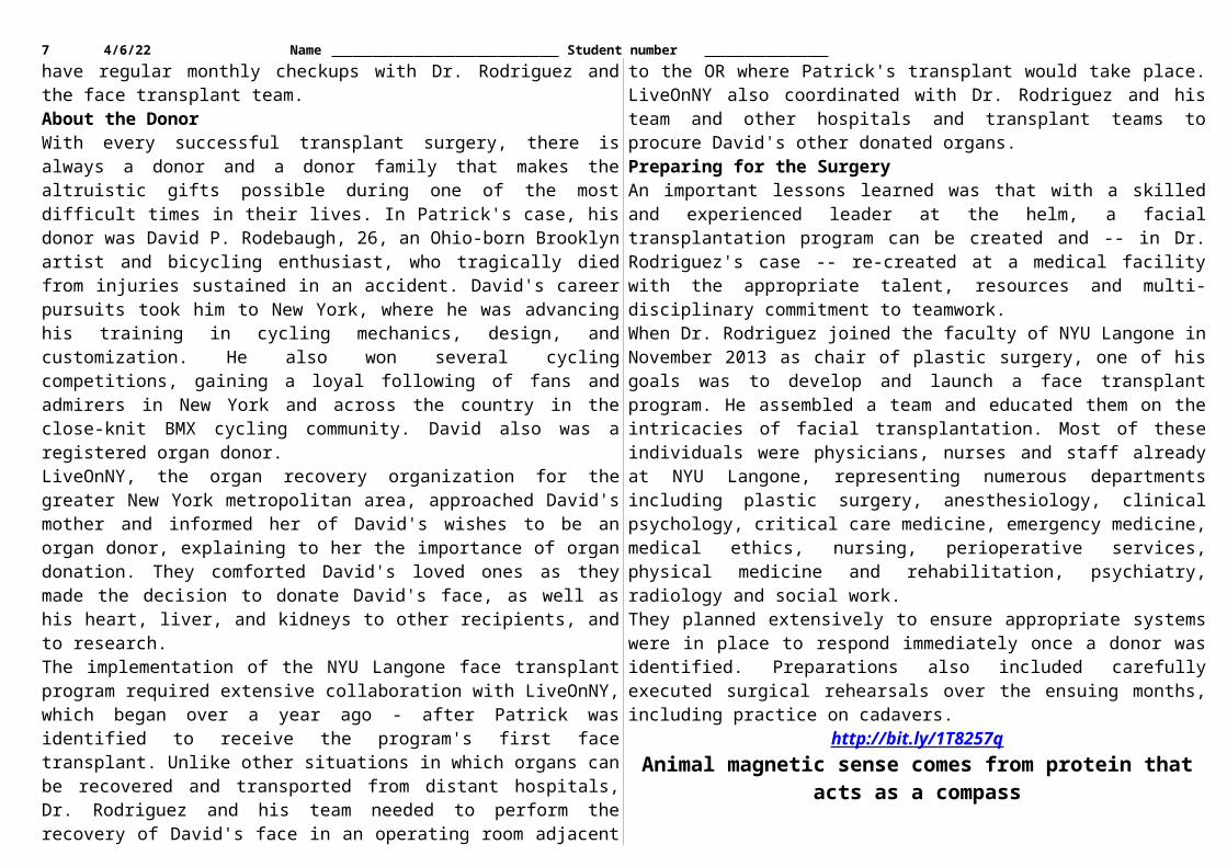
Patrick, like all transplant patients, will need to remain on anti-rejection medication for the rest of his life to prevent transplant rejection. Patrick will also rely on his family and friends -- particularly his fellow firefighters in Senatobia -- to support him in his recovery and his transition back to his hometown after he is discharged from the hospital. He will also have regular monthly checkups with Dr. Rodriguez and the face transplant team.About the DonorWith every successful transplant surgery, there is always a donor and a donor family that makes the altruistic gifts possible during one of the most difficult times in their lives. In Patrick's case, his donor was David P. Rodebaugh, 26, an Ohio-born Brooklyn artist and bicycling enthusiast, who tragically died from injuries sustained in an accident. David's career pursuits took him to New York, where he was advancing his training in cycling mechanics, design, and customization. He also won several cycling competitions, gaining a loyal following of fans and admirers in New York and across the country in the close-knit BMX cycling community. David also was a registered organ donor.LiveOnNY, the organ recovery organization for the greater New York metropolitan area, approached David's mother and informed her of David's wishes to be an organ donor, explaining to her the importance of organ donation. They comforted David's loved ones as they made the decision to donate David's face, as well as his heart, liver, and kidneys to other recipients, and to research.The implementation of the NYU Langone face transplant program required extensive collaboration with LiveOnNY, which began over a year ago - after Patrick was identified to receive the program's first face transplant. Unlike other situations in which organs can be recovered and transported from distant hospitals, Dr. Rodriguez and his team needed to perform the recovery of David's face in an operating room adjacent to the OR where Patrick's transplant would take place. LiveOnNY also coordinated with Dr. Rodriguez and his team and other hospitals and transplant teams to procure David's other donated organs.Preparing for the SurgeryAn important lessons learned was that with a skilled and experienced leader at the helm, a facial transplantation program can be created and -- in Dr. Rodriguez's case -- re-created at a medical facility with the appropriate talent, resources and multi-disciplinary commitment to teamwork.When Dr. Rodriguez joined the faculty of NYU Langone in November 2013 as chair of plastic surgery, one of his goals was to develop and launch a face transplant program. He assembled a team and educated them on the intricacies of facial transplantation. Most of these individuals were physicians, nurses and staff already at NYU Langone, representing numerous departments including plastic
5 5/20/23 Name Student number surgery, anesthesiology, clinical psychology, critical care medicine, emergency medicine, medical ethics, nursing, perioperative services, physical medicine and rehabilitation, psychiatry, radiology and social work.They planned extensively to ensure appropriate systems were in place to respond immediately once a donor was identified. Preparations also included carefully executed surgical rehearsals over the ensuing months, including practice on cadavers.
http://bit.ly/1T8257qAnimal magnetic sense comes from protein that acts as a compass
Quick – can you tell where north is? Animals as diverse as sea turtles, birds, worms, butterflies and wolves can, thanks to sensing Earth’s magnetic field.
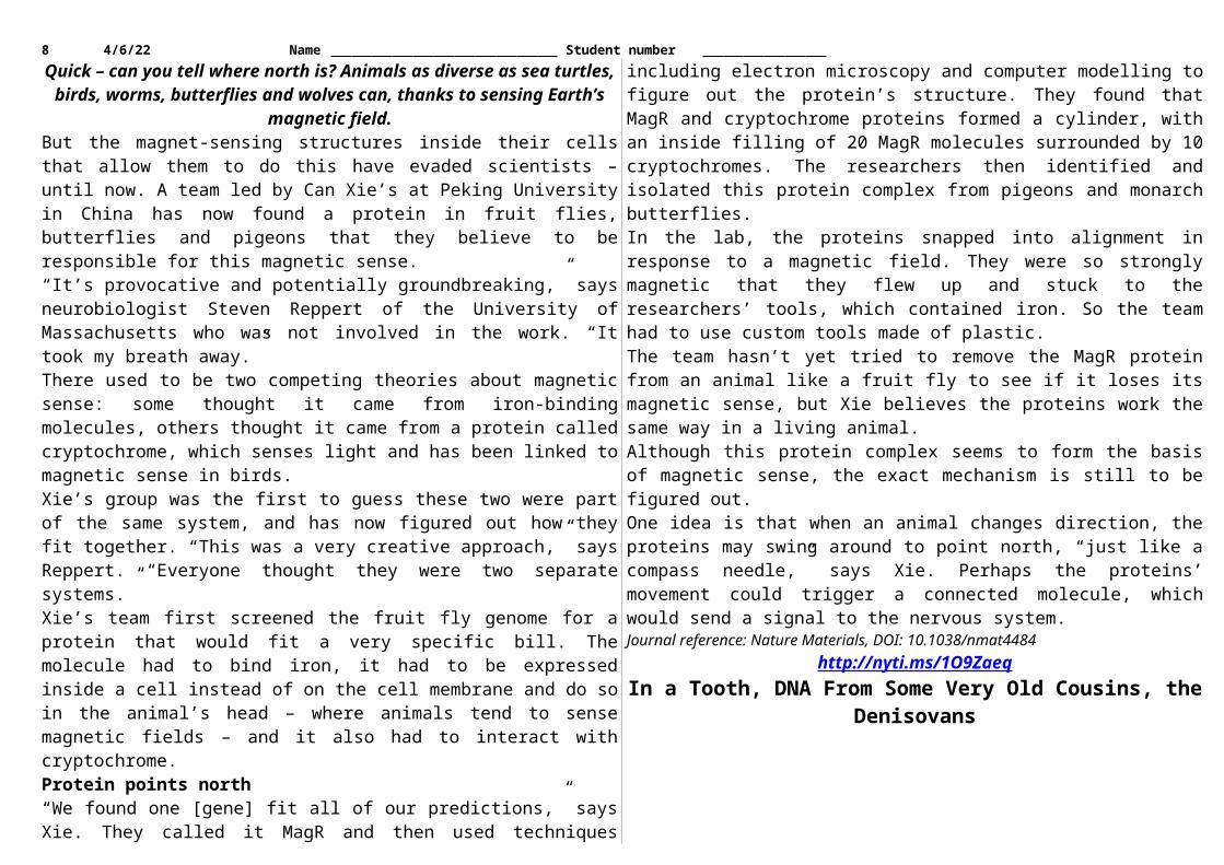
But the magnet-sensing structures inside their cells that allow them to do this have evaded scientists – until now. A team led by Can Xie’s at Peking University in China has now found a protein in fruit flies, butterflies and pigeons that they believe to be responsible for this magnetic sense.“It’s provocative and potentially groundbreaking,” says neurobiologist Steven Reppert of the University of Massachusetts who was not involved in the work. “It took my breath away.”There used to be two competing theories about magnetic sense: some thought it came from iron-binding molecules, others thought it came from a protein called cryptochrome, which senses light and has been linked to magnetic sense in birds.Xie’s group was the first to guess these two were part of the same system, and has now figured out how they fit together. “This was a very creative approach,” says Reppert. “Everyone thought they were two separate systems.”Xie’s team first screened the fruit fly genome for a protein that would fit a very specific bill. The molecule had to bind iron, it had to be expressed inside a cell instead of on the cell membrane and do so in the animal’s head – where animals tend to sense magnetic fields – and it also had to interact with cryptochrome.Protein points north“We found one [gene] fit all of our predictions,” says Xie. They called it MagR and then used techniques including electron microscopy and computer modelling to figure out the protein’s structure. They found that MagR and cryptochrome proteins formed a cylinder, with an inside filling of 20 MagR molecules surrounded by 10 cryptochromes. The researchers then identified and isolated this protein complex from pigeons and monarch butterflies.In the lab, the proteins snapped into alignment in response to a magnetic field. They were so strongly magnetic that they flew up and stuck to the researchers’ tools, which contained iron. So the team had to use custom tools made of plastic.
The team hasn’t yet tried to remove the MagR protein from an animal like a fruit fly to see if it loses its magnetic sense, but Xie believes the proteins work the same way in a living animal.Although this protein complex seems to form the basis of magnetic sense, the exact mechanism is still to be figured out.One idea is that when an animal changes direction, the proteins may swing around to point north, “just like a compass needle,” says Xie. Perhaps the proteins’ movement could trigger a connected molecule, which would send a signal to the nervous system.Journal reference: Nature Materials, DOI: 10.1038/nmat4484
http://nyti.ms/1O9ZaeqIn a Tooth, DNA From Some Very Old Cousins, the DenisovansA tooth fossil discovered in a Siberian cave has yielded DNA from a vanished
branch of the human tree, mysterious cousins called the Denisovans, scientists said Monday.
Their analysis pushes back the oldest known evidence for Denisovans by 60,000 years, suggesting that the species was able to thrive in harsh climates for thousands of generations. The results also suggest that the Denisovans may have bred with other ancient hominins, relatives of modern humans whom science has yet to discover.Todd Disotell, a molecular anthropologist at New York University who was not involved in the new study, said the report added to growing evidence that our species kept company with many near relatives over the past million years. The world, Dr. Disotell said, “was a lot like Middle-earth.”
The Denisova 8 molar, from a hominin species whose remains were found in Siberia. Its age has been estimated at 110,000 years. Bence Viola
“There you’ve got elves and dwarves and hobbits and orcs,” he continued. On the real earth, “we had a ton of hominins that are closely related to us.”The Denisovans are named after the cave where their bones were found in the Altai Mountains in southern Siberia. Every summer, a team of Russian scientists led by Anatoly Derevianko of the Russian Academy of Sciences explores the cave, unearthing thousands of bone fragments.Before the latest discovery, Denisovans were known only from DNA in another tooth and a finger bone found in the cave in 2008. Analysis had shown them to be at least 50,000 years old.
6 5/20/23 Name Student number In 2010, Dr. Derevianko and his colleagues reported that the genetic material in the bone and the tooth belonged to the same lineage of hominins, which they called Denisovans.With virtually no bones to study, scientists struggle to guess what the Denisovans were like. Their closest relatives were the Neanderthals, those stocky, big-brained hominins who hunted big game in Europe and western Asia 300,000 to 40,000 years ago. Scientists estimate that Neanderthals and Denisovans diverged on the human family tree 400,000 years ago.Since their initial discoveries, the Russian researchers have sifted through more bone fragments from Denisova. In 2013, the scientists reported the discovery of a Neanderthal toe bone in the cave with enough DNA to reconstruct the entire genome. The newest batch of Denisovan DNA comes from a tooth discovered in 2010. Svante Paabo of the Max Planck Institute for Evolutionary Anthropology in Leipzig, Germany, and his colleagues described it in the Proceedings of the National Academy of Sciences.The new tooth, called Denisova 8, yielded only a modest amount of DNA. But the scientists gathered enough to draw some important conclusions.Denisova 8, it turns out, is much older than the previously discovered remains. The researchers estimated its age at 110,000 years.Bence Viola, a paleoanthropologist at the University of Toronto and a co-author of the new study, said the Denisovans either lived near the cave at least 110,000 to 50,000 years ago, or came into the region at least twice.They must have been resourceful to endure in Siberia for so long, Dr. Viola said. “It’s not a very pleasant environment,” he noted.Dr. Viola and his colleagues got another intriguing clue about the Denisovans when they tallied up the variations in the DNA sample. The Denisovans, they found, had almost as much genetic diversity as modern Europeans even though the remains were found in a single cave. “You actually see more diversity in the Denisovans than you’ve seen in Neanderthals from Spain to the Altais, and that, I think, is pretty astonishing,” Dr. Viola said.He speculated that the Neanderthals became inbred because ice age glaciers drove them into isolated refuges in southern Europe. The Denisovans, though, were able to move south through large regions of Asia that were not covered by glaciers.Another clue that Denisovans traveled far south of Siberia is in the DNA of living humans. Chunks of Denisovan DNA are found in Australian aborigines, New Guineans and Polynesians. Some of the DNA in the Denisova 8 tooth hints at an even older interbreeding. While most of the genetic material in the tooth bears a close kinship with Neanderthals, some of it seems only distantly related to Neanderthal or human DNA.
One possible explanation, Dr. Paabo said in an interview, is that Denisovans interbred with another hominin species that lived in Asia. It is conceivable that this hominin was a species already known from fossil discoveries, such as Homo erectus. But it could also be a related species.“If you would have told me five years ago I would be talking about species we don’t have any fossils for, I would have thought you were crazy,” Dr. Disotell said.
http://bit.ly/1QXC8YOAnti-booze drug may flush out dormant HIV and could lead to
cureIt could be just what HIV researchers the world over have been waiting for – a non-toxic drug that will drive the virus from its hiding places around the body.
What is it? The well-known anti-alcohol drug, Antabuse.The drug, also called disulfiram, has been given to alcoholics for decades, making them vomit if they drink at the same time – a strong disincentive. But now a small clinical trial suggests this drug also flushes out dormant HIV from its hiding places in infected people.If bigger trials support the finding, the drug could be a vital step towards a cure for HIV. Today’s antiretroviral drugs are powerful enough to eradicate HIV from the blood, but the virus can hide in a dormant state elsewhere in the body, and re-emerge if a person stops taking these drugs. As a consequence, people with HIV have to stay on these drugs for the rest of their lives.One strategy would be to somehow wake up the dormant virus, flushing it from its hiding places so that it can be killed off once and for all. For this “kick and kill” strategy to work, drugs are needed to prod the virus into life.Viral activityUntil now, the main candidates have been a class of drugs called histone deacetylase inhibitors, but these have too many toxic side effects to be a realistic option. Disulfiram, by contrast, can be given harmlessly for long periods – as long as people avoid alcohol. “Disulfiram is very safe and can easily be given for weeks or months,” says Sharon Lewin of the University of Melbourne, Australia, whose team has tested the drug on 30 people with HIV over a period of three days.Over that time, they found an increase in HIV gene expression in all of their study group. The researchers think this is a sign that dormant virus had been woken up, although they can’t yet be sure if dormant cells are responsible.Kick and kill
7 5/20/23 Name Student number Disulfiram alone won’t do the trick, but it is a promising candidate for combining with other drugs, says Asier Sáez-Cirión of the Pasteur Institute in Paris.With a “kick and kill” strategy a second drug would be needed to kill the reawakened virus, either completely curing a person, or reducing the virus to such a low level that their own immune system can keep it under control, without the need for antiretroviral drugs. “Having the virus there but at low levels would itself be a great outcome,” says Lewin.The team is now hunting for a suitable second drug because current antiretroviral drugs do not kill infected cells, they only prevent the virus from infecting new ones. Lewin says the hope is that disulfiram and a second drug could be given over a finite period, and then a person could be healthy and drug-free.Although other approaches have seemingly cured HIV before, these have so far all ended in disappointment when the virus bounced back after being undetectable.Journal reference: The Lancet HIV, DOI: 10.1016/S2352-3018(15)00226-X
http://www.bbc.com/news/uk-northern-ireland-34839453Pancreatic cancer treatment 'breakthrough' - Ulster UniversityA new treatment for pancreatic cancer could significantly increase survival
rates, Ulster University has claimed.It said the treatment could lead to a five-fold reduction in tumour size.It involves injecting tumours with oxygen micro bubbles that are coated with a drug which is then activated by ultrasound. Pancreatic surgeon Mark Taylor said the researchers' work was "a very exciting development". "If this local treatment can actually allow us to operate, then we have a five-fold increase in survival," he told BBC Northern Ireland's Good Morning Ulster.Pancreatic cancer is one of the hardest tumours to detect and treat. Just under 9,000 people are diagnosed with it in the UK every year. It has the lowest five-year survival rate of any common cancer and one that has barely improved in 40 years.Mr Taylor said pancreatic cancer survival rates were low because it is a disease that tends to have few symptoms and, when these present, it is at a very late stage.Also, many of the symptoms are vague - simple heartburn, indigestion, unexplained weight loss. This can make it difficult for doctors in general to diagnose it at an early stage. The new treatment offered fresh hope to patients."The potential is that we can reduce the size of these tumours by this type of targeted local therapy which would then allow resectional surgery to take place to remove the tumour," Mr Taylor said.
"Eighty per cent of people have unresectable tumours and if you are able to give a targeted treatment without the side effects of that treatment in the rest of the body, then that helps prolong survival and that is an excellent chance."The university said it was a "major breakthrough that can open up more treatment options, even for advanced forms of the disease".Prof John Callan, who led the research at the university's biomedical laboratories in Coleraine said this was "a highly novel and targeted technique" and "one of the most promising advances in pancreatic cancer research for decades". "We can selectively target the tumour and spare healthy tissue making this a highly targeted therapy with reduced side-effects," he said."This really is a groundbreaking development and one of the most promising advances in pancreatic cancer research for decades." "We are hopeful that within the next one to two years we can start to begin clinical trials with this technology."
http://www.eurekalert.org/pub_releases/2015-11/ru-btr111715.phpBlood test results vary from drop to drop in fingerprick tests
Rice study: 6 to 9 drops of blood may be needed for consistent measurementsHOUSTON - When it comes to needles and drawing blood, most patients agree that bigger is not better. But in the first study of its kind, Rice University bioengineers have found results from a single drop of blood are highly variable, and as many as six to nine drops must be combined to achieve consistent results.The study, which appears online this week in the American Journal of Clinical Pathology, examines the variation between blood drops drawn from a single fingerprick. The results suggest that health care professionals must take care to avoid skewed results as they design new protocols and technologies that rely on fingerprick blood."We began looking at this after we got some surprising results from our controls in an earlier study," said lead investigator Rebecca Richards-Kortum, Rice's Malcolm Gillis University Professor and director of Rice 360°: Institute for Global Health Technologies."Students in my lab are developing novel, low-cost platforms for anemia, platelet and white blood cell testing in low-resource settings, and one of my students, Meaghan Bond, noticed there was wide variation in some of the benchmark tests that she was performing on hospital-grade blood analyzers."The benchmark controls are used to gauge the accuracy of test results from the new technology under study, so the variation among the control data was a sign that something was amiss.
8 5/20/23 Name Student number What wasn't immediately clear was whether the readings resulted from a problem with the current experiments or actual variations in the amount of hemoglobin, platelets and white blood cells (WBC) in the different drops of blood.Richards-Kortum and Bond designed a simple protocol to test whether there was actual variation, and if so, how much. They drew six successive 20-microliter droplets of blood from 11 donors. As an additional test to determine whether minimum droplet size might also affect the results, they drew 10 successive 10-microliter droplets from seven additional donors.All droplets were drawn from the same fingerprick, and the researchers followed best practices in obtaining the droplets; the first drop was wiped away to remove contamination from disinfectants, and the finger was not squeezed or "milked," which can lead to inaccurate results. For experimental controls, they use venipuncture, the standard of care in most hospitals, to draw tubes of blood from an arm vein.Each 20-microliter droplet was analyzed with a hospital-grade blood analyzer for hemoglobin concentration, total WBC count, platelet count and three-part WBC differential, a test that measures the ratio of different types of white blood cells, including lymphocytes and granulocytes. Each 10-microliter droplet was tested for hemoglobin concentration with a popular point-of-care blood analyzer used in many clinics and blood centers."A growing number of clinically important tests are performed using fingerprick blood, and this is especially true in low-resource settings," Bond said. "It is important to understand how variations in fingerprick blood collection protocols can affect point-of-care test accuracy as well as how results might vary between different kinds of point-of-care tests that use fingerprick blood from the same patient."Bond and Richards-Kortum found that hemoglobin content, platelet count and WBC count each varied significantly from drop to drop."Some of the differences were surprising," Bond said. "For example, in some donors, the hemoglobin concentration changed by more than two grams per deciliter in the span of two successive drops of blood."Bond and Richards-Kortum found that averaging the results of the droplet tests could produce results that were on par with venous blood tests, but tests on six to nine drops blood were needed to achieve consistent results."Fingerprick blood tests can be accurate and they are an important tool for health care providers, particularly in point-of-care and low-resource settings," Bond said. "Our results show that people need to take care to administer fingerprick tests in a way that produces accurate results because accuracy in these tests is increasingly
important for diagnosing conditions like anemia, infections and sickle-cell anemia, malaria, HIV and other diseases."The research was supported by the National Science Foundation and the Bill & Melinda Gates Foundation's Grand Challenges in Global Health Initiative.A copy of the AJCP study is available at: http://ajcp.ascpjournals.org/content/144/6/885.abstract
http://www.bbc.com/news/science-environment-34832284Frontal brain wrinkle linked to hallucinations
A study of 153 brain scans has linked a particular furrow, near the front of each hemisphere, to hallucinations in schizophrenia.
By Jonathan Webb Science reporter, BBC NewsThis fold tends to be shorter in those patients who hallucinate, compared with those who do not.It is an area of the brain that appears to have a role in distinguishing real perceptions from imagined ones.Researchers say the findings, published in Nature Communications, might eventually help with early diagnosis.The brain wrinkle, called the paracingulate sulcus or PCS, varies considerably in shape between individuals. It is one of the final folds to develop, appearing in the brain only just before birth."The brain develops throughout life, but aspects such as whether the PCS is going to be a particularly prominent fold - or not -may be apparent in the brain at an early stage," said Jon Simons, a neuroscientist at the University of Cambridge, UK."It might be that a reduction in this brain fold gives somebody a predisposition towards developing something like hallucinations later on in life."If further work shows that the difference can be detected before the onset of symptoms, for example, Dr Simons said it might be possible to offer extra support to people who face that elevated risk.But he stressed that schizophrenia is a complicated phenomenon. Hallucinations are one of the main symptoms, but some patients are diagnosed on the basis of other irregular thought processes.Image copyright Garrison et al, Nature CommunicationsImage caption The PCS is a furrow tucked inside the front of the brain, in the region highlighted in yellow. This illustration shows the inward-facing surface of the right hemisphere"We've known for some time that disorders like schizophrenia are not down to a single region of the brain. Changes are seen throughout various different areas.
9 5/20/23 Name Student number "To be able to pin such a key symptom to a relatively specific part of the brain is quite unusual."Monitoring realityDr Simons and his colleagues used data from the Australian Schizophrenia Research Bank, including structural MRI scans revealing the detailed physical dimensions of 153 individual brains: 113 people with schizophrenia and 40 healthy controls.Because this database includes other important information about the subjects, the team was able to choose its samples very carefully. The schizophrenia patients, for example, were split into those with a history of hallucinations (79 people) and those without (34) - but the two groups were closely matched in other ways."We're selecting patients to put into each group such that those two groups are... as directly comparable as possible," Dr Simons told the BBC. Factors such as the individuals' age, sex, medication and even whether they were left- or right-handed were all taken into account."So as close as we can get it, the only difference between those two groups is that one group experiences hallucinations and the other one doesn't."In the brain scans, the team looked for differences in the PCS because they knew from a previous study that the length of this fold showed a correlation with people's "reality monitoring" ability.And sure enough, this was reflected in the patients suffering hallucinations: on average, they had a PCS that was about 2cm shorter than the patients without hallucinations, and 3cm shorter than the healthy controls.Turning this measurement around, the team calculated that a 1cm decrease in the length of the furrow corresponded to a 20% increase in the risk of experiencing hallucinations.
As with other brain folds, the PCS (shown in red) can vary considerably in different people Garrison et al, Nature Communications
The study's first author, Jane Garrison, said that although other factors were certainly at play when a brain generates hallucinations, this was an important observation."We think that the PCS is involved in brain networks that help us recognise information that has been generated ourselves," she explained. "People with a shorter PCS seem less able to distinguish the origin of such information, and appear more likely to experience it as having been generated externally."Hallucinations are very complex phenomena that are a hallmark of mental illness and, in different forms, are also quite common across the general population."There is likely to be more than one explanation for why they arise, but this finding seems to help explain why some people experience things that are not actually real."Stephen Lawrie, a professor of psychiatry at the University of Edinburgh, was not involved with the research but has studied brain structure in relation to schizophrenia and hallucinations.He said the new findings were thoroughly researched and quite surprising - partly because, although schizophrenia is known to affect frontal parts of the brain like the PCS, hallucinations in particular are often associated with other areas that control perception and language. "There's quite a strong literature showing that auditory hallucinations are related to dysfunction or structural disruption in language areas of the brain," Prof Lawrie told BBC News."I think the value of this is that it probably helps us think slightly more broadly about hallucinations in schizophrenia, in terms of it not just being about language areas of the brain - but involving a more distributed network of regions, and implicating, in particular, cognitive control or higher-order cognitive functioning."
http://www.eurekalert.org/pub_releases/2015-11/uoc--cpn111015.phpCommon pigeon: Not just a bird brain, but a brainy bird
Study finds pigeons uncommonly good at distinguishing cancerous from normal breast tissue
SACRAMENTO, Cal - If pigeons went to medical school and specialized in pathology or radiology, they'd be pretty good at distinguishing digitized microscope slides and mammograms of normal vs. cancerous breast tissue, a new study from researchers at the University of California, Davis and The University of Iowa has found."With some training and selective food reinforcement, pigeons do just as well as humans in categorizing digitized slides and mammograms of benign and malignant human breast tissue," said Richard Levenson, professor of pathology and laboratory medicine at UC Davis Health System and lead author of the study.
10 5/20/23 Name Student number "The pigeons were able to generalize what they had learned, so that when we showed them a completely new set of normal and cancerous digitized slides, they correctly identified them," Levenson said. "Their accuracy, like that of humans, was modestly affected by the presence or absence of color in the images, as well as by degrees of image compression. The pigeons also learned to correctly identify cancer-relevant microcalcifications on mammograms, but they had a tougher time classifying suspicious masses on mammograms -- a task that is extremely difficult, even for skilled human observers."The pigeons' successes and difficulties provide a window into how physicians process visual cues present on slides and x-rays to diagnose and classify disease risk. This work also suggests that pigeons' remarkable ability to discriminate between complex visual images could be put to good use as trained medical image observers, to help researchers explore image quality and the impact of color, contrast, brightness, and image compression artifacts on diagnostic performance.The study appears online Nov. 18 in PLOS ONE.Outstanding learnersAlthough a pigeon's brain is no bigger than the tip of an index finger, it turns out that the neural pathways involved, including the basal ganglia and cortical-striatal synapses, operate in ways very similar to those at work in the human brain.According to Edward Wasserman, professor of psychological and brain sciences at The University of Iowa, co-author of the study, the common pigeon (Columba livia) has a tremendous capacity to discriminate and categorize a wide range of objects and images."Research over the past 50 years has shown that pigeons can distinguish identities and emotional expressions on human faces, letters of the alphabet, misshapen pharmaceutical capsules, and even paintings by Monet vs. Picasso," Wasserman said. "Their visual memory is equally impressive, with a proven recall of more than 1,800 images."When Levenson learned about Wasserman's earlier research on the visual short-term memory capacities of pigeons and people, conducted with UC Davis Center for Mind and Brain Director Steven Luck, he wondered how pigeons would perform on pathology slides. And a new collaboration began.Pigeons especially adept at discriminating breast cancer slidesFor the study, each pigeon learned to discriminate cancerous from non-cancerous images and slides using traditional "operant conditioning," a technique in which a bird was rewarded only when a correct selection was made; incorrect selections were not rewarded and prompted correction trials. Training with stained pathology slides included a large set of benign and cancerous samples from routine cases at UC Davis Medical Center.
Some birds, for example, first learned to recognize benign or malignant samples in full color at low magnification (4X) and then progressed to medium (10X) and high (20X) magnifications. They also were tested using monochrome samples to eliminate color and brightness as potential cues, as well as samples with different levels of image compression, a procedure commonly used to reduce the size of digital data sets.To rule out the possibility that the birds were relying on rote memorization on the tests, brand-new samples were presented and food was dispensed regardless of whether the pigeons made a correct selection. And, indeed, the pigeons performed virtually as well on images that they had never been shown before, indicating that they had, in an extremely narrow sense, learned pathology. "The birds were remarkably adept at discriminating between benign and malignant breast cancer slides at all magnifications, a task that can perplex inexperienced human observers, who typically require considerable training to attain mastery," Levenson said. "Pigeons' accuracy from day one of training at low magnification increased from 50 percent correct to nearly 85 percent correct at days 13 to 15.Wasserman, who has conducted studies on pigeons for over 40 years, found the pigeons especially adept at discerning pathology slides."The pigeons learned to discriminate benign from cancerous slides as fast in this research as in any other study we've conducted on pigeons in our laboratory," Wasserman said. "In fact, when we showed a cohort of four birds a set of uncompressed images, an approach known as "flock-sourcing," the group's accuracy level reached an amazing 99 percent correct, higher than that achieved by any of the four individual birds."Density on mammograms a challenge for pigeonsFor the mammogram study, the birds were trained to detect images with and without microcalcifications and to discriminate the presence of malignancy in breast masses using a similar process. Their accuracy averaged 84 percent for images with microcalcifications that they had been trained upon, and 72 percent for novel images -- a level of performance on par with human radiologists and radiology residents who were given the same cases to review.The birds, however, had difficulty evaluating the malignant potential of breast masses (without microcalcifications) detected on mammograms, a task the authors acknowledge as "very challenging." Human radiologists achieved an accuracy rate of about 80 percent when viewing images of the relatively subtle masses used in this study. But, the pigeons took many weeks -- instead of days that they had needed to master the histopathology tasks -- to learn to classify the breast masses in the mammogram training set. More strikingly, after the training phase, when
11 5/20/23 Name Student number they were finally shown novel, previously unseen images, the birds utterly failed to perform at a level better than chance."The data suggest that the birds were just memorizing the masses in the training set, and never learned how to key in on stellate margins and other features of the lesions that can correlate with malignancy," Levenson said. "But, as this task reflects the difficulty even humans have, it indicates how pigeons may be faithful mimics of the strengths and weaknesses of humans in viewing medical images."Pigeons as human surrogates?After years of education and training, physicians can sometimes struggle with the interpretation of microscope slides and mammograms. Levenson, a pathologist who studies artificial intelligence for image analysis and other applications in biology and medicine, believes there is considerable room for enhancing the process."While new technologies are constantly being designed to enhance image acquisition, processing, and display, these potential advances need to be validated using trained observers to monitor quality and reliability," Levenson said. "This is a difficult, time-consuming, and expensive process that requires the recruitment of clinicians as subjects for these relatively mundane tasks."Pigeons' sensitivity to diagnostically salient features in medical images suggest that they can provide reliable feedback on many variables at play in the production, manipulation, and viewing of these diagnostically crucial tools, and can assist researchers and engineers as they continue to innovate."Other authors of this study include E.A. Krupinski from Emory University and V.M. Navarro at the University of Iowa.This research was funded by grants from the National Institutes of Health (MH47313, EY019781, and MH076226).
http://www.eurekalert.org/pub_releases/2015-11/afot-tau111815.phpTel Aviv Univ discovery may redefine classifications in the animal
kingdomResearch finds a close cousin of the jellyfish evolved into a microscopic parasite
that lives in fishChildren are taught that all living organisms -- from animals, plants, and fungi to bacteria and single-celled organisms -- belong to specifically different categories of organic life. A new discovery by Tel Aviv University researchers and international collaborators is poised to redefine the very criteria used to define and classify these animals.Researchers have found that a close cousin of the jellyfish has evolved over time into a microscopic parasite. The finding represents the first case of extreme evolutionary degeneration of an animal body.
The research was led by Prof. Dorothée Huchon of TAU's Department of Zoology and Prof. Paulyn Cartwright of the University of Kansas, in collaboration with Prof. Arik Diamant of Israel's National Center for Mariculture and Prof. Hervé Philippe of the Centre for Biodiversity Theory and Modelling, CNRS, France. It was published this week in the Proceedings of the National Academy of Sciences.What makes a myxozoanThe international research used genome sequencing to find that myxozoans, a diverse group of microscopic parasites that infect invertebrate and vertebrate hosts, are actually are highly degenerated cnidarians -- the category or phylum that includes jellyfish, corals and sea anemones."These micro-jellyfish expand our basic understanding of what makes up an animal," said Prof. Huchon. "What's more, the confirmation that myxozoans are cnidarians demands the re-classification of myxozoa into the phylum cnidaria."Despite the radical changes in its body structure and genome over millions of years, the myxozoa have retained some of the basic characteristics of the jellyfish, including the essential genes to produce the jellyfish stinger."The myxozoa are microscopic -- only a few cells measuring 10 to 20 microns across -- and therefore biologists assumed that they were single-celled organisms," said Prof. Huchon. "But when we sequenced their DNA, we discovered the genome of an extremely strange macroscopic marine animal."Real-world applicationsThe discovery of the dramatic change from macroscopic marine animal to microscopic parasite is interesting on its own, but it may also have commercial applications, as myxozoa commonly plague commercial fish stock such as trout and salmon."Some myxozoa cause a neurological problem in salmon called 'whirling disease,'" said Prof. Huchon. "These fish parasites cause tremendous damage to the fish industry, and unfortunately there is no general treatment against them. We hope that our data will lead to a better understanding of the biology of these organisms and the development of more effective drugs to fight against myxozoa."The researchers are currently studying the evolution in myxozoa of genes that form the stinging organ of jellyfish. The study was funded by the National Science Foundation, the Binational Science Foundation, and the Israel Science Foundation.
http://www.eurekalert.org/pub_releases/2015-11/b-eop111715.phpEvidence of probable transmission of bird flu virus between two
unrelated individualsHospital acquired (nosocomial) infection most likely route of transmission, say
12 5/20/23 Name Student number Previous reports of person to person transmissions have all occurred in family clusters, suggesting that either common exposures or genetic susceptibility might contribute to the infection. The study describes two patients who shared the same ward in a district hospital in Zhejiang Province, China in February 2015.The first (index) case was a 49 year old man who became ill after buying two chickens from a live poultry market for the wedding ceremony of his elder daughter. He developed a fever, cough, and sore throat and was admitted to a district hospital on 18 February. He was diagnosed with H7N9 virus on 24 February and was admitted to a specialist hospital ward with intensive care facilities. He died of multi-organ failure on 20 April.The second case, a 57 year old man with a history of chronic lung disease (COPD), developed flu-like symptoms after staying on the same ward of the district hospital as the index case for five days (18 to 23 February). He was diagnosed with H7N9 virus on 25 February and died of respiratory failure on 2 March.A total of 38 close contacts of both cases, including family members and health workers, were tested for the virus.Two samples taken from the chickens purchased by the index patient as well as five of 11 samples from the live poultry market he visited were positive for H7N9 virus.The second patient had no history of poultry exposure for 15 days prior to his illness. Samples from his home, from chickens raised by his neighbours, and a local chicken farm were all negative for H7N9 virus.Yet the genetic sequence of H7N9 virus from the second patient was nearly identical to that from the index patient, and genetically similar to the virus samples taken from the live poultry market visited by the index patient.The researchers stress that they cannot completely rule out an unidentified environmental exposure that might explain the H7N9 infection in the second patient.However, because no other common exposure was identified, they say "it seems most likely that the H7N9 virus was transmitted from the index case to the second case during their stay on the same ward."Their findings also strongly suggest that the live poultry market is the most probable source of influenza H7N9 virus infection for the index case.They say these results "should raise our concern about the increasing threat to public health" and they call for better training and hospital hygiene as well as enhanced surveillance of both patients with influenza-like illness in hospitals and chickens in live poultry markets.
"We should not accept nosocomial transmission, of any pathogen, in any setting," say experts from the Netherlands in an accompanying editorial.Well described and researched case reports, such as today's study, are vital to keep researchers focused on promoting the wellbeing of patients in hospitals and other healthcare settings, they write.We must remain alert for (re)emerging infections, including avian influenza, particularly when we still cannot tell how risks to humans will evolve. We also need to invest more in clinical, epidemiological, and virological research to unravel the risks posed by sporadic human infections with any avian influenza virus," they conclude."First and foremost, however, we should do no harm to our patients, and so should not accept nosocomial transmission, of any pathogen, in any setting."
http://www.bbc.com/news/health-34744851Dawn of gene-editing medicine?
Does the smiling face of Layla Richards mark a new era in genetic medicine that could change all our lives?
By James Gallagher Health editor, BBC News websiteHer story is simply remarkable and a world first.On the day before her first birthday, Layla's parents were told that all treatments for her leukaemia had failed and she was going to die.The determination of her family, doctors and a biotechnology company led to her being given an experimental therapy that had previously been tried only in mice.Now, just months after her family was told her cancer was incurable, Layla is not only alive, but a happy, giggling child with no trace of leukaemia in her body.The "miracle" treatment was a tiny vial filled with genetically engineered immune cells that were designed to kill her cancer.There's no doubt this is exciting stuff and it raises questions about the future of medicine. There is already talk of a revolution - of using similar techniques to treat a range of cancers, but also inherited diseases such as sickle cell anaemia.But we have been here before. Around the turn of the millennium, over-excited scientists and journalists were proclaiming that gene therapy was going to transform the world. It hasn't happened, so has the "miracle" really changed anything?Prof Adrian Thrasher, from Great Ormond Street Hospital, told me: "There was a lot of hype that was unrealistic at the time, the technologies were very new and it's taken 15-20 years for those technologies to mature. "I think we're seeing the fruits of those early studies right now, so I think this is real."DNA
13 5/20/23 Name Student number All types of gene-based therapy involve changing the blueprint of life - our DNA, which contains the instructions for building and running every part of the human body. In the early incarnations of these therapies, new DNA was inserted into the cells of patients who had missing or defective instructions in their DNA.The most famous cases were boys with so-called bubble boy syndrome in the 1990s. They had no immune system and had to live in completely sterile conditions due to a defect in a gene called IL2RG. This was successfully replaced by using a virus to "infect" cells with a healthy copy of the DNA, but ultimately trials were abandoned after patients developed leukaemia. The problem was the DNA was being inserted almost at random and in such a way that it disrupted the natural functioning of some cells and they became cancerous.What has happened since then is precision. The viruses being used can place DNA into safer sites in the genome and three key technologies have arrived on the scene.Zinc fingers, Talens and Crispr all share the same general concept - they act as a type of satnav that finds its way to specific sites in our DNA and a pair of molecular scissors that can edit the DNA. They have opened up a whole new field - genetic engineering - in which not only can new information be inserted, the code of life that is already there can be rewritten.This is exactly what happened in Layla's case. White blood cells were taken from a donor. Talens were used to engineer protection against anti-cancer drugs being given to the patient and to stop them attacking healthy tissue. And a virus was used to insert a new gene that would make it attack leukaemia cells.Zinc fingers are already being tested in HIV-positive patients. The aim is to take the patient's cells out of the body, give them HIV protection, and then put them back in. Crispr has attracted its own controversy after being used to edit the DNA of an embryo for the first time.Prof Waseem Qasim, who was involved in treating Layla's leukaemia, told the BBC: "The technology is moving very fast, the ability to target very specific regions of the genome has suddenly become much more efficient."The technology itself has got enormous potential to correct other conditions where cells are engineered and given back to patients or to provide new properties to cells that allow them to be used in a way we can only imagine at the moment."Re-arming the immune system to target cancer and a wide range of inherited disorders is in the sights of doctors. It will be easiest for them to use cells that can be taken out of the body, modified and reinserted rather than trying to edit them still in the body. So diseases of the blood or immune system - such as beta thalassaemia or sickle cell anaemia - will be easier targets than kidney or heart defects.
Prof Thrasher is predicting an "explosion" in the use of such genetic engineering in the next 10 years. He argues: "In the past, we had treatments for a very small number of patients and now it's going into mainstream medicine. "We're at the point now where we can treat many more patients, we can see the breadth of this expanding and to me, that's the exciting part." After the overhyped false dawn fifteen years ago, gene-editing is now, it seems, about to arrive.
http://www.eurekalert.org/pub_releases/2015-11/byu-ceh111815.phpChemical engineers have figured out how to make vaccines faster
System uses freeze-dry concept for 'just-add-water' solutionResearchers at Brigham Young University have devised a system to speed up the process of making life-saving vaccines for new viruses. Their concept is to create the biological machinery for vaccine production en masse, put it in a freeze-dried state and stockpile it around the country. Then, when a new virus hits, labs can simply add water to a 'kit' to rapidly produce vaccines."You could just pull it off the shelf and make it," said senior author Brad Bundy, associate professor of chemical engineering. "We could make the vaccine and be ready for distribution in a day." The research, published in Biotechnology Journal, demonstrates the ability to store the drug and vaccine-making machinery for more than a year.Traditional systems to produce vaccines for pandemic influenza strains require heavy engineering and specialized equipment that only a few labs across the country have on hand. These traditional systems are also time-consuming, taking months to execute.Bundy's idea is a new angle on the emerging method of 'cell-free protein synthesis,' a process that combines DNA to make proteins needed for drugs (instead of growing protein in a cell). His lab is creating a system where the majority of the work is done beforehand so vaccine kits can be ready to go and be activated at the drop of a dime."It will not only provide a quicker response to pandemics, but it will also make protein-based drugs more available to third-world countries where production and refrigerated storage can be problematic," added William Pitt, a study coauthor and fellow BYU professor of chemical engineering.While the team is now testing their version of the cell-free, recombinant DNA process for vaccine production, they've already successfully demonstrated it for at least one anti-cancer protein (onconase).The researchers believe their method can significantly reduce investment of time and money towards future drug production and, in turn, reduce treatment expenses for patients. "The drugs today are changing," Bundy said. "The lifesaving cancer drugs we have now, the drugs for arthritis, the drugs with the greatest impact, are
14 5/20/23 Name Student number made out of proteins, not small chemical molecules. This method takes full advantage of that to provide a quicker, more personal response."
http://www.eurekalert.org/pub_releases/2015-11/uos-vm111715.php'Rat vision' may give humans best sight of all
Humans have the best of all possible visual worlds because our full stereo vision combines with primitive visual pathways to quickly spot danger, a study led by
the University of Sydney has discovered.The surprising finding published today in Current Biology shows that in humans and other primates, information from the eyes is not only sent to the visual cortex for the complex processing that allows stereoscopic vision, but also could feed directly into deep brain circuits for attention and emotion."The brain cells that we identified suggest that human and other primates retain a visual pathway that traces back to the primitive systems of vertebrates like fish and frogs," said University of Sydney's Professor Paul Martin from the Sydney Medical School, who led the team that made the discovery."These connections may not have been lost during evolution of humans and other primates after all," says Martin, who speculates that primates have the best of all possible visual worlds: full stereo vision, and the ability to quickly spot and respond to danger.The ability of the primate visual system to generate 3-D pictures of its surroundings is well recognised -- that's what enables humans to play a game of tennis, and enjoy other fine motor skills such as threading a needle.To do this, primates have two forward-facing eyes that capture the same view from slightly different angles, and a visual system that keeps information from each eye separate until it reaches the brain's visual cortex. There, complex processing combines the two views of the same scene to create 3-D vision.Rodents, on the other hand, are more preoccupied with detecting and avoiding predators, and their visual systems reflect this: their eyes are on each side of their head, scanning different fields of view, and stereo vision is poor.In both primates and rodents, messages from the two eyes enters the brain through a small structure called the lateral geniculate nucleus or LGN, which is made of slivers of nerve cells, arranged like sponge in a layer cake. And whereas in rodents LGN cells may fire in response to one or both eyes, until now, neuroscientists had thought that in primates, LGN cells fired only in response to inputs from a single eye.Now, the Martin team has found a subset of cells, squeezed in between the main LGN layers in marmoset monkeys that fire in response to inputs from both eyes. The properties and connections of these 'two-eye' cells resemble cells in the rodent LGN.
"At first we thought we'd made a mistake, but we repeated the experiment, and we were right -- the cells responded to inputs from either eye," says Natalie Zeater, a CIBF PhD Student and lead author on the paper.What's more, in rodents the two-eye cells hook into sub-cortical areas of the brain such as the amygdala that help process emotion and fear responses, and areas that play a role in an animal's ability to spot salient events in its environment -- an approaching cat for instance. How these primitive circuits work exactly is still mysterious. But they can be traced back in vertebrate brains from fish to frogs to rodents. The available evidence suggests that they trigger an alarm circuit that makes the brain more attentive to important visual cues -- those to do with danger or food, for instance."There is no doubt that processing of complex visual information in the cerebral cortex is what enables uniquely human behaviours," says Martin. "But these two-eye cells suggest that other types of visual information are just as important -- they allow the human species to survive to engage in the complex behaviours."The researchers plan to delve deeper into the function of the two-eye cells, initially investigating whether there are the same direct connections between two-eye cells and emotion-processing centres as there are in rats.
http://www.eurekalert.org/pub_releases/2015-11/kift-cie111915.phpCrack it! Energy from a fossil fuel without carbon dioxide
IASS and KIT develop a technology to produce hydrogen from methane without carbon dioxide emissions
The production of energy from natural gas without generating carbon dioxide emissions could fast become a reality, thanks to a novel technology developed by researchers of the Institute for Advanced Sustainability Studies (IASS) in Potsdam and the Karlsruhe Institute of Technology (KIT). In a joint project initiated by Nobel Laureate and former IASS Scientific Director Professor Carlo Rubbia, the two institutions have been researching an innovative technique to extract hydrogen from methane in a clean and efficient way. After two years of intensive experiments the proof-of-principle has now been provided. With the experimental reactor running reliably and continuously, the future potential of this technology has become apparent.The combustion of fossil fuels to produce electricity, power car engines or generate heat is a major source of harmful carbon dioxide emissions. In particular methane -- the main component of natural gas -- is a widely used fossil fuel whose worldwide production is forecasted to rise dramatically in the coming decades. Left unchecked, this continued reliance on conventional fossil fuel technologies will greatly hamper our efforts at mitigating climate change. This is why researchers at the IASS and KIT have decided to investigate an alternative and
15 5/20/23 Name Student number more sustainable approach: what if we could extract the energy content of methane, in the form of hydrogen, without generating any carbon dioxide in the process.Instead of burning methane (CH4), its molecular components, hydrogen (H2) and carbon (C), can be separated in a process called 'methane cracking'. This reaction occurs at high temperatures (750°C and above) and does not release any harmful emissions.The first product, hydrogen, is an energy vector best known for its clean combustion and high energy density per unit mass. In fact, many view it as an important component of a future, sustainable energy system. Envisaged applications include fuel cells, electricity generation and hydrogen-powered vehicles. But hydrogen is already today an important industrial commodity, used in large quantities for the production of ammonia -- a key precursor for the fertiliser industry. Yet most of the world's hydrogen production is currently based on conventional technologies like steam methane forming (SMR), which also uses natural gas as feedstock but releases significant amounts of carbon dioxide in the process. Indeed, carbon dioxide emissions from the ammonia industry alone amount to approximately 200 million tons per year -- by comparison, Germany generates around 800 million tons of carbon dioxide per year.While hydrogen is the main output of methane cracking, its by-product, solid black carbon, is also an increasingly important industrial commodity. It is already widely employed in the production of steel, carbon fibres and many carbon-based structural materials. The black carbon derived from the novel cracking process is of high quality and particularly pure powder. Its value as a marketable product therefore enhances the economic viability of methane cracking. Alternatively, black carbon can be stored away, using procedures that are much simpler, safer and cheaper than the storing of carbon dioxide.Methane cracking itself is not an entirely new idea: in the last two decades, many experiments in different institutions have been carried out that have proven its technical feasibility. But these past attempts were limited by issues such as carbon clogging and low conversion rates.The IASS and KIT have decided to build on this knowledge base and go one step further, setting up an experimental reactor that could demonstrate the potential of methane cracking and overcome previous obstacles. The starting point is a novel reactor design, as proposed by Carlo Rubbia and based on liquid metal technology. Fine methane bubbles are injected at the bottom of a column filled with molten tin. The cracking reaction happens when these bubbles rise to the surface of the liquid metal. Carbon separates on the surface of the bubbles and is deposited as a powder at the top end of the reactor when they disintegrate. This
idea was put to the test during a series of experimental campaigns that ran from late 2012 to the spring of 2015 in KIT's KALLA (KArlsruhe Liquid Metal LAboratory). Researchers were able to evaluate different parameters and options, such as temperature, construction materials and residence time. The final design is a 1.2-metre-high device made of a combination of quartz and stainless steel, which uses both pure tin and a packed bed structure consisting of pieces of quartz."In the most recent experiments in April 2015, our reactor operated without interruptions for two weeks, producing hydrogen with a 78% conversion rate at temperatures of 1200°C. In particular the continuous operation is a decisive component of the kind of reliability that would be needed for an industrial-scale reactor" said Professor Thomas Wetzel, head of the KALLA laboratory at KIT. The innovative reactor is resistant to corrosion, and clogging is avoided because the microgranular carbon powder produced can be easily separated. The reactor thus guarantees the technical preconditions that would be needed for the continuous operation of an industrial-scale reactor.While these remain laboratory-scale experiments, researchers can extrapolate from them to gain insights into how methane cracking could be integrated into the energy system and, more specifically, what its contribution to sustainability could be. To this end, the IASS is collaborating with RWTH Aachen University to conduct a life cycle assessment (LCA) of a hypothetical commercial methane cracking device based on a scaling-up of our prototype. Notably, we assume that some of the produced hydrogen is used to generate the required process heat. The compared hydrogen production technologies were steam methane reforming (SMR) and water electrolysis coupled with renewable electricity. With respect to emissions of carbon dioxide equivalent per unit of hydrogen, the LCA showed that methane cracking is comparable to water electrolysis and more than 50% cleaner than SMR.Furthermore, IASS researchers have also analysed the economic aspects of methane cracking. At this stage, cost estimates are uncertain, since methane cracking is not yet a fully mature technology. However, preliminary calculations show that it could achieve costs of 1.9 to 3.3 euro per kilogram of hydrogen at German natural gas prices, and without taking the market value of carbon into consideration."Our experimental results as well as the environmental and economic assessments all point to methane cracking as a clear candidate option in our portfolio of measures to transform the energy system," said Professor Carlo Rubbia. "This could be a gap-bridging technology, making it possible to tap into the energy potential of natural gas while safeguarding the climate and facilitating the integration of a clean energy carrier like hydrogen."
16 5/20/23 Name Student number In the next phase of the process, the IASS and KIT will focus on optimising some aspects of the reactor design, such as the carbon removal process, and progressively scaling it up to accommodate higher flow rates.
http://nyti.ms/1I5VQ2PHow Gourds Survived When Even
Mastodons Went ExtinctHuman domestication may have helped
save the wild ancestors of pumpkins, gourds and squash from extinction.
By SINDYA N. BHANOO NOV. 19, 2015Eons ago, wild gourds and squash relied on very large animals to disperse their seeds in dung. Seeds have been found in fossilized mastodon dung dating back 30,000 years.When mastodons and other large mammals became extinct 10,000 years ago, squash and gourds could have, too, said Logan Kistler, a molecular anthropologist at the University of Warwick in England and one of the study’s authors.So how did the plants survive?In a study published in Proceedings of the National of Academy of Sciences, Dr. Kistler and his colleagues analyzed the genomes of 46 modern day mammals — mice to humans to elephants. The smaller a mammal is, they found, the more taste receptors it has for bitterness.Gourd seeds, approximately 12,000 years old, recovered from fossilized mastodon dung
in Florida. Lee NewsomThat suggests that smaller mammals have never developed an appetite for wild squash and gourds, which have a bitter taste. “The wild forms of these plants were probably unpalatably bitter and toxic for small mammals,” Dr. Kistler said.Instead, squash and gourds may have found safe haven with humans, who began clearing large areas of land for themselves. These provided favorable conditions at a crucial time for the plants. “Any sort of clearing or burned out area,” Dr. Kistler said. “That’s the sort of niche they favor.”
Infection with a species of parasitic worm increases the fertility of women, say scientists.
By James Gallagher Health editor, BBC News website
A study of 986 indigenous women in Bolivia indicated a lifetime of Ascaris lumbricoides, a type of roundworm, infection led to an extra two children. Researchers, writing in the journal Science, suggest the worm is altering the immune system to make it easier to become pregnant. Experts said the findings could lead to "novel fertility enhancing drugs".Nine children is the average family size for Tsimane women in Bolivia. And about 70% of the population has a parasitic worm infection. Up to a third of the world's population also lives with such infections. But while Ascaris lumbricoides increased fertility in the nine-year study, hookworms had the opposite effect, leading to three fewer children across a lifetime.Prof Aaron Blackwell, one of the researchers , from the University of California Santa Barara, told the BBC News website: "The effects are unexpectedly large." He said women's immune systems naturally changed during pregnancy so they did not reject the foetus.Prof Blackwell said: "We think the effects we see are probably due to these infections altering women's immune systems, such that they become more or less friendly towards a pregnancy." He said using worms as a fertility treatment was an "intriguing possibility" but warned there was far more work to be done "before we would recommend anyone try this".Prof Rick Maizels, a specialist in parasitic worms and the immune system, told the BBC News website: "It's horrifying that the hookworm effects are so profound, half of women by 26 or 28 have yet to fall pregnant and that's a huge effect on life." Bacterial and viral infections try to outpace the immune system by having explosive population growth.But Prof Maizels said parasites did the opposite, "growing slowly and trying to suppress the immune system", which is why they make vaccines less effective and lower levels of allergies. He suggested hookworm may also be causing anaemia and leading to infertility that way.'Very surprising'Prof Allan Pacey, a fertility scientist at the University of Sheffield, told the BBC: "It is very surprising and intriguing to find that infection with this particular species of roundworm actually enhances fertility." He said drugs had been tried to alter a woman's immune system to boost IVF, but without success.He added: "Whilst I wouldn't want to suggest that women try and become infected with roundworms as a way of increasing their fertility, further studies of the immunology of women who do have the parasite could ultimately lead to new and novel fertility enhancing drugs."
http://www.eurekalert.org/pub_releases/2015-11/lu-epd111315.phpElectronic plants developed at Linköping University
17 5/20/23 Name Student number Researchers at Linköping University in Sweden have created analog and digital
electronics circuits inside living plants.The group at the Laboratory of Organic Electronics (LOE), under the leadership of Professor Magnus Berggren, have used the vascular system of living roses to build key components of electronic circuits.The article featured in the journal Science Advances demonstrates wires, digital logic, and even displays elements - fabricated inside the plants - that could develop new applications for organic electronics and new tools in plant science.Plants are complex organisms that rely on the transport of ionic signals and hormones to perform necessary functions. However, plants operate on a much slower time scale making interacting with and studying plants difficult. Augmenting plants with electronic functionality would make it possible to combine electric signals with the plant's own chemical processes. Controlling and interfacing with chemical pathways in plants could pave the way to photosynthesis-based fuel cells, sensors and growth regulators, and devices that modulate the internal functions of plants."Previously, we had no good tools for measuring the concentration of various molecules in living plants. Now we'll be able to influence the concentration of the various substances in the plant that regulate growth and development. Here, I see great possibilities for learning more," says Ove Nilsson, professor of plant reproduction biology and director of the Umeå Plant Science Center, as well as a co-author of the article.The idea of putting electronics directly into trees for the paper industry originated in the 1990s while the LOE team at Linköping University was researching printed electronics on paper. Early efforts to introduce electronics in plants were attempted by Assistant Professor Daniel Simon, leader of the LOE's bioelectronics team, and Professor Xavier Crispin, leader of the LOE's solid-state device team, but a lack of funding from skeptical investors halted these projects.Thanks to independent research money from the Knut and Alice Wallenberg Foundation in 2012, Professor Berggren was able to assemble a team of researchers to reboot the project. The team tried many attempts of introducing conductive polymers through rose stems. Only one polymer, called PEDOT-S, synthesized by Dr. Roger Gabrielsson, successfully assembled itself inside the xylem channels as conducting wires, while still allowing the transport of water and nutrients. Dr. Eleni Stavrinidou used the material to create long (10 cm) wires in the xylem channels of the rose. By combining the wires with the electrolyte that surrounds these channels she was able to create an electrochemical transistor, a transistor that converts ionic signals to electronic output. Using the xylem transistors she also demonstrated digital logic gate function.
Dr. Eliot Gomez used methods common in plant biology - vacuum infiltration - to infuse another PEDOT variant into the leaves. The infused polymer formed "pixels" of electrochemical cells partitioned by the veins. Applied voltage caused the polymer to interact with the ions in the leaf, subsequently changing the color of the PEDOT in a display-like device - functioning similarly to the roll-printed displays manufactured at Acreo Swedish ICT in Norrköping.These results are early steps to merge the diverse fields of organic electronics and plant science. The aim is to develop applications for energy, environmental sustainability, and new ways of interacting with plants. Professor Berggren envisions the potential for an entirely new field of research:"As far as we know, there are no previously published research results regarding electronics produced in plants. No one's done this before," he says.Professor Berggren adds, "Now we can really start talking about 'power plants' - we can place sensors in plants and use the energy formed in the chlorophyll, produce green antennas, or produce new materials. Everything occurs naturally, and we use the plants' own very advanced, unique systems."Electronic Plants, Eleni Stavrinidou, Roger Gabrielsson, Eliot Gomez, Xavier Crispin, Ove Nilsson, Daniel T Simon, Magnus Berggren, Science Advances 20 November 2015.
http://www.eurekalert.org/pub_releases/2015-11/uot-uot112015.phpU of T research sheds new light on mysterious fungus that has
major health consequencesResearchers at the University of Toronto examined fungi in the mucus of
patients with cystic fibrosis and discovered how one particularly cunning fungal species has evolved to defend itself against neighbouring bacteria.
A regular resident of our microbiome - and especially ubiquitous in the lungs of cystic fibrosis patients -the Candida albicans fungus is an "opportunistic pathogen."This means it usually leaves us alone, but can turn against us if our immune system becomes compromised. In fact, this fungus is among the most common causes of bloodstream infections, such as sepsis.As the population living with weakened immune systems has risen substantially over the past two decades - people living with HIV, having organ transplants or undergoing cancer chemotherapy are some examples - opportunistic fungal pathogens like this one have become an even greater threat.
18 5/20/23 Name Student number This is especially alarming considering we don't have any surefire anti-fungal drug to stop them."Fungi have a staggering impact on human health, infecting billions of people around the world and killing 1.5 million every year - that's in the range of tuberculosis and malaria," says Leah Cowen, lead researcher on the study, University of Toronto Molecular Genetics professor and Canada Research Chair in Microbial Genomics and Infectious Disease. "And yet, they are underappreciated and not well understood."
Candida albicans in its round and filamentous (stringy) shapes. University of TorontoCandida albicans is a particularly wily fungus. Its signature maneuver is shapeshifting - it can morph from a round, single-celled yeast into a long stringy structure, allowing it to adapt to different environments and making it exceptionally harmful.For this study, researchers analyzed 89 mucus samples from 28 cystic fibrosis patients, using both high-throughput genetic sequencing as well as culture-based analysis. Candida albicans was predictably prevalent.What surprised the researchers, however, was that some of this fungi began shifting into its stringy shape without any environmental cue - usually this transformation (called filamentation) doesn't happen spontaneously, but is triggered by the presence of certain substances, such as blood.To see if there could be a genetic explanation, the researchers sequenced the genomes of these samples and found a common denominator. All but one had genetic mutations in a gene known to repress the change shape - called NRG1."This was a smoking gun," says Cowen. "This gene makes a protein that stops filamentation - like a brake. Because of these genetic mutations, the fungi lost this brake and were not able to stop these long strings from forming."To find out why certain strains of this fungus would have developed this genetic variation, researchers looked to neighbouring bacteria. As part of an ongoing battle between microbes, certain bacteria, which are also found in cystic fibrosis patients, secrete molecules preventing the fungus from changing into its stringy shape.The researchers tried exposing the mutated fungus to these bacterial rivals. Instead of responding to the bacterial signals, the fungus kept to its stringy form. The researchers believe these fungi have evolved to counter the tactics of their bacterial rivals."We think the interaction between bacteria and fungus drove this," says Cowen. "Usually losing control isn't a very good thing, but in this case it may be a great defense mechanism for Candida. These fungi have essentially learned to ignore the bacteria."
This study was published today in the journal PLOS Pathogens. It was part of a large interdisciplinary Canadian Institutes of Health Research grant, involving researchers across disciplines - clinicians, molecular biologists, evolutionary biologists and bioinformaticians collaborated on a variety of microbiome-focused studies.Cowen is continuing research into the impact of fungal pathogens in cystic fibrosis patients, who are unable to clear microbes from their airways and suffer reduced lung function as a result.We still have no cure for this fatal genetic disease. She is also seeking to better understand the role of fungi in variety of other conditions.
Three new cases of Ebola have been confirmed in Liberia less than three months after the country was declared free of the virus, the World Health
Organization (WHO) has said.The three patients include a 10-year-old boy from Paynesville, a suburb of the capital Monrovia. All people with the symptom have been isolated, WHO spokeswoman Margaret Harris told the BBC.Liberia has seen more than 10,000 Ebola cases and more than 4,000 deaths. The country's Health Minister Bernice Dahn said six of the boy's relatives and other high-risk contacts have been taken to an Ebola Treatment Unit in Paynesville."The hospital is currently decontaminating the unit. All of the healthcare workers who came in contact with the patient have been notified," she said.On Thursday night, before the new case emerged, President Ellen Johnson Sirleaf delivered a speech praising "the resilience" of Liberians in overcoming the epidemic. The country was first declared Ebola-free on 9 May, but new cases emerged in June resulting in two deaths. It was declared free of the virus again on 3 September.Liberia recorded its first Ebola case in March last year and analysts believe the latest cases are a serious set-back for the country. The new cases in Liberia was announced just days after Guinea, where the epidemic started, said it had no more Ebola cases. If no more cases are detected for 42 days, it would be declared free of the virus. Sierra Leone was declared free of Ebola on 7 November.
19 5/20/23 Name Student number More than 11,000 people have died of the disease since December 2013, the vast majority of them in Liberia, Guinea and Sierra Leone.
http://www.eurekalert.org/pub_releases/2015-11/uoc--efr112015.phpElectric fields remove nanoparticles from blood with easeEngineers at the University of California, San Diego developed a new
technology that uses an oscillating electric field to easily and quickly isolate drug-delivery nanoparticles from blood.
The technology could serve as a general tool to separate and recover nanoparticles from other complex fluids for medical, environmental, and industrial applications.Nanoparticles, which are generally one thousand times smaller than the width of a human hair, are difficult to separate from plasma, the liquid component of blood, due to their small size and low density. Traditional methods to remove nanoparticles from plasma samples typically involve diluting the plasma, adding a high concentration sugar solution to the plasma and spinning it in a centrifuge, or attaching a targeting agent to the surface of the nanoparticles. These methods either alter the normal behavior of the nanoparticles or cannot be applied to some of the most common nanoparticle types."This is the first example of isolating a wide range of nanoparticles out of plasma with a minimum amount of manipulation," said Stuart Ibsen, a postdoctoral fellow in the Department of NanoEngineering at UC San Diego and first author of the study published October in the journal Small. "We've designed a very versatile technique that can be used to recover nanoparticles in a lot of different processes."This new nanoparticle separation technology will enable researchers -- particularly those who design and study drug-delivery nanoparticles for disease therapies -- to better monitor what happens to nanoparticles circulating in a patient's bloodstream. One of the questions that researchers face is how blood proteins bind to the surfaces of drug-delivery nanoparticles and make them less effective. Researchers could also use this technology in the clinic to determine if the blood chemistry of a particular patient is compatible with the surfaces of certain drug-delivery nanoparticles."We were interested in a fast and easy way to take these nanoparticles out of plasma so we could find out what's going on at their surfaces and redesign them to work more effectively in blood," said Michael Heller, a nanoengineering professor at the UC San Diego Jacobs School of Engineering and senior author of the study.The device used to isolate the drug-delivery nanoparticles was a dime-sized electric chip manufactured by La Jolla-based Biological Dynamics, which licensed the original technology from UC San Diego. The chip contains hundreds of tiny electrodes that generate a rapidly oscillating electric field that selectively
pulls the nanoparticles out of a plasma sample. Researchers inserted a drop of plasma spiked with nanoparticles into the electric chip and demonstrated nanoparticle recovery within 7 minutes. The technology worked on different types of drug-delivery nanoparticles that are typically studied in various labs.The breakthrough in the technology relies on designing a chip that can work in the high salt concentration of blood plasma. The chip's ability to pull the nanoparticles out of plasma is based on differences in the material properties between the nanoparticles and plasma components. When the chip's electrodes apply an oscillating electric field, the positive and negative charges inside the nanoparticles reorient themselves at a different speed than the charges in the surrounding plasma. This momentary imbalance in the charges creates an attractive force between the nanoparticles and the electrodes. As the electric field oscillates, the nanoparticles are continually pulled towards the electrodes, leaving the rest of the plasma behind. Also, the electric field is designed to oscillate at just the right frequency: 15,000 times per second."It's amazing that this method works without any modifications to the plasma samples or to the nanoparticles," said Ibsen.This work was supported by the National Cancer Institute.Full paper: "Recovery of Drug Delivery Nanoparticles from Human Plasma Using an Electrokinetic Platform Technology," by Stuart Ibsen, Avery Sonnenberg, Carolyn Schutt, Rajesh Mukthavaram, Yasan Yeh, Inanc Ortac, Sareh Manouchehri, Santosh Kesari, Sadik Esener, and Michael J. Heller. The paper was published in the Oct. 14, 2015 issue of the journal Small.