Page 1
Scientia Amazonia, v. 7, n.2, B1-B14, 2018
Revista on-line http://www.scientia-amazonia.org ISSN:2238.1910
Biotecnologia
B1
Antitumor and antibacterial activity of white and yellow venoms of Crotalus
durissus ruruima tested individually
Ilia Gilmara Carvalho dos Santos1,2*, Patrícia Danielle Oliveira de Almeida3, Maria Carolina
Scheffer de Souza1, Leilane Bentes de Sousa3, Aguyda Rayany Cavalcante Barbosa4, Juliana
Luiza Varjão Lameiras1,2, Emerson Silva Lima3, Marne Carvalho de Vasconcellos3, Cecilia
Veronica Nunez5, Antônio Luiz Ribeiro Boechat Lopes2, Consuelo Latorre Fortes-Dias6,
Maria Cristina Dos-Santos1,2
Abstract The aim of this study was to evaluate the antitumor and antibacterial potential of the yellow and white
venoms of the Amazonian rattlesnake Crotalus durissus ruruima tested individually. The yellow
venoms had antibacterial activity against Staphylococcus aureus. The pool of yellow venoms was
cytotoxic to SK-Mel-103, MCF-7, HCT-116 tumor cell lines and MCR-5 cell lines. The white
venoms did not exhibit any cytotoxicity to all the cell lines tested. Analysis of the damage index in
HCT-116 cells revealed that yellow venoms cause DNA damage and double-stranded DNA breaks in
this cell line. In conclusion, there is intrapopulational variation in C. durissus ruruima venoms; unlike
the white venoms, the yellow venoms have antimicrobial activity and are cytotoxic and genotoxic to
tumor cell lines. These findings indicate that the venoms are potential sources of components for new
drugs to combat cancer.
Keywords: Amazonian rattlesnake, venoms, cytotoxicity, anti-Staphylococcus aureus, antitumor
activity.
Resumo
O objetivo deste estudo foi avaliar o potencial antitumoral e antibacteriano dos venenos amarelos e
brancos da serpent amazônica Crotalus durissus ruruima testados individualmente. Os venenos
amarelos tiveram atividade antibacteriana contra Staphylococcus aureus. O pool de venenos amarelos
foi citotóxico para as linhagens tumorais SK-Mel-103, MCF-7 e HCT-116 e para a linhagem não
tumoral MCR-5. Os venenos brancos não exibiram citotoxicidade para todas as linhas celulares
testadas. A análise do índice de danos nas células HCT-116 revelou que os venenos amarelos causam
danos no DNA e quebra de DNA de fita dupla nesta linhagem celular. Em conclusão, há variação
intrapopulacional em venenos de C. durissus ruruima; ao contrário dos venenos brancos, os venenos
amarelos têm atividade antimicrobiana e são citotóxicos e genotóxicos para as linhas celulares
1 Multi-institutional Graduate Program in Biotechnology, Institute of Biological Sciences, Federal University of
Amazonas, Manaus, AM, 69077-000, Brazil. *Corresponding author: +55 92 992003719. E-mail:
[email protected] 2 Immunochemistry Laboratory, Parasitology Department, Institute of Biological Sciences, Federal University of
Amazonas, Manaus, AM, 69077-000 Brazil 3 Biological Activity Laboratory, Faculty of Pharmaceutical Sciences, Federal University of Amazonas, AM,
69077-000, Brazil 4 Graduate Program in Basic and Applied Immunology, Laboratory of Infectious Diseases and Immunology
(IDI), Federal University of Amazonas, Manaus, AM,69077-000, Brazil, AM, 69077-000, Brazil 5 Bioprospection and Experimental Biology Laboratory, Department for Technology and Innovation, Amazonian
National Research Institute, Manaus, AM, 69067-375, Brazil 6 Enzymology Service, Research and Development Division, Ezequiel Dias Foundation, Belo Horizonte, MG,
30510-010, Brazil
Page 2
Scientia Amazonia, v. 7, n.2, B1-B14, 2018
Revista on-line http://www.scientia-amazonia.org ISSN:2238.1910
Biotecnologia
B2
tumorais. Esses achados indicam que os venenos são fontes potenciais de componentes para novos
medicamentos no combate ao câncer.
Palavras-chave: cascavel amazônica, venenos, citotoxicidade, anti-Staphylococcus aureus, atividade
antitumoral.
1. Introduction Cancer is a very serious public health
problem and in 2015 alone was responsible for
the deaths of 8.8 million people worldwide
(WHO, 2017). Various therapies are used to
treat the condition, including radiation,
surgery, chemotherapy, immunotherapy and
hormone therapy, of which the most widely
used nowadays is chemotherapy. However,
one of the main obstacles associated with
chemotherapy is that patients very often do not
respond to the treatment or sometimes develop
resistance after the initial treatment, as well as
serious side effects (KUMAR et al., 2013).
Another major public health concern is
bacterial infections, which are among the ten
most common causes of death around the
world. The resistance of certain clinically
important pathogenic agents to antimicrobials
is a consequence of the indiscriminate use of
these drugs and is considered the main reason
for the increase in the morbidity and mortality
of infectious diseases (SANTOS, 2004). For
example, the Gram-positive bacteria
Staphylococcus aureus, which is commonly
found in the squamous epithelium lining the
nasal cavity, is currently resistant to
methicillin, an antibiotic widely used to treat
infections by antibiotic-resistant bacteria
(LOWY, 1998; FOSTER, 2004).
It is therefore imperative to develop
new, more potent, less toxic drugs based on
natural sources to treat not only infectious
diseases but also cancer. Various classes of
promising natural molecules that are toxic to
pathogens and tumor cells have been identified
(KOH et al., 2006; PERUMAL SAMY et al.,
2006; KUMAR et al., 2013; PERUMAL
SAMY et al., 2017), including proteins and
peptides from the venom of scorpions
(CONDE et al., 2000), spiders (BENLI;
YIGIT, 2008), bees (HEGAZI et al., 2015),
wasps (JALAEI et al., 2014) and snakes.
Researchers have concentrated their
efforts on characterizing the structure of snake
venom proteins with potential biological
activities that could be used to produce new
medicines (WHITE, 2000).
Brazil, a country with some of the
greatest biodiversity on the planet
(MITTERMEIER et al., 2005), is home to
various genera and species of snakes in the
families Elapidae, Viperidae and Colubridae.
Notable among these are the subspecies of the
rattlesnake Crotalus durissus, whose venoms
are a source of biologically active agents with
antifungal, antileishmanial, antiplasmodial,
antiviral, antibacterial and antitumor activities
(DIZ FILHO et al., 2009; SOARES et al.,
2010; BARROS et al., 2011; MULLER et al.,
2012; QUINTANA et al., 2012; VARGAS et
al., 2013; BARROS et al., 2015; NEVES et al.,
2015).
When the venom and the toxins
isolated from Crotalus durissus terrificus, the
South American rattlesnake, were tested
separately they conferred resistance to
infection by the dengue and yellow fever
viruses on Vero E6 cells (MULLER et al.,
2012). Furthermore, one of the toxins isolated
from this venom exhibited antiparasitic activity
and inhibited development of Plasmodium
falciparum dose-dependently (MALUF et al.,
2016).
The cytotoxic effect of fractions
isolated from C. durissus terrificus venom on
the following cell tumor lines has been
evaluated in several studies: MEL (murine
erythroleukemia), Hs578T (human mammary
duct), SK-LU-1 (lung adenocarcinoma), CHO-
K1 (Chinese hamster ovary), RT2
(glioblastoma), GH3 (benign pituitary
adenoma), SK-MES-1 (human lung) (CORIN
et al., 1993; RUDD et al., 1994; DA SILVA et
al., 1997; TAMIETI et al., 2007; SOARES et
al., 2010; HAN et al., 2014).
The enzyme L-amino acid oxidase
(LAO) was isolated from the venom of
Crotalus durissus cascavella, the rattlesnake
found in Northeastern Brazil, and exhibited
activity against the Gram-negative bacteria
Xanthomonas axonopodis pv passiflorae, the
Gram-positive bacteria Streptococcus mutans
Page 3
Scientia Amazonia, v. 7, n.2, B1-B14, 2018
Revista on-line http://www.scientia-amazonia.org ISSN:2238.1910
Biotecnologia
B3
and promastigote forms of Leishmania
amazonensis in vitro (TOYAMA et al., 2006).
Snakes of the subspecies Crotalus
durissus ruruima, the rattlesnake found in
Northern Brazil, secret two types of venom:
white and yellow. A study of both types of
venom found that a mixture of white venoms
was lethal and triggered coagulant, myotoxic,
edema-forming and myolytic activities. In
addition to the activities triggered by the white
venoms, a mixture of yellow venoms induced
hemorrhage and necrosis and, in a test using
casein as the substrate, exhibited proteinolytic
activity (DOS-SANTOS et al., 1993). When
tested individually, the venoms making up
these mixtures had different biological
activities and different strengths,
demonstrating the existence of
intrapopulational variability among C. durissus
ruruima venoms (DOS-SANTOS et al., 2005).
In an attempt to identify new bioactive
agents, this study sought to evaluate the
antitumor, antimicrobial and other biological
activities of white (Cdr110 and Cdr173) and
yellow (Cdr68 and Cdr69) venoms from C.
durissus ruruima tested individually.
2. Material and methods
2.1. Venoms Venoms were collected from four adult
snakes of the subspecies C. durissus ruruima
from Boa Vista (RR) kept in the Poisonous
Animals Unit at the Amazonas Institute of
Tropical Medicine. The snakes were
anesthetized with carbon dioxide gas before
the venom was extracted. The venoms were
collected individually, separated according to
color (white or yellow), filtered through a
0.45µm Millipore filter, lyophilized and stored
at -20°C. The yellow venoms were identified as
Cdr68 and Cdr69, and the white venoms as
Cdr110 and Cdr173.
The pool of venoms from C. durissus
terrificus came from snakes from the state of
Minas Gerais and was identified as Cdt.
2.2. Assessment of the in vitro cytotoxic
potential 2.2.1. Assessment of
cytotoxicity by the Alamar Blue
method The Alamar Blue assay was performed
following AHMED et al. (1994) in 96-well
plates. The cells were cultured in DMEM
medium (Gibco®, Life Technologies, USA)
supplemented with 10% fetal bovine serum
(FBS) (Gibco®, Life Technologies, USA), 50
U/mL of penicillin and 50 µg/mL of
streptomycin (Invitrogen) and incubated at
37°C, 5% CO2. To determine the IC50 values
(the concentration of venom that causes 50%
cell death), cells plated at a density of 5x10³
cells/well were treated with the venoms at
concentrations of 100 to 1.56 µg/mL. The test
was performed in triplicate. Doxorubicin (5
μg/mL) (Sigma) was used as a positive cell
death control, and the cell culture medium (cell
diluent) as a negative control. After 72 h of
treatment, 10 μL of 0.4% Alamar Blue®
solution (Sigma) were added to each well, and
fluorescence was measured in a microplate
reader (DTX800 Beckman Coulter) after 2 h of
exposure.
Cytotoxicity was initially assessed
with samples of Cdr110, Cdr173 and Cdt
individually and a pool of venoms
(Cdr68+Cdr69) in three human tumor cell lines
(SK-Mel 103, a melanoma line; HCT-116, a
colorectal carcinoma line; and MCF-7, a breast
adenocarcinoma line) and two human non-
tumor cell lines (MRC-5, a human fibroblast
line, and THP-1, a monocyte line). The Cdr68
and Cdr69 venoms were then tested
individually in the HCT116 and MRC-5 lines
at concentrations of 10 to 0.15 µg/mL.
2.2.2. Assessment of cytotoxicity by the
MTT method THP-1 cells were cultured at 37°C in a
humidified 5% CO2 incubator in RPMI 1640
medium (Gibco®, Life Technologies, USA)
supplemented with 10% FBS (Gibco®, Life
Technologies, USA) and 1% 2mM L-
glutamine and penicillin/streptomycin
(Gibco®, Life Technologies, USA). To induce
monocytes to differentiate into adherent
macrophages, the cells were resuspended in
fresh medium containing phorbol myristate
acetate (PMA) at 200 ng/mL. The cells were
distributed between the 96 wells at a density of
100,000 cells/well and incubated for 48 h. The
macrophages were washed with fresh medium
and left to rest for 2 days with a daily change
of medium.
The THP-1 cells were treated with 1,
10, 100 and 1000µg of the Cdr68, Cdr69,
Page 4
Scientia Amazonia, v. 7, n.2, B1-B14, 2018
Revista on-line http://www.scientia-amazonia.org ISSN:2238.1910
Biotecnologia
B4
Cdr110, Cdr173 or Cdt venoms for 48 h. The
controls were treated with 5 µM staurosporine
or medium. After the treatment period, the
media containing the venoms and controls
were removed and 90 µL of RPMI and 10 µL
of MTT (5 mg/mL) were added to each well.
The cells were incubated for 3 h, and the
medium was then removed. A volume of 100
µL of DMSO was added, and the plate was left
for 20 minutes under shaking so that all the
formazan crystals dissolved.
Absorbance at 560 nm, which is
linearly proportional to the number of live
cells, was read in an ELISA reader.
2.3. Assessment of genotoxicity – comet
assay Alkaline and neutral comet assays
were performed following SINGH et al.
(1988). MRC-5 or HCT-116 (2 x 10⁵) cells
were plated in 24-well plates. After 24 h, when
the cells had adhered to the slides, they were
treated with the Cdr68 3 µg/mL, Cdr68 3
µg/mL, DMSO 0.2% (negative control) or
doxorubicin 5 µg/mL (positive control) for 3 h.
The assay was performed with 10 µL of the
cell suspension after the treatment. The
suspension was homogenized with 100 µL of
0.5% low melting point agarose (Sigma) and
dissolved in PBS (phosphate buffered saline)
pH 7.4. Cells suspended in agarose were
spread on microscope slides previously
prepared with 1.5% normal melting point
agarose. A cover glass was placed over the
cells, which were kept at 4°C for 5 minutes.
The cover glasses were then removed, and the
slides were immersed in a lysis solution
containing 2.5 M NaCl, 100 mM EDTA, 1%
Triton X-100 and 10 mM Tris pH 10 for 24 h
at 4ºC. After lysis, the slides were
electrophoresed in 300 mM NaOH/1 mM
EDTA buffer pH 13 for 20 min (20 V or 300
mA). After the electrophoretic run, the slides
were washed in neutralizing buffer (0.4 M
Tris-HCl pH 7.5) for 15 minutes. They were
then dried at room temperature, immersed in
ethanol for 3 minutes and dried again at room
temperature.
The procedure for the comet assay in
neutral pH was the same as for the alkaline
assay except for the electrophoresis solution,
which was prepared with sodium acetate and
Tris–HCl, pH 8.5, and the electrophoretic run
(20 minutes, 20 v or 300 mA)
(WOJEWÓDZKA et al., 2002). Each slide was
stained with 50 µL of ethidium bromide (20
µg/mL) and analyzed immediately in a LEICA
fluorescence microscope. All the steps were
performed in the dark.
2.4. Morphological analysis by
hematoxylin-eosin staining Cell morphology was analyzed
following Wang et al. (2009). The HC-T116
cells were plated on 24-well plates (7x10⁴
cells/mL). After 24 h, the cells were treated
with Cdr68 or Cdr69 at concentrations of 0.5
and 5 µg/mL. After a 72 h incubation period
the cells were trypsinized and 100 µL of each
sample was cytocentrifuged at 2000 rpm for 5
minutes. The cells were fixed with methanol
for 1 minute, and the slides were stained with
hematoxylin and eosin. Changes in cell
morphology were assessed using an optical
microscope (Eclipse Ni, Nikon) and Nis-
Elements 4.30.01 (Nikon).
2.5. Assessment of antimicrobial activity
2.5.1. Assessment of antimicrobial
activity by the disk diffusion
method The following bacteria from the
American Type Culture Collection (ATCC)
were used for the antimicrobial activity assays:
Gram-negative –Pseudomonas aeruginosa
ATCC 27853, Escherichia coli ATCC 25922
and Klebsiela pneumoniae ATCC 700603;
Gram-positive –Staphylococcus aureus ATCC
25923 and Staphylococcus epidermidis ATCC
12228.
The agar disk diffusion assay was
performed with disks containing the crude
Cdr68, Cdr69, Cdr110 or Cdr173 venoms
following Bauer et al. (1966) and the
CLINICAL AND LABORATORY
STANDARDS INSTITUTE (2012).
The inocula were prepared in a 0.85 %
saline solution by the direct colony suspension
method. The turbidity of the inoculum was
compared and adjusted to the 0.5 McFarland
test standard.
After homogenization, the inoculum
was plated on a Mueller-Hinton agar culture
medium (DIFCO). The disks containing the
Page 5
Scientia Amazonia, v. 7, n.2, B1-B14, 2018
Revista on-line http://www.scientia-amazonia.org ISSN:2238.1910
Biotecnologia
B5
venoms and the controls were then applied to
the surface of the agar. The plates were
incubated at 35°C (±2ºC) for 24 h.
The presence of a clear halo without
bacterial growth around the disk was
considered to indicate antimicrobial activity.
The diameters of the halos (inhibition zones)
were measured in mm. All the assays were
performed in triplicate, and antimicrobial
activity was expressed as the mean of the
diameters of the halos for a given
concentration.
2.5.2. Microdilution test The venoms that exhibited
antimicrobial activity in the disk diffusion
assay were tested by the broth microdilution
technique (ELOFF, 1998; CLINICAL AND
LABORATORY STANDARDS INSTITUTE,
2003). Serial dilutions of the Cdr68 and Cdr69
venoms (1000 to 0.12 µg/mL) were performed,
and 95 µL of each dilution were added to each
well. A Staphyloccus aureus ATCC 2592
inoculum was prepared by adjusting the
turbidity of the suspension to the 0.5
McFarland test standard and diluting it 10
times. The inoculum (5 µL) was added to each
well immediately after the venoms were added.
The plates were incubated at 37°C for 24 h,
and the absorbance at 625 nm was then read in
a spectrophotometer.
2.6. Statistical analysis The results were expressed as mean ±
standard deviation. The means were compared
by one-way analysis of variance (ANOVA)
followed by Tukey’s post-test. The IC50 was
determined by nonlinear regression. The
analysis was performed with GraphPad Prism
6.0. A significance level of p<0.05 was used.
3. Results
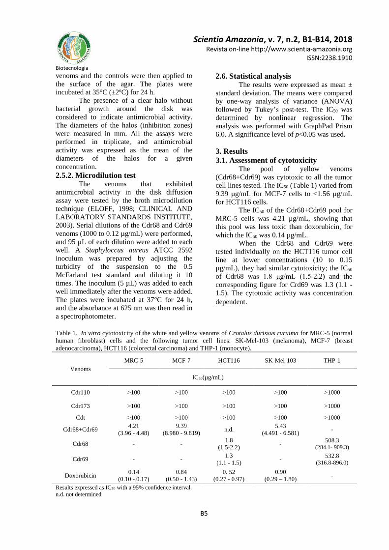
3.1. Assessment of cytotoxicity The pool of yellow venoms
(Cdr68+Cdr69) was cytotoxic to all the tumor
cell lines tested. The IC50 (Table 1) varied from
9.39 µg/mL for MCF-7 cells to <1.56 µg/mL
for HCT116 cells.
The IC50 of the Cdr68+Cdr69 pool for
MRC-5 cells was 4.21 µg/mL, showing that
this pool was less toxic than doxorubicin, for
which the IC50 was 0.14 µg/mL.
When the Cdr68 and Cdr69 were
tested individually on the HCT116 tumor cell
line at lower concentrations (10 to 0.15
µg/mL), they had similar cytotoxicity; the IC50
of Cdr68 was 1.8 μg/mL (1.5-2.2) and the
corresponding figure for Crd69 was 1.3 (1.1 -
1.5). The cytotoxic activity was concentration
dependent.
Table 1. In vitro cytotoxicity of the white and yellow venoms of Crotalus durissus ruruima for MRC-5 (normal
human fibroblast) cells and the following tumor cell lines: SK-Mel-103 (melanoma), MCF-7 (breast
adenocarcinoma), HCT116 (colorectal carcinoma) and THP-1 (monocyte).
Venoms
MRC-5 MCF-7 HCT116 SK-Mel-103 THP-1
IC50(µg/mL)
Cdr110 ˃100 ˃100 ˃100 ˃100 >1000
Cdr173 ˃100 ˃100 ˃100 ˃100 >1000
Cdt ˃100 ˃100 ˃100 ˃100 >1000
Cdr68+Cdr69 4.21
(3.96 - 4.48)
9.39
(8.980 - 9.819) n.d.
5.43
(4.491 - 6.581) -
Cdr68 - - 1.8
(1.5-2.2) -
508.3 (284.1- 909.3)
Cdr69 - - 1.3
(1.1 - 1.5) -
532.8 (316.8-896.0)
Doxorubicin 0.14
(0.10 - 0.17)
0.84
(0.50 - 1.43)
0. 52
(0.27 - 0.97)
0.90
(0.29 – 1.80) -
Results expressed as IC50 with a 95% confidence interval.
n.d. not determined
Page 6
Scientia Amazonia, v. 7, n.2, B1-B14, 2018
Revista on-line http://www.scientia-amazonia.org ISSN:2238.1910
Biotecnologia
B6
3.2. Assessment of genotoxicity – comet
assay Cdr68 and Cdr69 (3μg/mL) were
tested for their capacity to damage DNA in
MRC-5 cells and HCT116 tumor cells using
the alkaline and neutral versions of the comet
assay.
In MRC-5 cells treated with Cdr68 or
Cdr69 the damage index (DI) in an alkaline pH
was higher than in the negative control
(p<0.0001) but lower than in the positive
control (p<0.0001). Cdr69 had greater
genotoxic potential than Cdr68 (p=0.0185). In
neutral pH, treatment with each venom caused
DNA damage, and the result was statistically
significant compared with the negative control
(p<0.0001). The difference between the
positive control and Cdr68 was not statistically
significant (p˃0.05) (Figure 1). In the alkaline
comet assay, the most common types of
damage were class 1 for venoms and class 2
for doxorubicin, while in the neutral comet
assay the most common were classes 2 and 3,
respectively (Figure 2).
Figure 1. Damage index in MRC-5 cells treated with Cdr68 and Cdr69 in alkaline and neutral comet assays. In
the alkaline comet assay both venoms had a lower damage index than the positive control, while in the neutral
assay only Cdr69 had a statistically significantly lower damage index than the positive control. Doxo –
Doxorubicin (positive control). DMSO- Dimethyl sulfoxide (negative control).
Figure 2. Frequency and distribution of damage classes in MRC-5 cells treated with Cdr68 and Cdr69 in alkaline
and neutral comet assays. The most frequent damage class for the venoms was class 1 in the alkaline assay and
class 2 in the neutral assay. Doxo – Doxorubicin (positive control). DMSO - Dimethyl sulfoxide (negative
control).
A statistically significantly higher DI
was observed for HCT-116 cells treated with
the venoms in the alkaline and neutral comet
assay than for the positive and negative
controls (p<0.0001). Cdr68 had a greater
genotoxic potential than Cdr69 (p< 0.0001) in
the alkaline assay, but in the neutral assay
there was no statistically significant difference
between the venoms (Figure 3). The most
common types of damage in the alkaline assay
were classes 2 and 3 for the venoms and class
2 for doxorubicin, while in the neutral comet
assay the most common were classes 4 and 3,
respectively (Figure 4).
Page 7
Scientia Amazonia, v. 7, n.2, B1-B14, 2018
Revista on-line http://www.scientia-amazonia.org ISSN:2238.1910
Biotecnologia
B7
Figure 3. Damage index for HCT-116 cells treated with Cdr68 and Cdr69 in alkaline and neutral comet assays.
In both assays the venoms had a higher damage index than the positive control. Doxo – Doxorubicin (positive
control). DMSO- Dimethyl sulfoxide (negative control).
Figure 4. Frequency and distribution of damage classes in HCT116 cells treated with Cdr68 and Cdr69 in
alkaline and neutral comet assays. The most frequent damage classes for the venoms were classes 2 and 3 in the
alkaline assay and class 4 in the neutral assay. Doxo – Doxorubicin (positive control). DMSO- Dimethyl
sulfoxide (negative control).
3.3. Morphological analysis by
hematoxylin-eosin staining Exposure of HCT116 cells to Cdr68 or
Cdr69 for 72 h caused morphological changes
such as cell shrinkage, pyknotic nuclei,
chromatin condensation and cytoplasmic
vacuolization, and in some cases cellular
remains were observed, showing that the cells
had been destroyed (Figure 5).
3.4. Antimicrobial activity The diameters
of the halos for the yellow venoms (Cdr68 and
Cdr69) measured 9.6 ± 1.52 (SD) mm and 10.6
± 0.57 (SD) mm, respectively, for the S. aureus
ATCC 25923 strain. There was no statistically
significant difference between the activities of
the two venoms for this strain. The
microdilution test was performed with the
same strain and Cdr68 and Cdr69 venoms at
concentrations of 1000, 500, 250, 125 and 62.5
µg/mL. Cdr69 had greater inhibitory potential
than Cdr68 (p ≤ 0.05). However, at a
concentration of 0.12 µg/mL the Cdr68 yellow
venom had greater inhibitory potential (p ≤
0.05), as shown in Figure 6.
4. Discussion The particular characteristics of each
type of cancer, the mechanisms of resistance to
cancer therapies and the broad spectrum of
side effects of cancer treatment make
prevention and treatment of this condition a
challenge (VENDRAMINI-COSTA et al.,
2016).
In the last three decades, many studies
have investigated the anti-cancer properties of
venoms, and this has led to the discovery of
various molecules with promising activities,
some of which are being tested in clinical trials
and may in future be the basis for cancer
therapy drugs (GOMES et al., 2010).
The present study assessed the
cytotoxicity of white (Cdr110 and Cdr173) and
yellow (Cdr68 and Cdr69) snake venoms,
Page 8
Scientia Amazonia, v. 7, n.2, B1-B14, 2018
Revista on-line http://www.scientia-amazonia.org ISSN:2238.1910
Biotecnologia
B8
which had been characterized in a previous
study (DOS-SANTOS et al., 2005). We found
that, in the concentrations tested, the white
venoms and the venom of C. durissus terrificus
were not toxic to the cell lines tested.
However, the mixture of yellow venoms was
toxic to SK-Mel-103, MCF-7 and HCT116
tumor cells, of which the last were the most
sensitive. Although the mixture was toxic to
MRC-5 cells, at low concentrations it only
showed a high cytotoxic effect for colorectal
carcinoma cells.
Figure 5. Optical micrographs showing the morphology of HCT116 cells after 72 h of treatment with Cdr68 or
Cdr69. H&E staining. (A) negative control; (B and C) cells treated with Cdr68 at concentrations of 0.5 and 5
μg/mL, respectively; (D and E) cells treated with Cdr69 at concentrations of 0.5 and 5 μg/mL, respectively.
Changes are indicated by arrows.
Studies carried out with venoms from
snakes in the families Elapidae, Crotalidae and
Viperidae have shown that these can cause
lysis in Yoshida sarcoma cells (BRAGANÇA
et al., 1967). Venoms from Bothrops jararaca
and C. durissus terrificus, which belong to the
family Viperidae, have been shown to act
directly on tumor cells and to induce an
inflammatory response mediated by the
cytokine TNF-α and chemokine CXCL-8 (DA
SILVA et al., 1996).
DA SILVA et al. (1997) assessed in
vivo the effect of C. durissus terrificus venom
on the growth of Ehrlich tumor. Although
treatment with the venom did not completely
eliminate the tumor cells, there was an increase
in the animals’ survival time and significant
macrophage stimulation. The authors suggest
that this effect may be due to activation of
inflammatory responses.
All possible types of DNA damage can
be detected by the alkaline comet assay, while
the neutral assay detects mainly double-
stranded DNA breaks (OLIVE, 1999).
The alkaline comet assay with MRC-5
cells treated with Cdr68 or Cdr69 showed that
these venoms caused less DNA damage than
doxorubicin. In the neutral comet assay, there
was no statistically significant difference
between Cdr68 and doxorubicin, showing that
these venoms damage DNA by causing
double-strand breaks. Cdr69 was less toxic
than Cdr68 and doxorubicin.
Page 9
Scientia Amazonia, v. 7, n.2, B1-B14, 2018
Revista on-line http://www.scientia-amazonia.org ISSN:2238.1910
Biotecnologia
B9
Figure 6. Percentage inhibition by Cdr68 and Cdr69 of growth of the Staphylococcus aureus 25923 strain. At
concentrations of 1000, 500, 250, 125 and 62.5 µg/mL the Cdr69 yellow venom had greater inhibitory potential
than Cdr68 (p ≤ 0.05). PC - Oxytetracycline (positive control). NC – Mueller Hinton culture medium (negative
control).
Analysis of the results of the alkaline
and neutral comet assays in the HCT116 cells
revealed that Cdr68 and Cdr69 had statistically
significantly higher DIs (class 4) than the
positive control, showing that they cause DNA
damage and double-stranded DNA breaks in
in this tumor cell line. Double-stranded DNA
breaks represent a threat to genome integrity
because they can result in chromosome
aberrations that simultaneously affect many
genes, leading to cell malfunctioning and,
consequently, cell death (VAN GENT et al.,
2001). Our results suggest that the cytotoxicity
of Cdr68 and Cdr69 is associated with DNA
damage. Nevertheless, further studies are
required to clarify the cell death mechanisms
induced by these venoms.
MARCUSSI et al. (2011) assessed the
genotoxicity of C. durissus terrificus crude
venom and the toxins isolated from this venom
to human lymphocytes. In the comet assay, all
the toxins tested (crotamine, crotoxin,
phospholipase A2 and crotapotin) and the crude
venom exhibited genotoxicity; the toxins,
however, exhibited greater genotoxicity than
the crude venom. The same authors also
evaluated the genotoxicity of the venoms of
the snakes Bothrops jararacussu, B. atrox, B.
moojeni, B. alternatus and B. brazili to human
lymphocytes. The crude venoms of B. brazili,
B. jararacussu and B. atrox all exhibited
genotoxic potential, and the latter two induced
five times more DNA breaks than the negative
control (MARCUSSI et al., 2013).
TAMIETI et al. (2007) reported
structural changes in actin filaments, the
endoplasmic reticulum and the nucleus as a
result of the action of C. durissus terrificus
venom on the CHO-K1 hamster ovarian cancer
cell line. These structural changes and the
DNA fragmentation the authors observed
suggest that the venom can induce apoptosis.
SOARES et al. (2010) observed
morphological changes in RT2 glioma cells
and GH3 benign pituitary adenoma cells
treated with crude C. durissus terrificus venom
or crotoxin isolated from this venom. These
included irregular cell shapes and cell
shrinkage in cells treated with the crude
venom. Cells treated with crotoxin exhibited
shrinkage, irregular shapes, condensation of
the nucleus and the formation of apoptotic
bodies, all changes characteristic of apoptosis.
OVCAR-8 ovarian carcinoma cells treated
with C. durissus cascavella venom showed a
reduction in cell volume, irregular shapes,
nuclear fragmentation, pyknotic nuclei and the
formation of apoptotic bodies. Activation of
caspases 3 and 7 in these cells confirmed that
the venom induces cell death by apoptosis
(ARAÚJO et al., 2016).
HCT-116 cells treated with Cdr68 and
Cdr69 displayed morphological changes such
as cell shrinkage, pyknotic nuclei, condensed
chromatin and cytoplasmic vacuolation, and in
Page 10
Scientia Amazonia, v. 7, n.2, B1-B14, 2018
Revista on-line http://www.scientia-amazonia.org ISSN:2238.1910
Biotecnologia
B10
some cases cellular remains were observed,
indicating apoptosis. However, as this is to our
knowledge the first study of morphological
changes in cells treated with C. durissus
ruruima venom, further specific studies are
required to clarify the mechanism of death
induced by these venoms.
Snake venoms contain a mixture of
biologically active substances, each of which
may exercise its action separately or jointly
with other components, producing a synergic
effect (LIPPS, 1995; RANGEL-SANTOS et
al., 2004). The use of venoms to treat tumors is
therefore not straightforward and can be
dangerous as venoms are complex mixtures of
proteins and peptides that can affect
homeostasis (LU et al., 2005). Some of these
difficulties could be overcome, however, if
venoms were used with nanoparticles, which
have shown great promise for treatment of
cancer patients. Nanoparticles bound to
anticancer agents can increase the
concentration of these agents in specific target
tissues and be phagocyted and/or endocyted.
Because of this internalization, the drug’s
efficiency increases and toxicity reduces,
increasing its therapeutic index (BARRATT,
2003).
Badr et al. (2014) assessed the effect of
Walterinnesia aegyptia venom on its own and
coupled to nanoparticles on human breast
cancer cells isolated from biopsies. In both
cases it inhibited proliferation, changed the cell
cycle and induced apoptosis, and when bound
to nanoparticles it increased the antitumor
effect. The IC50 for the venom on its own and
for the venom bound to the nanoparticles was
50 ng/mL and 20 ng/mL, respectively,
indicating the potential of nanoparticle-bound
venoms in cancer treatment.
In the present study, Cdr173, Cdr110,
Cdr68, Cdr69 and Cdt were also evaluated for
antimicrobial activity. While Cdr173, Cdr110
and Cdt did not exhibit any activity against the
bacterial strains tested, Cdr69 and Cdr68 both
exhibited activity against S. aureus 25923, but
the latter had the greater activity (p ≤ 0.05).
The first reports of the antimicrobial
activity of snake venoms were in 1948 and
1968, in studies using venoms from snakes in
the families Elapidae and Viperidae
(GLASER, 1948; ALOOF-HIRSCH et al.,
1968). The venoms of Naja spp. and
Hemachatus haemachatus were shown to
contain direct lytic factor, indicating that they
could break down the phospholipid membranes
of S. aureus and E. coli, respectively (ALOOF-
HIRSCH et al., 1968).
Various proteins have been isolated
from crotalic venoms, including crotoxin, L-
amino acid oxidase and crotamine, and their
antimicrobial activity has been investigated
and confirmed (OLIVEIRA et al., 2003;
TOYAMA et al., 2006; OGUIURA et al.,
2011). Further studies of the venoms of snakes
in the North of Brazil are therefore needed to
gain a better understanding of their action
against tumor cells and antibiotic-resistant
bacteria. Our research group is already
fractionating and isolating proteins from the
white and yellow venoms of the Amazonian
snake C. durissus ruruima and evaluating the
cytotoxicity and antibacterial activity of these
fractions.
Acknowledgments The authors would like to express their
gratitude to the CNPq (National Council for
Scientific and Technological Development) for
awarding a productivity grant to Maria Cristina
dos Santos (303032/2016-2); to the FAPEAM
(State of Amazonas Research Foundation) for
providing a doctoral fellowship for Ilia
Gilmara Carvalho dos Santos; and to
FAPEMIG (State of Minas Gerais Research
Foundation) for providing a research and
technological development incentive grant for
Consuelo Latorre Fortes-Dias.
References
AHMED, S. A.; GOGAL, R. M.; WALSH, J. E. A
new rapid and simple non-radioactive assay to
monitor and determine the proliferation of lymphocytes an alternative to [3H] thymidine
incorporation assay. Journal of immunological methods, v. 170, n. 2, p.
211-224, 1994.
ALOOF-HIRSCH, S.; DE VRIES, A.; BERGER, A. The direct lytic factor of cobra venom:
purification and chemical characterization. Biochimica et Biophysica Acta (BBA) -
Protein Structure, v. 154, n. 1, p. 53-60,
Page 11
Scientia Amazonia, v. 7, n.2, B1-B14, 2018
Revista on-line http://www.scientia-amazonia.org ISSN:2238.1910
Biotecnologia
B11
1968. http://dx.doi.org/10.1016/0005-
2795(68)90257-2.
ARAÚJO, L. S.; ROCHA, D. D.; VIANA, D. A.;
SILVEIRA, J. A. M.; VASCONCELOS-FILHO, F.
S. L.; WILKE, D. V.; BORGES-NOJOSA, D. M.; O’ PESSOA, C.; MORAES, M. O.;
EVANGELISTA, J. S. A. M. Crotalus durissus cascavella VENOM TOXICITY TO MAMMALIAN
CELLS. Veterinária e Zootecnia v. 23, n. 3, p. 465-475, 2016.
BARRATT, G. Colloidal drug carriers:
achievements and perspectives. Cellular and Molecular Life Sciences CMLS, v. 60, n. 1,
p. 21-37, January 01 2003.
BARROS, G. A. C.; PEREIRA, A. V.; BARROS, L.
C.; JR, A. L.; CALVI, S. A.; SANTOS, L. D.;
BARRAVIERA, B.; FERREIRA, R. S. In vitro activity of phospholipase A2 and of peptides
from Crotalus durissus terrificus venom against amastigote and promastigote forms of
Leishmania (L.) infantum chagasi. Journal of Venomous Animals and Toxins including
Tropical Diseases, v. 21, n. 1, p. 1-9, 2015.
BARROS, L.; SOARES, A.; COSTA, F.; RODRIGUES, V.; FULY, A.; GIGLIO, J.;
GALLACCI, M.; THOMAZINI-SANTOS, I.; BARRAVIERA, S.; BARRAVIERA, B.; FERREIRA
JUNIOR, R. Biochemical and biological
evaluation of gyroxin isolated from Crotalus durissus terrificus venom. Journal of
Venomous Animals and Toxins including Tropical Diseases, v. 17, p. 23-33, 2011.
BENLI, M.; YIGIT, N. Antibacterial activity of
venom from funnel web spider Agelena labyrinthica (Araneae: Agelenidae). Journal of
Venomous Animals and Toxins including Tropical Diseases, v. 14, p. 641-650, 2008.
BRAGANÇA, B. M.; PATEL, N. T.; BADRINATH, P. G. Isolation and properties of a cobravenom
factor selectively cytotoxic to yoshida sarcoma
cells. Biochimica et Biophysica Acta (BBA) - General Subjects, v. 136, n. 3, p. 508-520,
1967. http://dx.doi.org/10.1016/0304-4165(67)90009-8.
CLINICAL AND LABORATORY STANDARDS
INSTITUTE. Methods for Dilution Antimicrobial Susceptibility Tests for Bacteria That Grow
Aerobically; ApprovedStandard—Sixth Edition. NCCLS document M7-A6 (ISBN 1-56238-486-
4). NCCLS, 940West. Valley Road, Suite 1400, Wayne, Pennsylvania 19087-1898 USA., 2003.
CLINICAL AND LABORATORY STANDARDS
INSTITUTE, C. Methods for Dilution Antimicrobial Susceptibility Tests for Bacteria
That Grow Aerobically. Approved Standard—
Sixth Edition. NCCLS document M7-A6 (ISBN 1-56238-486-4). NCCLS, 940 West. Valley
Road, Suite 1400, Wayne, Pennsylvania 19087-1898 USA, 2012.
CONDE, R.; ZAMUDIO, F. Z.; RODRıGUEZ, M. H.; POSSANI, L. D. Scorpine, an anti-malaria
and anti-bacterial agent purified from scorpion
venom. FEBS Letters, v. 471, n. 2, p. 165-168, 2000/04/14/ 2000.
http://dx.doi.org/10.1016/S0014-5793(00)01384-3.
CORIN, R. E.; VISKATIS, L. J.; VIDAL, J. C.;
ETCHEVERRY, M. A. Cytotoxicity of crotoxin on murine erythroleukemia cellsin vitro.
Investigational New Drugs, v. 11, n. 1, p. 11-15, 1993.
DA SILVA, R. J.; FECCHIO, D.; BARRAVIEIRA, B. Antitumor effect of snake venoms. Journal
of Venomous Animals and Toxins, v. 2, p.
79-90, 1996.
DA SILVA, R. J.; FECCHIO, D.; BARRAVIEIRA,
B. EFFECT OF Crotalus durissus terrificus (LAURENTI, 1768) venom on the evolution of
ehrlich ascites tumor. Journal of Venomous
Animals and Toxins, v. 3, p. 324-341, 1997.
DIZ FILHO, E. B. S.; MARANGONI, S.;
TOYAMA, D. O.; FAGUNDES, F. H. R.; OLIVEIRA, S. C. B.; FONSECA, F. V.;
CALGAROTTO, A. K.; JOAZEIRO, P. P.;
TOYAMA, M. H. Enzymatic and structural characterization of new PLA2 isoform isolated
from white venom of Crotalus durissus ruruima. Toxicon, v. 53, n. 1, p. 104-114,
2009. http://dx.doi.org/10.1016/j.toxicon.2008.10.02
1.
DOS-SANTOS, M. C.; ASSIS, E. B.; MOREIRA, T. D.; PINHEIRO, J.; FORTES-DIAS, C. L.
Individual venom variability in Crotalus durissus ruruima snakes, a subspecies of
Crotalus durissus from the Amazonian region.
Toxicon, v. 46, n. 8, p. 958-961, 2005. http://dx.doi.org/10.1016/j.toxicon.2005.06.00
8.
DOS-SANTOS, M. C.; FERREIRA, L. C. L.; DA
SILVA, W. D.; FURTADO, M. D. F. D. Caracterizacion de las actividades biologicas de
Page 12
Scientia Amazonia, v. 7, n.2, B1-B14, 2018
Revista on-line http://www.scientia-amazonia.org ISSN:2238.1910
Biotecnologia
B12
los venenos ‘amarillo’ y ‘blanco’ de Crotalus
durissus ruruima comparados con el veneno de Crotalus durissus terrificus. Poder neutralizante
de los antivenenos frente a los venenos de
Crotalus durissus ruruima. Toxicon, v. 31, n. 11, p. 1459-1469, 1993.
http://dx.doi.org/10.1016/0041-0101(93)90211-Z.
ELOFF, J. N. A sensitive and quick microplate method to determine the minimal inhibitory
concentration of plant extracts for bacteria.
Planta Med, v. 64, p. 711-713, 1998.
FOSTER, T. J. The Staphylococcus aureus “superbug”. Journal of Clinical Investigation, v. 114, n. 12, p. 1693-1696,
2004.
GLASER, H. S. R. Bactericidal Activity of Crotalus Venom in Vitro. Copeia, v. 1948, n.
4, p. 245-247, 1948.
GOMES, A.; BHATTACHARJEE, R. M.; BISWAS,
A. K.; DASGUPTA, S. C.; GIRI, B. Anticancer potential of animal venoms and toxins. Indian
Journal of Experimental Biology, v. 48, p.
93-103, 2010.
HAN, R.; LIANG, H.; QIN, Z.; LIU, C. Crotoxin
induces apoptosis and autophagy in human lung carcinoma cells in vitro via activation of
the p38 MAPK signaling pathway. Acta
Pharmacologica Sinica, v. 35, p. 1323-1332, 2014.
HEGAZI, A. G.; EL-FEEL, M.; ABDEL-RAHMAN, E.; AL-FATTAH, A. Antibacterial activity of bee
venom collected from apis mellifera carniolan
pure and hybrid races by two collection methods. Int. J. Curr. Microbiol. App. Sci,
v. 4, n. 4, p. 141-149, 2015.
JALAEI, J.; FAZELI, M.; RAJAIAN, H.;
SHEKARFOROUSH, S. S. In vitro antibacterial effect of wasp (Vespa orientalis) venom.
Journal of Venomous Animals and Toxins
including Tropical Diseases, v. 20, n. 1, p. 22, May 20 2014.
KOH, D. C.; ARMUGAN, A.; JEYASEELAN, K. Snake venom components and their
applications in biomedicine. Cellular and
Molecular Life Sciences, v. 63, p. 3030-3041, 2006.
KUMAR, S.; SARKAR, P.; JAIN, R. Venoms can be a boon for cancer patients. Forum on
Immunopathological diseases and
Therapeutics, v. 4, p. 255-273, 2013.
LIPPS, B. V. Eleventh World Congress on
animel, plant and microbial toxins Tel Aviv,
Israel 2–7 October 1994 Abstract of presentations lectures. Toxicon, v. 33, n. 3, p.
262, 1995.
LOWY, F. D. Staphylococcus aureus infections.
New England journal of medicine, v. 339, n. 8, p. 520-532, 1998.
LU, Q.; CLEMETSON, J. M.; CLEMETSON, K. J.
Snake venoms and hemostasis. Journal of Thrombosis and Haemostasis, v. 3, n. 8, p.
1791-1799, 2005.
MALUF, S. C.; MAS, C. D.; OLIVEIRA, E. B.;
MELO, P. M.; CARMONA, A. K.; GAZARINI, M.
L.; HAYASHI, M. A. F. Inhibition of malaria parasite Plasmodium falciparum development
by crotamine, a cell penetrating peptide from the snake venom. Peptides, v. 78, p. 11-16,
2016. http://dx.doi.org/10.1016/j.peptides.2016.01.0
13.
MARCUSSI, S.; SANTOS, P. R. S.; MENALDO, D. L.; SILVEIRA, L. B.; SANTOS-FILHO, N. A.;
MAZZI, M. V.; DA SILVA, S. L.; STÁBELI, R. G.; ANTUNES, L. M. G.; SOARES, A. M. Evaluation
of the genotoxicity of Crotalus durissus
terrificus snake venom and its isolated toxins on human lymphocytes. Mutation
Research/Genetic Toxicology and Environmental Mutagenesis, v. 724, n. 1–
2, p. 59-63, 2011.
https://doi.org/10.1016/j.mrgentox.2011.06.004.
MARCUSSI, S.; STÁBELI, R. G.; SANTOS-FILHO, N. A.; MENALDO, D. L.; SILVA
PEREIRA, L. L.; ZULIANI, J. P.; CALDERON, L. A.; DA SILVA, S. L.; GREGGI ANTUNES, L. M.;
SOARES, A. M. Genotoxic effect of Bothrops
snake venoms and isolated toxins on human lymphocyte DNA. Toxicon, v. 65, n.
Supplement C, p. 9-14, 2013/04/01/ 2013. https://doi.org/10.1016/j.toxicon.2012.12.020.
MITTERMEIER, R. A.; FONSECA, G. D.;
RYLANDS, A. B.; BRANDON, K. Uma breve história da conservação da biodiversidade no
Brasil. Megadiversidade, v. 1, n. 1, p. 14-21, 2005.
MULLER, V. D. M.; RUSSO, R. R.; OLIVEIRA CINTRA, A. C.; SARTIM, M. A.; DE MELO
Page 13
Scientia Amazonia, v. 7, n.2, B1-B14, 2018
Revista on-line http://www.scientia-amazonia.org ISSN:2238.1910
Biotecnologia
B13
ALVES-PAIVA, R.; FIGUEIREDO, L. T. M.;
SAMPAIO, S. V.; AQUINO, V. H. Crotoxin and phospholipases A2 from Crotalus durissus terrificus showed antiviral activity against
dengue and yellow fever viruses. Toxicon, v. 59, n. 4, p. 507-515, 2012.
http://dx.doi.org/10.1016/j.toxicon.2011.05.021.
NEVES, M. S.; SOUSA, D. R. T.; SOCORRO, M. P.; FERREIRA, B. C.; FROTA, M. Z. M.; SOUZA,
J. V. B.; LOZANO, L. L. L. Evaluation of
antifungal activity of snake venoms from the Amazon forest. Journal of Yeast and
Fungal Research, v. 6, n. 2, p. 11-16, 2015.
OGUIURA, N.; BONI-MITAKE, M.; AFFONSO,
R.; ZHANG, G. In vitro antibacterial and
hemolytic activities of crotamine, a small basic myotoxin from rattlesnake Crotalus durissus. The Journal of Antibiotics, v. 64, p. 327-331, 2011.
OLIVE, P. L. DNA damage and repair in individual cells: applications of the comet assay
in radiobiology. International Journal of
Radiation Biology, v. 75, n. 4, p. 395-405, 1999/01/01 1999.
OLIVEIRA, D. G.; TOYAMA, M. H.; MARTINS, A. M. C.; HAVT, A.; NOBRE, A. C. L.;
MARANGONI, S.; CÂMARA, P. R.; ANTUNES,
E.; DE NUCCI, G.; BELIAM, L. O. S.; FONTELES, M. C.; MONTEIRO, H. S. A.
Structural and biological characterization of a crotapotin isoform isolated from Crotalus
durissus cascavella venom. Toxicon, v. 42, n.
1, p. 53-62, 2003/07/01/ 2003. https://doi.org/10.1016/S0041-
0101(03)00100-4.
PERUMAL SAMY, R.; PACHIAPPAN, A.;
GOPALAKRISHNAKONE, P.; THWIN, M. M.; HIAN, Y. E.; CHOW, V. T.; BOW, H.; WENG, J.
T. In vitro antimicrobial activity of natural
toxins and animal venoms tested against Burkholderia pseudomallei. BMC Infectious
Diseases, v. 6, n. 1, p. 100, June 20 2006.
PERUMAL SAMY, R.; STILES, B. G.; FRANCO,
O. L.; SETHI, G.; LIM, L. H. K. Animal venoms
as antimicrobial agents. Biochem Pharmacol, v. 134, p. 127-138, 2017.
QUINTANA, J. C.; CHACÓN, A. M.; VARGAS, L.; SEGURA, C.; GUTIÉRREZ, J. M.; ALARCÓN, J.
C. Antiplasmodial effect of the venom of Crotalus durissus cumanensis, crotoxin
complex and Crotoxin B. Acta Tropica, v.
124, n. 2, p. 126-132, 2012. http://dx.doi.org/10.1016/j.actatropica.2012.0
7.003.
RANGEL-SANTOS, A.; DOS-SANTOS, E. C.; LOPES-FERREIRA, M.; LIMA, C.; CARDOSO, D.
F.; MOTA, I. A comparative study of biological activities of crotoxin and CB fraction of venoms
from Crotalus durissus terrificus, Crotalus durissus cascavella and Crotalus durissus
collilineatus. Toxicon, v. 43, n. 7, p. 801-810,
2004. http://dx.doi.org/10.1016/j.toxicon.2004.03.01
1.
RUDD, C. J.; VISKATIS, L. J.; VIDAL, J. C.;
ETCHEVERRY, M. A. In vitro comparison of
cytotoxic effects of crotoxin against three human tumors and a normal human epidermal
keratinocyte cell line. Investigational New Drugs, v. 12, n. 3, p. 183-184, 1994.
SANTOS, N. Q. A resistência bacteriana no contexto da infecção hospitalar. Texto
Contexto Enferm, v. 13, p. 64-70, 2004.
SINGH, N. P.; MCCOY, M. T.; TICE, R. R.; SCHNEIDER, E. L. A simple technique for
quantitation of low levels of DNA damage in individual cells. Experimental Cell
Research, v. 175, n. 1, p. 184-191,
1988/03/01/ 1988. http://dx.doi.org/10.1016/0014-
4827(88)90265-0.
SOARES, M.; PUJATTI, P.; FORTES-DIAS, C.;
ANTONELLI, L.; SANTOS, R. Crotalus durissus terrificus venom as a source of antitumoral agents. Journal of Venomous Animals and
Toxins including Tropical Diseases, v. 16, p. 480-492, 2010.
TAMIETI, B. P.; DAMATTA, R. A.; COGO, J. C.; DA SILVA, N. S.; MITTMANN, J.; PACHECO-
SOARES, C. Cytoskeleton, endoplasmic
reticulum and nucleus alterations in CHO-K1 cell line after Crotalus durissus terrificus (South
American rattlesnake) venom treatment. Journal of Venomous Animals and Toxins
including Tropical Diseases, v. 13, p. 56-
68, 2007.
TOYAMA, M. H.; TOYAMA, D. D. O.; PASSERO,
L. F. D.; LAURENTI, M. D.; CORBETT, C. E.; TOMOKANE, T. Y.; FONSECA, F. V.; ANTUNES,
E.; JOAZEIRO, P. P.; BERIAM, L. O. S.; MARTINS, M. A. C.; MONTEIRO, H. S. A.;
Page 14
Scientia Amazonia, v. 7, n.2, B1-B14, 2018
Revista on-line http://www.scientia-amazonia.org ISSN:2238.1910
Biotecnologia
B14
FONTELES, M. C. Isolation of a new l-amino
acid oxidase from Crotalus durissus cascavella venom. Toxicon, v. 47, n. 1, p. 47-57, 2006.
http://dx.doi.org/10.1016/j.toxicon.2005.09.00
8.
VAN GENT, D. C.; HOEIJMAKERS, J. H.;
KANAAR, R. Chromosomal stability and the DNA double-stranded break connection. Nat
Rev Genet, v. 2, n. 3, p. 196-206, 2001.
VARGAS, L. J.; QUINTANA, J. C.; PEREAÑEZ, J.
A.; NÚÑEZ, V.; SANZ, L.; CALVETE, J. Cloning
and characterization of an antibacterial L-amino acid oxidase from Crotalus durissus cumanensis venom. Toxicon, v. 64, p. 1-11, 2013.
VENDRAMINI-COSTA, D. B.; ALCAIDE, A.;
PELIZZARO-ROCHA, K. J.; TALERO, E.; ÁVILA-ROMÁN, J.; GARCIA-MAURIÑO, S.; PILLI, R.
A.; DE CARVALHO, J. E.; MOTILVA, V. Goniothalamin prevents the development of
chemically induced and spontaneous colitis in
rodents and induces apoptosis in the HT-29 human colon tumor cell line. Toxicology and
Applied Pharmacology, v. 300, n.
Supplement C, p. 1-12, 2016/06/01/ 2016. https://doi.org/10.1016/j.taap.2016.03.009.
WHITE, J. Bites and stings from venomous animals: a global overview. Therapeutic
drug monitoring, v. 22, n. 1, p. 65-68, 2000.
WHO. Cancer:. Disponível em: <
http://www.who.int/cancer/en >. Acesso em: 07.08.2017.
WOJEWÓDZKA, M.; BURACZEWSKA, I.; KRUSZEWSKI, M. A modified neutral comet
assay: Elimination of lysis at high temperature
and validation of the assay with anti-single-stranded DNA antibody. Mutation Research
- Genetic Toxicology and Environmental Mutagenesis, v. 418, n. 1, p. 9-20, 2002.