SCIENTIFIC ARTICLES A comparative study of tooth apexification in the dog George Pierre Citrome, DDS, MS; Edward J. Kaminski, PhD; and Michael A. Heuer, DDS, MS, Chicago Calcium hydroxide, collagen-calcium phosphate gel, and blood clot were compared as inducers of periapical calcification in the immature nonvital teeth of dogs. This investigation showed that calcium hydroxide accelerated hard tissue bridging of the open apex irrespective of complete resolution of initially induced inflammatory state. Collagen-calcium phosphate gel inhibited the reparative process of the initial inflammatory lesion leading to extensive destruction of the periapical tissues with no evidence of apexification. Locally induced blood clot maintained the initial inflammatory state and did not result in hard tissue bridging of the open apex. The investigation confrmed the use of the dog as an effective animal model for meeting the criteria of the experimental design. Apexification procedures to treat the nonvital immature root canal with a wide open apex have come into common use in the modern clinical practice of endodontics. Although calcium hydroxide pastes are the root canal filling materials most often used to induce apexification,'- ~e one school of thought questions the necessity of placing any filling mate- rial in the root canal to fulfill this objective. Nygaard-Ostby ':~ reported that the creation of an apical blood clot by over-instrumenting sterilized necrotic roots caused continued api- cal root closure. Moodnick ~' pro- posed that removing the bulk of necrotic pulpal tissue and filling with gutta-percha short of the apex could provide healing at the apex. Ham and others" have shown that both calcium hydroxide and the induction of a blood clot would lead to apexifi- cation of immature root canals in the monkey. Some investigators recently have introduced new materials to provide more predictable success. Tricalcium phosphate ceramic material used as a resorbable implant material to stim- ulate apical closure has been success- ful in experiments on monkeys'; and equally successful when compared with use of calcium hydroxide in human studies."; However, apical closure was not achieved with this material in an investigation in which dogs were used." Among the newer materials, col- lagen-calcium phosphate gel appears promising. In 1976, Nevins and others '~ published a report on the use of this gel as a filling material in pulpless teeth with open apexes in monkeys. When compared with a calcium hydroxide paste, this sub- stance promoted a rapid and more physiological apexification. Several of the teeth filled with the collagen gel "appeared to be revitalized with various forms of hard and soft connective tissue, including cemen- tum, bone, and dentin derived from lacerated remnants of uninflamed pulp tissue. '''~ The authors con- cluded that this material may be a step in the direction of revitalizing human pulpless teeth. Aside from the contention that several different techniques will lead to apexification of wide open root canal apexes, careful perusal of the literature has disclosed several varia- tions in the research methodology relating to experimental design, choice of animal model, and inter- pretation of the experimental data. Experimental design includes those conditions to be controlled 290
Transcript
SCIENTIFIC ARTICLES
A comparative study of tooth apexification in the dog
George Pierre Citrome, DDS, MS; Edward J. Kaminski, PhD; and Michael A. Heuer, DDS, MS, Chicago
Calcium hydrox ide , co l lagen-ca lc ium phospha te gel, and b lood clot were c o m p a r e d as inducers of periapical calcification in the immatu re nonvital tee th of dogs. This invest igat ion showed that calcium h y d r o x i d e accelerated hard t issue br idging of the o p en apex irrespective of comple te resolu t ion of initially induced in f l ammatory state. Col lagen-ca lc ium phospha t e gel inhibi ted the repara t ive process of the initial i n f l ammatory lesion leading to extens ive des t ruc t ion of the periapical tissues with no ev idence of apexif icat ion. Locally induced b lood clot main ta ined the initial i n f l ammatory state and did not resul t in hard tissue br idging of the open apex. T h e invest igat ion c o n f r m e d the use of the dog as an effective animal mode l for meet ing the criteria of the exper imen ta l design.
Apexification procedures to treat the nonvital immature root canal with a wide open apex have come into common use in the modern clinical practice of endodontics. Although calcium hydroxide pastes are the root canal filling materials most often used to induce apexification,'- ~e one school of thought questions the necessity of placing any filling mate- rial in the root canal to fulfill this objective. Nygaard-Ostby ':~ reported that the creation of an apical blood clot by over-instrumenting sterilized necrotic roots caused continued api- cal root closure. Moodnick ~' pro- posed that removing the bulk of necrotic pulpal tissue and filling with gutta-percha short of the apex could provide healing at the apex. Ham and others" have shown that both calcium hydroxide and the induction of a blood clot would lead to apexifi-
cation of immature root canals in the monkey.
Some investigators recently have introduced new materials to provide more predictable success. Tricalcium phosphate ceramic material used as a resorbable implant material to stim- ulate apical closure has been success- ful in experiments on monkeys'; and equally successful when compared with use of calcium hydroxide in human studies."; However, apical closure was not achieved with this material in an investigation in which dogs were used."
Among the newer materials, col- lagen-calcium phosphate gel appears promising. In 1976, Nevins and others '~ published a report on the use of this gel as a filling material in pulpless teeth with open apexes in monkeys. When compared with a calcium hydroxide paste, this sub-
stance promoted a rapid and more physiological apexification. Several of the teeth filled with the collagen gel "appeared to be revitalized with various forms of hard and soft connective tissue, including cemen- tum, bone, and dentin derived from lacerated remnants of uninflamed pulp tissue. '''~ The authors con- cluded that this material may be a step in the direction of revitalizing human pulpless teeth.
Aside from the contention that several different techniques will lead to apexification of wide open root canal apexes, careful perusal of the literature has disclosed several varia- tions in the research methodology relating to experimental design, choice of animal model, and inter- pretation of the experimental data.
Experimental design includes those conditions to be controlled
290
JOURNAL OF E N D O D O N T I C S VOL 5, N O 10, OCTOBER 1979
before the apexification procedures are performed. Studies have been done on root canals that, before the actual apexilication procedures, have ranged from those in intact healthy teeth '' ~:~ '~ to those that have been experimentally infected and made necrotic." ....... Similarly, stud- ies have been published in which the experimental teeth have contained fully developed root canals in which the apex was perforated to simulate a blunderbuss c a n a l " ' : " " to immature root canals with naturally occurring wide open apexes.-' ...........
The choice of animal model in these investigations also is controver- sial. Basically, three species have been used: humans ~ ' ' ' ' ; > mon- keys," ............ and dogs. ' ......... Mon- keys have been used extensively in these studies because of the evolu- tionary resemblance to humans, in particular the resemblance anatomi- cally to the human dentition. I-low- ever, this outward resemblance may be deceiving according to Torneck and others :'" who, in 1973, studied the effect of injury to the pulp with oral contaminat ion on developing monkey incisors. In spite of filing the walls of the root canals to remove residual fragments of pulp tissue and leaving the teeth open to oral sali- vary contaminat ion tbr up to 95 days, dentin and cementum were deposited at the periapex leading to apexification of the open apexes. The data suggest that monkeys, when compared with humans, have greater recuperative powers. Specifically, monkey pulpal tissue is highly resis- tant to the effects of oral contamina- tion; despite the presence of severe inflammatory disease, the periapical tissues of the monkey still allow for repair of hard tissue in the form of dentin, cementum, and bone.
Several investigators have found a close similarity in healing processes of teeth and supporting structures between humans and dogs. -''--'~ In 1932, Orban : ' neatly summarized whv dogs make ideal animal models: "Dogs' teeth are more sensitive to any kind of injury than human teeth. This mav be due to a greater permeabili ty of hard structures, den- tin and cementum in the dog than in humans, and that if t reatment in the thr more sensitive dog proves satis- factory it can be expected to be satisfactory in humans."
A third shortcoming in some of these studies relates to interpretation of the experimental data with respect to the histological evidence which is subjective to one degree or another. Histological evidence of hard tissue depositiori around the apex of a root canal can be seen in a section stained with hematoxvlin and eosin. Howev- er. under these conditions it is impos- sible to conclude objectively whether this deposition of hard tissue oc- curred before, during, or after the apexification procedure. The only way to confirm that the deposition of hard tissue observed is the result of the experiment is to label the tissue with a biological marker before and after the apexitication procedure is performed. The dye Procion Red, when viewed in unstained decalci- fied sections under ultraviolet light and with a specific excitation filter, has biologically marked actively cal- cifying connective tissues?"
An analysis of the results obtained in this investigation has been ensured by simulating a typical clinical situa- tion, namely, necrotic immature root canals; using an animal (the dog) that is similar to humans in biologi- cal response; and assessing hard tissue repair with Procion dye, a vital marker.
In applying the foregoing princi- ples, the purpose of this investigation was to study histologically the effects of various techniques to induce peri- apical calcifications in the teeth of dogs with incompletely formed apexes and with necrotic pulps. Specifically, a comparison of the standard material, calcium hydrox- ide, with collagen-calcium phos- phate gel was made. As a control, the induction of a blood clot without any filling materials also was investi- gated.
MATERIALS AND METHODS
Materials
The double-rooted premolars of two 5-month-old beagle dogs were used, giving a total of 20 experimen- tal teeth. Preliminat2, ,' radiographs showed incompletely formed roots. Intraperitoneal injections of Procion Brilliant Red dye (2~ solution, 100 mg/kg body weight) were adminis- tered to each animal at four intervals six weeks apart starting three weeks before the pulpectomies, then at three and nine weeks after the experi- mental procedure, and shortly before the animals were killed. Before the apexification procedures were begun, the pulps were rendered necrotic by performing pulpectomies and leav- ing them open to oral salivary contaminat ion for a week. The necrotic root canals were then debrided and treated with 1% aque- ous parachlorophenol and sealed with amalgam for another week.
Methods
Treatment of the open apex with the selected procedure was per- formed with use of rubber dam isola- tion and sterile techniques. Five of
291
JOURNAL OF ENDODONT1CS VOL 5, NO 10, OCTOBER 1979
the prepared teeth were simply instrumented beyond the apex to induce a blood clot (this was verified by blood seeping into the canal); eight of the prepared teeth were filled with a collagen-calcium phos- phate gel prepared in the manner described by Nevins and othrs 'x and the remaining seven teeth were filled with a calcium hydroxide-saline paste. All the teeth then were double-sealed with a stop of base plate gutta-percha followed by a Class I amalgam filling.
All treatments were performed with the animals under general anes- thesia; injections of sodium pento- barbital were used.
Periapical radiographs of the ex- perimental teeth were taken at regu- lar intervals to moni tor clinical progress of hard tissue pathological conditions or repair. The animals were killed at the end of 11 weeks when large radiolucent areas oc- curred at the apexes of several of the teeth. The tissues were fixed with a saline and Formalin infusion tech- nique according to procedures used by Kozlowski. ~ Block dissections of the jaws were made and decalcified in Perenyi's solution. ~' The speci- mens were embedded in paraffin. Semi-serial sections were cut into a thickness of 5 g at the plane of the root apex. Alternate slides were stained with hematoxylin and eosin. Adjacent alternate slides were left unstained for viewing with ultravio- let light with use of excitation filter BGI2* and barrier filter 53* in a Zeiss photomicroscope.*
Cri ter ia
The criteria of successful treat- ment were lack of inf lammatory cells and edema in the periapical tissues, continuity of the fiber pat tern of the
292
Table 1 �9 Response of the periapical tissue to the experimen- tal procedure.
Periapical tissue response*
Procedure None Mild Severe
Induction of a blood 0 1 4 clot (5)4
Collagen (8) 0 0 8 Calcium hydroxide (7) 5 1 1
"Number of icrth r the response in each exp~:rlmental group Frotal number of teeth in parentheses
Table 2 �9 Evidence of apexification and cementogenesis.
*Numl~r of tcclh showing the response in t'ach e~tperimenlal group fFo la l numi-~r of teelh m partuthest 's
periodontal ligament, and no resorp- tion of cementum or dentin.
Adverse periapical reactions were classified as being either mild or severe int lammator T reactions. Mild inflammation was interpreted as being caused by the presence of moderate populations of inflamma- tory cells with minimal loss of the continuily of the periodontal liga- ment. Severe inflammation was characterized by the presence of ei- ther granulomatous tissues, ab- scesses, or cysts concomitant with destruction of the periodontal liga- ment a, nd resorption of cementum or dentin, or both.
The criteria of successful apexifi- cation and active cementogenesis at the apex were evaluated by looking for the presence of orange fluorescent bands in the unstained sections under ultraviolet light, because Pro- cion dye labels actively calcifying tissues. As four injections of the
Procion dye were administered at six-week intervals, the number of fluorescent lines (to a maximum of four) were recorded as an index of the quant i ty of cementum laid down.
R E S U L T S
The response of the periapical tissues to the experimental proce- dures with respect to inf lammatory changes and hard tissue deposition is shown in Tables 1 and 2.
Of the five blood clot controls, the predominant lesion was granuloma- tous. One specimen showed only mild inflammation, and one showed cementogenesis (Fig 1).
All eight collagen-calcium phos- phate gel specimens disclosed severe inf lammatory lesions (Fig 2). Acute abscesses, granulomas, and cysts causing extensive destruction of the periapical tissues were characteristic responses. Resorption of both cemen-
~ " �9 ~ �9 ~ " - ~ " " 1
," ~
. , ,~ j ~ ,
f
" i : d .
...% ,,#.. . ~ , .
| . . . , ' . ~ , .",~r 211
JOURNAL OF E N D O D O N T I C S V O I 5. N O 10, OCTOBER 1979
A
o i"
:i! . . . . . .
,,' . 1 !.,..~.~..
'., '.," . _
8 C
D E
Fziq l - A , blood clot spe~ tmcn shou,itlg cementum on lateral aspect of one ~ide q/" root, as zcell as internal re~orptton o/dentm on its medial a.~/,ect. Other ~tde ~!f root shou,~ resorptton ~!/'~ernentum and dentin. Nvttce cellular in~,aswn of root ,anal space ( I I&E, ortg mag • 35). B, ht.~her mag- n(fication of lateral a~pect ?f root m P't_~ure 1..4, shou,m~ detatl of cementum (H&E, (,rt~ mag x 140). (2, corresponding unstained ,ectton o]'Ftgure 1, B. :%SJtt~efour bands ,!]'fluores, ence m cementum confirming actwe cementogenests ( l, Z 3, and 4), attd on O' one fluore.~cent band m dentin (1)), confirmtn s that no neu" dentin ha3 been deposited after experimental procedures (ultrautolet hs o~t.g mac x 140). D, ht.~her ma.~ni/tcation qf other ~tde of mot m P'tir I, A..~'howing re~,arpttot~ oJ hard tt~sue~ (H&E, orz~ mag x 140). E, corre~pomhn.r umtamrd sec- tton o/Figure 1, D, ,or!firmm.o., re~orptmn ~f cementum. Only one parttal band q /quorescen~e can be ~een (arrow). Al.so nottce one.fluorescent band m dentin (D ), which was present m all exper-
imental teeth (ultrmloh't. ~Jrtir mas • 1.t0). 2 9 3
JOURNAL OF ENDODONTICS I VOI. 5, NO 10, OCTOBER 1979
Fig 2-Le/t: collaten-calctum phosphate .qel spectrnen showm,g ~ranuloma at apex and earth, sq]i tissue mvaswn o]" root ~anal space (It&E. orl~ mag • 3.5). Center, htgher maqmficatwn o[ &/t ~zde 0/ root m Fzgure 2. left, showing ertemwe lesorptton of cementum and destruction of attachment ap-
paratt~ (H&E. mt~ ma.~ • 100). Rtght, corre~pond~n,~ unstained section Of Ft.gure Z center, showing on('~ mie band oJ /tuorescence m cementum (arrow). thus mdtcatm.~ lesorptwn t('cementum or lack o]'cement,,~eneszs takm~ place (ultravwlet h.~ht, ort.q mat •
tum and dentin occurred fi'equentty. Destructive granulomatous soft tis- sue invasion of the root canal space extended to a few millimeters from the apex of the root. No evidence of apexitication or cementogenesis was observed (Fig 2).
Five of the seven calcium hydrox- ide specimens showed no evidence of inflammation and destruction of the periodontal supporting apparatus. Two specimens showed inflammato- ry changes. All specimens had apex- ification and apical cementogenesis (Fig 3, 4). The hard tissue bridge was identified as cementum or amor- phous calcified matter, or both.
D I S C U S S I O N
To confirm that the cementogene- sis and apexification that were observed were newly deposited, un-
stained sections were examined un- der ultraviolet light. All of the calcium hydroxide specimens showed evidence of four fluorescent bands indicating active cementogen- esis (Fig 4, C). As the Procion dye was injected at four intervals (the first injection was administered three weeks preoperatively), an untreated control tooth should show four fluo- rescent bands in both the cementum and the dentin (Fig 5). A comparison of the calcium hydroxide sections with the unoperated control tooth, as viewed under ultraviolet light, showed two important features. First, the bands of ultraviolet fluores- cence in the calcium hydroxide sections were much more widely spaced than in the unoperated control specimen. This contirmed that a quantity of cementum, greater than normal, was being deposited
between each injection interval in the calcium hydroxide-treated teeth compared with the unoperated con- trol teeth. The second important feature was that all the experimental teeth showed only one fluorescent band in the dentin corresponding to the first preoperative injection of the dye. This showed that the experi- mental teeth were made necrotic; no viable odontoblasts were left to produce dentin.
The hard tissue bridging at the apexes of the teeth showed under ultraviolet light as a mass of orange fluorescence with no neat bands demarcating the different periods of injection (Fig 4, D). It can be in- ferred that the hard tissue deposition in this area was laid down rapidly and in a disorganized fashion. Corre- sponding sections stained with hema- toxylin and eosin reflected the disor-
2 9 4
JOURNAL OF ENDODONTICS i VOL 5, NO 10, OCTOBER 1979
t"tg 4-..1. calcmm h~dr,,wdr ~l, ectmen .~hm~ me h, th cementqgene.~t~ and ape~t/ieattmt. .\'ot~ce pre.~enee o]'ca/cmm hrdr,,~trle ~rl ~tal~ h,Jth m r,,,t ~anal space attd tn permpzcal area (arr,,;r Also n,ttee ab.~ence O/'mflarnmatt~ (IIC.~E, ,~rl~ ma~ X 3.5 j B. h(~her ma_~n?/icatmn '4 h'/! s~de u/root m t"tgure 4..'l..shoa'ln.~ detdd o~ (et.*~e'nlo.~e'tte~t~ (IIC.~E, ortg mao.. • 140). C. ~m-
responding unstmm'd seetton o/ l:l~me 4. B. sho~,tll.~ /;~ut l~'ttlt /) ~pamted band~ ,4"flu,,r~ ~,'m~ (1. 2. "]. 4 L corl/trt~lm.e continued acttl'r trmcntor (ultraz'tolet hsdht, orl~ mag >(140). 1).
htgher magn?fieatton o] hard tt~ue hrte/~to~ m l:teure 4..-1. shmcmg + t a d (deale!]ted t t~u , ( l l & E . or~ e ma~ • 14t)). E. ~,r~c~p,mdme unstained ~ectton of Fttcure 4. I). sh,lzcmg ma~ '4
.[luorestence (otlJirtntrt:~ atltl'e ta/t (fit att,n tn area (,~(urtmg zvtthln re/afire/l" short time (tt/tra; l,,-
let h~ht, ortg mag • 140).
!Ftg 3-Calc tum hydroxide ~pec~men ~howm~ hard tt~.~ue bttc/~m.~ tn pre~ence '4"a p,rtapl-
cal granuloma. .\',tree /,re~enc~ ,,l'r hr- dro~tde crj~tal~ ttt r,jut canal .~pace r l l & E .
ort.~ ma~ • ~5 )
A B
C D
2 9 5
JOURNAL OF ENDODONTICS I VOL 5, NO 10, OCTOBER 1979
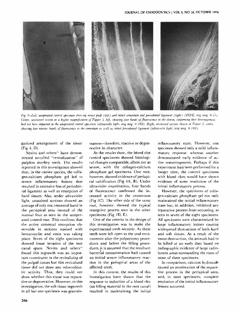
l-~g 5-Lef t . umJperated control specimen showme intact pulp (&fi ) and intact cementum and periodontal h+ament (right) ( l t&l:; ort.e ma+ x 35). Center. unstained section at a ht.~her ma.gmlicatmn qf Ftcure 5, le]'t, showzng.]our bands o]'fluorescence m the dentin, con/irmmq that d,'ntmo.~enest.~ had not been trnpatred tn the unoperated control specimen (ultra~'wlet hght, ortg mag • 140). Rtght. un~tamed sectton ~ho~cn in Fto, nre' 5. ~entrr. shou.,mg J'our narrow hand3 ?[fluorescence m the cementum a~ a'ell aa intact periodontal hgament (ultmt'mlet h.eht , ort~ ma~ X 140).
ganized arrangement of the tissue (Fig 4, D).
Nevins and others '~ have demon- strated so-called "revitalization" of pulpless monkey teeth. The results reported in this investigation showed that, in the canine species, the colla- gen-calcium phosphate gel led to severe inflammatory lesions that resulted in extensive loss of periodon- tal ligament as well as resorption of hard tissues. Also, under ultraviolet light, unstained sections showed an average of only one cemental band in the periapical area instead of the normal four as seen in the unoper- ated control root. This confirms that the active cementa[ resorption ob- servable in sections stained with hematoxylin and eosin was taking place. Seven of the eight specimens showed tissue invasion of the root canal space. Nevins and others" found this ingrowth was an impor- tant constituent in the revitalizing of the pulpal tissues but this revitalized tissue did not show any odontoblas- tic activity. Thus, they could not show whether this tissue was repara- tive or degenerative. However, in this investigation, the soft tissue ingrowth in all but one specimen was granulo-
matous-therefore, reactive or degen- erative in character.
As the results show, the blood clot control specimens showed histologi- cal changes comparable, albeit not as severe, with the collagen-calcium phosphate gel specimens. One root, however, showed evidence of periapi- cal calcification (Fig IA, B). Under ultraviolet examination, four bands of fluorescence confirmed the in- creased activity in the cementum (Fig IC). The other side of the same root, however, showed the typical resorptive process seen in the other specimens (Fig ID, E).
One of the criteria in the design of this investigation was to make the experimental teeth necrotic. As these teeth were left open to the oral envi- ronment after the pulpectomy proce- dures and before the filling proce- dures, it is assumed that the resultant bacter~'al contamination had caused an initial severe inflammatory reac- tion in the periapical areas of the affected teeth.
In this context, the results of this investigation have shown that the response to induction of a blood clot (no filling material in the root canal) resulted in maintaining the initial
inflammatory state. However, one specimen showed only a mild inflam- matory response, whereas another demonstrated early evidence of ac- tive eementogenesis. Perhaps if this experiment had been performed for a longer time, the control specimens with blood clots would have shown evidence of some resolution of the initial inflammatory process.
However, the specimens of colla- gen-calcium phosphate gel not only maintained the initial inflammatory state but, in addition, inhibited any reparative process from occurring, as seen in seven of tile eight specimens. All specimens were characterized bv large inflammatory lesions causing widespread destruction of both hard and soft tissues. As a result of the tissue destruction, the animals had to be killed at an early date based on radiographic evidence of large radio- lucent areas surrounding the roots of some of these specimens.
In comparison, calcium hydroxide caused an acceleration of the repara- tive process in the periapical area, and, in most specimens, complete resolution of the initial inflammatory lesions occurred.
296
JOURNAL OF ENDODONTICS VOL 5, NO 10, OCTOBER 1919
S U M M A R Y A N D
C O N C L U S I O N S
T h c c o m p a r a t i v e effects of cal-
c i u m h y d r o x i d e , c o l l a g e n - c a l c i u m
p h o s p h a t e gel, a n d the f o r m a t i o n o f
a b lood clot as i n d u c e r s o f p e r i a p i c a l
ca lc i f i ca t ions in t he i m m a t u r e n o n v i -
tal t ee th of dogs were i n v e s t i g a t e d .
C e n t r a l to th is i n v e s t i g a t i o n was
the e v a l u a t i o n o f the s u i t a b i l i t y o f
the dog as a n idea l a n i m a l m o d e l in
this field of s tudy . In a d d i t i o n , the
vi ta l dye P roc ion Br i l l i an t Red was
e v a l u a t e d as to its e f fec t iveness as a
t e m p o r a l m a r k e r to i d e n t i f y pos top -
e r a t i ve ca lc i f i ca t ions .
T h e fo l lowing c o n c l u s i o n s were
m a d e f rom this i n v e s t i g a t i o n :
- C a l c i u m h y d r o x i d e a c c e l e r a t e d
h a r d t issue b r i d g i n g o f t he o p e n apex
w h e t h e r the in i t i a l i n f l a m m a t o r y
s ta te in the p e r i a p i c a l a r ea was
c o m p l e t e l y reso lved or was still in
ev idence .
- C o l l a g e n - c a l c i u m p h o s p h a t e gel
i n h i b i t e d a n y r e p a r a t i v e process o f
the in i t i a l i n f l a m m a t o r y lesion, lead-
ing to ex t ens ive d e s t r u c t i o n of the
p e r i a p i c a l a n d s u p p o r t i n g tissues,
w i th no e v i d e n c e o f apex i f i c a t i on .
- I n d u c t i o n of a b l o o d clot
resu l ted in m a i n t a i n i n g the in i t ia l
i n f l a m m a t o r y s t a t e a n d d i d no t
resul t in h a r d t issue b r i d g i n g of the
o p e n apex .
- P r o c i o n Br i l l i an t R e d dye was a n
effect ive t e m p o r a l m a r k e r of h a r d
t issue ca lc i f i ca t ions .
- T h e dog was a n ef fec t ive a n i m a l
m o d e l for m e e t i n g the c r i t e r i a of the
e x p e r i m e n t a l des ign i n c l u d i n g t he
a v a i l a b i l i t y of o p e n apexes a n d a
nec ro t i c a n d sens i t ive l~iological
r e sponse to the effects of ora l
c o n t a m i n a t i o n .
*Carl Zeiss. Overkocken/~,Vurtt. West Ger- many.
This stud'," was supporwd in part by H. Sicher Foundation and Grant RR-05311 USPH-NIIt.
Dr. Citrc,me is in private practice, Ottav,'a, Canada. l)r. Kaminski and Dr. Heuer are assistant professors, Northwestern University Dental School, 311 E Chicago Ave, Chicago 611611. Requests for repricms should be sent m l)r. E. J. Kaminski.
References 1. Frank, A L. "l'herapy for the divergent
pull)less tooth by continued apical formation. .JADA 72:87-93, 1966.
2. Michanowicz, J R , and Michanowiez, A.E. A conse~,ative approach and procedure to fill an incompletely formed root using calcium hydroxide as an adjunct. J Dent Child 34:42-47, 1967.
3. Heithersay, G.S. Stimulation of root formation in incompletely developed pulpless teeth. Oral Surf 29:620-63(), 197(I.
4. Holland, R., and others. Healing process of teeth with wide open apices: histological study, Bull Tokyo Dent Coil 12(4):333-'338, 1971.
5. Cvek, M. "l'reamlenl of non-vital pcrma- c,ent incisors with calcium M, droxide. Fc,llnw- up of periapical repair anti apical closure of immature roots. Odonml Rev 23:27-44, 1972
6 Klein, S.H., and Levy, B.A. Hismlogic evaluation of induced apical closure of a human pulpless tooth. Oral Surf 38:954-959, 1974.
7. Piekoff, M,D.. and Trott, J.R. Apexilica- tic.n: report of a case. J Endod 2:182-183. 1976.
8. Steiner, . JC, and Van Hassel, HJ. Experimental root apexification in primates. Oral Surf 31:409-413, 1971.
10. Torneck, CD.: Smith, .J.S.: and Grin- dall, P. Biologic efects of endodontic proce- dures on developing incisor teeth. Effects of debridement procedures and calcium hydrox- ide-camphorated parachloropfienol paste in the treatment of experimentally induced pulp and p~:riapical disease. Oral Surf 35:541-554, 1973.
I1. Ham, J.W.; Patterson, S.S.; and Mit- chell, I).F. Induced apical closure of immature pulpless teeth in monkeys. Oral Surg 33:438- 449, 1972.
12. Binnic, WH., and Rowe, A.H.R. A histological studv of the pcriapical tissues of incompletely formed pulpless teeth filled ,.~.ith calcium hvdroxide. J Dent Res 52:1110-1116, 1973.
13. Nygaard-Ostby, BN. The role of the blood clot in cndodontic therapy, Acta ()don- tol Stand 19(3):323-353. 1961.
14. Mc,odc,ick, R.M. Clinical correlations of the developmec,t of the root apex and surrounding structures, Oral Sur R 16:600-607, 1963.
15. Kr J.F., and others Induced apical closure of permanent teeth in adult primates using a remrbable form of tricalcium phosphate ceramic. J l"ndod 1:102-100, 1973.
16. Roberts, S.C., .Jr., and Brilliant. ,J.l). Tricaleium phosphate as an adjum't to apk'al closure in pulpless permanent teeth ,1 Endnd 1:263-269, 1973.
17. Maser, T.F, ,Jr The effects of beta phase tricalcium phosphate on the pulp and periapk:al supporting apparatus in dogs. "l'hesis, Northwestern University, 1975.
18. Ncvins, AJ , and others. Revitalization of pulpless npcn apex teeth in rhesus monke':,'s, using collagen-calcium phosphate gel. .1 Endod 2:139-165, 1976.
20. Torneck, D.C,; Smith, J.S.; and Grun- dall, P Biologic effects of ec,dodontic proce- dures on developing incisor teeth. Efli:ct of pulp injury and oral contamination Oral Surf 35:378-388, 1973.
21. Gonlieb, B.; Baron, S.L.; and (_:rook, J.H. Endodontics. St. Louis. C. V. Mosby Co., 1950, pp 25-46.
22. Matsumiya, S., and Kitamura. M. ltisto-pathological and histo-bacterioh)gical studies of the relation between the condition of sterilization of the interior of the root canal and the healing process of periapical tissue in experimentally infected root canal treatment Bull Tokyo Dent Coil 1:1-19, 1960
23. Orban, B. The problem of root canal treatment..JAI)A 19:1384-13118, 1932.
24. Kozlowski. D. The effect of tricalcium phosphate cvramic as a pulp capping agent over minimal exposures in dogs. Thesis, Northwestern University, p 28, 1976.
2f) Drury, R.A.B., and Wallington. E.A Carleton's histological technique. New York, Toronto. ()xfl,rd University Press, 1967, p 140.


![Soal Uts Anestesi 2015 [Cementum 2013]](https://static.documents.pub/doc/80x56/577c7dbe1a28abe0549fb9bf/soal-uts-anestesi-2015-cementum-2013.jpg)


![Microanalysis of Root Cementum in Patients with Rapidly ......at the exposed cementum [4]. Chemical analysis of the exposed cementum has shown an increase in calcium, magnesium, and](https://static.documents.pub/doc/80x56/5f237b2b5d795a336e24c740/microanalysis-of-root-cementum-in-patients-with-rapidly-at-the-exposed-cementum.jpg)

![Cementum in Disease[Nalini]](https://static.documents.pub/doc/80x56/55cf9d52550346d033ad2077/cementum-in-diseasenalini.jpg)

