19
Scientific Inquiry Year 9-10 Liana Walters Brighton Secondary School

Scientific Inquiry
Year 9-10
Liana Walters
Brighton Secondary School

Science Journal
Note: While doing this experiment, planning stage and report, I was recovering from a surgery I had on my
back from a few weeks prior. As a result, I wasn’t at school and wasn’t able to start this until later. There
were some days where I wasn’t able to check the bacteria in the incubator. That is why my Journal seems very
spread out. Sorry for the inconvenience.
8/6/21
Today I came up with ideas for what I should do an experiment on and what interested me to see if there was
something I could do that I liked. I came up with ideas about doing a experiment on bones and bone structures
but I didn’t come far with the restrictions there were at school and where I would be able to get materials from.
I thought about doing something that had to do with chemistry as I like chemsity and am interested by it. One
of my ideas was to do something with fire and changing colours but I didn’t like it. Then I had an idea to do
an experiment with cells and repairing cells. I did like the idea but when I asked my teacher, I couldn’t do it
with the time we had. Also while coming up with ideas I didn’t really know what direction I wanted to do in
and turned down most of my thoguhts as they were to out landish. I asked my teacher for some help about
what topic I should do and she told me of past students projects. I was intrigued by this one project were a girl
measured the different strenghs of handsanitiser on different strains of bacteria. I was keen on doing my own
version of it but my mind still thought of possible ideas so I didn’t choose that idea.
10/6/21
Today I was basically doing the same things I did two days ago and decided to try and do something that
involves space and the planets. I didn’t really do much though as I was trying to figure out what to do and
how I would do it.
17/6/21
Today I was still looking at different experiments I could do and still hadn’t made up my mind on what I
should do for the report. After a lot of thinking I decided on expanding the effect of handsanitiser on bacteria,
to come up with my own question. I kept thinking to myself that I was copying this girl’s idea but I was simply
inspired by it and wanted to make my own variation of it with a different goal in mind. With a final idea, I
came up with possible questions I could base my research on such as, the effects of mutliple types of hand
sanitiser brands on one strain of common hand bacteria or the effect of one hand sanitiser on multiple types
of hand bacteria. I chose the second option “The effect of one hand sanitiser on multiple hand bacteria strians”.
21/6/21
Today I researched different types of bacteria that is found on the hand but all of the ones I found were not
safe to be used in a school setting. I also looked at different ways I could do this experiment online. I found a
website about university students doing the same thing that I intend to do and looked through it. I saw that
they put there hands on different things like shopping trollies and bathroom doors as well as having some
wash their hands before putting it on the petri dish and someone not washing their hands at all. I found this
very interesting and looked more into it and found out that the students were able to tell what types of bacteria
were growing which I thought was really cool. Apart from that I didn’t really do much. I also learned what
Gram-Negative and Gram-Postive bacteria means.
Gram-Negative bacteria is when a bacteria has a cell membrane around it to protect itself from the white blood
cells so they won’t kill it allowing the bacteria to grow and cause infections such as pneumonia. While Gram-
Postive bacteria don’t have a cell membrane allowing white blood cells the abilitiy to destroy them. Gram-
Negative bacteria goes red when it has the chemical process Gram, applied to it. Whereas Gram-Positive
bacteria stains blue when it goes through the same process. Bothe Gram-Negative and Gram-Positive Bacteria
are treated with antibiotics suited for the type of bacteria and if its Gram-Negative or not. They also have
different types of infection with Gram-Negative bacteria being more dangerous.
The websites I used to look for different bacteria: 2021. A comparison of the bacteria found on the hands of ‘homemakers’ and neonatal intensive care unit nurses. [online]
Available at: <https://www.ncbi.nlm.nih.gov/pmc/articles/PMC2062569/> [Accessed 21 June 2021].

n.d. Normal bacterial flora on hands. [online] Available at: <https://www.ncbi.nlm.nih.gov/books/NBK144001/> [Accessed 21
June 2021]. n.d. Pseudomonas putida and Pseudomonas fluorescens Species Group Recovery from Human Homes Varies Seasonally and by
Environment. [online] Available at: <https://www.ncbi.nlm.nih.gov/pmc/articles/PMC4449118/> [Accessed 21 June 2021]. n.d. Staphylococcus warneri: Skin Commensal and a Rare Cause of Urinary Tract Infection. [online] Available at:
<https://www.ncbi.nlm.nih.gov/pmc/articles/PMC7263002/> [Accessed 21 June 2021]. n.d. Enterobacter aerogenes and Enterobacter cloacae; versatile bacterial pathogens confronting antibiotic treatment. [online]
Available at: <https://www.ncbi.nlm.nih.gov/pmc/articles/PMC4435039/> [Accessed 21 June 2021]. Cdc.gov. n.d. Klebsiella pneumoniae in Healthcare Settings | HAI | CDC. [online] Available at:
<https://www.cdc.gov/hai/organisms/klebsiella/klebsiella.html> [Accessed 21 June 2021]. Betterhealth.vic.gov.au. n.d. Staphylococcus aureus - golden staph - Better Health Channel. [online] Available at:
<https://www.betterhealth.vic.gov.au/health/conditionsandtreatments/staphylococcus-aureus-golden-staph> [Accessed 21 June
2021]. MSD Manual Consumer Version. n.d. Overview of Gram-Negative Bacteria - Infections - MSD Manual Consumer Version.
[online] Available at: <https://www.msdmanuals.com/en-au/home/infections/bacterial-infections-gram-negative-
bacteria/overview-of-gram-negative-bacteria> [Accessed 21 June 2021].
The website where I found the Univeristy Investigation: CNA. n.d. In pictures: The bacteria living on your hands right now. [online] Available at:
<https://www.channelnewsasia.com/news/singapore/bacteria-living-on-your-hands-10067062> [Accessed 21 June 2021].
22/6/21
Today I told my teacher about all the research I did and she said that all the bacteria I looked at wasn’t safe to
do an experiment with. She then said she would ask my schools lab supervisor, if there was any bacteria
available for me to use. That was also when she wrote down the risk assessment. I decided on that I would do
four different types of bacteria if my school had them and if not, I had to research what types of safe bacteria
lived on the hands and tell my teacher so she could order them in.
24/6/21
Today I was given the all go for the experiment and was given a list of all of the types of bacteria strains my
school had. These strains were:
Bacillus subtilis
Staphylococcus epidermidis
Escherichia coli K12 strain
Micrococcus luteus
I was also told to research them when I got home so that when I was doing the experiment I knew what the
types of bacteria were. When I got home that night, I lost the stickynote that had the types of bacteria on it so
I didn’t know what to research. I also forgot to ask my teacher for the types of bacteria the school had via
email.
28/6/21
Today was the day I did the experiment, when I got to class, my teacher gave me a run down on what to do
and how to do it. Then I put on a lab coat and safety glasses and washed my hands before I touched any of the
equipment I was using. I did wear gloves for some of it but then I asked my teacher if I could take them off
and she said it was fine, as long as I was really careful.
The first thing I did was cut up a bunch of little discs from the fitler paper that would go in the petri dishes
with the bacteria. I put the little discs into a plastic bowl and put another bowl ontop of that when I was done,
to minimise the contact the discs had with the air. I then put a bit of hand sanitiser into one bowl and distilled
water into another one. Seeing as I didn’t know how to put the bacteria into the petri dish, how to put the filter
paper in and so forth, I had my teacher show me how to do it. I tried doing it myself and made a few testers.
From doing the testers, I was able to come to a decision of putting black lines on the backside of the petri dish
so that I would know where to place the filter paper instead of putting them anywhere.

Here are photos of plates with lines and without:
Figure 1: I found it easier to place the filter paper discs into the right places with the lines spliting the petri dish into quadrants
instead of estimating where I should place them.
I also came to the decision to use 0.5ml f the bacteria instead of 1ml as I found that it was too much and made
the filter paper discs to move around. Once I was confident with making the samples, I repeated what I had
done with testers and tried my best to make the filter paper be placed in the middle of one of the four quadrants.
I did two hand sanitiser petri dishes and one control with distilled water. The bacteria I used for the first dishes
was Micrococcus luteus.
I wasn’t able to do all of the petri dishes today as I didn’t have enough time to do them inbetween my lunch
and next lesson so I put the ones I had done in the incubator and decided I would do the rest of them tomorrow.
When I got home that night I did some research on the bacteria I was using:
Bacillus subtilis – An aerobic Gram-Positive, Rod shaped bacteria that is spore forming and found in soil and
vegetation. The optimal temperature for Bacillus subtilis to thrive is from 25-35 degress celcius and only takes
around 48h for it to grow. The B. subtilis produces numerous amounts of emzymes that deteriorate a variety
of substances, allwoing the bacteria to thrive and prosper in an ever changing enviroment.
Staphylococcus epidermidis – This bacterium is a Gram-Positive, member of the Staphylococcus family and
is commonly found on the skin on the face. Staphylococcus epidermidis doesn’t produce any harmful toxins
but can work as a conductor for Staphylococcus aureus, enhancing its resistance to antibiotics and
pathogenic success making it more dangerous than it originally was.
Escherichia coli K12 strain – A Rob shaped bacteria that has no nuclear membrane because it is a prokryate.
This is a rapidly growing bacterium and can start to grow within an hour and reproduces by binary fission
creating two cells within a short span of time. Though Escherichia coli is a Gram-Positive bacteria, it possess
the characteristics of a Gram-Negative bacteria as it is enveloped by two membranes.
Micrococcus luteus – A spheriacl, Gram-Positive bacteria that isn’t usually pathogenic and are common
inhabitants of the human skin and body. Micrococcus luteus has even been said to be a key part in keeping
balance between Gram-Positive and Gram-Negative bacteria that lives on the skin. As well as other types of
viruses.
The websites I used: Microchemlab.com. n.d. Bacillus subtilis | Microchem Laboratory. [online] Available at:
<https://microchemlab.com/microorganisms/bacillus-subtilis> [Accessed 28 June 2021].
Microbial Cell Factories. n.d. Bacillus subtilis: a universal cell factory for industry, agriculture, biomaterials and medicine.
[online] Available at: <https://microbialcellfactories.biomedcentral.com/articles/10.1186/s12934-020-01436-8> [Accessed 28
June 2021].
PLOS PATHOGENS. n.d. Staphylococcus epidermidis — the 'accidental' pathogen. [online] Available at:
<https://www.nature.com/articles/nrmicro2182> [Accessed 28 June 2021].
PLOS PATHOGENS. n.d. Staphylococcus epidermidis—Skin friend or foe?. [online] Available at:
<https://journals.plos.org/plospathogens/article?id=10.1371/journal.ppat.1009026> [Accessed 28 June 2021]

Uniprot.org. n.d. Escherichia coli (strain K12). [online] Available at: <https://www.uniprot.org/proteomes/UP000000625>
[Accessed 28 June 2021].
Sciencedirect.com. n.d. Escherichia Coli K 12 - an overview | ScienceDirect Topics. [online] Available at:
<https://www.sciencedirect.com/topics/medicine-and-dentistry/escherichia-coli-k-12> [Accessed 28 June 2021].
Encyclopedia Britannica. n.d. Micrococcus | bacteria genus. [online] Available at:
<https://www.britannica.com/science/Micrococcus> [Accessed 28 June 2021].
29/6/21
Today I used the same method of making the test dish I did yesterday with the rest of the bacterial strains I
had. While doing each petri dish, I realised that when I placed the coated filter paper onto the agar, I touched
the end of my tweezers on the bacteria. Once I realised this, I had already done 5 plates and I couldn’t change
the plates so I decided that I would be more careful when putting the filter paper on the bacteria coated agar.
I was also told by my teacher that I should clean the tweezers by using the flame of a bunsen burner and that’s
what I did. I was also given more tweezers so I could use them as I waited for the other tweezers to cool down.
Once I finished doing all of the bacteria and used up all of the petri dishes, I put them all on the bottom shelf
of the incubator so that I know those plates were put in the day after the first ones I did.
Today was also the day I took photos of the plates to see the if there was any growth. When I was looking at
the Micrococcus luteus petri dishes, I realised that when I picked them up to take a photo of the growth, the
bacteria liquid would move and get into the filter paper. This was human error and it resulted in some of the
samples to not show any results.
The photos I took:
Figure 2: The 2 photos to the left are the control for Micrococcus luteus and the two on the right are the hand sanitiser sample.
Figure 3: This is how I set up the incubator, Mondays batch of petri dishes are on the first shelf and Tuesdays batch of petri dishes
are on the bottom.
30/6/21
Today I took photos again and saw some growth on some of the plates but there wasn’t a lot all together. There
was like a cloudy coating in some areas which I believe is the bacteria growing. Also I noticed the same
problem I had noticed the day before, everytime I picked up a petri dish to look at the growth on the agar, the
bacteria liquid would move around and get on the coated filter paper. It didn’t happen to all of the petri dishes
which is lucky but I was still disappointed that it happened to some of them. When I got home I realised I
didn’t take photo’s of the Staphylococcus Epidermis which was a pretty big mistake.

Here are the photos I took:
Bacillus subtilis (Tuesday):
Figure 4: The photo on the left are the control and the two photos on the right are the hand sanitiser samples.
Escherichia coli K12 strain (Tuesday):
Figure 5: The two photos on the left are the control and the two photos on the right are the hand sanitiser samples.
Micrococcus luteus (Monday):
Figure 6: The two photos on the left are the control and the two photos on the right are the hand sanitiser samples.
2/7/21
Today I was sick and couldn’t go to school so I had my teacher take photos of the petri dishes for me and I
was going to measure the inhibition zone but I wanted to wait until I went back to school to do it as I could
get a better reading that way. Also my teacher told me that she put the petri dishes in the fridge to stop the
bacteria growing so that I could see them when I got back from holidays.
Inhibition zone – An area around the filter cirlces that has no bacteria growing.
19/7/21

Today I measured the inhibition zone. I did this by finding the closest point the bacteria was to the filter paper,
then I drew a line between the center of the filter paper and the bacteria and multiplied it by two to get the
diameter. Some of the samples didn’t show an inhibition zone so I just left them a zero. Some of the samples
were hard to tell were the inhibition zone was but when the plates were placed against a light, they were
visiable.
Here are some photos I took of the inhibition zone:
Figure 7: Both of these photos are testers I did with Micrococcus luteus, I did this so I could understand how to spread the bacteria
and how to place the filter paper discs on the agar. There was 1ml of bacteria on the plates but was changed to 0.5ml because there
was to much excess bacteria liquid left on the plate. This caused the filter circles to move around.

Risk Assessment Form

How effective is hand sanitiser on bacteria found on hands?
Introduction Each type of hand sanitiser have different levels of alcohol, the sanitisers that have higher percentages of
ethanol can kill bacteria more efficiently. The SA Health Alcohol-Based Hand Rub fact sheet states that hand
sanitisers should have an “achohol concerntration of 60 – 80%” to be deemed bacteria resistant to an extent.
The fact sheet also states that both ethanol and isopropanol have showed to hold the ability to isolate the
activity of bacteria, fungi and some types of viruses. Scientists have conducted numerous experiments for the
effectiveness of different ratios of alcohol in sanitisers and found that isopropanol has stronger effects against
bacteria than ethanol. Hand Sanitiser that contains 60% isopropanol holds the similar in-vitro activity against
bacterium as hand sanitiser containing 77% ethanol. That doesn’t necessarily mean that isopropanol is stronger
than ethanol with all bacteria strains.
For hand sanitiser to effectively disrupt and kill bacteria cells, the ethanol and isopropanol attach themselves
onto the water-based membrane that surrounds the bacteria cell. The alcohol is then able to breakdown the
membrane as the molecules in the proteins and a membrane of the cell easily bond with the molecules of the
alcohol, tearing the molecules away from one another. The alcohol in hand sanitiser is an amphiphile chemical
compound, meaning it possess both hydrophilic and lipophilic properties, make killing bacteria cells highly
effective as the fatty amino acids and water are attracted to the alcohol molecules. Leaving the bacteria
cells defenceless against hand sanitisers.
The hand sanitiser that was used in this experiment is a gel based sanitiser and has an alchohol concentration
of 70%. This infers that this sanitiser is able to inhibit the growth of bacteria and holds the abiltiy to eradicate
microorganisms, referred to as ‘antimicrobial activity’ in scientific terms. The bacteria that will be used and
tested in this experiment are Bacillus subtilis, Staphylococcus epidermidis, Escherichia coli K12 strain,
Micrococcus luteus. All of which don’t hold much resistance to hand sanitisers. Taking all of this into
consideration, I believe that once the hand sanitiser is placed in the petri dish, the alcohol will break down the
cell membrane of the bacteria and kill its core. This will inhibit the growth of the bacteria around the filter
discs leaving a space were no bacteria has grown.
Aim To investigate the effectiveness of hand sanitiser on bacteria that’s commonly found on the surfaces of human
hands.
Variables Independent variable – The type of bacteria used throughout the experiment.
Dependant variable – The area of inhibition around the filter paper disc.
Controlled Variable Method of Control
The hand sanitiser brand The hand sanitiser that is used will be the same type on all of the
bacteria.
The number of petri dishes for
each strain
Each strain of bacteria was given a certain number of petri dishes as well
as one controll.
The number of paper discs in
each petri dish
The petri dish was split up into four different quadrants with the filter
paper disc placed in the middle.
The amount of bacteria in the
petri dish
The amount of bacteria in each petri dish was 0.5ml to keep it consistant.
The time the bacteria spends in
the incubator
Once the petri dishes were placed in the incubator, the petri dishes were
left in there for 4 days.
The temperature of the
incubator
The temperature of the incubator was left at 35°c constantly.

The distance between each
paper disc
The filter paperdiscs were placed in the center of each quadrant to keep
the same distance between them all.
Materials/Equipment Bacillus subtilis (B5B)
Staphylococcus epidermidis(S.albus) (B2)
Escherichia coli K12 strain (B1)
Micrococcus luteus(s.Lutea) (B3B)
Filter Paper
Hand sanitiser
Petri dishes (25 including testers)
Permanent marker
Tweezers
Plastic weigh trays
Distilled water
Teat Pippets
Parafilm
Hole punch
Labels
Spreader
Ruler
Scissors
Gloves
Bunsen Burner
Heat proof mat
Saftey Glasses
Lab Coat
Glass beaker
Incubator
Phone (to take photos)
Figure 8: The materials used in the experiment minus the busen burner,
lab coat, safety glasses, incubator, phone, ruler and heat proof mat.
*The petri dishes were made the day before the experiment*
Method
1. 5mm in diameter discs were cut out of the filter paper with the hole punch and then placed into a plastic
weigh tray. Another weigh tray was placed ontop to minimise he contact the filter paper had with the
air.
2. Hand sanitiser and distilled water were added into seperate plastic weigh trays.
Figure 9: The bowl in the top right corner is distilled water and the bowl in the bottom left corner is the hand sanitiser.

3. Then lines were drawn on the underside of the petri dish to section off four quadrants. All the petri
dishes had this done.
4. The pippet was used to extract 0.5 ml of the bacteria and then was placed on the petri dish as quickly
as possible without rushing to minimise the contact it had to the air.
5. The bacteria was the spread in a circular motion with the sterile spreader to get an even coat on the
petri dish. The spreader was then thrown out after each use.
6. One filter paper disc was picked up with the tweezers and placed into the hand sanitiser or distilled
water. Then the discs were placed into a quadrant in the petri dish, with the discs as close to the centre.
Figure 10: A new pair of tweezers were used to pick up the filter paper discs when dealing with a new strain of bacteria
and when the tip of the tweezer would touch the agar plate covered in bacteria to stop it from spreading.
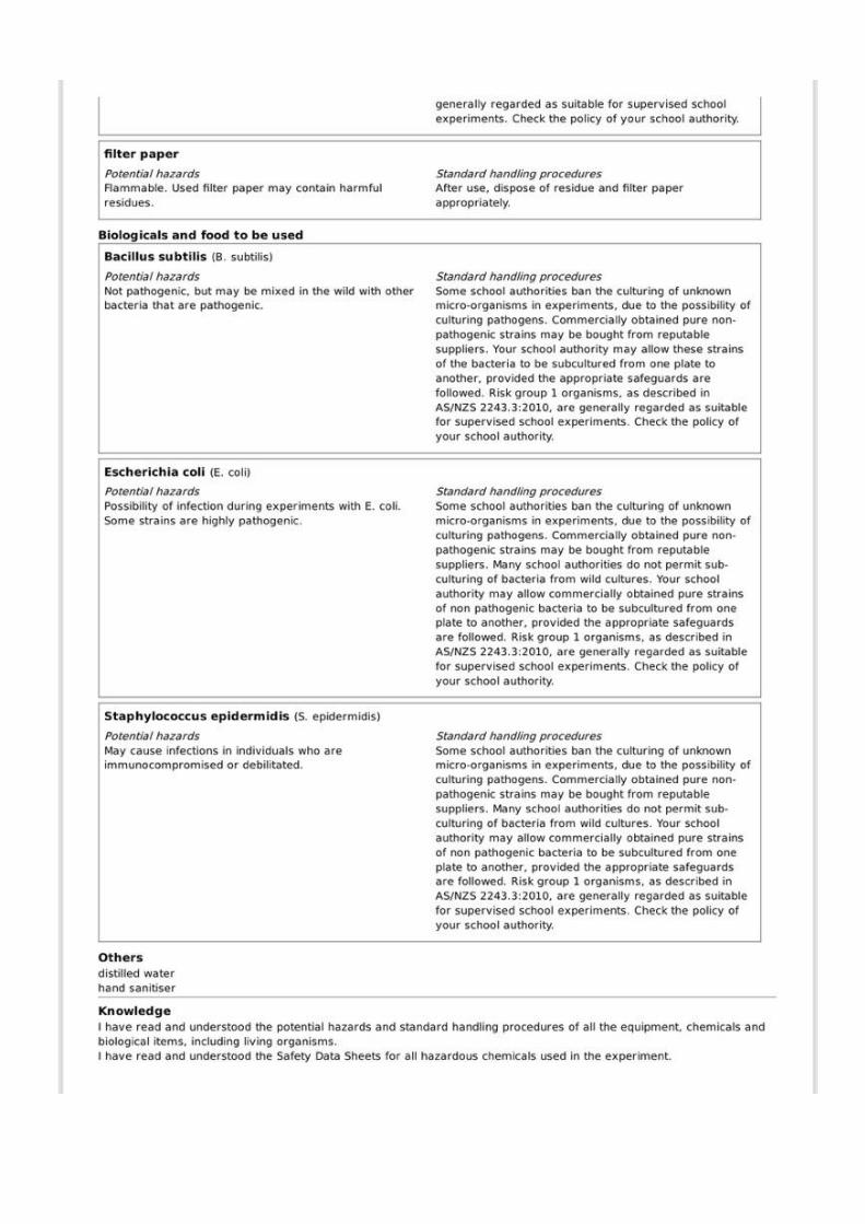
7. Once the lid was placed on the petri dish, the parafilm was wrapped around the edge to kept the petri
dish together.
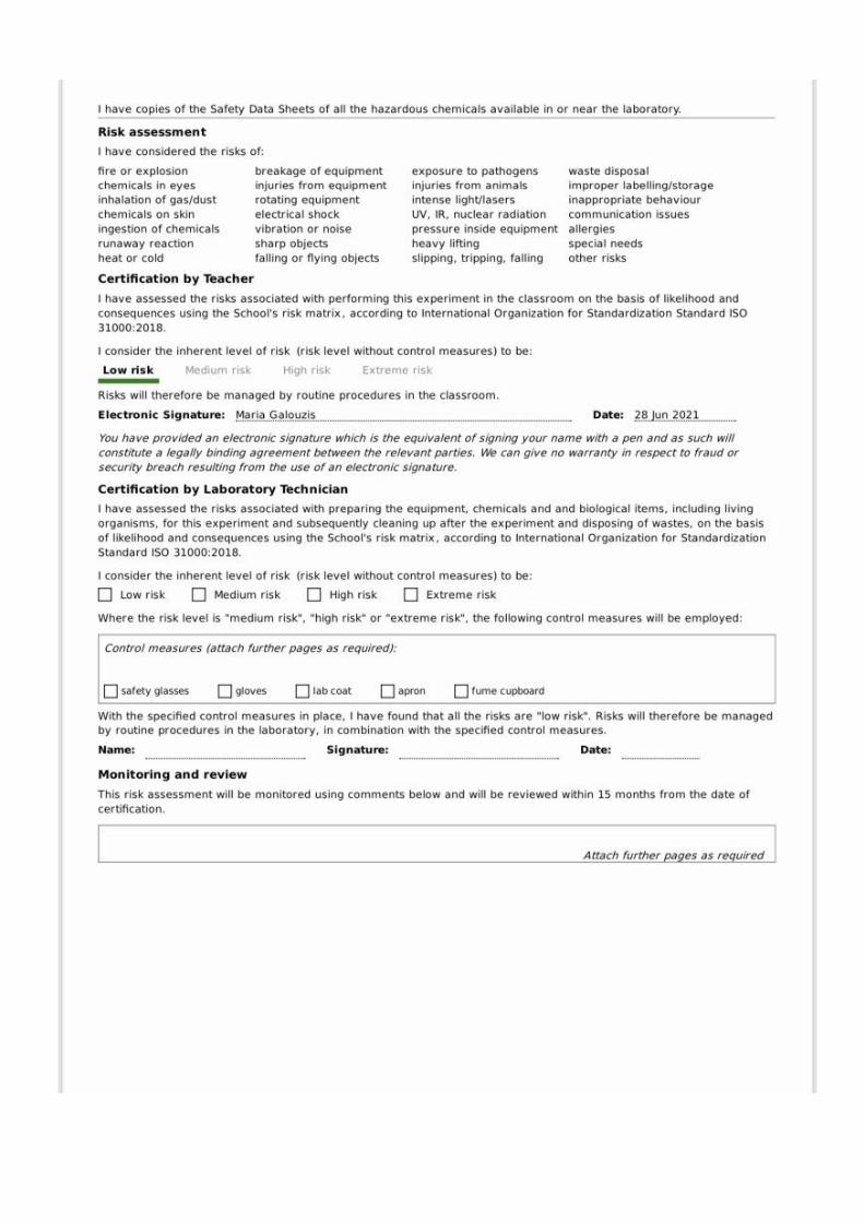
8. A label was written that had the strain classification and whether the dish was a control or had the
handsanitiser on it (HS).
Figure 11: Each type of bacteria had 3 hand sanitiser plates, 1 plate of control and were done in bacteria specific groups
to avoid confusion.
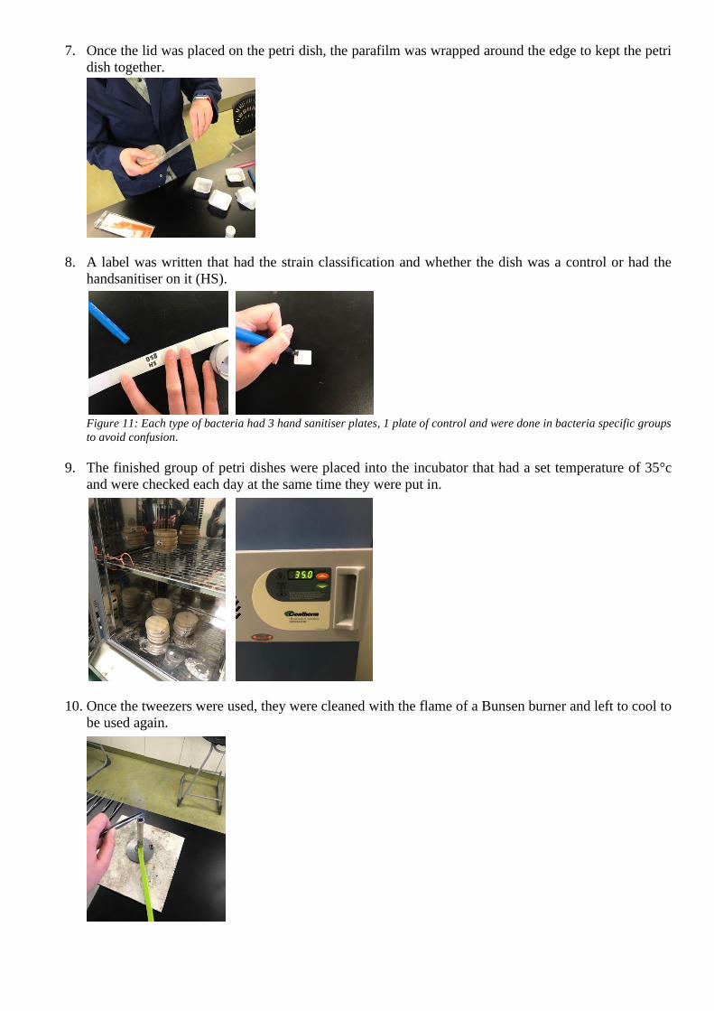
9. The finished group of petri dishes were placed into the incubator that had a set temperature of 35°c
and were checked each day at the same time they were put in.
10. Once the tweezers were used, they were cleaned with the flame of a Bunsen burner and left to cool to
be used again.

Results
Table 2: The resultant zones of inhibition for each replicate and average
Type of
bacteria
Zone of inhibition
(mm)
Zone of
inhibition
Average
Photo
Plate
1
Plate
2
Plate
3
Plate 1 Plate 2 Plate 3
Staphylococcus
epidermidis
(HS)
10
10
10
8
10
12
8
8
10
12
0
0
98 ÷ 12 =
8.17
Average =
8.17
Staphylococcus
epidermidis
(Control)
0
0
0
0
0 ÷ 4 = 0
Average = 0
Escherichia
coli K12 strain
(HS)
0
0
0
0
32
16
12
8
20
14
10
18
130 ÷ 12 =
10.83
Average =
10.83mm
Escherichia
coli K12 strain
(Control)
26
30
0
0
56 ÷ 4 = 14
Average =
14mm
Micrococcus
luteus (HS)
12
0
0
0
8
10
10
10
10
10
10
0
80 ÷ 12 =
6.67
Average =
6.67mm
Micrococcus
luteus
(Control)
0
0
0
0
0 ÷ 4 = 0
Average =
0mm
Bacillus
subtilis (HS)
0
0
0
0
8
0
0
0
10
0
0
0
18 ÷ 12 =
1.5
Average =
1.5mm
Bacillus
subtilis
(Control)
10
8
0
0
18 ÷ 4 = 4.5
Average =
4.5mm

Discussion
The results showed that 3 out of the 4 bacteria didn’t grow around the hand sanitiser coated filter discs.
However, the bacteria that continued to grow around the discs was Bacillus subtilis with only having results
from 2 of the 12 filter discs. The zone of inhibition from the 2 discs were 8mm and 10mm, possessing the
same data as the control sample of the same bacteria. The average zone of inhibition for Bacillus subtilis was
1.5mm and is the most resistant to a hand sanitiser that contains 70% alcohol compared to the other 3 bacteria.
Staphylococcus epidermidis, Escherichia coli and Micrococcus luteus all produced data that showed a range
of zones of inhibition. Both Staphylococcus epidermidis and Micrococcus luteus, zones of inhibition across
the 3 handsanitiser plates all ranged between 8mm and 12mm. These results inferred that these 2 types of
bacteria weren’t able to stop the hand sanitiser to break away the cell membrane that was protecting the
bacteria cell. This is why the bacteria that surrounded the filter paper discs weren’t able to thrive due to the
alcohol in the sanitiser stopping the growth of the bacteria. The averages for these types of bacteria are 8.17mm
and 6.67mm.
The bacteria that allowed the handsanitiser to destroy most of the bacteria cells around the paper discs was
Escherichia coli. 1 of this bacteria’s plates did not show any results because of an error that occurred while it
was in the process of creation, this error had consistantly been shown to repeat itself on a few other agar plates
over the experimenting period. However, the 2 other handsanitiser test plates showed some promising data by
day four of incubation. The first plate had the zone of inhibition range between 8mm and 32mm, and the
second plate had the zone of inhibition range between 10mm and 20mm. These ranges were the most spread
out than the other 3 bacteria, and because of that, made this bacteria the lest resistant towards this type of
bacteria. This means that the cell membrane of the bacteria wasn’t able to stop the alcohol from destroying it
before the bacteria had the chance to reproduce.
The prediction that was made at the start of this experiment would be around 75% accurate because 3 of the
4 bacteria strains were able to produce results and the other 1 wasn’t able to do just that.
The concept of human error occurs quite a few times throughout the experiment and is evident on the results.
Some of the sample plates showed that the filter paper discs had moved from their intended quadrants,
allowing bacteria to get on it and stop the hand sanitiser from working properly. Human error is also present
on the filter discs that hadn’t moved but still had the bacteria grow on it. This is because once the petri dishes
were moved into the incubator and when they were observed inbetween each day, the excess bacteria moved
across the agar and coated the circles. Resulting in bacteria to form on top of some of the filter papers. Another
human error occurred when the petri dishes were being made, once the coated discs were placed onto the layer
of bacteria, the tweezers would touch the bacteria. Allowing it to travel onto the end of the tweezers and
tranferrer onto the paper discs in the plastic weigh tray when a new disc was needed.
Evaluation
After observing all the potential errors that had occurred while I was doing this experiment, I now know how
I can avoid them the best I can to make this experiment better and show a higher quality set of results. Such
as letting the plates grow for the full amount of time, instead of checking on them every day, which avoids the
possiblitiy of bacteria growing over the filter paper on the agar. There are still a few things I would need to
work on to make this experiment run smoother but even so, the results that I managed to collate can be helpful
in the field of Science. I say this because I was able to find out that Escherichia coli wasn’t able to protect its
cells from the alcohol content in the hand sanitiser used.
Questions that can be made off of this experiment are ‘The effects of different strenghted hand sanitiser on
Escherichia coli?’ or something along those lines. That question can also be used for the other 3 types of
bacteria giving researcher the opportunity to discover what percentage of alcohol in hand sanitiser can
disintergrate the cell membrane allowing the alcohol molecules to breakdown the bacteria’s cell structure
before it can reproduce and cause harm to the human body.

Conclusion
This experiment produced results that showed that Escherichia coli wasn’t able to hold strong defences against
the alcohol as its cells were being destroyed. Whereas, Bacillus subtilis was able to stop the hand sanitiser
from breaking it’s cell membrane, giving the bacteria enough time to reproduce and overpower the hand
sanitiser. These result’s demonstrated the effectiveness of hand sanitiser against 4 types of hand bacteria and
allowed for potential research and data to be accumulated. This data has the abiltiy to help researchers develop
more accurate views and ideas about each of the bacteria used and tested in this experiment.
The hypothesis stated at the beginning of this report had been consistently proven over the course of the testing
and recording phases of this experiment. An area of inhibition had appeared around the filter paper discs in
the petri discs, some might not have been the biggest but there were areas where no bacteria had formed. There
was a few petri dishes that had bacteria grow around the filter paper discs but was the result of human error
not because the hand sanitiser wasn’t able to kill the bacteria around it. Overall this experiment had produced
promising results that were in alignment with the hypothesis and aim intened, deeming this experminet
successful with the occasional human error.
Report Word count: 1,778

Reference List
Therapeutic Goods Administration (TGA). n.d. Hand sanitisers: Information for consumers. [online]
Available at: <https://www.tga.gov.au/hand-sanitisers-information-consumers> [Accessed 19 July 2021].
n.d. [online] Available at: <https://www.safetyandquality.gov.au/our-work/infection-prevention-and-
control/national-hand-hygiene-initiative-nhhi/what-hand-hygiene/alcohol-based-handrubs> [Accessed 19
July 2021].
Medicalnewstoday.com. n.d. In vivo vs. in vitro: What is the difference?. [online] Available at:
<https://www.medicalnewstoday.com/articles/in-vivo-vs-in-vitro> [Accessed 19 July 2021].
Sciencedirect.com. n.d. Antimicrobial Activity - an overview | ScienceDirect Topics. [online] Available at:
<https://www.sciencedirect.com/topics/engineering/antimicrobial-activity> [Accessed 19 July 2021].
Sahealth.sa.gov.au. n.d. [online] Available at:
<https://www.sahealth.sa.gov.au/wps/wcm/connect/e10fc9804fb5b324b085ba5cbc1ea1e9/FactSheet-
Alcohol-based-hand-rubs_V4.1-phcs-ics-
20190530.pdf?MOD=AJPERES&CACHEID=ROOTWORKSPACE-e10fc9804fb5b324b085ba5cbc1ea1e9-
nwKsf76> [Accessed 19 July 2021].
n.d. [online] Available at: <https://www.ncbi.nlm.nih.gov/pmc/articles/PMC6255669/> [Accessed 19 July
2021].
Medicalnewstoday.com. n.d. Bacteria: Types, characteristics, where they live, hazards, and more. [online]
Available at: <https://www.medicalnewstoday.com/articles/157973#types> [Accessed 19 July 2021].
Cdc.gov. n.d. Klebsiella pneumoniae in Healthcare Settings | HAI | CDC. [online] Available at:
<https://www.cdc.gov/hai/organisms/klebsiella/klebsiella.html> [Accessed 21 June 2021].
Betterhealth.vic.gov.au. n.d. Staphylococcus aureus - golden staph - Better Health Channel. [online]
Available at: <https://www.betterhealth.vic.gov.au/health/conditionsandtreatments/staphylococcus-aureus-
golden-staph> [Accessed 21 June 2021].
MSD Manual Consumer Version. n.d. Overview of Gram-Negative Bacteria - Infections - MSD Manual
Consumer Version. [online] Available at: <https://www.msdmanuals.com/en-au/home/infections/bacterial-
infections-gram-negative-bacteria/overview-of-gram-negative-bacteria> [Accessed 21 June 2021].
CNA. n.d. In pictures: The bacteria living on your hands right now. [online] Available at:
<https://www.channelnewsasia.com/news/singapore/bacteria-living-on-your-hands-10067062> [Accessed
21 June 2021].
Microchemlab.com. n.d. Bacillus subtilis | Microchem Laboratory. [online] Available at:
<https://microchemlab.com/microorganisms/bacillus-subtilis> [Accessed 28 June 2021].
Uniprot.org. n.d. Escherichia coli (strain K12). [online] Available at:
<https://www.uniprot.org/proteomes/UP000000625> [Accessed 28 June 2021].
Murugesu, J., n.d. Hand sanitiser. [online] New Scientist. Available at:
<https://www.newscientist.com/definition/hand-sanitiser/> [Accessed 20 July 2021].
Sciencing. n.d. How Does Alcohol Kill Bacteria?. [online] Available at: <https://sciencing.com/alcohol-kill-
bacteria-5462404.html> [Accessed 21 July 2021].
Sciencedirect.com. n.d. Amphiphiles - an overview | ScienceDirect Topics. [online] Available at:
<https://www.sciencedirect.com/topics/materials-science/amphiphiles> [Accessed 21 July 2021].
2021. A comparison of the bacteria found on the hands of ‘homemakers’ and neonatal intensive care unit
nurses. [online] Available at: <https://www.ncbi.nlm.nih.gov/pmc/articles/PMC2062569/> [Accessed 21
June 2021].
n.d. Normal bacterial flora on hands. [online] Available at:
<https://www.ncbi.nlm.nih.gov/books/NBK144001/> [Accessed 21 June 2021].
n.d. Pseudomonas putida and Pseudomonas fluorescens Species Group Recovery from Human Homes
Varies Seasonally and by Environment. [online] Available at:
<https://www.ncbi.nlm.nih.gov/pmc/articles/PMC4449118/> [Accessed 21 June 2021].
n.d. Staphylococcus warneri: Skin Commensal and a Rare Cause of Urinary Tract Infection. [online]
Available at: <https://www.ncbi.nlm.nih.gov/pmc/articles/PMC7263002/> [Accessed 21 June 2021].
n.d. Enterobacter aerogenes and Enterobacter cloacae; versatile bacterial pathogens confronting antibiotic
treatment. [online] Available at: <https://www.ncbi.nlm.nih.gov/pmc/articles/PMC4435039/> [Accessed 21
June 2021].

Microbial Cell Factories. n.d. Bacillus subtilis: a universal cell factory for industry, agriculture,
biomaterials and medicine. [online] Available at:
<https://microbialcellfactories.biomedcentral.com/articles/10.1186/s12934-020-01436-8> [Accessed 28
June 2021].
PLOS PATHOGENS. n.d. Staphylococcus epidermidis — the 'accidental' pathogen. [online] Available at:
<https://www.nature.com/articles/nrmicro2182> [Accessed 28 June 2021].
PLOS PATHOGENS. n.d. Staphylococcus epidermidis—Skin friend or foe?. [online] Available at:
<https://journals.plos.org/plospathogens/article?id=10.1371/journal.ppat.1009026> [Accessed 28 June 2021]
Uniprot.org. n.d. Escherichia coli (strain K12). [online] Available at:
<https://www.uniprot.org/proteomes/UP000000625> [Accessed 28 June 2021].
Sciencedirect.com. n.d. Escherichia Coli K 12 - an overview | ScienceDirect Topics. [online] Available at:
<https://www.sciencedirect.com/topics/medicine-and-dentistry/escherichia-coli-k-12> [Accessed 28 June
2021].
Encyclopedia Britannica. n.d. Micrococcus | bacteria genus. [online] Available at:
<https://www.britannica.com/science/Micrococcus> [Accessed 28 June 2021].
n.d. Different responses of planktonic and attached Bacillus subtilis and Pseudomonas fluorescens to
sanitizer treatment. [online] Available at: <https://pubmed.ncbi.nlm.nih.gov/10419210/> [Accessed 21 July
2021].
n.d. Enterobacter aerogenes and Enterobacter cloacae; versatile bacterial pathogens confronting antibiotic
treatment. [online] Available at: <https://www.ncbi.nlm.nih.gov/pmc/articles/PMC4435039/> [Accessed 21
July 2021].


![What Ever Happened to Scientific Inquiry? Notions of Scientific... · What Ever Happened to Scientific Inquiry? A Look at Evolving Notions of Inquiry ... 2012, p. 2]). Scientific](https://static.documents.pub/doc/80x56/5e3e71273eec9a4bfd7dddce/what-ever-happened-to-scientific-inquiry-notions-of-scientific-what-ever-happened.jpg)


