Page 1
Volume 5, Issue 10, October – 2020 International Journal of Innovative Science and Research Technology
ISSN No:-2456-2165
IJISRT20OCT209 www.ijisrt.com 574
Screening for Actinomycetes from Government
Science College Campus and Study of their
Secondary Metabolites
Dr. KAVITHA.B, M.Phil., Ph.D
Associate Professor and Head
Department of Microbiology
Government Science College
Nrupathunga road, Bangalore
VISHNU PRIYANKA.N, M.Sc
Department of Microbiology
Government Science College
Nrupathunga road, Bangalore
ZEBA KHANUM, M.Sc
Department of Microbiology
Government Science College
Nrupathunga road, Bangalore
Abstract:- Actinomycetes are a group of organisms
which have characteristics of both bacteria and fungi,
hence, they are also called as ‘Actinobacter’ and ‘Ray
fungi’. Actinomycetes are known for producing novel
secondary metabolites like enzymes, anti-biotics, anti-
cancerous agents and play major role in recycling of
organic matter. In this present research study,
actinomycetes were isolated from 11 different soil
samples from different places from college campus by
serially diluting and spread plate technique on SCA
media. 22 actinomycete isolates were obtained, which
were identified by gram staining and biochemical tests
using Bergey’s manual. The secondary metabolites of the
isolated actinomycetes were screened for anti-microbial
activity against 8 clinical pathogens by perpendicular
streak method (primary screening) and agar well
diffusion method (secondary screening). 2 actinomycetes
showed mild anti-microbial activity against Proteus
vulgaris and Staphylococcus aureus. By using lysozyme
method, DNA from selected 8 actinomycetes was isolated
and on study of effect of lysozyme concentration on the
quantity of DNA using 3 isolates and it was found that
.5mg/1ml (15mg/10ml) lysozyme concentration yields
highest amount of DNA. Also RAPD PCR was performed
on isolate number 4 and its DNA was successfully
amplified.
Keywords:- Actinomycetes, Bergey’s manual, anti-microbial
activity, DNA, lysozyme, nanodrop, RAPD PCR.
I. INTRODUCTION
Actinomycetes are known for decomposing resistant
organic materials such as chitin, a complex sugar found in
the outer skeleton of insects and elsewhere are common soil
microorganisms generally called "thread or ray bacteria."
They are gram positive, have a high G+C base composition,
and are mycelium forming organisms. Nucleic materials
consist of a helical structure with a sugar backbone attached
through the bases adenine – thymine (A+T) and guanine –
cytosine (G+C). Compared with the DNA of other
organisms, actinomycetes have a high percentage of guanine
– cytosine bases i.e., upto 70.80%. In growth habit, many
actinomycetes resemble fungi but are smaller. “The most
common genus of actinomycetes in soil is Streptomyces that
produces straight chains or coils of spores or conidia. More
than one-half of the antibiotics used in human medicine,
including aureomycin, chloromycetin, kanamycin,
neomycin, streptomycin, and terramycin, have been
produced from soil actinomycetes.” (Singh V, Haque S,
Singh H, Verma J, Vibha K, Singh R, Jawed A and Tripathi
CKM, 2016). The smell of freshly turned soil is due to
metabolic end products called geosmins that are produced
by these organisms and move through soil as unseen
volatiles.
“Actinomycetales can be found mostly in soil and
decaying organic matter, as well as in living organisms such
as humans and animals. They form symbiotic nitrogen fixing
associations with over 200 species of plants, and can also
serve as growth promoting or biocontrol agents, or cause
disease in some species of plants. Actinomycetales can be
found in the human urogenital tract as well as in the
digestive system including the mouth, throat, and
gastrointestinal tract in the form of Helicobacter without
causing disease in the host. They also have wide medicinal
and botanical applications, and are used as a source of many
antibiotics and pesticides.” (Singh V, Haque S, Singh H,
Verma J, Vibha K, Singh R, Jawed A and Tripathi CKM,
2016).
Characteristics of Actinomycetes
The Actinomycetes have a hyphal diameter of
approximately 1µm.These organisms reproduce by asexual
spores called conidia when they are naked or
sporangiospores when enclosed in a sporangium. Although
these spores are not heat-resistant, they are resistant to
desiccation and aid survival of the species during periods of
drought. These filamentous bacteria are mainly harmless soil
organisms, although a few are pathogenic for humans
Page 2
Volume 5, Issue 10, October – 2020 International Journal of Innovative Science and Research Technology
ISSN No:-2456-2165
IJISRT20OCT209 www.ijisrt.com 575
(Streptomyces somaliensis causes actinomycetoma of
human), other animals (Actinomyces bovis causes lumpy-
jaw disease of cattle), or plants (Streptomyces scabies
causes common scab in potatoes and sugar beets). In soil
they are saprophytic and chemoorganotrophic, and they have
the important function of degrading plant or animal resides.
Economic Importance of Actinomycetes
“Actinomycetes have gained the greatest importance in
recent years as producers of therapeutic substances. Many of
the Actinomycetes have the ability to synthesize metabolites
which hinder the growth of bacteria; these are called
antibiotics, and, although harmful to bacteria are more or
less harmless when introduced into the human or animal
body. Antibiotics have great therapeutical and industrial
value. The past decade has seen considerable interest in the
Actinomycetes as producers of antibiotic substances. The
successful use in chemotherapy of streptomycin,
chloromphenicol aureomycin and terramycin all metabolites
of the Actinomycetes, has stimulated the search for new
Actinomycetes and new antibiotics among the
Actinomycetes.”
(https://www.biologydiscussion.com/bacteria/actinomycetes-
economic-importance-and-reproduction/58664)
Distribution and Mode of Nutrition of Actinomycetes
The Actinomycetes are essentially mesophilic and
aerobic in their requirements for growth and thus resemble
both bacteria and fungi. They along with other
microorganisms, form the soil microflora and produce
powerful enzymes by means of which they are able to
decompose organic matter.
The Actinomycetes grow slowly and on artificial
media produce hard and chalky colonies. They are
particularly abundant in forest soil because of the abundance
of organic matter. They occur mainly in soils of neutral pH,
although some prefer acidic or alkaline soil. The
Actinomycetes can grow in soils having less water content
than that needed for most others bacteria. They are capable
of utilizing a large number of carbohydrates as energy
sources.
Most of the Actinomycetes attack proteins and
polypeptides, and are also able to utilize nitrates and
ammonia as sources of nitrogen. Nearly all synthesize
vitamin B12 when grown on media containing cobalt salts,
and many are able to synthesize rather complex organic
molecules which have antibiotic properties. The
Actinomycetes grow slowly and on artificial media produce
hard and chalky colonies. They are particularly abundant in
forest soil because of the abundance of organic matter. They
occur mainly in soils of neutral pH, although some prefer
acidic or alkaline soil. The Actinomycetes can grow in soils
having less water content than that needed for most others
bacteria. They are capable of utilizing a large number of
carbohydrates as energy sources (Berdy, 2005; Singh and
Tripathi, 2011).
Somatic Structures of Actinomycetes
“They begin their development as unicellular
organisms but grow into branched filaments or hyphae
which grow profusely by producing further branches
constituting the mycelium. The width of thehyphae is
usually 1 µm. The delicate mycelia often grow in all
directions from a central point and produce an appearance
that has been compared with the rays of sun or of a star.”
“Therefore, the Actinomycetes are also called ‘ray
fungi’. They are Gram-positive. The protoplasm of the
young hyphae appears to be undifferentiated, but the older
parts of the mycelium show definite granules, vacuoles and
nuclei. Many Actinomycetes at first produce a very delicate,
widely branched, mycelium that may embed itself into the
soil, or, if grown in culture, into the solid medium. This kind
of mycelium is therefore called the ‘substratum or primary
mycelium’.”
“After a period of growth, hyphae of a different kind
develop, which raise themselves up from the substratum
mycelium and grow into the air. These are called aerial
hyphae, and the corresponding mycelium is the aerial or
secondary mycelium. The aerial mycelium may be white
yellow, violet, red, blue, green, or grey and many form
pigments that are excreted into the medium. Therefore, the
Actinomycetes are also called ‘ray fungi’. The protoplasm of
the young hyphae appears to be undifferentiated, but the
older parts of the mycelium show definite granules, vacuoles
and nuclei.”
“After a period of growth, hyphae of a different kind
develop, which raise themselves up from the substratum
mycelium and grow into the air. These are called aerial
hyphae, and the corresponding mycelium is the aerial or
secondary mycelium. The aerial mycelium may be white
yellow, violet, red, blue, green or grey and many form
pigments that are excreted into the medium.”
“The aerial mycellium is usually slightly wider than
the substratum mycelium. The aerial hyphae possess an
extra ceil wall layer (sheath). The hyphal tip undergoes
septation within this sheath to form a chain of conidia.
Conidial cell contains a plump, deeply staining, oval or rod-
shaped nuclear
body.”(https://www.biologydiscussion.com/bacteria/actinom
ycetes-economic-importance-and-reproduction/58664)
Isolation of genomic DNA from Actinomycetes
Actinomycetes produce important drugs such as
antibiotics, immunosuppressants, and antitumor compounds.
“The isolation of genomic DNA is imperative for the
understanding of the biosynthesis of these compounds and
has led to the rational design of new analogs. Actinomycetes
are Gram-positive bacteria, making DNA isolation difficult
due to their resistance to cell lysis. Most methods use
lysozyme and sodium docecyl sulfate (SDS) for cell
disruption. To further increase lysis, glycine is often
incorporated into media to minimize peptidoglycan cross-
linking; muramidases such as mutanolysin or grinding of
mycelia are also commonly used. Compared with DNA
Page 3
Volume 5, Issue 10, October – 2020 International Journal of Innovative Science and Research Technology
ISSN No:-2456-2165
IJISRT20OCT209 www.ijisrt.com 576
isolation methods for Escherichia coli, most methods are
time-consuming or low yielding, or give low-quality DNA.
An improved method for DNA isolation from Streptomyces
species using achromopeptidase, lysozyme, and SDS for cell
lysis that results in higher yield compared with current
standard methods. When incubated simultaneously with
lysozyme disrupting glycosidic linkages in the polymer, the
resulting bacterial structures are more susceptible to SDS
lysis. The increase in cell lysis would lead to an increase in
DNA concentration for purification in the later stages of the
protocol.” (Jasmina Nikodinovic, Kevin D. Barrow and Jo-
Anne Chuck, University of New South Wales, Sydney and
University of Western Sydney, Penrith South, Australia,
BioTechniques 35:932-936, November 2003).
II. MATERIALS and METHODS
Collection of Soil Sample
The soil sample was collected from eleven different
places from the campus of Government Science College in
sterile petri pates at a depth of 1-2 cm below soil surface
with sterile spatula and forceps.
Places of soil sample collection
Opposite to KV hall
Opposite to chemistry lab
Biotechnology garden
Near canteen
Near library
Central flag area
College ground
Earthworm casts 1
Earthworm casts 2
Botany garden
Gate garden pots
Serial Dilution of Soil Samples
1g of all soil samples was weighed and dissolved in 10
ml of sterile distilled water and was serially diluted upto 10-6
dilution using 9ml of sterile distilled water.
Spread Plate Technique
Media used for culturing Actinomycetes
STARCH CASEIN AGAR
Soluble Starch – 10.0g
Casein – 0.30g
KNO3 – 2.00g
MgSO4 .7H2O – 0.05g
K2HPO4 – 2.00g
NaCl – 2.00g
CaCO3 – 0.02g
FeSO4 .7H2O – 0.01g
Agar – 20g
Distilled water – 1000ml
At p.H-7.0
Approximately 20 ml of sterile media was poured into
the sterile plates and allowed to solidify. Once the media
gets solidified 0.1 ml of 10-2, 10-4 and 10-6 dilution of soil
sample was pipetted on the center of the agar plate. Dip a L-
shaped glass spreader into alcohol and then flame the glass
spreader over a bunsen burner. Then by using the sterile
glass spreader the sample was evenly spread on the agar
surface by carefully rotating the petri dish underneath at an
angle of 45o at the same time. For each dilution of every soil
sample duplicates of agar plates were made. The plates were
incubated at 28-300C for 48-72hrs. After the incubation the
plates were observed for growth of colonies of
actinomycetes.
Pure Culture
The isolated colonies of actinomycetes were pure
cultured on slants of starch casein agar using sterile
inoculaton loop and then further subcultured on plates for
subsequent steps of experiments.
Bio-Chemical Tests
Tests were carried out for identification of the isolates
(Williams et al., 1989; Holt et al., 1994). Catalase Test
Transfer a small amount of bacterial colony to a surface
of clean, dry glass slide using a loop or sterile wooden
stick
Place a drop of 3% H2O2 to the slide and mix.
A positive result is the rapid evolution of oxygen (within
5-10 sec.) as evidenced by bubbling.
A negative result is no bubbles or only a few scattered
bubbles.
Dispose of your slide in the biohazard glass disposal
container.
Oxidase Test
Requirements for Oxidase test : Moist filter paper with
the substrate (1% tetramethyl-p-phenylenediamine
dihydrochloride), or commercially prepared paper disk,
wooden wire or platinum wire.
Take a filter paper soaked with the substrate tetramethyl-
p-phenylenediamine dihydrochloride.
Moisten the paper with a sterile distilled water.
Pick the colony to be tested with wooden or platinum
loop and smear in the filter paper.
Observe inoculated area of paper for a color change to
deep blue or purple within 10-30 seconds.
Screening of Secondary metabolite for Anti-microbial
Property
Primary Screening
Primary screening for evaluating the antimicrobial
potential of the isolated actinomycetes was performed by
perpendicular streak method against pathogenic bacterial
strains. “Isolates were screened for antagonism studied by
inoculating a single streak of the pure producer organism in
the middle of the assay media ( MHA media) plate. The
plates were incubated for 4 days at 28o C and subsequently
seeded with “test” organism by a single streak at a 90o angle
to the steak of the “producer strain” and finally the plates
were incubated for 1-2 days at 28o C. The microbial
interactions were analyzed by determining the distance of
Page 4
Volume 5, Issue 10, October – 2020 International Journal of Innovative Science and Research Technology
ISSN No:-2456-2165
IJISRT20OCT209 www.ijisrt.com 577
inhibition measured in mm.” (Singh V, Haque S, Singh H,
Verma J, Vibha K, Singh R, Jawed A and Tripathi CKM,
2016)
Clinical Pathogens (Test Organisms) used
Proteus vulgaris
Staphylococcus aureus
Pseudomonas aeruginosa
Bacillus subtilis
Escherichia coli
Enterococcus aerogens
Klebsiella pneumonia
Candida albicans
Secondary Screening
“Isolates showing “moderate” to “ good” inhibition
activity were selected for secondary screening, which was
performed by agar well method. Pure and active cultures of
isolates selected for secondary screening experiment were
grown in X-medium (soybean meal-10g/L, CaCO3-g/L,
MgSO4.7H2O-0.5g/L, (NH4)2HPO4-0.5g/L, NaCl-3g/L,
K2HPO4-1g/L, glycerol-15ml/L Ph-6.9 to 7.0),and incubated
at 28oC for 3-5 days and cellular growth was confirmed by
visible pellets, clumps, aggregates or turbidity in the culture
broth. The culture broths were centrifuged separately and
filtrates (100µl) were used to evaluate the antimicrobial
activity against the above mentioned test micro-organisms
by agar well diffusion method.”(Singh V, Haque S, Singh H,
Verma J, Vibha K, Singh R, Jawed A and Tripathi CKM,
2016).
Isolation Of DNA From Pure Culture
“The isolates , were cultured in 30ml of nutrient broth
medium (beef extract-2g/L,yeast extract 2g/L,peptone-
5g/L and sodium chloride-8g/L pH-7.1 after sterilisation)
Cells were harvested by centrifugation (5min,4000xg),
washed (2x10ml of 10% (w/v) dextrose.
The pellet was resuspended in 10ml of lysis solution
(0.3M dextrose,25mM EDTA,25mM Tris-HCl ,pH-7.5 )
Lysozyme (10mg) was added as crystalline solid
(powdered form) to the bacterial suspension and
incubated at 370C for 20 mins.
For checking for variation in DNA concentration
lysozyme concentration was varied ( 5mg, 10mg and
15mg).
12% SDS (1ml) was then added with further incubation
at 550C for 1.5hr.
After addition of 5M NaCl (3.6ml) and
chloroform(15ml) ,the sample was rotated end-over-end
for 20 mins at 6 rpm .
After centrifugation (20mins,5000xg),the aqueous phase
was transferred with a pipette into a clean tube .
DNA was precipitated by addition of 1volume of chilled
isopropanol and centrifuged for 10-20mins at
5000rpm.The pellet was rinsed with 1ml
70%(v/v)ethanol.
The air dried DNA was dissolved in minimum volume of
prewarmed buffer containing 10mM Tris;Hcl and 10mM
EDTA at 600C,pH ;7.4.
Quality of DNA were determined by agarose gel
electrophoresis and quantified using Nanodrop.”
(Jasmina Nikodinovic, Kevin D. Barrow and Jo-Anne
Chuck, University of New South Wales, Sydney and
University of Western Sydney, Penrith South, Australia,
BioTechniques 35:932-936, November 2003).
Agarose Gel Electrophoresis
1 g of agarose is dissolved in 100 mL 1xTAE in
Microwave for 1-3 min until the agarose is completely
dissolved.
Agarose solution is cooled to about 50 °C.
Ethidium bromide (EtBr) to a final concentration of
approximately 0.2-0.5 μg/mL is added .
Agarose is poured into a gel tray with the well comb in
place and let sit at room temperature for 20-30 mins,
until it has completely solidified.
Once solidified, it is placed in the electrophoresis unit
and covered with 1xTAE buffer.
Molecular weight ladder of 1kb is loaded into the first
lane of the gel.
Samples are loaded into the additional wells of the gel.
The gel is run ar 50-100V until the dye line is
approximately 75-80% of the way down the gel.
The DNA fragments are visualized under UV light using
UV transilluminator.
Quantification of isolated DNA by Nanodrop for study of
lysozyme concentration variation
Procedure
Double click on the desktop NanoDrop™ 2000 software
icon and select the application of interest. Follow the
prompts for instrument initialization.
Establish a Blank using the appropriate buffer. Pipette 1-
2 µl of the blanking buffer onto the bottom pedestal,
lower the arm and click the Blank button. The blank
solution used is TE buffer.
Wipe away the blank and enter the sample ID in the
appropriate field. Pipette 1-2 µl of sample and hit
Measure.
Note the readout.
Amplification of isolated DNA by RAPD
Protocol of RAPD
Thaw the DNA, PCR master mix and primer vials
completely on ice.
Prepare a cocktail of the reaction components
excluding template DNA samples in a vial as follows:
COMPONENTS QUANTITY
Nuclease free water 35µl
2X PCR master mix 50µl
Template -
Random Primer 5µl
Page 5
Volume 5, Issue 10, October – 2020 International Journal of Innovative Science and Research Technology
ISSN No:-2456-2165
IJISRT20OCT209 www.ijisrt.com 578
Now aliquot 18µl of the above cocktail into a fresh vial
for each sample.
Add 2µl of the respective template DNA using fresh tip
for each sample.
Mix the contents and incubate in the thermal cycler after
setting the following conditions.
PCR conditions:
94o c 2 minutes Initial denaturation
94o c 1 minute Denaturation
40
cycles 45o c 1 minute Annealing
72o c 1 minute 30
seconds
Extension
72o c 7 minutes Final extension
After the reaction is complete, add 2µl of gel loading dye
to each vial and load it onto 1.5% agarose gel.
III. RESULTS and DISSCUSION
Isolation of Actinomycetes from soil by spread plate
method
The media used for isolation of actinomycetes is SCA (
starch casein agar) by using spread plate technique.
Highest number of actinomyetes were isolated from
the soil sample collected from botany garden (6), then from
the soil sample collected near KV hall (4) and no
actinomycete was isolated from soil sample collected near
chemistry lab.
Least number of actinomycetes were isolated near
canteen (1), library (1), central flag area (1) and earthworm
treated sample 1 (1).
The total number of isolated from 11 soil samples are
22. The results are tabulated in the table 1.
Table 1: Isolation of Actinomycetes from soil by spread plate method
Place of soil sample collected Number of colonies isolated
Opposite to KV hall 04
Opposite to chemistry lab 00
Biotechnology garden 02
Near canteen 01
Near library 01
Central flag area 01
College ground 02
Earthworm casts 1 01
Earthworm casts 2 02
Botany garden 06
Garden pots 02
Organism Description (colony characteristics)
On totally 22 isolates of actinomycetes were isolated from 11 soil samples which showed varied morphological variations
from colony colour to colony features.
The various characters are tabulated in table 2.
Table 2: Organism Description
Organism
number
Colony
size in
mm
Colony
colour
pigmentation Colony
shape
Elevation Margin Consistency Opacity Gram
character
01 4 mm Pale yellow Orangish
brown
Irregular Raised Undulate Hard Opaque Gram +ve
02 5 mm Chalk white Yellow Circular Flat Irregular Powdery Opaque Gram +ve
03 3 mm Cream Cream Circular Submerged Curvy Hard Opaque Gram +ve
04 2.5 mm Cream Purple Circular Flat Undulate Hard Opaque Gram +ve
Page 6
Volume 5, Issue 10, October – 2020 International Journal of Innovative Science and Research Technology
ISSN No:-2456-2165
IJISRT20OCT209 www.ijisrt.com 579
05 4.5 mm Chalk white Yellowish
slightly pink
Irregular Raised Undulate Powdery Opaque Gram +ve
06 2 mm Chalk white Yellow Circular Flat Entire Powdery Opaque Gram +ve
07 3 mm Chalk white Yellow Circular Raised Entire Hard Opaque Gram +ve
08 2.5 mm Creamish
white
Light purple Circular Flat Entire Hard Opaque Gram +ve
09 3 mm Grey Creamish
yellow
Circular Raised Entire Powdery Opaque Gram +ve
10 1 mm White Cream Irregular Raised Curvy Powdery Opaque Gram +ve
11 6 mm White Yellow Circular Raised Entire Hard Opaque Gram +ve
12 4 mm Creamish
grey
Creamish
yellow
Irregular Raised Undulate Hard Opaque Gram +ve
13 5 mm Creamish
yellow
Brown Circular Raised Entire Hard
slightly
mucoid
Opaque Gram +ve
14 4 mm White Creamish
yellow
Circular Flat Entire Hard Opaque Gram +ve
15 1 mm Yellow Light yellow Circular Raised Entire Hard Opaque Gram +ve
16 3 mm Brick red Maroon red Circular Raised Entire Hard Opaque Gram +ve
17 2 mm Off white Cream Circular Raised Entire Hard Opaque Gram +ve
18 3 mm Golden
yellow
Yellow Irregular Raised Undulate Hard Opaque Gram +ve
19 0.9 mm Golden
yellow
Creamish
yellow
Irregular Raised Curvy Hard Opaque Gram +ve
20 1 mm Pinkish red Pinkish
yellow
Irregular Flat Undulate Hard Opaque Gram +ve
21 3 mm Grey Light yellow Irregular Raised Irregular Hard Opaque Gram +ve
22 1 mm Pink Orangish red Irregular Flat Curvy Hard Opaque Gram +ve
Bio-chemial Tests
Biochemical tests were performed on isolated
actinomycetes for identification.
Actinomycetes showing positive catalase test were
isolate number 1,4,7 and 8.Those showing negative catalase
test were isolate number 2,3,5 and 6.
Actinomycetes showing positive for oxidase test were
isolate number 1,3,5,6,7 and 8.Those showing negative for
oxidase test were isolate number 2 and 4.
The results ar tabulated in table 3.
Table 3: Biochemical test results
Organism number Catalase test Oxidase test
01 +ve +ve
02 -ve -ve
03 -ve +ve
04 +ve -ve
05 -ve +ve
06 -ve +ve
07 +ve +ve
08 +ve +ve
Antimicrobial Screening
Primary screening
All 22 isolated actinomycetes were screened primarily
by perpendicular streak method against 8 clinical pathogens.
Out of the 22 isolates 2 isolates (9 and 14) showed inhibition
activity against 2 clinical pathogens
(Proteus vulgaris and Staphylococcus aureus ).
The results are tabulated in table no. 4
Page 7
Volume 5, Issue 10, October – 2020 International Journal of Innovative Science and Research Technology
ISSN No:-2456-2165
IJISRT20OCT209 www.ijisrt.com 580
Table 4: Primary screening for antimicrobial activity
Isolate no. P. vulgaris S. aureus P. aeruginosa B. subtilis E. coli E.
aerogens
K.
pneumonia
C. albicans
01 -ve -ve -ve -ve -ve -ve -ve -ve
02 -ve -ve -ve -ve -ve -ve -ve -ve
03 -ve -ve -ve -ve -ve -ve -ve -ve
04 -ve -ve -ve -ve -ve -ve -ve -ve
05 -ve -ve -ve -ve -ve -ve -ve -ve
06 -ve -ve -ve -ve -ve -ve -ve -ve
07 -ve -ve -ve -ve -ve -ve -ve -ve
08 -ve -ve -ve -ve -ve -ve -ve -ve
09 +ve (4 mm) +ve (2 mm) -ve -ve -ve -ve -ve -ve
10 -ve -ve -ve -ve -ve -ve -ve -ve
11 -ve -ve -ve -ve -ve -ve -ve -ve
12 -ve -ve -ve -ve -ve -ve -ve -ve
13 -ve -ve -ve -ve -ve -ve -ve -ve
14 +ve (2 mm) +ve (2 mm) -ve -ve -ve -ve -ve -ve
15 -ve -ve -ve -ve -ve -ve -ve -ve
16 -ve -ve -ve -ve -ve -ve -ve -ve
17 -ve -ve -ve -ve -ve -ve -ve -ve
18 -ve -ve -ve -ve -ve -ve -ve -ve
19 -ve -ve -ve -ve -ve -ve -ve -ve
20 -ve -ve -ve -ve -ve -ve -ve -ve
21 -ve -ve -ve -ve -ve -ve -ve -ve
22 -ve -ve -ve -ve -ve -ve -ve -ve
Secondary Screening
The isolates (9 and 14) that showed inhibition activity
in primary screening were selected to perform secondary
screening by agar well diffusion method against the clinical
pathogens ahainst which the isolates showed inhibition
activity.
Page 8
Volume 5, Issue 10, October – 2020 International Journal of Innovative Science and Research Technology
ISSN No:-2456-2165
IJISRT20OCT209 www.ijisrt.com 581
The isolates showed very less inhibition activity
against the pathogens.
The results are tabulated in table no. 5
Table: 5 Secondary screening
Isolate number Pathogen Inhibition zone
09 P. vulgaris > 1 mm
S. aureus -ve
14
P. vulgaris > 1 mm
S. aureus -ve
Quantification of isolated DNA by Nanodrop for
variation of lysozyme concentration results
The isolated DNA from all the selected 8 organisms
(isolate number 1,2,3,4,5,6,7 and 8) was run on 1% agore
gel electrophorosis and from these 3 organisms ( isolate
number 4,6 and 8) were selected for testing the effect of
lysozyme concentration to quantify the concentration of
DNA and to standardize the protocol.
In isolate 04 organism isolated DNA concentration was
seen maximum with lysozyme concentration 15mg and least
with 10mg concentration.
In isolate 06 organism isolated DNA concentration was
seen maximum with lysozyme concentration 5mg amd least
with 15mg concentration.
In isolate 08 organism isolated DNA concentration was
seen maximum with lysozyme concentration 15mg and least
with 10mg concentration.
Therefore , maximum concentration of DNA was seen
by the usage of 15mg lysozyme concentration.
The quantificaton of DNA was done by using
Nanodrop technology and the hence results are depicted in
the table 6 and are represented on a bar graph (figure 1).
Page 9
Volume 5, Issue 10, October – 2020 International Journal of Innovative Science and Research Technology
ISSN No:-2456-2165
IJISRT20OCT209 www.ijisrt.com 582
Table 6: Nanodrop DNA Concentration Variation by
varying lysozyme concentration results
Organism Place of
isolation
Conc. Of
lysozyme/ 10ml
of lysis buffer
Conc. Of
DNA in ng/
µl
04 a
Near KV Hall
5mg 192
04 b 10mg 144.7
04 c 15mg 618.5
06 a
Biotechnology
garden
5mg 250.9
06 b 10mg 249.3
06 c 15mg 89.2
08 a
Near Library
5mg 63
08 b 10mg 38.8
08 c 15mg 90.5
FIGURE:01
Lane 1: ladder (1kb) (FROM DOWN TO TOP)
Lane 2: 8a (5mg)
Lane 3: 8b (10mg)
Lane 4: 8c (15mg)
Lane 5: 6a (5mg)
Lane 6: 6b (10mg)
Lane 7: 6c (15mg)
Lane 8: 4a (5mg)
Lane 9: 4b (10mg)
Lane 10: 4c (15mg)
Amplification of Isolate no. 4 varied DNA by change
in lysozyme concentration by RAPD
Lane 1: ladder (1kb) (FROM DOWN TO TOP)
Lane 2: amplified DNA of 4a (5mg)
Lane 3: amplified DNA of 4b (10mg)
Lane 4: amplified DNA of 4c (15mg)
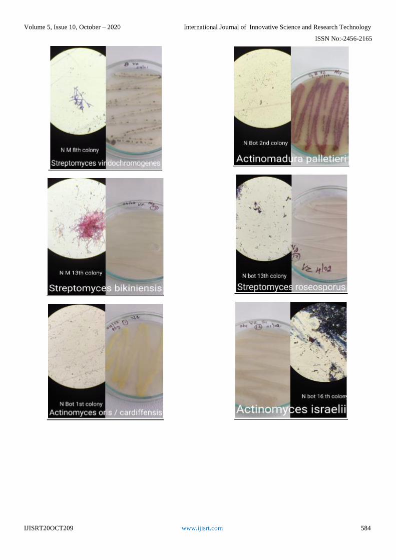
Identification of isolated actinomycetes using
Bergey’s manual The isolated actinomycetes were identified based on
their colony morphology and their microscopic morphology
using Bergey’s Manual of Systematic Bacteriology Volume
five – The Actinobacteria.
The identified actinomycetes are tabulated in table
number 7
Table:7 Identification of Actinomycetes
ISOLATE
NUMBER
IDENTIFIED NAME
01 Streptomyces griseus
02 Streptomyces tandae
03 Streptomyces griseorubens
04 Streptomyces purpureus
05 Nacordiapis alba
06 Streptomyces lateritius
07 Streptomyces griseoauranticus
08 Actinomyces gerencseriae
09 Streptomyces fradiae
10 Actinomyces bovis
11 Not identified
12 Streptomyces viridochromogens
13 Gordonia amarae
14 Streptomyces bikiniensis
15 Actinomyces oris / cardiffensis
16 Actinomadura palletieri
Page 10
Volume 5, Issue 10, October – 2020 International Journal of Innovative Science and Research Technology
ISSN No:-2456-2165
IJISRT20OCT209 www.ijisrt.com 583
17 Streptomyces albogriseolus
18 Actinomyces turicensis
19 Micromonospora carbonacea
20 Streptomyces roseosporus
21 Actinomyces israelii
22 Streptomyces violaceoruber
Page 11
Volume 5, Issue 10, October – 2020 International Journal of Innovative Science and Research Technology
ISSN No:-2456-2165
IJISRT20OCT209 www.ijisrt.com 584
Page 12
Volume 5, Issue 10, October – 2020 International Journal of Innovative Science and Research Technology
ISSN No:-2456-2165
IJISRT20OCT209 www.ijisrt.com 585
IV. CONCLUSION
The eleven soil samples collected from different places
in the college campus were serially diluted to isolate
Actinomycetes .As a result of which 22 Actinomycetes were
isolated.
Antimicrobial study on all 22 isolates was done
through primary (perpendicular streak method) and
secondary (agar well diffusion method)) screening against
08 clinical pathogens. Two actinomycetes were found to
show only mild anti-microbial property against Proteus
vulgaris.
The genomic DNA of first 8 Actinomycetes was
successfully isolated using lysozyme protocol. Their
respective DNA band were viewed on agarose gel.The
isolate 04, 06 and 08 were selected to study the effect of
variation of lysozyme concentration (5mg,10mg and 15mg)
on the amount of DNA .As a result we conclude that 15 mg
lysozyme concentration yields the maximum amount of
DNA (except for isolate 06). RAPD PCR was performed on
all three variations of DNA isolated from isolate number 04
and it was successfully amplified.
Identification of Actinomycetes was done using
Bergey’s manual of systematic bacteriology volume 5- The
Actinobacteria by studing their colony and microscopic
morphology.
As a part of the project DNA sequencing of the two
isolates that showed anti-microbial activity was supposed to
be conducted and also BLAST and FASTA of DNA
sequence was to be done. This was not conducted due to the
pandemic conditions of COVID-19.
AKNOWLEDGEMENT
At the outset, we would like to express our deep sense
of gratitude to Dr. C.N. Lokappa Gowda, Principal,
Government Science College, for his kind co-operation and
support and accordance of carrying out our project in
campus and outside campus.
We express our sincere thanks to Dr. Kavitha .B, Head
of the Department, Microbiology, for her constant effort and
creditable guidance all through the project and correcting us
through out the project and dealing with us patientfully.
We express our sincere thanks and gratitude to all the
faculty members of department of microbiology, who gave
us valuable guidance and information regarding project. A
special mention to Dr. Anuroopa, for helping us during the
genetics part of the project.
A very special thanks to Dr. Prasad sir of Sangene
Biotech, for letting us work in his laboratory and for his
constant guidance through out the project and also Sneha,
student assistant of Sangene Biotech, who helped us with
conduction of every experiment of project.
At last we would like to thank our friends and family
for their constant support towards us for conducting the
project peacefully.
REFERENCES
[1]. Abd-Alla, M. H., El-Sayed, E. S. A., and Rasmey, A.
H. M. (2013). Biosynthesis of L-glutaminase by
Streptomyces variabilis ASU319 isolated from
rhizosphere of triticum vulgaris. Univ. J. Microbiol.
Res. 1, 27–35. doi: 10.13189/ujmr.2013.010301.
[2]. Abd-Alla, M. H., Rasmey, A. H. M., El-Sayed, E. S.
A., El-Kady, I. A., and Yassin, I. M. (2016). Biosynthesis of anti-inflammatory immunosuppressive
metabolite by Streptomyces variabilis ASU319. Eur. J.
Biol. Res. 6, 152–169.
[3]. Aoyagi, T., Hatsu, M., Kojima, F., Hayashi, C.,
Hamada, M., and Takeuchi, T. (1992). Benarthin: a
new inhibitor of pyroglutamyl peptidase. i taxonomy,
fermentation isolation and biological activities. J.
Antibiot. 45, 1079–1083. doi:
10.7164/antibiotics.45.1079.
[4]. Arishima, M., Sakamoto, J., and Sato, T. (1956). An
antibiotic Streptomyces no. 689 strain. I. Taxonomic
studies. Nippon Nogei Kagaku Kaishi 30, 469–471.
doi: 10.1271/nogeikagaku1924.30.8_469.
[5]. Assaf, N.A. and W.A. Dick. 1993. Spheroplast
formation and plasmid isolation from Rhodococcus
spp. BioTechniques 15:10101012.
[6]. Baltz, R. H. and T. J. Hosted. 1996. Molecular
genetic methods for improving secondary-metabolite
production in Actinomycetes. Trends Biotechnol.
14:245-250.
[7]. Baltz, R. H. 2001. Genetic methods and strategies for
secondary metabolite yield improvement in
Actinomycetes. Antonie van Leeuwenhoek 79:251-
259.
[8]. Berdy, J. (2005). Bioactive microbial metabolites, a
personal view. J. Antibiot. 58, 1–26. doi:
10.1038/ja.2005.1.
[9]. Brockmann, H., and Musso, H. (1954). Antibiotics
from actinomycetes. XXIX. Geomycin 2. Chem. Ber.
87, 1779–1799. doi: 10.1002/cber.19540871128.
Page 13
Volume 5, Issue 10, October – 2020 International Journal of Innovative Science and Research Technology
ISSN No:-2456-2165
IJISRT20OCT209 www.ijisrt.com 586
[10]. Cho, H., Beale, J. M., Graff, C., Mocek, U.,
Nakagawa, A., Omura, S., et al. (1993). Studies on
the biosynthesis of the antibiotic reductiomycin in
Streptomyces xanthochromogenus. J. Am. Chem. Soc.
115, 12296–12304. doi: 10.1021/ja00079a009.
[11]. de los Reyes-Gavilan, C.G., J.F. Aparicio, C.
Barbes, C. Hardisson, and J. Sanchez. 1988. An
exocytoplasmic endonuclease with restriction function
in Streptomyces antibioticus. J. Bacteriol. 170:1339-
1345.
[12]. Ezaki, T. and S. Suzuki. 1982. Achromopeptidase for
lysis of anaerobic gram positive cocci. J. Clin.
Microbiol. 16:844-846.
[13]. Griffiths, P. J. F., and Ellis, G. P. (1972).
Benzopyrones—VI1: the ultraviolet absorption spectra
of chromone and 2-substituted chromones.
Spectrochim. Acta A Mol. Spectros. 28, 707–713. doi:
10.1016/0584-8539(72)80039-4.
[14]. Hayakawa, M. (2008). Studies on the isolation and
distribution of rare actinomycetes in soil.
Actinomycetologica 22, 12–19. doi:
10.3209/saj.SAJ220103.
[15]. Hayakawa, M., Sadakata, T., Kajiura, T., and
Nonomura, H. (1991). New methods for the highly
selective isolation of Micromonospora and
Microbispora from soil. J. Fermen. Bioeng. 72, 320–
326. doi: 10.1016/0922-338X(91)90080-Z.
[16]. Hazato, T., Naganawa, H., Kumagai, M., Aoyagi, T.,
and Umezawa, H. (1979). Beta-Galactosidase-
inhibiting new isoflavonoids produced by
actinomycetes. J. Antibiot. 32, 217–222. doi:
10.7164/antibiotics.32.217.
[17]. Holt, J. G., Krieg, N. R., Sneath, P. H. A., Staley, J.
T., and Williams, S. T. (1994). Bergey's Manual of
Determinative Bacteriology, 9th Edn. Baltimore, MD;
Philadelphia; Hong Kong; London; Munch; Sydney;
Tokyo: William and Wilkins.
[18]. Hopwood, D. A., and Wright, H. M. (1973). A
plasmid of Streptomyces coelicolor carrying a
chromosomal locus and its inter-specific transfer. J.
Gen. Microbiol. 79, 331–342. doi: 10.1099/00221287-
79-2-331.
[19]. Hunter, I.S. 1985. Gene cloning in Streptomyces, p.
19-44. In D.M. Glover (Ed.), DNA Cloning, A
Practical Approach, Volume II. IRL Press, Oxford.
[20]. Jasmina Nikodinovic, Kevin D. Barrow and Jo-
Anne Chuck, University of New South Wales, Sydney
and University of Western Sydney, Penrith South,
Australia, BioTechniques 35:932-936, November
2003.
[21]. Kang, M. J., Strap, J. L., and Crawford, D. L. (2010). Isolation and characterization of potent
antifungal strains of the Streptomyces violaceusniger
clade active against Candida albicans. J. Ind.
Microbiol. Biotechnol. 37, 35–41. doi:
10.1007/s10295-009-0641-9.
[22]. Kekuda, P. T. R., Onkarappa, R., and Jayanna, N.
D. (2014). Characterization and antibacterial activity
of a glycoside antibiotic from Streptomyces variabilis
PO-178. Sci. Technol. Arts Res. J. 3, 116–121. doi:
10.4314/star.v3i4.17.
[23]. Kieser, T., M.J. Bibb, M.J. Buttner, K.F. Chater,
and D.A. Hopwood. 2000. Practical Streptomyces
Genetics. The John Innes Foundation, Norwich,
England.
[24]. Kim, C., Lee, K., Kwon, O., Park, D., and Shimazu,
A. (1995). Isolation of rare actinomycetes on various
types of soil. Kor. J. Appl. Microbiol. Biotechnol. 23,
36–42.
[25]. Kim, C., Lee, K., Kwon, O., Yoo, I., and Shimazu,
A. (1994). Selective isolation of actinomycetes by
physical pretreatment of soil sample. Kor. J. Appl.
Microbiol. Biotechnol. 22, 222–225.
[26]. Kurtböke, D. I., Chen, C. F., and Williams, S. T. (1992). Use of polyvalent phage for reduction of
streptomycetes on soil dilution plates. J. Appl.
Bacteriol. 72, 103–111. doi: 10.1111/j.1365-
2672.1992.tb01810.
[27]. Kutchma, A.J., M.A. Roberts, D.B. Knaebel, and
D.L. Crawford. 1998. Small-scale isolation of
genomic DNA from Streptomyces mycelia or spores.
BioTechniques 24: 452-456.
[28]. Lee, Y.K., Kim, H.W., Liu, H.K. and H.K. Lee. 2003. A simple method for DNA extraction from
marine bacteria that produce extracellular materials. J.
Microbiol. Methods 52: 245-250.
[29]. Leonard, R.B. and K.C. Carroll. 1997. Rapid lysis of
gram positive cocci for pulse field electrophoresis
using achromopeptidase. Diagn. Mol. Pathol. 6:288-
291.
[30]. Li, S., S. Norioka, and F. Sakiyama. 1997.
Purification, staphylolytic activity, and cleavage sites
of a-lytic protease from Achromobacter lyticus. J.
Biochem. 122:772-778.
[31]. Madigan, M. T., Martiko, J. M., and Parker, J. (1997). “Antibiotics: isolation and characterization,” in
Brock Biology of Microorganisms, 8th Edn., ed M. T.
Madigan (New Jersey: Prentice-Hall International
Inc.), 440–442.
[32]. Marques, D. A. V., Santos-Ebinuma, V. D. C., de
Oliveira, P. M. S., de Souza Lima, G. M., Araújo, J.
M., Lima-Filho, J. L., et al. (2014). Screening of wild
type Streptomyces isolates able to overproduce
clavulanic acid. Braz. J. Microbiol. 45, 919–928. doi:
10.1590/S1517-83822014000300022.
[33]. Mordarska, H., S. Cebrat, B. Blach, and M.
Goodfellow. 1978. Differentiation of nocardioform
Actinomycetes by lysozyme sensitivity. J. Genet.
Microbiol. 109:381-384.
[34]. Ogawa, H., S. Imai, A. Satoh, and M. Kojima. 1983.
An improved method for the preparation of
Streptomycetes and Micromonospora protoplasts. J.
Antibiot. 36:184-186.
[35]. Ohba, K., Nakayama, H., Furihata, K., Shimazu,
A., Endo, T., Seto, H., et al. (1987). Nitropeptin, a
new dipeptide antibiotic possessing a nitro group. J.
Antibiot. 40, 709–713.
[36]. Okami, Y., and Hotta, K. (1988). “Search and
discovery of new antibiotics,” in Actinomycetes in
Biotechnology, eds M. Goodfellow, S. T. Williams, and
M. Mordarski (London: Academic Press), 37–67.
Page 14
Volume 5, Issue 10, October – 2020 International Journal of Innovative Science and Research Technology
ISSN No:-2456-2165
IJISRT20OCT209 www.ijisrt.com 587
[37]. Onda, M., Konda, Y., Hinotozawa, K., and Omura,
S. (1982). The alkaloid AM-6201 from Streptomyces
xanthochromogenus. Chem. Pharmaceut. Bull. 30,
1210–1214. doi: 10.1248/cpb.30.1210.
[38]. Otake, N., Seto, H., Nakayama, H., Endo, T., Oba,
K., Iwata, M., et al. (1988). Antibiotic 6257
manufacture with Streptomyces for Pyricularia oryzae
infection control in rice. Jpn. Kokai Tokkyo Koho. JP
63126495 (Accessed May 30, 1988).
[39]. Praveen, V., Tripathi, D., Tripathi, C. K. M., and
Bihari, V. (2008). Nutritional regulation of
actinomycin-D production by a new isolate of
Streptomyces sindenensis using statistical methods.
Indian J. Exp. Biol. 46, 138–144.
[40]. Ramalingam, V., and Rajaram, R. (2016).
Antioxidant activity of 1-hydroxy-1-norresistomycin
derived from Streptomyces variabilis KP149559 and
evaluation of its toxicity against zebra fish Danio rerio.
RSC Adv. 6, 16615–16623. doi:
10.1039/C5RA22558B.
[41]. Rao, R.N., M.A. Richardson, and S. Kuhstoss. 1987.
Cosmid shuttle vectors for cloning and analysis of
Streptomyces DNA. Methods Enzymol. 153:166-198.
[42]. Rodriguez, E. and R. McDaniel. 2001. Combinatorial
biosynthesis of antimicrobials and other natural
products. Curr. Opin. Microbiol. 4:526-534.
[43]. Sanchez, J., C. Barbes, A. Hernandez, C.R. de los
Reyes-Gavilan, and C. Hardisson. 1985. Restriction-
modification systems in Streptomyces antibioticus.
Can. J. Microbiol. 31:942-946.
[44]. Shibamoto, N., Terasawa, T., Okamoto, R., Shin, T.,
and Murao, S. (1993). Novel postproline
endopeptidase and its manufacture with Streptomyces
species. Jpn. Kokai Tokkyo Koho. Patent No. JP
05244947, Indexing: Fermentation and Bioindustrial
Chemistry (Section 16-4).
[45]. Shirling, E. B., and Gottlieb, D. (1966). Methods for
characterization of Streptomyces sp. Int. J. Syst.
Bacteriol. 16, 313–340. doi: 10.1099/00207713-16-3-
313.
[46]. Singh, V., and Tripathi, C. K. M. (2011). Olivanic
acid production in fed batch cultivation by
Streptomyces olivaceus MTCC 6820. Res. J.
Pharmaceut. Biol. Chem. Sci. 2, 726–731.
[47]. Singh, V., Praveen, V., Banga, J., and Tripathi, C.
K. M. (2009). Antimicrobial activities of microbial
strains isolated from soil of stressed ecological niches
of Eastern Uttar Pradesh, India. Indian J. Exp. Biol. 47,
298–303.
[48]. Singh V, Haque S, Singh H, Verma J, Vibha K,
Singh R, Jawed A and Tripathi CKM (2016)
Isolation, Screening, and Identification of Novel
Isolates of Actinomycetes from India for Antimicrobial
Applications. Front. Microbiol. 7:1921. doi:
10.3389/fmicb.2016.01921.
[49]. Takahashi, Y., and Omura, S. (2003). Isolation of
new actinomycete strains for the screening of new
bioactive compounds. J. Gen. Appl. Microbiol. 49,
141–154. doi: 10.2323/jgam.49.141.
[50]. Tanaka, Y., and Omura, S. (1990). Metabolism and
products of actinomycetes. An introduction.
Actinomycetolgica 4, 13–14. doi: 10.3209/saj.4_13.
[51]. Williams, S. T., Goodfellow, M., and Alderson, G.
(1989). “Bergey's manual of systematic bacteriology,”
in Genus Streptomyces Waksman and Henrici 1943,
339AL, Vol. 4, eds S. T. Williams, M. E. Sharpe, and J.
G. Holt (Baltimore, MD: Williams and Wilkins),
2452–2492.
[52]. Wu, R. Y. (1984). Studies on the Streptomyces SC4. II
Taxonomic and biological characteristics of
Streptomyces strain SC4. Bot. Bull. Acad. Sin. 25,
111–123.
[53]. .Yanagida, T. and H. Ogawara. 1980.
Deoxyribonucleases in Streptomyces. J. Antibiot.
33:1206-1207.
[54]. Zhang, L., Zhang, J., Yang, W., and Bai, G. (2008).
Classification of Streptomyces strain Z314 and
purification of its product pravastatin. Wei Sheng Wu
Xue Bao 48, 33–37.
[55]. https://www.biologydiscussion.com/bacteria/actinomy
cetes-economic-importance-and-reproduction/58664.