Screening for Iron Deficiency Anemia in Childhood and Pregnancy Update of the 1996 U.S. Preventive Task Force Review Agency for Healthcare Research and Quality Contract Number 290-02-0024, Task Order Number 1 for the US Preventive Services Task Force Oregon Evidence-based Practice Center Oregon Health and Science University 3181 SW Sam Jackson Park Road Portland, Oregon 97239 April 21, 2006 Publication No. AHRQ 06-0590 1
Transcript
Evidence Synthesis Number 40
Screening for Iron Deficiency Anemia in Childhood and Pregnancy: Update of the 1996 U.S. Preventive Services Task Force Review
Prepared for: Agency for Healthcare Research and Quality U.S. Department of Health and Human Services 540 Gaither Road Rockville, MD 20850 www.ahrq.gov Contract Number 290-02-0024 Prepared by: Oregon Evidence-based Practice Center Oregon Health and Science University 3181 SW Sam Jackson Park Road Portland, Oregon 97239 AHRQ Publication No. 06-0590-EF-1 April 2006
Abstract Background. To update its 1996 guidelines, the U.S. Preventive Services Task Force
(USPSTF) commissioned this brief update of the evidence on selected questions about
screening for iron deficiency anemia (IDA) in children, adolescents, and pregnant
women.
METHODS: We searched relevant databases, Web sites, journals, and reference lists for
systematic reviews, epidemiologic surveys, and controlled trials published in 1995 or
later that contained new information about the prevalence, diagnosis, natural course, or
treatment of iron deficiency anemia in asymptomatic persons in developed countries.
One investigator rated the quality of included trials and summarized their results in
tables.
RESULTS: In the U.S., the average prevalence IDA in target groups is: Infants 1-2 years
(6 to 17 per 1000), teenage girls (1.5%), nonpregnant females of reproductive age (2% to
5%). Factors associated with a higher prevalence include prematurity and low birth
weight, black or Mexican-American race, Alaskan native heritage, recent immigration,
poverty and, among teenage girls, fad dieting or obesity. The prevalence among pregnant
women is not known.
For cognitive and school outcomes, trials of iron supplementation for iron
deficiency anemia have had mixed results. Most trials conducted in high-risk groups
within developed countries did not demonstrate any benefit for infants and preschool
children, but one trial in high-risk infants demonstrated a transient benefit.
2
Introduction
Iron deficiency anemia has been associated with psychomotor and cognitive
abnormalities and poor school performance in children, and with poor pregnancy
outcome in pregnant women. In 1996, the U.S. Preventive Task Force (USPSTF)
recommended one-time screening for iron deficiency anemia using hemoglobin or
hematocrit for pregnant women and for high-risk infants, but not for other groups.1 The
Task Force recommended against routine testing for anemia in other children and in
adults because of low prevalence, cost, and potential adverse effects of iron therapy.2
We undertook a limited review of recent literature to assist the USPSTF in
updating its recommendations. This review was focused on key questions addressing
gaps in the evidence that were identified in the USPSTF’s 1996 review of screening for
and treating iron deficiency anemia. Specifically, the critical key questions were:
• Is there direct evidence that screening for iron deficiency in asymptomatic children
results in improved behavioral, motor, or cognitive development and/or growth?
• Does early iron supplementation in infants, children, adolescent girls, or pregnant
women with iron deficiency anemia improve these outcomes?
• What are the adverse effects of screening for iron deficiency anemia?
• What are the adverse effects of iron supplementation?
For this review, we focused on studies conducted in developed countries that addressed
one or more of these questions.
3
Background
Prevalence of Iron Deficiency and Iron Deficiency Anemia
Iron deficiency is the most common nutritional disorder worldwide. Severe or
prolonged iron deficiency can cause iron deficiency anemia (IDA). The prevalence of
IDA is sensitive to the age at testing and the diagnostic criteria used.
The hemoglobin concentration and hematocrit are the principal screening tests for
detecting anemia. Hemoglobin can be measured quickly and accurately on a few drops of
blood.3 Data on infants aged 6-12 months are sparse. For infants aged 1-2 years and 3-5
years, most studies use cut-offs for serum hemoglobin (Hgb) of <110 g/L and <112 g/L,
respectively. Typical cut-off values for females are <118 g/L for 12- 14-year-olds and
<120 g/L for 15- 39-year-olds.4, 5
These cut-off values were chosen by consensus or based on statistical analysis of
the distribution of laboratory values in the population. Some experts argue that normal
limits for Hgb and for iron studies should be based on analysis of the response to iron
therapy, but efforts to define cut-off values in this manner have not yielded definitive
results.6-8
Most cases of anemia are due to causes other than iron deficiency. When anemia
is diagnosed, additional tests can determine whether iron deficiency is the cause. Centers
for Disease Control and Prevention (CDC) analysts diagnose iron deficiency when two or
more of the following tests are abnormal: free erythrocyte protoporphyrin (>1.24 μmol/L
red blood cells), transferrin saturation (<14% for 12-15 year-olds or <15% for 16-39
year-olds), and serum ferritin (<12 μg/L).4, 9
4
While the CDC criteria are arbitrary, they have been used consistently across
several analyses of the National Health and Nutrition Examination Survey (NHANES)
(1988-1994 and 1999-2000), making comparisons across time and between demographic
groups possible. Table 1 shows that the prevalence of IDA in infants aged 1-2 years (2%
to 3%) and in females aged 12-19 years (2%) did not change substantively between these
time periods.
Table 1. Prevalence of iron deficiency anemia in selected populations – United States, National Health and Nutrition Examination surveys, 1988-1994 and 1999-2000*
1988-1994 1999-2000 Sex / Age group (yrs) No. % (95% CI†) No. % (95% CI) Both sexes 1-2 1339 3 (2-4) 319 2‡ (0-4) Females§ 12-49 5982 4 (3-5) 1950 3 (2-4) 12-19 1486 2 (1-3) 1001 2 (1-3) 20-49 4495 5 (4-6) 949 4 (2-5) 50-69 2034 2 (1-3) 611 3‡ (0.5-5) > 70 1630 2 (1-3) 394 1‡ (0-2) Data from MMWR 2002.10 *All racial/ethnic groups. †Confidence interval. ‡Unreliable; relative standard error (i.e., standard error/prevalence estimate) is > 30%. §Non-pregnant only.
Not all studies use the CDC case definitions. The positive predictive value of a
low hemoglobin for IDA varies with age and with the cut-off values used for case
definition. Among children aged 12-35 months in NHANES III, the positive predictive
value of Hgb concentration <110 g/L for iron deficiency was 29% (95% CI, 20–38%),
and the sensitivity was 30% (95% CI, 20–40%). Changing the diagnostic cutoff point to
5
Hgb <107 g/L resulted in a positive predictive value of 38% (95% CI, 24–52%) but
lowered the sensitivity to 15% (95% CI, 7–22%).8
Table 2 illustrates how the positive predictive value varies with age and with the
cut-offs used to define IDA. In the Avon longitudinal study of pregnancy and childhood
(ALSPAC), investigators developed criteria for the diagnosis of IDA based on the
distributions of Hgb and ferritin levels in their own sample. By these (ALSPAC) criteria,
5% of infants 12 months or 18 months of age had a low Hgb value, and 10% or 12% of
these infants, respectively, proved to have iron deficiency anemia. Using the World
Health Organization (WHO) or Institute of Medicine (IOM) criteria, the apparent
prevalence of anemia was between 17% and 18%, but the prevalence of IDA and the
positive predictive value of a low Hgb value were much lower in infants 12 months of
age than at 18 months of age.
Table 2. Percentage of infants with iron deficiency anemia at 12 and 18 months of age using different case definitions*
Risk factors among infants. The risk of iron deficiency anemia is high during the
second year of life because of increased iron requirements related to rapid growth.11, 14-16
Premature and low birth weight infants and infants with history of prolonged stay in the
neonatal unit are at particularly high risk of developing iron deficiency anemia before 1
year of age.17 Among term infants younger than 1 year, however, the prevalence of IDA
is low, and Hgb and serum ferritin are uncorrelated.7, 18
Risk factors for developing IDA in the second year of life include the use of non-
iron-fortified formula in the first year of life (without therapeutic iron supplementation);
exclusive breastfeeding with no or erratic iron supplementation after 6 months of age;
and the introduction of cow’s milk before 1 year of age.5 The prevalence of IDA
increases between 12 and 18 months of age as these factors come into play.
At present, about 97% of formula sold in the United States is iron-fortified.19
Randomized and nonrandomized controlled trials, observational studies, and time series
studies have demonstrated substantial reductions in the incidence of iron deficiency and
IDA in healthy infants fed iron-fortified formula, iron-fortified cereal, or breast milk with
iron-fortified cereal added at 4-6 months, compared with infants fed cow’s milk or
unfortified formula.
U.S. data on the impact of race, ethnicity, and socioeconomic factors on the risk
of developing IDA in infancy are surprisingly sparse. The Pediatric Nutrition
Surveillance System (PedNSS) measures hemoglobin levels in a national sample of
infants from families participating in the Special Supplemental Nutrition Program for
Women, Infants, and Children (WIC), but does not perform iron-related measures. In the
8
2003 PedNSS report, the most recent to be published, 16.2% of infants aged 6-11 months
had Hgb < 110 g/L, and 15% of children aged 12–17 months had Hgb < 110 g/L. The
overall prevalence of anemia in PedNSS children declined from 15.8% in 1994 to 12.8%
in 2003. The highest prevalence of anemia was among black infants (19.0%).20 This
survey establishes that black infants have a higher risk of anemia, but the proportion of
cases that are related to iron deficiency is unknown.
In developing countries, and therefore among some groups of immigrants to
North America, blood loss due to parasitic infection or malaria is a common cause of iron
deficiency.21 Native American infants and recent immigrants from Cuba are also at risk
for IDA. A study of First Nations communities in Canada determined the prevalence of
anemia (defined as Hgb < 110 g/L) among 9-month-old infants to be 31.9%, and
estimated that the prevalence of IDA to be 5.6% to 10.8%, based on Hgb < 110 g/L and a
low mean cell volume as proxy measures for IDA.22 A 1998 Pan American Health
Organization report estimated that IDA affects 40% to 50% of Cuban children aged 1-3
years.23
Risk factors among adolescent girls and adult women. Females of childbearing age
require additional iron. Heavy menstrual blood loss (≥ 80 mL/mo) and pregnancy are
associated with higher iron requirements.5, 24
Race, income, education, and other socioeconomic factors are associated with IDA
in girls and women. In NHANES III, Mexican-American women aged 12-39 years were
at higher risk of having IDA (6.2% ± 0.8%) than non-Hispanic white women of the same
age (2.3%± 0.4%), a difference that was marked among poor women but small for
9
women with higher household incomes (Table 3)9 and which could not be accounted for
by differences in dietary intake of iron. We did not find an analysis of risk factors among
black women.
Table 3. Prevalence of iron deficiency anemia in relation to poverty in Mexican-American and non-Hispanic white women aged 12-39 years
Poverty Income Ratio*
Mexican-American (n = 1194) % ± S.E.
Non-Hispanic white (n = 1183) % ± S.E.
≤1.3 6.9 ± 1.3 1.9 ± 0.8 >1.3–1.85 8.8 ± 2.2 4.9 ± 1.8 >1.85–3.0 4.4 ± 1.6 2.2 ± 0.8 >3.0 2.6 ± 0.9 1.9 ± 0.6 (not significant) * Ratio of the total household income divided by the poverty threshold for the year of the interview. Data from NHANES III9
Eating disorders are also associated with IDA. An analysis of NHANES III data
on 9698 children aged 2-16 years found that overweight and obesity were associated with
a higher risk of IDA; in a logistic regression model controlling for age, gender, ethnicity,
poverty status, and parental education level, children who were overweight were 2.3
times as likely to be iron-deficient (2.3; 95% CI, 1.4-3.9, respectively) as were those who
were not overweight.25 Adolescent girls who try to control their weight may
inadvertently limit their iron intake. In Britain in the 1980s, the prevalence of IDA in
adolescent girls was higher among girls who bought snacks at local shops instead of
eating school lunches or bringing food from home.17
Complications of Iron Deficiency Anemia
As early as the 1960s, researchers demonstrated that, in general, decreased
hemoglobin alone does not have readily apparent adverse effects unless it is below 10
10
g/dL (100 g/L).26-28 Persons with markedly reduced hemoglobin levels are at risk for
cardiopulmonary and other complications. Screening is intended to find milder degrees
of anemia before such complications have developed.
Infants and children. Several cross-sectional and case-control studies have
demonstrated an association between IDA and psychomotor and cognitive abnormalities
and poor school performance in children.17, 29-35 For example, in a recent cross-sectional
analysis of NHANES III data, 71% of iron-deficient children had below-average math
scores, versus 49% of children who had normal iron status.34 Scores of tests on reading,
block design, and digit span did not differ. After adjustment for age, gender, race,
poverty status, caretaker education, and lead status, iron-deficient children were 2.4 times
as likely to have low math scores (95% CI, 1.1-5.2; p=0.03). The effect was strongest
among girls aged 12-16.
Several causal hypotheses have been proposed to explain this association. The
oldest is that the brain functions poorly in IDA because of decreased oxygen delivery to
tissues. According to this theory, correction of anemia could reverse the neurocognitive
deficits seen in cross-sectional studies. An alternative hypothesis is that iron deficiency
leads to increased absorption of lead, which can also cause brain damage.
Another alternative hypothesis is that, in the fetus, infant, and toddler, iron
deficiency may cause abnormal metabolism of neurotransmitters or hypomyelination,
leading to irreversible or very slowly reversible neurocognitive deficits. Evidence for
this hypothesis comes primarily from animal studies.36, 37 Investigators seeking
supporting evidence in humans have measured auditory brainstem responses and visual
evoked potentials in a cohort of Chilean children who were diagnosed to have IDA as
11
infants. At the time of initial diagnosis at 6, 12, or 18 months of age, infants with IDA
had slower transmission through the auditory brainstem pathway than healthy controls.
Although IDA was diagnosed and treated early, at 4 years of age the children who had
IDA as infants still had slower transmission than healthy infants.38
A recent critical review identified seven longitudinal studies in which low
hemoglobin levels in early childhood were linked to poor cognitive development or
school achievement in later childhood.35 (Two of these studies were available in 1996.)
The older studies were small (range 20-41 anemic children) and the iron status of the
anemic children was not clear.
One of the recent longitudinal studies using records from the WIC were linked to
school records in Dade County, Florida.39 The outcome variable for the analysis was
mild or moderate mental retardation on the basis of criteria used by the Florida
Department of Education for special education placement. About 69% of the sample
(n=3,771) were black, 23% were Hispanic, and 7% were white. After adjustment for
birth weight, maternal education, sex, race-ethnicity, age of mother, and age of child,
there was a significant association between Hgb level at entry into the WIC program and
the probability of mental retardation at age 10 (odds ratio 1.28; 95% CI, 1.05-1.60).
The other recent study40 was a 10-year follow-up of a cohort of Costa Rican
children, the subject of previous reports in infancy and at 5 years of age. In this cohort,
48 children who had severe iron deficiency in infancy were compared with 114 children
who had good iron status in infancy. At ages 11-14 years, the children who had severe
iron deficiency as infants still had worse scores on intelligence tests (101.8 ±2.0 vs.
104.6±1.3) and on a variety of tests of cognitive function, despite having similar Hgb
12
levels in adolescence. Parents of children in the severe iron deficiency group were more
likely to report behavior problems.
It is difficult to prove that the relationship between anemia and developmental
abnormalities in longitudinal studies is causal. Many other factors associated with
abnormal neurocognitive development are also associated with iron deficiency. These
include nutritional factors, such as intake of iodine, zinc, and other micronutrients;
environmental factors (e.g., exposure to lead); prematurity and low birth weight;
caretaker characteristics (e.g., maternal education, household income); and other
socioeconomic factors.35, 41 In all cross-sectional studies, iron-deficient children and
their families differed in nutritional status, income, education, and other factors from the
comparison groups.35 Most longitudinal studies did not include enough children to
control for all environmental variables that could be associated with iron deficiency and
with the outcomes.35
Socioeconomic factors are so strongly associated with cognitive outcomes, and so
highly inter-correlated, that the ability of statistical adjustment to eliminate confounding
is uncertain. In the Dade County study, for example, maternal education was a powerful
predictor of mental retardation after adjustment for other risk factors. Compared with
maternal education greater than 12 years, the adjusted odds associated with only 12 years
of maternal education and less than 12 years were 8.32 (95% CI, 1.12-62.0) and 11.9
(95% CI, 1.63-88.1), respectively. In the Costa Rican study, maternal IQ and education
were strongly associated with children’s IQ and with cognitive abnormalities.
Screening is most likely to influence neurodevelopmental outcomes if it is done at
an age when IDA is present and development is still normal.14 Investigators from the
13
AVON longitudinal study of pregnancy and childhood sought to identify the best age for
screening by examining the relationship between serum Hgb and developmental
outcomes, measured at age 18 months.11, 14 Delayed development by age 18 months was
associated with anemia at 8 months of age. However, most abnormalities that would lead
to a diagnosis of iron deficiency without anemia resolved spontaneously by 12 or 18
months of age.
Pregnancy. Numerous observational studies have reported an association between
severe to moderate anemia (hemoglobin <9–10 g/dL) and poor pregnancy outcome.2
However, the relationship between maternal iron deficiency or IDA during pregnancy
and birth outcome is not well understood. Older studies, including three large,
population-based studies, evaluated the relationship between Hgb or Hct and low birth
weight or premature birth without assessing the iron status of the mother. Recent cohort
studies42 and reviews,13, 43 including a critical review of studies published between 1966
and 1999,44 emphasize that the relationship of maternal Hgb to birth weight is U-
shaped—that is, low and high Hgb values are markers for poor birth outcomes. In white
women, maternal hemoglobin values of 105–125 g/L were associated with the lowest rate
of LBW. For black women, the rate of low birth weight was lowest for maternal
hemoglobin values of 85–95 g/L, but this estimate is based on data that are now over 25
years old. In the first trimester, IDA is associated with a greater than two-fold increase in
the risk of preterm delivery. In the third trimester, however, lower Hgb and Hct levels
are not associated with higher rates of low birth weight or preterm delivery.
14
Maternal IDA might have other complications. One prospective, longitudinal
human study found an association between low umbilical cord serum ferritin
concentrations and poor performance on mental and psychomotor tests at 5 years of
age.45 Low postpartum Hgb or Hct levels may be associated with postpartum
depression.46
Postpartum maternal IDA may also be associated with developmental delay in
children. A controlled trial of iron therapy in young, South African mothers with IDA,
published in 2005, compared non-anemic mothers with anemic mothers administered
either placebo (25 mg ascorbic acid and 10 μg folate) or daily iron treatment (125 mg
FeSO4) plus ascorbate and folate).47 All mothers had full-term, normal birth weight
infants (n = 81) and were enrolled in the study at 6-8 weeks postpartum. At baseline,
anemic mothers tended to be less responsive to, and more controlling of, their infants
than non-anemic mothers. Infants of anemic mothers were delayed at 10 weeks in hand-
eye movement and overall development. Infants whose mothers were anemic in the early
postpartum period scored worse on developmental tests at 10 weeks and 9 months of age.
At 9 months, anemic mothers in the placebo group were significantly more negative
toward their babies, engaged less in goal setting, and were less responsive than non-
anemic mothers in the control group.
15
Methods
Problem Formulation
Members of the USPSTF defined the scope of this update with input from Agency
for Healthcare Research and Quality (AHRQ) and Evidence-based Practice Center (EPC)
personnel.
Search for New Studies
The search was focused on the following key questions:
• Is there direct evidence that screening for iron deficiency in asymptomatic children
aged 6-12 months results in improved health outcomes; that is, abnormal infant
behavior, growth, and development (longer-term outcomes)?
• Is there evidence that early iron supplementation in infants, children, adolescent girls,
or pregnant women with iron deficiency anemia improves these outcomes?
• What are the adverse effects of screening for iron deficiency anemia?
• What are the adverse effects of iron supplementation?
EPC personnel searched the Cochrane Database of Systematic Reviews (2005,
v.2), Cochrane CENTRAL (2005, v.2), reference lists of review articles, and tables of
contents of leading pediatric journals for studies published 1995 or later that contained
new information about the prevalence, diagnosis, natural course, or treatment of iron
deficiency anemia in asymptomatic persons. We also searched the web site of the Iron
Deficiency Project Advisory Service Working Group on Iron Deficiency Anemia in
Children < 2 (http://www.micronutrient.org/idpas/WorkingGroup.html), which
maintains bibliographies and reprints of articles about the prevalence and cognitive
consequences of iron deficiency in developing countries.
Articles that met the following criteria were included in this update:
1) The study was a systematic review, prospective cohort study, controlled trial,
quasi-experimental study with concurrent controls, or case-control study; not a
case series, case report, or comparison with historical controls.
2) The study was not included in the 1996 review.
3) The study was rated at least “fair-quality” using the USPSTF criteria for internal
validity.48
Synthesis
Eligible studies were rated and abstracted by one investigator. Because several
recent meta-analyses were available, the investigator did not conduct a new quantitative
synthesis; instead the focus was on reporting the results of a critical appraisal of trials
published since the USPSTF’s 1996 guideline. USPSTF members also reviewed key
studies identified in the review.
Results
Is there direct evidence that screening for iron deficiency anemia in
asymptomatic children aged 6-12 months results in improved health
outcomes; that is, abnormal infant behavior, growth, development (longer-
term outcomes)?
17
We did not find any controlled trials of screening for IDA. In the United States,
an uncontrolled, practice-based study conducted at the pediatric resident clinic at Johns
Hopkins University described the results of an effort to implement the screening strategy
recommended by the American Academy of Pediatrics.49 Of 1358 clinic patients aged 9-
36 months who were screened, 343 (25%) had an Hgb level less than 110 g/L. About
half of these infants had mild anemia (Hgb 106 to 109 g/L). Of these, 75 were prescribed
iron and anemia resolved by 6 months of follow-up in 55 (73%). Another 25 were not
prescribed iron, and anemia resolved by 6 months in 21 (84%). For those who had Hgb
levels <106 g/L, 61 of 90 (68%) treated children resolved by 6 months, versus 6 of 15
(40%) untreated children.
Is there evidence that early iron supplementation in infants, children,
adolescent girls, or pregnant women with iron deficiency anemia improves
outcomes?
Infants and children. Improved growth and weight gain with 3-6 months of iron
supplementation have been reported consistently in placebo-controlled trials of anemic,
malnourished children in developing countries.
Whether treatment is also associated with improvements in cognition, behavior,
and motor development is less clear. The U.S. Preventive Services Task Force, in 1996,
noted that trials of treatment of infants with IDA to improve neurodevelopmental
outcomes had conflicting results. Some of the trials reporting a benefit had serious
flaws.50, 51 A Cochrane review, published in 2001, found seven trials of treatment in
18
19
children up to 3 years of age; all these trials were published prior to 1994 and had been
cited in the Task Force’s review (Table 4).52 The Cochrane review concluded that there
was a lack of clear evidence of a beneficial effect on psychomotor development.
A review by Grantham-McGregor and colleagues included trials as well as
observational studies.35 With the exception of one trial published in 1993,53 short-term
and longer-term trials found no benefit of iron supplementation on intelligence tests, tests
of cognitive function, or other neurodevelopmental outcomes, and the observational
studies had not adequately addressed potential confounders.
Table 4. Neurodevelopmental outcomes in children under age 3 with iron deficiency anemia (articles included in Cochrane Review and 1996 USPSTF reviews) Trial (Funding Source) Setting Subjects
Age (mos.) N
Design Characteristics
Treatment (duration) Control
Follow-up Assess-
ment Time Results Oski and Honig, 197854
Pediatric Clinic in
New York
IDA (Hgb<10.
5 g/dl)
9-29 24 DBRCT. No details of allocation process available.
IM iron dextran complex
once.
Placebo of IM sterile saline once.
BSID, BIB. 1 wk. Improved alertness,
gross and fine motor
coordination. Lozoff, 198255
Community volunteers, Guatemala
IDA (Hgb<10.
5 g/dl)
6-24 28 DBRCT. Allocation by investigators not connected with study.
Oral iron for 7 days.
Placebo. BSID. 6 -8 days
NS trend in PDI, no
difference in MDI.
Lozoff, 198729
- Anemia (Hgb<=10.5g/dl)
- 52 DBRCT. Allocation by investigators not connected with study.
IM or oral iron for 7
days.
Placebo. BSID. 7 days
No difference.
Walter, 198933
Community sample,
Chile
IDA (Hgb<10.
5g/dl)
12 39 Unclear. Method of allocation unclear.
Ferrous sulphate 45 mg day for 10 days.
Placebo. BSID. 3 mos.
No difference.
Kimmons, un-published
General pediatric
clinic, U.K.
IDA (Hgb<10.
6g/dl, MCV<73)
6 -24 42 DBRCT. Allocation by nurse not connected with study.
IM Iron dextran
complex.
Placebo. BSID. 1 wk. No difference.
Aukett, 198656
Community Sample
IDA (Hgb 8-11g/dl)
17-19 110 DBRCT. Allocation by investigators not connected with study.
Oral iron plus
vitamin C.
Placebo. DDST, weight
change.
8-9 wks.
No difference.
Idjradinata, 199353
Pediatric clinic in
Indonesia
IDA (Hgb<=10.5g/dl)
12-18 50 DBRCT. Oral iron for 4
months.
Placebo. BSID, weight, length.
4 mos.
BSID = Bayley Scales of Infant Development, DBRCT= Double-blinded randomized controlled trial, DDST = Denver Development Screening Test, Hgb = Hemoglobin, IDA = Iron deficiency anemia, IM= Intramuscular, MCV = Mean corpuscular volume, MDI = Mental Development Index, NS = Not significant, PDI = Psychomotor Development Index, USPSTF = U.S. Preventive Services Task Force
PDI + 18.4 MDI + 18.8
20
The conflicting results in trials of treatment for IDA have called into question the reversibility of neurodevelopmental
abnormalities associated with IDA, and have led to increased interest in preventing rather than remediating iron deficiency. Until
recently, few trials of primary prevention examined neurocognitive or behavioral endpoints. Moreover, as many studies do not
distinguish between high-risk subjects who have IDA and those who have iron deficiency without anemia, it is very difficult to
distinguish the effect of prophylactic iron supplementation from the effect of treatment of existing iron deficiency.
A recent meta-analysis57 combined 17 trials of iron supplementation in infants or in children up to 12 years of age. Sixteen
trials were published 2001 or earlier and one was unpublished. Ten of the trials were conducted in developing countries, two were
from the United States,31, 58 two were from the United Kingdom,56, 59 one was from Canada,60 and one was from Greece.61 The main
measure was the “mental development score,” derived by combining any available scores from the Bayley Mental Development
Index (MDI), Stanford Binet Test, Peabody Picture Vocabulary Test (PPVT), IQ, and cognition tests. Overall, the standard mean
difference in mental development scores was 0.30 (95% CI, 0.15 to 0.46, p< 0.001), a difference equivalent to equivalent to 1.5 to 2
points on a scale of 100. Separate analyses for mental development tests and motor development found no statistically significant
differences.
The trials pooled in this meta-analysis included diverse subjects, settings, clinical interventions, and outcome measures.
Because of this heterogeneity, the overall pooled result has little or no applicability to the United States. In subgroup analyses, the
improvement in mental development scores was attributable to five trials in children aged 7 years and older (standard mean difference,
21
0.44; 94% CI, 0.21-0.66, p<0.0001) conducted in India (2), Thailand (1), and Indonesia (2). The effect was small and statistically not
significant for infants under 2 years of age (0.15, CI 0.04-0.34, p=0.128). The effect is intermediate for children between 2 and 5
years of age.62 More recent trials in developing countries have had mixed results (Table 5 and Evidence Table 1, Panel 1).63-65
Table 5. Recent trials of iron supplementation on cognitive or motor development in developing countries Trial / Design
Subjects / Setting
Results
Lozoff, 200363 Partly randomized
1657 infants 6 or fewer months old Chile
Bayley Scales and Fagan test of Infant Intelligence at 12 months. No difference in PDI or MDI scores at 12 months. On Fagan test, mean looking time was longer in the no-iron-added group (1.39 ± 0.02 seconds vs. 1.46 ± 0.04).
Lind, 200464 RCT
680 full-term infants aged 6 months Indonesia
Bayley Scores at 12 months. PDI in Iron group 106 ± 11 vs. placebo group 103 ± 10.8. MDI in Iron group 101 ± 9.3 vs. placebo group 99 ± 10. No difference in Behavioral Rating Scale.
MDI = Mental Development Index, PDI = Psychomotor Development Index, RCT = Randomized controlled trial
In their review, Grantham-McGregor35 and colleagues identified three prevention trials conducted in developed countries59, 60,
66 that measured motor, cognitive, or behavioral function. One60 of these, conducted in Canada, was reviewed by the USPSTF
previously. In this trial, infants from very low-income families were randomized to take iron-fortified versus unfortified formula.
22
Those who were fed iron-fortified formula had significantly higher Bayley motor scores at 9 and 12 months. By 18 months there was
no longer any effect, but by that time 46% of the subjects had been lost to follow-up.
Trials conducted in developed countries and published since the last USPSTF review are summarized in Table 6 (see Evidence
Table 1, Panel 2 for more details). Two trials (which were included in the review by Grantham-McGregor and colleagues) evaluated
iron-fortified formula in low socioeconomic status areas of the United Kingdom.59, 66 We also identified a Canadian trial of iron
supplementation in infants 1 to 6 months of age67 and one of preschoolers conducted in nine day care centers in Athens, Greece,68
neither of which has been included in previous systematic reviews.
Table 6. Effect of iron supplementation on infants in developed countries Trial / Design
Subjects / Setting
Results
Developed countries Williams, 199966 RCT
100 full-term infants aged 6-8 months Low-income England
Griffith’s scale at 18 and 24 months. No differences at 18 months. At 24 months, global developmental quotient fell 5.4 points more in the non-fortified group than in the fortified group (P<0.05).
Morley, 199959
RCT
493 full-term infants aged 9 months. England
MDI and PDI at 18 months. No differences.
23
Trial / Design
Subjects / Setting
Results
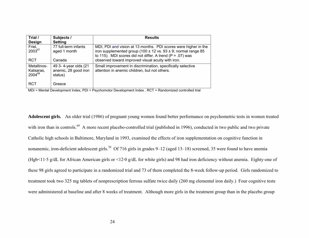
Friel, 200367 RCT
77 full-term infants aged 1 month Canada
MDI, PDI and vision at 13 months. PDI scores were higher in the iron supplemented group (100 ± 12 vs. 93 ± 9; normal range 85 to 115). MDI scores did not differ. A trend (P = .07) was observed toward improved visual acuity with iron.
Metallinos-Katsaras, 200468 RCT
49 3- 4-year olds (21 anemic, 28 good iron status) Greece
Small improvement in discrimination, specifically selective attention in anemic children, but not others.
MDI = Mental Development Index, PDI = Psychomotor Development Index , RCT = Randomized controlled trial
Adolescent girls. An older trial (1986) of pregnant young women found better performance on psychometric tests in women treated
with iron than in controls.69 A more recent placebo-controlled trial (published in 1996), conducted in two public and two private
Catholic high schools in Baltimore, Maryland in 1993, examined the effects of iron supplementation on cognitive function in
nonanemic, iron-deficient adolescent girls.70 Of 716 girls in grades 9–12 (aged 13–18) screened, 35 were found to have anemia
(Hgb<11·5 g/dL for African American girls or <12·0 g/dL for white girls) and 98 had iron deficiency without anemia. Eighty-one of
these 98 girls agreed to participate in a randomized trial and 73 of them completed the 8-week follow-up period. Girls randomized to
treatment took two 325 mg tablets of nonprescription ferrous sulfate twice daily (260 mg elemental iron daily.) Four cognitive tests
were administered at baseline and after 8 weeks of treatment. Although more girls in the treatment group than in the placebo group
24
noticed changes in stool color (65% v 10%, p<0.001), the percentage of girls who correctly guessed their group assignment did not
differ significantly between groups (62% in treatment compared with 45% of controls, p=0.18).
The four tests were the Symbol Digit Modalities Test (SDMT); Visual Search and Attention Test (VSAT); Brief Test of
Attention (BTA); and the Hopkins Verbal Learning Test (HVLT). Only subjects who completed the study were included in the
analysis. Treatment had no effect on the SDMT, VSAT, or BTA, all of which are considered to be tests of attention. The authors
described the other test, HVLT, as follows:
“The HVLT is a 12-item, semantically categorized word-list learning test with three free recall trials, a delayed recall trial, and
yes/no recognition; participants are read the same list of words three times and each time are asked to repeat as many words as they
can recall; 20 minutes later they are asked to say which words they remember, and are read 24 words which include the original 12
words plus 12 semantically related and unrelated words.”70
By the author’s description, the HVLT, which is considered a test of learning, is scored in three parts—the sum of the three
recall trials (the “total recall score”), the delayed recall trial, and the recognition part. The article reported an average baseline HVLT
total recall score for the treatment and placebo groups, but did not report the post-treatment scores. The authors state that “on the total
recall score of the HVLT (sum of trials 1–3), girls who took iron showed significant improvement over baseline and end of treatment
25
compared with the control group (p<0·02). Baseline performance on the HVLT accounted for 93% of the variability in post-
intervention scores, whereas treatment condition accounted for the remainder. However, there were no significant differences
between groups in other components of the HVLT (delayed recall, yes/no recognition).” From the published results, it is impossible
to determine the magnitude of effect or how many girls’ scores improved, stayed the same, or worsened in each group.
Pregnant women. A large body of data suggests that iron supplements are effective in improving the hematologic indices of
pregnant women, but there is limited evidence that improving hematologic indices in anemic women results in improved clinical
outcomes for the mother, fetus, or newborn. Clinical trials have reported that iron supplements in healthy pregnant women with initial
hemoglobins ≥ 10 g/dL are efficacious in correcting red cell indices and iron stores, but they do not improve birth weight, length of
gestation, or other outcome measures when compared with placebo or with no supplements.71-77 A 2005 review of community-based
interventions to improve perinatal and maternal outcomes in developing countries also found no clear evidence that iron
supplementation improved maternal and perinatal or neonatal outcomes.21
A Cochrane review found that iron supplementation appears to prevent low hemoglobin at birth or at 6 weeks postpartum, but
concluded there were no reliable data from controlled trials about the pregnancy outcomes for either mother or baby.78 The largest trial
included in the Cochrane review compared selective versus routine supplementation in 2693 pregnant Finnish women.79 The initial
results of this trial showed a statistically significant increased likelihood of cesarean section (OR 1.36; 95% CI, 1.04 -1.78) and of
26
post-partum blood transfusion (OR 1.68; 95% CI, 1.05 -2.67) in the selective supplementation group compared with routine
supplementation. The authors attributed the increased cesarean sections and blood transfusion rates to possible anxiety by midwives
and obstetricians about low hematocrit values in the selectively supplemented group. Selective supplementation was associated with a
significantly reduced risk of stillbirth after 28 weeks’ gestation and of death in the first 7 days after birth. There were also fewer
women in this group who complained of side effects from the medication. In a 7-year follow-up study, fewer infants in the selective
supplementation group had been hospitalized for convulsions (OR 0.44; 95% CI, 0.25-0.79).80
The Cochrane review did not include an American trial published in 2003 of iron supplementation in 275 low-income pregnant
women who had a hemoglobin concentration >110 g/L and a ferritin concentration >20 g/L.81 The women were enrolled before 20
weeks of gestation and randomly assigned to receive a monthly supply of capsules containing either 30 mg iron as ferrous sulfate or
placebo until 28 weeks of gestation. For unclear reasons, women assigned to the placebo group had higher baseline pre-pregnancy
weight (77.9kg ± 24.3 vs. 72.5kg ± 20.3, p=0.04) and initial ferritin levels (49.4µg/L vs. 44.7µg/L, p=0.0168). Of these 275 women,
62 were excluded from the analysis because the investigators could not obtain birth weights. For the 213 newborns included in the
analysis, birth weight < 2,500 grams occurred less frequently in the iron supplementation group (4.3% vs. 16.7%, p=0.003), though
the risk of preterm delivery was not different (12.8% vs. 12.5%.)
What are the adverse effects of screening for iron deficiency anemia?
27
We did not identify any studies of the harms of screening for IDA.
What are the adverse effects of iron supplementation?
Many infants dislike the taste of oral iron preparations or have gastrointestinal side effects. The likelihood of response to
treatment for iron deficiency anemia identified by screening is unclear because so many families or infants do not accept treatment and
because the rate of spontaneous resolution is high. In the population-based Millennium Baby Study cohort from Glasgow, UK, for
example, 147 children aged 13 months were offered treatment because they had a low Hgb, ferritin, MCV, MCH, or high
protoporphyrin level.7 Of these, 124 families accepted the first bottle of oral iron, but only 83 accepted a second bottle 3 weeks later.
Thirty-one families reported that iron had caused diarrhea and many infants refused to go back on iron-fortified formula milk. A
practice-based UK general practitioner study noted similar problems.82
Many controlled trials of iron have examined whether new forms of iron replacement, combinations of micronutrients, and
iron regimens requiring less frequent dosing can improve compliance and iron status in infants,83-92 preschool children,93, 94 school
children (including adolescents),95-100 and pregnant women.101, 102
Adverse effects of iron therapy include unpleasant gastrointestinal symptoms (e.g., nausea and constipation) that are dose-
related and, at normal doses, reversible. Iron therapy can cause complications of excessive iron storage in patients with an underlying
iron storage disorder (e.g., idiopathic hemochromatosis).103, 104 A potential hazard of iron supplements is unintentional overdose by
28
children in the home. Iron overdose is a concern because it has been observed even in the context of controlled trials and screening
programs in which parents were instructed in the safe storage and use of iron-containing products.
Iron supplements accounted for 30% of fatal pediatric pharmaceutical overdoses occurring between 1983 and 1990.105 In
1997, to reduce the danger of overdosage, the U.S. Food and Drug Administration (FDA) required unit-dose packaging for iron-
containing dietary supplement and drug products that contain 30 milligrams (mg) or more of iron per dosage unit.106 According to an
analysis of American Association of Poison Control Center Toxic Substance Surveillance System (AAPCCTSSS) abstracts, the
regulations were associated with a reduction in deaths of children from iron ingestions. From 1983 through 2000, a review of the
AAPCCTSSS system showed that at least 43 children had died from the ingestion of iron supplements, but only one child was
reported to have died from the ingestion of an iron-containing product from 1998 through 2002 while the regulation was in effect.107
In October 2003, the FDA withdrew this requirement in response to a court ruling in the case of Nutritional Health Alliance
versus FDA, in which the Court concluded that the Federal Food, Drug, and Cosmetic Act does not provide the FDA with authority to
require manufacturers of iron-containing dietary supplement and drug products to use unit-dose packaging for poison prevention
purposes.108 All iron-containing supplements must carry the warning “Accidental overdose of iron-containing products is a leading
cause of fatal poisoning in children under 6.”
Other potential adverse effects of iron mentioned in the literature (e.g., birth defects, cancer, heart disease, metabolic
imbalances of other minerals, and harmfully high hemoglobin levels) have not been proven. A recent meta-analysis of 28 placebo-
29
controlled trials of iron supplements in 7,892 infants and children found no increased risk of infection (1.02 [95% CI, 0.96-1.08]) and
an 11% increase in the risk of diarrhea corresponding to one episode per 20 children per year (95% CI, 1.01-1.23; P=0.04).109
Recommendations of Other Groups
In 1998, the Centers for Disease Control and Prevention (CDC) published recommendations to prevent and control iron
deficiency.5 In addition to pregnant women and high-risk infants, the CDC recommended screening among high-risk preschool
children and among nonpregnant women of childbearing age. The CDC also recommended “universal iron supplementation to meet
the iron requirements of pregnancy.”
The American Academy of Pediatricians (AAP) recommends checking hemoglobin (or hematocrit) between 9 and 12 months
of age and then 6 months later, and, for patients at high risk, once a year from age 2 to 5 years. Screening may be universal or
selective depending on the prevalence of IDA in the local or demographic population. The AAP also recommends that adolescent girls
be screened for anemia during all routine physical examinations.16
The 2005 AAP breastfeeding guidelines recommend continuing breastfeeding for at least the first year of life and beyond,
while introducing complementary foods rich in iron beginning around 6 months of age. Preterm and low birth weight infants and
infants with hematologic disorders, or infants who had inadequate iron stores at birth, generally require iron supplementation before 6
30
months of age. The AAP recommends that infants weaned before 12 months of age receive iron-fortified infant formula instead of
cow’s milk.110
The American Academy of Family Physicians (AAFP) recommends screening for IDA by obtaining hemoglobin and/or
hematocrit levels in infants aged 6 to 12 months who are living in poverty, or who are black, Native American, or Alaska Native,
immigrants from developing countries, preterm and low birth weight infants, and infants whose principal dietary intake is unfortified
cow's milk.
The American College of Obstetricians and Gynecologists (ACOG) recommends prenatal screening for all women at the
earliest prenatal visit and early in the third trimester. Screening of older children or nonpregnant adolescents and adults is not
recommended.
The Veterans Administration/Department of Defense guideline panel found insufficient evidence to recommend for or against
routinely supplementing iron for all pregnant women who are not anemic. They recommended supplementation with at least 50 mg
elemental iron (325 mg ferrous sulfate) twice a day in all pregnant women diagnosed with anemia (hematocrit <30).
Summary and Discussion
31
Iron deficiency anemia is a serious problem worldwide. Although it is less common in developed countries such as the United
States and Canada, where the use of iron supplemented formula is widespread, within these countries it is still a major concern among
vulnerable populations, particularly in recent immigrants, native Americans, and poor African-American communities.
While iron deficiency anemia in infancy is a marker for poor neurocognitive development subsequently, screening and early
treatment have not consistently improved these outcomes. Two leading hypotheses have been advanced to explain these disappointing
results. One is that prevention of neurodevelopmental consequences of iron deficiency anemia may require acting to prevent iron
deficiency in the first place rather than detection and treatment of existing iron deficiency. At present there is little evidence to
support this hypothesis, but additional studies, particularly in developing countries, may confirm it. The second hypothesis is that
prevention of neurodevelopmental consequences may require screening and early treatment of multiple nutritional deficiencies, rather
than iron deficiency anemia alone.
In adolescent girls, a few provocative studies indicate that early detection or prevention of iron deficiency might improve
social adjustment and cognitive function. In pregnant women, there are no reliable data from controlled trials about the pregnancy
outcomes for either mother or baby.78
Table 7 summarizes the main findings of this update.
32
Table 7. Summary of the evidence
Question Group(s) Findings Comments What is the burden of illness of iron deficiency anemia in the U.S.?
All Overall, prevalence now and in the mid-1990s is similar. Prevalence in the groups targeted for screening are: Infants 1- 2 years (7%), teenage girls (9%), non-pregnant females of reproductive age (12%).
-
Prevalence All Estimated prevalence in the groups targeted for screening are: Infants 1-2 years (6 to 17 per 1000), teenage girls (1.5%), non-pregnant females of reproductive age (2% to 5%). The prevalence among pregnant women is uncertain.
-
Risk Factors All Risk factors include female sex, black or Mexican-American race, Alaskan native heritage, recent immigration, poverty, and among teenage girls, fad dieting or obesity. Premature and low birth weight infants are at high risk.
-
Complications Infants and children
In cross-sectional and longitudinal cohort studies, iron deficiency anemia as an infant is associated with long-term psychomotor and cognitive abnormalities, poor school performance, and mental retardation. However, confounding due to environmental, socioeconomic, and other nutritional factors cannot be excluded.
-
Pregnant women Low or high hemoglobin values in the first trimester are associated with an increased risk of premature birth. Maternal anemia may be associated with poorer parental interaction and poorer developmental outcomes at 9 months of age.
-
All Severe iron deficiency anemia is symptomatic (fatigue, reduced endurance) and reduces work productivity.
-
Is there direct evidence that screening for iron deficiency anemia results in improved health outcomes?
Infants and children
(Poor.) In controlled trials and time-series studies, screening and treatment for iron deficiency anemia can reduce the prevalence of iron deficiency anemia in high-risk populations, but there are no data that directly link screening with better neurodevelopmental outcomes.
-
33
Question Group(s) Findings Comments Does early iron supplementation in people with (or at risk for) iron deficiency anemia improve neurodevelopmental outcomes?
Infants and children
(Fair.) The results of trials are mixed. Most trials conducted in high-risk groups within developed countries did not demonstrate any benefit for infants and preschool children, but in one trial in high-risk infants there was a transient benefit.
-
Older children (Fair.) Iron supplementation for infants aged 3-6 months improves growth and weight gain in anemic, malnourished children aged 7-10 years in developing countries. There is no evidence directly relevant to screening in the U.S.
-
Adolescent girls (Poor.) In girls who had iron deficiency but not iron deficiency anemia, iron supplementation improved performance on tests of verbal learning, but the magnitude or duration of the benefit was not clear.
-
Pregnant girls and women (effect on the mother)
(Poor.) In one small trial conducted in Baltimore in the early 1980s, supplementation improved short-term memory and attention span. In a South African trial, treatment of anemic mothers enrolled at 6-8 weeks postpartum improved parenting (Parent/Caregiver Involvement Scale) assessed at 9 months.
-
Pregnant girls and women (effect on the child)
(Fair.) Most studies of treatment found no effect on birth outcome. The most recent trial found a substantial increase in birth weight, but had important flaws.
-
What are the adverse effects of screening for iron deficiency anemia?
Infants and children
No evidence. Potential harms: cost, time, anxiety, false-positives
34
35
Question Group(s) Findings Comments What are the adverse effects of iron supplementation?
Infants and children
(Good.) Accidental overdose is the most serious potential adverse event. Diarrhea is a common side effect (incidence rate difference 0.05 episodes/child year, -0.03 to 0.13; P=0.21). Cohort studies have reported no important adverse effects with iron-fortified formula, nor were serious side effects reported in the clinical trials of iron-fortified food or formula.
No new evidence of additional harms. There is good evidence that overdose of iron can cause fatal poisoning.
Pregnant women (Poor.) In one Finnish trial of pregnant women, routine iron supplementation led to a higher cesarean section rate, but this result has not been verified.
-
References 1. U.S. Preventive Services Task Force. Routine iron supplementation during pregnancy.
JAMA. 1993;270:2848–2854.
2. U. S. Preventive Services Task Force. Screening for Iron Deficiency Anemia-Including
Iron Prophylaxis. Guide to Clinical Preventive Services, Second Edition. Baltimore:
Willliams and Wilkins; 1996.
3. Conway A, Hincliffe R, Earland J, Anderson L. Measurement of Haemoglobin using
single drops of skin puncture blood: is precision acceptable? J Clin Path. 1998;51:248-
250.
4. Looker A, Dallman P, Carroll M, al. e. Prevalence of Iron deficiency in the United States.
JAMA. 1997;277:973–976.
5. Recommendations to prevent and control iron deficiency in the United States. Centers for
Disease Control and Prevention. Morbidity & Mortality Weekly Report.
Recommendations & Reports. 1998;47(RR-3):1-29.
6. Domellof M, Dewey KG, Lonnerdal B, Cohen RJ, Hernell O. The diagnostic criteria for
iron deficiency in infants should be reevaluated. Journal of Nutrition.
2002;132(12):3680-3686.
7. Wright C, Kelly J, Trail A, Parkinson K, Summerfield G. The diagnosis of borderline
iron deficiency: results of a therapeutic trial. Arch. Dis. Child. 2004;89:1028 - 1031.
8. White K. Anemia Is a Poor Predictor of Iron Deficiency Among Toddlers in the United
States: For Heme the Bell Tolls. Pediatrics. 2005;115(2):315-320.
9. Frith-Terhune AL, Cogswell ME, Khan LK, Will JC, Ramakrishnan U. Iron deficiency
anemia: higher prevalence in Mexican American than in non-Hispanic white females in
36
the third National Health and Nutrition Examination Survey, 1988-1994. Am. J. Clinical
Nutrition. 2000;72(4):963-968.
10. Iron Deficiency --- United States, 1999--2000. MMWR. 2002;51(40):897-899.
11. Sherriff A, Emond A, Hawkins N, Golding J, the ALSPAC Children in Focus Study
Team. Haemoglobin and ferritin concentrations in children aged 12 and 18 months. Arch.
Dis. Child. 1999;80(2):153-157.
12. Pilch SM, Senti FR, eds. Assessment of the iron nutritional status of the US population
based on data collected in the second National Health and Nutrition Examination Survey,
1976-1980. Rockville, MD: Life Sciences Research Office, Federation of American
Societies for Experimental Biology; 1984.
13. Scholl TO. Iron status during pregnancy: setting the stage for mother and infant. Am J
Clin Nutr. 2005;81(5):1218S-1222.
14. Sherriff A, Emond A, Bell JC, Golding J, Team AS. Should infants be screened for
anaemia? A prospective study investigating the relation between haemoglobin at 8, 12,
and 18 months and development at 18 months. Archives of Disease in Childhood.
2001;84(6):480-485.
15. Sadler S. Iron deficiency in eight-month-old babies. Professional Care of Mother &
Child. 1996;6(3):68-69.
16. American Academy of Pediatrics. Iron deficiency. Pediatric Nutrition Handbook. Fourth
ed. Elk Grove Village, IL: American Academy of Pediatrics; 1998.
17. Wharton BA. Iron deficiency in children: detection and prevention. British Journal of
Haematology. 1999;106(2):270-280.
18. Emond AM, Hawkins N, Pennock C, Golding J. Haemoglobin and ferritin concentrations
in infants at 8 months of age. Archives of Disease in Childhood. 1996;74(1):36-39.
37
19. Kohli-Kumar M. Screening for Anemia in Children: AAP Recommendations—A
<6 283 DBRCT. High dropout rate. 20.5% dropped out before any outcome data were gathered; 225, 204, 186, and 154 remained at 6-, 9-, 12-, and 15-month assessments.
Iron-fortified formula.
Regular formula.
BSID. 15 Better psychomotor scores (+6.4) at 9 and 12 months but not at 15 months; no differences in cognition or behavior.
Williams, 199966
Low-income, high-risk area neighborhood clinic, Birmingham, England.
Term infants on cow’s milk. 14% were anemic at baseline.
6-8 100 SBRCT. Fair quality. 85/100 included in analysis due in part to exclusions after randomization.
Formula for 18 mos., then cow’s milk until 24 mos.
Cash payment sufficient to buy 500 ml of cow’s milk daily for 24 mos.
Griffith’s. 18, 24 No difference at 18 months. At 24 months, + difference in global developmental quotient (+5.4 points).
49
Panel 2. Supplementation in developed countries (continued)
Trial Setting Subjects Age
(mos.) N Design
Characteristics Treatment (duration) Control
Follow-up Assess-ment
Time (mos.) Results
Morley, 199959
Average-risk area, Leicester, Norwich, and Nottingham, England
Healthy full-term infants on pasteurized cow’s milk.
9 493 SBRCT. Fair-quality. 428/493 included in analysis.
Formula containing 0.9 mg/liter iron or formula containing 1.2 mg/liter iron.
Continue on cows’ milk (estimated to contain 0.05 mg iron/liter).
BSID. 18 No differences.
Friel, 200367
Average-risk area, Newfoundland, Canada
Healthy , breastfed infants
1 77 DBRCT. Fair-to-poor; small, underpowered study with a high dropout rate. 77, 56, 51, and 44 infants were seen at 1, 3.5, 6, and 12 mos.
Oral iron syrup from 1 to 6 mos. of age.
Placebo as syrup alone.
BSID. 13 PDI (100 ± 12 vs. 93 ± 9; normal range 85 to 115) MDI scores did not differ. Trend (P =.07) for improved visual acuity.
Metallinos-Katsaras, 200468
Low-income, high-risk area day-care centers in Athens, Greece.
Healthy children except for low iron intake.
3-4 years
49 RCT. Fair quality. Allocation concealment, blinding not discussed.
MV for 2 mos., then MV plus either 15mg iron in the form of ferrous fumarate or placebo for 2 mos.
Placebo. Reaction time, a continuous performance task (CPT), and 3 oddity learning (OL) tasks.
2 In ITT analysis, for ‘‘suspected anemic’’ subjects only, treatment improved reaction time (P<0.05), speed of discrimination P<0.05), and CPT efficiency (P<0.10).
BSID = Bayley Scales of Infant Development, DBRCT = Double-blinded randomized controlled trial, ITT = Intention to treat, MDI = Mental Development Index, MV = Multivitamin, PDI = Psychomotor Development Index, RCT = Randomized controlled trial, SBRCT = Single-blinded randomized controlled trial