SCREENING FORM Demographics Complete Questions 1-4 for all patients who are screened. 1. Gender: Male Female 2. Age in years: ____ years 3. Ethnicity: Hispanic or Latino NOT Hispanic or Latino Unknown 4. Race: Select ALL that apply. NOTE: If the race(s) cannot be obtained from the patient, the patient's family, or from a source document, select “not reported”. A. American Indian or Alaskan Native B. Asian C. White D. Black or African Native E. Native Hawaiian or Pacific Islander F. Not reported Inclusion/Exclusion Criteria To be completed on all patients who are screened. 1. Please select yes or no to indicate whether patient meets the following inclusion criteria? 1) Participant must have at least five minutes of chest pain or equivalent (chest tightness; pain radiating to left, right, or both arms or shoulders, back, neck, epigastrium, jaw/throat; or unexplained shortness of breath, syncope/presyncope, generalized weakness, nausea, or vomiting thought to be of cardiac origin) at rest or during exercise within 24 hours of ED presentation, warranting further risk stratification, as determined by an ED attending. Yes No 2) Participant is able to provide written Informed Consent Yes No 3) Participant is <75 years of age, but > 40 years of age Yes No 4) Participant is able to perform a breath hold of at least 10 seconds Yes No 5) Participant is in sinus rhythm Yes No 2. Please select yes or no to indicate whether patient meets the following exclusion criteria 1) New diagnostic ischemic ECG changes ST-segment elevation or depression > 1 mm or T-wave inversion > 4 mm in two or more anatomically adjacent leads or left bundle branch block Yes No
Transcript
SCREENING FORM
Demographics Complete Questions 1-4 for all patients who are screened. 1. Gender: Male
Female 2. Age in years: ____ years
3. Ethnicity: Hispanic or Latino NOT Hispanic or Latino Unknown
4. Race: Select ALL that apply.
NOTE:
If the race(s) cannot be obtained from the patient, the patient's family, or from a source document, select “not reported”.
A. American Indian or Alaskan Native B. Asian C. White D. Black or African Native E. Native Hawaiian or Pacific Islander F. Not reported
Inclusion/Exclusion Criteria To be completed on all patients who are screened. 1. Please select yes or no to indicate whether patient
meets the following inclusion criteria?
1) Participant must have at least five minutes of chest pain or equivalent (chest tightness; pain radiating to left, right, or both arms or shoulders, back, neck, epigastrium, jaw/throat; or unexplained shortness of breath, syncope/presyncope, generalized weakness, nausea, or vomiting thought to be of cardiac origin) at rest or during exercise within 24 hours of ED presentation, warranting further risk stratification, as determined by an ED attending.
Yes No
2) Participant is able to provide written Informed Consent Yes No 3) Participant is <75 years of age, but >40 years of age Yes No 4) Participant is able to perform a breath hold of at least
10 seconds Yes No
5) Participant is in sinus rhythm Yes No 2. Please select yes or no to indicate whether patient
meets the following exclusion criteria
1) New diagnostic ischemic ECG changes
ST-segment elevation or depression > 1 mm or T-wave inversion > 4 mm in two or more anatomically adjacent leads or left bundle branch block
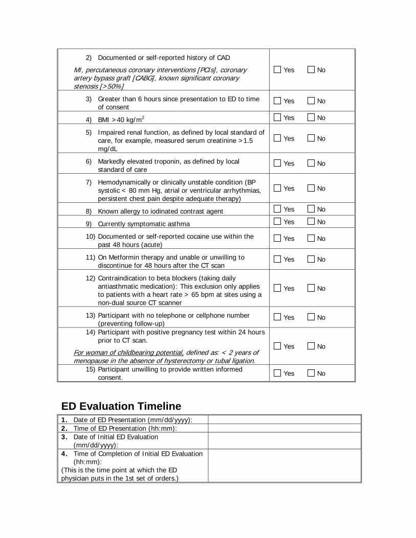
3) Greater than 6 hours since presentation to ED to time of consent
Yes No
4) BMI >40 kg/m2 Yes No
5) Impaired renal function, as defined by local standard of care, for example, measured serum creatinine >1.5 mg/dL
Yes No
6) Markedly elevated troponin, as defined by local standard of care
Yes No
7) Hemodynamically or clinically unstable condition (BP systolic < 80 mm Hg, atrial or ventricular arrhythmias, persistent chest pain despite adequate therapy)
Yes No
8) Known allergy to iodinated contrast agent Yes No
9) Currently symptomatic asthma Yes No
10) Documented or self-reported cocaine use within the past 48 hours (acute)
Yes No
11) On Metformin therapy and unable or unwilling to discontinue for 48 hours after the CT scan
Yes No
12) Contraindication to beta blockers (taking daily antiasthmatic medication): This exclusion only applies to patients with a heart rate > 65 bpm at sites using a non-dual source CT scanner
Yes No
13) Participant with no telephone or cellphone number (preventing follow-up)
Yes No
14) Participant with positive pregnancy test within 24 hours prior to CT scan.
For woman of childbearing potential, defined as: < 2 years of menopause in the absence of hysterectomy or tubal ligation.
Yes No
15) Participant unwilling to provide written informed consent. Yes No
ED Evaluation Timeline 1. Date of ED Presentation (mm/dd/yyyy): 2. Time of ED Presentation (hh:mm): 3. Date of Initial ED Evaluation
(mm/dd/yyyy):
4. Time of Completion of Initial ED Evaluation (hh:mm):
(This is the time point at which the ED physician puts in the 1st set of orders.)
Randomization & Consent 1. Date of Consent (mm/dd/yyyy): 2. Time of Consent (hh:mm): 3. Was the patient randomized? Yes No*
If yes, SOC arm Interventional arm
Randomization number _________________
If no, reason: markedly positive troponin positive pregnancy test patient withdrew consent other, specify____________
4. Date of Randomization (mm/dd/yyyy): 5. Time of Randomization (hh:mm):
MEDICAL HISTORY FORM
Cardiac Risk Factors and Medications 1. Hypertension Yes No Not Reported
2. Diabetes mellitus None Insulin requiring Non-insulin Not Reported
3. Hypercholesterolemia/hyperlipidemia Yes No Not Reported
4. Cocaine use Never Former Recent (last use >48 hours)
5. Tobacco use Never Former Current Not Reported
6. First degree relative with CAD/ACS/AMI: (male < 55 yrs, female < 65 yrs)
Yes No Not Reported
7. Home medications: a. ACE-inhibitors/ARB Yes No b. Aspirin Yes No c. Nitrates Yes No d. Beta-blockers Yes No e. Calcium channel blocker Yes No f. Statins Yes No g. Niacin/fibrates Yes No h. Insulin Yes No i. Oral hypoglycemics Yes No
Medical History 1. Heart failure Yes No Not Reported
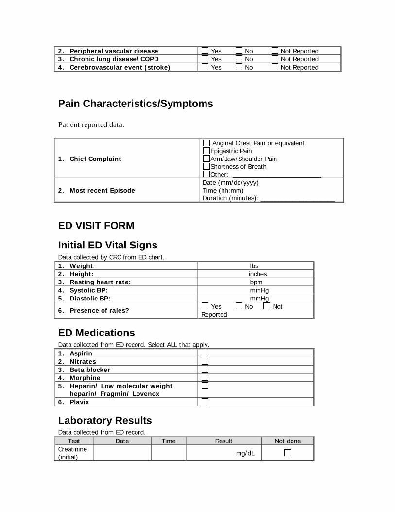
2. Peripheral vascular disease Yes No Not Reported 3. Chronic lung disease/COPD Yes No Not Reported 4. Cerebrovascular event (stroke) Yes No Not Reported
Pain Characteristics/Symptoms
Patient reported data:
1. Chief Complaint
Anginal Chest Pain or equivalent Epigastric Pain Arm/Jaw/Shoulder Pain Shortness of Breath Other: _________________________
2. Most recent Episode Date (mm/dd/yyyy) Time (hh:mm) Duration (minutes): _____________________
ED VISIT FORM
Initial ED Vital Signs Data collected by CRC from ED chart. 1. Weight: lbs 2. Height: inches 3. Resting heart rate: bpm 4. Systolic BP: mmHg 5. Diastolic BP: mmHg
6. Presence of rales? Yes No Not Reported
ED Medications Data collected from ED record. Select ALL that apply. 1. Aspirin 2. Nitrates 3. Beta blocker 4. Morphine 5. Heparin/ Low molecular weight
heparin/ Fragmin/ Lovenox
6. Plavix
Laboratory Results Data collected from ED record.
Test Date Time Result Not done Creatinine (initial) mg/dL
Test Troponin classification Date Time Result Range Not done
Troponin (initial)
T I
ug/ml
Normal Enter Range:___
Borderline Enter Range:___
Elevated Enter Range:___
Troponin (2nd)
T I
ug/ml
Normal Enter Range:___
Borderline Enter Range:___
Elevated Enter Range:___
Troponin (3rd)
T I
ug/ml
Normal Enter Range:___
Borderline Enter Range:___
Elevated Enter Range:___
Biomarker Testing
Did the patient consent to biomarker testing? Yes No
If yes,
Draw #1: Was blood collected and stored? If less than 3 tubes collected, fill out protocol deviation form. Yes No
Date (mm/dd/yyyy): Time (hh:mm):_____________
Red tube specimen ID no _____________ How many daughter tubes? ______ Purple tube specimen ID no _____________ How many daughter tubes? ______
Green tube specimen ID no _____________ How many daughter tubes? ______ Draw #2: Was blood collected and stored? If less than 3 tubes collected, fill out protocol deviation form.
Yes No Date (mm/dd/yyyy): Time (hh:mm):______________
Red tube specimen ID no _____________ How many daughter tubes? ______
Purple tube specimen ID no _____________ How many daughter tubes? ______
Green tube specimen ID no _____________ How many daughter tubes? ______ Draw #3: Was blood collected and stored? If less than 3 tubes collected, fill out protocol deviation form.
Yes No
Date (mm/dd/yyyy): _____________ Time (hh:mm):__________________
Red tube specimen ID no _____________ How many daughter tubes? ______
Purple tube specimen ID no _____________ How many daughter tubes? ______
Green tube specimen ID no _____________ How many daughter tubes? ______
ECG FORM
Initial 12-Lead ECG Interpretation Information entered into eCRF by CRC.
1. Date of initial ECG (mm/dd/yyyy): 2. Time of initial ECG (hh:mm):
Manufacturer: Siemens GE Philips Other ____________________ Model: Somatom Sensation 64 Lightspeed 64 Lightspeed VCT Dual Source Flash ICT Other _____________________
B. Hybrid Imaging (SPECT or PET) Yes No
C. Time CT ordered (hh:mm) D. Time CT performed (hh:mm) E. Time of CT interpretation (hh:mm)
2. Calcium Scan If no, give reason and fill out protocol violation form
Yes No
3. CTA Yes No 4. Intra CCTA vitals:
Average HR during scan: bpm
5. Pre-procedure medications: Yes No
a. Beta blocker IV
Yes No If yes, Metoprolol Other _________ dose: ________ mg
b. Beta blocker PO
Yes No Metoprolol Atenolol_________
dose: _________mg c. Nitroglycerin SL Yes No
If yes, dose:
6. Contrast agent:
Isovue Omnipaque Optiray Vispaque Other
Specify:_____________
a. concentration mg iodine per ml
300 320 350 370
7. Completion of the CT scan
Yes No If no, indicate why not done: Medical reason Patient refusal/withdrawal Equipment failure Other If other, specify: ______________
8. Prospectively gated/triggered cardiac CTA scan Yes No
9. Retrospectively gated cardiac CTA scan Yes No 10. Dose Length Product of CTA only: mGY cm 11. Total Dose Length Product mGY cm 12. CTDI Volume of CTA mGy CT Test Subject Physician Reading Form To be completed by CT reader with signature next to physician ID on paper CRF; information entered on form by CRC. 1. Reading physician ID: (Please have CT
Reader initial paper CRF)
2. Calcium Score 3. Coronary CTA Fill out the appropriate box to indicate the level of stenosis. If level of stenosis equals stent, please fill out the protocol violation form.
a. Left Main normal 0% 1-49% (non-significant/mild or minor) 50-99% (significant/severe) 100% (occluded)
indeterminate stent** b. LAD (any) normal 0% 1-49% (non-significant/mild or
Uninterpretable: 5. LV functional analysis performed: Yes No
Global LV function Abnormal Normal Global LV function in % ______ Regional Wall Motion Abnormality: Yes No If yes, match stenosis territory Anterior/apex Inferior/posterior Lateral
6. If retrospectively gated, was tube modulation technique or a similar radiation safety technique used?
_____(Mega Bq/millicuries) 13. Was a Reinjection performed? administered activity /unit of activity
Yes No If yes, fill out the following: Tracer
Technetium Tetrafosmin Thallium
_____(Mega Bq/millicuries)
14. Was a Rubidium test performed? rest stress
Yes No If yes, administered activity /unit of activity -_____(Mega Bq/millicuries) administered activity /unit of activity -_____(Mega Bq/millicuries)
15. Completion of Protocol
Not done Done and completed Performed but not completed Baseline Heart rate ____ bpm Peak Heart Rate __________ bpm Systolic Blood Pressure at rest _____ mmHg Diastolic Blood Pressure at rest _____mmHg Systolic Blood Pressure at peak stress ____ mmHg Diastolic Blood Pressure at peak stress ____mmHg
Reached target HR?* Yes No NA If no, was it converted to pharmacologic? Yes No If yes, choose the agent used: Dobutamine Dipyridamole Adenosine Regadenoson Did the patient develop symptoms of possible CAD (including CP, SOB) Yes No
equivocal (if the answer is yes or equivocal, go to qs 15)
16. If there were symptoms of possible CAD, enlist any other symptoms perceived by the patient.
Chest Pain Yes No If yes, did the chest pain limit exercising capacity? Yes No Shortness of Breath Yes No Hypotension Yes No VT or non sustained VT Yes No Patient request Yes No Abnormal BP response Yes No Other Yes No If yes, specify:
17. Was the perfusion in the anterior/apical myocardial territory normal?
Yes No
18. Was the perfusion in the lateral myocardial territory normal? Yes No
19. Was the perfusion in the infero-posterior myocardial territory normal?
Yes No
20. Resting gated LVEF
Not done _________ EF % Normal Abnormal
21. Post stress gated LVEF _________ EF % Normal Abnormal
22. Transient Ischemic Dilatation Yes No Not Mentioned 23. Was additional nuclear imaging performed? Yes No
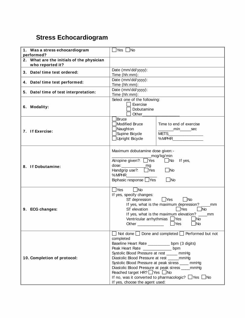
Stress Echocardiogram
1. Was a stress echocardiogram performed?
Yes No
2. What are the initials of the physician who reported it?
3. Date/time test ordered: Date (mm/dd/yyyy): Time (hh:mm):
4. Date/time test performed: Date (mm/dd/yyyy): Time (hh:mm):
5. Date/time of test interpretation: Date (mm/dd/yyyy): Time (hh:mm):
6. Modality:
Select one of the following: Exercise Dobutamine Other_________________
7. If Exercise:
Bruce Modified Bruce Naughton Supine Bicycle Upright Bicycle
Time to end of exercise _______min_____sec METS________________ %MPHR______________
8. If Dobutamine:
Maximum dobutamine dose given:-__________________mcg/kg/min Atropine given?: Yes No If yes, dose:___________mg Handgrip use?: Yes No %MPHR______________ Biphasic response Yes No
9. ECG changes:
Yes No If yes, specify changes: ST depression Yes No If yes, what is the maximum depression? ____mm ST elevation Yes No If yes, what is the maximum elevation? ____mm Ventricular arrhythmias Yes No Other ____________ Yes No
10. Completion of protocol:
Not done Done and completed Performed but not completed Baseline Heart Rate __________ bpm (3 digits) Peak Heart Rate _____________ bpm Systolic Blood Pressure at rest _____ mmHg Diastolic Blood Pressure at rest _____mmHg Systolic Blood Pressure at peak stress ____ mmHg Diastolic Blood Pressure at peak stress ____mmHg Reached target HR? Yes No If no, was it converted to pharmacologic? Yes No If yes, choose the agent used:
Dobutamine Dipyridamole Adenosine Regadenoson Did the patient develop symptoms of possible CAD (including CP, SOB)
Yes No equivocal (if the answer is yes or equivocal, go to qs 11)
11. If there were symptoms of possible CAD, enlist any other symptoms perceived by the patient.
Chest Pain Yes No If yes, did the chest pain limit exercising capacity? Yes No Shortness of Breath Yes No Hypotension Yes No VT Yes No Patient request Yes No Abnormal BP response Yes No Other Yes No If yes, specify:
12. Was the wall motion in the anterior/apical segment normal on stress?
Yes No
13. Was the wall motion in the lateral segment normal on stress? Yes No
14. Was the wall motion in the infero-posterior segment normal on stress? Yes No
15. Resting LVEF
_____ EF% Normal Abnormal Scar existing on rest exam Yes No If yes, Anterior/apical inferior/posterior Lateral
16. Stress LVEF _____ EF% Normal Abnormal If abnormal, severely decreased (<35% EF) Yes No
17. LV Dilatation at Peak Stress Yes No Not Mentioned
18. ECHO Results:
Normal Abnormal If abnormal, specify:
Inducible ischemia MI/scar (no ischemia) Both
19. Was another Stress Echocardiogram done? Yes No
Transthoracic Echocardiogram (rest)
To be entered on form by CRC 1. Was a resting transthoracic
echocardiogram performed? Yes No
2. What are the initials of the physician who performed the test?
3. Date/time test ordered: Date (mm/dd/yyyy): Time (hh:mm):
4. Date/time test performed: Date (mm/dd/yyyy): Time (hh:mm):
5. Date/time of test interpretation: Date (mm/dd/yyyy): Time (hh:mm):
6. Was the wall motion in the anterior/apical segment normal? Yes No
7. Was the wall motion in the lateral segment normal? Yes No
8. Was the wall motion in the infero-posterior segment normal? Yes No
9. LV function (Ejection fraction %)
__________%
10. Results:
Normal Abnormal If abnormal, specify:
11. Was a resting transthoracic echocardiogram done again? Yes No
Exercise ECG Stress Test (Non-imaging only) To be entered on form by CRC.
1. Was an exercise ECG stress test performed? Yes No
2. What are the initials of the physician who performed the test?
3. Date/time test ordered: Date (mm/dd/yyyy): Time (hh:mm):
4. Date/time test performed: Date (mm/dd/yyyy): Time (hh:mm):
5. Date/time of test interpretation: Date (mm/dd/yyyy): Time (hh:mm):
6. Type of Exercise protocol
Bruce Modified Bruce Naughton Supine Bicycle Upright Bicycle
7. Functional Capacity Time to end of Treadmill __________min________sec METS________________ %MPHR______________
8. Completed protocol?
Not done Done and completed Performed but not completed Peak Heart Rate __________ bpm (3 digits) Baseline Heart Rate ________ bpm Reached target HR? Yes No NA If no, was it converted to pharmacologic? Yes No If yes, choose the agent used: Dobutamine Dipyridamole Adenosine Regadenoson
Symptoms of possible CAD (including CP, SOB) Yes No equivocal (If the answer is yes or equivocal, go to qs.
9)
9. If there were symptoms of possible CAD, enlist any other symptoms perceived by the patient.
Chest Pain Yes No If yes, did the chest pain limit exercising capacity? Yes No If yes, Ischemic Equivocal Shortness of Breath Yes No Hypotension Yes No VT or non sustained VT Yes No Patient request Yes No Abnormal BP response Yes No Other Yes No If yes, specify:
10. Results:
Negative Positive Borderline If positive or borderline, select as appropriate: ST depression Yes No If yes, what was the maximum? _____ ST elevation Yes No If yes, what was the maximum? _____ Ventricular arrhythmias Yes No Other Yes No If yes, specify:______
11. Was exercise ECG stress test done again? Yes No
Cardiac Catheterization To be entered by the CRC
1. Was Cardiac Catheterization done?
Yes No
2. Initials of physician who interpreted test:
3. Date/time test ordered: Date (mm/dd/yyyy): Time (hh:mm):
4. Date/time test performed: Date (mm/dd/yyyy): Time (hh:mm):
5. Date/time of test interpretation: Date (mm/dd/yyyy): Time (hh:mm):
6. Were there any complications to the procedure (as per cath lab ACC/NCDR instruction)?
Yes No If yes, fill out adverse event form.
7. Mark the appropriate box to indicate the level of stenosis in each vessel: a. Left Main normal 0% 1-49% (non-significant/mild
or minor) 50-99% (significant/severe) 100% (occluded)
indeterminate stent note: ________ b. LAD (any) normal 0% 1-49% (non-significant/mild
or minor) 50-69% (moderate) 70-99% (significant/severe) 100%
(occluded) indeterminate stent note: ________
c. LCX (any) normal 0% 1-49% (non-significant/mild or minor) 50-69% (moderate)
7) SAE? Yes No 8) Was patient withdrawn from study due to AE? Yes No
9) Relationship to study procedure
1 - Not related 2 - Unlikely Related 3 - Possibly Related 4 - Probably Related 5 - Definitely Related
10) Relationship to contrast (1-5)
1 - Not related 2 - Unlikely Related 3 - Possibly Related 4 - Probably Related 5 - Definitely Related
11) Relationship to underlying disease
1 - Not related 2 - Unlikely Related 3 - Possibly Related 4 - Probably Related 5 - Definitely Related
12) Action (1-5)
1 - No Action Taken 2 - Medication Given 3 - Non-drug therapy given 4 - ED visit 5 - Hospitalization /prolonged
hospitalization
13) Outcome (1-5)
1 - Recovered 2 – Recovered with sequelae 3 - Ongoing 4 – Death 5 - Unknown
14) Did the subject have another AE? Yes No
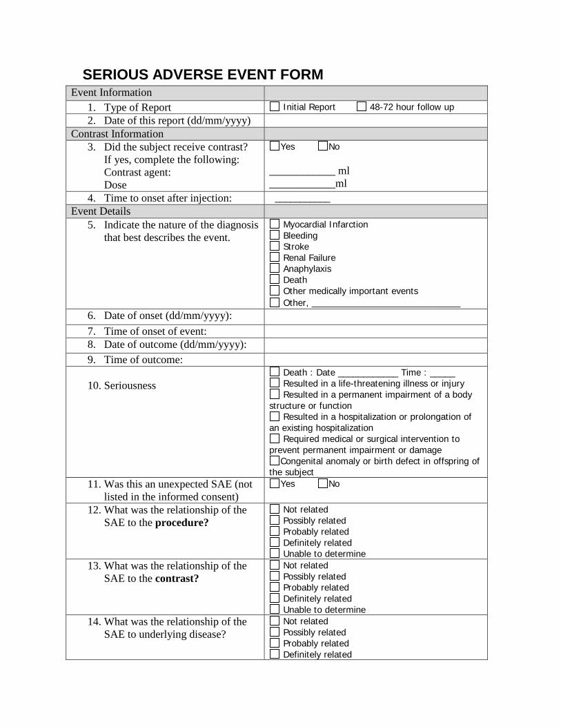
SERIOUS ADVERSE EVENT FORM Event Information
1. Type of Report Initial Report 48-72 hour follow up 2. Date of this report (dd/mm/yyyy)
Contrast Information 3. Did the subject receive contrast?
If yes, complete the following: Contrast agent: Dose
Yes No ____________ ml ____________ml
4. Time to onset after injection: ___________ Event Details
5. Indicate the nature of the diagnosis that best describes the event.
Myocardial Infarction Bleeding Stroke Renal Failure Anaphylaxis Death Other medically important events Other, ___________________________
6. Date of onset (dd/mm/yyyy): 7. Time of onset of event: 8. Date of outcome (dd/mm/yyyy): 9. Time of outcome: 10. Seriousness
Death : Date ____________ Time : _____ Resulted in a life-threatening illness or injury Resulted in a permanent impairment of a body
structure or function Resulted in a hospitalization or prolongation of
an existing hospitalization Required medical or surgical intervention to
prevent permanent impairment or damage Congenital anomaly or birth defect in offspring of
the subject 11. Was this an unexpected SAE (not
listed in the informed consent) Yes No
12. What was the relationship of the
SAE to the procedure? Not related Possibly related Probably related Definitely related Unable to determine
13. What was the relationship of the SAE to the contrast?
Not related Possibly related Probably related Definitely related Unable to determine
14. What was the relationship of the SAE to underlying disease?
Not related Possibly related Probably related Definitely related
Unable to determine
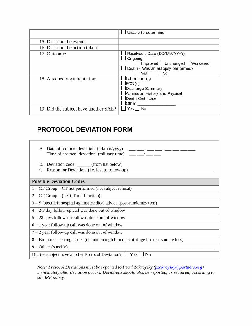
15. Describe the event: 16. Describe the action taken:
17. Outcome: Resolved : Date (DD/MM/YYYY) Ongoing
Improved Unchanged Worsened Death - Was an autopsy performed?
Yes No 18. Attached documentation: Lab report (s)
ECG (s) Discharge Summary Admission History and Physical Death Certificate Other _________________
19. Did the subject have another SAE? Yes No
PROTOCOL DEVIATION FORM
A. Date of protocol deviation: (dd/mm/yyyy) ___ ___ - ___ ___- ___ ___ ___ ___ Time of protocol deviation: (military time) ___ ___: ___ ___
B. Deviation code: ______ (from list below) C. Reason for Deviation: (i.e. lost to follow-up)___________________________________
Possible Deviation Codes 1 – CT Group – CT not performed (i.e. subject refusal) 2 – CT Group – (i.e. CT malfunction) 3 – Subject left hospital against medical advice (post-randomization) 4 – 2-3 day follow-up call was done out of window 5 – 28 days follow-up call was done out of window 6 – 1 year follow-up call was done out of window 7 – 2 year follow-up call was done out of window 8 – Biomarker testing issues (i.e. not enough blood, centrifuge broken, sample loss) 9 – Other: (specify) ________________________________________________________________
Did the subject have another Protocol Deviation? Yes No
Note: Protocol Deviations must be reported to Pearl Zakroysky ([email protected]) immediately after deviation occurs. Deviations should also be reported, as required, according to site IRB policy.
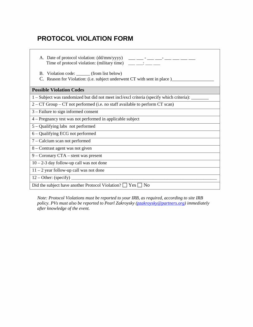
A. Date of protocol violation: (dd/mm/yyyy) ___ ___ - ___ ___- ___ ___ ___ ___ Time of protocol violation: (military time) ___ ___: ___ ___
B. Violation code: ______ (from list below) C. Reason for Violation: (i.e. subject underwent CT with sent in place )_________________
Possible Violation Codes 1 – Subject was randomized but did not meet incl/excl criteria (specify which criteria): _______ 2 – CT Group – CT not performed (i.e. no staff available to perform CT scan) 3 – Failure to sign informed consent 4 – Pregnancy test was not performed in applicable subject 5 – Qualifying labs not performed 6 – Qualifying ECG not performed 7 – Calcium scan not performed 8 – Contrast agent was not given 9 – Coronary CTA – stent was present 10 – 2-3 day follow-up call was not done 11 – 2 year follow-up call was not done 12 – Other: (specify) ________________________________________________________________
Did the subject have another Protocol Violation? Yes No
Note: Protocol Violations must be reported to your IRB, as required, according to site IRB policy. PVs must also be reported to Pearl Zakroysky ([email protected]) immediately after knowledge of the event.