17
DIAGNOSIS OF VERTEBROBASILAR INSUFFICIENCY USING TRANSCRANIAL DOPPLER ULTRASOUND Sean Steward February 20, 2013
| Date post: | 13-Dec-2015 |
| Category: |
Documents |
| Upload: | hollie-walters |
| View: | 222 times |
| Download: | 0 times |
DIAGNOSIS OF VERTEBROBASILAR INSUFFICIENCY USING
TRANSCRANIAL DOPPLER ULTRASOUND
Sean StewardFebruary 20, 2013
Overview
Vertebrobasilar Insufficiency Signs and Symptoms Risk Factors
Tests Transcranial Doppler Ultrasound Case Study QA statistics of TCD Conclusion

Vertebrobasilar Insufficiency
Vertebrobasilar insufficiency (VBI) is a posterior circulation transient ischemic attack (TIA) caused by intermittent vertebral artery occlusion that is induced by a head rotation or extension
VBI also may result from large vessel atherosclerotic disease, dissection, cervical compressive lesions, and subclavian steal phenomenon
Signs and Symptoms for VBI
Loss of vision in part or all of both eyes Double vision Vertigo (spinning sensation) Numbness or tingling Nausea and vomiting Slurred speech Loss of coordination, dizziness or confusion Trouble swallowing A drop attack — sudden generalized
weakness Ucdavis.edu

Risk Factors for VBI
Smoking Hypertension Hyperlipidemia Older age Gender: men have a higher risk before
age 75; women have a high risk after 75 Family history Genetic factors
Ucdavis.edu
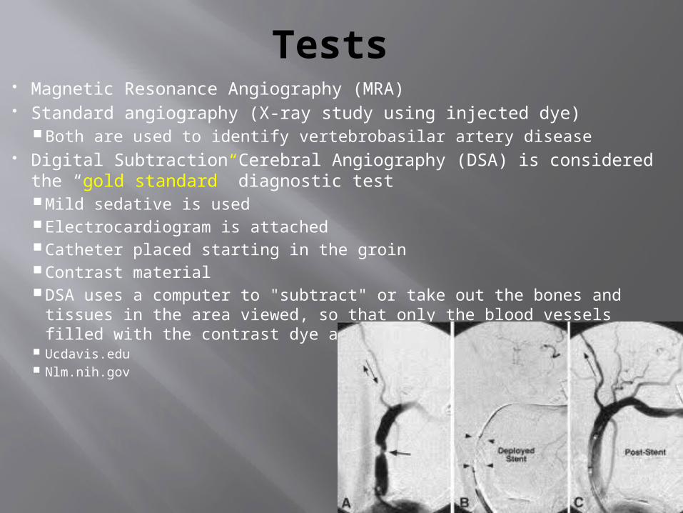
Tests Magnetic Resonance Angiography (MRA) Standard angiography (X-ray study using injected dye)
Both are used to identify vertebrobasilar artery disease Digital Subtraction Cerebral Angiography (DSA) is considered the
“gold standard” diagnostic test Mild sedative is used Electrocardiogram is attached Catheter placed starting in the groin Contrast material DSA uses a computer to "subtract" or take out the bones and tissues in
the area viewed, so that only the blood vessels filled with the contrast dye are seen
Ucdavis.edu Nlm.nih.gov

Transcranial Doppler
Typically use a 2 MHz probe Angle of insonation is zero
degrees (3) most common windows
utilized: Transforamenal (A) Transtemporal (B) Transorbital (C)
Standard method of quantifying velocity is time averaged mean velocity (TAMV) McPharlin & Rumwell
A
B
C

Transcranial Doppler
Accurate vessel identification Appropriate sample volume and depth Direction and velocity of blood flow
McPharlin & Rumwell

Case Study
50 year old Caucasian male Presented with one year of positional vertigo
and ataxic gait when extending his neck Positional vertigo is a sudden sensation of
spinning with head movements Ataxic gait is an unsteady, uncoordinated walk
CT and MRI were WNL TCD performed
nlm.nih.gov thefreedictionary.com ncbi.nlm.nih.gov
Case Study
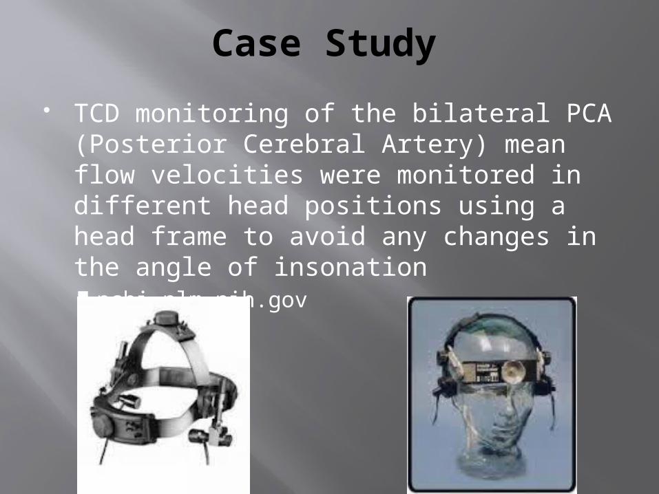
TCD monitoring of the bilateral PCA (Posterior Cerebral Artery) mean flow velocities were monitored in different head positions using a head frame to avoid any changes in the angle of insonation ncbi.nlm.nih.gov
Case Study
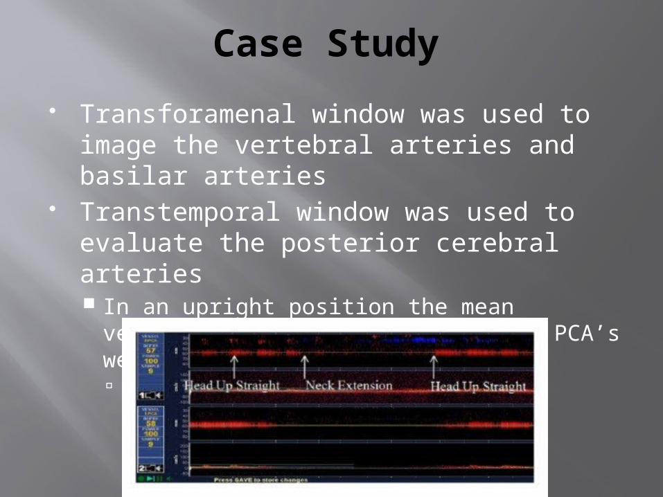
Transforamenal window was used to image the vertebral arteries and basilar arteries
Transtemporal window was used to evaluate the posterior cerebral arteries In an upright position the mean velocities for
the right and left PCA’s were 18cm/sec and 19cm/sec ncbi.nlm.nih.gov

Case Study
After the TCD imaging was performed: An electronystagmography (ENG) was performed
which was negative Electronystagmography (ENG) is used to evaluate people
with vertigo (a false sense of spinning or motion that can cause dizziness) and certain other disorders that affect hearing and vision
An MRI of his cervical spine was done to rule out myelopathy Myelopathy is a term used to describe symptoms related
to spinal cord compression Cervical (neck) myelopathy is most common
hopkinsmedicine.org piedmonthospital.org ncbi.nlm.nih.gov

Case Study
Review: (2) MRI’s (Both negative) CT (Negative) ENG (Negative) ENT (Ear nose and throat work up – Negative) TCD (Showed positive results…thank you very
much) Patient refused spinal surgery and is being
treated conservatively
ncbi.nlm.nih.gov
QA Statistics
According to a series that was done in 1992 that compared TCD with DSA (Gold Standard) in 20 patients the results are as follows for TCD: Sensitivity – 87% Specificity – 80% Positive Predictive Value – 93% Negative Predictive Value – 67%
The results proved that TCD is a useful screening method in patients with VBI to detect large vessel disease of the intracranial vertebrobasilar system
ncbi.nlm.nih.gov

Conclusion
VBI is a very difficult disease to diagnose TCD should be the new “Gold Standard”
diagnostic test for evaluating VBI’s It’s noninvasive
No sedative, catheter, contrast dye, ECG Relatively inexpensive test Can go bedside if needed Proven QA numbers

References
Alnami, I., Siddiqui, M. & Saqqur, M. (2012, November).Case Reports in Medicine. TheDiagnosis of Vertebrobasilar Insufficiency Using Transcranial Doppler Ultrasound.Retrieved from: http://www.ncbi.nlm.nih.gov/pmc/articles/PMC3509548/
Ataxic gait (n.d.) The free dictionary. Retrieved from: http://medical-dictionary.thefreedictionary.com/ataxic+gait
Benign positional vertigo (n.d.).Medline Plus.Retrieved from: http://www.nlm.nih.gov/medlineplus/ency/article/001420.htm
Cerebral angiography (n.d.).Medline Plus.Retrieved from: http://www.nlm.nih.gov/medlineplus/ency/article/003799.htm
References
Electronystagmography (ENG) (n.d.) John Hopkins Medicine. Retrieved from:http://www.hopkinsmedicine.org/healthlibrary/test_procedures/neurological/electronystagmography_eng_92,P07659/
McPharlin, M., & Rumwell, C. (2009). Vascular Technology: An Illustrated Review. Pasadena, CA:Davies Publishing, Inc.
Myelopathy (n.d.) Piedmont Healthcare. Retrieved from:http://www.piedmonthospital.org/SpineCenter/Myelopathyctr.aspx
UCDavis Health System. Vertebrobasilar insufficiency. Retrieved
from:http://www.ucdmc.ucdavis.edu/vascular/diseases/vertebrobasilar.html


