23
| Date post: | 16-Dec-2015 |
| Category: |
Documents |
| Upload: | anderson-johnson |
| View: | 218 times |
| Download: | 4 times |


Search for cellular attachment receptors used by a GII.4 Norovirus Dijon
Gaurav Dutta DwivediSupervisors: Niklas Arnberg and Carin Årdahl
Introduction
Norovirus are a group of viruses that cause non bacterial gastroenteritis in humans.
The first outbreak of Norovirus infection was reported in Norwalk, Ohio in 1968.
Human Norovirus infection is popularly known as winter vomiting disease.
Norovirus outbreaks frequently occur in community surroundings.
Noroviruses are predominantly infectious and highly stable in the environment and immunity following infection generally is short-lived.

DehydrationNausea and Vomiting
Diarrhea
Fever
Abdominal cramping
Symptoms of Norovirus infection
Incubation period is 24 to 48 hours, with the symptoms lasting 12 to 60 hours.
Infected hosts can shed virus in stool for up to 2 weeks.

Transmission of Norovirus

Noroviruses infect individuals with a functional alpha-1, 2-fucosyltransferase (FUT2) gene and are designated as secretor-positive and those with defected FUT2 gene are termed as secretor-negative ,they are resistant to the common GII.4 strain, however they can still get infected with other Norovirus strains.
Human Noroviruses recognize histo-blood group antigens (HBGAs) that are expressed on the surface of mucosal epithelial cells.

Norovirus Classification
The genogroup II, genotype 4 Noroviruses, designated GII.4, are currently responsible for 70–80% of Norovirus outbreaks worldwide.
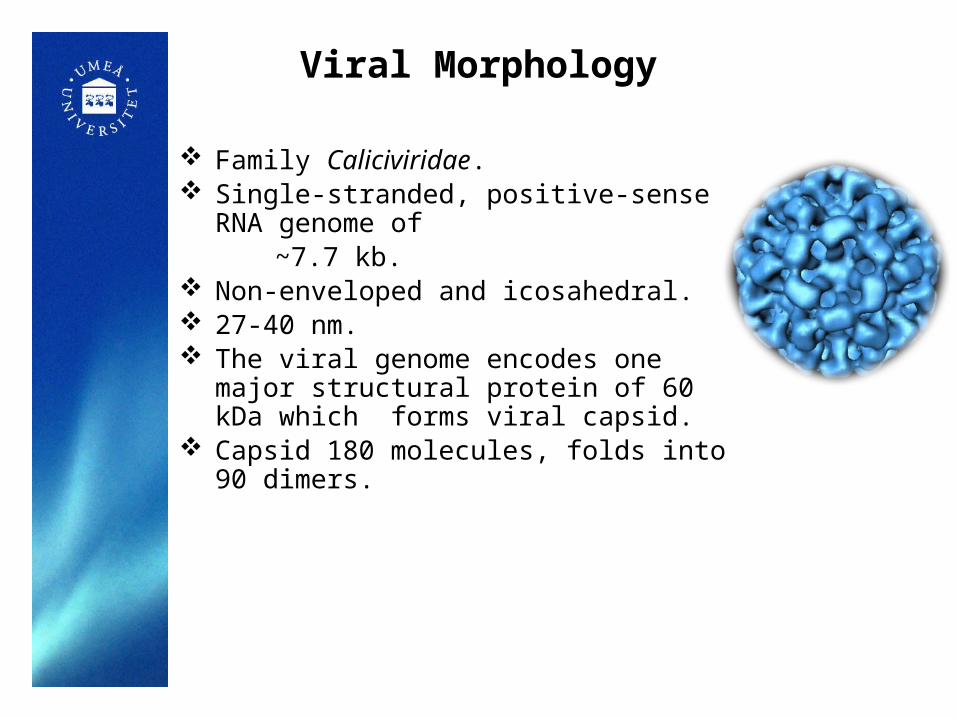
Viral Morphology
Family Caliciviridae. Single-stranded, positive-sense RNA genome of ~7.7 kb. Non-enveloped and icosahedral. 27-40 nm. The viral genome encodes one major structural
protein of 60 kDa which forms viral capsid. Capsid 180 molecules, folds into 90 dimers.

Norovirus genome comprises of three open reading frames (ORFs).
ORF1 encodes the non-structural proteins that are crucial for virus replication, ORF2 and ORF3 encode a major capsid protein VP1 and a minor structural protein VP2, respectively.
VP1 consists of shell domain (S) and the protruding domain (P).
P domain is further divided into two subdomains known as P1 and P2.
Unpublished observations indicate that the presence of specific integrin-binding motifs plays a role in interactions for binding to integrins and allows virus particles attachment to the host cells.

Aim
To explore cellular receptors used by the GII.4 Human Norovirus (NoV) Dijon Virus Like Particles (VLPs) and analyse its binding characteristics across various integrin expressing cells.

Why we used Norovirus like particles (NoV-LPs) in the present study ?
The study of NoV has been hampered by the lack of an efficient cell culture system, which is still not available for Human NoV.

MethodsThe Dijon NoV-LPs used were produced in Sf9 insect cells.
The VLPs were clarified using ultracentrifugation and sucrose density gradient methods , finally the purity was confirmed by using SDS-PAGE and Western blot.
Flow cytometry was used for evaluation of binding characteristics of NoV-LPs across various integrin expressing cells.

Production of VLPs
Sf9 cell culture
40%
Sucrose density gradient
SDS-PAGE
Incubate at 130 rpm,28 ˚C, 5 days
Centrifuge at 3000 g,30 minUltracentrifuge supernatant at 30000 rpm,2hrs,4˚C
Collect the fractions1 2 3 4 5
Western blot
20%
30%
50%60%
Ultracentrifuge at 320000 rpm,4hrs,4˚C.
Loading on gel
Collected fractions

FACS BD LSRII
Cell culture
96 well plate
Centrifuge
NoV-LPs
Wash 3 X with 100 µl of PFN and centrifuge every time.
Detach cells and Transfer 200 000 cells/well.
Incubate with primary and secondary antibody
rabbit-anti-NoV
goat-anti-rabbit Alexa Flour 647
Incubate with added VLPs
Wash 3X with 100 µl of PFN and transferInto FACS tubes and analyse the binding.
Wash with PFN buffer and incubatewith the antibodies
Binding Assay
Centrifuge at 1300 rpm,4˚C for 5 mins

SDS-PAGE showing Fractions1-9 of the produced VLPs
SDS-PAGE of cell lysate from the sf9 cell culture
Results
Pooled 3-5 Pooled 6-7

Protein gel of pooled concentrated GII.4 Dijon (100K, Amicon filter)

Binding characteristics of NoV-LPs to integrin expressing cells
Figure 1 and Figure 2 represents the binding percentage of various integrin expressing cells to NoV-LPs .
Figure 1 Figure 2

Western blot for the produced VLPs
Figure 3
Detection of produced NoV-LPs. Figure 3 showing the Western blot results of Dijon NoV-LPs in various concentrations probed with primary antibody (rabbit-anti-NoV, serum) and secondary antibody (HRP-conjugated-swine-anti-rabbit).

Conclusion
In conclusion, the production and characterization of GII.4 NoV-LPs in insect cells was validated in this study.
Western blot detected NoV-LPs using NoVs raised antibodies from rabbit sera.
This receptor binding study indicates approximately 2-6 times increased binding of NoV-LPs to various integrin expressing cells signifying the importance of integrins as candidate receptors for NoV.

Future directions
Repetition of binding experiments with more integrin expressing cell lines.
Immortalization and replication studies using human cells.
Blocking studies for analyzing Norovirus binding.

Acknowledgement
Carin Årdahl thesis supervisor.
Professor Niklas Arnberg Division of Virology, Umeå university.
Division of Medical Microbiology, Department of Clinical and Experimental Medicine, LiU(Linköping University), Linköping, Sweden.


Datum Sidfot 23
Reference: www.Slideshare.com


![GII Presentation - Week2 Dr2 · Measuring Gender InequalityMeasuring Gender Inequality • Gender Inequality Index (Index (GII) [UNDP] – The index may not differentiate these two](https://static.documents.pub/doc/80x56/5f38e5f2d8822b3ec4466be2/gii-presentation-week2-measuring-gender-inequalitymeasuring-gender-inequality.jpg)


