An Interesting Case of Orange Colored Sweat SS Lakshmanan Chapter 178 Figure 1: Colored sweat over the face Figure 2: Stained undergarment CASE REPORT A 35-year-old female presented with history of colored sweat over the face and neck along with staining of her dresses and undergarments. e colored sweat causing discoloration of face was easily removable with soap and water, but would reappear later. She was a housewife. No history of prolonged exposure to any drugs, no recent history of intake of any colored foods, no history of exposure to any dyes, paints, coal or any other chemicals. Urine, saliva and tear color was normal. All baseline investigations including urine examination were found to be normal. • Urine homogentisic acid levels were normal • Bleeding and clotting time were normal • Liver function tests were normal • Blood and urine cultures were negative • Scrapings from the skin and biopsy results were inconclusive (Figure 3). DISCUSSION Sweat glands are small tubular structures of the skin that produce sweat. ere are two kinds of sweat glands: (1) eccrine and (2) Apocrine sweat glands, which are referred to as sudoriferous glands. Eccrine and Apocrine Glands e eccrine glands are found all over the body and function throughout our life. ey secrete a clear, odorless fluid that serves to regulate body temperature. 1 Apocrine glands develop during puberty and are most active throughout adulthood and are located in the armpits, areolar, genital, and anal areas. Apocrine glands secrete a thick, milky sweat that, once broken down by bacteria, is the main cause of body odor (Figure 4). 2 ABSTRACT A 35-year-old female presented with colored staining of her dresses, undergarments and colored sweat over face for 15 days (Figures 1 and 2). On inspection, the secretion was odorless and yellowish orange in color. On investigation, no significant laboratory abnormalities were noted. Urine examination was normal. Grams stain and cultures of skin, scrapings from the affected area and skin biopsy was done. Section 27 Case Studies
Transcript
An Interesting Case of Orange Colored Sweat
SS Lakshmanan
Chapter 178
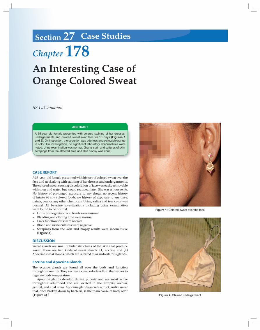
Figure 1: Colored sweat over the face
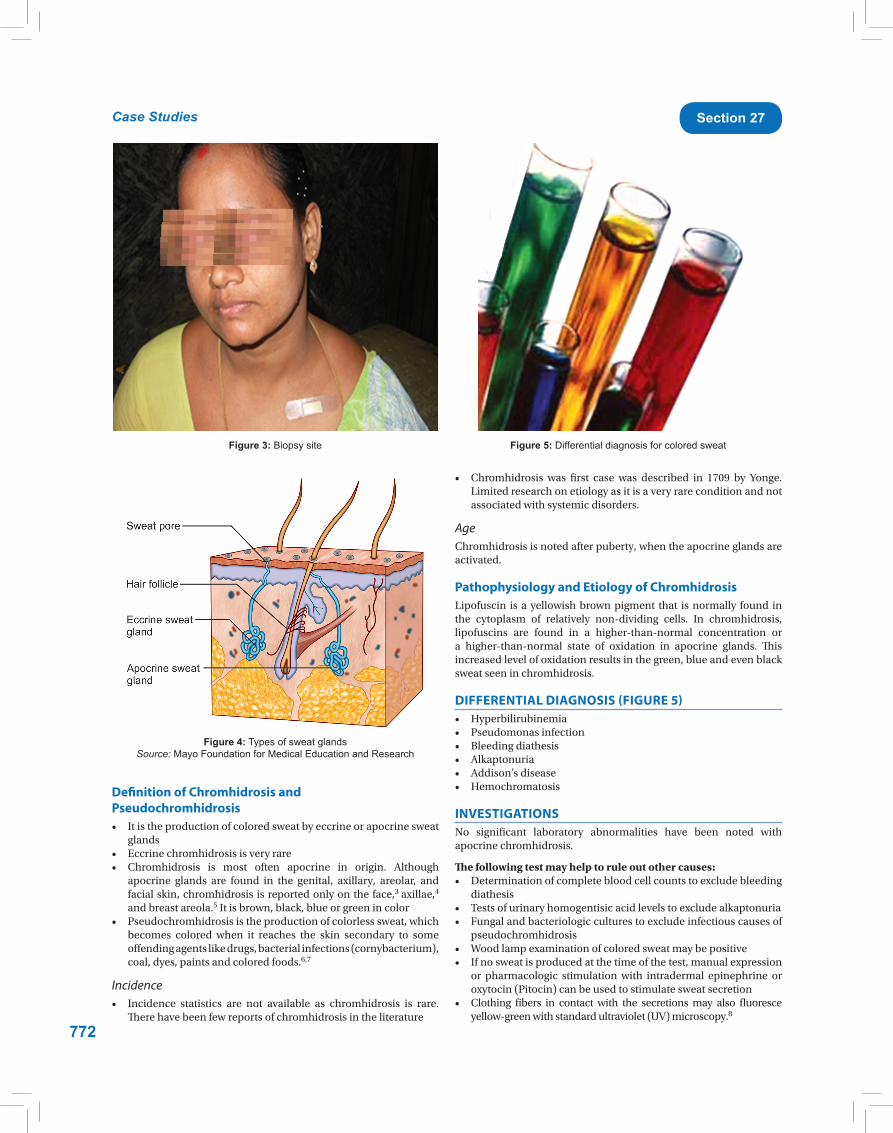
Figure 2: Stained undergarment
CASE REPORTA 35-year-old female presented with history of colored sweat over the face and neck along with staining of her dresses and undergarments. The colored sweat causing discoloration of face was easily removable with soap and water, but would reappear later. She was a housewife. No history of prolonged exposure to any drugs, no recent history of intake of any colored foods, no history of exposure to any dyes, paints, coal or any other chemicals. Urine, saliva and tear color was normal. All baseline investigations including urine examination were found to be normal.• Urinehomogentisicacidlevelswerenormal• Bleedingandclottingtimewerenormal• Liverfunctiontestswerenormal• Bloodandurinecultureswerenegative• Scrapings from the skin and biopsy results were inconclusive
(Figure 3).
DISCUSSIONSweat glands are small tubular structures of the skin that produce sweat. There are two kinds of sweat glands: (1) eccrine and (2) Apocrine sweat glands, which are referred to as sudoriferous glands.
Eccrine and Apocrine GlandsThe eccrine glands are found all over the body and function throughout our life. They secrete a clear, odorless fluid that serves to regulate body temperature.1
Apocrine glands develop during puberty and are most active throughout adulthood and are located in the armpits, areolar, genital, and anal areas. Apocrine glands secrete a thick, milky sweat that, once broken down by bacteria, is the main cause of body odor (Figure 4).2
ABSTRACT
A 35-year-old female presented with colored staining of her dresses, undergarments and colored sweat over face for 15 days (Figures 1 and 2). On inspection, the secretion was odorless and yellowish orange in color. On investigation, no significant laboratory abnormalities were noted. Urine examination was normal. Grams stain and cultures of skin, scrapings from the affected area and skin biopsy was done.
Section 27 Case Studies
772
Case Studies Section 27
Definition of Chromhidrosis and Pseudochromhidrosis• Itistheproductionofcoloredsweatbyeccrineorapocrinesweat
glands• Eccrinechromhidrosisisveryrare• Chromhidrosis is most often apocrine in origin. Although
apocrine glands are found in the genital, axillary, areolar, and facial skin, chromhidrosis is reported only on the face,3 axillae,4
and breast areola.5Itisbrown,black,blueorgreenincolor• Pseudochromhidrosisistheproductionofcolorlesssweat,which
becomes colored when it reaches the skin secondary to some offending agents like drugs, bacterial infections (cornybacterium), coal, dyes, paints and colored foods.6,7
Incidence• Incidence statistics are not available as chromhidrosis is rare.
There have been few reports of chromhidrosis in the literature
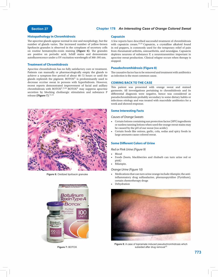
Figure 3: Biopsy site
Figure 4: Types of sweat glands Source: Mayo Foundation for Medical Education and Research
Figure 5: Differential diagnosis for colored sweat
• Chromhidrosis was first case was described in 1709 by Yonge.Limitedresearchonetiologyasitisaveryrareconditionandnotassociated with systemic disorders.
Pathophysiology and Etiology of ChromhidrosisLipofuscin isayellowishbrownpigment that isnormally found inthe cytoplasm of relatively non-dividing cells. In chromhidrosis,lipofuscins are found in a higher-than-normal concentration or a higher-than-normal state of oxidation in apocrine glands. This increased level of oxidation results in the green, blue and even black sweat seen in chromhidrosis.
or pharmacologic stimulation with intradermal epinephrine or oxytocin(Pitocin)canbeusedtostimulatesweatsecretion
• Clothing fibers in contact with the secretionsmay also fluoresceyellow-green with standard ultraviolet (UV) microscopy.8
773
Chapter 178 An Interesting Case of Orange Colored SweatSection 27
CapsaicinA few reports have described successful treatment of chromhidrosis with capsaicin cream.13,14 Capsaicin, a crystalline alkaloid foundin red peppers, is commonly used for the temporary relief of pain fromrheumatoidarthritis,osteoarthritis,andneuralgias.Capsaicindepletes neurons of substance P, a neurotransmitter important inapocrinesweatproduction.Clinicalrelapseoccurswhentherapyisstopped.
Pseudochromhidrosis (Figure 8)The causative factor has to be removed and treatment with antibiotics as infection is the most common cause.
COMING BACK TO THE CASEThis patient was presented with orange sweat and stained garments. All investigations pertaining to chromhidrosis and its differential diagnosis were negative, hence was considered as pseudochromhidrosis probably secondary to some dietary habits or infectious etiology and was treated with macrolide antibiotics for a week and showed response.
Some Interesting Facts
Causes of Orange Sweats• Certainlotionscontainingsunprotectionfactor(SPF)ingredients
or sunless tanning lotions when used the orange sweat stains may be caused by the pH of our sweat (too acidic).
• Certain foods likeonions,garlic, cola, sodasandspicy foods inlarge amounts cause colored sweat.
Some Different Colors of Urine
Red or Pink Urine (Figure 9)• Blood• Foods (beets, blackberries and rhubarb can turn urine red or
inflammatory drug sulfasalazine, phenazopyridine (Pyridium),certain chemotherapy drugs
• Dehydration
Figure 6: Oxidized lipofuscin granules
Figure 7: BOTOXFigure 8: A case of topiramate induced pseudochromhidrosis which
subsided after drug removal15
Histopathology in ChromhidrosisThe apocrine glands appear normal in size and morphology, but the number of glands varies. The increased number of yellow-brown lipofuscin granules is observed in the cytoplasm of secretory cells on routine hematoxylin-eosin staining (Figure 6). The granules are positive on periodic acid, Schiff stains and demonstrate autofluorescenceunderaUVexcitationwavelengthof360–395nm.
Treatment of ChromhidrosisApocrine chromhidrosis has no fully satisfactory cure or treatment. Patients can manually or pharmacologically empty the glands toachieve a symptom-free period of about 48–72 hours or until theglands replenish the pigment. BOTOX is predominantly used to decrease eccrine sweat in persons with hyperhidrosis. However, recent reports demonstrated improvement of facial and axillary chromhidrosis with BOTOX.9,10 BOTOX may suppress apocrine secretion by blocking cholinergic stimulation and substance Prelease (Figure 7).11,12
774
Case Studies Section 27
Figure 9: Pink urine caused by food products which turned clear on removal of the offending agent
Figure 10: Orange urine caused by drugs
Figure 11: Showing different colors of chromhidrosis and pseudochromhidrosis
Blue or Green Urine• Blue diaper syndrome is an autosomal recessive metabolic
disorder caused by a defect in tryptophan absorption in children• Green urine sometimes occurs during urinary tract infections
caused by pseudomonas bacteria.
WE CAN SWEAT IN DIFFERENT COLORSSweatcanoccurindifferentcolors.ItiseitherclassifiedasChromhidrosisandpseudochromhidrosis. InChromhidrosis thecommonthread isthat the color sweat is produced in the gland. This differentiates it from pseudochromhidrosis where clear sweat is produced and mixed with a colored agent when it reaches the skin (Figure 11).
CONCLUSIONBoth chromhidrosis and pseudochromhidrosis are rare.Diagnosisof chromhidrosis is based on biopsy and autofluorescence of both skin specimens and stained clothing, while diagnosis of pseudochromhidrosis is based primarily on history, and successful treatment with antibiotics.
REFERENCES 1. Folk GE, Semken A.The evolution of sweat glands. Int J of bioclim
10. Beer K, Oakley H. Axillary chromhidrosis: report of a case, reviewof the literature and treatment considerations. J Cosmet Dermatol.2010;9(4):318-20.


![Blood, Sweat & Tears - [Book] the Best of Blood, Sweat & Tears](https://static.documents.pub/doc/80x56/577c780e1a28abe0548e8be9/blood-sweat-tears-book-the-best-of-blood-sweat-tears.jpg)


