53
Section B - Protein Section B - Protein Structure Structure
| Date post: | 01-Jan-2016 |
| Category: |
Documents |
| Upload: | chester-tyler |
| View: | 25 times |
| Download: | 1 times |

Section B - Protein Section B - Protein StructureStructure
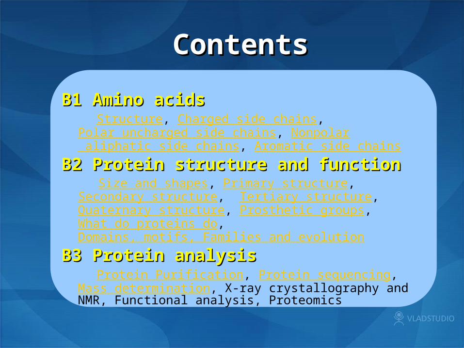
ContentsContents
B1 Amino acidsB1 Amino acids Structure, Charged side chains,
Polar uncharged side chains, Nonpolar aliphatic side chains, Aromatic side chains
B2 Protein structure and function B2 Protein structure and function Size and shapes, Primary structure, Secondary structure,
Tertiary structure, Quaternary structure, Prosthetic groups, What do proteins do, Domains, motifs, Families and evolution
B3 Protein analysis B3 Protein analysis Protein Purification, Protein sequencing,
Mass determination, X-ray crystallography and NMR, Functional analysis, Proteomics

B1 Amino acids—B1 Amino acids—StructureStructure
4. A few proteins contain nonstandard amino acids that are formed by post-translational modification of proline and lysine.
1. a-carbon is chiral ( 手性; asymmetric) except in glycine (R is H)
2. Amino acids are dipolar ions (zwitterions , 兼性离子 ) in aqueous solution and are amphoteric.
3. The side chains (R) differ in size, shape, charge and chemical reactivity
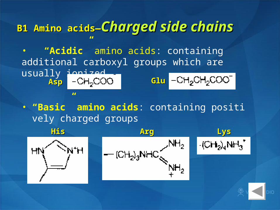
B1 Amino acids—B1 Amino acids—CCharged side chainsharged side chains
• “Acidic” amino acids: containing additional carboxyl groups which are usually ionized
AspAsp GluGlu
HisHis LysLys ArgArg
• “Basic” amino acids: containing positively charged groups
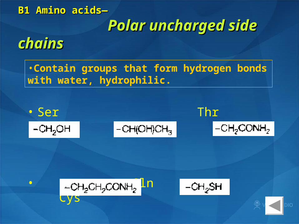
B1 Amino acids—B1 Amino acids—
Polar uncharged side chainsPolar uncharged side chains
• Ser Thr Asp
• Gln Cys
•Contain groups that form hydrogen bonds with water, hydrophilic.

B1 AminoB1 Amino acids—acids— Nonpolar aliphatic side chainsNonpolar aliphatic side chains
• Gly Ala Val Leu
• Ile Met Pro

B1 Amino acids— B1 Amino acids— Aromatic side Aromatic side chainschains
Phe Tyr
Trp
•Accounts for most of UV absorbance of proteins at 280 nm hydrophobic (疏水的 )


B2 Protein structure and function—B2 Protein structure and function—
Size and shapesSize and shapes
1. A few thousands Daltons (x 103) to more than 5 million Daltons (x 106)
2. Some proteins contain bound nonprotein materials (prosthetic groups or other macromolecules), which accounts for the increased sizes and functionalities of the protein complexes.

• Two broad classes of protein may be distinguished:
① Globular( most enzymes)
② Fibrous protein( silk protein, 角蛋白 keratin)
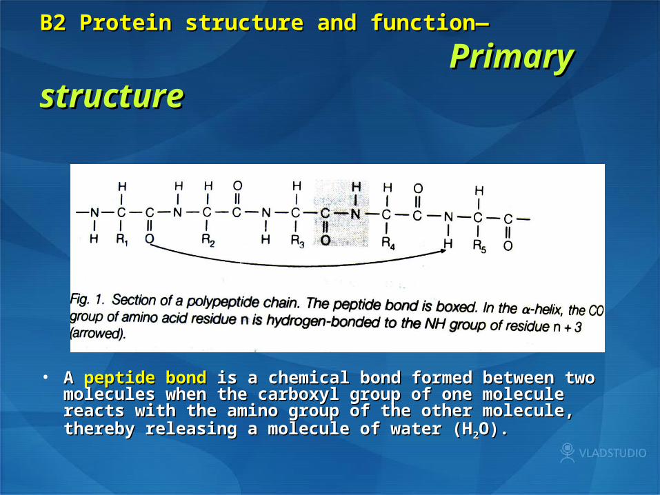
B2 Protein structure and function—B2 Protein structure and function—
PrimaryPrimary structurestructure
• A A peptide bondpeptide bond is a chemical bond formed between two is a chemical bond formed between two molecules when the carboxyl group of one molecule reacts molecules when the carboxyl group of one molecule reacts with the amino group of the other molecule, thereby releasing a with the amino group of the other molecule, thereby releasing a molecule of water (Hmolecule of water (H22O).O).
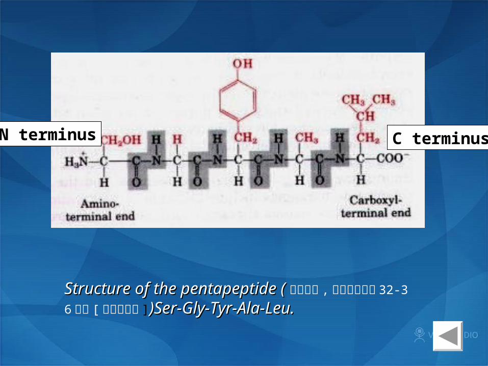
Structure of the pentapeptide (Structure of the pentapeptide ( 胸腺喷丁 , 促胸腺生成素 32-36 五肽 [ 免疫调节剂 ])Ser-Gly-Tyr-Ala-Leu. )Ser-Gly-Tyr-Ala-Leu.
N terminus C terminus

B2 Protein structure and function—B2 Protein structure and function—
Secondary structureSecondary structure
•a-helix:a-helix: right-handed 3.6 aa per turn hydrogen bond N-H···O=C

•b-sheet:
hydrogen bonding of the pepetide bond N-H and C=O groups to the complementary groups of another section of the polypeptide chain

B2 Protein structure and function—B2 Protein structure and function—
Tertiary structureTertiary structure
• The different sections of a-helix, b-sheet, other minor secondary structure and connecting loops of a polypeptide fold in three dimensions

Noncovalent interaction between side chains that hold the tertiary structure together: van der Waals forces, hydrogen bonds, electrostatic salt bridges, hydrophobic interactionsCovalent interaction: disulfide bonds
Denaturation of protein by disruption of its 2o and 3o structure by heat and extremes of pH will lead to a random coil conformation

B2 Protein structure and function—B2 Protein structure and function—
Quaternary structureQuaternary structure
CAM kinase II
Many proteins are composed of two or more polypeptide chains (subunits)
•Many proteins are composed of two or more polypeptide chains (subunits). These subunits may be identical or different. The same forces which stabilize tertiary structure hold these subunits together. This level of organization called quaternary structure.

( a) Primary stucture

Advantages of the quaternary structure:
1. It allows very large protein molecules to be made, such as tubulin.
2. It can provide greater functionality to a protein by combining different activities into a single entity.
3. The interactions between the subunits can often be modified by binding of small molecules and lead to the allosteric effects seen in enzyme regulation.

B2 Protein structure and function—B2 Protein structure and function—
Prosthetic groupsProsthetic groups
CoenzymesCoenzymes • vitamins: NAD+ (B3) · NADP+ (B3) · Coenzyme A (B5) · THF / H4F
(B9), DHF, MTHF · Ascorbic acid (C) · Menaquinone (K) · Coenzyme F420
• non-vitamins: ATP · CTP · SAM · PAPS · GSH · Coenzyme B · Coenzyme M · Coenzyme Q · Methanofuran · BH4 · H4MPT
Organic prosthetic groupsOrganic prosthetic groups • vitamins: TPP / ThDP (B1) · FMN, FAD (B2) · PLP / P5P (B6) · Bioti
n (B7) · Methylcobalamin, Cobamamide (B12)• non-vitamins: Haem / Heme · Lipoic acid · Molybdopterin · PQQ
Metal prosthetic groupsMetal prosthetic groups • Ca2+ · Cu2+ · Fe2+, Fe3+ · Mg2+ · Mn2+ · Mo · Ni2+ · Se · Zn2+

•Covalently or noncovalently attached to many conjugated proteins (结合、共轭蛋白) , and give the proteins chemical functionality. Many are co-factors in enzyme reactions.
Fig. Since not enough energy is liberated in oxidation of succinate and transfer of e to ubiquinon no free hence no gain in free energy results in no pumping of protons in complex II.

B2 Protein structure and function—B2 Protein structure and function—
What do proteins doWhat do proteins do
Enzymes Signaling Transport and storage Structure and movemrnt Nutrition Immunity Regulation
B2 Protein structure and function—B2 Protein structure and function—
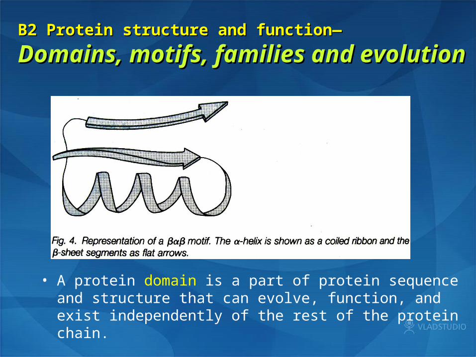
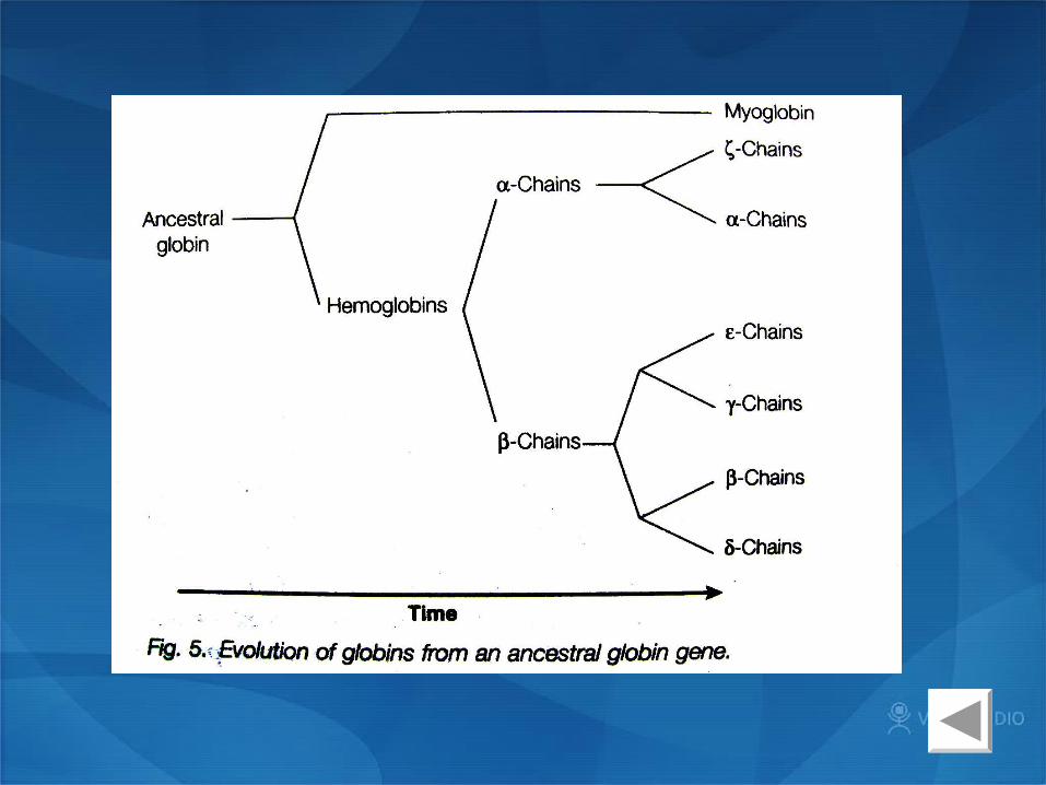
Domains, motifs, families and evolutionDomains, motifs, families and evolution
• A protein domain is a part of protein sequence and structure that can evolve, function, and exist independently of the rest of the protein chain.

• Structural motif, a pattern in a protein structure formed by the spatial arrangement of amino acids
• A protein family is a group of evolutionarily related proteins, and is often nearly synonymous with gene family.
Pyruvate kinase, a protein from three domains

B3 Protein analysis— B3 Protein analysis— Protein Protein PurificationPurification
• Gel filtration chromatography
• Ion-exchange chromatography
• Isoelectric focusing
• Electrophoresis
• Hydrophobic interaction chromatography
• Affinity chromatography
• Overexpression

Gel filtration chromatographyGel filtration chromatography<size>

Ion-exchange Ion-exchange chromatographychromatography
<size and charge>
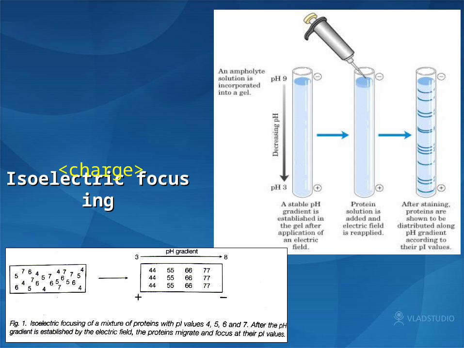
Isoelectric focusingIsoelectric focusing<charge>
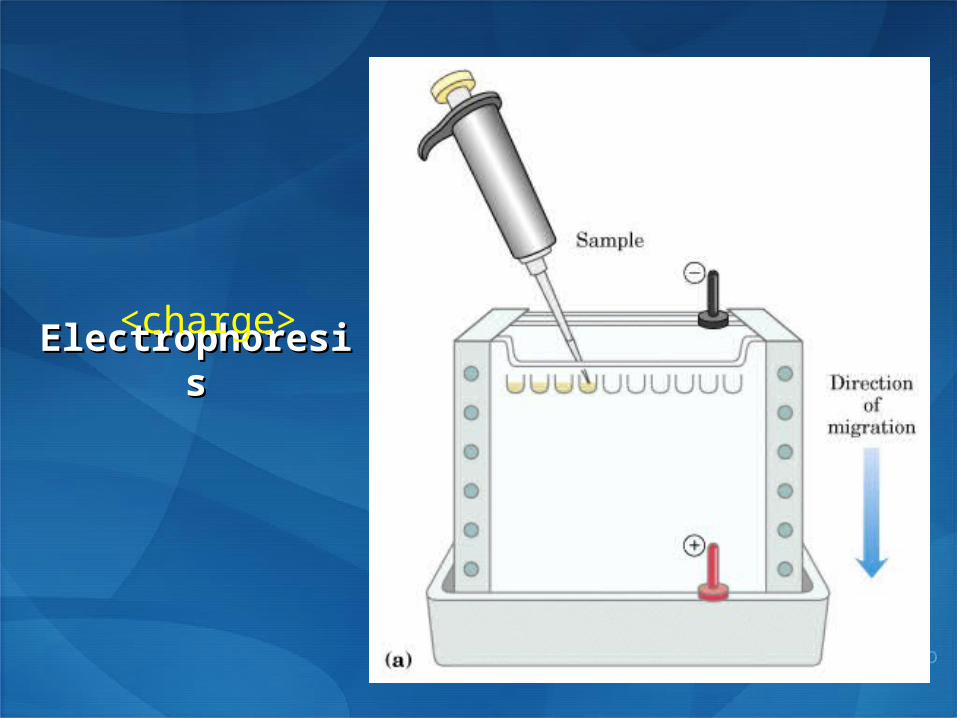
ElectrophoresisElectrophoresis<charge>
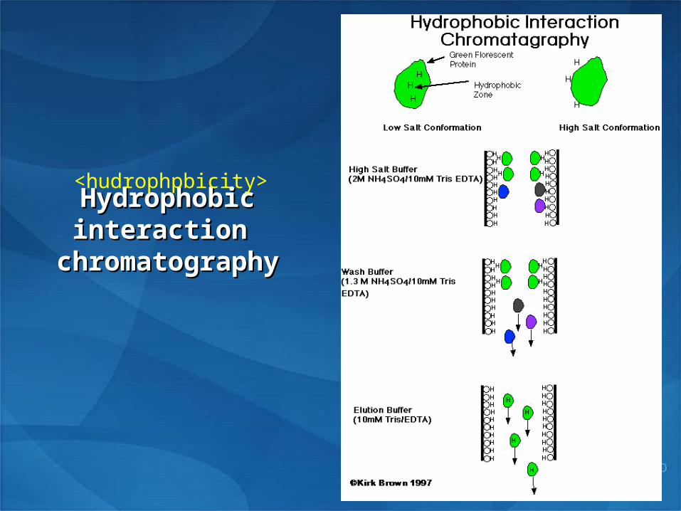
Hydrophobic interaction Hydrophobic interaction chromatographychromatography
<hudrophpbicity>

Affinity Affinity chromatographychromatography
< affinity>

• In the laboratory, the protein encoded by a gene is sometimes In the laboratory, the protein encoded by a gene is sometimes expressed in increased quantity. This can come about by increasing expressed in increased quantity. This can come about by increasing the number of copies of the gene or increasing the binding strength the number of copies of the gene or increasing the binding strength of the promoter region.of the promoter region.
OverexpressionOverexpression
ExampleExample :: Escherichia coli T7 promoter lac operator lacI coding sequence His · tag IPTG (a lactose analog)
Vector
T7 promoter
His · tag
lac operator
B3 Protein analysis— B3 Protein analysis— Protein Protein sequencingsequencing
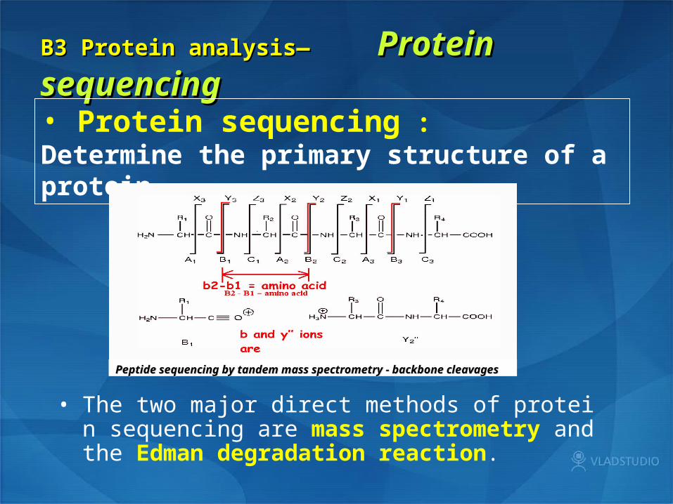
• Protein sequencing : Determine the primary structure of a protein
• The two major direct methods of protein sequencing are mass spectrometry and the Edman degradation reaction.
Peptide sequencing by tandem mass spectrometry - backbone Peptide sequencing by tandem mass spectrometry - backbone cleavagescleavages

Edman degradation Mechanism
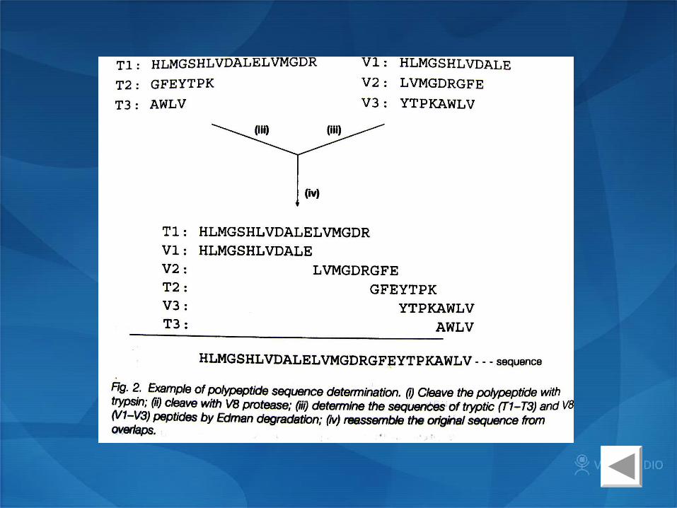
• Edaman degradation:1. Performed in an automated protein sequencer2. Determine the sequence of a polypeptide from
N-terminal amino acid one by one.3. Expensive and laborious

Specific enzyme/ chemical cleavage: • Trypsin cleaves after lysine( K) or arginine ( R)• V8 protease cleaves after glutamic acid (E)• Cyanogen bromide cleaves after methionine (M)

Most protein sequences are deduced from the DNA/cDNA sequence
Direct sequencing: determine the N-terminal sequences or some limited internal sequence construction of an oligonulceotide or antibody probe fishing the gene or cDNA

B3 Protein analysis— B3 Protein analysis— Mass Mass determinationdetermination
• Gel filtration chromatography
• Electrophoresis ( SDS-PAGE)
• Mass spectrometry
Gel filtration chromatography and SDS-PAGE
•Comparing of the unknown protein with a proper standard
•Popular SDS-PAGE: cheap and easy with a 5-10% error
•SDS: sodium dodecyl sulfate, makes the proteins negatively charged and the overall charge of a protein is dependent on its mass.
Mass spectrometry:
•Molecules are vaporized and ionized, and the degree of deflection (mass-dependent) of the ions in an electromagnetic field is measured
•extremely accurate, but expensive
•MALDI can measure the mass of proteins smaller than 100 KDa
•Helpful to detect post-translational modification
•Protein sequencing: relying on the protein data base

B3 Protein analysis— B3 Protein analysis—
X-ray crystallography and NMRX-ray crystallography and NMR
•Measuring the pattern of diffraction of a beam of X-rays as it pass through a crystal. The first hand data obtained is electron density map, the crystal structure is then deduced.
•A very powerful tool in understanding protein tertiary structure.
•Many proteins have been crystallized and analyzed.
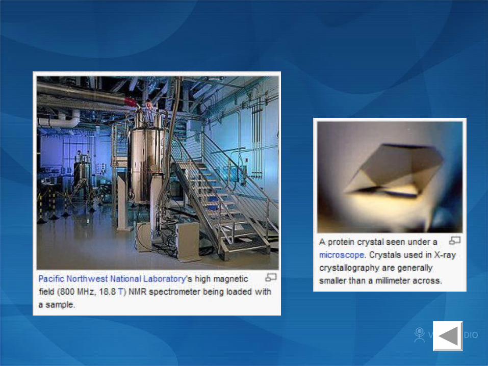
X-ray crystallographyX-ray crystallography
•Measuring the relaxation of protons after they have been excited by radio frequencies in a strong magnetic field.
•Measure protein structure in liquid but not in crystal.
•Protein measured can not be larger than 30 KDa.
NMRNMR

B3 Protein analysis— B3 Protein analysis— Functional Functional analysisanalysis
• Tertiary and quaternary structural determination is still a relatively cost and laborious procedure.
• Computational methods will allow the prediction of both structure and possible function from simple amino acid sequence information.
• Understanding of the true function of a protein still requires its isolation and biochemical and structure characterization.
• Identification of all the other proteins with which a protein interacts in the cell is another important aspect of functional analysis.
• If the gene foe the protein can be inactivated by mutagenesis or deleted be recombinant DNA techniques, then the phenotype of the resulting mutant can be studied.

B3 Protein analysis— B3 Protein analysis— ProteomicsProteomics
• Proteomics is the identification and analysis of the total protein complement expressed by any given cell type under defined conditions.
• Two-dimensional Protein Electrophoresis
• peptide mass fingerprint

Two-dimensional Protein Electrophoresis (2DE)

Analysis of peptide mass fingerprint of the tryptic digest of P. falciparum actin I by MALDI mass spectrometry. a. Spectrum (500-3500 Da) with zoom-scan (1850-1990 Da) showing N-terminal fragment at 1872 Da and fragment containing histidine 73 at 1947 Da. b. The table shows selected peptide matches from three experiments. N-terminal fragment and fragment containing histidine 73 are in bold.

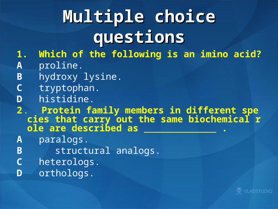
Multiple choice Multiple choice questionsquestions
1. Which of the following is an imino acid? A proline. B hydroxy lysine. C tryptophan. D histidine. 2 . Protein family members in different species that c
arry out the same biochemical role are described as .
A paralogs. B structural analogs. C heterologs. D orthologs.

3. Which of the following is not a protein secondary structure?
A α-helix.B triple helix. C double helix. D ß-pleated sheet. 4 . In isoelectric focusing, proteins are separated .A in a pH gradient. B in a salt gradient. C in a density gradient. D in a temperature gradient.

5 . Edman degradation sequences peptides . . .
A using a cDNA sequence.
B according to their masses.
C From the C-terminus to the N-terminus.
D from the N-terminus to the C-terminus.

THANK YOU !


![. Protein Structure Prediction [Based on Structural Bioinformatics, section VII]](https://static.documents.pub/doc/80x56/56649d575503460f94a35877/-protein-structure-prediction-based-on-structural-bioinformatics-section.jpg)


