Page 1
TOMÁS MARIA COSTA ALMEIDA PINTO DE SOUSA
SEDATIVE EFFECT OF ACEPROMAZINE MICRO
DOSE IN YINTANG ACUPOINT IN DOGS
Advisor: Ph.D. Lénio Ribeiro
Co Advisor: M.S. Luís Resende
Universidade Lusófona de Humanidades e Tecnologias
Faculdade de Medicina Veterinária
Lisboa
2015
Page 2
1
TOMÁS MARIA COSTA ALMEIDA PINTO DE SOUSA
SEDATIVE EFFECT OF ACEPROMAZINE MICRO
DOSE IN YINTANG ACUPOINT IN DOGS
Universidade Lusófona de Humanidades e Tecnologias
Faculdade de Medicina Veterinária
Lisboa
2015
Dissertation to obtain the degree of Master
in Veterinary Medicine in the course of
Integrated Master in Veterinary Medicine
given by Universidade Lusófona de
Humanidades e Tecnologias
Advisor: Ph.D. Lénio Ribeiro
Co Advisor: M.S. Luís Resende
Jury Members
President: PhD Laurentina Pedroso
Examiner: PhD Leandro Gardel
Advisor: PhD Lénio Ribeiro
Page 3
2
Dedicatory
This work is to be dedicated primarily to my closest family, especially, my parents and
also grandmother "Mami" which is not alive to witness the completion of my studies with this
study. I hope they may be proud since all the effort they endured to pay and to support my
studies merge into what I am and will be as a professional upon completion of my degree. A
high school professor once said to me that the world does not give you opportunities but it is
you who give yourself opportunities to become what you want. So with that said, I had the
luck to be able to pursue my dreams thanks to my family and for that I will be eternally
grateful.
A part is to be dedicated to my professors that have taught throughout my education. I
remember some with more sympathy than others but all deserve my respect with no exception
since one can always learn something from another person. Many times I wanted to write and
thank them personally but most of the times, one simply lets the moment pass and this is my
way of being grateful. Professors are of the utmost importance and become examples to
students in ways many don't realize and they should always reach out to make students better
not only at knowledge but at themselves. Those special that reached to me, i will be, forever
grateful.
It is to be dedicated also to all my friends who cared and supported me throughout life
giving me the security needed to engage on the obstacles and adventures of life. A special
mention is to be made to Raquel Redol Silva which has been a part of me for the greater part
of my education as a friend or loved one, being there through good and bad moments helping
me to get up and keep my eyes on the road.
Page 4
3
Acknowledgements
As author of this study I am forever grateful to my advisor PhD Lénio Ribeiro for
patiently counselling me throughout the whole process and co advisor Professor Luís Resende
for his active participation in the study.
A special thanks is to be given to PhD Stélio Pacca Loureiro Luna for being the source
of inspiration for this study and one great professor and researcher that lecture me and also
advised me in the study and provided my foreign internship in Brazil. I would also like to
thank the institution, Universidade Estadual Paulista - UNESP in Botucatu for providing the
wonderful and overwhelming final professional experience required to obtain my degree. I am
thankful to everyone i have worked with in UNESP, but I would like to give a special warm
thanks to Nadia Crosignani, Natache Garofalo, Mariana Werneck, Jéssica Rodrigues, Marina
Cayetano Evangelista, Luciana Kinoshita, Monica Midon, Nuno Emanuel, Elizabeth Carvalho
and Carol Hagy Girotto for all the friendship that i miss so much and for the support you gave
me for the study, internship and life in Brazil.
I am thankful to my professors from Universidade Lusófona de Humanidades e
Tecnologias and the institution itself for providing my education and actively supporting my
study.
Page 5
4
Resumo
Introdução. A acupuntura representa uma parte integral e muito importante da
medicina tradicional Chinesa, sendo utilizada à mais de 2500 anos. A técnica consiste em
introduzir agulhas extremamente finas em pontos específicos do corpo denominados pontos
de acupunctura, induzindo múltiplas respostas biológicas que activam o sistema nervoso. Os
efeitos benéficos da fármacopuntura já foram provados em estudos científicos, dentro dos
quais, o efeito analgésico, é reconhecido no tratamento de dor pela World Health
Organization e National Institutes of Health. O mecanismo de acção da acupunctura por
detrás da analgesia permanece ainda um pouco incerto.
A medicina tradicional Chinesa representa mais um tipo de filosofia que procura
equilibrar o corpo através de uma perspectiva que não é baseada em evidências anatómicas,
fisiológicas ou bioquímicas. Este facto, torna a compreensão e aceitação da acupuntura muito
difícil por parte dos praticantes de medicina ocidental.
Teorias e estudos científicos, dos quais alguns envolvem imagiologia cerebral, aludem
ao facto do sistema nervoso estar envolvido na transmissão dos sinais criados pela acupuntura
a áreas-alvo.
Pomeranz em 1987, sugeriu uma teoria onde a estimulação da acupuntura activa as
fibras aferentes A-δ e C no músculo levando à transmissão do sinal à medula espinal
causando a libertação local de opióides endógenos. Os nervos aferentes continuam a
propagação do sinal para o mesencéfalo desencadeando a libertação para a medula espinal, de
neurotransmissores como a serotonina, dopamina e noradrenalina que por sua vez levam à
inibição e supressão da transmissão da dor. O sinal ao chegar ao hipotálamo e à glândula
pituitária desencadeia a libertação de hormona adrenocorticotrófica (ACTH) e endorfinas.
Esta teoria foi fortemente confirmada por outros estudos científicos.
A libertação de opióides endógenos em resposta à acupuntura é uma das principais
teorias para a explicação do mecanismo de acção da acupuntura e é denominada pela
comunidade científica como a teoria neurohormonal.
Técnicas recentes de imagiologia do sistema nervoso central permitiram avaliar
padrões de expressão aquando a estimulação pela acupuntura. Hiesh et al., 2001 através do
uso de tomografia de emissão de positron, desvendou que durante a estimulação eléctrica do
acuponto Hegu ocorria activação do hipotálamo, periaqueducto cinzento e da insula. A
Page 6
5
activação dessas áreas ocorria com menor intensidade com o uso de estimulação mecânica, no
entanto, a estimulação em pontos falsos de acupuntura não desencadeou a activação. Outras
regiões foram descobertas por Biella et al., 2001 que demonstrou, através de estimulação dos
pontos Zusanli e Chize, a activação do cíngulo anterior esquerdo, giro frontal superior, giro
frontal inferior medial direito e ainda a activação bilateral do cerebelo e insula.
Hui et al., 2000, usando ressonância magnética funcional, reforçou a hipótese de que a
dor e a acupuntura possuem vias sobrepostas no sistema nervoso central. Wu et al., 2002
suportou também esta hipótese demonstrando que através de estimulação elétrica do acuponto
Yanglingquan e de um falso ponto, ocorria activação das vias centrais da dor. No entanto, só a
estimulação elétrica no verdadeiro acuponto Yanglingquan é que causou activação do
hipotálamo, córtex somatosensorial primário e desactivação do segmento rostral do córtex
cingular anterior, sugerindo que o sistema límbico-hipotalâmico é modelado pela
electroacupuntura. Ainda que importante, estudos científicos de neuroimagem em acupuntura
representam apenas meras explorações da rede de sinalização neuronal causada pela
acupuntura.
Na acupuntura diferentes pontos causam diferentes efeitos terapêuticos. Zhang et al.,
2004 usando ressonância magnética funcional examinou a activação de diferentes regiões do
sistema nervoso central por diferentes acupontos, Zusanli/Sanyinjiao e
Yanglingquan/Chengsan. Observações demonstraram que ambas as combinações de pontos
causaram activação de áreas somatosensoriais primárias e secundárias, a insula, o cerebelo, o
tálamo e o putámen. No entanto, cada combinação de pontos possuía para além das áreas
comuns, activação de áreas específicas.
Na acupuntura existem vários métodos para estimular os pontos sendo que a
fármacopuntura é o método mais comum de injecção utilizado em acupuntura. De acordo com
a medicina tradicional Chinesa este método combina a eficácia da estimulação do acuponto
com as propriedades farmacológicas das drogas aplicadas, potenciando o estímulo mecânico
no acuponto e produzindo efeitos similares àqueles causados pelo uso dessas drogas em
terapias convencionais. A fármacopuntura usa subdoses de drogas ou pequenas quantidades
de extractos herbais medicinais. Este uso de micro doses provou-se útil em medicina
veterinária, Alvarenga et al., 1998 injectou uma subdose de prostaglandina (0.5mg/kg) no
acuponto Bai-Hui, induzindo luteólise em éguas tão eficazmente como a dose convencional
Page 7
6
(5mg/kg) aplicada intramuscularmente, contribuindo ainda, para a redução de efeitos
secundários associados à dose convencional.
Na medicina tradicional Chinesa o acuponto Yin Tang tem efeito sedativo em humanos
e animais. Em 2002, Luna et al., demonstraram que a injecção de 0.01 mg/kg de
acepromazina no acuponto Yin Tang reduzia em 32% a quantidade necessária de tiopental
para indução de anestesia em cães, enquanto que a dose convencional de 0.1 mg/kg de
acepromazina causava uma redução de 51%. Cassu et al., 2014 demonstrou que 0.01 mg/kg
de xilazina, administrada no acuponto Yin Tang, produzia efeitos sedativos clinicamente
relevantes com a vantagem de reduzir os efeitos secundários associados ao uso de α2-
agonistas quando comparado com um grupo tratado com a dose convencional de 0.1 mg/kg de
xilazina.
O mecanismo de acção da fármacopuntura continua por esclarecer apesar desta técnica
ser amplamente utilizada na prática clínica. A injecção de substratos líquidos nos acupontos
causa uma alteração da configuração espacial dos tecidos. Esta alteração aliada às
características do substrato, estimulam o acuponto, o que por sua vez desencadeia a activação
do sistema nervoso. Chen et al., 2014, demonstrou que a distribuição de neurónios
expressores de c-fos em ratos, aos diferentes substratos injectados no acuponto Zusanli era
semelhante entre substratos embora a intensidade da expressão dependesse do tipo de
substrato utilizado. A distribuição dos neurónios expressores de c-fos observou-se
primariamente na lamina II do corno dorsal da medula espinal.
Sedação é um estado caracterizado por uma depressão central acompanhada de
sonolência. O paciente normalmente não está ciente dos estímulos exteriores mas responde a
estímulos dolorosos. É importante que a sedação ajude a aliviar a ansiedade e a diminuir o
stress associado à manipulação e todo o processo associado à anestesia, ou seja, é uma parte
essencial de um protocolo anestésico completo.
Diferentes classes de sedativos produzem uma extensa variedade de respostas
comportamentais entre as espécies de animais. Fenotiazínicos são drogas eficientes na
sedação de cães.
A acepromazina é o sedativo mais comumente utilizado na medicina veterinária, e
sendo mais potente que outros derivados fenotiazínicos, produz sedação a partir de baixas
doses. A sua administração produz algum relaxamento muscular mas não produz qualquer
Page 8
7
tipo de efeito analgésico, sendo necessário adicionar um opióide para bloquear a resposta
nociceptiva em casos de procedimentos dolorosos.
Fenotiazínicos causam diferentes níveis de efeitos anticolinérgicos, antiespasmódicos,
anti-histamínicos e ainda de bloqueio α-adrenérgico. O bloqueio dos receptores α1 causa a
hipotensão normalmente associada ao uso destas drogas. O bloqueio de receptores de
dopamina causa depleção de catecolaminas no centro de termorregulação do hipotálamo,
originando assim a perda da termorregulação que por sua vez, pode levar à hipotermia. Os
fenotiazínicos produzem sedação e tranquilização por inibição dos receptores centrais
dopaminérgicos (D2). Perifericamente, os fenotiazínicos bloqueiam a noradrenalina nos
receptores α-adrenérgicos.
A administração de acepromazina causa efeitos dramáticos no sistema cardiovascular
tanto em animais conscientes, como em animais anestesiados, no entanto, tem pouco efeito
sobre a função pulmonar. O uso desta droga deve ser feito com precaução, especialmente
quando se procura usar a acepromazina como agente de contenção em animais agitados, já
que esta pode fazer os animais mais propensos a reagir a estímulos sensoriais.
Objectivo. Este estudo procura avaliar o nível de sedação e os efeitos sobre a
frequência cardíaca, frequência respiratória, temperatura rectal e pressão arterial resultantes
da injecção no acuponto Yin Tang, de 0.005 mg/kg de acepromazina e comparar os resultados
com aqueles da administração intramuscular de 0.05 mg/kg de acepromazina.
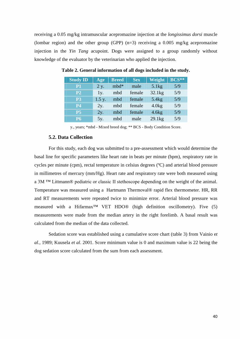
Materiais e Métodos. Foram submetidos ao estudo um total de 6 cães de raça
indeterminada com idades compreendidas entre 1 e 5 anos de idade e pesos entre 4 e 33 kg.
3 cães foram submetidos à técnica de fármacopuntura com injecção no acuponto Yin Tang de
0.005 mg/kg de acepromazina e 3 cães à técnica de injecção intramuscular de 0.05 mg/kg de
acepromazina. O ponto de acupuntura Yin Tang encontra-se na depressão presente na linha
dorsal média entre as sobrancelhas do cão.
Foram recolhidos, para todos os animais, valores basais para a frequência cardíaca,
frequência respiratória, temperatura rectal, pressão arterial sistólica e pressão arterial
diastólica antes de aplicar a técnica de sedação. 30 minutos após a injecção do fármaco uma
Page 9
8
nova recolha de parâmetros foi efectuada e um score de sedação atribuído a cada animal
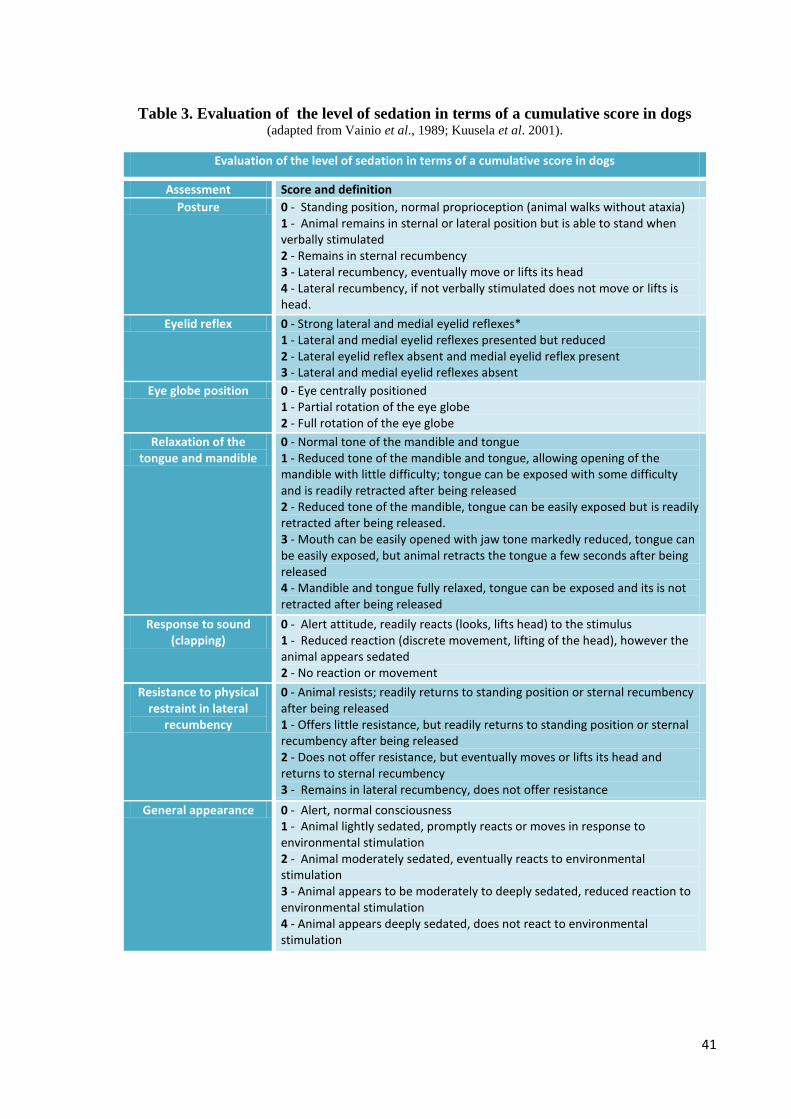
conforme a tabela de score de sedação de Vainio et al., 1989; Kuusela et al., 2001.
Os resultados foram submetidos a análise estatística através do software IBM®
SPSS® statistics versão 23. Aos dados foi aplicado o teste de independência não-paramétrico
Kruskal-Wallis para testar as hipóteses. As correlações entre dados foram avaliadas através do
teste não paramétrico de Spearman.
Resultados. Os scores de sedação obtidos para cada grupo estão descritos na tabela 5.
As medições dos parâmetros de frequência cardíaca, frequência respiratória, temperatura
rectal, pressão arterial sistólica e pressão arterial diastólica recolhidas antes da sedação e 30
minutos após a sedação estão descritas na tabela 6 para o grupo de fármacopuntura e na tabela
7 para o grupo de injecção intramuscular.
Tabela 5. Método de Sedação aplicado e respectivo Score de Sedação.
Método de Sedação ID Score de sedação
Injecção intramuscular (G1) P1 14/22
P4 1/22
P6 7/22
Fármacopuntura no Yin Tang (GPP) P2 2/22
P3 4/22
P5 1/22
ID, identificação do animal; G1, grupo de injecção intramuscular; GPP, grupo de injecção no acuponto Yin Tang
Tabela 6. Parâmetros medidos no grupo de fármacopuntura antes e após sedação.
Fármacopuntura GPP
P2 BL* P2 AS** P3 BL P3 AS P5 BL P5 AS
HR 100 100 124 120 165 111 bpm
RR 48 26 58 28 16 16 cpm
RT 39,4 38,8 39 38,5 39,7 38,6 ºC
mSP 216,4 147,4 185,8 148,6 139,8 129,8 mm/Hg
mDP 134,6 77,8 92 69 79,6 79,6 mm/Hg
HR, frequência cardíaca; RR, frequência respiratória; RT, temperatura rectal; mSP, média da pressão arterial
sistólica; mDP, média da pressão arterial diastólica; *, basal; **, após sedação; GPP, grupo de injecção no
acuponto Yin Tang
Page 10
9
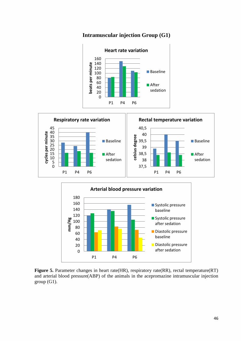
Tabela 7. Parâmetros medidos no grupo de injecção intramuscular antes e após sedação.
Injecção Intramuscular G1
P1 BL* P1 AS** P4 BL P4 AS P6 BL P6 AS
HR 80 84 150 129 110 104 bpm
RR 28 16 24 18 40 16 cpm
RT 38,9 38,4 40 38,6 39,5 38,4 ºC
mSP 120,4 127,2 140,2 135 155,6 106 mm/Hg
mDP 64 71,2 83,2 75,8 72 45,8 mm/Hg
HR, frequência cardíaca; RR, frequência respiratória; RT, temperatura rectal; mSP, média da pressão arterial
sistólica; mDP, média da pressão arterial diastólica; *, basal; **, após sedação; G1, grupo de injecção
intramuscular.
Análise estatística não mostrou diferença significativa (valor p < 0.05) entre os scores
de sedação do grupo de injecção no acuponto Yin Tang e o grupo de injecção intramuscular.
No entanto, os resultados no grupo de fármacopuntura apresentam de uma forma generalizada
um score de sedação mais baixo. A análise estatística não mostrou diferença significativa
(valor p < 0.05) entre os parâmetros recolhidos após sedação no grupo de injecção no
acuponto Yin Tang e no grupo de injecção intramuscular.
Discussão. A amostra presente neste estudo demonstra-se insuficiente para uma
correta análise estatística dos dados. Devido a restrições impostas em prol do bem estar
animal, apenas cães da rotina diária do hospital poderiam ser alvos deste estudo, tornando a
amostra relativamente heterogénea. As restrições também não permitiram a presença de um
grupo de controlo, no entanto, estudos anteriores por Luna et al., 2002; 2008; Godoi et al.,
2013, usando técnicas semelhantes observaram que a injecção das drogas ou de solução salina
num ponto falso, não produzia efeito ou apenas um efeito reduzido quando comparado com a
injecção num verdadeiro ponto de acupuntura.
Manipulação do animal levaria certamente à adulteração do próximo score de sedação
e como tal, o score de sedação foi medido apenas uma vez aos 30 minutos. No estudo a
avaliação do score seguiu uma ordem específica, começando na avaliação do aspecto geral,
seguido da postura, resistência à contenção física, posição do globo ocular, reflexo palpebral,
relaxamento da mandíbula e língua e resposta ao som. A escala de score cumulativo de
sedação em cães usada neste estudo foi escolhida porque permitia avaliar objetivamente
características que também são usadas para monitorizar animais durante a anestesia. Permitiu
Page 11
10
ainda, quantificar eficazmente o nível de consciência, o estado de alerta e o controlo motor
dos animais. Outras escalas de sedação, como a escala de sedação Ramsay, poderiam também
ser usadas no estudo com o mesmo nível de eficácia e fiabilidade, no entanto, estas
apresentam um maior nível de subjetividade e apenas detetam mudanças moderadas no
comportamento dos animais. A escala de sedação usada no estudo não possui critérios para
avaliar o nível de agitação dos animais e isto representa um problema no uso desta escala. Os
cães P2 e P3 no grupo de fármacopuntura demonstraram, antes da sedação, sinais de agitação
e reatividade excessiva para com os estímulos sensoriais. A técnica de fármacopuntura, tendo
em conta que nenhum animal apresentou sinais de agitação após a sedação, pode ter sido
eficaz no alívio da agitação, reduzindo a reactividade a estímulos sensoriais.
Estatisticamente, não houve diferença significativa entre métodos de sedação
sugerindo assim, que de um ponto de vista teórico, os métodos de sedação são similares. No
entanto, a observação clínica durante o estudo denotou uma diferença entre grupos, sendo que
o grupo de fármacopuntura apresenta, comparativamente ao grupo de injecção intramuscular,
um score de sedação mais baixo, ou seja, uma sedação mais leve.
A variação do score de sedação no grupo de injecção intramuscular de acepromazina é
grande, já que o score mínimo foi de 1 num total de 22 e o score máximo de 14 em 22. Está
descrito por BSAVA Small Animal Formulary (2011) que a acepromazina não é fiável para
sedação, quando usada sozinha, podendo assim explicar a variação tão ampla dos valores
obtidos neste estudo.
A acepromazina, devido a ser o sedativo mais amplamente usado na medicina
veterinária, foi a droga de escolha para o protocolo deste estudo. Outros sedativos mais
recentes poderão ter um potencial maior na sedação de cães. Um estudo anterior por Cassu et
al., 2014 administrou, em cães, 0.01 mg/kg de xilazina no acuponto Yin Tang, induzindo um
efeito sedativo clinicamente relevante em cães, reduzindo ainda os efeitos secundários
associados ao uso de α2-agonistas.
A sedação leve que ocorreu no grupo de fármacopuntura pode ser resultado de uma
dose ineficaz de acepromazina. Como tal, o ajuste da dose deve ser feito em estudos
posteriores de forma a determinar a dose ideal de acepromazina, necessária para atingir um
nível eficaz de sedação.
Page 12
11
Ainda que anedótico, a comparação dos resultados obtidos após sedação entre o grupo
de fármacopuntura e o grupo de injecção intramuscular pode ser útil e como tal, elações
clínicas podem ser feitas a partir da evidência. As variações nos parâmetros de frequência
cardíaca, frequência respiratória, temperatura rectal, pressão arterial sistólica e pressão arterial
diastólica medidos após sedação em ambos os grupos, demonstram de um ponto de vista
clínico, uma semelhança de efeito, pois ambos causaram uma diminuição dos valores basais
para limites fisiológicos mais compatíveis com um cão em repouso. Este efeito similar entre
técnicas, originado pelo uso da fármacopuntura é demonstrado por Alvarenga et al., 1998;
Silva & Luna, 1999; Nie et al., 2001; Luna et al., 2008; Cassu et al., 2014.
Neste estudo, a injecção de 0.005 mg/kg de acepromazina administrada no ponto de
acupuntura Yin Tang causou, com sucesso um efeito sedativo leve nos animais, moldando o
comportamento dos mesmos para um estado mais relaxado. O grupo de fármacopuntura não
apresentou nenhum dos efeitos secundários descritos para a acepromazina. Tendo em conta
este facto e o facto da amostra ser pequena, podemos apenas especular que este método
parece ser seguro de usar na prática clínica.
A fármacopuntura combina o efeito da acupuntura com os efeitos dos fármacos. Neste
estudo o fármaco usado foi a acepromazina, uma droga que causa inibição dos receptores
centrais dopaminérgicos (D2) que são responsáveis pela modulação do comportamento e por
sua vez, a sedação. O ponto Yin Tang é indicado para sedação mas o mecanismo envolvendo
essa sedação é vago. Perifericamente, os fenotiazínicos como a acepromazina bloqueiam a
noradrenalina nos receptores α-adrenérgicos. Estudos experimentais por Han et al., 1979;
Wang, Jiang & Can, 1994; Zhu et al., 1997, demonstram, em ratos, níveis reduzidos de
noradrenalina no cérebro após electroacupuntura. Isto demonstra que tanto a droga como o
efeito causado pela acupuntura vão actuar nos receptores α-adrenérgicos. Fenotiazínicos tem
grande afinidade para receptores α1-adrenérgicos, que por sua vez, facilitam a sinalização
nociceptiva. Isto pode explicar o porquê da acepromazina não produzir efeito analgésico. Por
outro lado a acupuntura causa a libertação de opióides endógenos, sugerindo um efeito
analgésico aditivo no uso da técnica de fármacopuntura. Estudos posteriores seriam
necessários para confirmar esta teoria e comparar a resposta a estímulos nocivos entre o grupo
de fármacopuntura e o grupo de injecção intramuscular de acepromazina.
As alterações dos parâmetros no grupo da fármacopuntura sugerem que o método
desencadeia actividade parasimpática e inibe a actividade simpática, e que estas alterações são
Page 13
12
similares àquelas observadas no grupo de injecção intramuscular, sugerindo então, que ambos
os métodos alteram o sistema nervoso autónomo.
Tendo em conta as observações, a injecção de acepromazina no ponto Yin Tang pode
provar-se útil no dia-a-dia da prática clínica se futuros estudos científicos forem efetuados
com vista em obter uma melhor compreensão sobre o método e a sua segurança. Este método
mostra-se promissor já que produziu efeitos sedativos clinicamente relevantes e reduziu em
90% os custos relativos á acepromazina, o que permite uma melhor gestão dos recursos pelas
clínicas e ainda permite ao animal a metabolização e excreção de uma menor quantidade de
droga.
Conclusão. Este estudo apenas serve como uma análise preliminar para futuras
investigações científicas na área da fármacopuntura já que todas as conclusões retiradas das
observações são meras especulações resultantes de um modelo animal deficiente disponível
na altura do estudo. Ainda assim, elações podem ser feitas baseadas nas evidências clínicas
observadas.
Um efeito sedativo leve foi observado nos animais usando o método de injecção de
acepromazina no acuponto Yin Tang e consequentemente ocorreram mudanças na frequência
cardíaca, frequência respiratória, temperatura rectal, pressão arterial e comportamento,
levando os animais a retornar a um estado de repouso e relaxamento, sugerindo que este
método talvez possa ser utilizado na prática clínica para sedação de cães. No entanto, este
método não deve ser utilizado quando o objetivo é a contenção de animais agressivos, já que
ambas as técnicas usadas no estudo demonstraram-se ineficazes neste ponto.
Devem ser realizados futuros estudos científicos com amostras fortemente controladas
para avaliar os benefícios e efeitos secundários que podem advir do uso das técnicas de
fármacopuntura.
Palavras-chave: Acepromazina, Acupuntura, Fármacopuntura, Sedação em Cães, Yin Tang.
Page 14
13
Abstract
Acupuncture is part of Chinese medicine and has been used for more than 2000 years.
Pharmacopuncture is a more recent way of practicing acupuncture by converging the
acupuncture effect with the drug effect. This method still needs much scientific research but
studies already show that is possible to administer a small amount of drug in specific
acupuncture points to obtain a similar effect than that of western medicine treatments where a
conventional dose is administered. This study, tried to scratch the surface of
pharmacopuncture's research by administering 1/10 of the acepromazine dose at Yin Tang, a
acupuncture point indicated for sedation in dogs, to a group of 3 dogs (GPP) and comparing
the sedation score, heart rate, respiratory rate, rectal temperature and arterial blood pressure,
before and after sedation, with a group of 3 dogs (G1) that received a intramuscular injection
of acepromazine at the dose of 0.05 mg/kg. The results, in a clinical perspective show that
pharmacopuncture method caused a similar effect when compared to the conventional dose.
Sedation by this method seems promising but further extensive scientific research must be
made do bring this methods to western practitioners in clinical practice.
Keywords: Acepromazine, Acupuncture, Pharmacopuncture, Sedation in Dogs, Yin Tang.
Page 15
14
Abbreviations & Symbols
ABP - arterial blood pressure
ACTH - adrenocorticotropic hormone
AS - after sedation
BA - broadmann area
BL - baseline
BL57 - bladder meridian point nº57
bpm - beats per minute
CNS - central nervous system
cpm - cycles per minute
CSF - cerebral spinal fluid
c-fos - proto-oncogene
DRG - dorsal root ganglion
DVC - dorsal vagal complex
EA - electroacupuncture
EMA - European Medicines Agency
FDA - Food & Drug Administration
fMRI - functional magnetic resonance imaging
GAP43 - growth-associated protein
GB34 - gallbladder meridian point nº 34
GPP - Yin Tang pharmacopuncture group
G1 - intramuscular injection group
GV1 - governor vessel meridian point nº 1
HR - heart rate
Hz - hertz
Page 16
15
ID - identification
IM - intramuscular
IV - intravenous
kg - quilogram
LI4 - large Intestine meridian point nº 4
LU5 - lung meridian point nº5
MAC - minimum alveolar concentration
MDR1 - multi drug resistance gene
mg - milligram
mm/Hg - millimetres of mercury
mDP - mean diastolic pressure
mSP - mean systolic pressure
NIH - National Institutes of Health
PCO2 - partial pressure of carbon dioxide
PET - positron emission tomography
phospho Erk 1/2 - phosphorylation of extracellular-signal-regulated kinases
PO2 - partial pressure of oxygen
RNA - ribonucleic acid
RR - respiratory rate
RT - rectal temperature
SA - sinoatrial
SC - subcutaneous
ST36 - stomach meridian point nº 36
TCM - traditional Chinese medicine
TENS - transcutaneous electric nerve stimulation
TSH - thyroid stimulating hormone
Page 17
16
Vd - volume of distribution
WHO - World Health Organization
ºC - Celsius degree
(n) - number
22G - gauge measure
25G - gauge measure
Page 18
17
Index
1. ANAESTHESIA _______________________________________________________________ 20
1.1. PREMEDICATION AND SEDATION ________________________________________________ 20
1.1.1. EVALUATING SEDATION _______________________________________________________ 21
1.1.2. SEDATION SCALES ___________________________________________________________ 21
1.2. ACEPROMAZINE ______________________________________________________________ 22
1.2.1. MECHANISM OF ACTION _______________________________________________________ 23
1.2.2. INDICATION ________________________________________________________________ 24
1.2.3. PHARMACOKINETICS AND PHARMACODYNAMICS __________________________________ 25
1.2.4. DOSAGE ___________________________________________________________________ 27
1.2.5. CONTRAINDICATIONS _________________________________________________________ 27
1.2.6. ADVERSE EFFECTS ___________________________________________________________ 27
2. ACUPUNCTURE ______________________________________________________________ 29
2.1. TRADITIONAL CHINESE MEDICINE APPROACH _____________________________________ 30
2.1.1. MERIDIANS _________________________________________________________________ 30
2.1.2. ACUPUNCTURE POINTS ________________________________________________________ 30
2.1.3. QI ________________________________________________________________________ 30
2.1.4. YIN-YANG _________________________________________________________________ 30
2.2. SCIENTIFIC RESEARCH _________________________________________________________ 30
2.2.1. NEUROPHYSIOLOGY APPROACH _______________________________________________ 30
2.2.2.CENTRAL NERVOUS SYSTEM IMAGING __________________________________________ 33
2.2.2.1.PET ______________________________________________________________________ 33
2.2.2.2. FMRI ____________________________________________________________________ 34
2.3. ACUPUNCTURE POINT SPECIFICITY ______________________________________________ 35
2.4. ACUPUNCTURE POINT STIMULATION METHODS ____________________________________ 36
2.4.1. PHARMACOPUNCTURE _______________________________________________________ 36
2.4.1.1. MECHANISM ______________________________________________________________ 38
3. OBJECTIVE __________________________________________________________________ 39
4. HYPOTHESIS ________________________________________________________________ 39
5. MATERIALS AND METHODS __________________________________________________ 39
5.1. ANIMALS ____________________________________________________________________ 39
5.2. DATA COLLECTION ___________________________________________________________ 40
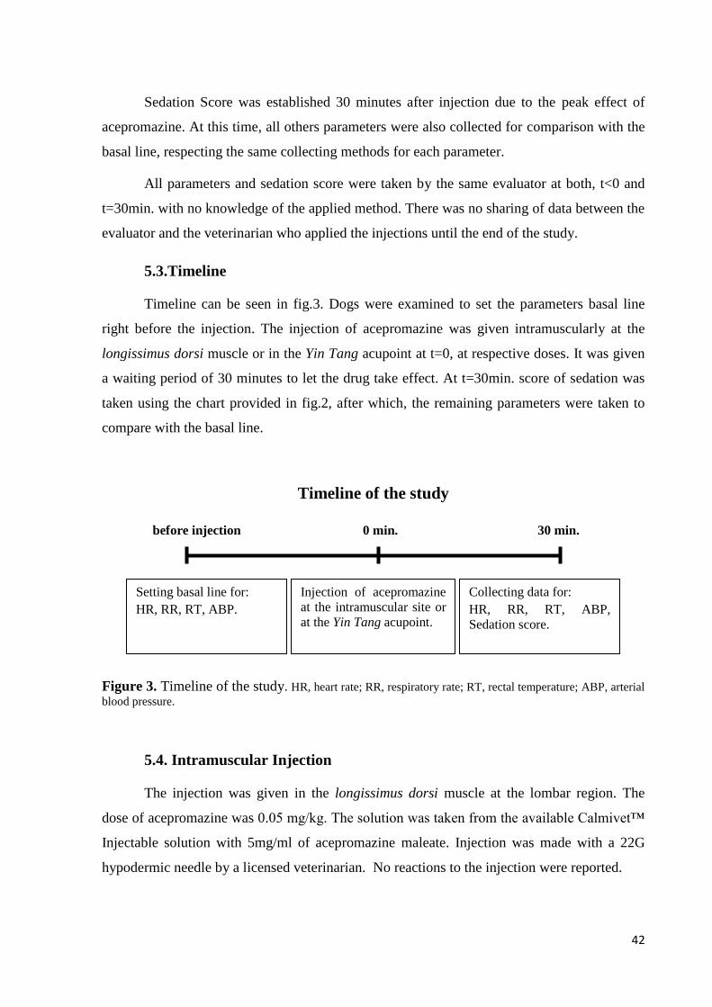
5.3.TIMELINE ____________________________________________________________________ 42
5.4. INTRAMUSCULAR INJECTION ___________________________________________________ 42
5.5. PHARMACOPUNCTURE INJECTION _______________________________________________ 43
5.6. DATA ANALYSIS ______________________________________________________________ 43
Page 19
18
6. RESULTS ____________________________________________________________________ 44
7. DISCUSSION _________________________________________________________________ 49
7.1. SEDATION SCORE _____________________________________________________________ 49
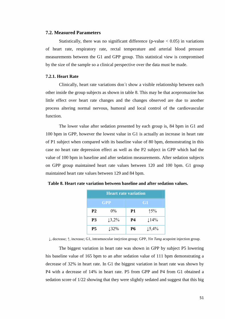
7.2. MEASURED PARAMETERS ______________________________________________________ 51
7.2.1. HEART RATE ________________________________________________________________ 51
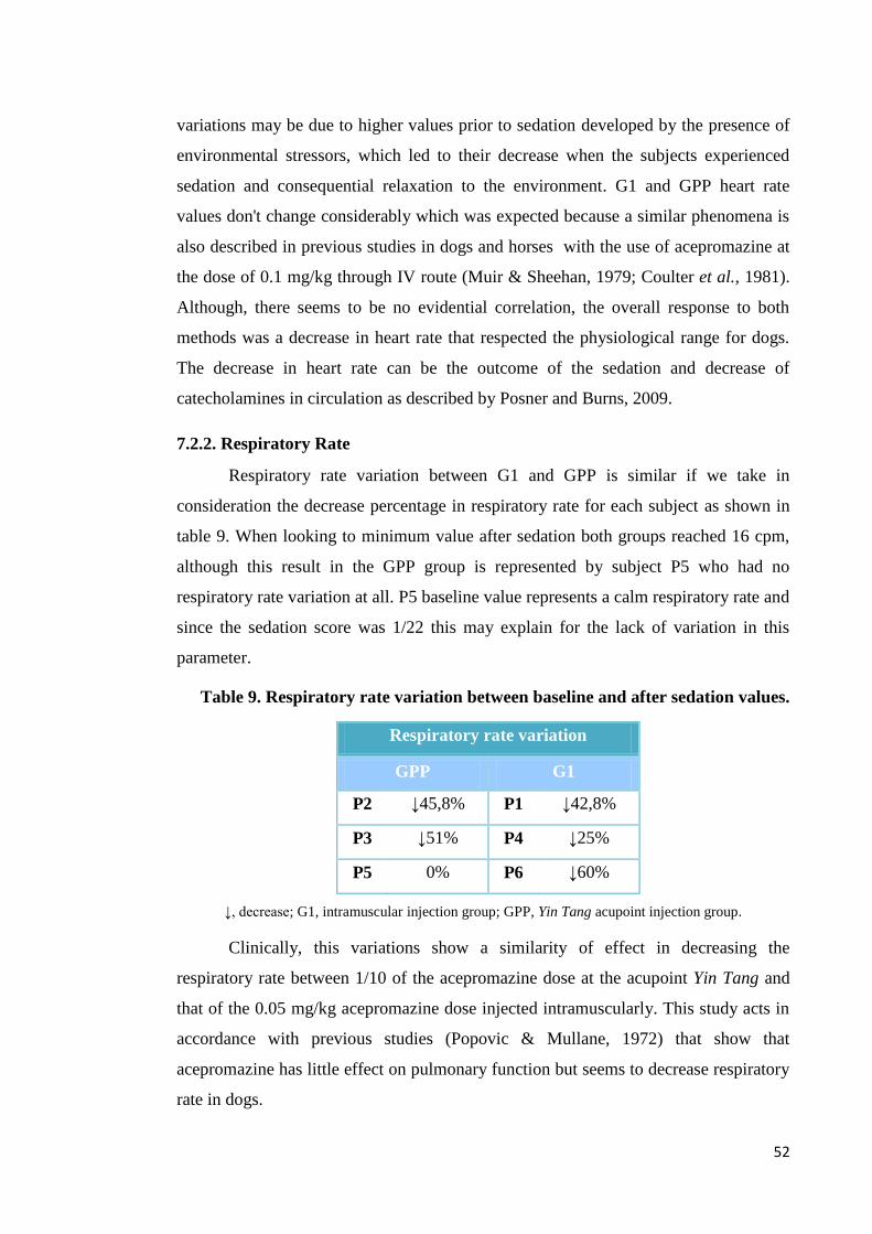
7.2.2. RESPIRATORY RATE __________________________________________________________ 52
7.2.3. RECTAL TEMPERATURE _______________________________________________________ 53
7.2.4. ARTERIAL BLOOD PRESSURE ___________________________________________________ 54
7.3. SYNOPSIS ____________________________________________________________________ 54
8. CONCLUSION ________________________________________________________________ 57
BIBLIOGRAFIA ________________________________________________________________ 59
Page 20
19
Table Index
Table 1. Relative receptor-binding affinities of phenothiazines. ______________________ 23
Table 2. General information of all dogs included in the study. ______________________ 40
Table 3. Evaluation of the level of sedation in terms of a cumulative score in dogs ______ 41
Table 4. General information for all dogs included in the study. _____________________ 44
Table 5. Individual animal sedation score and respective applied sedation method. ______ 44
Table 6. Parameters collected in the pharmacopuncture group. ______________________ 45
Table 7. Parameters collected in the intramuscular injection group. ___________________ 45
Table 8. Heart rate variation between baseline and after sedation values. ______________ 51
Table 9. Respiratory rate variation between baseline and after sedation values. _________ 52
Table 10. Rectal temperature variation between baseline and after sedation values. ______ 53
Table 11. Arterial blood pressure variation between baseline and after sedation values. ___ 54
Figure Index
Figure 1. Chemical Structure of acepromazine ___________________________________ 22
Figure 2. Effect of acepromazine (0.1 mg/kg intramuscularly) on mean arterial pressure in
dogs anaesthetized with isoflurane (2%) ________________________________________ 25
Figure 3. Timeline of the study. HR, heart rate; RR, respiratory rate; RT, rectal temperature;
ABP, arterial blood pressure. _________________________________________________ 42
Figure 4. Location of the Yin Tang acupoint in dogs. ______________________________ 43
Figure 5. Parameter changes in heart rate(HR), respiratory rate(RR), rectal temperature(RT)
and arterial blood pressure(ABP) of the animals in the acepromazine intramuscular injection
group (G1). _______________________________________________________________ 46
Figure 6. Parameter changes in heart rate(HR), respiratory rate(RR), rectal temperature(RT)
and arterial blood pressure(ABP) of the animals in the acepromazine pharmacopuncture group
(GPP). ___________________________________________________________________ 47
Page 21
20
1. Anaesthesia
The term anaesthesia comes from Greek an-, "without" and -aisthēsis, "sensation"
meaning insensibility (Thurmon & Short, 2007). It is used to describe the reversible process
of depression of the central nervous system (CNS) with drugs that produce unconsciousness
and a reduced or absent response to noxious stimuli (Jones, 1999).
Anaesthesia is a wide subject including many concepts like analgesia,
tranquilization/sedation, narcosis, hypnosis, local analgesia, regional analgesia, limb
analgesia, general anaesthesia, balanced anaesthesia and dissociative anaesthesia amongst
others.
For the purpose of this work, the focus will be on the concept of sedation.
Sedation is a state characterized by central depression accompanied by drowsiness.
The patient is generally unaware of its surroundings but responsive to painful manipulation
(Thurmon & Short, 2007).
Analgesia is also an important concept, meaning a state of relief, reduced or abolished
perception of pain (Jones, 1999). It's important since some sedatives like acepromazine do not
possess analgesic effect (Lukasik, 1999), so its use, should be complement with analgesic
drugs like opioids for minor painful procedures (Lemke, 2007).
1.1. Premedication and Sedation
Premedication is an essential part of a complete anaesthetic protocol. It is important to
help relieve anxiety and decrease stress associated with handling and the whole process of
anaesthesia. One of the major advantages is, the increased safety of the process and of the
staff during restraint and introduction of intravenous catheters. The drugs used in
premedication contribute to smooth induction and recovery from anaesthesia (Lukasik, 1999).
Premedication and sedation should be used to reduce anxiety, produce mild to
moderate sedation and provide analgesia if required for painful manipulations or before
surgery. It must also target an increase in muscle relaxation, decrease saliva and airway
secretions, reduce side effects of subsequently administered drugs, suppress vomiting and
regurgitation, decrease the amount of drug needed to cause unconsciousness for induction and
contribute to analgesia postoperatively (Lukasik, 1999).
Page 22
21
The different classes of sedatives produce a wide variety of behavioural responses
among the species. Phenothiazines and α2-agonists are effective sedatives in dogs and cats.
Benzodiazepines are effective sedatives in ferrets, rabbits, swine and birds but are not reliable
for cats and young dogs. Dose requirements vary widely among species (Lemke, 2007). It's
important for sedatives to take effect before anaesthesia induction otherwise an overdose of
induction and maintenance drugs may occur. Patients should be given 15 to 30 minutes in a
quiet area to let the drugs take effect (Lukasik, 1999). Commonly used drugs for
premedication and sedation are the phenothiazines, benzodiazepines and α2-agonists. They
can also be combined with opioids to produce the added effect of analgesia (Lemke, 2007).
Since 1950's phenothiazines are used in veterinary medicine as tranquilizers, even
though this class, originally was used as an antipsychotic drug for treatment of schizophrenia.
(Posner & Burns, 2009).
Acepromazine is the most commonly used phenothiazine in veterinary medicine
(Lukasik,1999; Posner & Burns, 2009). Other phenothiazines used on rare occasions include
chlorpromazine, promazine, promethazine, trimeprazine and methotrimeprazine (Lukasik,
1999).
1.1.1. Evaluating Sedation
Sedation is a drug induced state for the patient. Patient needs, differ upon
clinical circumstances and can be specific for each patient changing over time even for
the same patient. All must be done to ensure patient safety and comfort, including
preventing excessive and prolonged sedation. Excessive or prolonged sedation is
problematic leading to increased risk of complications. To prevent such consequences,
sedation must be measured accurately and that has to be done using robust tools that
assess precise and accurately the state of the patient. For that tools must include a wide
range of behaviours. This approach leads to a patient-directed approach increasing
safety for patient and practitioners (Sessler, Grap & Ramsay, 2008).
1.1.2. Sedation Scales
In the past a subjective tool called the Ramsay Sedation scale was introduced,
allowing to evaluate precisely the level of consciousness during titration of sedative
medications in an ICU (Ramsay et al., 1974). Ever since, more and more tools have
been developed, validated, and applied in clinical and research environments to
Page 23
22
monitor level of consciousness or arousal, as well as to evaluate cognition, agitation
and other parameters (Sessler, Grap & Ramsay, 2008). These include the Sedation
Agitation Scale (SAS) (Riker, Picard & Fraser, 1999), the Motor Activity Assessment
Scale (MAAS) (Devlin et al., 1999), the Richmond Agitation-Sedation Scale
(RASS)(Sessler et al., 2002) and others.
In order for such tools to be effective, they must be accurate, reliable, easy to
apply and repeatable by multiple evaluators (Sessler, Grap & Ramsay, 2008).
Desirable features of a good sedation scale have been enumerated and include:
rigorous multidisciplinary development, ease of administration, recall and
interpretation, well defined discrete criteria for each level, sufficient sedation levels for
effective drug titration, assessment of agitation and demonstration of inter-rater
reliability and evidence for validity in relevant patient populations (Sessler, 2004).
Implementation of a sedation assessment tool can have a positive effect on
precision of sedative administration (Costa et al., 1994; Botha & Mudholkar, 2004),
with greater frequency of appropriate sedation level and lower incidence of over-
sedation, reduction in sedative and analgesic drug doses, shorter duration of
mechanical ventilation, and even reduced use of vasopressor drugs. Implementation of
strategies that incorporate scheduled assessment for agitation, have been associated
with a reduction in agitation and even fewer nosocomial infections (De Jonghe, 2005;
Chanques et al., 2006). Use of a sedation scale is an integral part of most patient-
focused management algorithms (Sessler, Grap & Ramsay, 2008).
1.2. Acepromazine
Acepromazine is the most widely used sedative in veterinary medicine. (Lemke, 2007)
The chemical name of acepromazine is 2-acetyl-10(3-dimethylaminopropyl)-
phenothiazine (fig.1).
Figure 1. Chemical Structure of acepromazine (Gross, 2001)
Page 24
23
Acepromazine is more potent than other phenothiazine derivatives and produces
sedation at relatively low doses. Administration produces some muscle relaxation but has no
analgesic effect (Lemke, 2007). Phenothiazine derivatives have little or no analgesic activity.
Tranquilization must be supplemented with analgesics and/or general anaesthetics to block
nociceptive responses during painful procedures (Posner & Burns, 2009).
1.2.1. Mechanism of action
Phenothiazines produce a wide spectrum of autonomic, endocrine and behavioural
effects. The behavioural effects caused by this drugs are caused primarily by blockade of
dopamine receptors in basal ganglia and in the limbic system. They inhibit conditioned
avoidance behaviour and decrease spontaneous motor activity in therapeutic doses and may
cause extrapyramidal effects like tremor, rigidity and catalepsy in higher doses (Lemke,
2007). Furthermore, phenothiazines have varying degrees of anticholinergic, antihistaminic,
antispasmodic and α-adrenergic blocking effects (Plumb, 2008). These type of sedatives have
also a great binding affinity for other types of receptors: adrenergic and muscarinic, as shown
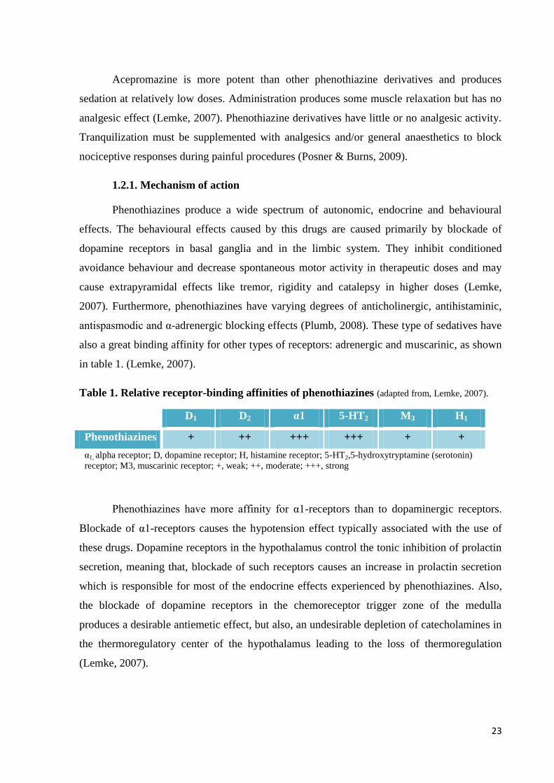
in table 1. (Lemke, 2007).
Table 1. Relative receptor-binding affinities of phenothiazines (adapted from, Lemke, 2007).
D1 D2 α1 5-HT2 M3 H1
Phenothiazines + ++ +++ +++ + +
α1, alpha receptor; D, dopamine receptor; H, histamine receptor; 5-HT2,5-hydroxytryptamine (serotonin)
receptor; M3, muscarinic receptor; +, weak; ++, moderate; +++, strong
Phenothiazines have more affinity for α1-receptors than to dopaminergic receptors.
Blockade of α1-receptors causes the hypotension effect typically associated with the use of
these drugs. Dopamine receptors in the hypothalamus control the tonic inhibition of prolactin
secretion, meaning that, blockade of such receptors causes an increase in prolactin secretion
which is responsible for most of the endocrine effects experienced by phenothiazines. Also,
the blockade of dopamine receptors in the chemoreceptor trigger zone of the medulla
produces a desirable antiemetic effect, but also, an undesirable depletion of catecholamines in
the thermoregulatory center of the hypothalamus leading to the loss of thermoregulation
(Lemke, 2007).
Page 25
24
Dopamine is mostly an inhibitory neurotransmitter which is responsible for the
regulation of fine motor control, prolactin secretion and behavioural regulation (Lemke,
2007). Dopamine receptors are included in the family of G-protein-coupled receptors (Lemke,
2007), (Posner & Burns, 2009) and are divided in two types: dopamine 1 (D1) receptors
located post-synaptically and dopamine 2 (D2) receptors located pre- and post-synaptically
(Lemke, 2007). They were classified based on their ability to inhibit or enhance the adenylate
cyclase activity (Lachowicz, 1997). Activating D1 receptors causes an increase in adenylate
cyclase activity and in intracellular levels of cyclic adenosine monophosphate (cAMP).
Counterwise, activation of D2 receptors causes a decrease in both, and can also activate other
pre-synaptic signal transduction pathways, decreasing calcium conduction and post-synaptic
signal transduction pathways, increasing potassium conduction. Behavioural effects are
mainly mediated by the D2 receptors family (Lemke, 2007). Phenothiazines produce sedation
and tranquilization by inhibition of central dopaminergic receptors (D2). Peripherally,
phenothiazines block norepinephrine at α-adrenergic receptors. (Posner & Burns, 2009).
1.2.2. Indication
Acepromazine is approved by Food and Drug Administration (FDA) for use in dogs,
cats and horses in North America and by European Medicines Agency (EMA) for cattle,
sheep, goats, pigs and horses in the European Union.
Clinical uses of acepromazine are usually restricted to healthy animals. The drug is
administered alone as a sedative for non-painful diagnostic procedures or in combination with
an opioid for painful diagnostic and minor surgical procedures. Acepromazine is also given
alone or in combination with opioids as a preanaesthetic to facilitate placement of IV
catheters and to reduce the dose of injectable and inhalation anaesthetics required to induce
and maintain anaesthesia. Small doses of acepromazine can also be given postoperatively,
provided that patients are hemodynamically stable and that pain has been managed
effectively. Acepromazine can be given SC, IM, IV, but the IM and IV routes are preferred
because uptake from SC sites can be erratic in patients with altered peripheral
circulation.(Lemke, 2007)
Page 26
25
1.2.3. Pharmacokinetics and Pharmacodynamics
In dogs given acepromazine and an opioid IM, onset of sedation is observed within 15
minutes, peak effects are observed within 30 minutes, and sedation lasts for 2 to 3 hours
(Cornick & Harstfield, 1992; Smith et al., 2001).
Acepromazine is metabolized by the liver, and unconjugated and conjugated
metabolites are excreted in the urine (Dewey et al., 1981).
In a comparative study, IM administration of acepromazine (0.2 mg/kg) to dogs
anesthetized with halothane or isoflurane decreased the MAC by 28% and 48% respectively
(Webb & O'Brien, 1988).
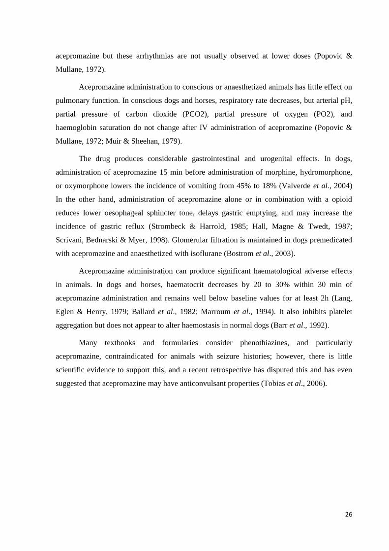
Acepromazine administration produces dramatic effects on the cardiovascular system
in both conscious and anaesthetized animals. In conscious dogs, stroke volume, cardiac
output, and mean arterial pressure decrease 20% to 25% after IV administration of
acepromazine (0,1 mg/kg), and mean arterial pressure is reduced for at least 2h (Coulter et al.,
1981; Stepien et al., 1995). Preanaesthetic administration of acepromazine (0.1mg/kg IM) also
decreases mean arterial pressure by 24% in dogs anaesthetized with isoflurane as shown in
fig.2 (Bostrom et al., 2003).
Figure 2. Effect of acepromazine (0.1 mg/kg intramuscularly) on mean arterial pressure in
dogs anaesthetized with isoflurane (2%) (Bostrom et al., 2003).
Heart rate does not change considerably in conscious dogs and horses administered
with acepromazine at the dose of 0.1 mg/kg through IV route (Muir & Sheehan, 1979; Coulter
et al., 1981). Increases in heart rate and sinus tachycardia can occur in some patients. At very
high doses (1mg/kg), bradycardia and sinoatrial (SA) block can occur in dogs given
Page 27
26
acepromazine but these arrhythmias are not usually observed at lower doses (Popovic &
Mullane, 1972).
Acepromazine administration to conscious or anaesthetized animals has little effect on
pulmonary function. In conscious dogs and horses, respiratory rate decreases, but arterial pH,
partial pressure of carbon dioxide (PCO2), partial pressure of oxygen (PO2), and
haemoglobin saturation do not change after IV administration of acepromazine (Popovic &
Mullane, 1972; Muir & Sheehan, 1979).
The drug produces considerable gastrointestinal and urogenital effects. In dogs,
administration of acepromazine 15 min before administration of morphine, hydromorphone,
or oxymorphone lowers the incidence of vomiting from 45% to 18% (Valverde et al., 2004)
In the other hand, administration of acepromazine alone or in combination with a opioid
reduces lower oesophageal sphincter tone, delays gastric emptying, and may increase the
incidence of gastric reflux (Strombeck & Harrold, 1985; Hall, Magne & Twedt, 1987;
Scrivani, Bednarski & Myer, 1998). Glomerular filtration is maintained in dogs premedicated
with acepromazine and anaesthetized with isoflurane (Bostrom et al., 2003).
Acepromazine administration can produce significant haematological adverse effects
in animals. In dogs and horses, haematocrit decreases by 20 to 30% within 30 min of
acepromazine administration and remains well below baseline values for at least 2h (Lang,
Eglen & Henry, 1979; Ballard et al., 1982; Marroum et al., 1994). It also inhibits platelet
aggregation but does not appear to alter haemostasis in normal dogs (Barr et al., 1992).
Many textbooks and formularies consider phenothiazines, and particularly
acepromazine, contraindicated for animals with seizure histories; however, there is little
scientific evidence to support this, and a recent retrospective has disputed this and has even
suggested that acepromazine may have anticonvulsant properties (Tobias et al., 2006).
Page 28
27
1.2.4. Dosage
Acepromazine dosage varies among the textbooks and formularies.
IM - doses for small dogs range from 0.05 to 0.2 mg/kg and those for larger dogs
range from 0.04 to 0.06 mg/kg (Lemke, 2007).
IM, IV - doses for dogs range from 0.01 to 0.1 mg/kg (Posner & Burns, 2009).
IV - dose 0.01 to 0.02 mg/kg administered slowly; IM - 0.01 to 0.05 mg/kg (BSAVA,
2011)
1.2.5. Contraindications
Acepromazine due to its hypotensive effect is relatively contraindicated in patients
with hypovolemia, shock (Plumb, 2008), trauma, cardiovascular disease (BSAVA, 2011),
severe dehydration or active bleeding (Posner & Burns, 2009). Acepromazine should be
avoided in animals below 3 months of age and animals with liver disease (BSAVA, 2011).
Paediatric patients are very susceptible to the hypotensive effect of acepromazine (BSAVA,
1999). Phenothiazines are relatively contraindicated in patients with tetanus or strychnine
intoxication due to effects on the extrapyramidal system. In dogs, acepromazine effects may
be individually variable and breed dependent. Dogs with the MDR1 mutations may develop a
more pronounced sedation that persists longer than normal. It could be prudent to reduce
initial doses 25% to determine the reaction of a patient identified or suspect of having the
mutation (Plumb, 2008).
Acepromazine should be used very cautiously as a restraining agent in aggressive dogs
as it may make the animal more prone to startle and react to noises or other sensory stimulus .
In geriatric patients, very low doses have been associated with prolonged effects of the drug
(Plumb, 2008).
1.2.6. Adverse effects
Hypotension due to acepromazine administration is well described and an important
consideration in therapy. Cardiovascular collapse secondary to bradycardia and hypotension
has been described in all major species. Dogs may be more sensitive to these effects than
other animals (Plumb, 2008). If profound hypotension occurs after acepromazine
administration, cardiovascular function should be supported by aggressive administration of
intravenous fluid. Treatment with vasopressors or catecholamines may be indicated if
Page 29
28
cardiovascular compromise is severe. Adrenaline is contraindicated in patients overdosed with
acepromazine. In the presence of α1-adrenergic blockade, adrenaline administration may lead
to unopposed β2-receptor activity. This effect augments vasodilatation and hypotension may
become more severe (BSAVA, 1999).
A resume of adverse effects are listed below: (BSAVA, 1999)
Hypotension
Hypothermia (by loss of thermoregulation)
α1- adrenergic blockade
Excessive vagal tone
Bradycardia
Organophosphate toxicity potentiation
Haematocrit decrease
Page 30
29
2. Acupuncture
Acupuncture is an important and integral part of traditional Chinese medicine (TCM)
for more than 2500 years (Wang, Kain & White, 2008; VanderPloeg & Yi, 2009; Chang,
2012). Among acupuncture therapies, the acupuncture induced analgesia effect has been
widely used to alleviate diverse pains (Zhao, 2008), this therapy was recognized as a
treatment for pain by World Health Organization (WHO) in 1996 (WHO, 2003) and by the
National Institutes of Health (NIH) in 1997 (NIH, 1998).
Acupuncture is a technique that makes use of hair thin needles introducing them at
specific locations in the body denominated acupuncture points or acupoints. Acupuncture
originates from the latin words acus meaning "needle" and pungere meaning "prick"
(Vanderloeg & Yi, 2009). This technique can induce multiple biological responses through
the activation of the neuronal system, and the therapeutic benefits of acupuncture treatments
have already been proven (Chien et al., 1998; Chao et al., 1999; Peng, 2002; Moazzami et al.,
2010; Chen et al., 2013). Acupuncture is widely used for pain management (Wang, Kain &
White, 2008) and even recognized for it by WHO and NIH. Nevertheless, the mechanism for
acupuncture-induced analgesia remains uncertain (Vanderloeg & Yi, 2009).
Traditional Chinese medicine is more a philosophy which focuses on balancing the
body. Its perspective is not based in anatomical, physiological, or biochemical evidence, thus
lacking on the evidence to be comprehended by western medicine practitioners (Wang, Kain
& White, 2008). In a research view, metaphors will continue to evade rigorous testing and
offer little evidence for the understanding of true acupuncture action mechanisms (Robinson,
2009). Numerous theoretical and experimental studies including brain imaging studies allude
to the nervous system being involved in transmission of acupuncture signals into target areas,
in which traditional nerve-reflex theory, gate control theory, and the neurotransmitter theory
are considered (Perlow, 1973; Dhond et al., 2008).
The traditional acupuncture mechanisms belonging to TCM are based on concepts
such as meridian, acupoint, Qi, yin-yang along others (Lindley & Cummings, 2006). Cheng,
(2014) suggested a possible interpretation of some concepts based on neurobiological and
fascia network models.
Page 31
30
2.1. Traditional Chinese Medicine approach
2.1.1. Meridians
Meridians are the major channels in the connective tissue fascia network in the body.
Acupuncture stimulation at sites along this network tends to produce stronger response than
other sites because of concentration of connective tissues and nerve endings (Cheng, 2014).
2.1.2. Acupuncture points
The traditional acupoints are the sites where acupuncture stimulation produces a
stronger response than neighbouring sites due to higher concentration of connective tissues
and nerve endings. These points were discovered through painstaking observation of ancient
researchers and clinicians. However, the difference between a traditional acupuncture point
and a non-acupuncture point lies on the intensity of response rather than structural
components (Cheng, 2014).
2.1.3. Qi
Qi refers to the signals within the channels that mediate the effects of acupuncture.
Although the nerve signal is the best understood one, other possible signals are the
propagation of mechanical force (Langevin & Yandow, 2002; Wang et al., 2007) and the
movement of paracrine-signalling molecules (Bai et al., 2011).
2.1.4. Yin-Yang
It refers to the maintenance of homeostasis true the balance between sympathetic and
parasympathetic branches from the autonomic nervous system (Cheng, 2014).
2.2. Scientific research
2.2.1. Neurophysiology approach
In the interest of closing the gap between TCM and western medicine for a better
understanding of how acupuncture works, a review of scientific literature focused on clinical
research, theories and clinical evidences will be presented.
It is important to state that there is no unified theory of acupuncture mechanism, but
rather multiple models and theories (Cheng, 2014)
Page 32
31
Early western theories were mainly dependent upon acupuncture ability to induce
neurologic signals along afferent nerves that in turn modulated spinal cord signal transmission
of pain (Wang, Kain & White, 2008; VanderPloeg & Yi, 2009).
In 1987, Pomeranz suggested a theory on acupuncture signalling based on his findings
in 1976, that naloxone administration is able to block analgesic effect of acupuncture in a
mouse. He suggested that acupuncture needle stimulation activates A-δ and C afferent fibers
in muscle which cause signal transmission to the spinal cord, resulting in local release of
endogenous opioids, dynorphin and enkephalin. Afferent pathways further propagate
signalling to the midbrain, triggering a sequence of excitatory and inhibitory mediators that
cause release of neurotransmitters, such as serotonin, dopamine, and norepinephrine onto the
spinal cord leading to pre and postsynaptic inhibition and suppression of pain transmission.
When this signal reaches the hypothalamus and pituitary, it triggers the release of
adrenocorticotropic hormone (ACTH) and endorphins (Pomeranz & Chiu, 1976). This theory
was strongly confirmed by several studies (Research Group of Acupuncture Anaesthesia,
1973,1974; Pomeranz & Chiu 1976; Lim et al., 1977; Clement-Jones et al., 1980; Cheng &
Pomeranz, 1981; Han & Terenius, 1982; Wu, 1995; Horrigan, 1996).
One of the main obstacles in the experimental study of the mechanism of acupuncture
anaesthesia is the difficulty in developing suitable animal models (Research Group of
Acupuncture Anaesthesia, 1974).
In 1973, Han et al. applied acupuncture stimulation to a rabbit for 30 minutes to
achieve an analgesic effect, then the cerebrospinal fluid (CSF) was removed and infused into
the lateral ventricle of an acupuncture-naive recipient rabbit. This resulted in an increased
pain threshold in the recipient rabbit. No increase was noted when saline or CSF from a
nonacupuncture control rabbit was infused into a acupuncture-naive recipient rabbit,
concluding that acupuncture induced analgesia was associated with the release of
neuromodulatory substances in the CSF (Research Group of Acupuncture Anaesthesia, 1974).
In the same year, Clement-Jones et al. demonstrated that 10 patients with chronic pain who
received electroacupuncture (EA), increased CSF levels for beta-endorphin significantly
(p<0.02) when compared to controls (Clement-Jones et al., 1980). Similarly, Sjolund and
Eriksson as well as Mayer, also showed increased levels of endorphins in CSF after EA
stimulation and the reversal of acupuncture analgesia by naloxone (Sjolund & Eriksson, 1977;
Mayer, 1977).
Page 33
32
Several subsequent studies supported the theory that acupuncture triggers the release
of endorphins and other endogenous opioids within the central nervous system (CNS) which
seems to be responsible for the analgesic properties of acupuncture (Peets & Pomeranz, 1978;
Lee & Beitz, 1993; Han, 2003)
Guo et al. in 1996 displayed that EA at low (2Hz) and high frequency (100Hz) caused
an increased c-fos expression in the arcuate nucleus of the rat. Low-frequency EA resulted in
a much higher c-fos expression when compared to high-frequency EA stimulation, and also
when compared to dry-needling in a control group. In situ hybridization studies showed that
low-frequency stimulation increased expression of messenger RNA (mRNA) for enkephalin
precursor protein, whereas, high-frequency stimulation caused a increased expression of
mRNA for dynorphin precursor protein (Guo et al., 1996 a,b). Even though it seems that
differential effects on c-fos expression occur by low and high-frequency EA stimulation, c-fos
expression can also be caused by non specific stimulations. Furthermore, mRNA levels may
not represent actual peptide levels (Wang, Kain & White, 2008).
The release of endogenous opioids in response to acupuncture is one of the leading
theories behind acupuncture's mechanism of action, and is denominated by the scientific
community as the "Neurohormonal Theory"(Wang, Kain & White, 2008).
Pan et al. observed if there was an overlap of central pathways in rats by comparing
noxious stimulation and electroacupuncture stimulation. They found that both induced a c-fos
expression in the anterior lobe of the pituitary gland, arcuate nucleus and in nearby
hypothalamic nuclei. A similar c-fos expression in the anterior lobe of the pituitary gland, was
shown in immobilization stress in awake rats. Although they all seem to have similar pituitary
gland activation, noxious simulation and EA involved different hypothalamic nuclei (Pan,
Castro-Lopes & Coimbra, 1996). In a follow up study, researchers found that fos-
immunoreactive cells activated by noxious stimulation and EA, co-localized with
adrenocorticotropic hormone (ACTH) or thyroid-stimulating hormone (TSH) and that
noxious stimulation and EA were associated with a similar increase in plasma ACTH and β-
endorphin. EA stimulation showed distinct increase c-fos expression at the hypothalamic
level in the mediobasal-nuclei and in the paraventricular nucleus. This researchers also
confirmed that a intact nociceptive primary afferent input is needed to transmit signal from
both, noxious stimulation and EA. They found that there was no activation of the
hypothalamic-pituitary-adrenocortical axis or increased plasma ACTH in rats, when afferent
Page 34
33
input was eliminated by sensory deafferentation. Thus, it seems to be an overlap in pain and
acupuncture central pathways (Pan, Castro-Lopes & Coimbra, 1997).
In 2010, Goldman et al. showed that stimulation at ST36, Zusanli acupoint in mice
induces purinergic receptor activation, which, in turn, inhibits pain transmission to the CNS
(Goldman et al., 2010). Purinergic receptor activation in the sciatic nerve increases the
synthesis of axonal growth-associated protein (GAP-43) in dorsal root ganglion (DRG)
sensory neurons (Arthur, Akassoglou & Insel, 2005). GAP-43 is the neural-specific protein
known to play a role in neuronal development and activity-dependent synaptic plasticity
(Aigner et al., 1995; Benowitz & Routtenberg, 1997). A recent study by Kim et al., 2012
investigating the effects of acupuncture stimulation on nervous system activation in mice and
rats, showed that acupuncture stimulation at ST36, Zusanli acupoint generates increased
neuronal response in terms of increased expression of GAP-43 and phospho Erk 1/2
activation in DRG sensory neurons and induction in c-fos expression in neurons of the dorsal
vagal compex (DVC) area. Sham acupuncture also causes a certain level of neuronal
response, although the extent of the responsiveness is weaker than that of acupuncture
stimulation.
2.2.2.Central Nervous System Imaging
Researchers, in an attempt to try to unfold the more complex aspects of the
acupuncture's effect, have focused in expression patterns in the CNS in order to examine
regions of the brain that are directly influenced by acupuncture stimulation (VanderPloeg &
Yi, 2009).
Nowadays technology advancement as allowed researchers to examine those patterns
through positron emission tomography (PET) and functional magnetic resonance imaging
(fMRI) (Wang, Kain & White, 2008).
2.2.2.1.PET
Using this technology, Hiesh et al., in 2001 studied the central activation caused by
acupuncture stimulation at LI4, Hegu acupoint, as well as stimulation by a non-classical
needling at a non-acupoint (sham acupuncture). The study consisted in PET imaging in 4
groups, one with needle insertion up to 1 cm at LI4 with EA stimulation at 4Hz, a second had
same EA stimulation at a sham-acupoint; a third had superficial insertion at LI4 with minimal
mechanic stimulation and a fourth had superficial insertion of the needle at a sham-acupoint.
Page 35
34
In the study they found that only the true acupuncture with the De-Qi sensation (EA 4Hz) at
LI4, significantly activated the hypothalamus, the periaqueductal gray and the insula. Minimal
stimulation at LI4 also activated both regions but in a lesser way. No activation was show in
both sham-acupuncture stimulations (Hiesh et al., 2001). "De Qi" sensation is frequently
described by patients as soreness, numbness, ache, fullness, or warm sensation that is
achieved during manipulation of acupuncture needles (Liu & Akira, 1994; Pomeranz, 1998).
Evidence of other regions involved in the acupuncture mechanism were discovered by
Biella et al. also in 2001, where they sequentially applied acupuncture and sham-acupuncture
at bilateral ST36, Zusanli and LU5, Chize acupoints during a pet scanning sequence. They
observed that acupuncture but not sham, activated the left anterior cingulum, superior frontal
gyrus, bilateral cerebellum and insula as well as the right medial and inferior frontal gyri
(Biella et al., 2001).
In 2005, Pariente et al., suggested that belief and anticipation could affect the
therapeutic outcome in humans. Using PET imaging, they reported that true and sham
acupuncture activated the right dorsolateral prefrontal cortex, anterior cingulated cortex and
the midbrain. They also found that only true acupuncture caused a greater activation in the
insula ipsilateral.
2.2.2.2. fMRI
Wu et al. established that traditional acupuncture stimulation caused activation in the
hypothalamus and nucleus accumbens but on the other hand caused deactivation in the rostral
part of the anterior cingulated cortex, the amygdale formation, and the hippocampus complex.
Superficial pricking caused activation at the primary somatosensory cortex, the thalamus and
the anterior cingulated cortex (Wu et al., 1999).
Hui et al. reported that acupuncture stimulation associated with De Qi sensation
deactivated the nucleus accumbens, hypothalamus, amygdale, hippocampus, para
hippocampus, ventral tegmental area, anterior cingular gyrus, caudate, putamen, temporal
lobe, and insula (Hui et al., 1999). In a follow up study, they found that subjects experiencing
De Qi sensation deactivated the frontal pole, ventromedial prefrontral cortex, cingulated
cortex, hypothalamus, reticular formation, and the cerebellar vermis. Furthermore, subjects
who experienced pain activated the anterior cingular gyrus, caudate, putamen, and the anterior
thalamus. Subjects that experienced both, pain and De Qi sensation had a mix of CNS
Page 36
35
responses with predominant activation of frontal pole, anterior, middle, and posterior
cingulate (Hui et al., 2000) reinforcing the hypothesis that pain and acupuncture have
overlapping central pathways. Other studies support this hypothesis. Wu et al.,2002 reported
that both true and sham EA at the GB34, Yanglingquan acupoint activated pain central
pathways on fMRI, but that only true EA activated hypothalamus, the primary somatosensory
cortex, and deactivated the rostral segment of the anterior cingulate cortex which led to their
insight that the hypothalamus-limbic system was modulated by EA (Wu et al.,2002). Zhan et
al., (2003) showed that true EA stimulation and not sham, can modify signals generated by
experimental cold pain stimulation. Only patients who received true EA reported a decrease in
pain (p<0.01) displaying a acupuncture-induced increase activity in the bilateral
somatosensory areas and medial prefrontal cortices and Brodmann area (BA32) and also, a
decrease in the contralateral primary somatosensory areas BA7 and BA24 (anterior cingulated
gyrus)(Zhang et al., 2003). Furthermore, in another study, Zang et al., showed that low
frequency (2Hz) and high frequency (100Hz) EA stimulation appear to be mediated by
different brain networks.
Napadow et al., 2005 study used fMRI to compare manual acupuncture with EA at 2
and 100 Hz and tactile control stimulation at ST36 in a group of volunteers. They reported
that EA produced more widespread fMRI signal changes than manual acupuncture, and the
latter, more than simple tactile stimulation. EA produced a considerable signal increase in the
anterior middle cingulate cortex, but only, low frequency EA (2Hz) activated the raphe area
(Napadow et al., 2005).
Essentially, neuroimaging studies in acupuncture are mere explorations of acupuncture
signal networking (Wang, Kain & White, 2008).
2.3. Acupuncture Point Specificity
TCM teaches that different acupoints produce different reactions and with the
common CNS patterns in the imaging studies involving acupuncture stimulation, a question is
born. Do different points produce different results?
Zhang et al., (2004) using fMRI, examined the different activation of CNS regions by
different TCM acupoints, ST36 Zusanli/ SP6 Sanyinjao acupoints or the GB34
Yanglingquan/BL57 Chengsan acupoint. They observed that both acupoints caused activation
of primary and secondary somatosensory areas, the insula, cerebellum, thalamus, and the
Page 37
36
putamen. Nevertheless, each acupoint had a specific pattern of activation in addition to the
common one. Zusanli/Sanyinjao acupoint activated the orbital frontal cortex along with
deactivation of the amygdala, while Yanglingquan/Chengsan acupoint activated the dorsal
thalamus with deactivation of the primary motor area and pre motor cortex (Zhang et al.,
2004).
2.4. Acupuncture Point Stimulation Methods
There are several ways to stimulate an acupoint according to TCM and each method
has particular characteristics and indications. Some methods are dry-needle,
hemoacupuncture, aquapuncture/pharmacopuncture, pneumoacupuncture, moxibuston,
electroacupuncture and implants (Xie's & Preast, 2007). For this work the focus will be in the
pharmacopuncture method.
2.4.1. Pharmacopuncture
It is the most common method of injection in acupuncture. It is the injection of fluids
and soluble products into acupoints. Sterile saline (aquapuncture), vitamin B12, homeopathic
remedies, patient's own blood, and local anaesthetics are the most commonly used in western
acupuncture practice. A variation using herbal medicines is used in China (Xie's & Preast,
2007).
According to Chinese authors who follow TCM, this technique combines the efficacy
of acupoint stimulation with the pharmacological effect of drugs, enhancing the mechanical
stimulus at acupoints and producing similar effects to those, with conventional drugs (Zhang,
Wu & Jiang, 2005; Zhu & Chen, 2005; Jin, Xu & Zheng, 2006) Pharmacopuncture uses
subclinical doses of drugs or small amounts of extracts of medicinal herbs (Kim & Kang,
2010). Pharmacopuncture's use of micro doses of drugs has proven useful in veterinary
practice. A subclinical dose of prostanglandine 0.5 mg/kg (1/10 of the conventional dose)
injected at the Bai-Hui acupoint induced luteolysis in mares as effectively as the conventional
dose (5 mg/kg) injected intramuscularly (Alvarenga et al., 1998) while significantly
decreasing the side effects associated with conventional dose (Nie et al., 2001). However, in a
follow up study, glucose or distilled water had no effect, suggesting a specific drug effect on
the acupoint (Luna et al., 1999). Using the same micro dose approach, Silva & Luna, (1999)
obtained the same weight gain in calves born from cows treated with 1/10 of the conventional
Page 38
37
dose of growth hormone (somatotrophin) when compared to a control group treated with the
conventional dose (500 mg) injected intramuscularly (Silva & Luna, 1999).
In TCM, the acupoint Yin Tang, has a sedative effect in humans and animals (Shoen,
2001; Ovechkin et al., 2003; Dos Santos et al., 2005). In 2002, Luna et al., study in dogs
showed that even though not statistically significant the injection of 0.01 mg/kg of
acepromazine (1/10 of the conventional dose) reduced in 32% the dose of thiopenthone
necessary for induction of anaesthesia when compared to the 51% reduction in animals treated
with the conventional dose of acepromazine (Luna et al., 2002).
The Ho-Hai acupoint, also named Chang Qiang (or GV1) has too a sedative
indication, specially for horses in alternative to the Yin Tang acupoint. Luna et al, (2008)
study in horses, demonstrated that injection of 1/10 of the conventional dose of acepromazine
(0.01 mg/kg) in GV1 caused sedation effect at 30 min. with long lasting effect (60 min.) when
compared with control groups. The conventional dose (0.1 mg/kg) given intramuscularly
caused a greater sedation but also a decrease in respiratory rate which supports the fact that
pharmacopuncture as the advantage to reduce undesirable side effects while producing similar
desirable effects (Luna et al., 2008).
Cassu et al., (2014) studied the sedative and clinical effects of pharmacopuncture with
xylazine in dogs. Pharmacopuncture with 1/10 of the conventional dose of xylazine (0.01
mg/kg) produced a clinically relevant sedative effect in dogs with the advantage of reducing
undesirable side effects associated with α2-agonists, including bradycardia, arrythmias, and
emesis shown in the group treated with the conventional dose (Cassu et al., 2013).
Santos Godoi et al., (2014) study in horses, compared the administration of 1/10 of the
conventional dose of acepromazine (0.01 mg/kg) at GV1 with acepromazine (0.1 mg/kg)
given intramuscularly to reduce stress-induced responses during transport. Pharmacopuncture
at GV1 reduced the stress induced response in the heart rate of horses, suggesting a possible
autonomic effect. However, it was not able to change other variables, such as transport
induced increases in cortisol, body temperature, and respiratory rate. On the other hand,
acepromazine given intramuscularly produced significant sedation and reduced the stress-
induced increase in respiratory rate during transportation without reducing the stress-induced
increase in cortisol.
Page 39
38
2.4.1.1. Mechanism
Pharmacopuncture mechanism is still very unclear even though this technique has
been widely used in clinical practice. Injection of the liquid substrates in the acupoints causes
local spatial configuration changes. Both, spatial configuration changes and the liquid
substrate characteristics stimulate the acupoint and activate the neuronal system (Chen et al.,
2014).
Chen et al., (2014) studied the number and distribution of neurons expressing c-fos
protein following the changes in spatial configuration caused by liquid substrate stimulation
in rats at the acupoint ST36 (Zusanli). These researchers used dry-needle and different liquid
substrates like, bee venom, normal saline, vitamin B1 and vitamin B12. The different liquid
substrates injected and the dry-needle insertion into ST36 induced c-fos expression in the L3,
L4 and L5 segments of the spinal cord. The group injected with bee-venom had more c-fos-
positive neurons in the dorsal horn than the other groups. All groups showed more c-fos
expression than the sham control group (dry needle in sham location). The study showed that
the distribution of c-fos-expressing neurons following stimulation by dry needle or the liquid
substrates was similar despite the difference in intensity and that it was primarily observed in
lamina II of the dorsal horn of the spinal cord (Chen et al., 2014).
Page 40
39
3. Objective
The study objective is to evaluate the level of sedation and the effects caused on
parameters like heart rate (HR), respiratory rate (RR), rectal temperature (RT) and arterial
blood pressure (ABP) resultant from administration of 1/10 of the acepromazine dose 0,05
mg/kg (0,005 mg/kg) in the Yin Tang acupoint and compare it to the administration of
acepromazine intramuscular dose of 0,05 mg/kg in dogs.
4. Hypothesis
Null Hypothesis H0 - Sedative effect is equal between the injection of 0,005 mg/kg of
acepromazine at the Yin Tang acupoint and the intramuscular injection of 0,05 mg/kg of
acepromazine.
Alternative Hypothesis H1 - Sedative effect is different between the injection of 0,005
mg/kg of acepromazine at the Yin Tang acupoint and the intramuscular injection of 0,05
mg/kg of acepromazine.
5. Materials and Methods
The study protocol was approved by the Animal Welfare Committee from Faculdade
de Medicina Veterinária belonging to Universidade Lusófona de Humanidades e Tecnologias
and all experiments were conducted in the University Veterinary Hospital.
5.1. Animals
In this study, six (6) dogs were used with ages between one (1) and five (5) years old.
The dogs were mixed breed, had a body condition of 5 in the scale of 9 from the body
condition score chart from WSAVA(2013) with weights ranging from 4 to 33 kg. In the study
sample, 4 dogs were female and 2 dogs were male. All dogs were evaluated before the study
in a pre anaesthetic exam making them suitable for surgical procedure: orquiectomy or
ovariohisterectomy, respectively. All animals had a signed consent by the owner. Detailed
information for each dog can be seen in table 2. No dog had history or symptoms of cardiac or
neurologic disease and all were in an normal state of hydration. No dog had alterations on
haematological and biochemical parameters. Dogs preformed a 12 hours fasting for solid food
and a 2 hours for liquids. Dogs (n=6) were divided in 2 groups, one group (G1) (n=3)
Page 41
40
receiving a 0.05 mg/kg intramuscular acepromazine injection at the longissimus dorsi muscle
(lombar region) and the other group (GPP) (n=3) receiving a 0.005 mg/kg acepromazine
injection in the Yin Tang acupoint. Dogs were assigned to a group randomly without
knowledge of the evaluator by the veterinarian who applied the injection.
Table 2. General information of all dogs included in the study.
Study ID Age Breed Sex Weight BCS**
P1 2 y. mbd* male 5.1kg 5/9
P2 1y. mbd female 32.1kg 5/9
P3 1.5 y. mbd female 5.4kg 5/9
P4 2y. mbd female 4.0kg 5/9
P5 2y. mbd female 4.6kg 5/9
P6 5y. mbd male 29.1kg 5/9
y., years; *mbd - Mixed breed dog; ** BCS - Body Condition Score.
5.2. Data Collection
For this study, each dog was submitted to a pre-assessment which would determine the
basal line for specific parameters like heart rate in beats per minute (bpm), respiratory rate in
cycles per minute (cpm), rectal temperature in celsius degrees (ºC) and arterial blood pressure
in millimetres of mercury (mm/Hg). Heart rate and respiratory rate were both measured using
a 3M ™ Littmann® pediatric or classic II stethoscope depending on the weight of the animal.
Temperature was measured using a Hartmann Thermoval® rapid flex thermometer. HR, RR
and RT measurements were repeated twice to minimize error. Arterial blood pressure was
measured with a Hifarmax™ VET HDO® (high definition oscillometry). Five (5)
measurements were made from the median artery in the right forelimb. A basal result was
calculated from the median of the data collected.
Sedation score was established using a cumulative score chart (table 3) from Vainio et
al., 1989; Kuusela et al. 2001. Score minimum value is 0 and maximum value is 22 being the
dog sedation score calculated from the sum from each assessment.
Page 42
41
Table 3. Evaluation of the level of sedation in terms of a cumulative score in dogs (adapted from Vainio et al., 1989; Kuusela et al. 2001).
Evaluation of the level of sedation in terms of a cumulative score in dogs
Assessment Score and definition
Posture 0 - Standing position, normal proprioception (animal walks without ataxia) 1 - Animal remains in sternal or lateral position but is able to stand when verbally stimulated 2 - Remains in sternal recumbency 3 - Lateral recumbency, eventually move or lifts its head 4 - Lateral recumbency, if not verbally stimulated does not move or lifts is head.
Eyelid reflex 0 - Strong lateral and medial eyelid reflexes* 1 - Lateral and medial eyelid reflexes presented but reduced 2 - Lateral eyelid reflex absent and medial eyelid reflex present 3 - Lateral and medial eyelid reflexes absent
Eye globe position 0 - Eye centrally positioned 1 - Partial rotation of the eye globe 2 - Full rotation of the eye globe
Relaxation of the tongue and mandible
0 - Normal tone of the mandible and tongue 1 - Reduced tone of the mandible and tongue, allowing opening of the mandible with little difficulty; tongue can be exposed with some difficulty and is readily retracted after being released 2 - Reduced tone of the mandible, tongue can be easily exposed but is readily retracted after being released. 3 - Mouth can be easily opened with jaw tone markedly reduced, tongue can be easily exposed, but animal retracts the tongue a few seconds after being released 4 - Mandible and tongue fully relaxed, tongue can be exposed and its is not retracted after being released
Response to sound (clapping)
0 - Alert attitude, readily reacts (looks, lifts head) to the stimulus 1 - Reduced reaction (discrete movement, lifting of the head), however the animal appears sedated 2 - No reaction or movement
Resistance to physical restraint in lateral
recumbency
0 - Animal resists; readily returns to standing position or sternal recumbency after being released 1 - Offers little resistance, but readily returns to standing position or sternal recumbency after being released 2 - Does not offer resistance, but eventually moves or lifts its head and returns to sternal recumbency 3 - Remains in lateral recumbency, does not offer resistance
General appearance 0 - Alert, normal consciousness 1 - Animal lightly sedated, promptly reacts or moves in response to environmental stimulation 2 - Animal moderately sedated, eventually reacts to environmental stimulation 3 - Animal appears to be moderately to deeply sedated, reduced reaction to environmental stimulation 4 - Animal appears deeply sedated, does not react to environmental stimulation
Page 43
42
Sedation Score was established 30 minutes after injection due to the peak effect of
acepromazine. At this time, all others parameters were also collected for comparison with the
basal line, respecting the same collecting methods for each parameter.
All parameters and sedation score were taken by the same evaluator at both, t<0 and
t=30min. with no knowledge of the applied method. There was no sharing of data between the
evaluator and the veterinarian who applied the injections until the end of the study.
5.3.Timeline
Timeline can be seen in fig.3. Dogs were examined to set the parameters basal line
right before the injection. The injection of acepromazine was given intramuscularly at the
longissimus dorsi muscle or in the Yin Tang acupoint at t=0, at respective doses. It was given
a waiting period of 30 minutes to let the drug take effect. At t=30min. score of sedation was
taken using the chart provided in fig.2, after which, the remaining parameters were taken to
compare with the basal line.
Timeline of the study
Figure 3. Timeline of the study. HR, heart rate; RR, respiratory rate; RT, rectal temperature; ABP, arterial
blood pressure.
5.4. Intramuscular Injection
The injection was given in the longissimus dorsi muscle at the lombar region. The
dose of acepromazine was 0.05 mg/kg. The solution was taken from the available Calmivet™
Injectable solution with 5mg/ml of acepromazine maleate. Injection was made with a 22G
hypodermic needle by a licensed veterinarian. No reactions to the injection were reported.
Setting basal line for:
HR, RR, RT, ABP.
Setting basal line for:
HR, RR, RT, ABP.
Injection of acepromazine
at the intramuscular site or
at the Yin Tang acupoint.
Injection of acepromazine
at the intramuscular site or
at the ying tang acupoint.
Collecting data for:
HR, RR, RT, ABP,
Sedation score.
Collecting data for:
HR, RR, RT, ABP,
Sedation score.
30 min. 0 min. before injection
Page 44
43
5.5. Pharmacopuncture Injection
The injection of 0.005 mg/kg of acepromazine in the Yin Tang acupoint was made by
a veterinarian certified by the IVAS (International Veterinary Acupuncture Society) for the
practice of acupuncture. This was the same veterinarian for both groups. The injection was
made with a 25G hypodermic needle. Preparation of the dilution from the available
Calmivet™ Injectable solution with 5mg/ml of acepromazine maleate was made before
application in the Yin Tang acupoint. The dilution was made with sterile solution of sodium
chloride at 0,9% concentration. No reactions to the injection were reported.
Yin Tang acupoint is located in a depression on the dorsal midline between the
eyebrows on the dog (fig.4) (Cassu et al., 2014).
Figure 4. Location of the Yin Tang acupoint in dogs. Drawing by Tomás Pinto de Sousa, 2015.
5.6. Data analysis
Statistical analysis was made using the IBM® SPSS
® statistics version 23 software.
The data analysis was made using a non-parametrical Kruskal-Wallis independence test to test
the hypothesis. Application of the non-parametrical Spearman test was also used to check
correlation between the data. Tables and figures were created using Microsoft Office Excel
2007® and Microsoft Office Power Point 2007®.
Page 45
44
6. Results
In the end of the study the sedation method was revealed to the evaluator for each
animal as shown in table 4.
Table 4. General information for all dogs included in the study.
Study ID Age Breed Sex Weight BCS**
Sedation
method
P1 2 y. mbd* male 5.1kg 5/9 IM
(1)
P2 1y. mbd* female 32.1kg 5/9 PP
(2)
P3 1.5 y. mbd* female 5.4kg 5/9 PP
(2)
P4 2y. mbd* female 4.0kg 5/9 IM
(1)
P5 2y. mbd* female 4.6kg 5/9 PP
(2)
P6 5y. mbd* male 29.1kg 5/9 IM
(1)
y., years; *mbd - Mixed breed dog;
** BCS - Body Condition Score; (1), intramuscular injection group; (2),
pharmacopuncture group.
Sedation score results are shown for each animal in its respective group in table 5.
Baseline parameters and its after sedation results are shown for the pharmacopuncture
injection at Yin Tang acupoint group in table 6. and for the intramuscular injection group at
table 7, respectively. Study ID's order in each group is random because animals were only
assigned to each group after all data had been collected from the entire population sample.
Table 5. Individual animal sedation score and respective applied sedation method.
Sedation method Study ID Sedation score*
Intramuscular injection (G1) P1 14/22
P4 1/22
P6 7/22
Pharmacopuncture at Yin Tang (GPP) P2 2/22
P3 4/22
P5 1/22
ID, animal identification; G1, intramuscular injection group; GPP, Yin Tang acupoint injection group
Page 46
45
Table 6. Parameters collected in the pharmacopuncture group.
Pharmacopuncture GPP
P2 BL* P2 AS** P3 BL P3 AS P5 BL P5 AS
HR 100 100 124 120 165 111 bpm
RR 48 26 58 28 16 16 cpm
RT 39,4 38,8 39 38,5 39,7 38,6 ºC
mSP 216,4 147,4 185,8 148,6 139,8 129,8 mm/Hg
mDP 134,6 77,8 92 69 79,6 79,6 mm/Hg
HR, heart rate; RR, respiratory rate; RT, rectal temperature; mSP, mean systolic pressure; mDP, mean diastolic
pressure; *, baseline; **, after sedation; GPP, Yin Tang acupoint injection group.
In the GPP group in table 6, maximum values after sedation for HR, RR, RT, mSP,
mDP parameters were 120 bpm, 28 cpm, 38,8ºC, 148,6 mm/Hg and 79,6 mm/Hg,
respectively. Minimum values after sedation for HR, RR, RT, mSP, mDP parameters were
100 bpm, 16 cpm, 38,5ºC, 129,8 mm/Hg and 69 mm/Hg, respectively.
Table 7. Parameters collected in the intramuscular injection group.
Intramuscular injection G1
P1 BL* P1 AS** P4 BL P4 AS P6 BL P6 AS
HR 80 84 150 129 110 104 bpm
RR 28 16 24 18 40 16 cpm
RT 38,9 38,4 40 38,6 39,5 38,4 ºC
mSP 120,4 127,2 140,2 135 155,6 106 mm/Hg
mDP 64 71,2 83,2 75,8 72 45,8 mm/Hg
HR, heart rate; RR, respiratory rate; RT, rectal temperature; mSP, mean systolic pressure; mDP, mean diastolic
pressure; *, baseline; **, after sedation; G1, intramuscular injection group.
In the G1 group in table 7, maximum values after sedation for HR, RR, RT, mSP,
mDP parameters were 129 bpm, 18 cpm, 38,6ºC, 135 mm/Hg and 75,8 mm/Hg, respectively.
Minimum values after sedation for HR, RR, RT, mSP, mDP parameters were 84 bpm, 16
cpm, 38,4ºC, 106 mm/Hg and 45,8 mm/Hg, respectively.
A graphical overview on the evolution of the measured parameters, heart rate,
respiratory rate, rectal temperature and arterial blood pressure is shown in fig.5 for the
acepromazine intramuscular injection group (G1) and for the Yin Tang pharmacopuncture
injection group (GPP) in fig.6, respectively.
Page 47
46
Intramuscular injection Group (G1)
Figure 5. Parameter changes in heart rate(HR), respiratory rate(RR), rectal temperature(RT)
and arterial blood pressure(ABP) of the animals in the acepromazine intramuscular injection
group (G1).
0 20 40 60 80
100 120 140 160
P1 P4 P6
bea
ts p
er m
inu
te
Heart rate variation
Baseline
After sedation
0 5
10 15 20 25 30 35 40 45
P1 P4 P6
cycl
es
pe
r m
inu
te
Respiratory rate variation
Baseline
After sedation
37,5
38
38,5
39
39,5
40
40,5
P1 P4 P6
cels
ius
de
gre
e
Rectal temperature variation
Baseline
After sedation
0
20
40
60
80
100
120
140
160
180
P1 P4 P6
mm
/Hg
Arterial blood pressure variation
Systolic pressure baseline
Systolic pressure after sedation
Diastolic pressure baseline
Diastolic pressure after sedation
Page 48
47
Yin Tang Pharmacopuncture Group (GPP)
Figure 6. Parameter changes in heart rate(HR), respiratory rate(RR), rectal temperature(RT)
and arterial blood pressure(ABP) of the animals in the acepromazine pharmacopuncture group
(GPP).
0 20 40 60 80
100 120 140 160 180
P2 P3 P5
bea
ts p
er m
inu
te
Heart rate variation
Baseline
After sedation
0 5
10 15 20 25 30 35 40 45 50 55 60 65
P2 P3 P5
cycl
es
pe
r m
inu
te
Respiratory rate variation
Baseline
After sedation
37,5
38
38,5
39
39,5
40
P2 P3 P5
Ce
lsiu
s d
egr
ee
Rectal temperature variation
Baseline
After sedation
0 20 40 60 80
100 120 140 160 180 200 220 240
P2 P3 P5
mm
/Hg
Arterial blood pressure variation
Systolic pressure
baseline
Systolic pressure
after sedation
Diastolic pressure
baseline
Diastolic pressure
after sedation
Page 49
48
In the study, statistically, there is no significant differences (p-value < 0.05) between
the Yin Tang sedation method group (GPP) and the intramuscular injection sedation method
(G1). However, the results show an overall lower sedation score in the pharmacopuncture
group (GPP) when compared to the G1 group as shown in table 5.
Statistical analysis showed no significant differences (p-value < 0.05) between the Yin
Tang sedation method group (GPP) and the intramuscular injection sedation method (G1) and
the after sedation measurements of heart rate, respiratory rate, rectal temperature and arterial
blood pressure.
Page 50
49
7. Discussion
The use of routine animals from the hospital due to animal welfare restrictions caused
the sample to be heterogeneous and may not contribute for the comparison of results between
groups. This study only had permission to use healthy animals undergoing selective surgery
so they could benefit from the use of the techniques applied in the study as a mean of
premedication. Also, restrictions did not allow for a control group with a placebo effect so
such group was not included in this study but other studies with similar techniques used
control groups with saline injection at false acupuncture points and observed that this groups
had no effect or a reduced effect when compared to injection at a true acupuncture point with
the respective drug used in the study (Luna et al., 2002; 2008; Godoi et al, 2013).
7.1. Sedation Score
Sedation score and the other parameters were only measured once in the timeline
because manipulation would most definitely alter the subsequent results after the first
measurement. The decision was to only take measurements at 30 minutes after injection
which is the time of the peak effect described for acepromazine by Cornick & Harstfield,
1992; Smith et al., 2001. Due to the complexity of the sedation score chart in table 3 a
specific order for assessment was considered to avoid excitement that could alter the overall
sedation score. The sedation score table shown in table 3 was applied in this study due to its
ability to assess objectively different characteristics also used to monitor animals during
anaesthesia. It allowed to effectively quantify consciousness, environmental awareness and
motor muscle control. Other tools could be used like the Ramsay sedation scale, Riker
sedation and agitation scale and Richmond agitation-sedation scale which are easier to use
and rely more on observation than manipulation of the patient which can be good when
applying it to daily routine. Nevertheless, they possess a more subjective overview over
patients and have a smaller range that makes it hard to notice slight changes in sedation
behaviour. In veterinary practice it is important to characterize patients control over motor
behaviour because it implies safety for the handlers during clinical procedures. This and the
ability to detect minor changes during sedation in patients, were decisive when choosing the
sedation scale to apply in this study. Nevertheless, this scale does not evaluate agitation of
patients representing a considerable downside when using this tool.
Page 51
50
Statistically, there was no significant difference between both methods of sedation
thus, one, could only speculate that the null hypothesis H0 that both are similar in efficacy is
true, however, it was watched, clinically, a relatively noteworthy difference, whereas the
injection of acepromazine at the Yin Tang acupoint created an overall lower sedation score in
its group than the acepromazine intramuscular injection group. This can be caused by a very
small sample and due to the high variation between sedation scores, especially in the
intramuscular injection G1 group, where the minimum value is 1 and the maximum value is
14 in a total of 22 points. This variation can also be an effect of the unreliability of
acepromazine when used alone for sedation purposes as described in BSAVA Small Animal
Formulary 7th edition (2011).
This study used acepromazine because it is the most widely used sedative in veterinary
medicine (Lemke, 2007) representing a good primary target for research in
pharmacopuncture, since results from this study may be helpful for a wider range of
practitioners worldwide. A previous study from Cassu et al., 2014 using 0.01mg/kg of
xylazine injected in the Yin Tang acupoint induced clinically relevant sedative effects in
dogs, reducing the undesirable side effects associated with α2-agonists. Nonetheless, other
sedatives are more recent (e.g. α2-agonists; benzodiazepines) deserving attention in future
researches with similar methods of pharmacopuncture for sedation in animals. Recent
sedatives may show more potential to sedate animals considering their specific drug
properties and their interaction with pharmacopuncture mechanism.
The light sedation observed in GPP group may be a result of an ineffective dose of
acepromazine. This study used 1/10 of the conventional dosage 0.05 mg/kg of acepromazine
in the attempt to produce a similar sedation effect. A previous study from Luna et al., 2002
used an acepromazine dose of 0.01 mg/kg in the Yin Tang acupoint in order to obtain a similar
effect in thiopental reduction for induction in dogs than that of 0.1 mg/kg of acepromazine
administered subcutaneously. Results demonstrated also a lighter but clinically relevant effect
suggesting that, in similarity with this study, future studies should be made to determine the
ideal dose of acepromazine injected in the Yin Tang acupoint to achieve a reliable level of
sedation. Any reduction in the use of drugs to obtain similar effects than that of conventional
therapies, contributes to a safer and healthier process for the patients justifying future research
in the area.
Page 52
51
7.2. Measured Parameters
Statistically, there was no significant difference (p-value < 0.05) in variations
of heart rate, respiratory rate, rectal temperature and arterial blood pressure
measurements between the G1 and GPP group. This statistical view is compromised
by the size of the sample so a clinical perspective over the data must be made.
7.2.1. Heart Rate
Clinically, heart rate variations don´t show a visible relationship between each
other inside the group subjects as shown in table 8. This may be that acepromazine has
little effect over heart rate changes and the changes observed are due to another
process altering normal nervous, humoral and local control of the cardiovascular
function.
The lower value after sedation presented by each group is, 84 bpm in G1 and
100 bpm in GPP, however the lowest value in G1 is actually an increase in heart rate
of P1 subject when compared with its baseline value of 80 bpm, demonstrating in this
case no heart rate depression effect as well as the P2 subject in GPP which had the
value of 100 bpm in baseline and after sedation measurements. After sedation subjects
on GPP group maintained heart rate values between 120 and 100 bpm. G1 group
maintained heart rate values between 129 and 84 bpm.
Table 8. Heart rate variation between baseline and after sedation values.
Heart rate variation
GPP G1
P2 0% P1 ↑5%
P3 ↓3,2% P4 ↓14%
P5 ↓32% P6 ↓5,4%
↓, decrease; ↑, increase; G1, intramuscular inejction group; GPP, Yin Tang acupoint injection group.
The biggest variation in heart rate was shown in GPP by subject P5 lowering
his baseline value of 165 bpm to an after sedation value of 111 bpm demonstrating a
decrease of 32% in heart rate. In G1 the biggest variation in heart rate was shown by
P4 with a decrease of 14% in heart rate. P5 from GPP and P4 from G1 obtained a
sedation score of 1/22 showing that they were slightly sedated and suggest that this big
Page 53
52
variations may be due to higher values prior to sedation developed by the presence of
environmental stressors, which led to their decrease when the subjects experienced
sedation and consequential relaxation to the environment. G1 and GPP heart rate
values don't change considerably which was expected because a similar phenomena is
also described in previous studies in dogs and horses with the use of acepromazine at
the dose of 0.1 mg/kg through IV route (Muir & Sheehan, 1979; Coulter et al., 1981).
Although, there seems to be no evidential correlation, the overall response to both
methods was a decrease in heart rate that respected the physiological range for dogs.
The decrease in heart rate can be the outcome of the sedation and decrease of
catecholamines in circulation as described by Posner and Burns, 2009.
7.2.2. Respiratory Rate
Respiratory rate variation between G1 and GPP is similar if we take in
consideration the decrease percentage in respiratory rate for each subject as shown in
table 9. When looking to minimum value after sedation both groups reached 16 cpm,
although this result in the GPP group is represented by subject P5 who had no
respiratory rate variation at all. P5 baseline value represents a calm respiratory rate and
since the sedation score was 1/22 this may explain for the lack of variation in this
parameter.
Table 9. Respiratory rate variation between baseline and after sedation values.
Respiratory rate variation
GPP G1
P2 ↓45,8% P1 ↓42,8%
P3 ↓51% P4 ↓25%
P5 0% P6 ↓60%
↓, decrease; G1, intramuscular injection group; GPP, Yin Tang acupoint injection group.
Clinically, this variations show a similarity of effect in decreasing the
respiratory rate between 1/10 of the acepromazine dose at the acupoint Yin Tang and
that of the 0.05 mg/kg acepromazine dose injected intramuscularly. This study acts in
accordance with previous studies (Popovic & Mullane, 1972) that show that
acepromazine has little effect on pulmonary function but seems to decrease respiratory
rate in dogs.
Page 54
53
7.2.3. Rectal Temperature
Rectal temperature values after sedation in G1 varied from 0,5ºC to as further
as 1,4 ºC reaching a minimum value of 38,4 ºC. In GPP, variation went from 0,5 ºC to
1,1 ºC reaching a minimum value of 38,5 ªC. Comparison of G1 and GPP values in
table 10 show that both groups had a similar effect on thermoregulation. One of the
adverse effects described for acepromazine is hypothermia and the loss of
thermoregulation.
Table 10. Rectal temperature variation between baseline and after sedation values.
Rectal temperature variation
GPP G1
P2 ↓1.5% P1 ↓1.2%
P3 ↓1.3% P4 ↓3.5%
P5 ↓2.8% P6 ↓2.8%
↓, decrease; ↑, increase; G1, intramuscular injection group; GPP, Yin Tang acupoint injection group.
Observations show that the groups had similar rectal temperatures and that no
subject at the time of data collection was below physiological rectal temperature
values described for dogs in BSAVA Manual of Small Animal Anaesthesia and
Analgesia, 1999. It would be interesting to collect more measurements over time
during the sedation period to analyse if the thermoregulation behaviour is similar in
both sedation methods. In G1, subject P4 showed a baseline value of 40 ºC compatible
with stress induced hyperthermia, having it dropped after sedation to a value of 38,6
ºC. The same behaviour was watched in GPP, where P5 had a baseline value of 39,7
ºC having it dropped after sedation to 38,6 ºC. Although P5 had a difference of 0,3 ºC
from P4, it also showed stress induced hyperthermia, which was reduced after sedation
to the same value than P4 (Olivier et al., 2003). Clinically, this suggests that the
injection of 1/10 of the dose of acepromazine at the Yin Tang acupoint may have the
ability to bring animals from a stress induced state to a more tranquilized state through
sedation.
Page 55
54
7.2.4. Arterial Blood Pressure
Baseline arterial blood pressure values are lower in G1 than in GPP but almost
all subjects showed a slight hypertension which may be attributed to the fact that
animals were in fact in a strange stressful environment. Interestingly, both methods
caused a similar decrease in arterial blood pressure. In G1 the minimum values for
systolic and diastolic pressure belong to P6 which went below recommend values for
awaken dogs having a moderate decrease in diastolic pressure. This accounts for the
only hypotensive phenomenon observed in this study. Considering this, the
intramuscular injection of acepromazine at the dose 0.05 mg/kg should be done with
caution always controlling arterial blood pressure. The injection of acepromazine at
the dose of 0.005 mg/kg at the Yin Tang acupoint had no hypotensive phenomenon
and caused a similar decrease in arterial blood pressure that never went below the
expected physiological range for dogs. This may suggest that this method is safe to
use, however more intensive study on the effects of the method should be done prior to
its use in routine practice.
Table 11. Arterial blood pressure variation between baseline and after sedation values.
Arterial blood pressure variation
GPP Systolic
pressure
Diastolic
pressure G1
Systolic
pressure
Diastolic
pressure
P2 ↓31,8% ↓42,1% P1 ↑5,64% ↑11,25%
P3 ↓20% ↓25% P4 ↓3,7% ↓8,9%
P5 ↓7,1% 0% P6 ↓31% ↓36,4%
↓, decrease; ↑, increase; G1, intramuscular injection group; GPP, Yin Tang acupoint injection group.
7.3. Synopsis
Even though anecdotal the comparison between the two groups, maximum and
minimum values after sedation of HR, RR, RT, mSP, mDP parameters which received
different sedation methods with the same pharmaceutical drug, acepromazine, G1 and GPP
show a decrease of the values to more acceptable physiological values and that they seem to
produce a similar effect on the parameters measured in this study, suggesting that maybe 1/10
of the dose of acepromazine administered in the acupoint Yin Tang can produce a similar
effect than that of 0.05 mg/kg acepromazine dose injected intramuscularly. This effect of
Page 56
55
pharmacopuncture is described and showed in earlier studies as Alvarenga et al., 1998; Silva
& Luna, 1999; Nie et al., 2001; Luna et al., 2008; Cassu et al., 2014.
In this study, the injection of 1/10 of the dose of acepromazine administered in the
acupoint Yin Tang successfully caused light sedation in GPP animals, shaping their behaviour
to a more relaxed state. This can be shown by the variations shown by each animal for HR,
RR, RT and ABP parameters.
GPP group did not show any of the described (BSAVA, 1999) undesirable side effects
for acepromazine like hypotension, bradycardia and hypothermia. In view of this, and as a
result of a small sample, we can only speculate that this method is safe to apply, although,
clinically the sedation itself is unreliable as in G1, but shows signs of tranquilization of the
animals returning them to more physiological state by shaping their behavioural response.
Acupuncture and by consequence pharmacopuncture mechanism of action remains
very unclear, nevertheless, and considering the observations in this study a theory will be
discussed. Pharmacopuncture combines the effects of acupuncture and drugs. In this case, the
drug used was acepromazine which causes inhibition of central dopaminergic receptors (D2),
that are responsible for shaping the behavioural response, thus contributing for the sedation
(Lemke, 2007). The Yin Tang acupoint is indicated for sedation, but the mechanism that leads
to sedation is vague.
Peripherally, phenothiazines block norepinephrine at alpha-adrenergic receptors
(Posner & Burns, 2009). Experimental studies in rats, showed decreased levels of
norepinephrine in the brain after EA (Han et al., 1979; Wang, Jiang & Can, 1994; Zhu et al.,
1997). This shows that both the drug and the effect caused by acupuncture will act at α-
adrenergic receptors. Phenothiazines have high affinity for α1-adrenergic receptors as shown
in table 1. This α1-adrenergic receptors in spinal dorsal horn facilitate nocioceptive signalling
whereas α2-adrenergic receptors suppress it (Millan, 2002). This may explain for the fact that
acepromazine has no analgesic effect. In the other hand, acupuncture causes the release of
endogenous opioids (e.g. β-endorphin) suggesting a possible added analgesic effect to the
pharmacopuncture method that would be interesting to prove in future studies by comparing
response to noxious stimuli between G1 and GPP. The alterations shown in the parameters by
pharmacopuncture suggest parasympathetic activity and inhibition of sympathetic activity
similar to the intramuscular injection of 0.05 mg/kg of acepromazine, thus proposing similar
pathways. This show us that both methods alter the autonomous nervous system.
Page 57
56
Acupuncture causes the release of β-endorphin a pituitary peptide (Grimm & Wagner, 2007)
showing hypothalamic-pituitary activation. Acepromazine also has hypothalamic-pituitary
activity by blocking dopamine receptors which cause release of prolactin in pituitary gland.
The adrenal gland medulla produces cathecolamines, epinephrine, norepinephrine, dopamine
(Squires, 2003) suggesting that it also plays a role in the action of both methods.
Summarizing, in pharmacopuncture mechanotransduction at the acupoint causes signalling
trough afferent pathways to the spinal dorsal horn, which in turn will cause central nervous
system activation at hypothalamic- pituitary-axis. This in turn will create systemic effects by
altering the autonomous nervous system influencing the adrenal gland and its hormones. The
specific mechanism is unclear but clinical observations suggest that pharmacopuncture
mechanism works by stimulation of specific acupuncture points that cause local effects and
specific systemic effects which work in synergy with drugs, contributing to their systemic
and/or specific local effects.
The injection of acepromazine at the Yin Tang acupoint may prove useful in daily
clinical practice if further research is done in providing a better understanding and safety
involving the method. In the eyes of this study, the method shows promising by causing
similar effects and reducing the costs of the drug to both the clinician and the owner of the
dog by 90% when compared with the intramuscular injection of 0.05 mg/kg of acepromazine,
making it possible for the clinician to reduce possible adverse effects and for the dog to
quickly metabolize and excrete the small amount of drug received. This may allow for the
drug to be used more often to tranquilize dogs that show excitatory behaviours and also
contribute to the economic inflow in the clinic. If a clinician was to use this method in a place
or circumstance where the animal could not be well monitored, this method may prove itself
to be safer to achieve tranquilization in dogs than the intramuscular injection of 0.05 mg/kg of
acepromazine according to observations in this study.
Page 58
57
8. Conclusion
This study will only serve as a preliminary approach to further studies in the areas of
pharmacopuncture and induced sedation through injection of acepromazine in the Yin Tang
acupuncture point since all conclusions are mainly mere speculations resultant from a
deficient animal model available at the time. Even so and trusting that not all is a statistical
truth, speculations can be made from a clinical point of view.
A light sedation effect was observed in the animals in this study using the Yin Tang
acupoint method and consequently, heart rate, respiratory rate, rectal temperature, arterial
blood pressure and behavioural changes occurred suggesting that, although, not statistically
proved, this method may be used to sedate dogs and return them to a more relaxed state.
However one should be very careful using this method if the aim is to control the motor
behaviour in more agitated and aggressive animals since acepromazine use in both methods
showed to be unreliable and unfitting for the job. Practitioners should consider maybe only
using the acepromazine injection at the Yin Tang acupoint method to tranquilize dogs which
show signs of agitation or stress response in a clinical environment since it is a relatively safe
method to use and a very small amount of drug for the dog metabolism to process and
eliminate.
Limitations encountered during this study, severely damaged the sample and
consequent interpretation of the results. During the time span of 7 months it was hard to
obtain dogs with the adequate criteria and a healthy condition prior to selective surgery.
Furthermore, it was difficult to conciliate the patients with the availability of the specialized
technician to apply the method. Another limitation, like the selection of the adequate dose was
unfortunate and came from the principle that 1/10 of the dose should cause similar effect than
that of the conventional dose. In this study, both methods caused an inadequate effect, so for
future studies, maybe the doses or the proportion of such doses should be reconsidered.
Further extensive studies with more controlled samples are required to evaluate the
future benefits and consequent side effects of this method but from this study perspective the
method may show itself promising to tranquilize not only healthy dogs but elderly dogs
and/or dogs that are somehow debilitated and/or have a concurrent disease which make them
a higher risk patient for normal sedation protocols. The pharmacopuncture effect of producing
similar effects using only 1/10 of conventional doses remains promising but nevertheless, it
should be confirmed with extensive critical scientific research at all levels of investigation in
Page 59
58
many areas of veterinary practice so it can be brought to the western medical point of view
and allow western practitioners to use it in routine practice.
Page 60
59
Bibliografia
Aigner, L.; Arber, S.; Kapfhammer, J.P.; Laux, T.; Schneider, C.; Botteri, F.; et al.
(1995) Overexpression of the neural growth- associated protein GAP-43 induces nerve
sprouting in the adult nervous system of transgenic mice. Cell; 83:269e278.
Alvarenga, M.A.; Ferreira, J.E.P.; Meira, C.; Luna, S.P.L.; Burns, P.J. (1998)
Induction of luteolysis in mares using a micro-dose of prostaglandin in the sacral lumbar
space (Bai Hui Acupoint). Equine Vet Sci; 18(3) 167-168.
Arthur, D.B.; Akassoglou, K.; Insel, P.A. (2005) P2Y2 receptor activates nerve growth
factor/TrkA signaling to enhance neuronal differentiation. Proc Natl Acad Sci
USA;102:19138e19143.
Bai, Y.; Wang, J.; Wu, J.P.; Dai, J.X.; Sha, O.; Tai Wai Yew, D.; et al. (2011) Review
of evidence suggesting that the fascia network could be the anatomical basis for acupoints and
meridians in the human body. Evid Based Complement Alternat Med.; 2011:260510.
Ballard, S.; Schults, T.; Knownacki, A.; Blake, J.; Tobin, T. (1982) The
pharmacokinetics, pharmacological responses and behavioural effects of acepromazine in the
horse. J Vet Pharmacol Ther, Mar; 5(1): 21-31.
Barr, S.C.; Ludders, J.W.; Looney, A.L.; et al. (1992) Platelet aggregation in dogs
after sedation with acepromazine and atropine and during subsequent general anesthesia and
surgery. Am J Vet Res; 53: 2067-2070.
Benowitz, L.I.; Routtenberg, A. (1997) GAP-43: an intrinsic determinant of neuronal
development and plasticity. Trends Neurosci; 20:84e91.
Biella, G.; Sotgiu, M.L.; Pellegata, G.; Paulesu, E.; Castiglioni, I.; Fazio, F. (2001)
Acupuncture produces central activations in pain regions. Neuroimage; 14:60–6.
Bostrom, I.; Nyman, G.; Kampa, N.; et al. (2003) Effects of acepromazine on renal
function in anesthetized dogs. Am J Vet Res; 64:590-598.
Cassu, R.N.; Melchert, A.; Canoa, J.T.B.; de Oliveira Martins, P.D. (2014) Sedative
and clinical effects of the pharmacopuncture with xylazine in dogs. Act Cir Bras; 29(1) 2014-
47.
Page 61
60
Chao, D.M.; Shen, L.L.; Tjen-A-Looi, S.; Pitsillides, K.F.; Li, P.; Longhurst, J.C.
(1999) Naloxone reverses inhibitory effect of electroacupuncture on sympathetic
cardiovascular reflex responses, Am J Physiol Cell Physiol, vol. 276, no. 6, pp.H2127–
H2134.
Chen, C.-Y.; Chern, R.-S.; Liao, M.-H.; Chang, Y.-H.; Hsu, J.-Y. C.; Chien, C.-H.
(2013) The possible neuronal mechanism of acupuncture: morphological evidence of the
neuronal connection between groin A-shi point and uterus, Evid Based Complement Alternat
Med, vol. 2013, ID:429186, 13 pp.
Chen, C.Y.; Lin, C.N; Chern, R.S.; Tsai, Y.C.; Chang, Y.H.; Chien, C.H. (2014)
Neuronal activity stimulated by liquid substrates injection at Zusanli (ST36) acupoint: The
possible mechanism of aquapuncture. Evid Based Complement Alternat Med, ID:627342, 7
pp.
Cheng, K.J. (2014) Neurobiological mechanisms of acupuncture for some common
illnesses: a clinician's perspective. J Acupunct Meridian Stud; 7(3): 105-114.
Cheng, R.S.; Pomeranz, B. (1980) Electroacupuncture analgesia is mediate by
sterospecific opiate receptors and is reversed by antagonists of type I receptors. Life
Sci;26:631–8.
Cheng, R.S.; Pomeranz, B. (1981) Monoaminergic mechanism of electroacupuncture
analgesia. Brain Res; 215:77–92.
Chien, C.-H.; Shieh, J.-Y.; Liao, M.-H.; Ling, E.-A.; Wen, C.-Y. (1998) Neuronal
connections between the auricular skin and the sympathetic pre- and postganglionic neurons
of the dog as studied by using pseudorabies virus, Neurosci Res, vol. 30, no. 2, pp. 169–175.
Clement-Jones, V.; McLoughlin, L.; Corder, R.; Lowry, P.J.; Besser, G.M.; Rees,
L.H.; et al. (1980) Increased beta-endorphin but not met-enkephalin levels in human
cerebrospinal fluid after acupuncture for recurrent pain. Lancet; 2:946–9.
Cornick, J.L.; Hartsfield, S.M. (1992) Cardiopulmonary and behavioural effects of
combinations of acepromazine/butorphanol and acepromazine/oxymorphone in dogs. J Am
Vet Med Assoc; 200:1952-1956.
Coulter, D.B.; Whelan, S.C.; Wilson, R.C.; et al. (1981) Determination of blood
pressure by indirect methods in dogs given acetylpromazine maleate. Cornell Vet; 71:75-84.
Page 62
61
Devlin, J.W.; Boleski, G.; Mlynarek, M.; Nerenz, D.R.; Peterson, E.; Jankowski, M.;
Horst, H.M.; Zarowitz, B.J. (1999) Motor Activity Assessment Scale: a valid and reliable
sedation scale for use with mechanically ventilated patients in an adult surgical intensive care
unit. Crit Care Med, 27:1271-1275.
Dewey E.A.; Maylin, G.A.; Ebel J.G.; et al. (1981) The metabolism of promazine and
acetylpromazine in the horse. Drug Metab Dispos; 9:30-36.
Dhond, R.P.; Yeh, C.; Park, K.;, Kettner, N.; Napadow, V. (2008) Acupuncture
modulates resting state connectivity in default and sensori-motor brain networks.
Pain.;136:407e418.
Dos Santos, J.G. Jr.; Tabosa, A.; do Monte, F.H.; Blanco, M.M.; de Oliveira Freire,
A.; Mello, L.E. (2005) Electroacupuncture prevents cognitive deficits in pilocarpine-epileptic
rats. Neurosci Lett;384: 234–8.
Goldman, N.; Chen, M.; Fujita, T.; Xu, Q.; Peng, W.; Liu, W.; et al. (2010) Adenosine
A1 receptors mediate local anti-nociceptive effects of acupuncture. Nat Neurosci;
13:883e888.
Grimm, K.A.; Wagner, A.E.(2007) Nervous System. In: Tranquilli, W.J.; Thurmon,
J.C.; Kurt, A.G., Lumb & Jones Veterinary Anesthesia and Analgesia (4th ed, pp. 153-168).
Blackwell Publishing Professional.
Gross, M.E. (2001) Tranquilizers, a2-adrenergic agonists, and related agents. In:
Adams HR. Veterinary Pharmacology and Therapeutics, (8th ed, pp.299-342). Ames: Iowa
State University Press.
Guo. H.F.; Tian, J.H.; Wang, X.; Fang, Y.; Hou, Y.; Han, J. (1996) Brain substrates
activated by electroacupuncture of different frequencies. I. Comparative study on the
expression of oncogene c-fos and genes coding for three opioid peptides. Brain Res Mol
Brain Res; 43:157–66.
Guo. H.F.; Tian, J.H.; Wang, X.; Fang, Y.; Hou, Y.; Han, J. (1996) Brain substrates
activated by electroacupuncture (EA) of different frequencies. II. Role of Fos/Jun proteins in
EA-induced transcription of preproenkephalin and pre-prodynorphin genes. Brain Res Mol
Brain Res; 43:167–73.
Page 63
62
Hall, J.A.; Magne, M.L.; Twedt, D.C. (1987) Effect of acepromazine, diazepam,
fentanyl-droperidol, and oxymorphone on gastroesophageal sphincter pressure in healthy
dogs. Am J Vet Res; 48:556-557.
Han, J.S. (2003) Acupuncture: neuropeptide release produced by electrical stimulation
of different frequencies. Neuroscience; 26:17–22.
Han, J.S.; Ren, M.F.; Tang, J.; Fan, S.G.; Xu, J.M.; Guan, X.M. (1979) The role of
catecholamine in acupuncture analgesia. Chin Med J (Engl); 92:793e800.
Han, J.S.; Terenius, L. (1982) Neurochemical basis of acupuncture analgesia. Annu
Rev Pharmacol Toxicol; 22:193–220.
Hashem, A.; Kietzmann, M.; Scherkl, R. (1992) The pharmacokinetics and
bioavailability of acepromazine in the plasma of dogs. Dtsch Tierarztl Wochenschr, Oct;
99(10): 396-8.
Horrigan, B. (1996) Acupuncture and the Raison D’etre for alternative medicine.
Altern Ther Health Med; 2:85–91.
Hsieh, J.C.; Tu, C.H.; Chen, F.P.; Chen, M.C.; Yeh, T.C.; Cheng, H.C.; et al. (2001)
Activation of the hypothalamus characterizes the acupuncture stimulation at the analgesic
point in human: a positron emission tomography study. Neurosci Lett;307:105–8.
Hui, K.K.S.; Liu, J.; Makris, N.; Gollub, R.L.; Chen, A.J.; Moore, C.I.; et al. (2000)
Acupuncture modulates the limbic system and subcortical gray structures of the human brain:
evidence from fMRI studies in normal subjects. Hum Brain Mapp; 9:13–25.
Hui, K.K.S.; Liu, J.; Marina, O.; Napadow, V.; Haselgrove, C.; Kwong, K.K.; et al.
(2005) The integrated response of the human cerebro-cerebellar and limbic systems to
acupuncture stimulation at ST 36 as evidenced by fMRI. Neuroimage; 27:479–96
Jin, J.J.; Xu, Y.L.; Zheng Y. (2006) Effects of acupoint-injection of Kaixilai on TG,
TC, MDA, SOD, AST and ALT in the patient of non-alcoholic fatty liver (Chinese).
Zhongguo Zhen Jiu; 26:100–2.
Jones, R.S. (1999) The Practice of Veterinary Anaesthesia and Analgesia. In:
Seymour, C.; Gleed, R., BSAVA Manual of Small Animal Anaesthesia and Analgesia (pp. 3-
7). British Small Animal Veterinary Association.
Page 64
63
Kim, J.; Kang, D.I. (2010) A descriptive statistical approach to the Korean
pharmacopuncture therapy. J Acupunct Meridian Stud; 3:141–9.
Kim, M.-H.; Park, Y.-C.; Namgung, U. (2012) Acupuncture-stimulated activation of
sensory neurons. J Acupunct Meridian Stud; 5(4): 148-155.
Lang, S.M.; Eglen, R.M.; Henry, A.C. (1979) Acetylpromazine administration: Its
effect on canine haematology. Vet Rec; 105:397-398.
Langevin, H.M.; Yandow, J.A. (2002) Relationship of acupuncture points and
meridians to connective tissue planes. Anat Rec.; 269:257e265.
Lee, J.H.; Beitz, A.J. (1993) The distribution of brain-stem and spinal cord nuclei
associated with different frequencies of electroacupuncture analgesia. Pain; 52:11–28.
Lemke, K.A.(2007) Anticholinergics and Sedatives. In: Tranquilli, W.J.; Thurmon,
J.C.; Kurt, A.G., Lumb & Jones Veterinary Anesthesia and Analgesia (4th ed, pp. 203-241).
Blackwell Publishing Professional.
Leung, L. (2012) Neurophysiological Basis of Acupuncture-induced Analgesia - An
Updated Review. J Acupunct Meridian Stud; 5(6): 261-270.
Lim, T.; Loh, T.; Kranz, H.; Scott, D. (1977) Acupuncture-effect on normal subjects.
Med J Aust;1:440–2.
Liu. G.; Akira, H. (1994) Basic principle of TCM. In: Liu. G.; Akira, H. (eds.)
Fundamentals of acupuncture and moxibustion. Tianjin, China: Tianjin Science and
Technology Translation and Publishing Corporation.
Lukasik, V.M. (1999) Premedication and Sedation. In: Seymour, C.; Gleed, R.,
BSAVA Manual of Small Animal Anaesthesia and Analgesia (pp. 71-83). British Small
Animal Veterinary Association.
Luna, S.P.; Angeli A.L.; Ferreira, C.L.; Lettry, V.; Scognamillo-Szabo, M. (2008)
Comparison of pharmacopuncture, aquapuncture and acepromazine for sedation of horses.
Evid Based Complement Alternat Med; 5:267–72.
Luna, S.P.L.; Alvarenga, M.A.; Joaquim, J.G.F.; Minichelli, M. (1999) The role of
Bai-Hui accupoint for induction of luteolysis in mares. International Congress on Veterinary
Acupuncture, 25., Lexington, Proceedings, p.29-32.
Page 65
64
Luna, S.P.L.; Maiante, A.A.; Xavier, F.; Pires-Osório, D.D.; Endo, Y.; Karasawa,
A.S.M. (2002) Effect of acupuncture on the thiopental dose for induction of anaesthesia in
dogs. R. bras. Ci. Vet; 9(1) 289-291.
Marroum, P.J.; Webb, A.I.; Aeschbacher, G.; et al. (1994) Pharmacokinetics and
pharmacodynamics of acepromazine in horses. Am J Vet Res;55:1428-1433.
Mayer, D.J. (1977) Antagonism of acupuncture analgesia in man by the narcotic
antagonist naloxone. Brain Res; 121:368–72.
Maylin, G.; Dewey, F.; Ebel, J.; Henion, J. (1978) The metabolism of promazine and
acetylpromazine in the horse. Drug Metab Dispos. Jan-Feb; 9(1): 30-6.
Millan, M.J. (2002) Descending control of pain. Prog Neurobiol.; 66:355e474.
Moazzami, A.; Tjen-A-Looi, S.C.; Guo, Z.-L.;. Longhurst, J.C (2010) Serotonergic
projection from nucleus raphe pallidus to rostral ventrolateral medulla modulates
cardiovascular reflex responses during acupuncture, J Appl Physiol, vol. 108, no. 5, pp. 1336–
1346.
Muir, W.W.(2007) Consideration for General Anesthesia. In: Tranquilli, W.J.;
Thurmon, J.C.; Kurt, A.G., Lumb & Jones Veterinary Anesthesia and Analgesia (4th ed, pp.
7-31). Blackwell Publishing Professional.
Muir, W.W.; Skarda R.T.; Sheehan, W. (1979) Hemodynamic and respiratory effects
of a xylazine-acetylpromazine drug combination in horses. Am J Vet Res; 40:1518-1522.
Napadow, V.; Makris, N.; Liu, J.; Kettner, N.W.; Kwong, K.K.; Hui, K.K. (2005)
Effects of electroacupuncture versus manual acupuncture on the human brain as measured by
fMRI. Hum Brain Mapp; 24:193–205.
National Institutes of Health. (1998) The NIH consensus development panel on
acupuncture. Acupuncture-NIH consensus conference. J Am Med Assoc; 280:1518e1524.
Nie, G.J.; Goodin, A.N.; Braden, T.D.; Wenzel, J.G. (2001) Luteal and clinical
response following administration of dinoprost tromethamine or cloprostenol at standard
intramuscular sites or at the lumbosacral acupuncture point in mares. Am J Vet Res;62:1285–
9.
Page 66
65
Olivier, B.; Zethof, T.; Pattij, T.; van Boogaert, M.; van Oorschot, R.; Leahy, C. et al.
(2003) Stress-induced hyperthermia and anxiety: pharmacological validation. Eur J
Pharmacol. Feb 28; 463(1-3):117-32.
Ovechkin, A.; Kim, K.S.; Lee, J.W.; Lee, S.M. (2003) Thermo-visual evaluation of the
Yin-Tang acupuncture point for intracranial hypertension syndrome. Am J Chin Med;
31:455–66.
Pan, B.; Castro-Lopes, J.; Coimbra, A. (1994) C-fos expression in the hypothalamo-
pituitary system induced by electroacupuncture or noxious stimulation. Neuroreport; 5:1649–
52.
Pan, B.; Castro-Lopes, J.; Coimbra, A. (1996) Activation of anterior lobe corticotrophs
by electroacupuncture or noxious stimulation in the anaesthetized rat, as shown by
colocalization of Fos protein with ACTH and beta-endorphin and increased hormone release.
Brain Res Bull;40:175–82.
Pan, B.; Castro-Lopes, J.; Coimbra, A. (1997) Chemical sensory deafferentation
abolishes hypothalamic pituitary activation induced by noxious stimulation or
electroacupuncture but only decreases that caused by immobilization stress. A c-fos study.
Neuroscience; 78:1059–68.
Pariente, J.; White, P.; Frackowiak, R.S.J.; Lewith, G. (2005) Expectancy and belief
modulate the neuronal substrates of pain treated by acupuncture. Neuroimage; 25:1161–7.
Peets, J.; Pomeranz, B.; (1978) CXBK mice deficient in opiate receptors show poor
electroacupuncture analgesia. Nature; 273:675–6.
Peng, L. (2002) Neural mechanisms of the effect of acupuncture on cardiovascular
diseases, Int Congr Ser, vol. 1238, pp. 71–77.
Perlow, B.W. (1973) Acupuncture: its theory and use in general practice. Proc R Soc
Med; 66:426e428.
Plumb, D.C. (2008) Plumb's Veterinary Drug Handbook (6th ed, pp.3-4). Blackwell
Publishing Professional.
Pomeranz, B. (1998) Scientific basis of acupuncture. In: Stux G, Pomeranz B, (eds.)
Basis of acupuncture. (4th ed.) Heidelberg: Springer-Verlag.
Page 67
66
Pomeranz, B.; Chiu, D. (1976) Naloxone blockade of acupuncture analgesia:
endorphin implicated. Life Sci; 19:1757–62.
Popovic, N.A.; Mullane, J.F.; Yhap, E.O. (1972) Effects of acetylpromazine maleate
on certain cardiorespiratory responses in dogs. Am J Vet Res; 33:1819-1824.
Posner, L.P.; Burns, P. (2009) Sedative Agents: Tranquilizers, Alpha-2 Agonists, And
Related Agents. In: Riviere, J.E.; Papich, M.G., Veterinary Pharmacology and Therapeutics
(9th ed, pp. 337-381). Wiley Blackwell.
Ramsay, M.A.; Savege, T.M.; Simpson, B.R.; Goodwin, R. (1974) Controlled sedation
with alphaxalone-alphadolone. BMJ, 2:656-659.
Ramsey, I. (2011) BSAVA Small Animal Formulary (7th ed, pp.1). British Small
Animal Veterinary Association.
Redman, H.C.; Wilson, G.L.; Hogan, J.E.(1973) Effect of chlorpromazine combined
with intermittent light stimulation in the electroencephalogram and clinical response of the
Beagle dog. Am J Vet Res 34:929-936.
Research Group of Acupuncture Anesthesia PMC. (1973) The effect of acupuncture
on human skin pain threshold. Chin Med J; 3:151–7.
Research Group of Acupuncture Anesthesia PMC. (1974) The role of some
neurotransmitters of brain in finger acupuncture analgesia. Sci Sin; 117:112–30.
Riker, R.R.; Picard, J.T.; Fraser, G.L. (1999) Prospective evaluation of the Sedation-
Agitation Scale for adult critically ill patients. Crit Care Med, 27:1325-1329.
Robinson, N.G. (2009) Making sense of the metaphor: how acupuncture works
neurophysiologically. J Equine Vet Sci; 29(8) 642-644.
Santos Godoi, T.L.O.; Villas-Boas, J.D.; Almeida, N.A.S.; Trigo, P.I.; Almeida, F.Q.;
Medeiros, M.A. (2014) Pharmacopuncture versus acepromazine in stress responses of horses
during road transport. J Equine Vet Sci; 34:294-301.
Schoen, A.M. (2001) Veterinary acupuncture: ancient art to modern medicine.(2nd
ed.) St. Louis: MO: Mosby.
Page 68
67
Scrivani, P.V.; Bednarski, R.M.; Myer, C.W. (1998) Effects of acepromazine and
butorphanol on positive-contrast upper gastrointestinal tract examination in dogs. Am J Vet
Res; 59:1227-1233.
Sessler, C.N. (2004) Sedation scales in the ICU. Chest, 126:1727-1730.
Sessler, C.N.; Gosnell, M.S.; Grap, M.J.; Brophy, G.M.; O’Neal, P.V.; Keane, K.A.;
Tesoro, E.P.; Elswick, R.K. (2002) The Richmond Agitation-Sedation Scale: validity and
reliability in adult intensive care unit patients. Am J Respir Crit Care Med, 166:1338-1344.
Sessler, C.N.; Grap, M.J.; Ramsay, M.A. (2008) Evaluating and monitoring analgesia and
sedation in the intensive care unit. Crit Care, 12(S3):S2
Silva, C.T.L.; Luna, S.P.L. (1999) Use of micro-dose of somatotropin to increase
weight gain in calves. Congresso Internacional de Acupuntura Veterinária; São Paulo.
Proceedings. 1, p.87.
Sjolund, B.H.; Ericksson, M. (1977) Increased cerebrospinal fluid levels of endorphins
after electroacupuncture. Acta Physiol Scand; 100:382–4.
Smith, L.J.; Yu J.K.; Bjorling D.E.; et al. (2001) Effects of hydromorphone or
oxymorphone, with or without acepromazine, on preanesthetic sedation, physiologic values,
and histamine release in dogs. J Am Vet Med Assoc; 218:1101-1105.
Squires, E.A. (2003). Applied Animal Endocrinology (1st ed., pp. 204-208) CAB
International.
Stepien, R.L.; Bonagura, J.D.; Bednarski, R.M.; et al. (1995) Cardiorespiratory effects
of acepromazine maleate and buprenorphine hydrochloride in clinically normal dogs. Am J
Vet Res; 56:78-84.
Strombeck, D.R.; Harrold, D. (1985) Effects of atropine, acepromazine, meperidine,
and xylazine on gastroesophageal sphincter pressure in the dog. Am J Vet Res; 46:963-965.
Thurmon, J.C.; Short, C.E.(2007) History and Overview of Veterinary Anesthesia. In:
Tranquilli, W.J.; Thurmon, J.C.; Kurt, A.G., Lumb & Jones Veterinary Anesthesia and
Analgesia (4th ed, pp. 3-7). Blackwell Publishing Professional.
Tobias, K.; Marioni-Henry, K.; Wagner, R. (2006) A retrospective study on the use of
acepromazine maleate in dogs with seizures. J Am Anim Hosp Assoc, Aug; 42(4) 283-9.
Page 69
68
Valverde, A.; Cantwell, S.; Hernandez, J.; et al. (2004) Effects of acepromazine on the
incidence of vomiting associated with opioid administration in dogs. Vet Anaesth Analg;
31:40-45.
VanderPloeg, K.; Xiaobin, Yi. (2009) Acupuncture in modern society. J Acupunct
Meridian Stud; 2(1): 26-33.
Wang, H.; Jiang, J.; Can, X. (1994) [Changes of norepinephrine release in rat’s
nucleus reticularis paragigantocellularis lateralis in acupuncture analgesia]. Zhen Ci Yan Jiu;
19:20e25.
Wang, J.; Dong, W.R.; Wang, C.L.; Yao, D.W.; Zhao, B.L.; Shen, B.L.; et al. (2007)
From meridians and acupoints to self-supervision and control system: a hypothesis of the 10th
functional system based on anatomical studies of digitized virtual human. Nan Fang Yi Ke Da
Xue Xue Bao;27:573e579.
Wang, S.-M.; Kain, Z.N.; White, P. (2008) Acupuncture analgesia: I. The scientific
basis. Pain Med; 106(2): 602-610.
Webb, A.I.; O'Brien, J.M. (1988) The effect of acepromazine maleate on the anesthetic
potency of halothane and isoflurane. J Am Anim Hosp Assoc; 24:609-613.
World Health Organization (1991). A proposed standard international acupuncture
nomenclature: Report of a WHO Scientific Group. Geneva, Switzerland: World Health
Organization.
World Health Organization (2003). Acupuncture: review and analysis of controlled
clinical trials. Geneva, Switzerland: World Health Organization.
Wu, B. (1995) Effect of acupuncture on the regulation of cell-mediated immunity in
patients with malignant tumors. Zhen Ci Yan Jiu; 20:67–71.
Wu, M.T.; Hsieh, J.C.; Xiong, J.; Yang, C.F.; Pan, H.B.; Chen, Y.C.; et al. (1999)
Central nervous pathway for acupuncture stimulation: localization of processing with
functional MR imaging of the brain—preliminary experience. Radiology; 212:133–41.
Wu, M.T.; Sheen, J.M.; Chuang, K.H.; Yang, P.; Chin, S.L.; Tsai, C.Y. et al. (2002)
Neuronal specificity of acupuncture response: an fMRI study with electroacupuncture.
Neuroimage; 16:1028–37.
Page 70
69
Zhang, A.L.; Wu, Y.; Jiang, X.L.; (2005) Analysis on therapeutic effect of acupoint
injection on chronic hepatitis B (Chinese). Zhongguo Zhen Jiu; 25:25–6.
Zhang, W.; Jin, Z.; Huang, J.; Ahang, Y.W.; Luo, F.; Chen, A.C.; et al. (2003)
Modulation of cold pain in human brain by electric acupoint stimulation: evidence from
fMRI. Neuroreport; 14:1591–6.
Zhang, W.T.; Jin, Z.; Luo, F.; Zhang, L.; Zeng, Y.W.; Han, J.S. (2004) Evidence from
brain imaging with fMRI supporting functional specificity of acupoints in humans. Neurosci
Lett; 354:50−3.
Zhao, Z.Q. (2008) Neural mechanism underlying acupuncture analgesia. Prog
Neurobiol;85:355e375.
Zhu, C.B.; Li, X.Y.; Zhu, Y.H.; Wu, G.C.; Xu, S.F. (1997) [Alteration of monoamine
contents in microdialysate following droperidol enhanced electroacupuncture]. Sheng Li Xue
Bao; 49:382e388.
Zhu, Y.H.; Chen, Y.H. (2005) On effects of acupoints and drugs in acupoint injection
treatment (Chinese). Zhongguo Zhen Jiu; 25:46–8.