HAL Id: hal-02090029 https://hal.archives-ouvertes.fr/hal-02090029 Submitted on 12 Apr 2019 HAL is a multi-disciplinary open access archive for the deposit and dissemination of sci- entific research documents, whether they are pub- lished or not. The documents may come from teaching and research institutions in France or abroad, or from public or private research centers. L’archive ouverte pluridisciplinaire HAL, est destinée au dépôt et à la diffusion de documents scientifiques de niveau recherche, publiés ou non, émanant des établissements d’enseignement et de recherche français ou étrangers, des laboratoires publics ou privés. Seeing Your Foot Move Changes Muscle Proprioceptive Feedback Rochelle Ackerley, Marie Chancel, Jean-Marc Aimonetti, Edith Ribot-Ciscar, Anne Kavounoudias To cite this version: Rochelle Ackerley, Marie Chancel, Jean-Marc Aimonetti, Edith Ribot-Ciscar, Anne Kavounoudias. Seeing Your Foot Move Changes Muscle Proprioceptive Feedback. eNeuro, Society for Neuroscience, 2019, 6 (2), pp.e0341-18. 10.1523/ENEURO.0341-18.2019. hal-02090029 brought to you by CORE View metadata, citation and similar papers at core.ac.uk provided by Archive Ouverte en Sciences de l'Information et de la Communication
Transcript
HAL Id: hal-02090029https://hal.archives-ouvertes.fr/hal-02090029
Submitted on 12 Apr 2019
HAL is a multi-disciplinary open accessarchive for the deposit and dissemination of sci-entific research documents, whether they are pub-lished or not. The documents may come fromteaching and research institutions in France orabroad, or from public or private research centers.
L’archive ouverte pluridisciplinaire HAL, estdestinée au dépôt et à la diffusion de documentsscientifiques de niveau recherche, publiés ou non,émanant des établissements d’enseignement et derecherche français ou étrangers, des laboratoirespublics ou privés.
Seeing Your Foot Move Changes Muscle ProprioceptiveFeedback
Rochelle Ackerley, Marie Chancel, Jean-Marc Aimonetti, Edith Ribot-Ciscar,Anne Kavounoudias
To cite this version:Rochelle Ackerley, Marie Chancel, Jean-Marc Aimonetti, Edith Ribot-Ciscar, Anne Kavounoudias.Seeing Your Foot Move Changes Muscle Proprioceptive Feedback. eNeuro, Society for Neuroscience,2019, 6 (2), pp.e0341-18. �10.1523/ENEURO.0341-18.2019�. �hal-02090029�
brought to you by COREView metadata, citation and similar papers at core.ac.uk
provided by Archive Ouverte en Sciences de l'Information et de la Communication
Seeing Your Foot Move Changes MuscleProprioceptive Feedback
Rochelle Ackerley,1,2 Marie Chancel,1,3 Jean-Marc Aimonetti,1 Edith Ribot-Ciscar,1� and AnneKavounoudias1*
https://doi.org/10.1523/ENEURO.0341-18.2019
1Aix-Marseille Université, Centre National de la Recherche Scientifique, Laboratoire de Neurosciences Sensorielles etCognitives - UMR 7260, Marseille 13331, France, 2Department of Physiology, University of Gothenburg, Göteborg40530, Sweden, and 3Department of Neuroscience, Karolinska Institutet, Stockholm 17177, Sweden
AbstractMultisensory effects are found when the input from single senses combines, and this has been well researchedin the brain. Presently, we examined in humans the potential impact of visuo-proprioceptive interactions at theperipheral level, using microneurography, and compared it with a similar behavioral task. We used a paradigmwhere participants had either proprioceptive information only (no vision) or combined visual and proprioceptivesignals (vision). We moved the foot to measure changes in the sensitivity of single muscle afferents, which can bealtered by the descending fusimotor drive. Visual information interacted with proprioceptive information, wherewe found that for the same passive movement, the response of muscle afferents increased when the proprio-ceptive channel was the only source of information, as compared with when visual cues were added, regardlessof the attentional level. Behaviorally, when participants looked at their foot moving, they more accurately judgeddifferences between movement amplitudes, than in the absence of visual cues. These results impact ourunderstanding of multisensory interactions throughout the nervous system, where the information from differentsenses can modify the sensitivity of peripheral receptors. This has clinical implications, where future strategiesmay modulate such visual signals during sensorimotor rehabilitation.
Key words: fusimotor drive; human; kinesthesia; movement perception; muscle proprioception
IntroductionPerception is multimodal by nature and the CNS inte-
grates multiple sensory sources to produce coherent per-cepts (Kavounoudias, 2017). Combining spatially andtemporally congruent multisensory cues is beneficial(Stein and Stanford, 2008), where combined vision andmuscle proprioception can improve perceptual or motor
responses (Tardy-Gervet et al., 1986; Rossetti et al., 1995;Van Beers et al., 1999; Sober and Sabes, 2003; Reuschelet al., 2010; Guerraz et al., 2012; Blanchard et al., 2013).These studies have shown that convergent inputs mustbe integrated properly to assess body configuration andany changes that may occur. Computational modeling, inparticular the theoretical Bayesian framework, provides
Received August 30, 2018; accepted February 14, 2019; First published March4, 2019.The authors declare no competing financial interests.
Author contributions: R.A., M.C., J.-M.A., and E.R.-C. performed research;R.A., M.C., and A.K. contributed unpublished reagents/analytic tools; R.A.,M.C., J.-M.A., E.R.-C., and A.K. analyzed data; R.A., M.C., J.-M.A., E.R.-C.,and A.K. wrote the paper; E.R.-C. and A.K. designed research.
Significance Statement
It is well known that multisensory processes occur in the brain, yet we know little about the consequencesof multisensory interactions at the spinal level. We recorded from single muscle afferents, while participantseither saw or did not see their foot moving. We show that adding visual information reduces muscle afferentfiring, probably via descending commands by fusimotor efference. These results impact sensorimotor rehabil-itation, where clinical strategies using exercises without visual feedback may promote proprioceptive training.
such an approach to predict perceptual enhancement dueto multisensory integration, by postulating that the multi-sensory estimate of an event is given by the reliability-weighted average of each single-cue estimate (Ernst andBanks, 2002; Landy et al., 2011). Bayesian predictionshave shown the optimal integration of vision and propri-oception when evaluating arm movements (Reuschelet al., 2010), positions in space (Van Beers et al., 2002;Holmes and Spence, 2005; Tagliabue and McIntyre,2013), and in performing pointing motor tasks (Sober andSabes, 2003).
Interactions between sensory systems are found in thebrain, including in the early stages of sensory informationprocessing (Kavounoudias et al., 2008; Cappe et al.,2009; Hagura et al., 2009; Helbig et al., 2012; Klemen andChambers, 2012). The sensitivity of muscle afferents canbe modulated via central efference, which may mean thatthe periphery is subject to multisensory influences. Thefusimotor system sends efferent �-motoneurons from thespinal cord to the intrafusal fibers of muscle spindles(Awiszus and Schäfer, 1989; Murphy and Martin, 1993;Ellaway et al., 2002, 2015), where the positional sensitivityof muscle afferents is changed by �-static fusimotor neu-rons and their velocity sensitivity by �-dynamic fusimotorneurons (Matthews, 1981).
Since direct recordings of � efferents are rare in hu-mans (Ribot et al., 1986), the influence of the fusimotordrive is classically assessed by recording the activity ofsingle muscle afferents, whose modulation can likely beindirectly supported by a change in the fusimotor drive.Through this approach, microneurographic studies haveshown that the fusimotor drive can influence muscle af-ferent firing depending on the attentional (Hospod et al.,2007; Ribot-Ciscar et al., 2009) or emotional (Ackerleyet al., 2017) context. Hospod et al. (2007) showed adecrease in the dynamic sensitivity of primary muscleafferents when a participant’s attention was selectivelydirected to the recognition of an imposed, complex, two-dimensional movement. Conversely, muscle afferent dy-namic sensitivity has been observed to increase when theproprioceptive attention task was specifically oriented to-wards the movement velocity (Ribot-Ciscar et al., 2009).These studies show an independent static or dynamicfusimotor control of muscle spindle sensitivity in humans,which depends on the behavioral context.
There are few studies on the influence of vision onmuscle proprioceptive sensitivity via the fusimotor drive.
Wessberg and Vallbo (1995) compared muscle afferentactivity from the hand during a visual tracking task thatconsisted of following a target displayed on a screen;during the reproduction of the same movement in theabsence of visual control, no difference was reported. Incontrast, Jones et al. (2001) showed that muscle afferentactivity decreased in a visuo-motor adaptation task,where the displacement of a visual target was shifted,making the visual information incongruent with proprio-ceptive information from the moving hand. The decreasein proprioceptive sensitivity was interpreted as a strategyfor resolving bisensory conflict. More recently, Dimitriou(2016) showed that the muscle spindle firing varied withadaptation state independently of muscle activity, makingthe � system a specific contributor to motor learning.
In these previous studies, vision was not directed to-wards the participant’s own moving body, but towards avisual target (displaced by the participant’s moving hand).In addition, these studies used active, rather than passivemovements. Active movements are more representative ofnatural body conditions; however, passive movements areideal to address muscle spindle sensitivity in the absence ofconcomitant activation of skeletomotor neurons (�-� coacti-vation). Presently, we investigated whether seeing your ownfoot move passively altered muscle proprioceptive feedbackand how it might be related to perceptual performance. Wedesigned a behavioral experiment to test whether move-ment amplitude discrimination was better when participantsviewed their foot moving, as compared to only having mus-cle proprioception when participants kept their eyes closed.Further, we examined changes in muscle spindle sensitivityto similar passively-imposed foot movements, varying bothvision and attention, where we hypothesized that muscleafferent firing would be modulated over these conditions.
Materials and MethodsThe present experiments were performed on healthy
human volunteers [human subjects were recruted at Aix-Marseille University], where written, informed consentwas obtained and a random experimental design wasused. The study was approved by the local ethics com-mittee [Comité de protection des personnes Sud-Méditerranée I, Marseilles] and performed in accordancewith the Declaration of Helsinki. The study consisted oftwo series of experiments to investigate the multisensoryeffects of visual and proprioceptive processing: one usingbehavioral psychophysics and the other using the in vivotechnique of microneurography. Fifteen volunteers (twomales; 26 years � 5 SD) took part in the first behavioralexperiment, and 13 (seven males; 26 years � 6 SD)different volunteers took part in the second microneuro-graphic experiment.
General experimental set-upIn both experiments, the participants were seated in a
semi-reclined armchair with their legs positioned in cush-ioned grooves, so that a standardized position could bemaintained without muscle activity. The knee joint was ata flexion angle of �120–130°. The right foot rested on astationary plate and the left foot rested and was held on apedal connected to a computer-controlled robot, allowing
This work was supported by the Agence Nationale de la Recherche, FranceGrant ANR12-JSH2-0005-01- Project: MULTISENSE (to A.K.). R.A. was sup-ported by a grant from the FP7-People-COFUND (Marie Curie Actions) of theEuropean Union, under Research Executive Agency Grant Agreement 608743.This publication reflects only the view of the authors and the European Unionis not liable for any use that may be made of the information contained herein.
*E.R.-C. and A.K. contributed equally to this work.Correspondence should be addressed to Anne Kavounoudias at
sinusoidal foot plantar flexion/dorsiflexion movements tobe imposed. The absence of concomitant muscle activitywas monitored throughout the two experiments by re-cording surface electromyography (EMG). A pair of sur-face electrodes (Ag–AgCl, interelectrode distance 2 cm)was placed over the tibialis anterior (TA) and another pairon gastrocnemius soleus (GS) muscle bellies during thebehavioral experiment. In the microneurographic experi-ment, pairs of surface electrodes were placed over the TA[corresponding to afferents originating in TA and extensordigitorum longus (EDL) muscles] and peroneus longus(PL; corresponding to afferents originating in PL) musclebellies. The location of each pair of electrodes was de-fined by asking the participant to isometrically contractthe muscle under consideration and palpation of the mus-cle belly. The EMGs were band-pass filtered (30–3000Hz), recorded with a high gain (5000�), and sampled at 10kHz. Autonomic responses were recorded through elec-trodermal activity (EDA), using two surface electrodesplaced on each side of the left hand (gain: 500�, band-pass: 0.1–100 Hz, sampling frequency: 500 Hz). Physio-logical data were stored on a digital tape recorder (DTR1802, Biologic) and processed off-line in Spike2 (Spike2Software, RRID:SCR_000903). During all experiments,participants wore noise-cancelling headphones (Bose) toprevent extraneous sounds.
Unitary muscle afferent recordingsThe in vivo technique of microneurography was used to
record from the left common peroneal nerve at the pop-liteal fossa in humans (Hagbarth and Vallbo, 1968; Ber-genheim et al., 1999). The nerve was located by palpation.Unitary muscle afferent activity was recorded differentiallyusing an insulated tungsten microelectrode (impedance0.3–1 M�, tip diameter �5 �m, length �30 mm; FHC).The recordings were monitored continuously using anoscilloscope and a loudspeaker. Neural activity was am-plified (100,000�) and band-pass filtered (300–3000 Hz)to ensure an optimal signal-to-noise ratio and sampled at20 kHz. Muscle afferents were identified as primary end-ings on the basis of their irregular spontaneous activity,their high dynamic sensitivity to ramp-and-hold move-ments, and their silencing during passive muscle short-enings (Edin and Vallbo, 1990). The activity from 24 singlemuscle spindle endings (21 Type Ia muscle afferents andthree Type II) was recorded, but due to a loss of unitstability over time in some recordings, we gained fulldatasets over all conditions (vision, no vision, attention,no-attention) from 16 units (all Type Ia). These originatedin the EDL (n � 10), PL (n � 3), and TA (n � 3) muscles.Microneurographic data were stored via digital tape re-corder (DTR 1802, Biologic), along with the physiologicaldata. Data were processed off-line by means of Spike2Software (RRID:SCR_000903).
ProcedureBehavioral experiment
Participants were required to discriminate the ampli-tude difference between two imposed movements of theirleft foot. The robot moved their foot up-and-down twice,which then returned to its initial position (set at 20° and
40° from typical maximal dorsal and plantar flexions, re-spectively). The velocity was fixed at 5°/s. One of themovements was always the same reference movement,corresponding to an amplitude angle of 6.4° between thefoot and the shin bone. Before each movement pair (re-peated 15 times), participants were orally instructed tokeep their eyes closed (“no vision,” proprioceptive-onlyinformation) or have them open (“vision,” combined andcongruent visuo-proprioceptive information); vision andno vision trials were randomized. In the vision condition,the participants were required to look at their left footmoving. Each trial included the reference movement at6.4° (given randomly the first or second movement) andanother “test” movement, which consisted of one ofseven possible angles (5.1°, 5.6°, 6°, 6.4°, 6.8°, 7.2°, or7.6°). These angles were chosen on the basis of a previ-ously defined pilot study (performed on four participantsnot included in the main experiment), in order to identifyangle amplitudes that make discrimination against the6.4° reference very difficult (6° and 6.8°) or rather easy(5.1° and 7.6°) or of intermediate difficulty (5.6° and 7.2°).Participants had to decide whether the first or the secondmovement was the largest in amplitude. They answeredorally “one” or “two,” after the movements had finished,when prompted by the experimenter. Each angle wastested 30 times (15 times with closed eyes and 15 timeswith opened eyes) and resulted in a total of 210 move-ment comparisons (30 repetitions � seven angles) perparticipant. All movement pairs were pseudo randomized.Three-minute breaks were systematically given after every20 pairs of movement comparisons and the experimenterregularly checked whether the subject needed to take anextra break at any time to prevent fatigue and loss ofmotivation.
Microneurographic experimentParticipants underwent similar passive foot displace-
ments at the level of the ankle, where a series of 30sinusoidal plantar flexion/dorsiflexion movements (5° am-plitude and 5°/s velocity, over �1 min) were imposedduring microneurographic recording. This longer footmovement protocol was chosen for the single unit micro-neurographic recording because it was important to ana-lyze muscle afferent firing in the absence of muscleactivity. A time pause of 30 s was given after each move-ment.
To investigate the effect of vision, the activity of eachmuscle afferent was recorded under four conditions pre-sented in a pseudo-randomized order using a 2 � 2factorial design, with vision (vision, no vision) and atten-tion (attention, no attention) as experimental factors.Visual information was manipulated by asking the partic-ipant either to keep their eyes closed (no vision condition),or their eyes open with the instruction to watch the move-ment of their foot (vision condition). Attention was manip-ulated by asking the participants either to simply relax andnot pay attention to their foot moving (no attention con-dition) or they were instructed to pay attention to themovement of their foot (attention condition). To make surethat the participants were attentive, the participant wasasked to judge whether it felt like the current sinusoidal
movements were of larger amplitude than the previousones. In fact, it was always the same passive movementimposed on the participant, to compare the response ofmuscle afferents to investigate a change in firing proper-ties of the afferent fibers depending on the experimentalconditions. Therefore, the same movement amplitude wasused over all the four experimental conditions in the mi-croneurographic study. We chose the lowest amplitude(5°) from the range of amplitudes previously tested in thepresent psychophysical study. Only one amplitude wasused to minimize the duration of the experiment, as thelonger the microneurographic recording, the higher therisk of losing the unit (e.g., due to electrode displacement)and thus not obtaining data. This is a common risk duringmicroneurography in humans, which was more likely tooccur presently due to the long-lasting trials used in thisstudy (30 cycles of 189 ankle movements, repeated). Inaddition, to avoid any implicit attention task, the no-attention and attention trials were blocked separately, andthe no-attention block always preceded the attentionblock.
Data analysisData were analyzed in MATLAB (RRID:SCR_001622)
and compared statistically in SPSS (RRID:SCR_002865)with a level of significance set at p � 0.05. For all statis-tical tests, effect sizes were determined using partial �2.See the statistical table (Table 1) for further details of thetests carried out.
Behavioral experimentIn order to evaluate and compare participants’ perfor-
mances across the two conditions (vision/no vision), weused an approach classically employed to estimate ve-locity discriminative thresholds of self-movements (Wich-mann and Hill, 2001; Ernst and Banks, 2002; Kingdomand Prins, 2009; Reuschel et al., 2010; Tagliabue andMcIntyre, 2013; Chancel et al., 2016a; Landelle et al.,2018). The psychometric data (i.e., the proportion of an-swers corresponding to movements found to be larger inamplitude than the reference) were fitted by a cumulativeGaussian function:
P�x� � � � �1 2�� 1
��2��
x
e�y ���2
2�2 dy
Here, x represents the movement angle (in degrees); ��
is the mean of the Gaussian, i.e., the point of subjective
equality (PSE), that corresponds to the stimulation inten-sity leading the participant to perceive no difference be-tween the reference and the test movements; and � isthe standard deviation (SD) of the curve (discriminationthreshold), which is inversely related to the participant’sdiscrimination sensitivity. A smaller � value correspondsto higher discrimination sensitivity in the task and wasused to measure their discrimination capability. The twoindices, PSE and �, characterize the participant’s perfor-mance, and � accounts for stimulus-independent errors(e.g., due to participant lapses) and was restricted tosmall values (0 � � � 0.06; Wichmann and Hill, 2001). Thisparameter is not informative about the perceptual deci-sion, thus we disregarded it for the subsequent analyses.Psignifit toolbox, implemented in MATLAB, was used to fitthe psychometric curves. In this fitting procedure, boot-strap analysis was performed and the goodness-of-fit ofthe chosen model (i.e., the Gaussian function) waschecked. As a result, the statistical power of the twoparameters obtained to describe each participant’s per-ception, mean and variance, was reinforced which leadsto a reliable comparison between the different conditionsboth within and between participants (Wichmann and Hill,2001). Since the � (�) values can be assimilated aspositively-skewed continuous variables modeled by a �distribution, we used a non-parametric generalized linearmodel for repeated measured (GzLM) to compare thesevariables between the vision and no vision conditions.
Microneurographic experimentThe nerve spikes were inspected carefully for their sin-
gle unit nature in an expanded time scale and then trans-formed into an instantaneous frequency curve (bin size �0.005 s). The mean curve was obtained by averaging theresponse to 29 sinusoidal movements, where the firstmovement was excluded because of a dynamic responsefrom the onset of the movement. Occasionally, someEMG activity (i.e., fluctuations in the steady EMG baseline)was found, despite the instruction for the participant torelax. When this occurred, the contaminated movementcycle was removed (Ackerley et al., 2017). This occurredin only 5/64 runs (16 units � four conditions) and for eachcase, at least 85% of cycles were included. Measureswere extracted from the averaged response, including themaximum and minimum frequency, and the differencebetween these two measures (“�”), which was used as anindex to characterize a unit’s response in each condition
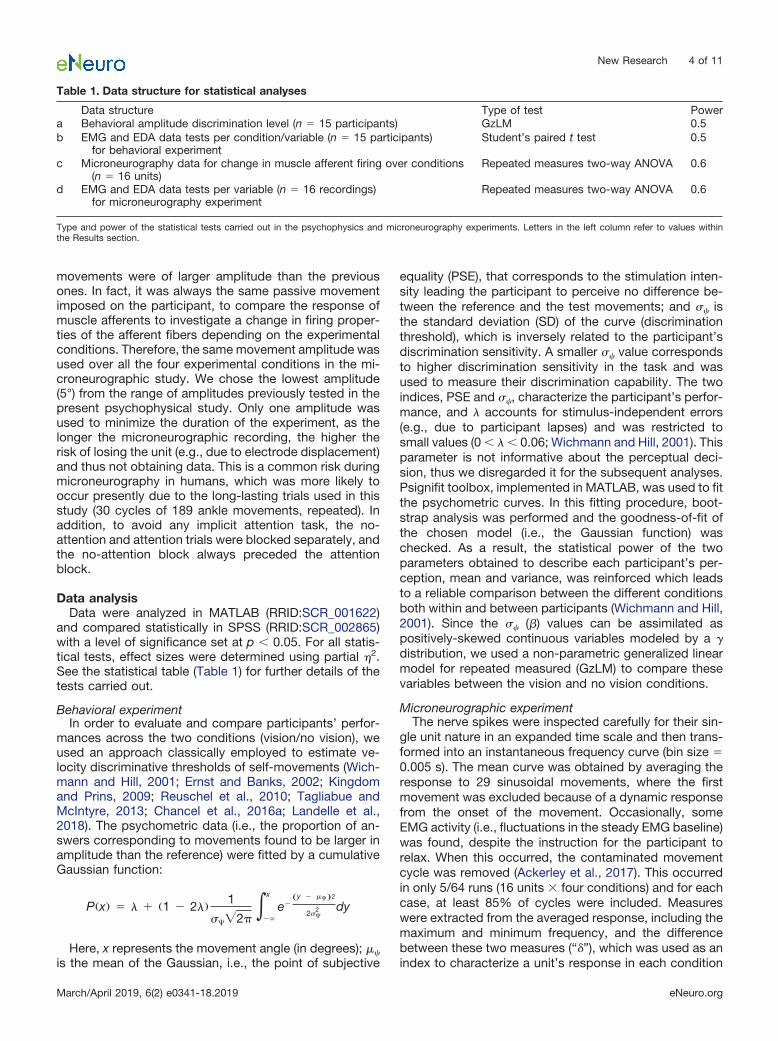
Table 1. Data structure for statistical analyses
Data structure Type of test Powera Behavioral amplitude discrimination level (n � 15 participants) GzLM 0.5b EMG and EDA data tests per condition/variable (n � 15 participants)
for behavioral experimentStudent’s paired t test 0.5
c Microneurography data for change in muscle afferent firing over conditions(n � 16 units)
Repeated measures two-way ANOVA 0.6
d EMG and EDA data tests per variable (n � 16 recordings)for microneurography experiment
Repeated measures two-way ANOVA 0.6
Type and power of the statistical tests carried out in the psychophysics and microneurography experiments. Letters in the left column refer to values withinthe Results section.
(Ackerley et al., 2017). This measure was used to quantifythe dynamic response of muscle afferents (Kakuda, 2000).
In line with other microneurographic studies of muscleafferent firing (Dimitriou, 2016), the data were normalized(z-transformed to give z-scores), so as to compare differ-ences across the conditions over the individual afferents.Here, we obtained the � per afferent/condition, which wasthen normalized by subtracting the mean �, and this wasdivided by the � SD, for that afferent. This produced thenumber of SDs by which each condition differed from themean value for each afferent tested. Statistical analyseswere conducted on these normalized data, on the wholepopulation of afferents, where the data were first checkedfor normality. A repeated measures two-way ANOVA wascarried out in SPSS, to determine the effects of visualinformation and attention, and any interaction betweenthese.
Physiological indexes in both experimentsThe EMG and EDA activity were used to investigate
whether the participant showed muscular or autonomicactivity in the experiments. The direct current offset wasremoved from the EDA data and the EDA and EMG datawere down-sampled to 2.5 kHz. For the psychophysicalexperiment, these data were separated by visual condi-tion, where data were epoched from the beginning of themovement to the end of a movement, per trial, resulting in105 total trials for the combined visuo-proprioceptive in-formation condition and 105 for the proprioceptive-onlycondition. For the microneurographic experiment, EMG(one EMG source was used, which depended on themuscle afferent recorded from) and EDA signals wereextracted, per condition per participant, from the durationof the sinusoidal movement. For both signals, areas underthe curves were measured to analyze the modulation ofphysiological signals across conditions. The mean values,per measure, were checked for normality and the visualconditions were compared by Student’s paired t tests inthe behavioral experiment and the visual/attention condi-
tions using repeated measures two-way ANOVA for themicroneurography experiment.
ResultsBehavioral measurement of effect of visualinformation on movement discrimination
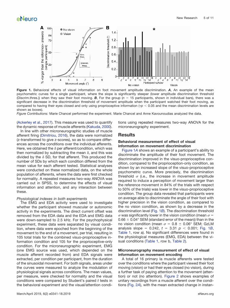
Figure 1A shows an example of a participant’s ability todiscriminate the amplitude of their foot movement. Thediscrimination improved in the visuo-proprioceptive con-dition, compared to the proprioception-only condition, asshown by an increased slope of the visuo-proprioceptivepsychometric curve. More precisely, the discriminationthreshold (i.e., the increase in movement amplituderequired to induce a perception of movement larger thanthe reference movement in 84% of the trials with respectto 50% of the trials) was lower in the visuo-proprioceptivecondition. The group data revealed that participants wereon average able to discriminate the angle of their foot withhigher precision in the vision condition, as compared tothe no vision condition, as shown by a decrease in thediscrimination level (Fig. 1B). The discrimination threshold was significantly lower in the vision condition (mean �0.66 � 0.04° SEM (standard error of the mean)) than in theno vision condition (mean � 0.8 � 0.06° SEM; GzLManalysis slope � 0.242, t � 3.31 p � 0.001; Fig. 1B,Table 1, row a). No significant differences were found inthe physiological measures (EMG, EDA) between the vi-sual conditions (Table 1, row b, Table 2).
Microneurography measurement of effect of visualinformation on movement encoding
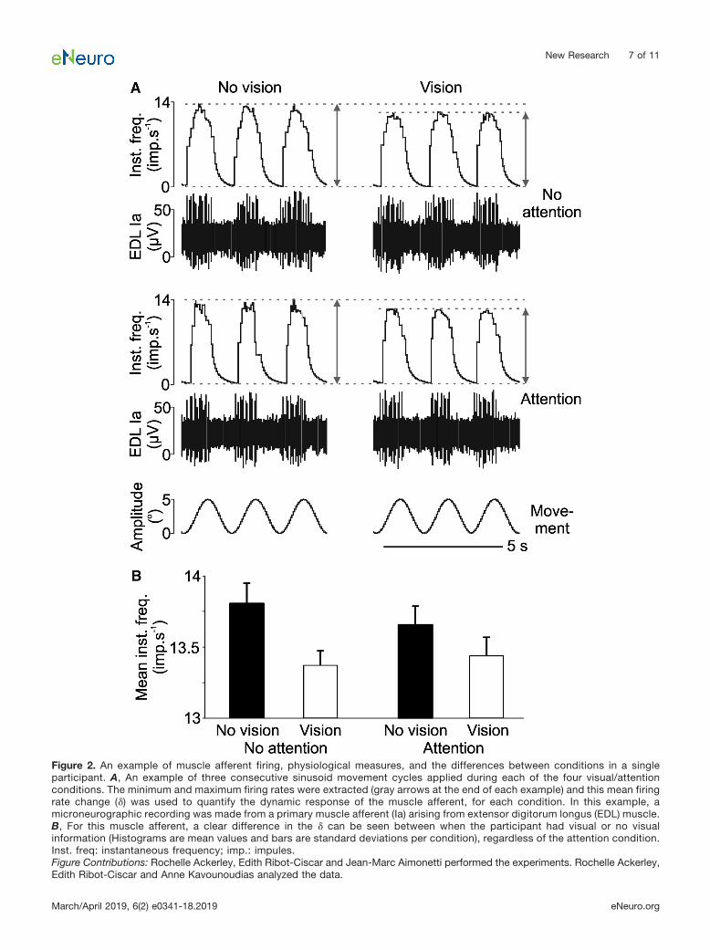
A total of 16 primary Ia muscle afferents were testedover the conditions where the participant viewed their footmoving (vision) or had their eyes closed (no vision), duringa further task of paying attention to the movement (atten-tion) or not (no attention). Figure 2 shows examples ofunitary recordings from a muscle afferent over the condi-tions (Fig. 2A), with the mean extracted change in instan-
Figure 1. Behavioral effects of visual information on foot movement amplitude discrimination. A, An example of the meanpsychometric curves for a single participant, where the slope is significantly steeper (lower amplitude discrimination threshold(Discrim.thres.)) when they saw their foot moving. B, For the group (n � 15 participants, shown in individual bars), there was asignificant decrease in the discrimination threshold of movement amplitude when the participant watched their foot moving, ascompared to having their eyes closed and only using proprioceptive information (�p � 0.05 and the mean discrimination levels areshown as boxes).Figure Contributions: Marie Chancel performed the experiment. Marie Chancel and Anne Kavounoudias analyzed the data.
New Research 5 of 11
March/April 2019, 6(2) e0341-18.2019 eNeuro.org
taneous firing (�) over the sinusoidal movement cycles percondition (Fig. 2B). It can be seen in, both the individualcycles and in the unit’s means, that there was a cleardifference between the vision conditions, where the meaninstantaneous firing frequency was lower with visual in-formation, in both attention and no attention conditions.
The same result was found in the group data (Fig. 3). Arepeated measures ANOVA on the � z-scores showed asignificant main effect of vision (F(1,15) � 20.36, p � 0.001,partial �2 � 0.58; Fig. 3), but no significant effect ofattention (F(1,15) � 0.19, p � 0.672, partial �2 � 0.01), noran interaction between visual information and attention(F(1,15) � 0.64, p � 0.435, partial �2 � 0.04; Table 1, rowc). Therefore, a significant increase in � was found whenvisual information was removed, but paying attention tothe movement did not make a difference in the muscleafferent firing in this paradigm.
The physiological data (EMG, EDA) showed no signifi-cant differences between the conditions (Table 1, row d,Table 3). Here, for both EMG and EDA data, we found nosignificant effect of having visual information, or not, andneither was there an effect of whether the participant paidattention to the movement or simply relaxed, nor an in-teraction of these factors.
DiscussionPresently, we investigated the effect of congruent visual
and/or proprioceptive signals on the processing of anklemovement. We found that visual information interactedwith proprioceptive information, as seen in behavioralmeasures and in the responses of single muscle afferents.When participants saw their moving foot, they were moreaccurate in judging movement amplitude. Further, wefound that the response from single muscle afferents wasincreased when the proprioceptive channel was the onlysource of sensory information, as compared to when theparticipant had the congruent visual input.
Enhancement of visuo-proprioceptive perceptionOur behavioral results confirmed that combining visuo-
proprioceptive information relating to self-body move-ments provided a perceptual enhancement, as there wasa significant decrease in the threshold for discriminationwhen additional visual information was available. Thiscorresponds well with many studies showing that com-
bining congruent visuo-proprioceptive stimulation en-hances the resulting perception, suggesting that bothvisual and proprioceptive cues are co-processed for kin-esthetic purposes (Tardy-Gervet et al., 1986; Rossettiet al., 1995; Van Beers et al., 1999; Reuschel et al., 2010;Guerraz et al., 2012; Blanchard et al., 2013). For example,using the classical mirror paradigm, Guerraz et al. (2012)reported that when participants looked at the reflection oftheir moving left arm in a mirror, they felt an illusion of aconcomitant displacement of their stationary, hidden rightarm. When a congruent muscle vibration was added on theresting right arm, i.e., simulating a movement in the samedirection as that of the visual moving arm, the velocity of theresulting illusion increased, showing the beneficial impact ofmultisensory inputs.
Vision and muscle proprioception may combine inmovement perception, but it does not mean that theweights allocated to each of these sensory cues areequal. For example, under artificial conflicting visuo-proprioceptive conditions, where visual cues of the par-ticipants were deviated using prisms and participants hadto place an unseen finger in the same position as theirseen finger, visual information has been shown to overridemuscle proprioceptive information under full-light condi-tions. In contrast, proprioception dominates when visioninput is severely reduced to a small light-emitting diode onthe end of their finger, viewed in darkness (Plooy et al.,1998). Therefore, the exact behavioral context must betaken into account. According to the theoretical Bayesianframework, the CNS allocates relative weights to eachsensory cue on its relative reliability to encode the per-ceptual event in a given context and their weighted com-bination can optimize the resulting perception (Ernst andBanks, 2002; Landy et al., 2011). Although our presentexperiment was not designed to test the optimality hy-pothesis, one may hypothesize that the discriminativeenhancement we found in the bisensory condition may beexplained by a weighted combination of both visual andproprioceptive information, as reported in other percep-tual tasks (Van Beers et al., 1999; Reuschel et al., 2010).
Dynamic muscle spindle sensitivity increases inabsence of vision
It is generally assumed that multisensory integrativemechanisms take place in the brain, but the present
Table 2. Mean values and statistics for the physiological measures during microneurography experiment
The mean values for the EMG and EDA, with the SEM, as shown for the microneurography experiment. The EMG and electrodermal responses are shown inarbitrary units (area under the curve) for the duration of the sinusoidal cycles per condition. There was no significant effect of vision, attention, or the interac-tion of these, as shown in the ANOVAs, where the partial �2 shows the size effects.
New Research 6 of 11
March/April 2019, 6(2) e0341-18.2019 eNeuro.org
Figure 2. An example of muscle afferent firing, physiological measures, and the differences between conditions in a singleparticipant. A, An example of three consecutive sinusoid movement cycles applied during each of the four visual/attentionconditions. The minimum and maximum firing rates were extracted (gray arrows at the end of each example) and this mean firingrate change (�) was used to quantify the dynamic response of the muscle afferent, for each condition. In this example, amicroneurographic recording was made from a primary muscle afferent (Ia) arising from extensor digitorum longus (EDL) muscle.B, For this muscle afferent, a clear difference in the � can be seen between when the participant had visual or no visualinformation (Histograms are mean values and bars are standard deviations per condition), regardless of the attention condition.Inst. freq: instantaneous frequency; imp.: impules.Figure Contributions: Rochelle Ackerley, Edith Ribot-Ciscar and Jean-Marc Aimonetti performed the experiments. Rochelle Ackerley,Edith Ribot-Ciscar and Anne Kavounoudias analyzed the data.
New Research 7 of 11
March/April 2019, 6(2) e0341-18.2019 eNeuro.org
findings show spinal effects, where visual signals wereassociated with decreases in the responses of the muscleafferents. We found that there was a decrease in thedepth of modulation (�) to repeated sinusoidal move-ments, when the participants viewed their foot moving.We verified that this change in muscle spindle sensitivitywas not due to involuntary muscle activity, as the leg EMGactivity recorded showed no significant differencesacross conditions. The effect of vision occurred indepen-dently of the attentional state of the participants, as ma-nipulated via direct instructions to attend or not, whereattention did not affect the �. Similarly, there was nosignificant interaction between vision and attention.Therefore, we postulate that in the present experimentalmanipulation, attentional effects do not account for thechanges in muscle spindle sensitivity in the different visualconditions. However, our manipulation of attention wasexplicit (i.e., we asked the participants to attend or not),which was in part constrained by the microneurographyconditions where the participants are required to remain
relaxed, and we were not able to confirm their attentionalload. It may have been the case that participants mayhave simply disregarded the instruction to either attend ornot attend; however, participants often reported the diffi-culty of the task, since the movements were actually allthe same, which suggested that they really followed theinstructions and executed the attentional task.
The absence of a change in muscle afferent activity withattention may appear contradictory with the results ofprevious studies, where it has been observed that afusimotor-induced sensitization of muscle spindles oc-curs during proprioceptive attention tasks (Hospod et al.,2007; Ribot-Ciscar et al., 2009). However, the previousexperiments were specifically designed to address theeffect of attention, while here it was only a controlledparameter, and a difference between the difficulties of thepresent and previous tasks likely accounts for this. Thepresent task was a simple comparison of movement am-plitude at the end of the sinusoidal movements betweenattention conditions, which is far easier than the recogni-
Figure 3. The mean effect of visual information and attention on muscle afferent movement encoding. The group data of Ia muscleafferents (n � 16) show a significant difference in the dynamic response of muscle afferents, as measured by the change in theminimum-to-maximum firing rates (�), which was normalized via z-transform (means and SEMs are shown). A main effect was foundfor having visual information, where the � was significantly lower with visual information, but no significant difference was found in theresponse between attention conditions, nor the interaction between vision and attention. Inst. freq. : instantaneous frequency.Figure Contributions: Rochelle Ackerley, Edith Ribot-Ciscar and Jean-Marc Aimonetti performed the experiments. Rochelle Ackerley,Edith Ribot-Ciscar and Anne Kavounoudias analyzed the data.
Table 3. Mean values and statistics for the physiological measures during behavioral experiment
EMG TA(mean � SEM)
EMG GS(mean � SEM)
EDA(mean � SEM)
Vision 9330 � 1146 8478 � 1766 8217 � 1595No vision 9237 � 1076 8343 � 1076 7827 � 1237Paired t test vision vs no vision t(13) � 0.92, p � 0.385,
partial �2 � 0.06t(13) � 1.82, p � 0.092,
partial �2 � 0.20t(13) � 0.64, p � 0.531,
partial �2 � 0.03
The mean values for the EMG and EDA, with the SEM, as shown for the behavioral experiment. The EMG and electrodermal responses are shown in arbitraryunits (area under the curve) for the total number of trials per condition. There was no significant effect of vision, as shown in the ANOVAs, where the partial�2 shows the size effects.
New Research 8 of 11
March/April 2019, 6(2) e0341-18.2019 eNeuro.org
tion of writing movements (Hospod et al., 2007) or clas-sifying different movement amplitudes or velocities(Ribot-Ciscar et al., 2009).
We postulate that during the visual conditions, wherethe participant viewed their foot moving, the propriocep-tive information, coupled with congruent visual signals,aids signal processing. In line with the Bayesian frame-work, a relative weighting of each visual and propriocep-tive cue may account for the perceptual enhancementobserved in our visuo-proprioceptive condition. Themodel predicts that if one sensory source becomes lessreliable, the weight of the other one increases (Ernst andBanks, 2002). In the current study, when only propriocep-tive information was available, the participant relied onone sensory source, where we found an increased sensi-tivity in firing of the muscle afferents. Conversely, whencongruent visual information was present, the relativevisual weight increased, while that of the proprioceptiondecreased. Vision plays a dominant role in spatial tasks,as reported in studies using the mirror paradigm, whereseeing the reflection of one’s moving arm in a mirror issufficient to induce an illusion of a concomitant displace-ment of the other stationary, hidden arm (Guerraz et al.2012). Furthermore, Chancel et al. (2016b) reported thatillusions induced using the mirror paradigm can survivedespite a marked visual impoverishment (obtained bycovering between 0% and 100% of the mirror); the mirrorillusion was significantly degraded only when the visualdegradation was �84%, suggesting that even restrictedvisual information is sufficient to provide relevant kines-thetic cues. Future studies may be conducted to explorewhether changing the reliability of the visual feedback byprogressively degrading visual information results in anincrease of muscle spindle sensitivity.
Previous microneurographic studies exploring the ef-fect of vision on muscle proprioceptive information usedexternal visual targets (Wessberg and Vallbo, 1995; Joneset al., 2001; Dimitriou, 2016), in contrast to our experimentwhere the participant viewed their own passive move-ment. Jones et al. (2001) described a decrease in muscleafferent firing rate during incongruent muscle afferent andvisual feedback, which was interpreted as a strategy forresolving bisensory conflict. Conversely, an increase inmuscle afferent firing was more recently observed duringa similar visuomotor task specifically during an imposedadaptation phase, making the fusimotor control a meansof adjusting the human proprioceptive system to motorlearning (Dimitriou, 2016). Interestingly, the latter studyalso observed a decrease in muscle spindle dynamicsensitivity in the washout stage, when visual feedbackwas again congruent with muscle feedback. In line withDimitriou (2016), we found that the fusimotor drive selec-tively decreased muscle spindle sensitivity when muscleafferent feedback was accompanied by congruent visualcues. Although at first glance they might seem disparate,taken together, these recent studies and our present oneaccounted for a fusimotor control of muscle spindle sen-sitivity independent of the concurrent muscle activity,which has long been debated (Vallbo et al., 1979). They allsuggest that muscle spindle sensitivity may change ac-
cording to its relevance to the context and, in particular,the presence or not of relevant visual cues.
The reweighting of proprioceptive information in theabsence of visual signals can be related to the modulationobserved in the primary somatosensory cortex dependingon concomitant visual signals (Blakemore et al., 2005;Helbig et al., 2012). Using a design inspired by the Bayes-ian framework, Helbig et al. (2012) showed that during atask of shape identification, activation of the primarysomatosensory cortex was modulated by the reliabilityof visual information within congruent visuo-tactile in-puts. The less reliable the visual information, the moreactivity in the primary somatosensory cortex increased.In line with the modality appropriateness model (Welchand Warren, 1986) and the Bayesian framework (Ernstand Banks, 2002), one can assume that crossmodalprocessing is more likely to occur within the sensorypathway corresponding to the most accurate signalregarding the task, since this sensory signal is sup-posed to get a greater weight compared to the otherless reliable signals.
Descending fusimotor influences from relevant visualcues may reduce the sensitivity of muscle afferents, re-flecting a decrease in the proprioceptive contribution toencode the actual movement. Indeed, watching a video ofone’s own hand in movement is sufficient to elicit anillusory movement of participant’s resting hand. By re-cording brain activity during this pure visually-inducedkinesthetic illusion, Kaneko et al. (2015) found that thelateral premotor (PM) cortex and the supplementary motorarea (SMA) were specifically activated together with theposterior parietal cortices and the insula. It is wellknown that the SMA and lateral PM are part of themotor system, with direct connections to M1, and de-scending output to the spinal cord (Dum and Strick,1991; He et al., 1995; Picard and Strick, 1996, 2001;Maier, 2002). Further, the SMA and the lateral PM werenot activated when participants viewed a video ofsomeone else’s own hand. Only relevant kinestheticvisual cues may therefore influence proprioceptive sen-sitivity through descending motor commands that canmodulate spinal fusimotor efference.
Functional significance of the fusimotor modulationOne may consider that the observed fusimotor effect is
small, as compared to in animals; however, it has beenrepeatedly observed in humans (Burg et al., 1975; Vallboand Hulliger, 1981; Vallbo and Al-Falahe, 1990; Gandeviaet al., 1994; Ribot-Ciscar et al., 2000, 2009; Jones et al.,2001; Hospod et al., 2007; Dimitriou, 2016; Ackerley et al.,2017) and has been considered as intriguing when com-pared to animal data, where muscle spindle firing ratesare ten times higher than in humans, as are the fusimotor-induced changes (Matthews, 1981). Whatever its amount,the observed effect was sufficient to significantly alteractivity of muscle afferents and hence may have a func-tional impact on the resulting perception.
Moreover, the fact that our two experiments have beendone with two different populations of participants may atfirst appear as a limitation of the present study. However,
New Research 9 of 11
March/April 2019, 6(2) e0341-18.2019 eNeuro.org
it is worth noting that there is commonly a high variabilityin the firing of muscle afferents, depending on the natureand number of intrafusal muscle fibers that are included,the fusimotor innervation received by the intrafusal fibers,and the location of the muscle spindle inside the muscle,where a receptor near the ankle joint will be more affectedby the movement than another located more proximally inthe EDL or TA muscles. Therefore, one can consider thatthe variability introduced by the use of different partici-pants does not overly influence the outcome, with respectto the intra-subject variability, due to the technical chal-lenge of recordings made in the same subject.
Our finding that afferent proprioceptive signals fromankle could be modulated by visual cues may be impor-tant for controlling postural balance (Burke and Eklund,1977; Massion, 1992; Kavounoudias et al., 2001). Highankle proprioceptive acuity has been observed to be pre-dictive of sport performance level in elite athletes such asdancers (Han et al., 2015b) and in balance performance ofthe elderly (Goble et al., 2011). Similarly, better ankleproprioception is correlated with reduced ankle injuries(Han et al., 2015a), while after a complete loss of somato-sensory afferents, deafferented patients present severedeficits in postural and motor tasks (Forget and Lamarre,1995). The central processing of ankle proprioceptiveinformation with other sensory information enables op-timal integration for balance control. When a source ofinformation is used for other purposes, for example, ifvision is used to track a target in the environment, theCNS uses a reweighting strategy relying on the mostreliable sources of information to optimize balance con-trol. We presently show that a relative reweighting ofvisual signals may occur by a recalibration at moreperipheral levels of ankle proprioceptive inputs, via adirect setting of muscle receptor sensitivity by the CNS.
The present results may have further clinical impact onsensorimotor rehabilitation. Different interventions areused to improve ankle proprioception and balance con-trol, particularly after ankle injury. While passive interven-tions, such as taping or compressing, do not seem toparticularly improve proprioception, active interventionswith task-specific paradigms are efficient, suggestingcentral processing modifications (Han et al., 2015b). Thepresent results suggest that removing visual informationmay optimize the intervention, by providing the brain withincreased proprioceptive information that may favor abetter recovery of balance control.
In conclusion, we show that muscle afferent sensitivitycan be altered in a context-dependent way via descend-ing influences. Specifically, we show that when proprio-ceptive signals from a foot movement are coupled withcongruent visual information, a decrease in muscle affer-ent firing was found. This decrease in the bisensory con-dition may reflect a re-weighting of the two sensory cuesin favor of the visual source. Our study shows that themechanisms of sensory reweighting are not limited tohigher-level neural control in the brain, but that there arealso spinal effects of multisensory processing betweenvisual signals and proprioceptive coding. This opens upthe opportunity for the study of other multisensory
effects below the level of the brain and impacts on ourunderstanding of multisensory interactions throughoutthe CNS, which may also provide clinical therapeuticstrategies for ameliorating visuo-sensorimotor distur-bances.
ReferencesAckerley R, Aimonetti JM, Ribot-Ciscar E (2017) Emotions alter
muscle proprioceptive coding of movements in humans. Sci Rep7:8465.
Awiszus F, Schäfer SS (1989) Re-afferent effects of individual staticand dynamic gamma-stimuli during maintained fusimotor stimula-tion. Brain Res 489:41–48.
Bergenheim M, Roll J-P, Ribot-Ciscar E (1999) Microneurography inhumans. In: Modern techniques in neuroscience research (Wind-horst U, Johansson H, eds), pp 801–819. New York: Springer-Verlag Berlin Heidelberg.
Blakemore SJ, Bristow D, Bird G, Frith C, Ward J (2005) Somato-sensory activations during the observation of touch and a case ofvision-touch synaesthesia. Brain 128:1571–1583.
Blanchard C, Roll R, Roll J-P, Kavounoudias A (2013) Differentialcontributions of vision, touch and muscle proprioception to thecoding of hand movements. PLoS One 8:e62475.
Burg D, Szumski AJ, Struppler A, Velho F (1975) Observations onmuscle receptor sensitivity in the human. Electromyogr Clin Neu-rophysiol 15:15–28.
Burke D, Eklund G (1977) Muscle spindle activity in man duringstanding. Acta Physiol Scand 100:187–199.
Cappe C, Morel A, Barone P, Rouiller EM (2009) The thalamocorticalprojection systems in primate: an anatomical support for multisen-sory and sensorimotor interplay. Cereb Cortex 19:2025–2037.
Dimitriou M (2016) Enhanced muscle afferent signals during motorlearning in humans. Curr Biol 26:1062–1068.
Chancel M, Blanchard C, Guerraz M, Montagnini A, Kavounoudias A(2016a) Optimal visuotactile integration for velocity discriminationof self-hand movements. J Neurophysiol 116:1522–1535.
Chancel M, Brun C, Kavounoudias A, Guerraz M (2016b) The kin-aesthetic mirror illusion: How much does the mirror matter? ExpBrain Res 234:1459–1468.
Dum RP, Strick PL (1991) The origin of corticospinal projections fromthe premotor areas in the frontal lobe. J Neurosci 11:667–689.
Edin BB, Vallbo AB (1990) Classification of human muscle stretchreceptor afferents: a Bayesian approach. J Neurophysiol 63:1314–1322.
Ellaway P, Taylor A, Durbaba R, Rawlinson S (2002) Role of thefusimotor system in locomotion. Adv Exp Med Biol 508:335–342.
Ellaway PH, Taylor A, Durbaba R (2015) Muscle spindle and fusimo-tor activity in locomotion. J Anat 227:157–166.
Ernst MO, Banks MS (2002) Humans integrate visual and hapticinformation in a statistically optimal fashion. Nature 415:429–433.
Forget R, Lamarre Y (1995) Postural adjustments associated withdifferent unloadings of the forearm: effects of proprioceptive andcutaneous afferent deprivation. Can J Physiol Pharmacol 73:285–294.
Gandevia SC, Wilson L, Cordo PJ, Burke D (1994) Fusimotor reflexesin relaxed forearm muscles produced by cutaneous afferents fromthe human hand. J Physiol 479:499–508.
Goble DJ, Coxon JP, Van Impe A, Geurts M, Doumas M, WenderothN, Swinnen SP (2011) Brain activity during ankle proprioceptivestimulation predicts balance performance in young and olderadults. J Neurosci 31:16344–16352.
Guerraz M, Provost S, Narison R, Brugnon A, Virolle S, Bresciani JP(2012) Integration of visual and proprioceptive afferents in kines-thesia. Neuroscience 223:258–268.
Hagbarth K, Vallbo AB (1968) Discharge characteristics of humanmuscle afferents during muscle stretch and contraction. Exp Neu-rol 22:674–694.
New Research 10 of 11
March/April 2019, 6(2) e0341-18.2019 eNeuro.org
Hagura N, Oouchida Y, Aramaki Y, Okada T, Matsumura M, SadatoN, Naito E (2009) Visuokinesthetic perception of hand movement ismediated by cerebro-cerebellar interaction between the left cere-bellum and right parietal cortex. Cereb Cortex 19:176–186.
Han J, Anson J, Waddington G, Adams R, Liu Y (2015a) The role ofankle proprioception for balance control in relation to sports per-formance and injury. Biomed Res Int 2015:842804.
Han J, Waddington G, Anson J, Adams R (2015b) Level of compet-itive success achieved by elite athletes and multi-joint propriocep-tive ability. J Sci Med Sport 18:77–81.
He SQ, Dum RP, Strick PL (1995) Topographic organization ofcorticospinal projections from the frontal lobe: motor areas on themedial surface of the hemisphere. J Neurosci 15:3284–3306.
Helbig HB, Ernst MO, Ricciardi E, Pietrini P, Thielscher A, Mayer KM,Schultz J, Noppeney U (2012) The neural mechanisms of reliabilityweighted integration of shape information from vision and touch.Neuroimage 60:1063–1072.
Holmes NP, Spence C (2005) Visual bias of unseen hand positionwith a mirror: spatial and temporal factors. Exp Brain Res 166:489–497.
Hospod V, Aimonetti J, Roll JP, Ribot-Ciscar E (2007) Changes inhuman muscle spindle sensitivity during a proprioceptive attentiontask. J Neurosci 27:5172–5178.
Jones KE, Wessberg J, Vallbo AB (2001) Directional tuning of humanforearm muscle afferents during voluntary wrist movements. JPhysiol 536:635–647.
Kakuda N (2000) Response of human muscle spindle afferents tosinusoidal stretching with a wide range of amplitudes. J Physiol527:397–404.
Kaneko F, Blanchard C, Lebar N, Nazarian B, Kavounoudias A,Romaiguère P (2015) Brain regions associated to a kinestheticillusion evoked by watching a video of one’s own moving hand.PLoS One 10:e0131970.
Kavounoudias A (2017) Sensation of movement: a multimodal per-ception. In: Sensation of movement (Grunbaum T, Christensen M,eds), pp 87–109. London: Routledge.
Kavounoudias A, Roll R, Roll JP (2001) Foot sole and ankle muscleinputs contribute jointly to human erect posture regulation. JPhysiol 532:869–878.
Kavounoudias A, Roll JPP, Anton JLL, Nazarian B, Roth M, Roll R(2008) Proprio-tactile integration for kinesthetic perception: anfMRI study. Neuropsychologia 46:567–575.
Kingdom FAA, Prins N (2009) Psychophysics: A practical introduc-tion. London: Academic Press.
Klemen J, Chambers CD (2012) Current perspectives and methodsin studying neural mechanisms of multisensory interactions. Neu-rosci Biobehav Rev 36:111–133.
Landelle C, El Ahmadi A, Kavounoudias A (2018) Age-related impair-ment of hand movement perception based on muscle propriocep-tion and touch. Neuroscience 381:91–104.
Landy M, Banks M, Knill D (2011) Ideal-observer models of cueintegration. In: Sensory cue integration (Trommershauser J, Kord-ing K, Landy M, eds), pp 5–29. Oxford, UK: Oxford UniversityPress.
Maier MA (2002) Differences in the corticospinal projection fromprimary motor cortex and supplementary motor area to macaqueupper limb motoneurons: an anatomical and electrophysiologicalstudy. Cereb Cortex 12:281–296.
Murphy PR, Martin HA (1993) Fusimotor discharge patterns duringrhythmic movements. Trends Neurosci 16:273–278.
Picard N, Strick PL (1996) Motor areas of the medial wall: a review oftheir location and functional activation. Cereb Cortex 6:342–353.
Picard N, Strick PL (2001) Imaging the premotor areas. Curr OpinNeurobiol 11:663–672.
Plooy A, Tresilian JR, Mon-Williams M, Wann JP (1998) The contri-bution of vision and proprioception to judgements of finger prox-imity. Exp Brain Res 118:415–420.
Reuschel J, Drewing K, Henriques DYP, Rösler F, Fiehler K (2010)Optimal integration of visual and proprioceptive movement infor-mation for the perception of trajectory geometry. Exp Brain Res201:853–862.
Ribot E, Roll JP, Vedel JP (1986) Efferent discharges recorded fromsingle skeletomotor and fusimotor fibres in man. J Physiol 375:251–268.
Ribot-Ciscar E, Rossi-Durand C, Roll JP (2000) Increased musclespindle sensitivity to movement during reinforcement manoeuvresin relaxed human subjects. J Physiol 523:271–282.
Ribot-Ciscar E, Hospod V, Roll JP, Aimonetti J (2009) Fusimotordrive may adjust muscle spindle feedback to task requirements inhumans. J Neurophysiol 101:633–640.
Rossetti Y, Desmurget M, Prablanc C (1995) Vectorial coding ofmovement: vision, proprioception, or both? J Neurophysiol 74:457–463.
Stein BE, Stanford TR (2008) Multisensory integration: current issuesfrom the perspective of the single neuron. Nat Rev Neurosci9:255–266.
Tagliabue M, McIntyre J (2013) When kinesthesia becomes visual: atheoretical justification for executing motor tasks in visual space.PLoS One 8:e68438.
Tardy-Gervet MF, Gilhodes JC, Roll JP (1986) Interactions betweenvisual and muscular information in illusions of limb movement.Behav Brain Res 20:161–174.
Vallbo Å, Al-Falahe N (1990) Human muscle spindle response in amotor learning task. J Physiol 421:553–568.
Vallbo ÅB, Hulliger M (1981) Independence of skeletomotor andfusimotor activity in man? Brain Res 223:176–180.
Vallbo AB, Hagbarth KE, Torebjörk HE, Wallin BG (1979) Somato-sensory, proprioceptive, and sympathetic activity in human pe-ripheral nerves. Physiol Rev 59:919–957.
Van Beers RJ, Sittig AC, Denier Van Der Gon JJ (1999) Localizationof a seen finger is based exclusively on proprioception and onvision of the finger. Exp Brain Res 125:43–49.
Van Beers RJ, Wolpert DM, Haggard P (2002) When feeling is moreimportant than seeing in sensorimotor adaptation. Curr Biol 12:834–837.
Welch R, Warren D (1986) Intersensory interactions. In: Handbook ofperception and human performance (Boff K, Kaufman L, ThomasJ, eds), pp 25.1–25.36. New York, NY: Wiley.
Wessberg J, Vallbo AB (1995) Human muscle spindle afferent activityin relation to visual control in precision finger movements. JPhysiol 482:225–233.
Wichmann FA, Hill NJ (2001) The psychometric function: I. Fitting,sampling, and goodness of fit. Percept Psychophys 63:1293–1313.