Segregation of Form, Color, Movement, and Depth: Anatomy, Physiology, and Perception Anatomical and physiological observations in monkeys indicate that the primate visual system consists of several separate and independent subdivisions that analyze differ- ent aspects of the same retinal image: cells in cortical visual areas 1 and 2 and higher visual areas are segregated into three interdigitating subdivisions that differ in their selectivity for color, stereopsis, movement, and orienta- tion. The pathways selective for form and color seem to be derived mainly from the parvocellular geniculate subdivi- sions, the depth- and movement-selective components from the magnocellular. At lower levels, in the retina and in the geniculate, cells in these two subdivisions differ in their color selectivity, contrast sensitivity, temporal prop- erties, and spatial resolution. These major differences in the properties of cells at lower levels in each of the subdivisions led to the prediction that different visual functions, such as color, depth, movement, and form perception, should exhibit corresponding differences. Human perceptual experiments are remarkably consistent with these predictions. Moreover, perceptual experiments can be designed to ask which subdivisions of the system are responsible for particular visual abilities, such as figurdground discrimination or perception of depth from perspective or relative movement-functions that might be difficult to deduce from single-cell response properties. P EOPLE WITH NORMAL COLOR VISION WILL PROBABLY FIND the left illustration in Fig. 1 less clear and three-dimensional than the one on the right. But it springs forth if you look at it through a blue filter, such as a piece of colored glass or cellophane. In the left version the gray and yellow are equally bright, or luminant, for the average person, whereas the right version has luminance-contrast information. The ability to infer distance and three-dimensional shape from a two-dimensional image is an exam- ple of a visual fiunction that can use luminance but not color differences. Depth from perspective and color perception are thus aspects of vision that seem to be handled by entirely separate channels in our nervous system. Even though intuition suggests that our vision can plausibly be subdivided into several components--color, depth, movement, form, and texture perception-our perception of any scene usually seems well unified. Despite this apparent wholeness, studies of anatomy, physiology, and human perception are converging toward the conclusion that our visual system is subdivided into several -- - The authors are members of the faculty, Depamnent of Neurobiology, Harvard Medical School, Boston, MA 02115. separate parts whose functions are quite distinct. In this article we summarize some of these anatomical, physiological, and human- perceptual observations. Physiological and Anatomical Studies Occasionally people with strokes suffer surprisingly specific visual losses-for example, loss of color discrimination without impair- ment of form perception, loss of motion perception without loss of color or form perception, or loss of face recognition without loss of the ability to recognize most other categories of objects or loss of color or depth perception (1). Such selectivity seems to indicate that the visual pathway is functionally subdivided at a fairly gross level. Anatomical and physiological studies in monkeys also support this idea of functional divergence within the visual pathway. They reveal major anatomical subdivisions at the earliest peripheral stages in the visual system as well as segregation of function at the highest known cortical stages, but until recently there was little information about corresponding subdivisions in the intermediate levels, the first and second cortical visual areas. Subdivisions at early stages in the miualpathway. It has been known for a century that the nerve fibers leaving the eyes diverge to provide input both to the lateral geniculate bodies and to the superior colliculi. The colliculus seems to be relatively more important in lower mammals than it is in primates, in which its main role is probably orientation toward targets of interest; here we will be Fig. 1. The same image at equiluminance (left) and non-equiluminance (right). Depth from perspective, spatial organization, and figurelground segregation are diminished in the equiluminant version. T o convince yourself that the left version does indeed contain the same information as the other, look at it through a piece of blue cellophane or glass. These two colors may not be close enough to your eqduminance point to be effective. Changing the light source may help.
Transcript
Segregation of Form, Color, Movement, and Depth: Anatomy, Physiology, and Perception
Anatomical and physiological observations in monkeys indicate that the primate visual system consists of several separate and independent subdivisions that analyze differ- ent aspects of the same retinal image: cells in cortical visual areas 1 and 2 and higher visual areas are segregated into three interdigitating subdivisions that differ in their selectivity for color, stereopsis, movement, and orienta- tion. The pathways selective for form and color seem to be derived mainly from the parvocellular geniculate subdivi- sions, the depth- and movement-selective components from the magnocellular. At lower levels, in the retina and in the geniculate, cells in these two subdivisions differ in their color selectivity, contrast sensitivity, temporal prop- erties, and spatial resolution. These major differences in the properties of cells at lower levels in each of the subdivisions led to the prediction that different visual functions, such as color, depth, movement, and form perception, should exhibit corresponding differences. Human perceptual experiments are remarkably consistent with these predictions. Moreover, perceptual experiments can be designed to ask which subdivisions of the system are responsible for particular visual abilities, such as figurdground discrimination or perception of depth from perspective or relative movement-functions that might be difficult to deduce from single-cell response properties.
P EOPLE WITH NORMAL COLOR VISION WILL PROBABLY FIND
the left illustration in Fig. 1 less clear and three-dimensional than the one on the right. But it springs forth if you look at it
through a blue filter, such as a piece of colored glass or cellophane. In the left version the gray and yellow are equally bright, or luminant, for the average person, whereas the right version has luminance-contrast information. The ability to infer distance and three-dimensional shape from a two-dimensional image is an exam- ple of a visual fiunction that can use luminance but not color differences. Depth from perspective and color perception are thus aspects of vision that seem to be handled by entirely separate channels in our nervous system.
Even though intuition suggests that our vision can plausibly be subdivided into several components--color, depth, movement, form, and texture perception-our perception of any scene usually seems well unified. Despite this apparent wholeness, studies of anatomy, physiology, and human perception are converging toward the conclusion that our visual system is subdivided into several
- - -
The authors are members of the faculty, Depamnent of Neurobiology, Harvard Medical School, Boston, MA 02115.
separate parts whose functions are quite distinct. In this article we summarize some of these anatomical, physiological, and human- perceptual observations.
Physiological and Anatomical Studies Occasionally people with strokes suffer surprisingly specific visual
losses-for example, loss of color discrimination without impair- ment of form perception, loss of motion perception without loss of color or form perception, or loss of face recognition without loss of the ability to recognize most other categories of objects or loss of color or depth perception (1). Such selectivity seems to indicate that the visual pathway is functionally subdivided at a fairly gross level.
Anatomical and physiological studies in monkeys also support this idea of functional divergence within the visual pathway. They reveal major anatomical subdivisions at the earliest peripheral stages in the visual system as well as segregation of function at the highest known cortical stages, but until recently there was little information about corresponding subdivisions in the intermediate levels, the first and second cortical visual areas.
Subdivisions at early stages in the miualpathway. I t has been known for a century that the nerve fibers leaving the eyes diverge to provide input both to the lateral geniculate bodies and to the superior colliculi. The colliculus seems to be relatively more important in lower mammals than it is in primates, in which its main role is probably orientation toward targets of interest; here we will be
Fig. 1. The same image at equiluminance (left) and non-equiluminance (right). Depth from perspective, spatial organization, and figurelground segregation are diminished in the equiluminant version. T o convince yourself that the left version does indeed contain the same information as the other, look at it through a piece of blue cellophane or glass. These two colors may not be close enough to your eqduminance point to be effective. Changing the light source may help.
Fig. 2. The primate lateral genicu- late body. This six-layered structure is the first stage in the visual system after the retina, and it consists of two distinct subdivisions, the ventral two magnocellular layers and the dorsal four pan~ocellular layers. The two eyes project to different layers in the interdigitating fashion shown: c indicates layers that are innervated by the contralateral eye; i indicates layers with input from the ipsilateral eye.
Fig. 3. Receptive fields for (left) typical color-oppo- nent parvocellular genicu- late neuron, excited over a small region by red light and inhibited over a larger region by green light and (right) typical broadband magnocellular neuron, excited by all wavelengths in the center and inhibited by all wavelengths in its surround.
concerned exclusively with the geniculo-cortical part of the visual system, which seems to be directly concerned with visual perception (2)-what we think of as seeing.
The primate lateral geniculate body is a six-layered structure, with two obviously different subdivisions: the four dorsal, small-cell (parvocellular) layers and the two ventral, large-cell (magnocellular) layers; these two subdivisions differ both anatomically and physio- logically. In 1920 Minkowski (3) discovered that each eye projects to three of the six layers in the peculiar alternating fashion shown in Fig. 2: each half-retina is mapped three times onto one geniculate body, twice to the parvocellular layers and once to the magnocellu- lar, and all six topographic maps of the visual field are in precise register (4).
The four parvocellular layers seem to be very similar, if not identical, anatomically and physiologically. But the magno- and parvocellular divisions are profoundly different, implying a major split in the visual pathway. This division is most obvious, and was first recognized, in the geniculate, but it does not originate there; the two geniculate subdivisions receive input from two intermixed but anatomically distinct types of retinal ganglion cells: type A cells are larger and project to the magnocellular division, and the smaller type B cells project to the parvocellular division (5). These two subdivisions of the visual pathway, which we will refer to as magno and parvo, are distinguishable both anatomically and physiological- ly. Whether this duality in the visual path arises even earlier, at the bipolar or horizontal cells in the retina, is not known. We can at least be reasonably certain that the two components must both derive their inputs from the same rods and cones and that the marked differences in response properties must therefore depend on the way the photoreceptor inputs are combined.
Though they differ significantly in their response characteristics, the magno and parvo systems do share some basic physiological properties. Their receptive fields (the regions of retina over which their impulse activity can be influenced) are all circularly symmetri- cal, and about 90% show center-surround opponency (6, 7); some cells are excited (impulse rate speeded up) by illumination of a small
retinal region and inhibited (impulse rate slowed down) by illumi- nation of a larger surrounding region, whereas others are the reverse, inhibited from the center and excited from the surround. Because of the antagonism between center and surround, large uniform spots produce feeble responses or none. This center- surround arrangement is found also at earlier levels, starting with the retinal bipolar cells. Clearly these cells are wired up so as to convert the information from the photoreceptors into information about spatial discontinuities in light patterns. This should not be surpris- ing, since we ourselves are very poor in judging overall levels of illumination, as anyone who tries doing photography without a light meter well knows-we are lucky if we can come within an f stop (a factor of 2) of the right exposure. On the other hand we can detect a spot that is as little as a few percent brighter or darker than its immediate surround.
The magno and parvo divisions nevertheless differ physiologically in four major ways--color, acuity, speed, and contrast sensitivity (7- 10).
Color. About 90% of the cells in the parvocellular layers of the geniculate are strikingly sensitive to differences in wavelength, whereas cells in the magnocellular layers are not. The three types of cones in the primate retina have broad, overlapping spectral sensitiv- ities and can be loosely termed red-, green-, and blue-sensitive, to indicate that their peak sensitivities are in the long-, middle-, and short-wavelength regions of the spectrum. Parvo cells are wave- length selective because they combine these cone inputs so as in effect to subtract them (Fig. 3, left). A typical parvo cell may, for example, receive excitatory inputs to its receptive field center from red cones only, and inhibitory inputs to its receptive field surround from green cones only. Such a cell will be excited by long wave- lengths (reds), inhibited by short wavelengths (blues and greens), and be unresponsive to some intermediate wavelength (yellow). Besides such red-on center, green-off surround cells, most of the other possibilities also occur, most commonly red cones antagonized by green, and blue versus the sum of red and green (that is, yellow). In contrast to the color selectivity of most parvo cells, magno cells (and also the remaining 10% of the parvo cells) sum the inputs of the three cone types, so that the spectral sensitivity curves are broad, and the response to a change in illumination is of the same type, either on or off, at all wavelengths (Fig. 3, right) (11). The magno system is thus in effect color-blind: as in black-and-white photogra- phy, two different colors, such as red and green, at some relative brightness will be indistinguishable.
Acuity. The second difference between magno and parvo cells is the size of their field centers. For both systems the average size of the receptive field center increases with distance from the fovea, consist- ent with the differences in acuity between foveal and peripheral vision. Yet at any given eccentricity, magno cells have larger receptive field centers than parvo cells, by a factor of 2 or 3.
Speed. Magno cells respond faster and more transiently than parvo cells. This sensitivity to the temporal aspects of a visual stimulus suggests that the magno system may play a special role in detecting movement. Many cells at higher levels in this pathway are selective for direction of movement.
Contmt. Shapley et al. (10) found that magno cells are much more sensitive than parvo cells to low-contrast stimuli. Both begin to respond when the center and surround brightnesses differ by only 1 or 2%, but with increasing contrast magno responses increase rapidly and level off at about 10 to 15% contrast, whereas parvo responses increase more slowly, and saturate at far higher contrasts.
These four major differences between the two subdivisions, in color, acuity, quickness, and contrast sensitivity, imply that they contribute to different aspects of vision. Exactly what aspects have become clearer recently, with new anatomical techniques that have
6 MAY 1988 ARTICLES 741
made it possible to follow these subdivisions farther into the central In the magno pathway, cells in layer 4B are orientation selective; nervous system and to correlate them with the response selectivity of that is, they respond best to lines of a particular orientation, and cells at later stages in each subdivision for more abstract stimulus most of them also show selectivity for the direction of movement features.
Continuation of the madno and parvo subdivisions in visual area 1. The segregation of the two pathways is perpetuated in the primary visual cortex (12) (Fig. 4). Cells in the magnocellular geniculate layers project to layer 4Ca, which projects in turn to layer 4B, which then projects to visual area 2 and to cortical area MT. Parvo cells project to layer 4CP, and from there the connections go to layers 2 and 3, and from there to visual area 2. The parvocellular division splits to form an additional subdivision in the upper layers of visual area 1. The first evidence for this fUrther subdivision came in 1978 when Wong-lley (13) stained visual area 1 for the mitochondrial enzyme cytochrome oxidase and saw alternating regions of light and dark staining. The dark regions are round or oval in sections cut parallel to the surface; they are most prominent in the upper layers (2 and 3) but are also faintly visible in layers 5 and 6. They turned out to represent pillar-like structures about 0.2 rnm in diameter, spaced 0.5 mm apart (14). We term these structures blobs because of their three-dimensional shape. Blobs are found only in the primary visual cortex; they occur in all primates that have been looked at, and in the prosimian Galago, but have not been found in other prosi- mians or any lower mammals (15).
Since layers 2 and 3 receive most of their inputs from parvo- recipient layer 4CP, both the blobs and the interblobs could be considered continuations of the parvo subdivision. Nevertheless the blobs should probably be thought of as a separate subdivision, because they have somewhat different inputs and very different response properties from the interblobs (16-18). The visual re- sponse properties of cells in the blobs suggest that they may also receive magnocellular input (1 7, 18).
Thus by the output stage of visual area 1 the magno system remains segregated, and the parvo system seems to have split into two branches. All three subdivisions, rnagnm4Ca+4B, parvc-+4Cp-+ interblob, and parvo(+magno?)+4Cp-+blob, then project to visual area 2.
These anatomically defined subdivisions in the primary visual cortex differ from each other in the kinds of visual information they carry ( la) , as in earlier stages.
(1 8, 19)-for example, a cell preferking horizontal lines may respond when an edge is moved upward but not when it moves downward. Like magnocellular geniculate cells, cells in 4B lack color selectivity.
In the interblobs, most, perhaps all, cells are also orientation selective. Unlike cells in layer 4B, most are not direction selective; 10 to 20% are end-stopped, responding to short but not long line or edge stimuli. The receptive fields are small, and the optimum line thickness is similar to the optimum spot size of cells in the geniculate parvocellular layers at the same eccentricity. This system may therefore be responsible for high-resolution form perception. Al- though anatomical evidence indicates that the interblob system receives its major input from the color-coded parvocellular genicu- late layers, most of the interblob cells are not explicitly color-coded: they show no color opponency and respond well to achromatic luminance contrast borders. Nevertheless, many of them respond to an appropriately oriented color-contrast edge regardless of the colors forming the edge or the relative brightness of the two colors. Similarly, they usually respond to lines or borders of any brightness contrast (light-on-dark or dark-on-light), even though the anteced- ent geniculate cells are either on-center or off-center but not both. This suggests that much of the color-coded parvocellular input is pooled in such a way that color contrast can be used to identify borders but that the information about the colors (including black versus white) forming the border is lost (20).
Blob cells are not orientation selective but are either color or brightness selective. The blob system thus seems to carry informa- tion complementary to the information carried by the interblob system. The brightness-selective (non-color-coded or broadband) blob cells have larger receptive field centers than the broadband geniculate cells but are otherwise similar-they are either excited or inhibited by small spots of light, and they respond less well to large spots, indicating surround inhibition. These broadband blob cells could receive input from either the magnocellular geniculate cells or from the broadband parvo cells, but the physiological properties of many of them would be more consistent with input from the rnagno system (17, 18). We assume that the color-opponent blob cells receive input from the color-opponent parvocellular geniculate cells,
Fig. 4. Diagram of the functional segregation of the primate visual system. MT, middle temporal lobe; V4, visual area 4; LGN, lateral geniculate body. Primary visual cortex
SCIENCE, VOL. 240
Fig. 5. Section parallel to the surface through visual areas 1 and 2 of a squirrel monkey, stained for cytochrome oxidase. Visual area 1 is on the left; the blobs appear as small round dots. In Visual area 2 the cyto- chrome-oxidase stain reveals a pattern of alternating thin, thick, and pale stripes.
though they differ from them in that their receptive field centers are larger and their color coding is doubly opponent-they give oppo- site responses to different parts of the spectrum in the center (say, on to red and off to green), and both types of center response are reduced when the spot is made larger.
The blob and interblob systems thus work in entirely different and complementary ways. Blob cells are explicitly color-coded, excited by colors in one region of the spectrum and inhibited by others, and not selective for stimulus orientation. Interblob cells are selective for stimulus orientation but mostly are not color selective, responding to a line or edge of the correct orientation regardless of its color. The strategy of carrying orientation information in a system that mostly pools color information and color-contrast information in a separate system that does not carry orientation information is probably more efficient than having single cells selective for both the orientation and color of a border. Nevertheless, as emphasized earlier, although most of the interblob cells are not overtly color selective, they probably receive their inputs from explicitly color-coded parvocellu- lar geniculate cells and are most likely not color-blind in the sense that the cells in the magno system probably are. Most interblob cells, even though they lose the information about the colors that form a border or the sign of the contrast of the border, should respond to color-contrast borders in which the two colors are equally bright; such borders would be invisible to magno cells.
Visual area 2. The main target of visual area 1 is visual area 2 (Brodrnann's area 18), which shows an equally intriguing pattern when stained for cytochrome oxidase (Fig. 5) (16, 21). Instead of small round dots, tangential sections show a pattern of stripes, much coarser than the blobs of visual area 1; these alternately dark and light stripes are several millimeters wide and run perpendicular to the border between visual areas 1 and 2, probably extending over the entire 8- to 10-rnrn width of visual area 2. The dark stripes are themselves of two types, thick and thin. The regularity of this pattern of thick, thin, and pale stripes varies from animal to animal and is clearer in New World monkeys than in Old World ones, at least partly because in Old World monkeys visual area 2 is buried in the lunate sulcus. Given three histologically defined regions in visual area 2 and the fact that visual area 1 has three kinds of subdivisions that project to other cortical areas, it was natural to ask if they were related. And indeed, from tracer injections into the three kinds of stripes in visual area 2, we found that the blobs are reciprocally connected to the thin stripes, the interblob regions to the pale stripes, and layer 4B to the thick stripes (Fig. 4) (18, 22).
The next step was to record from cells in visual area 2, to learn whether the three subdivisions carry different types of visual infor- mation. We did indeed find marked differences, which were consist-
6 MAY 1988
ent with the properties of cells in the antecedent subdivisions of visual area 1 (18, 23).
Cells in the thin s t r i~es showed no orientation selectivitv. and , , over half were color-coded, just as we had found in the blobs. As in the blobs, most of the color-coded cells were doubly opponent, with two antagonistic inputs to their centers, and surround antagonism for both of these center inputs. About half of the thin-stripe cells, both broadband and color 'opponent, exhibited an additional prop: erty not seen in the blob cells: the receptive field centers were big- ger, yet optimum spot sizes were about the same. A typical cell might respond best, say, to a 0.5" diameter spot, give no response at all to 2" or 4" spots (indicating surround antagonism), and yet respond actively to the 0.5" spot anywhere within an area about 4" in diameter. ~ h e s k cells can bcbroadband or color opponent. Several years ago Baizer, Robinson, and Dow (24) described this kind of broadband cell, which they called "spot cells," in visual area -,
Cells in the pale stripes are orientation selective but not direction selective. At least half of them are end-stopped; this represents a dramatic increase in the proportion of end-stopping over what is seen in visual area 1. We have argued that end-stopping, like center- surround antagonism, is an efficient way of encoding information about shape (23). Like cells in the interblobs, pale-stripe cells are not explicitly color-coded, and we expect that they would respond to color-contrast borders at all relative brightnesses, though we have not yet tested this.
In the thick stripes the great majority of cells likewise show orientation selectivity, but are seldom end-stopped. The most consistent response selectivity we see in the thidr stripes is for stereoscopic depth-most cells respond poorly to stimulation of either eye alone but vigorously when both eyes are stimulated together, and for most cells the responses are extremely sensitive to variations in the relative horizontal positions of the stimuli in the two eyes (retinal disparity). Poggio and Fischer (25) had seen similar disparity-tuned cells in visual area 1 in alert monkeys, predominantly in layer 4B, the layer that projects to the thick stripes of visual area. 2. In the thick stripes we find the same three basic
Fig. 6. (A) Loss of depth from parallax at equiluminance. The position of the middle bar is made to vary with the observer's head position. In this case, the center bar appears to lie in front of the reference bars, except when the bars are made equiluminant with the background. ( 0 ) Two frames of a movie in which the movement of dots generates the sensation of a three-dimensional object. The dots appear to lie on the surface of a sphere (which you can see by stereo-viewing these two frames). ALL sensation of depth is lost when the dots are equiluminant with the background.
ARTICLES 743
Fig. 7. Cnmputer-generated images in which shape is generated by shad- ing. In the middle image the two colors are equiluminant, and the three-dimensional shape is harder to discern than in the other two images, which have luminance-contrast.
classes of cells described by Poggio and Fischer--cells selective for near stimuli, far stimuli, or stimuli falling on exactly corresponding retinal points. Like cells in the pale stripes, these cells show no color selectivity; moreover, we would predict that these cells would be like their magnocellular and would not respond to color- contrast borders when the colors are equally bright, though we have not yet tested responses to equiluminant color-contrast borders either in layer 4B of visual area 1 or in the thick stripes of visual area 'l A.
Other studies (26, 27) have not reported such a clean segregation of cells with different physiological properties in visual area 2, or as clear a correlation of physiological subtypes with the three types of stripes. How clear the functional segregation is in visual area 2 remains to be resolved, but we suspect that these differences are due to choice of classification criteria (23). , ,
Higher visual areas. Meanwhile, explorations of visual areas be- yond 1 and 2 are helping close the gap between the functions suggested by electrophysiological studies and what clinical obser- vations imply about the segregation of various functions in the human visual system. The response properties of cells at levels beyond visual area 2 suggest that the segregation of functions begun at the earliest levels is perpetuated at the highest levels so far studied. Indeed, the segregation-seems to become more and more pro- nounced at each successive level, so that subdivisions that are interdigitated in visual areas 1 and 2 become segregated into entirely separate areas at still higher levels. One higher visual area in the middle temporal lobe, MT, seems to be specialized for the analysis of movement and stereoscopic depth (28). It receives input not only from layer 4B in visual area 1 (29), which is also rich in directionality and disparity selectivity, but also from the thick stripes in visual area 2 (26, 30), which, as already described, contain many cells selective fo; binocular disparity (23): Another higher visual area, visual area 4, has been reported to contain a preponderance of color-selective cells (31), but just how specialized visual area 4 is for color is still unclear since many of the cells show some selectivity for orientation. Visual area 4 receives input from the color-coded thin stripes in visual area 2 and possibly from the pale stripes (26, 30, 32). The notion that there is a higher visual area devoted largely to the processing of color information is consistent with the clinical observation that patients with strokes in the posterior inferior occipital lobe (perhaps in a region homologous to visual area 4) can lose color perception without impairment of form or movement perception.
There are strong suggestions that these channels remain segregat- ed through still higher levels in the brain (33). From lesion studies Pohl (34) and Ungerleider and Mishkin (35) have defined two
f~u~ctionally distinct divisions of visual association areas: the tempo- ral-occipital region, necessary for learning to identifj objects by their appearance, and the parieto-occipital region, needed for tasks in- volving the positions of objects, a distinction they refer to as "where" versus "what." Visual area 4 preferentially projects to the temporal division and MT primarily to parietal cortex (36). Thus the temporal visual areas may represent the continuation of the parvo system, and the parietal areas the continuation of the magno pathway. There can be little doubt that in the next few years work on the dozen or so areas north of the striate cortex will greatly enhance our understanding of vision in general.
Human Perception Despite many gaps, the picture beginning to emerge from the
anatomical and electrophysiological studies summarized above is that the segregation begun in the eye gives rise to separate and independent parallel pathways. At early levels, where there are two major subdivisions, the cells in these two subdivisions exhibit at least four basic diEerences--color selectivity, speed, acuity, and contrast sensitivity. At higher stages the continuations of these pathways are selective for quite different aspects of vision (form, color, move- ment, and stereopsis), thus generating the counterintuitive predic- tion that different kinds of visual tasks should differ in their color, temporal, acuity, and contrast characteristics. To test this prediction, we asked whether the differences seen in the geniculate can be detected in conscious human visual perception by comparing the color, temporal, spatial, and contrast sensitivities of different visual functions. Many of these questions, not surprisingly, have already been asked, and the answers are wonderfully consistent with the anatomy and physiology. For several decades psychologists have accumulated evidence for two channels in human vision, one chromatic and the other achromatic, by showing that different tasks can have very different sensitivities to color and brightness contrast. Given what we know now about the electrophysiology and the anatomy of the subdivisions of the primate visual system, we can begin to try to correlate the perceptual observations with these subdivisions (37). Though at higher cortical levels there seem to be three subdivisions, possibly with some mixing of magno and parvo inputs to the blob system, the most important distinction is probably between the magno system (rnagnw+4Ca-4B+MT) and the parvo-derived subdivisions (parvw+4Cp-interblobs-pale stripes- visual area 4i) and [parvo(+rnagno?)+4C~+blobs-.thin stripes+visual area 41. In our discussion of human perception we will, therefore, stress the distinctions between functions that seem to
SCIENCE, VOL. 240
Fig. 8. Gibson's corridor il- lusion. [From (47) with per- mission, copyright 1950, Houghton Mimin] At equi- luminance the image no longer appears to recede into the distance, and the cylinders all appear to be the same size, as indeed they actually are.
be carried exclusively by the magno system and those that seem to be carried by the parvo-derived pathways.
From the fact that the magno system is color-blind and is faster than the parvo system, we can predict that discrimination of color and discrimination of brightness should have different temporal properties. This is indeed so: in 1923 Ives (38) showed that people can follow brightness alternations at much faster rates than pure color alternations.
The high incidence of movement and direction selectivity in MT suggests that this area may be particularly concerned with move- ment perception. Because anatomically MT receives its major inputs from layer 4B of the primary visual cortex and from the thick stripes of visual area 2, both part of the magno pathway, one would predict that human movement perception should somehow reflect magno characteristics: color blindness, quickness, high contrast sensitivity, and low acuity. Perceptual experiments indicate that movement perception does indeed have these characteristics. First, it is im- paired for patterns made up of equiluminant colors: Cavanagh, Tyler, and Favreau (39) found that if they generated moving red and green sinewave stripes, "the perceived velocity of equiluminous gratings is substantially slowed . . . the gratings often appear to stop even though their bars are clearly resolved . . . the motion is appreciated only because it is occasionally noticed that the bars are at some new position" (39, p. 897; 40). Second, movement perception is impaired at high spatial frequencies, consistent with the lower acuity of the magno system. Campbell and Maffei (41) viewed slowly rotating gratings and found a loss of motion perception at the highest resolvable frequencies, "At a spatial frequency of 16 and 32 cyclesldeg a strange phenomenon was experienced, the grating was perceived as rotating extremely slowly and most of the time it actually appeared stationary. Of course, the subject could call upon his memory and deduce that the grating must be moving for he was aware that some seconds before the grating had been at a particular 'clock-face position.' Even with this additional information that the grating must be rotating the illusion of 'stopped motion' persisted" (41, p. 714). What is most surprising about the perception of both the equiluminant stripes and the very fine stripes is that even though the sensation of movement is entirely, or almost entirely, lost, the stripes themselves are still clearly visible-they are clear enough that changes in their position can be seen, even though they do not seem to be moving. Last, movement can be vividly perceived with very rapidly alternating or very low contrast images (37, 41). Thus, as summarized in Table 1, the properties of human movement percep- tion are remarkably consistent with the properties of the magno system.
Finding cells in the thick stripes of visual area 2 and in MT that are tuned to retinal disparity suggests that the magno system is also involved in stereoscopic depth perception. Consistent with this, Lu and Fender (42) found that subjects could not see depth in equiluminant color-contrast random-dot stereograms even though the dots making up the stereogram remained perfectly clear (43). This finding has been disputed, but we found that differences in results can arise from variations in subjects' equiluminance points with eccentricity, which make it difficult to achieve equiluminance across the visual field. Like movement perception, stereopsis fails for stereograms containing only high, but resolvable, spatial frequen- cies, but it is not diminished for rapidly alternating or very low contrast stereograms (37) (Table 1).
Deduction of further magno or parvo functions fivm perceptual tests. Since the functions that electrophysiological studies had suggested should be carried by the magno system did indeed show all four distinguishing characteristics of that system, we decided to ask whether other visual functions, ones not predicted by single-cell response properties, might also manifest some or all of these properties.
If a particular magno cell sums red and green inputs, there will be a red : green ratio at which the red and green will be equally effective in stimulating the cell. This need not imply that every magno cell has the same ratio of red to green inputs and therefore necessarily the same equiluminance point. Nevertheless, the fact that movement and stereopsis fail at equiluminance implies that, for a given observer, the null ratio must be very similar for the majority of his cells responsible for that function. Kriiger (44) found that of 33 magnocellular geniculate cells studied in two monkeys, 75% were unresponsive to a moving color-contrast border at a particular relative brightness-a brightness ratio that was very close to a human observer's equiluminant point. Thus not only do individual cells in the magno system seem to be color-blind, but the properties of stereopsis and movement perception indicate that the magno system as a whole is color-blind. [There is, however, currently some disagreement about whether the magno system is inactive at equilu- minance (45).] People with the most common forms of color
Display
Percept at equiluminance +-. IT: 4 :TI Flg. 9. Linking by movement is lost at equilurninance. AU nine of the ambiguous motion squares appear to move in synchrony, even though any one seen alone could be seen moving either horizontally or vertically. This linking disappears at equiluminance, and the dots move every which way.
6 MAY 1988 ARTICLES 745
blindness, due to the lack of one of the three cone pigments, are not nearly as color-blind as the magno system appears to be. They still have two cone types to compare, and so they confuse only a small fraction of possible color pairs and can differentiate most color pairs at all relative brightnesses.
Since both motion perception and stereoscopic depth perception are lost at equiluminance, we suspected that the ability to use relative motion as a depth cue might also be lost. Relative motion is a very powerful depth cue: when an observer moves his head back and forth or moves around in his environment, the relative motion of objects provides information about their distance. In the experiment shown in Fig. 6A, the position of the middle bar was coupled to head movement, and the middle bar appeared to be either behind or in front of the reference bars, depending on whether its movement was the same as, or contrary to, the head movement. When the bars were made equiluminant with the background, all sensation of depth disappeared (37).
Relative movement of different parts of a three-dimensional object is also a powerful depth cue. Figure 6B shows two frames of a movie in which random dots move, some to the right and some to the left, as if they were pasted on a rotating spherical surface. The movie gives a powerful sensation of a rotating spherical surface- unless the dots are equiluminant with the background, and then all sensation of depth is lost (37), and the dots seem to dance aindessly. Thus depth from motion, both from viewer parallax and from object motion, seems also to depend on luminance contrast and could well be a function of the magno system. Consistent with this idea, we could see depth from motion at very low levels of luminance contrast (37).
The retinal image is of course two-dimensional, and to capture the three-dimensional relationships of objects the visual systems uses many kinds of cues besides stereopsis and relative motion-perspec- tive, gradients of texture, shading, occlusion, and relative position in the image. We wondered whether the sensation of depth from any of these other cues might also exhibit magno characteristics. It
seemed especially likely that the ability to perceive depth from shading might be carried by an achromatic system, because shading is almost by definition purely luminance-contrast information; that is, under natural lighting conditions a shaded region of an object has the same hue as the unshaded parts, simply darker. But in biology just because something could, or seemingly even should, be done in a certain way does not mean that it will be. Nevertheless, Cavanagh and Leclerc (46) found that the perception of three-dimensional shape from shading indeed depends solely on luminance contrast. That is, in order to produce a sensation of depth and three- dimensionality, shadows can be any hue as long as they are darker than unshaded regions of the same surface. Many artists seem to have been aware of this; for example, in some of the self-portraits of Van Gogh and Matisse the shadows on their faces are green or blue, but they still convey a normally shaped face. Black-and-white photographs of these paintings (taken with film that has approxi- mately the same spectral sensitivity as humans) confirm that the shadows are actually darker that1 the unshaded parts. The converse can be seen in Fig. 7; here the green shadows do not convey a sensation of depth and shape when they are the same brightness as the blue but do when they are darker (when the blue is darker, the blue parts are interpreted as shadowed).
Perspective was well known to artists by the time of the Renais- sance and is a powefil indicator of depth. Converging lines or gradients of texture are automatically interpreted by the visual system as indicating increasing distance from the observer; thus the image in Fig. 8 (47) looks like a corridor receding into the distance despite the conflicting information from other depth cues, the absence of stereopsis or relative motion, which tells us we are looking at a flat surface. The perception of depth from perspective probably underlies many illusions: the two cylinders in Fig. 8 are the same size (and they each cover the same area on your retina), but are perceived by most people as being unequal.
We found that when images with strong perspective are rendered in equiluminant colors instead of black and white, the depth
Table 1. Summary of the correlations between human psychophysical results and the physiological properties of the three subdivisions of the primate geniculo-cortical visual system. A check indicates that the psychophysical results are consistent with the physiology, and a blank indicates that such an experiment has not been done.
Linking properties Linking by movement J J Linking by J J J J
collinearity (illusory borders)
Figurelground discrimination
J
Panlo System
Parvo + Interblob pathway
I C ~ l o r Contrast Temporal Spatial selec- sensi- reso- reso-
P h ~ s i o l o g ~ tivity tivity lution lution
yes low slow high Human perception
Shape discrimination Orientation J J J J
discrimination Shape discrimination J J J J
Parvo+ (Magno?) -+ Blob ~athway
I Color Contrast Temporal Spatial selec- sensi- reso- reso-
Physiology tivity tivity lution lution
yes high slow low Human perception
Color perception Color determination Flicker photometry
J J
746 SCIENCE, VOL. 240
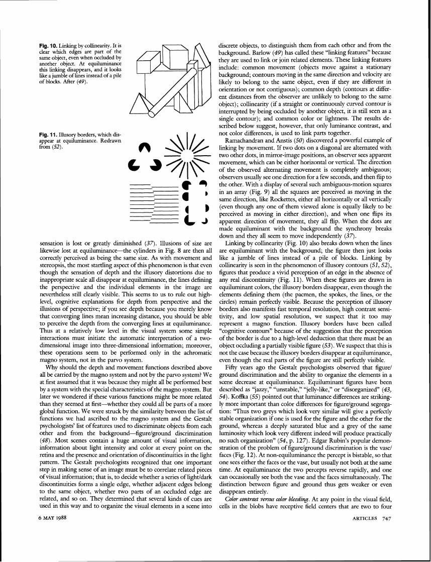
Fig. 10. Linking by collinearity. It is clear which edges are part of the same object, even when occluded by another object. At equiluminance this linking disappears, and it looks like a jumble of lines instead of a pile of blocks. After (49).
Fig. 11. Illusory borders, which dis- appear at equiluminance. Redrawn from (52).
sensation is lost or greatly diminished (37). Illusions of size are likewise lost at equiluminance-the cylinders in Fig. 8 are then all correctly perceived as being the same size. As with movement and stereopsis, the most startling aspect of this phenomenon is that even though the sensation of d e ~ t h and the illusorv distortions due to " inappropriate scale all disappear at equiluminance, the lines defining the perspective and the individual elements in the image are neveEtheiess still clearly visible. This seems to us to rule ou; high- level, cognitive explanations for depth from perspective and the illusions of perspective; if you see depth because you merely know that converging lines mean increasing distance, you should be able to perceive the depth from the converging lines at equiluminance. Thus at a relatively low level in the visual system some simple interactions must initiate the automatic interpretation of a two- dimensional image into three-dimensional information; moreover, these operations seem to be performed only in the achromatic magno system, not in the panro system.
Why should the depth and movement functions described above all be carried by the magno system and not by the parvo system? We at first assumed that it was because they might all be performed best by a system with the special characteristics of the magno system. But later we wondered if these various functions might be more related than they seemed at first-whether they could all be parts of a more global function. We were struck by the similarity between the list of functions we had ascribed to the magno system and the Gestalt psychologists' list of features used to discriminate objects from each other and from the background-figurelground discrimination (48). Most scenes contain a huge amount of visual information, information about light intensity and color at every point on the retina and the presence and orientation of discontinuities in the light pattern. The Gestalt psychologists recognized that one important step in making sense of an image must be to correlate related pieces of visual information; that is, to decide whether a series of lightldark discontinuities forms a single edge, whether adjacent edges belong to the same object, whether two parts of an occluded edge are related, and so on. They determined that several kinds of cues are used in this way and to organize the visual elements in a scene into
discrete objects, to distinguish them from each other and from the background. Barlow (49) has called these "linking features" because they are used to link or join related elements. These linking features inciude: common movement (objects move against a stationary background; contours moving in the same direction and velocity are likely to belong to the same object, even if they are different in orientation or not contiguous); common depth (contours at differ- ent distances from the observer are unlikely to belong to the same object); collinearity (if a straight or continuously curved contour is interrupted by being occluded by another object, it is still seen as a single iontour); and common color or lightness. The results de- scribed below suggest, however, that only luminance contrast, and not color differences, is used to link parts together.
Ramachandran and Anstis (50) discovered a powerful example of linking by movement. If two dots on a diagonal are alternated with two other dots, in mirror-image positions, an observer sees apparent movement, which can be either horizontal or vertical. The direction of the observed alternating movement is completely ambiguous; observers usually see one direction for a few seconds, and then flip to the other. With a display of several such ambiguous-motion squares in an array (Fig. 9) all the squares are perceived as moving in the same direction, like Rockettes. either all horizontallv or all verticalk (even though any one of them viewed alone is equally likely to be perceived as moving in either direction), and when one flips its apparent direction of movement, they all flip. When the dots are made equiluminant with the background the synchrony breaks down and they all seem to move independently (37).
Linking by collinearity (Fig. 10) also breaks down when the lines are equiluminant with the background; the figure then just looks like a jumble of lines instead of a pile of blocks. Linking by collinearity is seen in the phenomenon of illusory contours (51,52), figures that produce a vivid perception of an edge in the absence of any real discontinuity (Fig. 11). When these figures are drawn in equiluminant colors, the illusory borders disappear, even though the elements defining them (the pacmen, the spokes, the lines, or the circles) remain perfectly visible. Because the perception of illusory borders also manifests fast temporal resolution, high contrast sensi- tivity, and low spatial resolution, we suspect that it too may represent a magno function. Illusory borders have been called "cognitive contours" because of the suggestion that the perception of the border is due to a high-level deduction that there must be an object occluding a partially visible figure (53). We suspect that this is not the case because the illusory borders disappear at equiluminance, even though the real parts of the figure are still perfectly visible.
Fifty years ago the Gestalt psychologists observed that figure1 ground discrimination and the ability to organize the elements in a scene decrease at equiluminance. Equiluminant figures have been described as "jazzy," "unstable," "jelly-like," or "disorganized" (43, 54). Ko&a (55) pointed out that luminance differences are striking- ly more important than color differences for figurelground segrega- tion: "Thus two greys which look very similar will give a perfectly stable organization if one is used for the figure and the other for the ground, whereas a deeply saturated blue and a grey of the same luminosity which look very different indeed will produce practically no such organization" (54, p. 127). Edgar Rubin's popular demon- stration of the problem of figurelground discrimination is the vase1 faces (Fig. 12). At non-equiluminance the percept is bistable, so that one sees either the faces or the vase. but usuallv not both at the same time. At equiluminance the two percepts reverse rapidly, and one can occasionally see both the vase and the faces simultaneously. The distinction between figure and ground thus gets weaker or even disappears entirely.
C o b contrast versus color bleeding. At any point in the visual field, cells in the blobs have receptive field centers that are two to four
6 MAY 1988 ARTICLES 747
Fig. 12. Rubin's demonstration of figureiground discrimination [after (48)l . In a luminance-contrast image like this you see either the vase or the faces but not both. At equilu- nlinance you can see both sin~ulta- neously, or they alternate very rapid- ly.
times larger than those in the interblobs (18). Since only the blobs seem to retain information about the sign of color contrast, we suspect that they are responsible for the-perception of the actual colors of objects, as opposed to the ability to use color or luminance contrast to perceive the borders of objects. This implies that color perception should have lower spatial resolution than form percep- tion. This difference in spatial resolution may explain a phenomenon of color perception described by Chevreul (56) in 1839 and by von Bezold (57) in 1876, the phenomenon of bleeding. The way two adjacent colors can affect each other depends on their geometrical arrangement. When two large regions of color abut, their apparent colors and lightnesses repel each other, each making the other look more like its complement, a phenomenon consistentwith the center1 surround antagonism in the blob system. For example, a gray spot surrounded by red will look slightly greenish, and the same gray surrounded by green will appear slightly reddish; surrounding the gray by white will make it appear dark, and surrounding it by black will make it seem lighter. This is called simultaneous contrast and can be seen in Fig. 13. Two colors can have exactly the opposite effect on each other if their geometrical arrangement is such that one forms a very fine pattern, such as fine stripes or dots, with the other as a background. In the lower half of Fig. 13, the mortar seems to bleed into the surrounding gray; the white mortar makes the gray look lighter, and the black mortar makes the same gray look darker. We suspect that bleeding occurs when a pattern is too fine to be resolved by the low acuity color system but not too fine for the higher-resolution form system. Thus you see a pattern, but the colors do not seem to conform to the pattern. We think that the interblob system and the magno system can both define shape, and we cannot predict whether one or the other is more important in defining the borders to which the color is assigned. Some observa- tions, however, suggest that the magno system can influence the spreading of color: color bleeding can be contained by illusory borders or by borders defined only by stereopsis; also, stationary patches of color can seem to move with moving luminance-contrast stimuli (58).
Of course a pattern can be too fine to be seen by either system, as in the microscopic dots used in magazine illustrations. In this case the individual dots cannot be seen, and the colors simply blend. Many artists of the Impressionist period were aware of the way the colors in a resolvable pattern can bleed; they often made dots or dabs of paint large enough to be seen, but small enough that their colors blended (59). The television industry takes advantage of these differences in spatial resolution by broadcasting the color part of the image at a lower resolution than the black and white part, thus red;cing the amount of information to be carried.
Why should the visual system be subdivided? Electrophysiological studies suggest that the magno system is responsible for carrying information about movement and depth. We extended our ideas about the possible functions of the magno system with perceptual studies and concluded that the magno system map have a more global function of interpreting spatial organization. Magno func-
tions may include deciding which visual elements, such as edges and discontinuities, belong to and define individual objects in the scene, as well as determining the overall three-dimensional organization of the scene and the positions of objects in space and movements of objects.
If the magno system covers such a broad range of functions, then what is the function of the tenfold more massive panro system? The color selectivity of the parvo system should enable us to see borders using color information alone and thus borders that might be camouflaged to the color-blind magno system. But defeating cam- ouflage may be only a small part of what the parvo system is specialized for. Experiments with fading of low contrast images (37) indicate that the magno system is not capable of sustained scrutiny, since images that can be seen by only the magno system disappear after a few seconds of voluntary fixation. Thus while the magno system is sensitive primarily to moving objects and carries informa- tion about the overall organization of the visual world, the parvo system seems to be important for analyzing the scene in much greater and more leisurely detail. These postulated hnctions would be consistent with the evolutionary relation of the two systems: the magno system seems to be more primitive than the parvo system (60) and is possibly homologous to the entire visual system of nonprimate mammals. If so, it should not be surprising that the magno system is capable of what seem to be the essential functions of vision for an animal that uses vision to navigate in its environ- ment, catch prey, and avoid predators. The parvo system, which is well developed only in primates, seems to have added the ability to scrutinize in much more detail the shape, color, and surface properties of objects, creating the possibility of assigning multiple visual attributes to a single object and correlating its parts. Indeed, if the magno system needs to use the various visual attributes of an object in order to link its parts together, this could preclude its being able to analyze the attributes independently. It thus seems reason- able to us that the p a r v w +temporal lobe system might be especially suited for visual identification and association.
Is the existence of separate pathways an accident of evolution or a useful design principle? Segregating the processing of different types of information into separate pathways might facilitate the interac- tions between cells carrying the same type of information. It might also allow each system to develop functions particularly suited to its specialization. If the parvo system did evolve after the magno system, by duplication of previously existing structures, it should not be surprising to find some redundancy in the properties of the two systems. Indeed, both seem to carry information about orienta-
Fig. 13. Simultaneous contrast versus bleeding. This phenomenon is shown for black and white, but it is also true for colors. When a spot is surrounded by another color or brightness, the apparent color of the spot tends toward the opposite, or comple- ment, of the surround. The exact opposite hap- pens when one color forms a fine pattern on the other; then the col- ors bleed.
SCIENCE, VOL. 240
tion, and perceptual experiments indicate that both systems can be - - used to determine shaue.
We have summarized the anatomical, phjrsiological, and psycho- logical evidence for segregation of function in the primate visual system. By comparing our own perceptual abilities with the electro- physiological properties of neurons in digerent subdivisions of the visual system, we may be able to deduce functions of particular visual areas, functions that might not have been obvious from electrophysiological observations alone. We can now go back to physiological experiments to test some of the ideas raised by the perceptual experiments.
REFERENCES AND NOTES
1. H. Lissauer, Arch. Psychiatr. Newenkr. 21, 22 (1890); J. Bodamer, ibid. 179, 6 (1947); A. R. Damasio, T. Yamada, H. Damasio, J. Corben, J. McKee, Neurolody 30, 1064 (1980); A. L. Pearlman, J. Birch, J. C. Meadows,Ann. Neurol. 5, 253 (1979); D. Verrey,Arch. Opbtbalnwl. (Parir) 8,289 (1888); R. Balint,Atonatsscbr. Psychiatr. Neurol. 25, 51 (1909); J. Zihl, D. Von Cramon, N. Mai, Brain 106, 313 (1983). Cortical loss of color perception and loss of face recognition usudp occur together, but each can occur independently.
2. H. Munk, Centralbl. Prakt. Augenbedk. 3, 255 (1879). 3. M. Minkowski,Arcb. Keurol. Psychiatr. 6, 201 (1920). 4. W. E. Le Gros Clark and G. G. Pennlan, Proc. R. Soc. Lond. Ser. B 114,291 (1934);
S. Brody, Proc. Kon. Ned. Akad. Wet. 37, 724 (1934); J. G. Malpeli and F. H. Baker, J. Cump. Neurol. 161, 569 (1975).
5. A. G. Leventhal, R. W. Rodieck, B. Dreher, Science 213, 1139 (1981). 6. S. W. Kuffler, J. Neuropbyswl. 16, 37 (1953). 7. T. N. Wiesel and D. H. Hubel ibid. 29, 1115 (1966); P. H. Schiller and J. G.
Malpeli, ibid. 41, 788 (1978). 8. R. L. De Valois, E. Abramov, G. H. Jacobs, J. Opt. Soc.Am. 56,966 (1966); R. L.
De Valois, D. M. Snodderly, Jr., E. W. Yund, N. K. Hepler, Sens. Process. 1, 244 (1977); A. M. Derrington, J. Krauskopf, P. Lennie, J. Pbyswl. (London) 357, 241 (1984); E. Kaplan and R. M. Shapley, ibid. 330,125 (1982); Proc. Natl. Acad. Sci. U.S.A. 83, 2755 (1986); B. Dreher, Y. Fukada, R. W. Rodieck, J. Pbyswl. (Lotldotl) 258, 433 (1976); T. P. Hicks, B. B. Lee, T. R. Vidyasagar, ibid. 337, 183 (1983); P. Gouras, ibid. 199, 533 (1968); ibid. 204, 407 (1969); F. deMonasterio and P. Gouras, ibid. 251, 167 (1975).
9. A. M. Derrington and P. Lennie, J. Pbyswl. (London) 357, 219 (1984). 10. R. Shapley, E. Kaplan, R. Soodak, Nature (London) 292, 543 (1981). 11. The magno system clearly combines the inputs from the red and the green cones,
but the contribution from the blue cones is so small that it is not clear whether the magno system receives any input at all from the blue cones. Also, magno cells are not completely broadband, in that their receptive field surrounds are often weighted toward the red (7).
12. D. H. Hubel andT. N. Wiesel, J. Cmnp. Neurol. 146,421 (1972); J. S. Lund, ibid. 147,455 (1973); - and R. G. Boothe, ibid. 159, 305 (1975).
13. M. Wong-Riley, personal communication. 14. A. E. Hendrickson, S. P. Hunt, and 1.-Y. Wu, Nature (London) 292, 605 (1981);
J. C. Horton and D. H. Hubel, ibid., p. 762. 15. J. C. Horton, Philm. Trans. R . Soc. London 304, 199 (1984); E. McGuinness, C.
MacDonald, M. Sereno, J. Allman, Soc. Neurosci. Abstr. 12, 130 (1986). 16. M. S. Livingstone and D. H. Hubel, Proc. Natl.Acad. Sci. U.S.A. 79,6098 (1982). 17. C. R. Michael, Soc. Neurosci. Abstv. 13, 2 (1987); D. Fitzpatrick, K. Itoh, I. T.
Diamond, J . Neurosci. 3,673 (1983); R. R . H. Tootell, S. L. Hamilton, E. Switkes, R. L. De Valois, Invest. Ophthalmol. Visual Sci. (suppl.) 26, 8 (1985).
18. M. S. Livingstone and D. H. Hubel, J. Neurosci. 4, 2830 (1984). 19. B. Dow, J. Neuropbyswl. 37, 927 (1974). 20. P. Gouras and J. Kriiger, ibid. 42, 850 (1979). 21. R. B. H. Tootell et al., Science 220, 737 (1983).
22. M. S. Livingstone and D. H. Hubel, J. Neurosci. 7, 3371 (1987). 23. D. H. Hubel and M. S. Livingstone, ibid., p. 3378. 24. J. S. Baiwr, D. L. Robinson, B. M. Dow, J. Neuropbysiol. 40, 1024 (1977). 25. G. F. Poggio and B. Fischer, ibid., p. 1392; G. F. Poggio, in L?ynamicAspects of
Neocmtical Functions G. M. Edelman, W. E. Gall, W. M. Cowan, Eds. (Wiley, Kew York, 1984), pp. 631-632.
26. E. A. DeYoe and D. C. Van Essen, Nature (London) 317, 58 (1985). 27. A. Burkhalter and D. C. Van Essen, J. Neurosci. 6, 2327 (1986). 28. R. Dubner and S. M. Zeki, BrainRes. 35,528 (1971); J. H . R. Maunsell and D. C.
Van Essen, J. Neuropbyswl. 49, 1148 (1983). 29. J. S. Lund, R. D. Lund, A. E. Hendrickson, A. H. Bunt, A. F. Fuchs, J . Comp.
Neural. 164, 287 (1975); W. B. Spatz, Brain Res 92, 450 (1975); L. G. Ungerlieder and M. Mishkin, J. Comp. Neurol. 188, 347 (1979).
30. S. Shipp and S. Zeki, Nature (London) 315, 322 (1985). 31. S. Zeki, ibid. 284, 412 (1980). 32. References (26) and (30) are in agreement that the thick stripes project to MT and
that the thin stripes project to visual area 4, but only (26) reports that the pale stripes project to visual area 4.
33. For a review, J. H. R. Maunsell, Mattem oflntelligence, L. M. Vaina, Ed. (Kluwer Academic, Norwell, MA, 1987), pp. 59-87.
34. W. Pohl,]. Comp. Pbyswl. Psychol. 82, 227 (1973). 35. L. G. Ungerleider and M. Mishkin, inAnalysis of Visual Behavior, D. J. Ingle, M. A.
Goodale, R. J. W. Mansfield, Eds. (MIT Press, Cambridge, MA, 1982), pp. 549- 586.
36. K. S. Rockland and D. N. Pandva. Brain Res. 179. 3 11979): R. Desimone. 1. Fleming, C. G. Gross, ibid. 184:h (1980); J. H. R. ~aun$;ll and D. C. V& Essen, J. Neurosci. 3, 2563 (1983).
37. M. S. Livingstone and D. H. Hubel, J. Xeurosci. 7, 3416 (1987). 38. H. E. Ives, J. Qt. Soc. Am. REV. Sci. Instr. 7, 363 (1923). 39. P. ~ a v a n a ~ h , C: W. Tyler, 0. E. Favreau, J. Opt. ~oc . Ant. 8, 893 (1984). 40. We found (37) that apparent movement as well as real movement disappeared at
equiluminance. This result is controversial, but we suspect that different findings map be due to difficulties in achieving equiluminance across the visual field.
41. F. W. Campbell and L. Maffei, ViswnKes. 21, 713 (1981). 42. C. Lu and D. H. Fender, Invest. Opbtbalnwl. 11, 482 (1972). 43. R. L. Gregory, Perctptwn 6, 113 (1977). 44. J. Kriiger, Exp. Brain Res. 30, 297 (1979). 45. Derrington and Lennie (9) have reported that magnocellular neurons are less
responsive than pmocellular neurons at equiluminance, but are not unresponsive. P. H. Schiller and C. L. Colby [Viswn Res. 23, 1631 (1983)] and A. C. Hurlbert, N. K. Logothetis, E. R. Charles, and P. H . Schiller [Soc. Neurosci. Abstr. 13, 204 (1987)l have reported that cells in the panJo system, not the magno system, become unresponsive at equiluminance. This issue clearly remains to be resolved and is discussed in (37).
46. P. Cavanagh and Y. Leclerc, Invest. Ophthalmol. Visual Sci. (suppl.) 26, 282 11985).
47. J. J. dibson, The Perception ofthe Visual World, L. Carmichael, Ed. (Houghton MiWin, Boston, 1950).
48. E. Rubin, Synsoplevede Fgurer (Glydendalska, Copenhagen, 1915). 49. H . B. Barlow, Proc. R. Soc. London 212, 1 (1981). 50. V. S. Ramachandran and S. A. Anstis, Perception 14, 135 (1985). 51. F. Schumann, Z. Psychol. 23, 1 (1900). 52. G. Kanizsa, Ria. Psh[ogia 49, 7 (1955). 53. R. L. Gregory, Nature (London) 238, 51 (1972). 54. S. Liebmann, Pychol. Fmsch. 9, 300 (1926). 55. K. Kotfka, Principles of G e d t Pychology (Harcow Brace, New York, 1935). 56. M. E. Chevreul, De la Loi du Contvaste Simultane' des Couleun (Pitios-Levrault,
Paris, 1839). 57. W. von Bemld, The Themy of Coh , S. R. Koehler, Transl. (Prang, Boston, 1876). 58. H. F. J. M. van Tuijl, Acta Pgchol. 39,441 (1975); K. Nakapama and S. Shimojo,
personal conmicat ion; V. S. Ramachandran, Nature (London) 328,645 (1987). 59. M. S. Livingstone, Sci. Am. 258, 78 (January 1988). 60. R. W. Guillery, Prod. BrainRes. 51. 403 (1979); S. M. Sherman, Prog. Psychobwl.