Page 1
Selective determination of urea using urease
immobilized on ZnO nanowires
Syed Usman Ali, Zafar Hussain Ibupoto, Salah Salman, Omer Nur,
Magnus Willander and Bengt Danielsson
Linköping University Post Print
N.B.: When citing this work, cite the original article.
Original Publication:
Syed Usman Ali, Zafar Hussain Ibupoto, Salah Salman, Omer Nur, Magnus Willander and
Bengt Danielsson, Selective determination of urea using urease immobilized on ZnO
nanowires, 2011, Sensors and actuators. B, Chemical, (160), 1, 637-643.
http://dx.doi.org/10.1016/j.snb.2011.08.041
Copyright: Elsevier
http://www.elsevier.com/
Postprint available at: Linköping University Electronic Press
http://urn.kb.se/resolve?urn=urn:nbn:se:liu:diva-74860
Page 2
Selective determination of urea using urease immobilized on ZnO nanowires
Syed M. Usman Ali1,*, Zafar Hussain Ibupoto1, Salah Salman2, Omer Nur1, Magnus Willander1, Bengt Danielsson2
1Physical Electronics and Nanotechnology Division, Department of Science and Technology, Campus Norrköping, Linköping University, SE-60174 Norrköping, Sweden.
2Acromed Invest AB, Magistratsvägen 10, SE-22643 Lund, Sweden
Abstract: Well-aligned zinc oxide (ZnO) nanowire arrays were fabricated on gold-coated
plastic substrates using a low-temperature aqueous chemical growth (ACG) method. The ZnO
nanowire arrays with 50-130 nm diameters and ~1 µm in lengths were used in an enzyme-
based urea sensor through immobilization of the enzyme urease that was found to be sensitive
to urea concentrations from 0.1 mM to 100 mM. Two linear sensitivity regions were observed
when the electrochemical responses (EMF) of the sensors were plotted vs. the logarithmic
concentration range of urea from 0.1 mM to 100 mM. The proposed sensor showed a
sensitivity of 52.8 mV/decade for 0.1- 40 mM urea and a fast response time less than 4 s was
achieved with good selectivity, reproducibility and negligible response to common
interferents such as ascorbic acid and uric acid, glucose, K+ and Na+ ions.
Index Terms: ZnO-nanowire arrays, electrochemical nanodevices, urease enzyme and urea
determination.
*Corresponding author.
Email address: [email protected]
Page 3
1. Introduction
Urea is an important marker for studies of renal function. The normal level of urea in serum is
from 15 to 40 mg/dl (or 1.7 - 8.3 mM) and level increases up to 100 mM under patho-
physiological conditions [1]. An increase in urea level in blood and urine can be caused by
renal failure, urinary tract obstruction, dehydration, shock, burns, and gastrointestinal
bleeding. Moreover, reduced urea level may be seen in hepatic failure, nephritic syndrome,
and cachexia. Determination of blood urea nitrogen is an important routine test widely used in
clinical laboratories. Several types of biosensors [2-5] are used for the detection and
estimation of urea based on urease. Urease is an important part in most enzymatic sensors
evolution to fulfil the growing requirement for the urea determination. Although colorimetric
and spectrometric methods are most commonly used [6-7], these methods are laborious and
not suitable for on line monitoring system. This inconvenience was overcome by using
electrochemical technique of sensing because electrochemical biosensors provide an attractive
means to analyze the content of a biological sample due to the direct conversion of a
biological event to an electric signal. The inherent advantages of electrochemical biosensors
are their robustness, easy miniaturization, excellent detection limits, also with small analyte
volumes and ability to be used in turbid biofluids with optically absorbing and fluorescing
compounds [8-9]. Moreover, due to the unique properties of nanostructures/nanomaterials in
the biosensing area, nanosensors offer some significant advantages owing to their small size
and high surface area to volume ratios allowing larger signals, better catalysis and the more
rapid movement of analytes through sensors. Recently, nanostructured materials such as zinc
oxide (ZnO) [10–14], cerium oxide (CeO2) [15], tin oxide (SnO2) [16], titanium oxide (TiO2)
[17], iron oxide (FeO2) [18] and zirconium oxide (ZrO2) nanoparticles [19] have been used for
fabrication of transducer surface because of their unique ability to promote faster electron
transfer between electrode and active site of desired enzyme. Among these nanomaterials,
Page 4
ZnO nanostructures receive growing attention due to their unique properties including high
specific surface area, high catalytic efficiency, strong adsorption ability, high isoelectric point
(IEP 9.5), wide band gap (3.37 eV), biocompatibility and high electron communication
features. Moreover, the high IEP of ZnO is advantageous for immobilizing enzymes with low
IEP through electrostatic interactions as reported in our earlier investigations for uric acid and
glucose detection [10, 13]. Furthermore, nontoxicity, high chemical stability and high electron
transfer capability make ZnO a promising material for immobilization of biomolecules
involved in electron or charge transfer without need for electron mediators [20]
In the present work, we have successfully demonstrated the potentiometric determination of
urea by using ZnO nanowire arrays fabricated on the gold-coated plastic substrates by using a
low-temperature aqueous chemical growth (ACG) method. Urease was immobilized on the
surface of the ZnO nanowires using a simple electrostatic process. The potentiometric
response vs. the Ag/AgCl reference electrode was found to be linear over a logarithmic
concentration range of 0.1 to 100 mM suitable for the common urea levels in blood serum.
The proposed sensor (Urs/ZnO/Au) has a fast response time of less than 4 s and retained good
enzymatic activity for more than three weeks when kept at 4 0C temperature when not in use.
2. Experimental details
2.1 Materials and reagent
Urease (E.C. 3.5.1.5 from jack bean 100 U/mg), Urea (ACS reagent.99.9 %), uric acid (Purity
was 99.8%), β-D-glucose (99.5%), zinc nitrate hexahydrate and hexamethylenetetramine were
purchased from Sigma Aldrich. Phosphate Buffer, 10 mM solution (PBS) was prepared from
Na2HPO4 and KH2PO4 (Sigma Aldrich) with sodium chloride in 0.135 mM and the pH was
adjusted to 7.4. A stock urea solution of 100 mM was prepared in PBS, and stored at 4 0C.
The low concentration standard solutions of urea were freshly prepared before the
measurements. All chemicals used (Sigma, Aldrich) were of analytical reagent grade.
Page 5
2.2 Fabrication of ZnO nanowire arrays on gold coated plastic substrate
To fabricate the ZnO nanowire arrays on the flexible plastic substrate, first the substrate was
cleaned with acetone, isopropanol and de-ionized water. After cleaning, a titanium thin film
with 20 nm thicknesses was evaporated as an adhesive layer on plastic substrate and then gold
(Au) thin film with 100 nm thickness was evaporated on it. To obtain well-aligned hexagonal
ZnO nanowire arrays on the electrode surface, we followed the low temperature ACG method
described in [21]. In the ACG method, substrates were spin-coated with seed solution and
annealed at 100 0C for 10 minutes. The seed solution consisted of 0.025 M zinc nitrate
hexahydrate [(Zn(NO3)26H2O)] mixed with 0.0025 M hexamethylenetetramine [C6H12N4] in
water. The substrates were placed in the seed solution using Teflon sample holders and kept
in an oven at 90 °C for 2-5 hours. A small part of the gold-coated plastic substrates were
covered to be used as contact area. After the growth was completed, the nanowires were
cleaned in de-ionized water and dried at room temperature. A typical SEM image of ZnO
nanowires grown using this procedure is shown in figure 1(a). It can be seen from the SEM
image that the ZnO nanowires were 50-130 nm in diameter and ~ 1µm in length with uniform
density and spatial distribution. The nanowires were perpendicular relative to the surface of
the substrate. The morphological and structural characteristics of the grown nanowires can be
controlled by adjusting the growth process parameters such as the concentration of the seed
solution, the reagent stoichiometry, the temperature and the pH of the growth solution [22].
2.3 Immobilization of urease on the ZnO-nanowires and measurements
Before the immobilization of urease enzyme on the surface of the ZnO-nanowires electrode,
the electrodes were rinsed with PBS to generate a hydrophilic surface. A urease solution was
prepared in phosphate buffer solution (PBS, 10mM, pH 7.4) containing 5 mg of urease per ml.
Page 6
In order to optimize the electrostatic adsorption and retained catalytic activity of urease,
electrodes were immersed in the urease solution for different time intervals like 10 minutes,
20 minutes, 30 minutes,60 minutes and 3 hours at room temperature and left in air for 30
minute to dry. It was found that an immersion time of 20 minutes was quite satisfactory to
form a uniform irreversible layer of urease with stable response. Shorter immersion time
resulted in incomplete immobilization with certain leakage of enzyme molecules and poor
sensor response. ZnO-nanowires with immobilized urease are shown in figure 1(b). After
completing these steps, the sensors were initially checked potentiometrically in different
concentrations of urea solutions with an Ag/AgCl reference electrode (3M KCl as filling
solution) purchased from Metrohm (Switzerland). The measurements were carried out by a
potentiometric method utilising two electrodes. ZnO-nanowires based electrode coated with
enzyme served as the working electrode and an Ag/AgCl electrode was used as the reference
electrode. A pH meter (Model 215, Denver Instrument) was used to measure the
potentiometric output voltage of the sensors presented here. For the time response
measurements, a model 363A potentiostat/galvanostat (EG & G, USA) was used. All enzyme
electrodes were stored in dry condition at 4 0C when not in use.
3. Results and discussion
3.1 Sensing mechanism and electrochemical measurements The elemental composition of the as-grown ZnO nanowires was analyzed by energy
dispersive X-rays spectroscopy as shown in figure 1(d). The peaks are attributed to the zinc
(Zn) and oxygen (O) elements while the peaks from gold (Au) are attributed to the substrate.
These results gave evidence that the ZnO nanowires were free from impurities. Most
electrochemical urea sensors are based on an enzymatic reaction catalyzed by urease. Figure
2 describes the sensing mechanism of the proposed urea sensor using immobilized urease on
ZnO-nanowires. It is known that urea can react directly with bare ZnO nanowires and
Page 7
hydrolyse, but the rate of the hydrolysis is not at all up to the extent of enzymatic hydrolysis.
The rate of urea hydrolysis with bare ZnO nanowires is slow and unpredictable and not
reproducible whereas the reaction rate of urea hydrolysis on the surface of ZnO nanowires
with immobilized urease is fast, repeatable and produces a reproducible electrochemical
response. Moreover, enzymatic urea detection is generally based on determining the products
of urea based hydrolysis formed in accordance with equation (1) given below.
As a result of this reaction one HCO3- and two NH4 + ions produced from the
uncharged urea. The ammonium ions react with the ZnO nanowire array and produce the
corresponding EMF change of the output of the sensor. The electrochemical measurements
were carried out using a two-electrode configuration consisting of the urease coated ZnO-
nanowires sensor as the working electrode and an Ag/AgCl reference electrode. The
electrochemical response was measured at room temperature (23 ± 2) 0C with the electrodes
immersed in the samples in a stirred small glass vessels. There are only few reports about the
urea sensing with ZnO as a matrix and then only using amperometric methods [23-24]. In this
work, we demonstrate a simple two-electrode potentiometric method for the urea
measurement using urease immobilized on ZnO nanowire arrays. The sensor as fabricated is
highly sensitive and showed sensitivity of 52.8 mv/decade to the concentration changes of
urea test solutions. The operational linear range and the stability of the sensors were evaluated
potentiometrically in urea solutions made in buffer (PBS pH 7.4) with concentration ranging
from 0.1 mM to 100 mM as shown in figure 3 (a). The tested sensor configuration showed
very interesting data which can be divided into two regions. The electrochemical response
(EMF) that was linear vs. the logarithmic concentration of urea going from 0.1 mM to 40 mM
Page 8
(region 1) and 50 mM to 100 mM (region 2) respectively as shown in the figure 3(b). The first
region (region 1) in figure 3 (b) includes the wide linear range from 0.1 mM to 40 mM. This
range is suitable for the detection of the urea because the normal level of urea is below 10
mM. The second region (region 2) indicates that the same proposed sensor can detect higher
concentrations of urea i.e. up to 100 mM with proper calibration although the underlying
reason for the abnormal response is not fully understood. In region 1 the ZnO-nanowire arrays
sensor approximately followed the Nernst’s expression
E = E0 − 0.05916V/ n log [Reduced] / [Oxidized]
The proposed sensor has exhibited a very fast response time noted over the whole
concentration range with 95% of the steady state voltage (EMF) achieved within ~ 4 s. The
calibration curve showing the sequential addition of urea solutions in a buffer solution is
shown in figure 4. It is very important to note that ZnO-nanowire arrays are relatively stable
around pH 7.4 and this gives these sensors good bio-compatibility in biological fluids and
species since most of the biological fluids are around pH of 7.4. The morphology of the
sensor surface was checked by scanning electron microscopy (SEM) right after measurements
as shown in figure in figure1 (c).
3.2 Sensor reproducibility, stability, measuring range and detection limit
The properties of a biosensor can be characterized by parameters like reproducibility,
measuring range, detection limit, response time and selectivity etc. The reproducibility is an
important characteristic for the performance evaluation of a biosensor. To evaluate
reproducibility characteristic and long term stability of the proposed sensors, we fabricated 8
sensors electrodes independently under the same conditions and immobilized the urease
enzyme on ZnO nanowires arrays. Due to high isoelectric point (IEP) of ZnO (~9.5) and low
IEP (~5.0) of urease, the proposed sensor retained its enzymatic activity due to their strong
Page 9
electrostatic interaction and could be reused several times with proper cleaning in a span of 2-
3 hours. The sensor response in 10 mM urea solution was more than 90 % reproducible till
eight times reused as shown in figure 5(a). The relative standard deviation of the fabricated
sensor electrode in standard urea solutions was less than 5 %. To evaluate the operational and
storage stability of the proposed sensors we have kept the sensors at 4 0C and periodically
tested them for more than three weeks; they retained up to 80% of their original activity and
still showed a good response to urea as shown in figure 5 (b). The measuring range of the
proposed sensor was obtained from the linear part of the calibration curves as shown in figure
3(a). The applicable measuring range of the proposed sensor is between 0.1 mM to 40 mM.
By extrapolating the linear parts of the calibration curve, the detection limit of the sensor
electrode can be calculated. In the present work, the detection limit of the sensor was 0.1 mM
which was calculated by the extrapolating of the two segments of the calibration curve shown
in figure 3(b).
3.3 Selectivity of the ZnO nanowires arrays based sensor
Selectivity is the most important characteristic which describes the specificity
towards the target ion in the presence of other ions (interfering ions). There are different
methods to determine selectivity of the potentiometric sensors [25]. These methods are such
as the separate solution method, the mixed solution method, the matched potential method,
and the unbiased selective coefficients. Instead of using the above mentioned methods, we
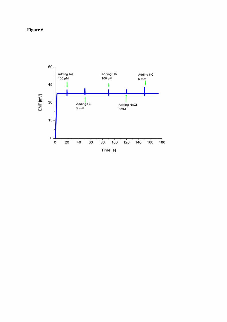
checked the selectivity and stability of the sensor by output response curve. The possible
interferences present in blood that may interfere with a urea biosensor include ascorbic acid
(AA) uric acid (UR), glucose (GL) and potassium (K+) and sodium (Na+) ions [26]. Hence,
ascorbic acid, uric acid, glucose, (K+) and (Na+) ions were selected to affirm the selectivity of
the proposed urea sensor. In the present work, upon adding of100 µM ascorbic acid, 5 mM
glucose, 100 µM uric acid, 5mM (Na+ ) and 5mM (K+) solutions in a 1 mM urea solution
Page 10
only minor signal changes were observed and this is probably due to instability/disturbance
caused by the successive addition of the solutions as shown figure 6. This was repeated
several times on new, independently prepared sensors and continued to show negligible signal
response to interferences. In practical measurements, however these changes in sensor
response can be neglected.
4. Effect of pH and temperature
The objective of this study is to determine the appropriate pH measurement environment for the newly
fabricated ZnO nanowire arrays based urea biosensor. The pH of the working medium affects the
activity of the urease based urea hydrolysis and the signal response (EMF) of the sensor electrode. The
pH dependence of sensor response was investigated in 10 mM urea solution over the pH range
from 4.5 to 9.5 as shown in figure 7(a). The experimental results indicate that the maximum
signal response of proposed urea sensor was obtained between 6.5 to 7.5 pH. Moreover, at pH
value higher than 8, the potential decreases due to the solubility of ZnO at pH ≥8 [27] and
precipitation of urea hydrolysis reaction. Thus, our experimental results showed that the
performance of the urea sensors was stable at pH around 7.4.
The effect of varying temperature on the sensor response in 20 mM urea solution was also
examined between 20 and 80 0C. As shown in Fig 7 (b), the response (EMF) gradually
increases with increasing of temperature and reaches to its maximum value at around 50 0C.
This is because at 50 0C the enzyme has optimum activity [28]. After 50 0C, the response
decreases which is caused by the natural thermal degradation of the enzymes. However, the
biosensor has showed a maximum response at around 50 0C. But during the experiments, it
has been observed that at temperature around 50 0C the response of the sensor although at
maximum was not well stable while at room temperature it was quite stable and exhibited
good long term performance. Moreover, it has also been observed that when we tried to test
Page 11
the sensor at 50 0C then the enzymatic activity degraded drastically and showed poor long
term performance. Therefore, room temperature (23±2) 0C was chosen for this work in order
to prevent possible solution evaporation, enzyme degradation at higher temperature and ease
of operation.
5. Conclusion
In this study, we have demonstrated a simple fabrication procedure for a highly
sensitive urea biosensor based on ZnO nanowire arrays which provided a suitable
microenvironment for enzyme loading and an easy immobilization procedure. The proposed
sensor retained its enzymatic activity due to strong electrostatic interaction between zinc
oxide and urease. The proposed biosensor showed a fast response with less than 4 s and has a
quite wide linear range which is divided into two linear regions that includes from 0.1 mM to
40 mM (region 1) and from 50 mM to 100 mM (region 2) respectively. During the
experiments, the sensor exhibited good performance in sensitivity, stability, selectivity,
reproducibility and negligible interference to the common interferents. All these advantageous
features can make the proposed biosensor applicable in medical, food or other areas. The
proposed device can also be employed to on-spot clinical diagnosis. It is also convenient to
assemble into portable chip based sensing devices suitable to unskilled users. Moreover, the
fabrication method is simple and can be extended to immobilize other enzymes and other
bioactive molecules with low isoelectric points for a variety of biosensor designs.
Page 19
Figure Captions
Fig. 1(a-d): Typical scanning electron microscopy (SEM) images of ZnO nanowires grown
on gold coated plastic substrate using low temperature chemical growth. Figure (a) showing
the ZnO-nanowires without urease immobilization and (b) with urease immobilization (c)
same sensor after measurements and (d) energy dispersive x-ray spectroscopic spectrum of
ZnO nanowires grown on gold coated plastic.
Fig. 2: Schematic diagram for the urea sensing set up using urease coated ZnO nanowires as
working electrode and Ag/AgCl as reference electrode with possible electrochemical reaction
near the working electrode.
Fig. 3(a-c): Calibration curves using urease coated ZnO-nanowires sensor electrode showing
the electrochemical response (EMF) at different urea concentrations (0.1 mM to 100 mM)
with Ag/AgCl reference electrode (a) Calibration curve between output response (EMF) vs.
Log of urea concentrations (b) Calibration curve of linear region 1 (c) Calibration curve of
linear region 2.
Fig. 4: Calibration curves showing the sequential addition of urea solutions.
Fig. 5: (a) Calibration curve showing the urease coated ZnO nanowires sensor
reproducibility/ reusability at room temperature after 2-3 hours span in 10 mM urea solution
(b) Calibration curve showing the study of response (EMF) with the influence of storage at
40C for three weeks.
Fig. 6: Calibration curve showing the study of interferences with time trace line of Output
response (EMF) change with time after adding 100 µM ascorbic acid (AA), 5 mM glucose
(GL) , 100 µM uric acid (UA), 5mM NaCl and 5 mM KCl solutions as an interfering
substances in 1 mM urea solution.
Page 20
Fig. 7: (a-b): Calibration curves showing the study of EMF response with the influence of
varying temperature and pH values.
Page 21
References:
[1] W. Rick, Klinische Chemie und Mikroskopie. Berlin: Springer–Verlag; 1990. p. 245–247.
[2] S.G. Ansari, Z. A. Ansari, H. K. Seo, G. S. Kim, Y. S. Kim, G. Khang, H. S. Shin, Urea
sensor based on tin oxide thin films prepared by modified plasma enhanced CVD, Sens.
Actuators B 132(2008) 265-271.
[3] P. R. Solanki, A. Kaushik, A.A. Ansari, G. Sumana and B. D. Malhotra, ZnO oxide-
chitosan nanobiocomposite for urea sensor, Appl. Phys. Lett. 93(2008) 163903-163906.
[4] A. P. Soldatkin, J. Montoriol, W. Sant, C. Martelet, N. J. Renault, A novel urea sensitive
biosensor with extended dynamic range based on recombinant urease and ISFETs, Biosens.
Bioelectron. 19(2003) 131-135.
[5] S. K. Jha, A. Topkar, S. F. D’Souza, Development of potentiometric urea biosensor based
on urease immobilized in PVA-PAA composite matrix for estimation of blood urea nitrogen
(BUN), J. Biochem. Biophys. Methods 70(2008) 1145-1150.
[6] H. L. Rosenthal, Determination of urea in blood and urine with diacetyl monoxime, Anal.
Chem. 27 (1955) 1980–1982.
[7] B. Xie, U. Harborn, M. Mecklenburg and B. Danielsson, Urea and lactate determined in 1-
uL whole blood with a miniaturized thermal biosensor. Clin. Chem. 40 (1994) 2282-2287.
[8] M. S. Wilson, Electrochemical immunosensors for the simultaneous detection of two
tumor markers, Anal. Chem. 77 (2005) 1496-1502.
[9] P. D’Orazio, Biosensors in clinical chemistry, Clin. Chim. Acta. 334 (2003) 41-69.
Page 22
[10] S. M. Usman Ali, N.H. Alvi, Z. Ibupoto, O. Nur, M. Willander, B. Danielsson, Selective
potentiometric determination of uric acid with uricase Immobilized on ZnO nanowires, Sens.
Actuators B Chem. 2 (2011) 241-247.
[11] S. M. Usman Ali, M.H. Asif , A. Fulati , O. Nur, M. Willander, C. Brännmark, P.
Strålfors, U. H. Englund, F. Elinder and B. Danielsson, Intracellular K+ determination with a
potentiometric microelectrode based on ZnO nanowires, Nanotechnology, IEEE Transaction
on , 10 (2011) 913-919.
[12] S. M. Usman Ali, O. Nur, M. Willander and B. Danielsson, Glucose detection with a
commercial MOSFET using a ZnO nanowires extended gate, Nanotechnology, IEEE
Transaction on, 8 (2009) 678-683.
[13] S. M. Usman Ali, O. Nur , M. Willander , B. Danielsson, A fast and sensitive
potentiometric glucose microsensor based on glucose oxidase coated ZnO nanowires grown
on a thin silver wire, Sens. Actuators B Chem. 145 (2010) 869–874.
[14] M. H. Asif, S. M. Usman Ali, O. Nur, M. Willander, C. Brännmark, P. Strålfors , U.
Englund , F. Elinder and B. Danielsson, Functionalized ZnO nanorod based intracellular
glucose sensor, Biosens. Bioelectron. 25 (2010) 2205–2211.
[15] A. A. Ansari, P. R. Solanki, B. D. Malhotra, Sol-gel derived nanostructured cerium oxide
film for glucose sensor, Appl. Phys. Lett. 92 (2008) 263901-263903.
[16] E. Topoglidis, Y. Astuti, F. Duriaux, M. Gratzel, J. R. Durrant, Direct electrochemistry
and nitric oxide interaction of heme proteins adsorbed on nanocrystalline tin oxide electrodes,
Langmuir. 19 (2003) 6894–6900.
[17] J. Yu, H. Ju, Preparation of porous titania sol-gel matrix for immobilization of
horseradish peroxidase by a vapor deposition method, Anal. Chem.74 (2002) 3579–3583.
Page 23
[18] A. Kaushik, P. R. Solanki, A. A. Ansari, S. Ahmad, B. D. Malhotra, Chitosan–iron oxide
nanobiocomposite based immunosensor for ochratoxin-A Electrochem. Commun.
10(2008)1364–1368.
[19] P. R. Solanki A. Kaushik , P.M. Chavhan , S.N. Maheshwari , B. D. Malhotra,
Nanostructured zirconium oxide based genosensor for Escherichia coli detection,
Electrochem. Commun. 11 (2009) 2272–2277.
[20] Z. Liu, Y. Liu, H. Yang, Y. Yang, G, Shen, R. Yu, A Mediator-Free Tyrosinase
Biosensor Based on ZnO Sol-Gel Matrix, Electroanalysis, 17 (2005) 1065–70.
[21] N. H. Alvi, S. M. Usman Ali, S. Hussain, O. Nur, and M. Willander, Fabrication and
comparative optical characterization of n-ZnO nanostructures (nanowalls, nanorods,
nanoflowers and nanotubes)/p-GaN white light emitting diodes, Scripta Materialia 64 (2011)
697-700.
[22] H .Zhang, D Yang, S. Li, X. Ma, Y. Ji, J. Xu, D. Qu, Controllable growth of ZnO
nanostructures by citric acid assisted hydrothermal process, Mater. Lett. 59 (2005) 1696
1700.
[23] A. Ali, A. A. Ansari, A. Kaushik, P. R. Solanki, A. Barik, M. K. Pandey, B. D. Malhotra,
nanostructured zinc oxide film for urea sensor, Mater. Lett. 63(2009) 2473-2475.
[24] S. G. Ansari, R. Wahab, Z. A. Ansari, Y. S. Kim, G. Khang, A. Al-Hajry, H. S. Shin,
Effect of nanostructure on the urea sensing properties of sol-gel synthesized ZnO, Sens.
Actuators B Chem. 137 (2009) 566-573
[25] M.R. Ganjali, P. Norouzi and M. Rezapour, Encyclopedia of Sensors; Potentiometric Ion
Sensors, American Scientific Publisher (ASP), Los Angeles: 8 (2006) 197-288.
Page 24
[26] J. W. Luo, M. Zhang, D. W. Pang, Selective and sensitive determination of uric acid at
DNA-modified graphite powder microelectrodes, Sens. Actuators B Chem. 106 (2005) 358–
362.
[27] A. Fulati, S. M. Usman Ali, M. Riaz, G. Amin, O. Nur and M. Willander, Miniaturized
pH sensors based on zinc oxide nanotubes/nanorods, Sensors 9 (2009) 8911-892.
[28] A. Arullan, C. A. Sastry, M. A. Hashim, Immobilization of urease on vermiculite,
Bioprocess Engg. 16(1997) 375-380.
Page 25
Corresponding author: Syed M. Usman Ali E-mail: [email protected] ;
Syed Muhammad Usman Ali received the B.E degree in Electronic Engineering from (DCET) NED University of Engineering & Technology Karachi, Pakistan in 1993 and the M Sc. (Electrical Engineering) in Power electronics and computer systems in 2000 from NED university of Engineering & Technology Karachi, Pakistan. Syed M. Usman Ali is an assistant Professor in Department of Electronic Engineering at NED University of Engineering and Technology Karachi, Pakistan. He is currently a PhD scholar in the Department of Science and Technology, (Physical Electronics and Nanotechnology Division) Campus Norrköping, Linköping University, SE-601 74 Norrköping, Sweden. His current research interests are based on ZnO nano-structures characterization and device development for technical and medical applications. He is also involved in the fabrication and characterization of micro and nano photonics devices (Nanoelectronics).
Zafar Hussain Ibupoto did M.Sc in Physical Chemistry in 2001 from S.A.L University, Khairpur Mirs, Sindh, Pakistan. Zafar Hussain Ibupoto is working as a lecturer in Dr. M.A Kazi Institute of Chemistry, University of Sindh, Jamshoro, Pakistan. He is currently joined as a PhD student in the Department of science and Technology, (Physical Electronics and Nano-technology Division) campus Norrköping, Linköping University, SE-601 74 Norrköping, Sweden. His ongoing research interests are based on ZnO nanostructures characterization and medical applications based on ZnO nanomaterials. .
Magnus Willander has M.Sc degrees from Lund University, (physics), Uppsala University (engineering physics) and Stockholm University (economy) and PhD degree in physics from Royal Institute of Technology in Stockholm. Dr Willander worked five years with electronic design in different industries in the 70s and 80s. In the 80s he did pioneering work on SiGe, SiC and polymer transistors as associate professor in Linköping University. In 1995 he was appointed to full professor in nanoscience in Gothenburg University, where he continued to work on more fundamental problems related to tunneling, collective phenomena like BEC, stochastic phenomena etc. In the beginning of 2000 Prof. Willander extended his work to more soft materials and liquids. Around 2002 he started his work on ZnO nanostructures. In 2005 Willander become professor in Linköping University where he has continued to work on ZnO nanostructures and its interaction with soft materials etc. During 2006 and 2009 he was also guest professor in Gothenburg University. He has also several times been guest scientist in nanoscience in Tokyo Institute of Technology, Tokyo. In the
Page 26
above mention research areas Prof. Willander has published numerous numbers of experimental and theoretical refereed articles and eight books.
Omer Nur completed the B.Sc. Honors in Physics during 1986 from the University of Khartoum, Sudan and the Ph.D. degree during 1996 in Device Physics from the University of Linköping, Sweden. His research interest is in device physics and technology. At present Dr. Omer Nur is an associate professor and holds a senior lecturer position at the Department of Science and Technology, Campus Norrköping, Linköping University, Sweden. His current research interest is synthesis, characterization and device development based on ZnO nano-structures for technical and medical applications. He has published over 120 articles in international journal and in reviewed conference proceedings.
Bengt Danielsson joined Pure and Applied Biochemistry, Lund University 1975 realizing various biosensor developments, such as the ‘enzyme thermistor’ and "enzyme transistors". He became PhD in biochemistry 1979 and associate professor (docent) in biochemistry 1982. He joined the medtech company Acromed Invest in Lund 2007. His current research interests are focused on bioanalysis and biosensor development and practical biomedical and environmental applications including miniaturized sensor-chips for home and in and ex vivo monitoring. Studies on thermometric and optical sensors as well as electrochemical and optothermal techniques has resulted in about 300 publications. Recent work involves nanotechnology (e.g. ZnO nanowires), bioaffinity arrays and micropattern formation studied by surface plasmon resonance, ellipsometry, scanning probe microscopy and chemiluminescent and fluorescent immuno- and molecular imprinting assays.
Salah Salman is a research assistant with Prof, Bengt Danielsson at Acromed Invest AB, Magistratsvägen 10, SE-22643 Lund, Sweden.