*lnlert~ive Care Unit and **Depl of Clini-al Bioohemistty, Royal Prince Alfred Hospital, Camperdown. NSW, Australia. tDept of Thoracic Medicine, Royal Nonh Shore Hospital, St. Leonards, NSW, Australia.
Sixteen weanling rats were fed a Se-free diet (Se-). Sixteen rats fed the same diet bad drinking water supplemented with 400 J.tg-1·1 sodium selenite (Se+). After 5 wee.ks, rats were killed after exposure to either 95 % 0 1 or air for 36 b.
Correspondence: F.H. Hawker, Intensive Care Unit, Royal Prince Alfred Hospital, Missenden Road, Camperdown, NSW 2050, Australia. Se concentration in blood, lung, liver, heart, muscle and spleen, and blood GSHPx
activity were higher in Se+ than in Se- groups. Pulmonary oedema developed in both 0 1-exposed groups, but was more severe in Se-01 group than in the Se+01 group, as judged by the presence of pleural effusions \1 out of 8 versus 0 out of 8), elevated lavage protein concentration (173±17 versus 120±14 jlg·ml·•), and higher wet/dry weight ratio (W:O) (5.8±().07 versus 5.4±0.01). W:O coJTCiated inversely with lung Se content in 0 1-
exposed rats. Both 01~xposed groups bad a reduction in tbe amount of less aggregated lavage phospholipid (PL) compared with the Se+air group. However, the Se-01 group had increased total PL, because of an increase In more aggregated PL.
Received: September 7 1992 Accepted after revision April 15 1993
We conclude tbat Se deficiency exacerbates pulmonary injury in 01~ rats, and that ol toxicity is associated with an altered physical form of alveolaJ' surfactant.
This work was supported by a grant from the ANZICS (Australian and New Zealand Intensive Care Society) Fowtdation. Eur Respir J., 1993, 6, 1317-1323.
Jnhalation of oxygen at high concentrations is toxic to healthy mammaJian lungs [I j. A probable cause of lung damage due to hyperoxia is the generation of oxygen free radicals. particularly superoxide anions, hydroxyl radicals and hydrogen peroxide CHPJ (Jl. These reactive 0 2 species cause deoxyribonucleic acid (DNA) damage, lipid peroxidation, and directly injure endothelial cells. Free radical damage has been implicated in the pathogenesis of other pulmonary dise-ases, including the adult respimtory distress syndrome (ARDS), where increased concentrations of H,02
have been found in expired breath [21. Biological systems have evolved endogenous defence
systems to protect against free radical damage. These include complex enzyme systems and tow-molecular weight free radical scavengers [1]. The superoxide dismutases (SODs) neutralize superoxide by conversion to 1:402 [3 }. The enzymes, catalase and glutathione peroxidase (GSHPx), both reduce intraceiJular Hz02 to water [3]. Glutathione peroxidao;e, but not caralase, aJso catalyses the breakdown of lipid hydroperoxides to biologically inert lipid alcohols, and thus confers protection against progressive lipid peroxidation.
Glutathione peroxidase is an 85,000 D tetrameric protein, with four atoms of the essential trace element selenium (Se) bound as selenocysteine moieties, which confer the catalytic activity [41. The essentiality of Se to animals and man is believed to be due to its integral structural role as part of GSHPx, and the activity of GSHPx in
blood and other body tissues is critically dependent on adequate Se status [5].
It is well-established that Se deficiency occurs in areas of low Se soil content, such as parts of China, New Zealand and Fmland [4], and in association with long-tenn artificial nutrition [6]. However. more recent observations show that low plasma Se concentrations are common in acute illness. A study of 175 consecutive patients admitted to an Intensive Care Unit showed 68% had plasma Se concentrations which were subnonnal [7]. Premature oeonates [8], and adults with asthma [9) and cystic fibrosis [10] also have evidence of Se deficiency. Prolonged 0 2 therapy is frequently required in the treatment of these patients, and the associated Se deficiency may have a potential role in augmenting the toxic pulmonary effects of 02'
Previous studies have shown that Se deficiency increases mortality due to hyperoxia in the rat [I l, 12]. However, specific changes in the ltmg resulting from inhalation of high concentrations of 0 2 in combination with Se deficiency have not been investigated, and no study has addressed the relationship between lung Se content and pulmonary oxygen toxicity.
Methods
Preparation of animals
A total of 32 weanling (3 week old) female, specific
1318 F.H. HAWKER ET AL.
pathogen-free (SPF), Wistar rats (weight 43-69 g) were obtained from the SPF Biological Unit, University of NSW, Little Bay, Australia. The animals were housed in sterile cages in a laminar flow unit and were allowed free access to irradiated food and acidified sterile water. Routine serological screening (Institute of Medical and Veterinary Science, Adelaide, Australia) did not reveal any evidence of infection with common pathogens during the study period.
All animals were fed Se and vitamin E deficient rat chow (ICN Biomedicals, Inc., Costa Mesa, CA, USA). The manufacturers state that this rat chow consists of 30% torula yeast, 59% sucrose ai)d 5% tocopherol stripped lard, with salt and vitamin fortification. All drinking water was supplemented with 400 IU d-alpha-tocopherol (Henkel Corp., Minneapolis, Minnesota, USA) per litre. This concentration was confirmed by the high-performance liquid chromatography (HPLC) method. The animals were weighed weekly for the duration of the study. Rats were divided into two groups of 16. The Se supplemented group (Se+) had drinking water supplemented with sodium selenite 400 j.ig·/-1• The Se deficient group (Se-) had no Se supplementation.
Assessment of Se status in response to the dietary regimen was perfonned after 4 weeks. The animals were lightly anaesthetized with 60 mg·kg·1 of intraperitoneal methobexital sodium (Eli Lilly, Australia). The tip of the tail was excised and 50 ~1 blood was collected in a heparinized capillary tube, for measurement of plasma and whole blood Se concentration and whole blood GSHPx activity. Two Se- animals died immediately after the injection of methohexital, presumably due to inadvertent intravascular injection.
The dietary regimen was continued for a further week. At this time, both Se-deficient and Se-supplemented animals were divided into two further groups, and placed in a sealed cage flushed with either air or 100% 0 2 (4 l·min·1)
for 36 h. To ensure similar 0 2 exposure times in. all animals, four separate experiments were conducted (8 animals·experimcnt1; 4 animals·cage·1). The 0 2 concenlr.ttion in the cage flushed with 0 2 was measured during each experiment and was 94.8±0.9%. Exhaled C02 was absorbed by soda lime canisters. Food and water were supplied ad libitwn. No Se supplementation was given for the period of 0 2 exposure.
After 36 h of exposure to either air or 0 2 the animals were deeply anaestl1elized witll intraperitoneal jnjection of methohexital sodium (100 mg·kg1). The thorax was opened by splitting the sternum, and any fluid present in the plewal cavities was aspirated into heparinized syringes and used for measurement of albumjn, protein, Se concentration and GSHPx activity. Blood was witlldrawn from the right ventricle for measurement of whole blood Se concentration and GSHPx activity, and plasma Se, albwnin, total protein and vitamin E concentration. Serum was tested for antibodies to common rat pathogens.
The animals were exsanguinated under anaestllesia, the trachea was cannulated and the lungs removed The liver, heart, spleen and a sample of skeletal muscle (vastus lateralis) were also removed for measurement of Se concentration.
Assessment of pulmonary oxidant injury
The right upper lobe (RUL) bronchus was tied, and the RUL removed and weighed before and after freeze drying for determination of the wet/dry weight ratio (W:D).
The left lung was lavaged three times, each witll 2 ml aliquots of 0.15 M saline (recovery 5.9±0.2 ml). Fifty microlitres of lavage fluid were diluted with 100 Ill crystal violet, and used for counting of total cells, macrophages, lymphocytes and polymorphonuclear ceUs in an improved Neubauer counting chamber. The remainder of tlle lavage fluid was spun at 150 xg (average) for 5 min at 4°C, to remove cells. The pellet was discarded. One ml of supematant was used for estimation of protein concentration by tlle metllod of LoWRY et aL [13], and the remainder was centrifuged at 1,000 xg for 25 min at 4°C to sediment tlle fraction tenned alv-1. Thls fraction contained tubular myelin and large multilamellar aggregates of surface active material, which are rich in surfactant associated protein A (SP-A). The supematant from the second centrifugation was tenned alv-2 and contained smaller unilamellar vesicles [14], minimal amounts of tubular myelin [15] and little SP-A.
After lavage tlle left main bronchus was tied and the left lung excised, freeze.<lried, weighed and then used for tissue Se measurement.
The right middle and lower lobes were inflated via the bronchial tree with 10% buffered formalin, at a constant pressure of 25 crnHzO for 48 h. Two blocks were taken from the fixed lobes and sections were stained with haematoxylin and eosin and examined microscopically.
Analytical methods
Blood samples were frozen and stored at -70°C before measurement. Blood and plasma Se were measured by flameless atomic absorption spectroscopy (Perlcin Elmer Z3030) using the Zeeman effect background correction [16]. Ashing and atomjzing temperatures were 1,400 and 2,400°C, respectively. Nickel chloride was used as tlle matrix modifier. Within run coefficient of variation (CV) was 2.5%, and between run CV was 5.4%, at a level of 1.27 ~mol·l·1 •
Tissues for Se analysis were freeze-dried and stored at -70°C. The Se concentrations of lung, liver, heart, muscle and spleen were measured in duplicate by hydride generation atomjc absorption spectrornetry, after digestion witll nitric acid and perchloric acid [ 17]. Reduction of Se to its gaseous hydride was achieved witll 0.6% NaBH4 in 0.5% NaOH.
Glutathione peroxidase was measured in whole blood, using reagents supplied in kit fonn (RAN-SEL UV Test, Randox Laboratories, Crumlin, Northern Ireland). The method was modified for use on a micro-centrifugal analyser (Multistat 111, Instrumentation Laboratories, Spokane, USA). Blood from nonsupplemented animals was diluted one hundred fold with diluting agent prior to assay, and from supplemented animals two hundred fold, to ensure that measurements were made in the linear range of the assay. Within run CV was 2.6%, and between run CV was 5.5%.
SELENIUM DEFICIENCY AND LUNG OXIDANT INJURY 1319
Phospholipids (PL) were extracted from the PLalv-1 pellet and lyophilized PLalv-2 by the method of BuoH and DYER [18]. Phosphorus content was measured by the method of BAR1l..ETI' [19], and total phospholipid calculated by multiplying phosphorus content by 25. Each sample was corrected for aliquot size and expressed per g dry lung (DL) weight (mg PL·g DL·1) [20].
Plasma and pleural fluid albumin concentrations were measured by bromcresol green binding [21], using the Parallel analytical system (American Monitor Corp., Indianapolis, Inc:J. USA). Plasma and pleural fluid protein concentrations were measured by the Biuret method [22], using the Parallel analytical system (American Monitor Corp., lndianapolis, Ind. USA).
Vitamin E concentration in plasma wa-; measured using a progranunable variable wavelength UV -visible detector, after separation by reverse phase HPLC [23].
Statistical analysis
Results are expressed as mean±standard error of the mean (sEM). Data we.re compared using Student's t-test. one-way analysis of variance with Fisher's least significant difference (LSD), simple regression analysis or Fisher's exact test as appropriate. Differences between groups were considered significant at p<O.OS. Data analysis was performed using the Number Cruncher Statistical System (NCSS) statistical package [24].
Results
The feeding regimen resulted in markedly different Se status in the two arms of the study, Se supplemented (Se+)
and Se deficient (Se-). Plasma and whole blood Se concentrations, whole blood GSHPx activity, and lung, liver, heart, skeletal muscle and spleen Se concentrations were lower in Se- than in Se+ rats (p<O.OOl for each) (table 1). Whole blood and plru.ma Se in a group of 11 rats from the same laboratory fed regular chow were 6.2±0.1 and 6.5±0.3 ~oH·', respectively. These were higher than the respective values in the Se supplemented experimental group (p<O.OO 1 ).
Despite the difference in Se status, both Se- and Se+ rats gained weight at the same rate for the 5 week study period (fig. 1). Mean total weight gain until immediately prior to
160
140
120
E 0)
100
.E ,Q>
80
~ 60
40
20
0 3 4 5 6 7 8
Age weeks Fig. I. - Weight gain of weanl.ing rats fed Se deficient (n=l4) and Se supplemented diets (n=l6) for five weeks. e: selenium deficient; 0: selenium supplemented.
Table 1. - Measurements of Se status of rats after 5 weeks of Se deficient and Se supplemented diet, and after exposure to 0 2 or air for 36 h
Plasma Se f.UllOl·/· 1
Whole blood Se f.UllOI·/·1
Whole blood GSHPx 103 units Lung Se
f!mol·g-1 dry wt Liver Se
f.UllOI·g-1 dry wt Heart Se
flmol·g-1 dry wt Muscle Se
f.UllOI· g-1 dry wt Spleen Se
f.UllOI·g-1 dry wt Pla~ma albumin
g·/·1 Plasma vit E
f!moJ./·'
Se deficient Se supplemented ---
Se- air Se- 0 2 Se+ air n=6 n=8 n=8
--------- ------- ------1.2±0.1 1.2±0.1 5.1±0.3*
1.0±0.2 1.1±0.1 4.3±0.2*
20.5±1.7 26.0±1.6 95.6±9.0*
6.1±0.4 7.1±0.4 13.6±0.7*
2.0±0.3 3.3±1.3 20.4±1.9*
5.6±0.5 6.2±0.3 17.4±1.2*
1.5±0.1 1.8±0.1 4.9±0.3*
9.8±0.7 10.8±0.6 18.9±1.3*
22.3±0.6 20.0±1.3f 23.7±1.0
8.0±0.5 10.1±0.5§ 7.0±0.4
Se+ 0 2 n=8 --. 5.3±0.2*
4.7±0.3*
107.8±6.9*
15.0±0.5*t
27.9±2.5*t
16.1±0.7*
5.0±0.4*
18.1±1.3*
22.3±0.6
9.4±0.6§
Data are presented as mean±sEM. *: p<O.Ol compared with both Se- groups; t: p<0.05 for Se+01 versus Se+air; t: p<0.05 for Se-02 versus other three groups; 1: p<O.OI for Se-02 and Se+02 versus Se+air; (ANOV A with Fisher's LSD). GSHPx: glutathione peroxidase; ANOV A: analysis of variance; LSD: least significant difference.
1320 F.H. HAWKER ET AL.
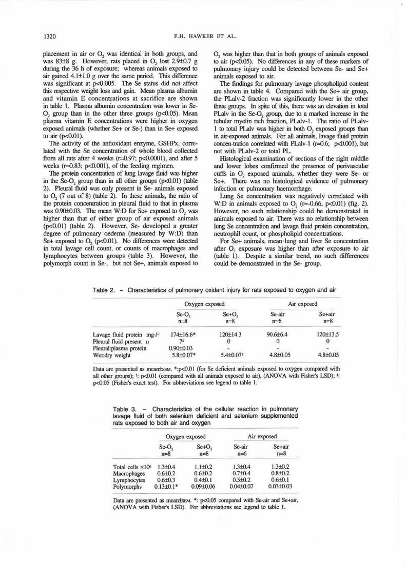
placement in air or 0 2 was identical in both groups, and was 83±8 g. However, rats placed in 0 2 lost 2.9±0.7 g during the 36 h of exposure; whereas animals exposed to air gained 4.1±1.0 g over the same period. This difference was significant at p<O.OOS. The Se status did not affect this respective weight loss and gain. Mean plasma albumin and vitamin E concentrations at sacrifice are shown in table 1. Plasma albumin concentration was lower in Se-02 group than in the other three groups (p<O.OS). Mean plasma vitamin E concentrations were higher in oxygen exposed animals (whether Se+ or Se-) than in Se+ exposed to air (p<O.Ol).
The activity of the antioxidant enzyme, GSHPx, correlated with the Se concentration of whole blood collected from all rats after 4 weeks (r=0.97; p<O.OOOI), and after 5 weeks (r=0.83; p<O.OOI), of the feeding regimen.
The protein concentration of lung lavage fluid was higher in the Se-02 group than in all other groups (p<O.Ol) (table 2). Pleural fluid was only present in Se- animals exposed to 0 2 (7 out of 8) (table 2). In these animals, the ratio of the protein concentration in pleural fluid to that in plasma was 0.90±{).03. 'The mean W:D for Se+ exposed to 0 2 was higher than that of either group of air exposed animals (p<0.01) (table 2). However, Se- developed a greater degree of pulmonary oedema (measured by W:D) than Se+ exposed to 0 2 (p<O.Ol). No differences were detected in total lavage cell count, or counts of macrophages and lymphocytes between groups (table 3). However, the polymorph count in Se-, but not Se+, animals exposed to
0 2 was higher than that in both groups of animals exposed to air (p<O.OS). No differences in any of these markers of pulmonary injury could be detected between Se- and Se+ animals exposed to air.
The findings for pulmonary lavage phospholipid content are shown in table 4. Compared with the Se+ air group, the PLalv-2 fraction was significantly lower in the other three groups. In spite of this, there was an elevation in total PLalv in the Se-02 group, due to a marked increase in the tubular myelin rich fraction, PLalv-1. The ratio of PLalvl to total PLalv was higher in both 0 2 exposed groups than in air-exposed animals. For all animals, lavage fluid protein concen-tration correla!ed with Pl..alv-1 (r=0.6; p<O.OOI), but not with PLalv-2 or total PL.
Histological examination of sections of the right middle and lower lobes confirmed the presence of perivascular cuffs in 0 2 exposed animals, whether they were Se- or Se+. There was no histological evidence of pulmonary infection or pulmonary haemorrhage.
Lung Se concentration was negatively correlated with W:D in animals exposed to 0 2 (r=-0.66, p<;O.Ol) (fig. 2). However, no such relationship could be demonstrated in animals exposed to air. There was no relationship between lung Se concentration and lavage fluid protein concentration, neutrophil count, or phospholipid concentrations.
For Se+ animals, mean ·lung and liver Se concentration after 0 2 exposure was higher than after exposure to air (table 1 ). Despite a similar trend, no such differences could be demonstrated in the Se- group.
Table 2. - Characteristics of pulmonary oxidant injury for rats exposed to oxygen and air
Oxygen exposed Air exposed
Se-02 Se+02 Se-air Se+air o=8 n=8 n=6 n=8
Lavage fluid protein mg·/·1 174±16.6* 120±14.3 90.6±6.4 120±13.5 Pleural fluid present n 71 0 0 0 Pleur.U:plasma protein 0.90±0.03 Wet:dry weight 5.8±{).07* 5.4±Q.07t 4.8±0.05 4.8±Q.05
Data are presented as mean±sEM. *:p<O.Ol (for Se deficient animals exposed to oxygen compared with all other groups); t: p<O.OI (compared with all animals exposed to air), (ANOVA with Fisher's LSD); t: p<0.05 (Fisher's exact test). For abbreviations see legend to table 1.
Table 3. - Characteristics of the cellular reaction in pulmonary lavage fluid of both selenium deficient and selenium supplemented rats exposed to both air and oxygen
Total cells xlQ6 Macrophages Lymphocytes Poly morphs
Oxygen exposed
Se-0 2 Se+02
n=8 n=8
1.3±{).4 0.6±Q.2 0.6±{).3
0.13±0.1*
1.1±0.2 0.6±Q.2 0.4±{).1
0.09±0.06
Air exposed ---Se-air Se+air
n=6 n=8
1.3±0.4 0.7±0.4 0.5±Q.2
0.04±0.07
1.3±0.2 0.8±{).2 0.6±Q.l
0.03±{).03
Data are presented as mean±sEM. *: p<0.05 compared with Se-air and Se+air, (ANOV A with Fisher's LSD). For abbreviations see legend to table l.
SELENIUM DEFICIENCY AND LUNG OXIDANT INJURY 1321
Table 4. - Characteristics of surfactant in pulrT1011a/y lavage fluid of bo1h Se deficient and Se supplemented rats exposed to bo1h air and oxygen (all samples from one Se+air animal and the Plalv-2 fraction from three Set02 animals were inadequate for analysis)
Data are presented as mean:fsEM. *: higher than all other groups (p<{l.05); t: higher than both groups of air-exposed animals, (ANOV A with Fisher's LSD). PL: phospholipid. For further abbreviations see legend to table 1.
Lung Se 1J.mOI·gm·1 dry wt Fig. 2. - Relationship between lWlg Se concentration and wet weight to dry weight ratio, both for Se supplemented (n=8) and Se deficient (n=8) rats exposed to 95% 02 for 36 h.
Discussion
The pulmonary injury caused by inhalation of high concentrations of 0 2 is similar in different animal species [3). It is characterized by an initial phase, when no morphological injury is apparent, followed by endothelial damage, proteinaceous pulmonary oedema, and an influx of platelets, macrophages and polymorphonuclear leucocytes into the lung [1, 3]. Abnormalities of alveolar surfactant have also been reported [251. After chronic 0 2 exposure, there is proliferation of type n pnewnocytes and subsequent development of interstitial fibrosis [lj. The pleural effusion is a characteristic fean.ue of pulmonary 0 2 toxicity .in the rat [3].
In this study, manifestations of pulmonary 0 2 toxicity were more severe in Se deficient animals. An alveolar protein leak and exudative pleural effusions were only present in Se- animals, and W:D (an index of lung water content) was higher in the Se- than in the Se+ group. Similar differences between the groups were obtained when the total lung water (g ~O·g-' dry lung weight), or the percentage of water in the lung were calculated. Although
our method of measuring W:D does not correct for the retained blood volume [26], the W:D for both 0 2 exposed groups were greater than that of control rat blood (5.27±0.03), indicating that at least some oedema was present After 0 2 exposure, alveolitis, characterized by an increased polymorph count in lavage fluid, only occurred in se· animals .
Differences in the amount and form of alveolar phospholipids were also observed. We processed the lavage fluid to obtain an aggregated fom1 of surfactant, which was significantly increased in amount in the Se-02 group. There was a less pronounced irtcrease in the Se+02 group, suggesting that the change in form of the surfactant was the result of pulmonary oxygen damage, rather than to Se deficiency. The less aggregated form of surfactant was reduced by both Se deficiency and oxygen exposure. This reduction in PLalv-2 in Se deficient animals exposed to air, when compared with Se supplemented animals breathing air, is the only evidence from this study that Se deficiency may have adverse effects in the absence of oxygen exposure.
A previous study in rats exposed to greater than 95% 0 2 for 64 h showed a similar increase in total PLalv [27]. However, other studies have yielded variable results, depending upon exposure conditions, animal species, and handling of the lavage fluid [25, 28]. Moreover, a similar change in disposition of surfactant has been docwnented by YOUNG et al. [25]. These changes may be mediated by SPA, an alveolar glycoprotein which associates with surfactant PL and calcium to form aggregates of PL and tubular myelin [29, 30]. SP-A has been observed to increase progressively in rats exposed to 85% 0 2 from 3 days postexposure [31J. Even after 36 b of 0 2 exposure, it is likely that SP-A is increased in the alveolus. lhis might explain the increase in aggregated surfactant, PLalv-1, that we have noted. On the other hand, we found that PLalv-1 correlated with lavage protein concentration. Leakage of serum protein into the alveolus may result in aggregation of surfactant PL, and may, therefore, be the cause of the increased Pl..alv-1 that we observed. Certainly, surfactant function is inhibited by serum proteins [32], although this inhibitory effect can be moderated in the presence of higher surfactant PL concentrations l33].
Seleniwn deficiency has been shown to result in increased mortality with hyperoxia in the rat [11, 12, 34]. CRoss et
1322 F.H. HAWKER ET AL.
al. (11] observed 35% mortality in Se deficient rats exposed to 80% 0 2 for 3 day.s, whereas survival was 100% in Se supplemented animals. Blood and tissue Se concentrations were not measured. Other studies have shown a lower median lethal dose (LDs0) for >95% 0 2 in Se deficient rats than in Se supplemented controls [12, 35]. Rats fed a Se deficient diet also show enhanced toxicity to hyperbaric 0 2 [35], and to paraquat [36].
It is likely that this augmenting effect of Se deficiency on pulmonary oxidant injury is due to the decreased Se concentration in lung tissue. Moreover, the inverse correlation observed between W:D and lung Se concentration in 0 2 exposed animals suggests that reduced levels of Se within the lung may play a role in the development of pulmonary oedema. However, we could not demonstrate a relationship between lung Se concentration and other marl<ers of pulmonary 0 2 toxicity. Furthermore, the finding of higher lung Se concentration after exposure to 95% 0 1
for 36 h than after air exposure in Se supplemented animals suggests that Se is redistributed into the lungs in response to oxidant stress. This has been confirmed in subsequent experiments (Hawker, unpublished data). This movement of Se into the lungs may be a protective mechanism with hyperoxia. No increase in lung Se in response to 0 2 breathing could be shown in Se deficient animals. This suggests that adequate Se status is critical for this adaptive response. No previous study of lung Se concentrations after 0 2 exposure has been reported.
Our study shows a close correlation between Se conpentration and GSHPx activity in whole blood The activity of GSHPx in lung and blood of the rat has been shown to be closely related [12, 37]. Although we did not measure GSHPx activity in pulmonary tissue, we speculate that limited availability of Se in the lung resulted in reduced lung GSHPx activity in Se deficient animals, as has been reported in previous studies of Se deficient rats [12, 35, 37, 38). Lungs from Se deficient rats have reduced capacity to metabolize infused tertbutyl hydroperoxide, an organic hydroperoxide usually metabolized by GSHPx [39]. These findings strongly suggest that Se deficient lung has reduced activity of GSHPx and, hence, limited capacity to metabolize Hz02 and other perox.ides. Under these circwnstances, it is likely that oxygen free radicals, particularly ~02, mediate the capillary endothelial injury. The resulting proteinaceous pulmonary oedema occurs earlier in Se supplemented animals. It is also possible that ~02 may have yet undescribed effects on surlactant metabolism, particularly with respect to SP-A.
Only one replacement dose of Se was used in this study. The Se dose of 400 J..lg·/·1 is similar to that used in other studies, when rats have been fed a baseline diet of Sedeficient chow [35). Whole blood and plasma Se concentrations in supplemented animals were 37% and 19% lower, respectively, than in other rats from this laboratory, fed different chow. Although these observations might suggest that our selenium supplementation regimen was suboptimal, there is wide variation in blood selenium levels in humans from different parts of the world Regaxdless of whether the Se status of our control animals was truly normal, deficient animals showed a marked and consistent increase in severity of markers of pulmonary oxygen toxicity by comparison.
This study does not address the question of whether Se repl!acement to higher levels would further increase protection from oxidant injury.
Possible confounding effects, which might have influenced our findings, include other nutritional deficiencies, infection, and known effects of Se deficiency on cardiac function.
Both groups were fed the same chow and vitamin E supplementation in drinking water, and Se was added only to the water of the supplemented group. Significant differences in protein calorie nutrition are unlikely as Se deficient and Se supplemented animals grew at the same rate. Plasma albumin concentration was lower in Se- animals exposed to oxygen than in Se- air exposed animals. We suggest that this does not represent nutritional deficiency, but rather is the result of albumin loss into pleural effusions and pulmonary oedema fluid. Plasma vitamin E concentrations were similar in both the Se- and Se+ groups, but tended to increase after 0 2 exposure. This, previously unreported, finding may represent an adaptive response to hyperoxia, and warrants further investigation. However, torula yeastbased diets, such as the one used in this study, are also defident in cysteine and methionine, which appear to have antioxidant activity in the nit [12]. Concomitant deficiency of these sulphur containing amino acids in both Se deficient and Se supplemented animals may have occurred, but cannot explain the differences between these groups.
It is possible, that Se deficiency might predispose to an increased incidence of infection. However, these SPF rats were housed, handled and fed under strictly sterile conditions, and there was no evidence of infection by common rat pathogens on serological testing or histological examination of the lungs.
Selenium deficiency has been associated with cardiomyopathy. Cardiac failure has been observed in Se deficient animals, in humans living in endemic areas of Se deficiency and in Se deficient long-term total parenteral nutrition (fPN) patients [39]. However, our findings do not support any suggestion that pulmonary oedema resulted from crudiac failure in this study. Firstly, the high protein concentrations in both pulmonary lavage fluid and the pulmonary effusions support a noncardiogenic cause. In addition, in a previous study, we observed no histological abnormality in the hearts of Se deficient rats exposed to a similar concentration of 0 2 for a similar period (Hawker, unpublished observations).
The findings in this study have potential clinical relevance. Low plasma Se concentrations have been reported in critically ill adults [7], and neonates [8). Both of these groups are frequently exposed to mechanical ventilation and high inspired 0 2 concentrations. There is evidence that the clinical course of patients with ARDS is aggravated by inhalation of high concentrations of 0 2 and the histological features of acute lung injury are worsened by hyperox.ia (40]. Abnormalities of surfactant metabolism are common in neonates, and have been observed in patients with ARDS.
The evidence presented in this study demonstrates that Se deficiency can augment pulmonary injwy from high concentrations of inspired oxygen. 'This suggests that dietary Se is important in the anti-oxidant defences of the rat lung. These fmdings may have implications in human disease states requiring high concentrations of inspired oxygen.
SELENIUM DEFICIENCY AND LUNG OXIDANT INJURY 1323
Aclcnow/edgements: The authors thank T .E. Nicholas and H.A. Barr for measurement of alveolar phospholipids, and J. Bums for glutathione peroxidase measurements.
References
I. Deneke SM, FanbUIE BL. - Normobaric oxygen toxicity of the lung. N Engl J Med 1980; 303: 76-86. 2. Sznajder JI, Fraiman A, Hall JB, et ut. - Increased hydrogen peroxide in the expired breath of patients with acute hypoxcmic respiratory failure. Chest 1989; 96: 606-612. 3. Klein J. - No.rmobaric pulmonary oxygen 1oxicity. Anestlt Analg 1990: 70: 195-207 .. 4. Combs GF, Combs SB. - The nutritional biochemistry of selenium. Ann Rev Nulr 1984; 4: 257-280. 5. Rotruck JT, Pope AL. Ganther HE, Swanson AB, Hafeman DG, Hoekstra WG. - Selenium: biochemical role as a component of glutathione peroxiclasc. Science 1973; 179: 58&-590. 6. Van Rij AM, Thomson CD, McKenzie JM, Robinson MF. - Selenium deficiency in total parenteral nulrition. Am J Clin Nutr 1979; 32: 2076-2085. 7. Hawker FH, Stewart PM, Snitch PJ. - Effects of acute iUness on selenium homeostasis. Crit Care Med 1990; 18: 442-446. 8. Lockitch G, Jacobsen B, Quigley G, Dison P, Pendray M. -Selenium deficiency in low birth weight neonates: an unrecognized problem. J Pediatr 1989; 114: 865-870. 9. Aatt A, Pearce N, Thomson CD, Sears MR., Robinson M, Beaslcy R. - Reduced selenium in asthmatic subjects in New Zealand. Thorax 1990; 45: 95-99. 10. Stead RJ, Redington AN, H.inks U, Clayton BE, Hodson ME, Batten JC. - Selenium deficiency and possible increased risk of carcinoma in adults with cystic fibrosis. lAncet 1985; ii: 862-863. 11 . Cross CE, Hasegawa G, Reddy KA. Omaya ST. - Enhanced lung toxicity of 02 in selenium-deficient rats. Res Comm Chem Path Phann 1977; 16: 695-706. 12. Fonnan HJ, Rotman El, Fisher AB. - Roles of selenium and sulfur-conta.ining amino acid~ in protection against oxygen toxicity. lAb Invest 1983; 49: 148-153. 13. Lowry OH. Roscbrough NJ, Farr AL. Rnndal.l RJ. - Protein measurement with the folin phenol reagent. J Bioi Chem 1951; 193: 265-275. 14. Magoon MW, Wrigbt JR, Baritussio A, et al. - Subfractionation of lung surfactant: implications for metabolism and surface activity. Biochim Biophys Acta 1983; 750: 18-31. 15. Thet LA, Oerch L, Massaro GO, Massaro D. - Changes in sedimentation of surfactant in ventilated excised rat lungs. Physical alterations in surfactant associated with the development and reversal of atelectasis. J Clin Invest 1979; 64: 600-608. 16. Pleban PA, Munyani A, Beacbum J. - Determination of selenium concentration and glutathione peroxidase activity in plasma and erythrocyteS. Clin Chem 1982; 28: 311- 316. 17. Hobbins WB. - Selenium determination by hydride generation. Varian Instruments at work. Number AA-11, January 1981. 18. Bligb EF, Dyer WJ. - A rapid method of total lipid extraction and purification. Can J Biochem 1959; 37: 911-917. 19. BartJett GR. - Phosphorus assay in column chromatography. J Bioi Chem 1959: 34: 466-468. 20. Nicholas TE. Barr HA. - Control of release of surfactant
phospholipids in the isolated perfused rat lung. J Appl Physiol: Respirat Environ Exercise Physiol 1981; 51: 90-98. 21. Rodkey FL. - Direct spectrophotomelric determination of albumin in human serum. Clin Chem 1965; 11: 47&-487. 22. Cannon DC, Olitzky I, Inkpen JA. - Clinical Chemistry: Principles and Technics. In: Hendry RJ, et al. eds. New York, Harper and Row, 1974; pp. 411-413. 23. Nilsson B, Johansson B. Jansson L, Holmberg L. -Determination of plasma alpha-tocopherol by high-performance liquid chromatography. J Chromatogr 1978; 145: 169-172. 24. Hintz.e JL. - Number Cruncher Statistical System (version 5.01). JL Hintz.e, Kaysville, USA, 1987. 25. Young SL, Crapo ID, Kremers JA, Brumley GW. -Pulmonary surfactant lipid production in oxygen-exposed rat lungs. lAb Invest 1982; 46: 570-576. 26. Morton LP, Yamashita J, Beazell J. - Measurement of pulmonary edema. Circ Res 1965; 16: 482-488. 27. Valimaki M, Pelliniemi T-T, Niinikoski J. - Oxygeninduced changes in pulmonary phospholipids in the rat. J Appl Physiol 1975; 39: 780-787. 28. Holm BA, Natter RH, Siegle J, Matalon S. - Pulmonary physiological and surfactant changes during injury and recovery from hyperoxia. J Appl Physiol 1985; 59: 1402- 1409. 29. WiUiams MC, Benson BJ. - Immunocytochemical localisation and identification of the major surfactant protein in adult rat lung. J Histochem Cytochem 1981; 29: 291- 305. 30. Hawgood S, Benson BJ, Hamilton RL. - Effect of a surfactant associated protein and calcium ions on the sbucture and surface activity of lung and surfactant lipids .. Biochemistry 1985; 24: 184-190. 31. Nogee LM, Wispe JR., Clack JC, Whitsett JA. - Increased synthesis and mRNA of surfactant protein A in oxygen exposed rats. Am J Respir Cell Mol Bio/1989; 1: 119-125. 32. Said SI, Avery ME, Davies KE, Bannerjee CM, EI-Gohary M. - Pulmonary surface activity in induced pulmonary oedema. J Clin Invest 1964; 44: 45&-464. 33. Holm BA, Notter RH, Finkelstein JN. - Surface property changes from interactions of albumin with natural lung surfactant and extracted lung lipids. Chem Phys Lipids 1985; 38: 287-298. 34. Jenkinson SG. Long RJ, Lawrence RA. - Endotoxin protects selenium-deficient rats from hyperoxia. 1 Clin lAb Med 1984; 103: 143-151. 35. Jenkinson SG, Jordan JM, Duncan CA. - Effects of selenium deficiency on glutathione-induced protection from hyperbaric hyperoxia in rat Am J Physioll989; 257: L393-L398. 36. Glass M, Sutherland MW, Forman HJ, Fisher AB. -Selenium deficiency potentiates paraquat-induced lipid peroxidation in isolated perfused rat lung. J Appl Physiol 1985; 59: 619-622. 37. Whanger PO, Butler JA. - Effects of various dietary levels of selenium as selenite or selenomethionine on tissue selenium levels and glutathione peroxida<;e activity in l"'clts. J Nutr 1988; 118: 846-852. 38. Jenkinson SG, Spence TH. Lawrence RA, Hill KE, Duncan CA, Johnson KH. - Rat lung glutathione release: response to oxidative stress and selenium deficiency. J Appl Physiol 1987; 62: 55-60. 39. Young VR. - Selenium: a case for its essentiality in man. N Engl J Med 1981; 304: 1228-1230. 40. Smith RA, Venus B, Masood S, Carter MC. - Effects of hyperoxia in the presence of acute lung injury. Cri/ Care Med 1990; 18: 198-202.