23 RRJOMS | Volume 5 | Issue 4 | August, 2017 Research & Reviews: Journal of Material Sciences e-ISSN: 2321-6212 p-ISSN: 2347-2278 DOI: 10.4172/2321-6212.1000191 Self-Healing Hydrogels as Biomedical Scaffolds for Cell, Gene and Drug Delivery Weizhong Yuan* and Chunyao Wang Department of Polymeric Materials, School of Materials Science and Engineering, Tongji University, Shanghai 201804, P. R. China Review Article INTRODUCTION Self-healing can be defined as a character to heal the micro- or macro-breakage both on surface and in interior autonomously, restoring virgin intensity and function of the material, which is adopted from the living body [1,2] . Just like cell’s proliferation and differentiation aiding organ’s self-recovery, bond’s linkage between atoms on two interfaces helps abiotic material’s self-healing. Over the past few years, self-healing and self-recovery have attracted much attention because it can keep the material durable and safer. Once breakage appeared, keep the fresh surface exposed to each other for a period of time, so that the material can heal itself [3] . On the macro level, the two parts of the fracture can form a surface whose crack is basically invisible, even smoother than new skin. On the micro level, the two are merged and permeated, finally get the compared strength with the initial one, which is shown in Figure 1. Self-healing would be ideal for vulnerable applications, such as surface coatings, supercapacitor and so on [4-7] . Hydrogels storing much water among 3D network are regarded as an important class of biomaterial based on the similar structure with extracellular matrix (ECM). Hydrogels can form in a variety of physical forms, including nanoparticles, microparticles, coatings, films and slabs [8] . However, hydrogel which usually possesses poor mechanical property is fragile to destroy the integrity of the structure. The biomaterials, with self-healing property, behave integrally all the time so that they cut off from the complicated environment. In other words, the self-healing endows the material more than one life. There is much potential relevance of self- healing hydrogels in the fields of biosensors, wound dressing, shape memory material and biomedical carrier [9-17] . Traditionally, damaged materials are repaired by welding, gluing, nailing or patching. Self-healing enables a material to repair crack itself with minimal interference. The following five steps illustrate the mechanism of self-healing for all kinds of materials: 1) A damage causes a crack in the material. 2) The resulting "mobile phase" is aroused by damage (in an ideal situation) or by external stimuli. 3) The damage can be gradually filled up because the directed substance transport to the damaged site and subsequent local mending reaction. 4) The distance between the molecules is near enough to ensure that the crack surface is connected by physical interaction and/or chemical bonds. 5) After the wound healing, the molecules distribute uniformly, resulting in the best mechanical performance of the perfect recovery [1] . However, the required condition may differ for different materials due to their intrinsic properties. There is no doubt that self-healing can be applied more easily to polymers than to metals or ceramics, due to the special molecular structure of the polymer and the range of temperatures they use. The healing agents in forth step is of ABSTRACT Self-healing polymeric hydrogels have the capability to recover their structures and functionalities upon injury, which are extremely attractive in emerging biomedical applications. Self-healing hydrogels with multifunctional properties as an important biomaterial class have been widely used in delivering cell, gene, drug and so on. Except high water content similar with extracellular matrix (ECM), hydrogels also need meet biocompatible scaffold requirements such as degradable, tough, injectable and good adhesiveness, and are developing more functional and smarter. Based on the significant studies of the biomedical scaffold in recent years, we review the widely used mechanism of the self-healing hydrogels, with focus on the design of the biocompatible scaffold and their relevant biomedical applications. The prospective regarding of the future potential development in functional and smart aspects is also delivered. Received: 01/09/2017 Accepted: 11/09/2017 Published: 21/09/2017 *For Correspondence Weizhong Yuan, Department of Polymeric Materials, School of Materials Science and Engineering, Tongji University, Shanghai 201804, P. R. China, Tel: 15821212297 . Email: [email protected]Keywords: Self-healing hydrogels; Extracellular matrix, Nanoparticles
Transcript
23RRJOMS | Volume 5 | Issue 4 | August, 2017
Research & Reviews: Journal of Material Sciences e-ISSN: 2321-6212 p-ISSN: 2347-2278
DOI: 10.4172/2321-6212.1000191
Self-Healing Hydrogels as Biomedical Scaffolds for Cell, Gene and Drug DeliveryWeizhong Yuan* and Chunyao Wang
Department of Polymeric Materials, School of Materials Science and Engineering, Tongji University, Shanghai 201804, P. R. China
Review Article
INTRODUCTIONSelf-healing can be defined as a character to heal the micro- or macro-breakage both on surface and in interior autonomously,
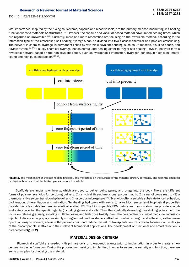
restoring virgin intensity and function of the material, which is adopted from the living body [1,2]. Just like cell’s proliferation and differentiation aiding organ’s self-recovery, bond’s linkage between atoms on two interfaces helps abiotic material’s self-healing. Over the past few years, self-healing and self-recovery have attracted much attention because it can keep the material durable and safer. Once breakage appeared, keep the fresh surface exposed to each other for a period of time, so that the material can heal itself [3]. On the macro level, the two parts of the fracture can form a surface whose crack is basically invisible, even smoother than new skin. On the micro level, the two are merged and permeated, finally get the compared strength with the initial one, which is shown in Figure 1. Self-healing would be ideal for vulnerable applications, such as surface coatings, supercapacitor and so on [4-7].
Hydrogels storing much water among 3D network are regarded as an important class of biomaterial based on the similar structure with extracellular matrix (ECM). Hydrogels can form in a variety of physical forms, including nanoparticles, microparticles, coatings, films and slabs [8]. However, hydrogel which usually possesses poor mechanical property is fragile to destroy the integrity of the structure. The biomaterials, with self-healing property, behave integrally all the time so that they cut off from the complicated environment. In other words, the self-healing endows the material more than one life. There is much potential relevance of self-healing hydrogels in the fields of biosensors, wound dressing, shape memory material and biomedical carrier [9-17].
Traditionally, damaged materials are repaired by welding, gluing, nailing or patching. Self-healing enables a material to repair crack itself with minimal interference. The following five steps illustrate the mechanism of self-healing for all kinds of materials: 1) A damage causes a crack in the material. 2) The resulting "mobile phase" is aroused by damage (in an ideal situation) or by external stimuli. 3) The damage can be gradually filled up because the directed substance transport to the damaged site and subsequent local mending reaction. 4) The distance between the molecules is near enough to ensure that the crack surface is connected by physical interaction and/or chemical bonds. 5) After the wound healing, the molecules distribute uniformly, resulting in the best mechanical performance of the perfect recovery [1]. However, the required condition may differ for different materials due to their intrinsic properties. There is no doubt that self-healing can be applied more easily to polymers than to metals or ceramics, due to the special molecular structure of the polymer and the range of temperatures they use. The healing agents in forth step is of
ABSTRACT
Self-healing polymeric hydrogels have the capability to recover their structures and functionalities upon injury, which are extremely attractive in emerging biomedical applications. Self-healing hydrogels with multifunctional properties as an important biomaterial class have been widely used in delivering cell, gene, drug and so on. Except high water content similar with extracellular matrix (ECM), hydrogels also need meet biocompatible scaffold requirements such as degradable, tough, injectable and good adhesiveness, and are developing more functional and smarter. Based on the significant studies of the biomedical scaffold in recent years, we review the widely used mechanism of the self-healing hydrogels, with focus on the design of the biocompatible scaffold and their relevant biomedical applications. The prospective regarding of the future potential development in functional and smart aspects is also delivered.
Weizhong Yuan, Department of Polymeric Materials, School of Materials Science and Engineering, Tongji University, Shanghai 201804, P. R. China, Tel: 15821212297 .
Research & Reviews: Journal of Material Sciences e-ISSN: 2321-6212 p-ISSN: 2347-2278
DOI: 10.4172/2321-6212.1000191
vital importance. Inspired by the biological systems, capsule and blood vessels, are the primary means transmitting self-healing functionalities to materials or structures [18]. However, the capsule and vascular-based material have limited healing times, which are regarded as irreversible [19]. Currently, more and more researches are focusing on the reversible method. According to the interaction type of the crosslinker, self-healing hydrogels can be divided into two classes: chemical and physical crosslinking. The network in chemical hydrogel is permanent linked by reversible covalent bonding, such as DA reaction, disulfide bonds, and acylhydrazone [20-25]. Usually chemical hydrogel needs stimuli and healing agent to trigger self-healing. Physical network form a reversible network based on the non-covalent bonds, such as hydrophobic interaction, hydrogen bonding, π-π stacking, metal-ligand and host-guest interaction [26-35].
Figure 1. The mechanism of the self-healing hydrogel. The molecules on the surface of the material stretch, permeate, and form the chemical or physical bonds so that the broken pieces restore to a whole.
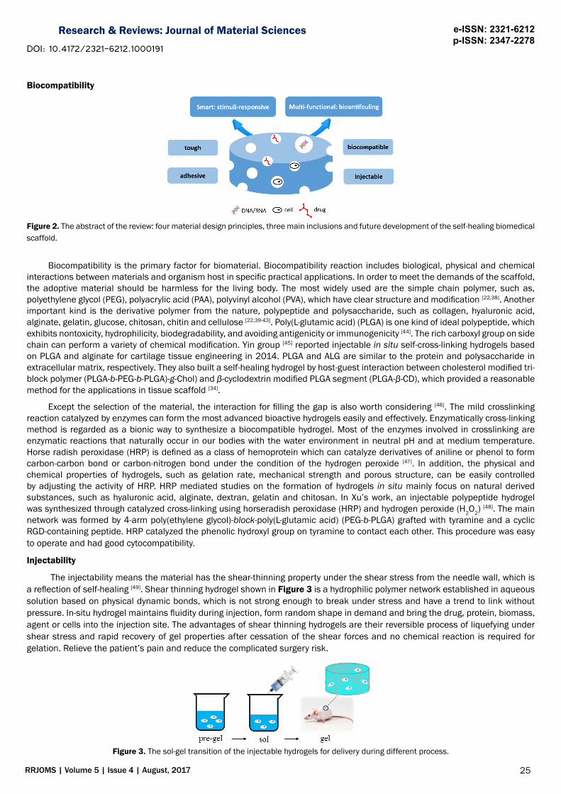
Scaffolds are implants or injects, which are used to deliver cells, genes, and drugs into the body. There are different forms of polymer scaffolds for cell/drug delivery: (1) a typical three-dimensional porous matrix, (2) a nanofibrous matrix, (3) a thermosensitive sol-gel transition hydrogel, and (4) a porous microsphere [36]. Scaffolds offer a suitable substrate for cell adhesion, proliferation, differentiation and migration. Self-healing hydrogels with easily tunable biochemical and biophysical properties provide many favorable features for medical scaffold [37]. The biocompatible ECM nature and porous structure provide enough and safe space for therapeutic agents (including gene) and cells. Then the gradually degrading crosslinking points help the inclusion release gradually, avoiding multiple dosing and high dose toxicity. From the perspective of clinical medicine, inclusions injected to tissue after prepolymer simply mixing formed random shape scaffold with certain strength and adhesion, so that make operation easy to operate, alleviate the patient's pain and reduce the risk of transplantation. This review focuses on the design of the biocompatible scaffold and their relevant biomedical applications. The development of functional and smart direction is prospected (Figure 2).
MATERIAL DESIGN CRITERIA Biomedical scaffold are seeded with primary cells or therapeutic agents prior to implantation in order to create a new
centers for tissue formation. During the process from mixing to implanting, in order to insure the security and function, there are some principles for choosing the material.
25RRJOMS | Volume 5 | Issue 4 | August, 2017
Research & Reviews: Journal of Material Sciences e-ISSN: 2321-6212 p-ISSN: 2347-2278
DOI: 10.4172/2321-6212.1000191
Biocompatibility
Figure 2. The abstract of the review: four material design principles, three main inclusions and future development of the self-healing biomedical scaffold.
Biocompatibility is the primary factor for biomaterial. Biocompatibility reaction includes biological, physical and chemical interactions between materials and organism host in specific practical applications. In order to meet the demands of the scaffold, the adoptive material should be harmless for the living body. The most widely used are the simple chain polymer, such as, polyethylene glycol (PEG), polyacrylic acid (PAA), polyvinyl alcohol (PVA), which have clear structure and modification [22,38]. Another important kind is the derivative polymer from the nature, polypeptide and polysaccharide, such as collagen, hyaluronic acid, alginate, gelatin, glucose, chitosan, chitin and cellulose [22,39-43]. Poly(L-glutamic acid) (PLGA) is one kind of ideal polypeptide, which exhibits nontoxicity, hydrophilicity, biodegradability, and avoiding antigenicity or immunogenicity [44]. The rich carboxyl group on side chain can perform a variety of chemical modification. Yin group [45] reported injectable in situ self-cross-linking hydrogels based on PLGA and alginate for cartilage tissue engineering in 2014. PLGA and ALG are similar to the protein and polysaccharide in extracellular matrix, respectively. They also built a self-healing hydrogel by host-guest interaction between cholesterol modified tri-block polymer (PLGA-b-PEG-b-PLGA)-g-Chol) and β-cyclodextrin modified PLGA segment (PLGA-β-CD), which provided a reasonable method for the applications in tissue scaffold [34].
Except the selection of the material, the interaction for filling the gap is also worth considering [46]. The mild crosslinking reaction catalyzed by enzymes can form the most advanced bioactive hydrogels easily and effectively. Enzymatically cross-linking method is regarded as a bionic way to synthesize a biocompatible hydrogel. Most of the enzymes involved in crosslinking are enzymatic reactions that naturally occur in our bodies with the water environment in neutral pH and at medium temperature. Horse radish peroxidase (HRP) is defined as a class of hemoprotein which can catalyze derivatives of aniline or phenol to form carbon-carbon bond or carbon-nitrogen bond under the condition of the hydrogen peroxide [47]. In addition, the physical and chemical properties of hydrogels, such as gelation rate, mechanical strength and porous structure, can be easily controlled by adjusting the activity of HRP. HRP mediated studies on the formation of hydrogels in situ mainly focus on natural derived substances, such as hyaluronic acid, alginate, dextran, gelatin and chitosan. In Xu’s work, an injectable polypeptide hydrogel was synthesized through catalyzed cross-linking using horseradish peroxidase (HRP) and hydrogen peroxide (H2O2)
[48]. The main network was formed by 4-arm poly(ethylene glycol)-block-poly(L-glutamic acid) (PEG-b-PLGA) grafted with tyramine and a cyclic RGD-containing peptide. HRP catalyzed the phenolic hydroxyl group on tyramine to contact each other. This procedure was easy to operate and had good cytocompatibility.
Injectability
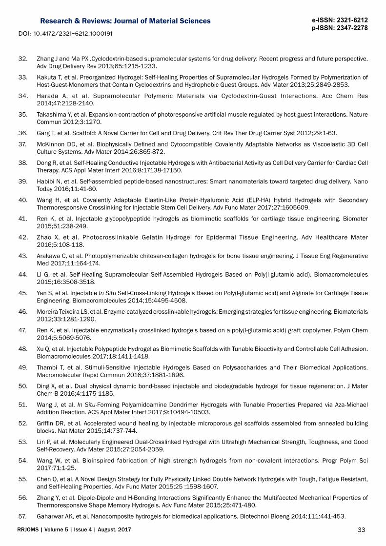
The injectability means the material has the shear-thinning property under the shear stress from the needle wall, which is a reflection of self-healing [49]. Shear thinning hydrogel shown in Figure 3 is a hydrophilic polymer network established in aqueous solution based on physical dynamic bonds, which is not strong enough to break under stress and have a trend to link without pressure. In-situ hydrogel maintains fluidity during injection, form random shape in demand and bring the drug, protein, biomass, agent or cells into the injection site. The advantages of shear thinning hydrogels are their reversible process of liquefying under shear stress and rapid recovery of gel properties after cessation of the shear forces and no chemical reaction is required for gelation. Relieve the patient’s pain and reduce the complicated surgery risk.
Figure 3. The sol-gel transition of the injectable hydrogels for delivery during different process.
26RRJOMS | Volume 5 | Issue 4 | August, 2017
Research & Reviews: Journal of Material Sciences e-ISSN: 2321-6212 p-ISSN: 2347-2278
DOI: 10.4172/2321-6212.1000191
Not every self-healing hydrogel can be used as injectable hydrogel. There are four basic principles to judge: 1) the spatial and temporal distribution of the active inclusion; 2) the ability to form and heal quickly after injection; 3) the constant release rate of the inclusion; 4) the matching rate of gel degradation to tissue development [50]. Juan Wang et al. synthesized and characterized the novel in situ-forming polyamidoamine (PAMAM) dendrimer hydrogels (DHs) with tunable properties prepared via highly efficient aza-Michael addition reaction [51]. Acetylation by acetic anhydride with different degree was performed on the basis of PAMAM dendrimer G5, so that hydrogel could be obtained with different curing time, rheological properties, network structure, expansion and decomposing properties, which is graphically depicted in Figure 4. Injectable hydrogels can provide a scaffold for in situ tissue regrowth and regeneration, yet gel degradation before tissue reformation limits the gels’ ability to provide physical support. Tuning of degradation rates based on local environment has been approached using hydrolytically and enzymatically degradable materials. However, decoupling cellular infiltration with decreases in material mechanical stability has proved extremely challenging. Griffin showed that this shortcoming can be circumvented through an injectable, interconnected microporous gel scaffold assembled from microgel building blocks whose chemical and physical properties can be tailored by microfluidic fabrication [52].
Figure 4. (A) Scheme of Acetylated G5 (G5-Acx) Synthesis and Aza-Michael Addition Reaction of G5 or G5-Ac with PEG-DA;(B-D) Effects of acetylation on morphologies, solidification time and disintegration of dendrimer hydrogels: (B) Solidification kinetics of dendrimer hydrogels as a function of the degree of acetylation and dendrimer concentration; (C) Disintegration in pH 7.4 PBS at 37 °C; (D) SEM micrographs (reproduced with permission) [51].
Mechanical Property
Hydrogels, due to its intrinsic structural inhomogeneity or lacking effective energy dissipation mechanism, usually have poor rheological and mechanical properties [53]. Although hydrogels seem to be fragile, there are many tissues such as (tendons, ligaments, meniscus and cartilage) even creatures (jellyfish and sea anemones) in hydrogel-like state [54]. They show excellent mechanical properties including softness, tenacity and impact resistance. These hydrogel-like tissues have fixed aggregation domains (some even include the crystal domain) in the mesoscale, induced by subtle, complicated, and multiple non-covalent interactions (intra-/inter-molecule). In this way, the combined mechanical strength of these hydrogel-like tissues has increased to an incredible level. Leaning from nature, the introduction of non-covalent crosslinking and proper molecular and structural design can deliver synthetic hydrogels to a set of attractive properties: improvements in strength, toughness, resilience, processability and dynamic adaptability.
27RRJOMS | Volume 5 | Issue 4 | August, 2017
Research & Reviews: Journal of Material Sciences e-ISSN: 2321-6212 p-ISSN: 2347-2278
DOI: 10.4172/2321-6212.1000191
In order to improve the mechanical properties of the gel, the usual method is to incorporate multiple crosslinking mechanism in a gel network structure [55,56]. For example, bond with lower strength, reversible and rapid response characteristics is used for rapid prototyping of the gel, and composite a higher strength, permanent, but slower reaction of bond is used to improve the stability of the gel. In previous studies, appears taking advantage of the molecular properties, such as the lower critical solution temperature (LCST), self-assembly and ionic crosslinking or forming supramolecular as the first line of crosslinking mechanism of rapid gelation. The method is selective to the molecules. A biodegradable polycation, mPEG-g-PEAD, was designed by Ding by simultaneously grafting arginine (Arg-OH) to polyethylene aspartate diglyceride (PED) and PEAD to monocarboxylic acid terminated PEG (mPEG-COOH). This polymer was then mixed with α-CD and heparin to form the supramolecular network. Rapid gelation is due to the simultaneous formation of branched polymer structures and biphysical crosslinking. The first crosslinking was formed by the inclusion of host-guest with the mPEG-graft and, and then self-assembly between the mPEG/α-CD composite string to generate the nanocrystalline domain as the main intersection point. At the same time, pendent arginine motifs with positive electricity and negatively charged heparin formed strong electrostatic interaction as the secondary crosslink.
Another method is to improve the mechanical properties and rheological properties of composites by mixing them with fillers [57,58]. Especially the filler in nanoscale has the size effect, specific surface area effect and quantum tunneling effect. A large number of active sites can be dispersed uniformly in the polymer matrix, and they provide connection bond with the matrix to enhance the hardness of the hydrogel. Shimon Unterman et al. [59] designed an injectable nanocomposite hydrogel based on dextran aldehyde and a poly(amido amine) dendrimer doped with phyllosilicate nanoplatelet fillers. Balance of components allows for exfoliation of nanoplatelets, significantly changing macromer solution flow, facilitating injection and manipulation. Importantly, rheological and mechanical effects were dependent on aspect ratio, with high aspect ratio nanoplatelets having much stronger effects on mechanics and low aspect ratio nanoplatelets having stronger effects on rheology, enabling nearly independent control of rheological and mechanical properties, abstracted in Figure 5.
Adhesion Property
Tissue adhesive materials play an important role in biomedical scaffold. The adhesion property can be divided into two classes, tissue attachment and cell affinity.
Tissue attachment: The self-adhesion of the material can overcome invasive wound closure device, such as sutures and staples, which usually have the disadvantage of inclusion-leakage [60]. In the case of low adhesion of tissue, such as lung and spleen, the stress concentration of the external material may lead to the failure of the operation, and the sustained nerve damage and pain may exist. The commonly used adhesives are usually bad for strong adhesion strength (fibrin glue) or biocompatibility (cyanoacrylate adhesive). To enhance the adhesion of synthetic materials to various surfaces, researchers have studied and developed materials inspired by systems found in nature. For structure, the micro/nanofibrillar can be adopted from the feet of geckos, where the directioned nanoscaled hairs form Van der Waals attraction more than weight. For constitution, the 3,4-dihydroxyphenylalanine (DOPA) secreted by mussels that hardens to form an adhesive plaque and byssal thread complex is used for reference, which enables these organisms to anchor themselves to surfaces in wet, saline and turbulent environments [61-64]. Catechols can transform to quinone and then bond with the material on the surface of a biological substrate, which can provide adhesion under the condition of enzymes (for example, tyrosinase), chemical oxidants (for example, high iodide) or in an aerobic alkaline environment [65]. The catechol moiety is capable of forming strong complexes with metal ions. The structure provides locus for hydrogen bonding, and can be consolidated by follow up interactions (for example, hydrophobic and steric) [66]. With DOPA or catechol analogues (for example, dopamine) has the very good application prospects in embryonic membrane seal, Achilles tendon repair, wound suture technology, cell and drug delivery. Lu Han et al. have designed the polydopamine-polyacrylamide (PDA-PAM) hydrogel, with super stretchability, high toughness, stimuli-free self-healing ability, cell affinity and tissue adhesiveness. The enough number of the non-oxidized catechol group was the deliberate design for adhesion (Figure 6) [67]. It is also worth noting that the current hydrogels can be repeatedly adhered to different surfaces for many times, without losing the strength of attachment.
Cell affinity: Cell−cell interactions and the matrix−cell interactions play fundamental roles in regulating cellular functions, including adhesion, morphogenesis, migration, proliferation, differentiation, and gene expression. The most widely studied adhesive peptide in the biomaterials field is the tri-amino acid sequence, arginine-glycine-aspartate, or “RGD” [68]. Because of the high activity of the isolated peptides, the binding ligands within ECM provide anchorages for cell adhesion with precise spatial and temporal control [69]. Zhou et al. introduced the RGD ligand into polymeric or supramolecular substrate through the same self-assembly mechanism to form Fmoc-peptide hydrogels [70]. This rapid gelling material was observed to promote adhesion of encapsulated dermal fibroblasts through specific RGD-integrin binding, and subsequently proliferation and proliferation of cells.
28RRJOMS | Volume 5 | Issue 4 | August, 2017
Research & Reviews: Journal of Material Sciences e-ISSN: 2321-6212 p-ISSN: 2347-2278
DOI: 10.4172/2321-6212.1000191
Figure 5. Morphology and dispersion of nanofillers. (A) Cryo-TEM of dispersions of each of four nanofillers in either 20% dextran-aldehyde or 24.6% PAMAM dendrimer macromer solutions. Scale bars = 100 nm;(B) Representative X-ray diffraction spectra of MMT and LAP nanoplatelets in 20% dextran−aldehyde or 24.6% dendrimer; (C) Varying polymer−nanoplatelet ratios result in varying exfoliation (reproduced with permission) [59] .
Figure 6. (A)Synthetic process and schematic structure of the polydopamine-polyacrylamide(PDA-PAM) hydrogel; B) Photo (1): without DA prepolymerization, the hydrogel could not form. Photo (2): after DA prepolymerization, the hydrogel was cured. Photo (3): the hydrogel firmly adhered on the author’s arm; (C) The self-healing process of polydopamine-polyacrylamide (PDA-PAM) hydrogel (reproduced with permission) [67].
29RRJOMS | Volume 5 | Issue 4 | August, 2017
Research & Reviews: Journal of Material Sciences e-ISSN: 2321-6212 p-ISSN: 2347-2278
DOI: 10.4172/2321-6212.1000191
BIOMEDICAL SCAFFOLD INCLUSIONThere are numerous original papers, academic reviews and monographs focused on biomedical scaffold inclusion. This part
introduces three main inclusions: cell, gene and drug.
Cell Delivery
People suffering from organ damage every day, tissue engineering has emerged to address this need by creating transplantable tissues or organs for therapy, especially for the organs without self-healing ability [71,72]. Hydrogels are heavily hydrated materials finding use in tissue regeneration efforts as extracellular matrix substitutes. Traditional tissue engineering methods use a “top-down” approach, in which cells are seeded onto a scaffold and are expected to populate and permeate in the scaffold. Because of the limited diffusion properties of biomimetic scaffolds usually thin or non-vascular tissues, such as skin, bladder, and cartilage can adopt this way. Currently, the emerging “bottom-up” approach focuses on the fabrication of microscale tissue building blocks with a specific microarchitecture and assembling these units to engineer larger tissue constructs from the bottom up [73]. The cells can be mixed with different shape of matrix to form tissue building blocks. Various methods include cell-encapsulating microgels, cell aggregation, cell sheet and cell printing.
Chondrocyte tissue engineering: Articular cartilage is an elastic body with a high water content. Its main function is to bear and distribute the load and protect the bone under the cartilage. However, articular cartilage has almost no self-healing ability, due to the lack of blood supply and the nutritional delivery of extracellular matrix. Currently, the best way of cartilage damage is to implant a functional tissue culture hydrogel for cell growth. Jianqi Wang et al. prepared a high-strength hydrogel, which consisted of 4-arm star PEG functionalized with vinyl sulfone and short dithiol crosslinker, whose strength could compare with natural cartilage. The hydrogel with cartilage cell inclusion has been subcutaneously injected into the mice with severe immunodeficiency for 12 weeks [74]. Chondrocytes proliferated and were well preserved in spatial and temporal distribution of extracellular matrix was found in the gel. The results have been shown in Figure 7.
Figure 7. A) Fabrication of hydrogel with ideal network structure; B) Quantitation of relative chondrocyte cell density from samples harvested at 3, 6, 12 weeks; C)-G): ECM components production at different time points in the chondrocytes/hydrogel constructs over 12 weeks. Shown are representative images of Alcian blue staining (C) Safranin O staining; (D) Mason's trichrome staining; (E) Sirius red staining; (F) Anti-Col II immunohistochemistry staining; (G) on frozen sections of chondrocytes/hydrogel constructs at indicated time points (reproduced with permission) [74].
30RRJOMS | Volume 5 | Issue 4 | August, 2017
Research & Reviews: Journal of Material Sciences e-ISSN: 2321-6212 p-ISSN: 2347-2278
DOI: 10.4172/2321-6212.1000191
Cardiac tissue engineering: Myocardial infarction is one of the death rate cause both in developing country and in developed country [75]. Myocardial tissue is conductive, whose main function is transmitting electrical signals to induce the heartbeat. The significant reduction of functional cardiac cells leads to the myocardiac infraction. The disease gives rise to a series of complicated process, such as cell apoptosis, scar forming and dysfunction of the cardiac in function, structure and mechanical property, leading to the cardiac standstill. Cardiac muscle for adults is regarded as the lacking self-healing tissue. At present drug therapy only can add years of survival. Researches have shown that the orderly coupling between electrical signal and macro contraction plays a vital role in the development and function of the heart. Electrical stimulation causes hyperpolarization and depolarization of cells. The cells that work with the strength of the electric field may produce a potential for contraction. Stimulation can promote the heart development of embryonic stem cells and enhance the phenotype of myocardial cells. To rebuild the contraction of the infarcted heart, the electroactive materials, such as carbon nanotube (CNT), polypyrrole (PPy), polyaniline (PANI), have been widely studied and applied in the cardiac tissue engineering. Haitao Cui et al. reported electroactive tetraaniline (TA) was introduced to synthesize thermosensitive PolyNIPAM hydrogels [76]. Studies showed that the addition of 2-methylene-1,3-dioxepane (MDO) provided the PolyNIPAM-based gel with biodegradability, and the introduction of tetraaniline endowed these copolymers with desirable electrical properties and antioxidant activities. The encapsulated H9c2 cells (rat cardiac myoblast) remained highly viable in the gel matrices. The gel formation and histological analysis of rats were performed by subcutaneous injection to observe biocompatibility. Furthermore, the proliferation and intracellular calcium transients of H9c2 cells were also studied with (and without) electrical stimuli. Both in vitro and in vivo experimental results showed that electroactive hydrogels can be used as injectable biomaterials for myocardial tissue engineering.
Gene Delivery
At present, scientists have already certified the unique set of DNA and RNA sequences of pathological genes by hybridizing a DNA strand with a sequence of complementary nucleic acids. Gene therapy offers the potential to stimulate tissue regeneration and treat disease through the overexpression or silencing of target genes, including cancer and infection [77,78]. Therefore, it is of great significance to combine the genetic agents with appropriate material in a definite and reversible way. However, its clinical application is limited by the deficiency of safe and efficient methods for gene delivery. There are at least three biological barriers, including the cellular membrane, the nuclear membrane and chromosomal integrity. Hydrogels are developed for the gene penetrated as delivery systems to achieve high gene transfection efficiency. Especially for the DNA- or RNA-functionalized hydrogels have been demonstrated for potential applications in drug release, cell-free protein production, and DNA immunotherapy [79]. Li et al. created a DNA nanohydrogel through self-assembly process using three kinds of building units, respectively termed Y-shaped monomer A with three sticky ends (YMA), Y-shaped monomer B with one sticky end (YMB), and DNA linker (LK) with two sticky ends, which is for the disulfide linkages [80]. Liao constructed stimuli-responsive DNA-acrylamide-based hydrogel microcapsules whose chain is crosslinked by the nucleic acid duplexes and the formation of an assembling thin film hydrogel on surfaces [81]. As a result, hundreds of therapeutic genes could be self-assembled into multifunctional hydrogels with controllable diameters, targeted delivery, and controllable release inside the target cells.
RNA-based therapeutics, such as small-interfering (siRNAs), microRNAs (miRNAs), antisense oligonucleotides (ASOs), aptamers, synthetic mRNAs and CRISPR-Cas9, there is great potential to attack genes and genetic products that are currently untreatable, and to produce a new treatment model in the disease [82-84]. Since the first RNA interference report (RNAi) in 1998, there has been great interest in applying RNAi therapy to inhibit histopathological gene expression. During this process, a small double-stranded RNA molecule, called a small interfering RNA (siRNA), enters the cell to interact with RNA-induced silent complexes (RISC), and keeps a complementary messenger RNA (mRNA) expression in silence [85]. The siRNA/RISC complex can effectively inhibit cell expression of gene targeting. The application of siRNA is limited because there is some delivery challenge: 1) stability against serum nucleases; 2) evasion of the immune system; 3) avoidance of non-specific interactions with serum proteins; 4) prevention of renal clearance; 5) exit from blood vessels to reach target tissues; 6) negatively charged hence negative cell entry. It is necessary to design a carrier to surmount these challenges. A common strategy to address these challenges is to combine the cationic polymers such as, polyethylene imine (PEI), for stabilizing the siRNA with the hydrophilic polymers for shielding the surface of delivery vehicles, usually polyethylene glycol.
Wang developed an injectable hydrogel that was formed using the host-guest assembly interaction between polyethylene amines (PEI) and polyethylene glycol (PEG) for siRNA delivery [86]. The composites formed by modified polymer have higher transfection rate and durability than that of PEI. In high concentrations, the hydrogel has the characteristics of shear thinning and can be repaired quickly after trauma. The mice were tested by injection of cy5-sirna hydrogel into the rat myocardial tissue, which resulted in the expression of the gene silencing of green fluorescent protein by ingestion of Cy5-siRNA.
Drug Delivery
ECM and ECM-like materials have great potential as drug carriers. The hydrogel scaffold combined with various growth factors (GFs), peptides or other drugs shows the ability to control cell growth, migration, and survival. Many methods have been developed to control the delivery of drugs, such as degradation-based delivery systems, affinity-based delivery systems, immobilized drug
31RRJOMS | Volume 5 | Issue 4 | August, 2017
Research & Reviews: Journal of Material Sciences e-ISSN: 2321-6212 p-ISSN: 2347-2278
DOI: 10.4172/2321-6212.1000191
delivery systems, and electrically controlled drug delivery systems [87]. According to these methods, a specific structure or function group of a polymer could be designed into a hydrogel to control degradation time, bind drugs, or react to the environment. The hydrogel matrix acts as a drug delivery library, slowly releasing the controlled drugs, making the drug concentration in the blood more stable over a long period of time, also applied to larger molecules, such as polypeptides and proteins. Because the degradation or erosion of hydrogels is essentially the cause of the package release, and the diffusion mechanism plays a limited role. Therefore, frequent and high doses of the drug can be avoided, leading to a decrease in toxicity and potential side effects. The hydrogel matrix creates a water environment that prevents early degradation of the drug, increasing the half-life of the drug. Therefore, improving the stability of hydrogels has great significance in extending the release time. The gel degradation time can be increased by increasing the density of the gel interconnection point, decreasing the length of the molecular chain, and changing the peptide sequence.
Malgosia et al. synthesized the hydrogel of a physical and chemical double crosslinking mechanism based on methylcellulose and tested its release rate and biocompatibility as a drug carrier protein [88]. The work demonstrated the practical application of this injectable, long-lasting drug delivery system, which spinal cord injury (SCI) in rats can benefit from.
Oyen showed an injectable amphipathic consisting of the hexapeptide H-FEFQFK-NH2 [89]. A small molecule, a peptide, a
protein that acts as a representative of three major drugs are visualized directly in vivo fluorescence and nuclear imaging. This study illustrates the release mechanism of peptide hydrogel system: the erosion of hydrogel is responsible for the controlled release.
The formation of functional blood vessels in damaged areas is essential for repairing skin wounds and repairing certain tissue types such as bone and skeletal muscle. The newly formed vasculature provides oxygen and nutrients to support cell growth and metabolism. The transmission of vascular growth factor (GFs) is widely used to treat vascular growth. However, exogenous GFs, usually in the form of recombinant proteins in the clinical application, are hampered by stability and curative effect. The directly injection usually brings many problems, such as GF’s rapid degradation/degeneration, short half-life in the body and carcinogenic risk for excessive consumption. Therefore, it is an effective treatment to deliver the coated growth factor hydrogel to the specified location. Hsieh proposed a new type of chitosan-fiber protein interpenetrating network (CF-IPN) to form hydrogels with good self-healing and angiogenesis [90]. Self-healing hydrogels have injectable characteristics and are degraded 70% in two weeks. The vascular endothelial cells cultured in CF hydrogel can form structures similar to capillaries. In addition, in the egg cell space of zebrafish, the injection of CF hydrogel promoted angiogenesis and saved the blood circulation of the ischemic hind limbs of mice.
SUMMARY AND OUTLOOKSelf-healing hydrogels provide a lot of advantages for biomedical scaffold due to the easy-to-modify and similar-to-ECM
structure. In order to meet the challenges for the cell, gene and drug delivery, material design principles have been developed here. Biocompatible is the basement for a biomaterial, while mechanical property is the key point that the hydrogel should overcome. Injectability and adhesion are adopted for convenience and safety.
Based on the above performance of self-healing hydrogel, it has wide application prospect in biological vector. In order to broaden the application of hydrogel, there is still much room for smarter and multifunctional self-healing hydrogels. Stimuli-responsive hydrogel, especially quickly responding under mild conditions, form an attractive approach for the fabrication of “smart” responsive surfaces, membranes, sensors with various transduction mechanisms, micro/nanoactuators, and capsules [91-93]. If the hydrogel is synthesized from stimuli-responsive polymers, then changes in the surrounding aqueous solution (for example, pH, ionic strength, temperature, electric field, solvent or magnetic field) may cause conformation transition of the polymer chains, which form strands between crosslinking points in the polymer gel. In the variation of the swelling degree of the gel material, the transition of the spherical to coils of these lines is regarded as the volume change of the gel material, which refers to changes of many properties of the materials: refractive index, permeability, elastic modulus, interfacial tension, adhesion, etc. Responsiveness is regarded as a manifestation of smart materials. Multifunctional properties, such as ultrahigh strength, superabsorbent, conductive, antifouling, antimicrobial, may be intergrated into the same hydrogel as well in the future for “one injection, multiple treatment” [94-99].
ACKNOWLEDGMENTSThe authors thank the financial supports of the Project Sponsored by the Scientific Research Foundation for the Returned
Overseas Chinese Scholars, State Education Ministry and the National Key Technology R&D Program (no. 2012BAI15B061).
REFERENCES1. Hager MD, et al. Self-Healing Materials. Adv Mater 2010;22:5424-5430.
2. Taylor DL, et al. Self-Healing Hydrogels. Adv Mater 2016;28:9060-9093.
32RRJOMS | Volume 5 | Issue 4 | August, 2017
Research & Reviews: Journal of Material Sciences e-ISSN: 2321-6212 p-ISSN: 2347-2278
DOI: 10.4172/2321-6212.1000191
3. Cordier P, et al. Self-healing and thermoreversible rubber from supramolecular assembly. Nature 2008;451:977-980.
4. Wang H, et al. A Mechanically and Electrically Self-Healing Supercapacitor. Adv Mater 2014;26:3638-3643.
5. Wu XL, et al. Biomass-Derived Sponge-like Carbonaceous Hydrogels and Aerogels for Supercapacitors. ACS Nano 2013;7:3589-3597.
6. Tao F, et al. Self-Healable and Cold-Resistant Supercapacitor Based on a Multifunctional Hydrogel Electrolyte. ACS Appl Mater Interf 2017;9:15541-15548.
7. Cho SH, et al. Self-Healing Polymer Coatings. Adv Mater 2009;21 :645-649.
8. Hoare TR and Kohane DS. Hydrogels in drug delivery: Progress and challenges. Polym 2008;49:1993-2007.
9. Le Goff GC, et al. Hydrogel microparticles for biosensing. European Polym J 2015;72:386-412.
10. DeLouise LA, et al.Hydrogel-Supported Optical-Microcavity Sensors. Adv Mater 2005;17:2199-2203.
11. Konieczynska MD, et al. On-Demand Dissolution of a Dendritic Hydrogel-based Dressing for Second-Degree Burn Wounds through Thiol-Thioester Exchange Reaction. Angewandte Chemie International Edition 2016;55:9984-9987.
12. Huang KT, et al. Zwitterionic nanocomposite hydrogels as effective wound dressings. J Mater Chem B 2016;4:4206-4215.
13. Thet NT, et al. Prototype Development of the Intelligent Hydrogel Wound Dressing and Its Efficacy in the Detection of Model Pathogenic Wound Biofilms. ACS Appl Mater Interf 2016;8:14909-14919.
14. Vashist A, et al. Recent advances in hydrogel based drug delivery systems for the human body. J Mater Chem B 2014;2:147-166.
15. Jiang Z-C, et al. Shape Memory Polymers Based on Supramolecular Interactions. ACS Appl Mater Interf 2017;9:20276-20293.
16. Lv T, et al. Self-Restoration of Superhydrophobicity on Shape Memory Polymer Arrays with Both Crushed Microstructure and Damaged Surface Chemistry. Small 2017;13:1503402.
17. Merino S, et al. Nanocomposite Hydrogels: 3D Polymer-Nanoparticle Synergies for On-Demand Drug Delivery. ACS Nano 2015;9:4686-4697.
18. Bekas DG, et al. Self-healing materials: A review of advances in materials, evaluation, characterization and monitoring techniques. Composites Part B: Engineering 2016;87:92-119.
19. Jin H, et al. Thermally Stable Autonomic Healing in Epoxy using a Dual-Microcapsule System. Advanced Materials 2014;26:282-287.
20. Taynton P, et al. Heat- or Water-Driven Malleability in a Highly Recyclable Covalent Network Polymer. Advanced Materials 2014;26:3938-3942.
21. Yu F, et al. Multifunctional Hydrogel with Good Structure Integrity, Self-Healing, and Tissue Adhesive Property Formed by Combining Diels Alder Click Reaction and Acylhydrazone Bond. ACS Appl Mater Interf 2015;7:24023-24031.
22. Shao C, et al. A Self-Healing Cellulose Nanocrystal-Poly(ethylene glycol) Nanocomposite Hydrogel via Diels-Alder Click Reaction. ACS Sust Chem Eng 2017;5:6167-6174.
23. Canadell J, et al. Self-Healing Materials Based on Disulfide Links. Macromolecules 2011;44:2536-2541.
24. Lu S, et al. An injectable and self-healing hydrogel with covalent cross-linking in vivo for cranial bone repair. J Mate Chem B 2017;5:3739-3748.
25. Kuhl N, et al. Acylhydrazones as Reversible Covalent Crosslinkers for Self-Healing Polymers. Adv Func Mater 2015;25:3295-3301.
26. Mihajlovic M, et al. Tough Supramolecular Hydrogel Based on Strong Hydrophobic Interactions in a Multiblock Segmented Copolymer. Macromolecules 2017;50:3333-3346.
27. Cao J, et al. Multiple Hydrogen Bonding Enables the Self-Healing of Sensors for Human Machine Interactions. Angewandte Chemie International Edition 2017;56:8795-8800.
28. Li M, et al. A Free-Standing and Self-Healable 2D Supramolecular Material Based on Hydrogen Bonding: A Nanowire Array with Sub-2-nm Resolution. Small 2017;13:1604077.
29. Bhattacharjee S and Bhattacharya S. Phthalate mediated hydrogelation of a pyrene based system: a novel scaffold for shape-persistent, self-healing luminescent soft material. J Mater Chem A 2014;2:17889-17898.
30. Rao YL, et al. Stretchable Self-Healing Polymeric Dielectrics Cross-Linked Through Metal-Ligand Coordination. J Am Chem Soc 2016;138 :6020-6027.
31. Miyamae K, et al. Expansion-Contraction, and Shape-Memory Properties of a Preorganized Supramolecular Hydrogel through Host-Guest Interactions. Angewandte Chemie International Edition 2015;54:8984-8987.
33RRJOMS | Volume 5 | Issue 4 | August, 2017
Research & Reviews: Journal of Material Sciences e-ISSN: 2321-6212 p-ISSN: 2347-2278
DOI: 10.4172/2321-6212.1000191
32. Zhang J and Ma PX .Cyclodextrin-based supramolecular systems for drug delivery: Recent progress and future perspective. Adv Drug Delivery Rev 2013;65:1215-1233.
33. Kakuta T, et al. Preorganized Hydrogel: Self-Healing Properties of Supramolecular Hydrogels Formed by Polymerization of Host-Guest-Monomers that Contain Cyclodextrins and Hydrophobic Guest Groups. Adv Mater 2013;25:2849-2853.
34. Harada A, et al. Supramolecular Polymeric Materials via Cyclodextrin-Guest Interactions. Acc Chem Res 2014;47:2128-2140.
35. Takashima Y, et al. Expansion-contraction of photoresponsive artificial muscle regulated by host-guest interactions. Nature Commun 2012;3:1270.
36. Garg T, et al. Scaffold: A Novel Carrier for Cell and Drug Delivery. Crit Rev Ther Drug Carrier Syst 2012;29:1-63.
37. McKinnon DD, et al. Biophysically Defined and Cytocompatible Covalently Adaptable Networks as Viscoelastic 3D Cell Culture Systems. Adv Mater 2014;26:865-872.
38. Dong R, et al. Self-Healing Conductive Injectable Hydrogels with Antibacterial Activity as Cell Delivery Carrier for Cardiac Cell Therapy. ACS Appl Mater Interf 2016;8:17138-17150.
39. Habibi N, et al. Self-assembled peptide-based nanostructures: Smart nanomaterials toward targeted drug delivery. Nano Today 2016;11:41-60.
40. Wang H, et al. Covalently Adaptable Elastin-Like Protein-Hyaluronic Acid (ELP-HA) Hybrid Hydrogels with Secondary Thermoresponsive Crosslinking for Injectable Stem Cell Delivery. Adv Func Mater 2017;27:1605609.
41. Ren K, et al. Injectable glycopolypeptide hydrogels as biomimetic scaffolds for cartilage tissue engineering. Biomater 2015;51:238-249.
42. Zhao X, et al. Photocrosslinkable Gelatin Hydrogel for Epidermal Tissue Engineering. Adv Healthcare Mater 2016;5:108-118.
43. Arakawa C, et al. Photopolymerizable chitosan-collagen hydrogels for bone tissue engineering. J Tissue Eng Regenerative Med 2017;11:164-174.
44. Li G, et al. Self-Healing Supramolecular Self-Assembled Hydrogels Based on Poly(l-glutamic acid). Biomacromolecules 2015;16:3508-3518.
45. Yan S, et al. Injectable In Situ Self-Cross-Linking Hydrogels Based on Poly(l-glutamic acid) and Alginate for Cartilage Tissue Engineering. Biomacromolecules 2014;15:4495-4508.
46. Moreira Teixeira LS, et al. Enzyme-catalyzed crosslinkable hydrogels: Emerging strategies for tissue engineering. Biomaterials 2012;33:1281-1290.
47. Ren K, et al. Injectable enzymatically crosslinked hydrogels based on a poly(l-glutamic acid) graft copolymer. Polym Chem 2014;5:5069-5076.
48. Xu Q, et al. Injectable Polypeptide Hydrogel as Biomimetic Scaffolds with Tunable Bioactivity and Controllable Cell Adhesion. Biomacromolecules 2017;18:1411-1418.
49. Thambi T, et al. Stimuli-Sensitive Injectable Hydrogels Based on Polysaccharides and Their Biomedical Applications. Macromolecular Rapid Commun 2016;37:1881-1896.
50. Ding X, et al. Dual physical dynamic bond-based injectable and biodegradable hydrogel for tissue regeneration. J Mater Chem B 2016;4:1175-1185.
51. Wang J, et al. In Situ-Forming Polyamidoamine Dendrimer Hydrogels with Tunable Properties Prepared via Aza-Michael Addition Reaction. ACS Appl Mater Interf 2017;9:10494-10503.
52. Griffin DR, et al. Accelerated wound healing by injectable microporous gel scaffolds assembled from annealed building blocks. Nat Mater 2015;14:737-744.
53. Lin P, et al. Molecularly Engineered Dual-Crosslinked Hydrogel with Ultrahigh Mechanical Strength, Toughness, and Good Self-Recovery. Adv Mater 2015;27:2054-2059.
54. Wang W, et al. Bioinspired fabrication of high strength hydrogels from non-covalent interactions. Progr Polym Sci 2017;71:1-25.
55. Chen Q, et al. A Novel Design Strategy for Fully Physically Linked Double Network Hydrogels with Tough, Fatigue Resistant, and Self-Healing Properties. Adv Func Mater 2015;25 :1598-1607.
56. Zhang Y, et al. Dipole-Dipole and H-Bonding Interactions Significantly Enhance the Multifaceted Mechanical Properties of Thermoresponsive Shape Memory Hydrogels. Adv Func Mater 2015;25:471-480.
57. Gaharwar AK, et al. Nanocomposite hydrogels for biomedical applications. Biotechnol Bioeng 2014;111:441-453.
34RRJOMS | Volume 5 | Issue 4 | August, 2017
Research & Reviews: Journal of Material Sciences e-ISSN: 2321-6212 p-ISSN: 2347-2278
DOI: 10.4172/2321-6212.1000191
58. Li Q, et al. Controlling Hydrogel Mechanics via Bio-Inspired Polymer-Nanoparticle Bond Dynamics. ACS Nano 2016;10:1317-1324.
59. Unterman S, et al. Hydrogel Nanocomposites with Independently Tunable Rheology and Mechanics. ACS Nano 2017;11:2598-2610.
60. Barrett DG, et al. Mechanically Robust, Negative-Swelling, Mussel-Inspired Tissue Adhesives. Adv Healthcare Mater 2013;2:745-755.
61. Lee BP and Konst S . Novel Hydrogel Actuator Inspired by Reversible Mussel Adhesive Protein Chemistry. Adv Mater 2014;26:3415-3419.
62. Azevedo S, et al. Bioinspired Ultratough Hydrogel with Fast Recovery, Self-Healing, Injectability and Cytocompatibility. Adv Mater 2017;29:1700759.
63. Skelton S, et al. Biomimetic adhesive containing nanocomposite hydrogel with enhanced materials properties. Soft Matter 2013;9:3825-3833.
64. Cencer M, et al. Effect of pH on the Rate of Curing and Bioadhesive Properties of Dopamine Functionalized Poly(ethylene glycol) Hydrogels. Biomacromolecules 2014;15:2861-2869.
65. Li L, et al. Novel Mussel-Inspired Injectable Self-Healing Hydrogel with Anti-Biofouling Property. Adv Mater 2015;27:1294-1299.
66. Ahn BK, et al. Surface-initiated self-healing of polymers in aqueous media. Nat Mater 2014;13:867-872.
67. Han L, et al. Tough, self-healable and tissue-adhesive hydrogel with tunable multifunctionality. NPG Asia Mater 20179:372.
68. Bellis SL .Advantages of RGD peptides for directing cell association with biomaterials. Biomater 2011;32:4205-4210.
69. Lee TT, et al. Light-triggered in vivo activation of adhesive peptides regulates cell adhesion, inflammation and vascularization of biomaterials. Nat Mater 2015;14:352-360.
70. Zhou M, et al. Self-assembled peptide-based hydrogels as scaffolds for anchorage-dependent cells. Biomater 2009;30:2523-2530.
71. Shen X, et al. Hydrogels based on cellulose and chitin: fabrication, properties, and applications. Green Chemistry 2016;18:53-75.
72. Seliktar D .Designing Cell-Compatible Hydrogels for Biomedical Applications. Sci 2012;336:1124-1128.
73. Lu T, et al. Techniques for fabrication and construction of three-dimensional scaffolds for tissue engineering. Int J Nanomed 2013;8:337-350.
74. Wang J, et al. Fabrication of injectable high strength hydrogel based on 4-arm star PEG for cartilage tissue engineering. Biomater 2017;120:11-21.
75. Jang J, et al. 3D printed complex tissue construct using stem cell-laden decellularized extracellular matrix bioinks for cardiac repair. Biomater 2017;112:264-274.
76. Cui H, et al. In Vitro Study of Electroactive Tetraaniline-Containing Thermosensitive Hydrogels for Cardiac Tissue Engineering. Biomacromolecules 2014;15:1115-1123.
77. Zhang J, et al. Poloxamine/fibrin hybrid hydrogels for non-viral gene delivery. J Tissue Eng Regenerative Med 2017;11:246-255.
78. Khvorova A and WattsJK . The chemical evolution of oligonucleotide therapies of clinical utility. Nat Biotech 2017;35:238-248.
79. Li J, et al. Functional nucleic acid-based hydrogels for bioanalytical and biomedical applications. Chem Soc Rev 2016;45:1410-1431.
80. Li J, et al. Self-assembly of DNA Nanohydrogels with Controllable Size and Stimuli-Responsive Property for Targeted Gene Regulation Therapy. J Am Chem Soc 2015;137:1412-1415.
81. Liao W-C, et al. pH- and ligand-induced release of loads from DNA-acrylamide hydrogel microcapsules. Chem Sci 2017;8 :3362-3373.
82. Dowdy SF. Overcoming cellular barriers for RNA therapeutics. Nat Biotech 2017;35 :222-229.
83. Gilam A, et al. Local microRNA delivery targets Palladin and prevents metastatic breast cancer. Nat Commun 2016;7:12868.
84. Conde J, et al. Local triple-combination therapy results in tumour regression and prevents recurrence in a colon cancer model. Nat Mater 2016;15:1128-1138.
35RRJOMS | Volume 5 | Issue 4 | August, 2017
Research & Reviews: Journal of Material Sciences e-ISSN: 2321-6212 p-ISSN: 2347-2278
DOI: 10.4172/2321-6212.1000191
85. Conde J, et al. Self-assembled RNA-triple-helix hydrogel scaffold for microRNA modulation in the tumour microenvironment. Nat Mater 2016;15:353-363.
86. Wang LL, et al. Guest-Host Assembled Polyethylenimine Hydrogel for siRNA Delivery. Biomacromolecules 18;2017:77-86.
87. Wei L, et al. Drug-carrier/hydrogel scaffold for controlled growth of cells. Eur J Pharmaceutics Biopharmaceutics 201178:346-354.
88. Pakulska MM, et al. Hybrid Crosslinked Methylcellulose Hydrogel: A Predictable and Tunable Platform for Local Drug Delivery. Adv Mater 2015;27 :5002-5008.
89. Oyen E, et al. In Vivo Imaging of the Stability and Sustained Cargo Release of an Injectable Amphipathic Peptide-Based Hydrogel. Biomacromolecules 2017;18.
90. Hsieh F-Y, et al. A novel biodegradable self-healing hydrogel to induce blood capillary formation. NPG Asia Mater 2017;9:363.
91. Buwalda SJ, et al. Hydrogels in a historical perspective: From simple networks to smart materials. J Controlled Release 2014;190:254-273.
92. Doring A, et al. Responsive hydrogels - structurally and dimensionally optimized smart frameworks for applications in catalysis, micro-system technology and material science. Chem Soc Rev 2013;42:7391-7420.
93. Tokarev I and Minko S. Stimuli-responsive hydrogel thin films. Soft Matter 2009;5:511-524.
94. Chang C, et al. Superabsorbent hydrogels based on cellulose for smart swelling and controllable delivery. Eur Polym J 2010;46:92-100.
95. Xing R, et al. An Injectable Self-Assembling Collagen-Gold Hybrid Hydrogel for Combinatorial Antitumor Photothermal/Photodynamic Therapy. Adv Mater 2016;28:3669-3676.
96. Shi Y, et al. A Conductive Self-Healing Hybrid Gel Enabled by Metal-Ligand Supramolecule and Nanostructured Conductive Polymer. Nano Lett 2015;15:6276-6281
97. Zhang X, et al. Mechanically strong and highly conductive graphene aerogel and its use as electrodes for electrochemical power sources. J Mater Chem 2011;21:6494-6497.
98. Murosaki T, et al. Antifouling properties of hydrogels. Sci Technol Adv Mater 2011;12:064706.
99. Ghavami Nejad A, et al. In Situ Synthesis of Antimicrobial Silver Nanoparticles within Antifouling Zwitterionic Hydrogels by Catecholic Redox Chemistry for Wound Healing Application. Biomacromolecules 2016;17:1213-1223.