Primary Seeding Testing the self-seeding hypothesis with a mathematical model Jacob G Scott 1,2 , David Basanta 1 , Philip Gerlee 3,4 & Alexander RA Anderson 1 1. Integrated Mathematical Oncology, H. Lee Moffitt Cancer Center, USA 2. Centre for Mathematical Biology, Oxford University, UK 3. Sahlgrenska Cancer Center, University of Gothenburg, Sweden 4.Mathematical Sciences, Chalmers University of Technology, Sweden Lungs and heart Brain Liver Gut Bladder and prostate Bone η γ , 20130011, published 20 February 2013 10 2013 J. R. Soc. Interface NATURE REVIEWS | CANCER VOLUME 12 | JULY 2012 | 445 Kim et al. (2009) Cell Metastatic disease accounts for the lion’s share of cancer deaths, yet it is a process that remains poorly understood. Many theories of metastasis have been posited in ‘cartoon’ form. These include the well known ‘seed soil’ hypothesis, the idea that removal of the primary tumor somehow increases the growth of metastasis and most recently, the ‘self-seeding’ hypothesis (right). In this work, we aim to test the ‘self-seeding’ hypothesis with a theoretical construct we recently posited (below). Evidence for ‘self-seeding’? Models of metastasis should not ignore known vascular connectivity Scott et al. Scott et al. A simple model derived from Norton et al. (Nature Med 2006) iterated on the vascular network Data on CTC prevalence in vascular network taken from literature: an opportunity for future personalization ? Results suggest Secondary Seeding is more likely the mechanism behind ‘self- seeding’. This suggests that treatment of subclinical micromets in specific organs (organ directed therapy) could be predicted to have clinical utility given patient specific parameters. Tumor simulation dynamics p p λ λ shedding rate λ return rate p
Transcript
Primary Seeding
Testing the self-seeding hypothesis with a mathematical model
Jacob G Scott1,2, David Basanta1, Philip Gerlee3,4 & Alexander RA Anderson1
1. Integrated Mathematical Oncology, H. Lee Moffitt Cancer Center, USA 2. Centre for Mathematical Biology, Oxford University, UK 3. Sahlgrenska Cancer Center, University of Gothenburg, Sweden 4.Mathematical Sciences, Chalmers University of Technology, Sweden
Lungs and heart
Brain
Liver
Gut
Bladder and prostate
Bone
!
"
#
, 20130011, published 20 February 201310 2013 J. R. Soc. Interface Jacob G. Scott, David Basanta, Alexander R. A. Anderson and Philip Gerlee growthsecondary metastatic deposits as drivers of primary tumour A mathematical model of tumour self-seeding reveals
References http://rsif.royalsocietypublishing.org/content/10/82/20130011.full.html#ref-list-1 This article cites 33 articles, 9 of which can be accessed free
(210 articles)biomathematics Articles on similar topics can be found in the following collections
Email alerting service hereright-hand corner of the article or click Receive free email alerts when new articles cite this article - sign up in the box at the top
http://rsif.royalsocietypublishing.org/subscriptions go to: J. R. Soc. InterfaceTo subscribe to
on March 11, 2013rsif.royalsocietypublishing.orgDownloaded from
Unifying metastasis — integrating intravasation, circulation and end-organ colonizationJacob Scott1,2, Peter Kuhn3 and Alexander R. A. Anderson1
Abstract | Recent technological advances that have enabled the measurement of circulating tumour cells (CTCs) in patients have spurred interest in the circulatory phase of metastasis. Techniques that do not solely rely on a blood sample allow substantial biological interrogation beyond simply counting CTCs.
1Integrated Mathematical Oncology, Moffitt Cancer Center, Tampa, Florida 33612, USA.2Oxford University Centre for Mathematical Biology, Mathematical Institute, Oxford OX1 3LB, UK.3Department of Cell Biology, The Scripps Research Institute, La Jolla, California 92037, USA.Correspondence to A.R.A.A. and J.S. e-mails: [email protected]; [email protected]:10.1038/nrc3287Published online 24 May 2012
In patients with advanced primary cancer, circulating tumour cells (CTCs)1 can be found throughout the entire vascular system2. When and where these CTCs form metastasis is not fully understood, and is currently the subject of intensive biological study. Paget’s well-known seed–soil hypothesis3 suggests that the ‘soil’ (the site of a metastasis) is as important as the ‘seed’ (the metastatic cells) in the determination of successful metastasis. The mechanism by which seeds are disseminated to specific soil has, to date, been a ‘known unknown’. We think that it is during this poorly understood phase of metastasis that we stand to answer important questions4.
We hypothesize that the rich variety of possible meta-static disease patterns not only stems from the physical aspects of the circulation but also from CTC hetero-geneity (FIG. 1). These seeds represent many different populations that are derived from a diverse population of competing phenotypes within the primary tumour5. Because such seeds need to pass through a system of physical and biological filters in the form of specific organs, the circulatory phase of metastasis could be modelled as a complex deterministic filter. In theory, until the evolution of a suitable seed, any number of CTCs could flow through the circulation and arrest at end organs without metastases forming. As tumour heterogeneity is thought to expand as the tumour pro-gresses, it follows that at some point a seed will come into existence that is suited to a specific soil within that patient’s body. If this seed is to propagate it must find its soil, a process that we hypothesize is governed by solvable physical rules that relate to the dynamics of the circulatory flow between different organs and how these organs filter (not only by size, but probably also by other biological mechanisms). Although these biologi-cal mechanisms are not yet known, we might be able to infer their existence by finding out which measurements
do not fit a model that is defined only by physical flow and filtration.
To begin the process of physical interrogation, we propose a model that represents the human circulatory system as a directed and weighted network, with nodes representing organs and edges representing arteries and veins. The novelty is only fully realized when combined with a heterogeneous CTC population (driven by primary tumour heterogeneity) modulated by the complex organ filter system (with physiologically relevant connections) under dynamic flow. Four important biological processes emerge from this representation. First, the shedding rate, which is defined as the rate at which the tumour sheds CTCs into the vasculature. Second, CTC heterogeneity, which is defined as the distribution of CTC phenotypes present in the circulation. Third, the filtration fraction, which is defined as the proportion (and type) of CTCs that arrest in a given organ. Fourth, the clearance rate, which is defined as the rate at which cancer cells are cleared from the blood and/or organ after arrest. Each of these biological processes is probably disease- and even patient-specific, and each is extremely poorly understood.
Using this representation to motivate the develop-ment of a mathematical model we can define both the concentration of CTCs and their phenotypic distribu-tion at any given point in the network, as well as organ-specific filtration values. To parameterize this model, characterization and enumeration of CTCs taken from a single patient at different time points and from differ-ent points in this network will need to be undertaken. A complete understanding of the model will also pro-vide information about the behaviour of the system as a whole. Specifically, the average lifespan of a CTC in a patient’s circulation will be able to be calculated with only a minimum of measurements. Although this seems to be a simple calculation, the scientific literature on this
COMMENT
NATURE REVIEWS | CANCER VOLUME 12 | JULY 2012 | 445
and inoculated into one mammary gland in mice to forma ‘‘donor’’ tumor mass. Unlabeled MDA231-LM2 cells were inoc-ulated into a contralateral mammary gland to form a ‘‘recipient’’mass of the same tumor (Figure 1A). After 60 days, the recipienttumors were excised and examined for the presence of seedingcells by means of ex vivo bioluminescence imaging (BLI).A majority (85%) of the recipient tumors showed extensive seed-ing by MDA231-LM2 cells (Figure 1B and Table 1). Tumorsformed by the more indolent MDA231 parental population wereas effective as MDA231-LM2 tumors at capturing seed cells(Figure 1B and Table 1). No seeding was observed in mock-inoc-ulated mammary glands within the same time period (Figure 1C).
Fluorescence microscopy analysis of MDA231 recipienttumors confirmed the presence of numerous GFP+ MDA231-LM2 seeding cells as distinct patches typically encompassingless than a quarter of a tumor section (Figure 1D and data notshown). When recipient tumors were generated using red-fluorescent protein (RFP)-labeled cells, the infiltrating GFP+ cells
were observed intermingling with resident RFP+ cancer cells andwith unlabeled areas of presumptive tumor stroma (Figure 1E).Quantitative RT-PCR analysis of firefly-luciferase mRNA levelin seeded recipient tumors revealed that seeder cells accountedfor 5%–30% of the recipient tumor mass (data not shown).
To establish the generality of this seeding phenomenon, weperformed similar experiments with different cancer cell lines.Recipient mammary tumors became seeded with high frequency(53% to 100% of mice) by donor tumors that were formed withbone-metastatic (MCF7-BoM2), lung-metastatic (MDA231-LM2), or brain-metastatic (CN34-BrM2) cells from different sub-types of breast cancer (basal, estrogen receptor-negativeMDA231 cells versus luminal, estrogen receptor-positive MCF7cells) or patient-derived malignant cell cultures (CN34 cells)(Figure 1B and Table 1). Seeding of a recipient tumor by its ownaggressive progeny was also observed between subcutaneoustumors formed by the human colon carcinoma line SW620 andits lung-metastatic derivative SW620-LM1, and between the
Figure 1. Seeding of Established Tumors by CTCs(A) A diagram of contralateral seeding experiment. Unlabeled and GFP/luciferase-expressing breast cancer cells were injected into contralateral No. 2 mammary
glands as a ‘‘recipient tumor’’ and a ‘‘donor tumor,’’ respectively.
(B) BLI of recipient tumors extracted from mice bearing the indicated GFP/luciferase-expressing donor tumors. Color-range bars: photon flux. LM2: a lung-meta-
static derivative of MDA231. MCF7-BoM2: a bone-metastatic derivative of MCF7. CN34-BrM2: a brain-metastatic derivative of pleural effusion CN34. PyMT:
cells derived from mammary tumors developed in MMTV-PyMT transgenic mice.
(C) BLI of tumor-free and tumor-bearing mammary glands from mice bearing GFP/luciferase-expressing donor tumors. n = 9–18. Error bars represent SEM.
(D) Frozen sections of seeded MDA231-LM2 tumors were visualized by fluorescence microscopy. An entire tumor section and a higher-magnification image (310)
of a selected field are shown.
(E) A contralateral seeding experiment was performed with RFP- and GFP-expressing MDA231-LM2 cells. Frozen sections from RFP-labeled tumors were
visualized under confocal microscopy at 320.
(F) A diagram to test mammary tumor seeding from lung metastases. GFP/luciferase-expressing MDA231-LM2 cells were injected intravenously. Once lung
metastases were established, unlabeled MDA231 cells were injected into a mammary gland No. 2.
(G) Left: burden of CTCs derived from lung metastases in mice described in (F). Relative levels of CTC were plotted against the luminescent signals of recipient
tumors. Right: BLI of three representative recipient tumors (i, ii, and iii) identified in the graph.
1316 Cell 139, 1315–1326, December 24, 2009 ª2009 Elsevier Inc.
CTCs to infiltrate tumors in response to this attraction (Fig-ure 3E).
To gain further insight into these attraction and infiltrationfunctions, we performed a trans-endothelial migration assay inwhich tumor cell-conditioned media were placed in the bottomwell of the chamber (Figure 4A). Media conditioned by MDA231breast carcinoma or A375 melanoma cells were several-foldmore active at stimulating the trans-endotheilal migration ofMDA231-LM2 cells than were media conditioned by MCF10Acells, a human breast epithelial cell line derived from untrans-formed tissue (Figure 4B). Similarly, A375-BoM2 melanoma cellsmigrated through endothelial cell layers more actively in responseto these cancer cell-conditioned media than to media condi-tioned by HaCat cells (Figure 4B), a human keratinocyte cell linerepresenting the most abundant cell type in skin epidermis.Media from MDA231 and MDA231-LM2 cultures were equivalentas a source of attraction in these experiments (Figure 4C), whichis consistent with the equivalent ability of these two cell lines toact as recipient tumors in self-seeding assays (refer to Figure 1Band Table 1).
MDA231-LM2 cells are more active at migrating through endo-thelial cell layers compared to parental MDA231 cells (Guptaet al., 2007). Conditioned media from either MDA231-LM2 or
MDA231 cells further stimulated the trans-endothelial migrationof MDA231-LM2 cells (Figure 4C). Parental MDA231 cellsshowed low trans-endothelial migration activity even in the pres-ence of media conditioned by tumor cells (Figure 4C). Similarly,the migration of A375-BoM2 cells through endothelial layerswas several-fold more efficient than that of the parental A375cells in the presence of conditioned media from A375 or A375-BoM2 (Figure 4C). These results demonstrated that cancer cellsrelease signals that attract their progeny across endotheliallayers. In addition, these results suggest that aggressive cancercells are superior to their more indolent counterparts in theirability to migrate in response to these signals.
Tumor-Derived Mediators of Cancer Cell AttractionTo identify candidate tumor-derived attractants for CTCs, wecompared the secreted levels of 180 cytokines in conditionedmedia. This analysis uncovered several cytokines whoseproduction was higher (IL-6, IL-8, oncostatin M, and vascularendothelial growth factor [VEGF]) or lower (CCL2) in MDA231and its derivatives than in MCF10A cells (Figures 5A, S2A, andS2B). IL-6 and IL-8 showed the sharpest increase. IL-8 wasalso the most abundantly secreted cytokine in A375 melanomacells compared to the HaCat cells. IL-6 and IL-8 are regulators
Figure 3. Tumor Attraction and Infiltration Functions(A) Unlabeled MDA231 cells were injected into a mammary gland No. 2. When tumors became palpable, LacZ/GFP/luciferase-expressing MDA231-LM2 cells
were introduced into the circulation by intracardiac injection.
(B) BLI of mice with seeded and unseeded tumors. Arrow, recipient tumor.
(C) Comparative tumor-seeding ability of MDA231 and MDA231-LM2 cells from the circulation. Luminescent signals from recipient tumors at the indicated time
points are shown.
(D) Luminescent signals of recipient tumors from mice injected with indicated cell lines were quantified 10 (MDA-231) and 5 (A375) days after injection. n = 6–10.
(E) A diagram summarizing two functions involved in tumor self-seeding.
Error bars in all cases represent SEM and p values were based on two-tailed Mann-Whitney test.
Cell 139, 1315–1326, December 24, 2009 ª2009 Elsevier Inc. 1319
Kim et al. (2009) Cell
and inoculated into one mammary gland in mice to forma ‘‘donor’’ tumor mass. Unlabeled MDA231-LM2 cells were inoc-ulated into a contralateral mammary gland to form a ‘‘recipient’’mass of the same tumor (Figure 1A). After 60 days, the recipienttumors were excised and examined for the presence of seedingcells by means of ex vivo bioluminescence imaging (BLI).A majority (85%) of the recipient tumors showed extensive seed-ing by MDA231-LM2 cells (Figure 1B and Table 1). Tumorsformed by the more indolent MDA231 parental population wereas effective as MDA231-LM2 tumors at capturing seed cells(Figure 1B and Table 1). No seeding was observed in mock-inoc-ulated mammary glands within the same time period (Figure 1C).
Fluorescence microscopy analysis of MDA231 recipienttumors confirmed the presence of numerous GFP+ MDA231-LM2 seeding cells as distinct patches typically encompassingless than a quarter of a tumor section (Figure 1D and data notshown). When recipient tumors were generated using red-fluorescent protein (RFP)-labeled cells, the infiltrating GFP+ cells
were observed intermingling with resident RFP+ cancer cells andwith unlabeled areas of presumptive tumor stroma (Figure 1E).Quantitative RT-PCR analysis of firefly-luciferase mRNA levelin seeded recipient tumors revealed that seeder cells accountedfor 5%–30% of the recipient tumor mass (data not shown).
To establish the generality of this seeding phenomenon, weperformed similar experiments with different cancer cell lines.Recipient mammary tumors became seeded with high frequency(53% to 100% of mice) by donor tumors that were formed withbone-metastatic (MCF7-BoM2), lung-metastatic (MDA231-LM2), or brain-metastatic (CN34-BrM2) cells from different sub-types of breast cancer (basal, estrogen receptor-negativeMDA231 cells versus luminal, estrogen receptor-positive MCF7cells) or patient-derived malignant cell cultures (CN34 cells)(Figure 1B and Table 1). Seeding of a recipient tumor by its ownaggressive progeny was also observed between subcutaneoustumors formed by the human colon carcinoma line SW620 andits lung-metastatic derivative SW620-LM1, and between the
Figure 1. Seeding of Established Tumors by CTCs(A) A diagram of contralateral seeding experiment. Unlabeled and GFP/luciferase-expressing breast cancer cells were injected into contralateral No. 2 mammary
glands as a ‘‘recipient tumor’’ and a ‘‘donor tumor,’’ respectively.
(B) BLI of recipient tumors extracted from mice bearing the indicated GFP/luciferase-expressing donor tumors. Color-range bars: photon flux. LM2: a lung-meta-
static derivative of MDA231. MCF7-BoM2: a bone-metastatic derivative of MCF7. CN34-BrM2: a brain-metastatic derivative of pleural effusion CN34. PyMT:
cells derived from mammary tumors developed in MMTV-PyMT transgenic mice.
(C) BLI of tumor-free and tumor-bearing mammary glands from mice bearing GFP/luciferase-expressing donor tumors. n = 9–18. Error bars represent SEM.
(D) Frozen sections of seeded MDA231-LM2 tumors were visualized by fluorescence microscopy. An entire tumor section and a higher-magnification image (310)
of a selected field are shown.
(E) A contralateral seeding experiment was performed with RFP- and GFP-expressing MDA231-LM2 cells. Frozen sections from RFP-labeled tumors were
visualized under confocal microscopy at 320.
(F) A diagram to test mammary tumor seeding from lung metastases. GFP/luciferase-expressing MDA231-LM2 cells were injected intravenously. Once lung
metastases were established, unlabeled MDA231 cells were injected into a mammary gland No. 2.
(G) Left: burden of CTCs derived from lung metastases in mice described in (F). Relative levels of CTC were plotted against the luminescent signals of recipient
tumors. Right: BLI of three representative recipient tumors (i, ii, and iii) identified in the graph.
1316 Cell 139, 1315–1326, December 24, 2009 ª2009 Elsevier Inc.
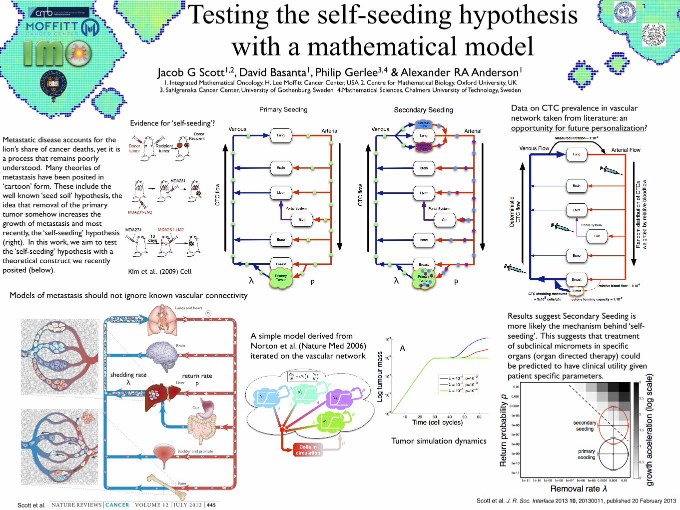
Metastatic disease accounts for the lion’s share of cancer deaths, yet it is a process that remains poorly understood. Many theories of metastasis have been posited in ‘cartoon’ form. These include the well known ‘seed soil’ hypothesis, the idea that removal of the primary tumor somehow increases the growth of metastasis and most recently, the ‘self-seeding’ hypothesis (right). In this work, we aim to test the ‘self-seeding’ hypothesis with a theoretical construct we recently posited (below).
Evidence for ‘self-seeding’?
Models of metastasis should not ignore known vascular connectivity
Scott et al.Scott et al.
A simple model derived from Norton et al. (Nature Med 2006) iterated on the vascular network
Data on CTC prevalence in vascular network taken from literature: an opportunity for future personalization?
Results suggest Secondary Seeding is more likely the mechanism behind ‘self-seeding’. This suggests that treatment of subclinical micromets in specific organs (organ directed therapy) could be predicted to have clinical utility given patient specific parameters.