Semenogelin I: a coagulum forming, multifunctional seminalvesicle proteinM. Roberta and C. Gagnonb,*
aChugai Research Institute for Molecular Medicine, Inc., 153-2 Nagai, Niihari-mura, Ibaraki, 300-0041 (Japan)bUrology Research Laboratory, Rm H6.46, Royal Victoria Hospital, 687 Pine Avenue West, Montreal, Quebec,H3A 1A1 (Canada), Fax +1 514 843 1457, e-mail: [email protected]
Received 21 October 1998; received after revision 15 December 1998; accepted 15 December 1998
Abstract. Human seminal plasma spontaneously coagu- products of Sg I may also have various biologicalfunctions. While the semenogelin I protein is unique tolates after ejaculation. The major component of thishuman and higher primates, it has recently been showncoagulum is semenogelin I, a 52-kDa protein expressedto belong to a gene family having a similar gene struc-exclusively in the seminal vesicles. Recently, a sperm
motility inhibitor has been found to be identical to ture but encoding widely differing proteins. The re-cently elucidated characteristics of the semenogelin Isemenogelin I, suggesting that it may also be a physio-
logical sperm motility inhibitor. The protein is rapidly gene as well as the biochemical and functional proper-ties of the encoded protein are reviewed, and an attemptcleaved after ejaculation by the chymotrypsin-like pro-
static protease prostate-specific antigen, resulting in liq- is made to integrate the various findings into a modelfor semen coagulation, sperm immobilization and po-uefaction of the semen coagulum and the progressive
release of motile spermatozoa. Some of the cleavage tential other functions.
Following ejaculation, human semen spontaneously co-agulates into a semisolid gelatinous mass, which thenliquefies within 5–20 min [1, 2]. This coagulum appearsas a dense network of narrow and long fibers that areapproximately 0.15 mm thick [3]. The major structuralcomponents of human semen coagulum have been de-scribed as disulfide-linked complexes of a predominant52-kDa protein, known as semenogelin I (Sg I), and twoforms of a less abundant Sg I-related protein of 71 and76 kDa (semenogelin II, Sg II), all originating from theseminal vesicle secretions [4–6]. Recently, novel activi-
ties have been shown to be associated with Sg I, includ-ing the property to block sperm motility [7] and toactivate sperm hyaluronidase [8].The past few years have thus given rise to a morecomprehensive body of information about this uniqueprotein through experiments originally performed ondifferent models but which eventually converged on thesame protein, namely Sg I. The present review reexam-ines some of the original work that developed in paral-lel, and attempts are made to integrate the more recentfindings which suggest a surprising variety in potentialfunctions for this protein in reproduction. While in thepresent review emphasis is placed on Sg I, whose func-tional properties have been more extensively studied,some properties of the closely related Sg II will also be* Corresponding author.
CMLS, Cell. Mol. Life Sci. Vol. 55, 1999 945Review Article
discussed, particularly as it relates to gene structure andpattern of expression where similarities suggest interest-ing evolutionary consequences. First, some of the origi-nal work on semen coagulation and on the seminalplasma sperm motility inhibitor (SPMI) will be re-viewed, and the more recently elucidated properties andfunction of the proteins will then be discussed in moredetail. It is first worth summarizing the original workon SPMI and Sg I in parallel to appreciate how bothstories originated from the isolation of a similar prote-olytic fragment of Sg I from seminal plasma.
Parallel paths converging on the same protein
Sg I, the predominant human semen coagulum proteinThe foundation for most of the work on Sg I and itsrole in human semen coagulation rests mainly on theresults of Lilja and co-workers. Their original studieslooked at the effect of various protease inhibitors on theprocessing of larger molecular mass forms of proteinsinto smaller polypeptides occurring during semen lique-faction [9–11]. This led to the identification of themajor coagulum components as disulfide-linkedpolypeptides of 52, 71 and 76 kDa [4, 5]. Most of thedegradation products derived from those proteins werefound to be basic polypeptides, the most basic of whichwas first purified and sequenced [9]. Prostatic proteaseswere found to be responsible for the degradation ofcoagulum proteins [5, 9, 10], and eventually the prote-olytic activity was associated with prostate-specific anti-gen (PSA) [12]. These findings led to the eventualmolecular cloning of the predominant 52-kDa seminalcoagulum component, thereafter referred to as se-menogelin I [13]. According to this sequence, severalpolypeptides isolated from seminal plasma [9, 14, 15]appeared to be derived from Sg I. The identity of theamino acids surrounding the deduced cleavage sitesprovided evidence that PSA had a chymotrypsin-likeactivity. Many of the earlier findings about Sg I havebeen previously reviewed [16]. We will therefore attemptin the present review to emphasize some of the morerecent data and also to integrate the findings with thoseoriginating from work on a sperm motility inhibitor.
SPMI, a sperm motility inhibitor associated with semencoagulumThe identification and characterization of some of theproperties of Sg I also originated from the study of asperm motility inhibitor present in seminal plasma. Be-fore the two proteins were found to be identical, thework on the latter had been initiated from studies onthe control sperm motility. Using a previously devel-oped model for the demembranation and reactivation
of sea urchin spermatozoa [17], Mohri and Yanagi-machi first showed that epididymal spermatozoa col-lected in physiological solutions were readily reactivatedfollowing demembranation [18]. On the other hand, thereinitiation of motility of ejaculated and then demem-branated rabbit spermatozoa was not possible. Interest-ingly, the motility of spermatozoa that had been washedon a Ficoll gradient before demembranation was reini-tiated following addition of Mg · adenosine triphos-phate (ATP) [19, 20], suggesting that some com-ponent(s) present in seminal plasma interfered with thereinitiation of motility. The seminal plasma of variousmammalian species was similarly found to contain amotility inhibitory activity [19]. Following these originalexperiments, a protein of 18–22 kDa associated withthis activity was isolated from human seminal plasmaand named seminal plasma motility inhibitor (SPMI)[21]. The finding that the purified protein was a potentdynein-ATPase inhibitor [21] suggests that this is themode of action of SPMI on demembranated spermato-zoa. On the other hand, purified SPMI was also foundto interfere with the motility of intact spermatozoa [22].However, the dose of SPMI required to completelyinhibit the motility of intact spermatozoa was found tobe more than 1000-fold higher than on demembranatedspermatozoa, suggesting that its effect on the motility ofintact spermatozoa is likely mediated through a differ-ent mechanism. The dose-dependent inhibitory activityof SPMI on intact spermatozoa caused a progressivedecrease in the percentage of motile spermatozoa, thecurvilinear velocity and the beat-cross frequency, with-out affecting the linearity. In a subsequent study, hu-man SPMI was found to originate exclusively from theseminal vesicles and to have a 9-fold higher specificinhibitory activity in this fluid than in seminal plasma[23]. The difference turned out to be explained by thepresence of a 52-kDa SPMI form in seminal plasmashortly after ejaculation which was rapidly transformedinto smaller mass forms during liquefaction [24]. Thiswas accompanied by an 80% reduction in inhibitoryactivity to levels that are compatible with normal spermmotility (B250 U/ml). The phenomenon could be re-constituted in vitro by incubating secretions isolatedfrom the seminal vesicles and prostate, thus demon-strating the prostatic origin of the processing factor.The serine protease inhibitors phenylmethyl sulfonylflu-oride (PMSF) and benzamidine and the metal chelatorsEDTA and 1,10-phenanthroline all partially preventedthe loss of inhibitory activity and processing intosmaller forms [24].The characteristics of SPMI processing after ejaculationwere highly reminiscent of the proteolysis of Sg I occur-ring during semen liquefaction, thus suggesting the phe-nomena might be related. This association becameapparent when all SPMI inhibitory activity and most
M. Robert and C. Gagnon Semenogelin is an inhibitor of sperm motility946
SPMI antigens were found to be associated with thecoagulated semen fraction [25]. Moreover, semen sam-ples that failed to liquefy spontaneously demonstratedhigh levels of SPMI inhibitory activity (1500 U/ml), thepresence of nondegraded SPMI antigens and containedsperm having poor motility. Addition of prostatic secre-tions to the nonliquefied semen samples resulted in amarked increase in sperm motility with a concomitantdegradation of high-mass SPMI antigens and a decreasein inhibitory activity. These findings thus provided anexplanation for the reported low sperm motility incoagulated semen shortly after ejaculation, and the pro-gressive increase in sperm motility as the protein precur-sor gets inactivated during semen liquefaction.The identical glandular origin of Sg I and SPMI, theircomparable molecular mass and processing by prostaticproteases displaying similar properties after ejaculationas well as the association with semen coagulum stronglysuggested that these two proteins were the same molec-ular entity. This was eventually confirmed when theprotein was purified from both seminal vesicle fluid andseminal plasma coagulum and the amino acid sequenceof three different peptides, the amino acid compositionand the mass were found to match very closely thevalues expected from the published complementaryDNA (cDNA) sequence of Sg I [7].
Characterization of Sg I gene and protein
Characterization of Sg I cDNAThe characterization of the Sg I cDNA chronologicallypreceded the purification of the proteins and will thusbe described first. The complete primary structures ofSg I and semenogelin-related proteins have been eluci-dated [13, 26]. An Sg I clone was originally isolated [13]by screening a cDNA expression library derived fromseminal vesicle transcripts with an antibody raisedagainst a 52-amino acid residue fragment of Sg I [9]. Afull-length cDNA containing an open reading frame of1386 nucleotides was thus isolated and characterized[13] (table 1). The resulting sequence encodes a precur-sor protein of 462 amino acid residues, which aftercleavage of a 23-residue signal peptide yields a matureprotein of 439 amino acids having an expected molecu-lar mass of 49.6 kDa and isoelectric point of 9.6 (fig. 1).The mature protein contains a single cysteine residue atposition 216 which is involved in the formation ofdisulfide-linked complexes [5, 13]. A cDNA for the71-kDa Sg-related protein was similarly obtained [26].Its characterization showed that it encodes a 559-residue protein, semenogelin II, that displays 78% iden-tity at the amino acid level with that of the 52-kDa SgI. Two cysteine residues are present at positions 136 and337. The difference in size between Sg I and Sg II is
related to the presence of an extended C-terminal in SgII. The 76-kDa protein, on the other hand, appears tobe a glycosylated form of the 71-kDa polypeptide Sg II[26, 27]. Sg I contains six repeats of 60 residues showinginternal sequence similarity (fig. 2), whereas Sg II con-tains eight such repeats. The exact function of theserepeats is unknown, but their structure is reminiscent ofrepeats found in rodent seminal vesicle proteins knownto serve as substrate units in transglutaminase reactions.Sg I has been shown to be expressed exclusively in theseminal vesicles, whereas Sg II transcripts have alsobeen detected in the epididymis [13], albeit at a muchlower expression level [26].
Gene structure of Sg I and IIThe structure of the Sg I and II genes has recently beenelucidated. Both genes are located on the long arm ofchromosome 20 at q12–13.1 and are relatively compact,spanning only 2.7 and 3.1 kb, respectively [28]. They arearranged in tandem, 11.5 kb apart, and are similarlycomposed of three exons that display more than 90%sequence identity [28, 29]. These three exons encode,respectively, the signal peptide, the secreted polypeptideand the 3% untranslated region (fig. 1). The flankingregions and introns demonstrate more than 80% iden-
Table 1. Characteristics of the semenogelin I gene and protein.
CMLS, Cell. Mol. Life Sci. Vol. 55, 1999 947Review Article
Figure 1. Schematic representation of the Sg I gene, transcript and protein. The basic structural elements of the Sg I gene showing thethree-exon structure typical of the REST-gene family are shown. Nucleotides are numbered according to EMBL accession numberZ47556. Dashed lines link corresponding sections of the gene, transcript and protein. The black box in Sg I protein represents the23-residue signal peptide. The single cysteine at position 216 is marked.
tity [28]. The intergenic sequences are composed ofhighly repetitive DNA sequences made up of long inter-spersed nucleotide sequences (LINES) such as L1 re-peats and Alu sequences [29]. The Sg I and II genes arebelieved to have originated from duplication of an 8-kbDNA fragment some 61 million years ago [29].While Sg I and II proteins are highly homologous [13,26], their primary structure shows little similarity toother abundant seminal vesicle secretory proteins re-sponsible for semen coagulation in rodent species. How-ever, recent studies on the structure of the genes thatencode these divergent proteins have highlighted inter-esting similarities. All appear to be members of a novelgene family that includes both Sg I and II, the ratseminal vesicle secretory proteins SVS-II, SVS-IV, SVS-V and guinea pig seminal vesicle proteins GP1 andGP2, the major seminal clotting protein in these species[26, 30]. The common characteristics of these genes liein their similar transcription unit made of three exons;the first one encoding the signal peptide, the second oneencoding the secreted protein and the third codon en-coding the 3% untranslated region [30]. Interestingly,significant sequence similarity exists exclusively withinexon I and exon III, whereas exon II is highly divergent.The results demonstrate that all these proteins arederived from a similar ancestral gene that evolvedrapidly by substitution of exon 2 to give rise to differentproteins [30]. These surprising findings suggest that, inspite of apparent disparity in amino acid sequence,
these various polypeptides may share similarities inthree-dimensional structure and/or functional domainsthat form the basis for intermolecular interactions in-volved in semen clotting or coagulation. This new fam-ily is collectively referred to as the REST (rapidlyevolving substrates for transglutaminase) gene familydue to the fact that most members have been found tobe good substrates for transglutaminases. Members ofthis family have a basic isoelectric point, contain severalinternal repeats and an abundance of lysine and glu-tamine residues which serve as donor and acceptor sitesin the transglutamination reaction.Until recently, Sg I was believed to be unique to hu-mans, since no homology to other known proteins inother species was found. However, the presence of aclose homologue in rhesus monkeys was suspected byimmunocytochemical study demonstrating the presenceof Sg immunoreactive material in the seminal vesicleand vas deferens [31]. The Sg II gene of the rhesusmonkey has recently been cloned [32]. As describedabove for human and rodent proteins, the transcriptionunit is similarly split into three exons encoding thesignal peptide, the mature protein and the 3% untrans-lated region. The rhesus monkey gene is about 22%larger than its human counterpart due to extension ofthe coding region by the presence of two additionalrepeats of 60 amino acid residues [32]. Interestingly, therhesus monkey Sg II lacks cysteine residues, thusdemonstrating that noncovalent interactions are respon-
M. Robert and C. Gagnon Semenogelin is an inhibitor of sperm motility948
sible for semen clotting or gellation and that the role ofintermolecular disulfide bonds, if any in this process, issecondary in those molecules which contain cysteineresidues [32]. Southern blot analysis has demonstratedthat Old World monkeys such as the rhesus monkeyand baboon and the New World monkey marmosetcontain two different Sg genes similar to Sg I and Sg II[32]. In contrast, only one Sg-hybridizing band wasfound in the New World monkey cotton-top tamarin.Cloning of the gene in this primate has revealed that itcontains a single Sg-related gene that is more similar tohuman Sg I (89% of nucleotides conserved) than tohuman or rhesus monkey Sg II (82%) [33]. The pre-dicted molecular mass of this extended Sg I is 66 kDa,or 32% higher than human Sg I. However, contrary tothe its human homologue, the cotton-top tamarinprotein contains 14 occurrences of a consensus sequencefor N-linked glycosylation. The potential for glycosyla-tion could thus significantly increase the actual molecu-lar mass of the protein and modify its isoelectric pointestimated at 9.0 [33]. Surprisingly, a probe for semeno-clotin, the major clot-forming protein in mouse, hy-bridized to a single restriction fragment under low
stringency in the cotton-top tamarin [32]. This suggeststhat while this New World monkey does not contain anSg II gene, it may contain a semenoclotin-related genepreviously thought to be unique to rodents [32]. Theevolutionary consequence of such a finding is that thegenome of a common ancestor to early mammaliansand primates likely contained both semenoclotin and Sggenes, but that murine species eventually lost Sg geneswhile most primates lost the semenoclotin gene. On theother hand, the cotton-top tamarin likely lost the origi-nal Sg II by substitution of a new exon in an alternativesplice site, giving rise to a semenoclotin-related gene[33].
Expression profile of Sg IRecently, a detailed localization study showed abun-dant semenogelin antigen expression in the cytoplasmof seminal vesicle glandular epithelial cells by immuno-cytochemistry using several different monoclonal andpolyclonal antibodies [34]. None of the antibodies usedin this study were found to be specific to either form ofSg, a finding not so surprising considering the very high
Figure 2. Structure of repetitive amino acid units in Sg I. Upper panel: The position of three different types of repeat present in Sg Iare shown along the Sg protein. Lower panel: alignment of amino acids within each type of repeat. Conserved residues are shadowed.The repeats are classified according to Lilja and Lundwall [26].
CMLS, Cell. Mol. Life Sci. Vol. 55, 1999 949Review Article
similarity in primary structure. On the other hand, theauthors were able to design oligonucleotide probes spe-cific for Sg I or Sg II, and these were used for in situhybridization [34]. Using these specific probes, intenseSg I and Sg II transcript signals were detected in thesame glandular epithelial cells shown to contain Sgantigens. Sg II transcripts were also detected in theepithelium of the cauda epididymis, whereas Sg I tran-scripts were absent [34]. Neither Sg I nor Sg II tran-scripts or antigens were found to be expressed in otherparts of the epididymis (caput, corpus) or in stromalcells. Accordingly, Sg antigens were absent from testicu-lar, caput and corpus spermatozoa but appeared on thesurface of cauda epididymis spermatozoa. Localizationof Sg antigens on freshly ejaculated spermatozoashowed that the protein localizes to the posterior head,midpiece and tail of spermatozoa [34], in agreementwith earlier findings [13]. The testis, prostate, femalegenital tract and numerous tissues tested were all foundto be devoid of Sg antigens in agreement with theresults of localization of SPMI antigens, shown to beexclusively located in the seminal vesicles [23].
Regulation of unique Sg I tissue expressionThe very restricted expression profile of Sg I as de-scribed above and the abundance of the protein in thesecretions of the seminal vesicles, a gland whose secre-tory function is androgen-dependent, argues in favor ofan androgen-regulated synthesis of this protein. Sg im-munoreactivity, as determined using the antibodyrecognizing the basic fragment of Sg I, was previouslyobserved on the epithelium of seminal vesicle specimensobtained from postpuberty subjects, but not on those ofa newborn or 5-year-old child [31]. This is in agreementwith preliminary observations from our laboratorymade during the collection of seminal vesicle fluid.Seminal vesicle fluid collected from two patients whounderwent radical prostatectomy and had followed apreoperative treatment with antiandrogens showed adramatic decrease in Sg I immunoreactivity, as mea-sured by immunoblotting, compared with those ob-tained from untreated patients (M. Robert and C.Gagnon, unpublished observations). Moreover, the re-ported Sg I gene sequence [28] contains nucleotide se-quences that are closely related to a reportedandrogen-response element consensus [35] in both thepromoter 5% upstream sequence and in the first intron ofthe Sg I gene (M. Robert and C. Gagnon, unpublishedobservations). Together, these observations stronglysuggest that the specific expression of Sg I in the semi-nal vesicle epithelium is androgen-dependent. On theother hand, the expression of Sg I exclusively in seminalvesicles [34] and not in other tissues that are alsoandrogen-responsive, such as the prostate, suggests that
there are additional factors that regulate this tightlyrestricted expression. The identification of regulatoryelements within the Sg genes by focussing on the specificand unique sequences identified by Lundwall [29] andthe identification of the associated regulatory DNA-binding proteins should allow elucidation of the mecha-nism of this unique tissue expression.
Purification and characteristics of Sg IWhile the isolation of smaller Sg I polypeptides [9, 21]from seminal plasma originally led to the identificationof a 52-kDa larger form in seminal vesicle and itsmolecular cloning, the purification of intact Sg I fromisolated seminal coagulum and seminal vesicle fluid wasreported only recently [7, 27]. In addition, Mandal andBhattacharyya previously reported the purification ofthe predominant semen coagulum protein [36]. Whilethe identity of the protein with Sg I was not directlydemonstrated, the similarities in molecular weight, pIand chromatographic behavior strongly suggest that itis Sg I. In all cases, proper solubilization of the proteinsin urea buffer, as well as reduction and alkylation ofcysteine residues prior to purification, was found to beessential to ensure good recovery of the proteins andprevent their degradation [7, 27, 36]. The successfulenrichment of Sg I by chromatography on heparin-Sep-harose [27] is likely due to its cation exchanger proper-ties at the alkaline pH used, as this is the type ofchromatography that proved useful in the other proto-cols [7, 36]. However, a specific affinity of Sg I forheparin cannot be excluded. One of the majordifficulties encountered during the purification of Sg Iwas the removal of degraded forms present in the start-ing material. These fragments had the tendency tocoelute with the 52-kDa precursor. Their successfulremoval could be achieved by an additional purificationstep using gel filtration on Superose [27], Sephacryl [36]or high-performance reverse-phase liquid chromatogra-phy [7]. The abundance of the 52-kDa Sg I polypeptideas observed by SDS-polyacrylamide gel electrophoresis(PAGE) in samples of seminal vesicle fluid and washedsemen coagulum, together with the low enrichment fac-tor (two- to fivefold) following purification, suggest thatthis protein may represent more than 20% and up to50% of the total proteins in these sources, respectively[7, 36]. In physiological solution, the purified Sg Iappeared to form aggregates of large molecular mass,which were not recovered during analysis on a gelfiltration column in buffer that did not contain urea [7].This is not a surprising finding considering the coagu-lating properties and low solubility of Sg I, and is likelythe consequence of the reported extensive noncovalentinteractions existing between Sg I and Sg II polypep-tides. Similar observations were also reported indepen-dently [27].
M. Robert and C. Gagnon Semenogelin is an inhibitor of sperm motility950
The amino acid composition of Sg I corresponds verywell with that predicted from the cDNA sequence [7,27]. Treatment of the purified protein with N-glycosi-dase F and specific staining using a glycoprotein detec-tion system demonstrated the absence of anyglycosylation [27]. While the site of processing by signalpeptidase had been suspected from analysis of theprotein sequence, the identification of a glutamineresidue (in the form of a pyrrolidone carboxylate orpyroglutamine) as the N-terminal residue of the pro-cessed protein was confirmed following digestion withpyroglutamate aminopeptidase [13]. This finding thusexplains the reported failure to obtain N-terminal se-quence information from the purified 52-kDa Sg Iprotein [7, 13]. The mass of Sg I, as measured by massspectrometry, is also in close agreement with that pre-dicted from the cDNA sequence (table 1) [7, 27]. Theslight difference of approximately 300 Da, between thetwo reports, is within reasonable experimental error andmay reflect a variation due to the different spectromet-ric method used (atmospheric pressure ionization-mass spectrometry (API-MS) vs. matrix-assisted laserdesorption ionization (MALDI)). The absence of a sig-nificant difference between the measured and expectedmass of Sg I confirms the absence of glycosylation andrules out the possibility that the protein undergoesextensive posttranslational modification.Sg I was shown to reversibly inhibit the motility ofintact spermatozoa in a dose-dependent manner [7].Incubation of the purified protein with prostate-specificantigen (PSA) caused degradation into smaller massforms and a concomitant 76% decrease in its inhibitoryactivity, as measured on demembranated spermatozoa.Similarly, PSA treatment also considerably reduced theinhibitory activity of Sg I on intact spermatozoa. Thedemonstration of the immobilizing effect of purified SgI on intact spermatozoa [7], at activity levels andprotein concentrations corresponding to those mea-sured in coagulated semen early after ejaculation [24],suggests that Sg I may be responsible for the lowmotility of spermatozoa in coagulated semen. The effectof Sg I on sperm motility was reversible when cells werewashed and thus appeared to be mediated by associa-tion of the protein with the sperm surface. The sperm-immobilizing activity of Sg I does not appear to beexclusively related to its high isoelectric point, sincecytochrome c, with a pI of 9.3, has no such activity atthe same concentration [7].The sperm-immobilizing activity of Sg I could bemapped within the N-terminal domain of the protein[37]. This segment of the protein appears highly con-served in Sg of other primates, suggesting that it mayplay a similar role in those species [33]. Because of itsvery high similarity with Sg I, Sg II also likely hassperm motility inhibitory activity. Accordingly, during
purification of Sg I, sperm-immobilizing activity hasalso been observed in fractions enriched in Sg II [7]. Itis thus tempting to speculate that Sg II, which has alsobeen shown to be expressed in the cauda epididymis aswell as on the surface of spermatozoa of the caudaepididymis [34], might be involved in sperm immobiliza-tion at this site, as recently suggested [33].The N-terminal domain of Sg I also contains the epi-tope recognized by an antibody raised against a 19-kDaSg I form present in liquefied seminal plasma [37] (table1). On the other hand, the epitope for the MHS-5monoclonal antibody appears to map to a more cen-trally located section of Sg I [37]. This antibody wasoriginally raised against whole spermatozoa but foundto recognize a sperm-coating antigen originating fromthe seminal vesicles [38]. The antigen recognized byMHS-5 antibody is referred to as seminal vesicle-spe-cific antigen (SVSA) and is present on a range ofpolypeptides. These also undergo extensive molecularprocessing during semen liquefaction and have massesvery similar to those from Sg I and II [38–40]. Whileseminal polypeptides reacting with this antibody werepurified, no biochemical information was available toallow the location of SVSA within the Sg molecule [41].However, the identification of a specific segment of Sg Ithat is recognized by this antibody further reinforces thesuspected identity of the 58-kDa SVSA with Sg I [13,37].Sg I was found to be cleaved readily by PSA [37]. Theeffect of zinc and 1,10-phenanthroline on the in vitrodegradation of Sg I by PSA and chymotrypsin led tothe postulation that Sg I may be a zinc-binding protein.These findings are in good agreement with those re-ported for the effect of those substances on semenliquefaction and degradation of Sg I by prostaticproteases [4, 11, 24, 25]. Zinc is an important ion inmany biological systems, and although it is usuallypresent only in trace amounts in most body fluids, zincis present in the prostate gland at the highest concentra-tion of any organ [42] and is secreted in semen atsimilarly high levels. However, its function in that fluidhas remained poorly understood. Interestingly, in coag-ulated semen, most of the seminal zinc was previouslyfound to be tightly associated with the washed coagu-lated protein mass [43], and to bind specifically tovarious polypeptides having identical electrophoreticmobility to Sg I [44]. Moreover, as semen liquefactionprogressed, zinc was shown to be associated with pro-gressively lower mass polypeptides (B25 kDa) of elec-trophoretic mobility, again very similar to those of theSg I degradation products [44]. These results thus sup-port the concept that Sg I may be a major zinc ligand.While this may not represent the physiological scenario,both Sg I and Sg II have recently been found to be goodsubstrates for the fibrin cross-linking transglutaminase,
CMLS, Cell. Mol. Life Sci. Vol. 55, 1999 951Review Article
factor XIII, in vitro [45], a finding that provides func-tional support for the similarity of Sg I and II to theREST gene family. Transglutaminase activities havebeen reported in human semen, and recently a noveltransglutaminase cDNA was cloned from a humanprostate cDNA [46]. However, factor XIII was used forthose experiments since this human prostatic transglu-taminase has not yet been isolated. Interestingly, thecross-linked high molecular mass complexes of Sg werefound to be resistant to PSA-mediated proteolytic di-gestion [45]. This may thus highlight the importance ofSg I and II conformation for proper processing by PSA,as previously suggested [37]. In addition, several Sgfragments released by PSA hydrolysis could be cross-linked by FXIIIa [45]. This may suggest that some ofthe proteolytic fragments are similarly cross-linked invivo and that the phenomenon might have functionalrelevance to sperm function. In any case, only a smallfraction of Sg I and II would be cross-linked in vivo,since no high molecular weight Sg complexes are ob-served in semen. However, the authors also speculatedthat some Sg could be cross-linked to spermatozoa andthus not be observable after removal of the cells [45].
Sg I as the main physiological substrate for PSAThe fragmentation of Sg I occurring after ejaculationhas been associated mainly with the proteolytic activityof PSA [10, 12, 13, 40]. Characterization of PSA activityusing nonphysiological or synthetic substrates [47, 48]and identification of the location of seminal peptidesderived from Sg [13, 16] had previously associated achymotrypsin-like activity to the enzyme in contrast tothe trypsin-like specificity of the other members of thekallikrein family of proteases, to which PSA belongs.Accordingly, Sg I was recently found to be cleavedpreferentially on the C-terminal side of several tyrosineand leucine residues [7, 37]. Similar findings of preferen-tial hydrolysis by PSA at tyrosine residues were recentlyreported using synthetic peptide substrates [49] or sub-strate phage display [50]. In the former study, PSA wasalso found to cleave synthetic substrates at glutamine,histidine and cysteine residues, albeit at a lower effi-ciency but with increased specificity for PSA when com-pared with other proteases. In Sg I, however, thefavored hydrolysis sites appear to be at the C-terminalof several tyrosine and leucine residues [13, 16, 37]. Theunusual specificity of PSA appears related to the pres-ence of a serine residue at the bottom of the substratebinding pocket instead of the aspartic acid residuefound in trypsin-like enzymes [51, 52]. PSA also seemsto differ from chymotrypsin in its preference for ty-rosine and leucine residues, whereas chymotrypsin pre-dominantly cleaves at the aromatic residuesphenylalanine, tryptophan and tyrosine and, only to a
lower extent, at leucine residues. The effect of variousprotease inhibitors and divalent cations on PSA-medi-ated cleavage of Sg I also highlights additional differ-ences with chymotrypsin [37]. Sg I appears to be arather specific and favored PSA substrate since PSAdoes not readily hydrolyze other proteins [37]. It is thuslikely that PSA cleaves Sg I preferentially at peptidebonds surrounded by specific residues [47], as suggestedfor Sg I [16, 37]. This is further substantiated by therecent demonstration that PSA displays preferences forsecondary subsites and that recognition of substrate ismediated by an extended binding site [50]. These clus-ters of amino acid may form specific three-dimensionalstructures that are preferentially recognized by the en-zyme. This strict specificity would explain the apparentlow activity of PSA on other substrates when comparedwith chymotrypsin [37, 47]. Together, these specific dif-ferences among hydrolysis site preferences and interac-tion with various inhibitors and ions demonstrate thatPSA is a specific Sg I processing enzyme that hydrolysesand displays unique catalytic properties in spite of itsclose relationship to other serine proteases of thekallikrein family.The serine protease inhibitor protein C inhibitor (PCI)has been recently shown to bind to both PSA and Sg IIin vitro and in vivo [53, 54] and to inhibit the PSA-me-diated degradation of Sg I and Sg II [54]. Interestingly,in the latter study, divalent cations and particularlyZn2+(0.01–0.1 mM) were found to significantly reducebinding of Sg II to both PCI and PSA [54]. Theseinteractions have so far only been demonstrated clearlywith Sg II. Caution must thus be exercised, since suchan interaction could be mediated by the carbohydrateresidues that appear to be present on the 76-kDa formof Sg II but absent from Sg I. However, if the interac-tion is mediated by the polypeptide backbone, then it ishighly probable that Sg I, PSA, PCI and seminal zincmay similarly interact to coordinate Sg I and IIprocessing.While PSA appears to be the main Sg I processingenzyme after ejaculation, the possibility that additionalproteases may also participate in the phenomenon can-not be excluded [24]. The human kallikrein hK2, whichis closely related to PSA [55, 56], and the recentlycharacterized prostasin [57], both of which are prostatictrypsin-like proteases that correspond well with theprostatic proteolytic activity inhibited by benzamidine[11, 24], are also proteases that may play a more mar-ginal role. The former possibility is especially likelyconsidering the recent demonstration that hK2 can de-grade Sg I and II in vitro at the carboxyl side ofarginine residues [58]. However, while these enzymescould contribute to cleave Sg I at alternative sites, theireffect is likely to be marginal, since PSA generates Sg Ifragments that are identical in size to those observed in
M. Robert and C. Gagnon Semenogelin is an inhibitor of sperm motility952
liquefied semen [7, 10, 37, 40, 59]. In addition, variousSg I fragments purified from liquefied seminal plasmawere shown to have terminal leucine or tyrosineresidues corresponding to cleavage by PSA [14, 15, 60].
Functions of Sg I
The major function of Sg I in the reproductive processhas so far been associated with the formation of theseminal coagulum [5, 16]. The finding that Sg I displayssperm immobilizing activity in vitro suggests that it mayalso be a physiological motility inhibitor at least shortlyafter ejaculation [7]. While these two functions may beintimately linked, their physiological role in the humanreproductive process and the mechanism of action hasnot been addressed extensively and remains of a ratherspeculative nature.
Sg I and semen coagulation and liquefactionThe phenomenon of semen coagulation occurs in mostmammals, but the mechanisms appear to vary widely[61]. The semen of dogs and cats does not coagulate,and interestingly these mammals lack seminal vesicles,the main source of coagulating substrates in most mam-malian species [61]. In some species, the formation of avaginal plug is believed to be essential to prevent theoutflow of semen from the female’s vagina. In rodents,it is also thought to prevent secondary inseminations ofspermatozoa by nondominant males [62]. Such a plugmay act as a reservoir and contribute to the gradualrelease of spermatozoa in species where the female doesnot possess a long cervix and cervical mucus, and wheresemen is deposited directly into the uterus. Similarly,the semisolid and adherent properties of the humansemen coagulum may also contribute to some extent toprevent outflow of spermatozoa from the female’s re-productive tract, until they are released progressivelyduring liquefaction. Whether the phenomenon of semencoagulation in humans is simply a remnant of ourevolution from a common earlier ancestor is not clearlyestablished. As described above, Sg I and Sg II havebeen found to belong to the REST gene family whoseseveral members are known transglutaminase substrates[30].While the primary structure and several biochemicalproperties of Sg I and Sg II have been elucidated, theprecise mechanism accounting for the formation of thegelatinous network of proteins found in the coagulumhas not yet been convincingly demonstrated. However,the integration of results derived from various studiesprovides some insights into some likely events. First, themechanism responsible for human semen coagulation isdifferent from the one involved in blood coagulation,
since factor XII, fibrinogen and prothrombin are allabsent from seminal plasma [61]. In addition, while inrodents transglutaminases from the coagulating glandcross-link protein substrates from the seminal vesicle toform the so-called copulatory plug [63], no such highmolecular weight complexes of Sg I and II resultingfrom covalent cross-linking of those proteins have beenobserved when coagulum components are analyzed bySDS-PAGE [5, 7, 25]. On the other hand, Sg I and II,the predominant components of the coagulum, areknown to form disulfide-linked high molecular weightcomplexes [5, 6, 12]. However, dithiothreitol does notcause apparent macroscopic changes on the coagulumstructure [5], suggesting that these disulfide interactionsdo not constitute the main force holding coagulumproteins together. Disulfide bridges thus likely play asupporting and indirect role in the coagulation andliquefaction of human semen, since semen liquefactionis enhanced in the presence of the reducing agent b-mercaptoethanol [6]. This effect is therefore likely medi-ated by a change in the three-dimensional structure ofSg I and II induced by reduction of disulfides, which inturn increases their sensitivity to proteolytic degrada-tion. The importance of noncovalent interactions inmaintaining the coagulum structure is highlighted bythe fact that denaturing agents such as guanidine-HCl(3 M) and urea (4–8 M) readily solubilize semen coagu-lum [5, 7, 27, 36].The exact mechanism by which noncovalent interac-tions between Sg I and other components trigger semencoagulation thus remains speculative. Fibronectin origi-nates from seminal vesicle fluid and, like Sg I, is en-closed in the coagulum structure and gets progressivelyfragmented and released into the fluid phase of semenduring semen liquefaction [12]. At ejaculation, prostaticcomponents such as zinc may induce a polymerizationreaction involving Sg I, Sg II and fibronectin by induc-ing conformational changes in these proteins, thus trig-gering coagulation [12]. The authors speculated that thismight arise by inducing an opening of fibronectindimers and consequent exposure of structures that haveaffinity for Sg. Alternately, one model [64] proposedthat interactions between sialoglycoproteins and metalions are responsible for coagulum formation [65, 66].According to this model, the initial stage of liquefactionwould occur by reduction of those metal ions by L-ascorbic acid. However, Sg I does not appear to beglycosylated, thus raising doubts about such a mecha-nism as the main pathway. It could nonetheless be acontributing factor, since Sg I and Sg II may formheterodimers [13], and the high molecular mass (76kDa) form of Sg II has been shown to be glycosylated[27]. Finally, another model proposed that coagulationoccurs through reticulation of proteins by formation ofstable links between fructose and monomer of vesicular
CMLS, Cell. Mol. Life Sci. Vol. 55, 1999 953Review Article
Figure 3. Model of semen coagulation and liquefaction based on Sg I properties. A simplified diagram of some of the components ofthe male reproductive tract is shown. Sg I and PCI are present exclusively in the seminal vesicles, whereas PSA and zinc ions originatefrom the prostate. At ejaculation, these components mix, and a conformational change in the Sg I, Sg II and fibronectin is triggeredby prostatic components (most likely Zn2+), causing the Sg molecules to interact with each other by a yet unknown mechanism,resulting in aggregation and semen coagulation. Spermatozoa are thus immobilized in the coagulum likely through the immobilizingactivity of Sg I. Chelation of zinc ions in the coagulum activates PSA, which gradually degrades Sg I and II, causing the release ofsmaller fragments and reversing the sperm motility inhibition. This in turn releases zinc and PCI in solution, which bind to andinactivate PSA to prevent further undesirable proteolysis. Fibronectin, which also appears to participate in the coagulum formation, isleft out for clarity and because its interaction with the other components remains unclear.
proteins, such as Sg, in the formation of semen coagu-lum [67].Semen liquefaction, on the other hand, is associatedwith the proteolytic degradation of Sg I and II, thepredominant coagulum components, since the disap-pearance of the gel structure parallels fragmentation ofthese proteins [10, 12, 24, 25, 39, 68, 69]. Fragmentationof Sg I and II probably reduces the extent of noncova-lent interactions between these proteins and fibronectin,thus causing liquefaction, potentially by allowing move-ment of interacting surfaces that were limited in theuncleaved backbone. This is reflected in the fact thatintact Sg molecules in seminal vesicle fluid or seminalcoagulum are insoluble due to the creation of a vastnetwork of protein aggregates, whereas Sg fragments ofliquefied semen are soluble. Originally, a collagenase-like activity [64], seminin and plasminogen activator[70] were associated with the liquefying activity. How-ever, it is now clear that PSA plays the major role indegrading the structural components of the coagulum,Sg I and Sg II [4, 10, 12, 37, 40, 69, 71]. In those studies,
purified PSA was shown to cleave the predominantcoagulum proteins in a manner essentially identical totheir degradation during semen liquefaction and in par-allel to the progressive release of motile spermatozoa.The predominant role of PSA in Sg I degradation andsemen liquefaction is further substantiated by the obser-vation that various polypeptides isolated from seminalplasma have terminal amino acid residues correspond-ing to cleavage of Sg I at leucine or tyrosine residues,sites associated with the chymotrypsin-like proteolyticactivity of PSA [13–15, 37, 60, 72].By integrating the various properties of semen coagu-lum and Sg I and II described above, it is possible topropose a mechanism for semen coagulation and lique-faction (fig. 3). Prior to ejaculation, Sg I and Sg IIpresent in the lumen of seminal vesicles may be in arandom, unordered arrangement and protected fromproteolysis by PCI also originating from the samegland. In prostatic secretions, zinc present at concentra-tions up to 7 mM [42] is likely to inhibit most of PSAactivity [37, 48, 73]. As mentioned previously, Sg I is
M. Robert and C. Gagnon Semenogelin is an inhibitor of sperm motility954
likely a zinc-binding protein. At ejaculation, Sg I mightchelate most of the free zinc contributed by prostaticsecretions. Chelation of such a high concentration ofzinc is not impossible considering that Sg I may bepresent in semen at concentrations as high as 0.4–0.6mM (20–30 mg/ml) and that each molecule may bindseveral zinc atoms. Zinc binding might induce structuralchanges in the Sg I and II molecules that would triggernoncovalent interactions with fibronectin and aggrega-tion of the proteins, culminating in the formation of theinsoluble protein complex of the coagulum and immobi-lization of spermatozoa. The resulting sudden decreasein free zinc concentration would cause PSA activationand release the interaction of Sg I and II with PCI,allowing PSA to cleave the coagulum proteins andcausing the gradual release of free PCI into the solubleliquefied phase [54] and gradual Sg I proteolysis. As SgI would become progressively hydrolyzed into smallerpolypeptides, interactions between the proteins wouldbe loosened, causing semen liquefaction. Zinc would bereleased due to structural changes around the metalcoordinating residues of the protein caused by thecleavage of the polypeptide backbone, and sperm motil-ity would increase gradually. In agreement with thishypothesis is the finding that several histidine residuesthat are the likely metal-binding residues in Sg I werefound in the proximity of various PSA cleavage sites[37]. Finally, the resulting increase in free zinc wouldagain inactivate PSA, thus preventing further undesir-able proteolysis of semen components and structuraldamage to spermatozoa or surrounding tissues. SolublePCI might also reform a complex with PSA, althoughonly a small proportion of total PSA is likely inacti-vated by PCI [53]. While this model remains speculativein nature, it provides a mechanistic link between variousproperties of Sg I and the observations on the role ofdivalent metal ions in the phenomenon of semen coagu-lation-liquefaction [4, 24, 25, 37, 44, 74], and supports aphysiological function for seminal zinc.
Sg I as a physiological sperm motility inhibitor?As described previously, a fragment of Sg I was origi-nally purified from seminal plasma as a factor thatcould inhibit the motility of demembranated spermato-zoa likely acting as a dynein-ATPase inhibitor [21].However, both degraded fragments of Sg I and thepurified intact protein were also found to inhibit intactsperm motility in vitro [7, 22]. This effect, together withthe observed change in inhibitory activity as Sg I getsdegraded during semen liquefaction and the ensuingincrease in sperm motility, suggests that Sg I may be aphysiological sperm motility inhibitor [7, 24, 25]. Theexact mechanism by which Sg I may exert its sperm-im-mobilizing activity on intact spermatozoa is still un-
known. Nonetheless, it is possible to draw someinferences as to the nature of this activity. Obviously,physical entrapment of spermatozoa within the coagu-lum structure may represent one direct form of immobi-lization by restricting movement of the sperm flagellum.However, such a mechanism is unlikely to explain thecomplete flagellum beating arrest observed in experi-ments using soluble purified Sg I [7]. Moreover, thesoluble 19-kDa Sg I fragment isolated from liquefiedseminal plasma was also shown to inhibit intact spermmotility [22], while it had obviously lost the ability toform a coagulated mass. There are thus additionaleffects that likely come into play in the mechanism ofsperm immobilization.As previously mentioned, immunolocalization studiesusing various anti-Sg monoclonal and polyclonal anti-bodies as well as the MHS-5 demonstrated the presenceof Sg antigens on the postacrosomal sheath of the head,midregion and tail of human spermatozoa [13, 34, 38].The localization of Sg antigens to sperm structuresinvolved in motility fits well with the sperm motilityinhibitory activity that has been associated with Sg I [7].While a more detailed microscopic study has not beenperformed, the findings that the motility of Sg I-immo-bilized spermatozoa can be recovered by washing thecells and that prostatic extracellular proteases inactivateSg I and allow recovery of sperm motility [7] argue infavor of a surface-mediated mode of action. Sg I thuslikely binds to the sperm surface in these regions andaffects motility through a mechanism yet to be eluci-dated. However, the integration of various studies al-lows speculation about a possible mechanism.Receptor-binding proteins are usually present in traceamounts in biological fluids, and subtle differences intheir concentrations trigger biological responses. Assuch, the mere abundance of Sg I in seminal plasmawould tend to argue against such a receptor-mediatedmechanism. On the other hand, the gradual decrease insperm-immobilizing activity of the 52-kDa Sg I proteinas it is cleaved suggests that a specific domain of theprecursor molecule may be involved in maximizing theinteraction between the active domain and its target siteon spermatozoa, assuming that those two activities(sperm binding and immobilization) are located on sep-arate domains. These observations share interestingsimilarities with previous findings concerning inhibin-like peptides. Originally, three different inhibin-likepeptides were isolated from seminal plasma during asearch for factors that could suppress pituitary follicle-stimulating hormone (FSH) secretion in vitro. Theircomplete amino acid sequences were elucidated andinterestingly, all turned out to have primary structurescorresponding to fragments of Sg I derived betweenresidues 45 and 136 [13, 14]; a-inhibin-92, -52 and -31correspond to residues 45–136, 85–136 and 85–115,
CMLS, Cell. Mol. Life Sci. Vol. 55, 1999 955Review Article
respectively (fig. 4). Moreover, Sg I fragments identicalto a-inhibin-92 and -52 were obtained following PSA-mediated degradation of SPMI precursor and corre-spond well to the presumptive active (sperm-immobilizing) domain of Sg I [37]. Perhaps not socoincidentally, a-inhibin-92 and a-inhibin-52 werefound to be 40- and 3.4-fold more active, respectively,than a-inhibin-31 in inhibiting FSH release in vitro [14].The observed decrease in the activity of inhibin-likepeptides as they are fragmented into smaller peptides isthus highly reminiscent of the drop of Sg I-immobilizingactivity that occurs during its processing by PSA [7].The affinity of larger SPMI and inhibin-like peptidesfor their respective target appears to be greater thanthat of smaller fragments and may thus be regulated byprogressive proteolysis. While the mechanism of actionof these peptides on the pituitary has not been clearlyestablished, direct binding of the peptides to humanpituitary membranes has been demonstrated [75]. More-over, cross-linking experiments using labeled a-inhibin-92 and pituitary membrane revealed the presence of aspecific 90-kDa complex [75]. It is therefore tempting tospeculate that Sg I could similarly bind to a specificacceptor protein present on spermatozoa. Whether thepituitary receptor of inhibin-like peptides bears anysimilarity to the target site of Sg I on human spermato-
zoa remains to be established. A similar study attempt-ing to identify such an Sg I ligand on the spermmembrane might yield clues to better define its actionon spermatozoa. Such an Sg I ‘receptor’ on the spermmembrane might modulate its suggested immobilizingactivity. Subsequent recovery of motility during lique-faction would then occur due to a decrease in affinityfor the receptor following degradation by PSA in vivo,or by removal of the protein by washing the cells invitro as we have observed [7].Alternately, it cannot be excluded that Sg I might bindto membrane components other than proteins. Mem-brane phospholipids could also be a binding site for SgI, as has been shown for a family of bovine seminalplasma proteins [76] and porcine seminal proteins calledspermadhesins [77, 78]. Interestingly, one member of thelatter family (AQN-3) has recently been shown to bealmost identical to a component of boar SPMI [79].While there is no obvious sequence homology betweenSg I and AQN-3, the fact that they are both spermmotility inhibitors having similar properties [7, 21, 80]suggests some possible structural and functional homol-ogy. Interaction of Sg I with membrane phospholipidsmight cause destabilization of the bilayer and subse-quent entry of extracellular components, such as ions,that might inhibit sperm motility in vivo. The composi-
Figure 4. Functional map of Sg I and polypeptides released by hydrolysis. The whole Sg I mature protein is represented: pQ indicatesthe pyroglutamyl residue at the N-terminal of the mature protein. Segments of Sg I that have been associated with specific biologicalactivity or reactivity with antibodies are highlighted with patterned boxes (see text). During semen liquefaction, PSA cleaves Sg I at theC-terminal of several leucine and tyrosine residues, releasing numerous polypeptides that may have various biological activities asstated. The peptides released are shown as a model only. Details of actual cleavage and derived peptides were previously described [16,37].
M. Robert and C. Gagnon Semenogelin is an inhibitor of sperm motility956
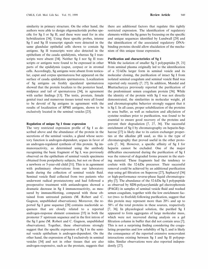
tion of the phospholipid bilayer of the sperm plasmamembrane is known to vary in different areas of thesperm surface. Preferential binding of Sg I to phospho-lipids that are predominantly present on the sperm tailand midpiece could provide an attractive explanationfor the reported binding of Sg I to those areas of thesperm surface [34]. While the hydropathy index of theboar SPMI/AQN-3 shows a rather hydrophilic charac-ter, there are segments of the protein that could poten-tially form small hydrophobic domains that couldinteract with the sperm membrane, thus causing itsdestabilization and resulting in changes in motility [79].A similar phenomenon in Sg I might also result inreversible interactions with the sperm membrane andeffects on sperm motility. Interestingly, a synthetic pep-tide derived from an antimicrobial protein was found tohave a reversible and dose-dependent sperm immobiliz-ing activity in vitro [81]. Its effect was found to bespecific, since five other cationic antimicrobial peptidesdid not similarly inhibit sperm motility. In addition, thepeptide did not cause detergent-type membrane disrup-tion but rather caused functional membrane changes.These characteristics are similar to those of Sg I or itsfragments, and it is tempting to speculate that they mayact similarly.The possibility of a large protein readily penetratinginto spermatozoa and reaching the dynein ATPase andacting similarly as on demembranated spermatozoa ap-pears unlikely. However, the isolation and identifica-tion, in human sperm nuclei, of a 19-kDa polypeptidethat appears to be a proteolytic product derived fromthe N-terminal domain of Sg [82] may suggest that atleast some fragments of the SPMI precursor may alsoenter the cell. In addition, we have observed that bothdetergent soluble and insoluble fractions of spermato-zoa washed twice on Percoll gradients to maximize theremoval of seminal plasma proteins and proteins looselyassociated with the sperm plasma membrane containedpolypeptides recognized by the SPMI antiserum (M.Robert and C. Gagnon, unpublished observations). In-terestingly, forms of mass greater than 20 kDa wereobserved in the insoluble fraction, supporting the no-tion that Sg I may have penetrated the cells and wasthus partially protected from PSA hydrolysis occurringin seminal plasma. Thus, specific mechanisms that allowthe penetration or translocation of seminal proteinsthrough sperm membranes may exist.While all the above observations do not yet allow us todraw conclusions about the exact target site of Sg I onspermatozoa, they do permit the elaboration of a poten-tial model of action. By integrating these various obser-vations, it is possible to postulate that under normalcircumstances, Sg I binds to an as yet uncharacterizedacceptor site on the flagellum of spermatozoa and im-mobilizes the cell. As the 52-kDa molecule gets de-
graded by PSA, its affinity for its acceptor site onspermatozoa is reduced by degradation of a ‘dockingdomain’, and the molecule is gradually released or re-mains bound in a degraded state that is no longeraffecting the motility of normal spermatozoa. Note thatsuch a mechanism would explain the progressive activa-tion of sperm motility during semen liquefaction interms of progressive inactivation of the inhibitory activ-ity of Sg I on sperm motility rather than by activationof motility by Sg I fragments or other seminal compo-nents. However, the participation of such activatingfactors cannot be ruled out.Alternately, in a subset of infertility cases where a largeproportion of spermatozoa remain immobilized in spiteof apparent normal semen liquefaction and Sg I pro-cessing into low mass fragments, a different mechanismmore similar to the effect of Sg on demembranatedspermatozoa might be involved. In such ejaculates, alarge proportion of spermatozoa may have membranedefects that allow Sg I fragments to reach the intracellu-lar space and directly interact with the axonemal dyneinarms and cause irreversible arrest of motility and associ-ated infertility. Interestingly, an earlier study [83]demonstrated that a motility inhibitor was present incytosolic extracts prepared from spermatozoa display-ing low motility, whereas extracts from normal sperma-tozoa did not contain such an inhibitor. Unfortunately,the lack of biochemical information on this immobiliz-ing factor does not allow us to conclude whether Sg Iwas responsible for this activity.The rationale for temporary sperm immobilization afterejaculation remains elusive. However, temporary spermimmobilization by Sg might serve as a mechanism toprevent energy exhaustion at times when sperm motilityis not required [33]. In addition, the bulk of seminalplasma is removed by passage of spermatozoa throughthe viscous cervical mucus. We may therefore speculatethat sperm immobilization by Sg I in the first fewminutes post-ejaculation might have beneficial effectsby providing sufficient time for seminal factors, affect-ing sperm function, to interact with spermatozoa beforecrossing the cervical mucus. Moreover, temporary im-mobilization of spermatozoa by Sg I and their physicalprotection from immune cells by entrapment within thecoagulum may also provide the necessary time for solu-ble seminal factors to elicit immunosuppressive reac-tions in the female reproductive tract before theprogressive release of spermatozoa.
Other functions for Sg I and its proteolytic fragments?As elaborated above, the main functions of Sg I appearto be related to the coagulation and liquefaction ofsemen and the concomitant early immobilization andsubsequent progressive release of motile spermatozoa in
CMLS, Cell. Mol. Life Sci. Vol. 55, 1999 957Review Article
Table 2. Established and potential functions of Sg I and/or of itsfragments.
Inhibin-like activity [14, 95][72, 85]Thyrotropin releasing hormone-like peptide[85]Activation of sperm capacitation[82]Role in the sperm nucleus
Zinc shuttling [87, 88]
is closely related to but different from Sg I [72, 85].Obviously, the exact physiological role of such bioactivepeptides as inhibin-like peptides and TRH-like peptidesin human semen remains to be established. However, itis possible that they bind to acceptor sites on spermato-zoa and modulate functions such as motility, hyperacti-vation, capacitation, egg-binding and so on.Alternatively, receptor sites might be within the femalereproductive tract and trigger immunosuppressive reac-tions or muscle contraction to facilitate sperm transportalong the reproductive tract, as observed for a kinin-like substance released by proteolysis of seminal vesicleproteins [86]. In addition, the isolation of a 19-kDapolypeptide in association with the human sperm nucleiand which appears to be derived from Sg I [82] maysuggest yet an additional role for the protein withinspermatozoa. This particular polypeptide had previ-ously been believed to be a sperm-specific histone. It isalso possible that Sg I and/or such a peptide fragment,through zinc-binding properties, may be responsible forthe observed exchange of zinc between seminal plasmaand spermatozoa [87, 88]. These polypeptides couldserve as molecular shuttles in the exchange of zinc ionsbetween seminal plasma and sperm nuclei, a phe-nomenon believed to be important form the integrity ofsperm DNA [87, 88]. Obviously, a more detailed assess-ment of the physiological relevance of all these activitiesin seminal plasma will require further investigation.Nonetheless, these observations underline the fact thatSg I is an important seminal protein that plays a centralrole in the formation of semen coagulum and probablyalso in regulating sperm motility shortly after ejacula-tion. At the same time, it may serve as a precursormolecule which gives rise to multiple bioactive polypep-tides having various functions.
Applications and benefits derived from Sg IThe unique properties, structure and pattern of expres-sion of the Sg I gene and protein suggest various poten-tial applications, some of which are already beingharnessed. The MHS-5 antibody, which recognizes aspecific Sg I epitope, is available commercially as animmunoassay kit for the detection of semen in forensicspecimens. Its value rests principally on its high specific-ity for semen, its stability and its presence in semenfrom normal and vasectomized men [38, 89, 90]. Inaddition, the same antibody has also been used as aspecific and reliable tool for the diagnostic of agenesisof the seminal vesicles and vas deferens [91], due to itsspecific reactivity with secretions of the seminal vesicles.One interesting and potentially promising applicationderived from Sg I and its derived proteolytic fragmenthas been recently proposed by Denmeade and co-work-ers [49] for the treatment of prostatic carcinoma. The
the female reproductive tract. In addition, the purifiedpredominant seminal coagulum proteins which appearidentical to Sg I and II have recently been shown to bepotent activators of sperm hyaluronidase activity [8].This suggests the potential participation of thoseproteins in the degradation of the egg’s envelope atfertilization and sperm penetration. Liquefied seminalplasma was found to retain the activity, suggesting thatcleaved forms of Sg I are also active. Thus, the possibil-ity that some of the numerous polypeptides released byPSA proteolysis of Sg I may have various biologicalactivities is attractive. This hypothesis is supported byvarious findings described below and by the fact thatPSA is a member of the kallikrein family of proteases,known for their growth factors and vasoactive polypep-tide-processing activities [84]. Possible functions forsuch bioactive peptides released by Sg I hydrolysisinclude the inhibin-like activity associated seminalplasma polypeptides that appear to be derived from Sgresidues 45 to 136 [13, 14] (table 2 and fig. 4). However,while binding of those peptides to specific acceptor siteson human pituitary membranes has been demonstrated[75], the targets and physiological effect of such inhibin-like activity on the reproductive tissues remain to beestablished. In addition, extended forms of thyrotropin-releasing hormone (TRH)-like peptides have been iso-lated from human seminal plasma [72]. These peptidescorrespond to sequences occurring between residues 350and 374 of Sg I and are thus likely derived therefrom.The N-terminal of one of these peptides occurs at aposition along the Sg I sequence that is consistent withcleavage by PSA at a leucine residue. The authors ofthis study noted that the peptides likely possess biologi-cal activity, since they observed the presence of anamide bond at the C-terminal, a feature believed to beessential for activity [72]. In addition, one such peptidewas recently shown to increase the level of sperm capac-itation [85]. However, the presence of a tryptophanresidue adjacent to the C-terminal end of the peptides inSg I instead of the usual glycine residue raises thepossibility that they are generated from a precursor that
M. Robert and C. Gagnon Semenogelin is an inhibitor of sperm motility958
strategy is based on harnessing the high specificity andaffinity of peptides derived from the Sg sequence forPSA. Peptide substrates that have been optimized forspecificity and affinity for PSA might be used as carriersfor the targeting of cytotoxic prodrugs. Ideally, thesewould be cleaved specifically in situ, releasing activedrug exclusively in PSA-secreting prostatic tissue as wellas metastatic prostate cancer cells while sparing non-prostatic normal tissues. Preliminary in vitro tests re-cently demonstrated that the cytotoxic prodrugdoxorubicin coupled to the carboxyl terminal of onesuch peptide could be specifically activated by PSAreleased from lymph node carcinoma of the prostate(LNCaP) cells in culture [92], resulting in cell death. Onthe other hand, TSU cells which do not secrete PSAwere unaffected by 10-fold higher doses of the prodrug.While this magic bullet strategy appears promising, itremains to be seen whether location-specific hydrolysisand cytotoxicity will be observed in vivo. In addition,the usefulness of the unique characteristics of the Sggenes and related genes of the REST family in theunraveling of evolutionary mysteries related to maleaccessory sex gland evolution and function is alreadybeing harnessed in various studies [30, 33, 93, 94].Finally, the high specificity and unique tissue distribu-tion of Sg I may make it an attractive candidate forfurther studies on gene expression, particularly as itrelates to the male accessory reproductive glands. Itmay allow the characterization of novel gene regulatorymechanisms in these androgen-dependent tissues andthe identification of specific transcription factors at playin glands whose functions remains widely unknown andunexplored.
Conclusion
Overall, the properties of Sg I and II and their degrada-tion products are gradually becoming clearer. Sg I ap-pears to be mainly involved in the coagulation of semenand the concomitant immobilization of spermatozoaobserved in semen shortly after ejaculation. Proteolyticprocessing by PSA causes semen liquefaction and aprogressive increase in sperm motility. Abnormal pro-cessing of Sg I by PSA may result in low sperm motilityand infertility. Recent findings on Sg I gene and proteinstructure, associated biochemical characteristics andmolecular processing are paving the way for furtherfunctional studies on the role of this protein and itsdegradation peptides. Such investigations are necessaryto better understand the various functions of whatappears to be a complex and multifunctional protein.The fact that multiple and very different activities canbe associated with the same precursor molecule and itsdegradation products is also of significant biological
interest. An attempt to integrate these functions and touncover the nature of the interactions and mechanismof action of these polypeptides during the reproductiveprocess would constitute a logical follow-up to previousstudies. Since the primary structure of the Sg gene andprotein is well established, mutation and deletion stud-ies followed by expression in a suitable experimentalsystem could be attempted to answer these interroga-tions. Such studies will possibly enable better under-standing of the modulation of sperm function byseminal plasma components, a biological fluid that hadbeen traditionally neglected and considered to be simplythe carrier of spermatozoa. The recent findings aboutSg I alone suggest that it is much more than that.Sustained developments in this field may consequentlylead to novel approaches to the diagnosis and treatmentof infertility, elucidation of gene expression regulationin male accessory sex glands as well as the developmentof pioneering therapies for the treatment of prostatecancer.
Acknowledgments. The authors are grateful to Dr. E. de Lami-rande and D. White for sustained help and advice and to LinaOrdonselli for secretarial assistance. The support of the MedicalResearch Council of Canada to C.G. is gratefully acknowledged.
1 Tauber P. F. and Zaneveld L. J. (1981) Coagulation andLiquefaction of Human Semen. In: Biochemical Andrology,pp. 153–166, Hafez E. S. E. (ed.), Mosby, St. Louis
2 Amelar R. D. (1962) Coagulation, liquefaction and viscosityof human semen. J. Urol. 87: 187–190
3 Zaneveld L. J., Tauber P. F., Port C., Propping D. andSchumacher G. F. (1974) Scanning electron microscopy of thehuman, guinea-pig and rhesus monkey seminal coagulum. J.Reprod. Fertil. 40: 223–225
4 Lilja H. and Laurell C. B. (1984) Liquefaction of coagulatedhuman semen. Scand. J. Clin. Lab. Invest. 44: 447–452
5 Lilja H. and Laurell C. B. (1985) The predominant protein inhuman seminal coagulate. Scand. J. Clin. Lab. Invest. 45:635–641
6 Chaistitvanich N. and Boonsaeng V. (1983) Molecular struc-ture of human seminal coagulum: the role of disulfide bonds.Andrologia 15: 446–451
7 Robert M. and Gagnon C. (1996) Purification and character-ization of the active precursor of a human sperm motilityinhibitor secreted by the seminal vesicles: identity with se-menogelin. Biol. Reprod. 55: 813–821
8 Mandal A. and Bhattacharyya A. K. (1995) Spermhyaluronidase activation by purified predominant and majorbasic human seminal coagulum proteins. Hum. Reprod. 10:1745–1750
9 Lilja H., Laurell C. B. and Jeppsson J. O. (1984) Characteri-zation of the predominant basic protein in human seminalplasma, one cleavage product of the major seminal vesicleprotein. Scand. J. Clin. Lab. Invest. 44: 439–446
10 Lilja H. (1985) A kallikrein-like serine protease in prostaticfluid cleaves the predominant seminal vesicle protein. J. Clin.Invest. 76: 1899–1903
11 Lilja H. and Weiber H. (1984) Synthetic protease inhibitorsand post-ejaculatory degradation of human semen proteins.Scand. J. Clin. Lab. Invest. 44: 433–438
12 Lilja H., Oldbring J., Rannevik G. and Laurell C. B. (1987)Seminal vesicle-secreted proteins and their reactions duringgelation and liquefaction of human semen. J. Clin. Invest. 80:281–285
CMLS, Cell. Mol. Life Sci. Vol. 55, 1999 959Review Article
13 Lilja H., Abrahamsson P. A. and Lundwall A. (1989) Se-menogelin, the predominant protein in human semen. Primarystructure and identification of closely related proteins in themale accessory sex glands and on the spermatozoa. J. Biol.Chem. 264: 1894–1900
14 Li C. H., Hammonds R. G. Jr., Ramasharma K. and ChungD. (1985) Human seminal alpha inhibins: isolation, character-ization and structure. Proc. Natl. Acad. Sci. USA 82: 4041–4044
15 Schneider K., Kausler W., Tripier D., Jouvenal K. and SpitellerG. (1989) Isolation and structure determination of two peptidesoccurring in human seminal plasma. Biol. Chem. Hoppe Seyler370: 353–356
16 Lilja H. (1990) Cell biology of semenogelin. Andrologia 22:132–141
17 Lindemann C. B. and Gibbons I. R. (1975) Adenosine triphos-phate-induced motility and sliding of filaments in mammaliansperm extracted with Triton X-100. J. Cell Biol. 65: 147–162
18 Mohri H. and Yanagimachi R. (1980) Characteristics of motorapparatus in testicular, epididymal and ejaculated spermatozoa.A study using demembranated sperm models. Exp. Cell Res.127: 191–196
19 de Lamirande E., Bardin C. W. and Gagnon C. (1983) Aprotininand a seminal plasma factor inhibit the motility of demem-branated reactivated rabbit spermatozoa. Biol. Reprod. 28:788–796
20 de Lamirande E. and Gagnon C. (1983) Aprotinin and a seminalplasma factor provide two new tools to study the regulation ofsperm motility. J. Submicrosc. Cytol. 15: 83–87
21 Iwamoto T. and Gagnon C. (1988) Purification and character-ization of a sperm motility inhibitor in human seminal plasma.J. Androl. 9: 377–383
22 Iwamoto T. and Gagnon C. (1988) A human seminal plasmaprotein blocks the motility of human spermatozoa. J. Urol. 140:1045–1048
23 Luterman M., Iwamoto T. and Gagnon C. (1991) Origin of thehuman seminal plasma motility inhibitor within the reproduc-tive tract. Int. J. Androl. 14: 91–98
24 Robert M. and Gagnon C. (1994) Sperm motility inhibitor fromhuman seminal plasma: presence of a precursor molecule inseminal vesicle fluid and its molecular processing after ejacula-tion. Int. J. Androl. 17: 232–240
25 Robert M. and Gagnon C. (1995) Sperm motility inhibitor fromhuman seminal plasma: association with semen coagulum.Hum. Reprod. 10: 2192–2197
26 Lilja H. and Lundwall A. (1992) Molecular cloning of epididy-mal and seminal vesicular transcripts encoding a semenogelin-related protein. Proc. Natl. Acad. Sci. USA 89: 4559–4563
27 Malm J., Hellman J., Magnusson H., Laurell C. B. and LiljaH. (1996) Isolation and characterization of the major gelproteins in human semen, semenogelin I and semenogelin II.Eur. J. Biochem. 238: 48–53
28 Ulvsback M., Lazure C., Lilja H., Spurr N. K., Rao V. V.,Loffler C. et al. (1992) Gene structure of semenogelin I and II.The predominant proteins in human semen are encoded by twohomologous genes on chromosome 20. J. Biol. Chem. 267:18080–18084
29 Lundwall A. (1996) The structure of the semenogelin gene locus– nucleotide sequence of the intergenic and the flanking DNA.Eur. J. Biochem. 235: 466–470
30 Lundwall A. and Lazure C. (1995) A novel gene family encodingproteins with highly differing structure because of a rapidlyevolving exon. FEBS Lett. 374: 53–56
31 Aumuller G., Seitz J., Lilja H., Abrahamsson P. A., von derKammer H. and Scheit K. H. (1990) Species- and organ-specifi-city of secretory proteins derived from human prostate andseminal vesicles. Prostate 17: 31–40
32 Ulvsback M. and Lundwall A. (1997) Cloning of the se-menogelin II gene of the rhesus monkey. Duplications of 360bp extend the coding region in man, rhesus monkey and baboon.Eur. J. Biochem. 245: 25–31
33 Lundwall A. (1998) The cotton-top tamarin carries an extendedsemenogelin I gene but no semenogelin II gene. Eur. J. Biochem.255: 45–51
34 Bjartell A., Malm J., Moller C., Gunnarsson M., Lundwell A.and Lilja H. (1996) Distribution and tissue expression ofsemenogelin I and II in man as demonstrated by in situhybridization and immunocytochemistry. J. Androl. 17: 17–26
35 Luke M. C. and Coffey D. S. (1994) Human androgen receptorbinding to the androgen response element of prostate specificantigen. J. Androl. 15: 41–51
36 Mandal A. and Bhattacharyya A. K. (1994) Isolation of thepredominant coagulum protein of human semen before lique-faction. Hum. Reprod. 9: 320–324
37 Robert M., Gibbs B. F., Jacobson E. and Gagnon C. (1997)Characterization of prostate-specific antigen proteolytic activityon its major physiological substrate, the sperm motility inhibitorprecursor/semenogelin I. Biochemistry 36: 3811–3819
38 Herr J. C., Summers T. A., McGee R. S., Sutherland W. M.,Sigman M. and Evans R. J. (1986) Characterization of amonoclonal antibody to a conserved epitope on human seminalvesicle-specific peptides: a novel probe/marker system for semenidentification. Biol. Reprod. 35: 773–784
39 McGee R. S. and Herr J. C. (1987) Human seminal vesicle-spe-cific antigen during semen liquefaction. Biol. Reprod. 37:431–439
40 McGee R. S. and Herr J. C. (1988) Human seminal vesicle-spe-cific antigen is a substrate for prostate–specific antigen (orP-30). Biol. Reprod. 39: 499–510
41 Herr J. C., Conklin D. J. and McGee R. S. (1989) Purificationof low molecular weight forms of seminal vesicle specific antigenby immunoaffinity chromatography on bound monoclonalantibody MHS-5. J. Reprod. Immunol. 16: 99–113
42 Lynch M. J., Masters J., Pryor J. P., Lindon J. C., Spraul M.,Foxall P. J. et al. (1994) Ultra high field NMR spectroscopicstudies on human seminal fluid, seminal vesicle and prostaticsecretions. J. Pharm. Biomed. Anal. 12: 5–19
43 Mandal A. and Bhattacharyya A. K. (1990) Biochemicalcomposition of washed human seminal coagulum in comparisonto sperm-free semen from the same donors. J. Reprod. Fertil.88: 113–118
44 Frenette G., Tremblay R. R. and Dube J. Y. (1989) Zinc bindingto major human seminal coagulum proteins. Arch. Androl. 23:155–163
45 Peter A., Lilja H., Lundwall A. and Malm J. (1998) SemenogelinI and semenogelin II, the major gel-forming proteins in humansemen, are substrates for transglutaminase. Eur. J. Biochem.252: 216–221
46 Grant F. J., Taylor D. A., Sheppard P. O., Mathewes S. L.,Lint W., Vanaja E. et al. (1994) Molecular cloning andcharacterization of a novel transglutaminase cDNA from ahuman prostate cDNA library. Biochem. Biophys. Res. Com-mun. 203: 1117–1123
47 Akiyama K., Nakamura T., Iwanaga S. and Hara M. (1987)The chymotrypsin-like activity of human prostate–specificantigen, gamma-seminoprotein. FEBS Lett. 225: 168–172
48 Watt K. W., Lee P. J., M’Timkulu T., Chan W. P. and LoorR. (1986) Human prostate-specific antigen: structural andfunctional similarity with serine proteases. Proc. Natl. Acad.Sci. USA 83: 3166–3170
49 Denmeade S. R., Lou W., Lovgren J., Malm J., Lilja H. andIsaacs J. T. (1997) Specific and efficient peptide substrates forassaying the proteolytic activity of prostate-specific antigen.Cancer Res. 57: 4924–4930
50 Coombs G. S., Bergstrom R. C., Pellequer J. L., Baker S. I.,Navre M., Smith M. M. et al. (1998) Substrate specificity ofprostate-specific antigen (PSA). Chem. Biol. 5: 475–488
51 Lundwall A. and Lilja H. (1987) Molecular cloning of humanprostate specific antigen cDNA. FEBS Lett. 214: 317–322
52 Vihinen M. (1994) Modeling of prostate specific antigen andhuman glandular kallikrein structures. Biochem. Biophys. Res.Commun. 204: 1251–1256
53 Christensson A. and Lilja H. (1994) Complex formationbetween protein C inhibitor and prostate-specific antigen invitro and in human semen. Eur. J. Biochem. 220: 45–53
54 Kise H., Nishioka J., Kawamura J. and Suzuki K. (1996)Characterization of semenogelin II and its molecular interactionwith prostate-specific antigen and protein C inhibitor. Eur. J.Biochem. 238: 88–96
M. Robert and C. Gagnon Semenogelin is an inhibitor of sperm motility960
55 Paradis G., Tremblay R. R. and Dube J. Y. (1989) Lookingfor human glandular kallikrein-1 in the prostate. Prostate 15:343–353
56 Deperthes D., Chapdelaine P., Tremblay R. R., Brunet C.,Berton J., Hebert J. et al. (1995) Isolation of prostatic kallikreinhK2, also known as hGK-1, in human seminal plasma. Biochim.Biophys. Acta 1245: 311–316
57 Yu J. X., Chao L. and Chao J. (1994) Prostasin is a novel humanserine proteinase from seminal fluid. Purification, tissue distri-bution, and localization in prostate gland. J. Biol. Chem. 269:18843–18848
58 Deperthes D., Frenette G., Brillard-Bourdet M., Bourgeois L.,Gauthier F., Tremblay R. R. et al. (1996) Potential involvementof kallikrein hK2 in the hydrolysis of the human seminal vesicleproteins after ejaculation. J. Androl. 17: 659–665
59 Lilja H. and Jeppsson J. O. (1985) Amino acid sequence of thepredominant basic protein in human seminal plasma. FEBSLett. 182: 181–184
60 Kausler W. and Spiteller G. (1992) Analysis of peptides ofhuman seminal plasma by mass spectrometry. Biol. MassSpectrom. 21: 567–575
61 Mann T. and Lutwak-Mann C. (1981) Male ReproductiveFunction and Semen. Themes and Trends in Physiology,Biochemistry and Investigative Andrology, Springer, Berlin
62 Aumuller G. and Seitz J. (1990) Protein secretion and secretoryprocesses in male accessory sex glands. Int. Rev. Cytol. 121:127–231
63 Williams-Ashman H. G. (1984) Transglutaminases and theclotting of mammalian seminal fluids. Mol. Cell. Biochem. 58:51–61
64 Lukac J. and Koren E. (1979) Mechanism of liquefaction of thehuman ejaculate. II. Role of collagenase-like peptidase andseminal proteinase. J. Reprod. Fertil. 56: 501–506
65 Polak B. and Daunter B. (1989) Seminal plasma biochemistry.IV: Enzymes involved in the liquefaction of human seminalplasma. Int. J. Androl. 12: 187–194
66 Polak B. and Daunter B. (1990) Seminal plasma biochemistry.III: Characterization of coagulum components. Eur. J. Obstet.Gynecol. Reprod. Biol. 35: 223–234
67 Montagnon D., Valtat B., Vignon F. and Koll-Back M. H.(1990) Secretory proteins of human seminal vesicles and theirrelationship to lipids and sugars. Andrologia 22 (Suppl. 1):193–205
68 Boonsaeng V. (1986) Molecular structure of human seminalcoagulum: the role of proteolysis. Andrologia 18: 252–258
69 Lee C., Keefer M., Zhao Z. W., Kroes R., Berg L., Liu X. X.et al. (1989) Demonstration of the role of prostate-specificantigen in semen liquefaction by two-dimensional electrophore-sis. J. Androl. 10: 432–438
70 Hisazumi H. (1970) Studies on the fibrinolytic activity in humansemen studied by gel filtration. Invest. Urol. 7: 410–421
71 Flickinger C. J., Herr J. C., McGee R. S., Sigman M., EvansR. J., Sutherland W. M. et al. (1990) Dynamics of a humanseminal vesicle specific protein. Andrologia 22: 142–154
72 Khan Z. and Smyth D. G. (1993) Isolation and identificationof N-terminally extended forms of 5-oxoprolylglutamylproli-namide (Glp-Glu-Pro-NH2), a thyrotropin-releasing-hormone(TRH)-like peptide present in human semen. Eur. J. Biochem.212: 35–40
73 Schaller J., Akiyama K., Tsuda R., Hara M., Marti T. and RickliE. E. (1987) Isolation, characterization and amino-acid se-quence of gamma-seminoprotein, a glycoprotein from humanseminal plasma. Eur. J. Biochem. 170: 111–120
74 Daunter B., Hill R., Hennessey J. and Mackay E. V. (1981) Sem-inal plasma biochemistry. I. Preliminary report: a possible mec-hanism for the liquefaction of human seminal plasma and itsrelationship to spermatozoal motility. Andrologia 13: 131–141
75 Ramasharma K. and Li C. H. (1987) Characteristics of bindingof human seminal alpha-inhibin-92 to human pituitary mem-branes. Proc. Natl. Acad. Sci. USA 84: 3595–3598
76 Desnoyers L. and Manjunath P. (1992) Major proteins of bovineseminal plasma exhibit novel interactions with phospholipid. J.Biol. Chem. 267: 10149–10155
77 Dostalova Z., Calvete J. J. and Topfer-Petersen E. (1995)Interaction of non-aggregated boar AWN-1 and AQN-3 withphospholipid matrices. A model for coating of spermadhesinsto the sperm surface. Biol. Chem. Hoppe Seyler 376: 237–242
78 Ensslin M., Calvete J. J., Thole H. H., Sierralta W. D.,Adermann K., Sanz L. et al. (1995) Identification by affinitychromatography of boar sperm membrane-associated proteinsbound to immobilized porcine zona pellucida. Mapping of thephosphorylethanolamine-binding region of spermadhesinAWN. Biol. Chem. Hoppe Seyler 376: 733–738
79 Iwamoto T., Hiroaki H., Furuichi Y., Wada K., Satoh M.,Osada T. et al. (1995) Cloning of boar SPMI gene which isexpressed specifically in seminal vesicle and codes for a spermmotility inhibitor protein. FEBS Lett. 368: 420–424
80 Iwamoto T., Tsang A., Luterman M., Dickson J., de LamirandeE., Okuno M. et al. (1992) Purification and characterization ofa sperm motility-dynein ATPase inhibitor from boar seminalplasma. Mol. Reprod. Dev. 31: 55–62
81 D’Cruz O. J., Pereira H. A. and Haas G. G. Jr. (1995) Spermimmobilizing activity of a synthetic bioactive peptide 20–44 of37-kDa cationic antimicrobial protein (CAP37) of humanneutrophils. J. Androl. 16: 432–440
82 Zalensky A. O., Yau P., Breneman J. W. and Bradbury E. M.(1993) The abundant 19-kilodalton protein associated withhuman sperm nuclei that is related to seminal plasma alpha-in-hibins. Mol. Reprod. Dev. 36: 164–173
83 de Lamirande E., Sherins R. J. and Gagnon C. (1986) Thepresence of a motility inhibitor within spermatozoa may explainthe poor sperm motility of some infertile men. J. Androl. 7:215–219
84 Clements J. A. (1989) The glandular kallikrein family ofenzymes: tissue-specific expression and hormonal regulation.Endocr. Rev. 10: 393–419
85 Huber A. E., Fraser H., del Rio-Garcia J., Kreil G. and SmythD. G. (1998) Molecular cloning in the marmoset shows thatsemenogelin is not the precursor of the TRH-like peptidepGlu-Glu-Pro amide. Biochim. Biophys. Acta 1387: 143–152
86 Fichtner J., Graves H. C., Thatcher K., Yemoto C. andShortliffe L. M. (1996) Prostate specific antigen releases akinin-like substance on proteolysis of seminal vesicle fluid thatstimulates smooth muscle contraction. J. Urol. 155: 738–742
87 Kjellberg S., Bjorndahl L. and Kvist U. (1992) Sperm chromatinstability and zinc binding properties in semen from men inbarren unions. Int. J. Androl. 15: 103–113
88 Kvist U., Kjellberg S., Bjorndahl L., Soufir J. C. and Arver S.(1990) Seminal fluid from men with agenesis of the Wolffianducts: zinc-binding properties and effects on sperm chromatinstability. Int. J. Androl. 13: 245–252
89 Herr J. C. and Woodward M. P. (1987) An enzyme-linkedimmunosorbent assay (ELISA) for human semen identificationbased on a biotinylated monoclonal antibody to a seminalvesicle-specific antigen. J. Forensic Sci. 32: 346–356
90 Keil W., Bachus J. and Troger H. D. (1996) Evaluation ofMHS-5 in detecting seminal fluid in vaginal swabs. Int. J. LegalMed. 108: 186–190
91 Calderon I., Barak M., Abramovici H., Gruener N., Yavez H.,Paz G. et al. (1994) The use of a seminal vesicle specific protein(MHS-5 antigen) for diagnosis of agenesis of vas deferens andseminal vesicles in azoospermic men. J. Androl. 15: 603–607
92 Denmeade S. R., Nagy A., Gao J., Lilja H., Schally A. V. andIsaacs J. T. (1998) Enzymatic activation of a doxorubicin-pep-tide prodrug by prostate-specific antigen. Cancer Res. 58:2537–2540
93 Hagstrom J. E., Fautsch M. P., Perdok M., Vrabel A. andWieben E. D. (1996) Exons lost and found. Unusual evolutionof a seminal vesicle transglutaminase substrate. J. Biol. Chem.271: 21114–21119
94 Lundwall A. (1996) The cloning of a rapidly evolving seminal-vesicle-transcribed gene encoding the major clot-formingprotein of mouse semen. Eur. J. Biochem. 235: 424–430
95 Ramasharma K., Sairam M. R., Seidah N. G., Chretien M.,Manjunath P., Schiller P. W. et al. (1984) Isolation, structureand synthesis of a human seminal plasma peptide with inhibin-like activity. Science 223: 1199–1202