21
SEPARATION OF DNA FRAGMENTS BY LENGTH
| Date post: | 17-Dec-2015 |
| Category: |
Documents |
| Upload: | jesse-bryant |
| View: | 219 times |
| Download: | 3 times |

SEPARATION OF DNA FRAGMENTS BY LENGTH
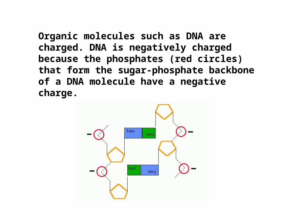
Organic molecules such as DNA are charged. DNA is negatively charged because the phosphates (red circles) that form the sugar-phosphate backbone of a DNA molecule have a negative charge.
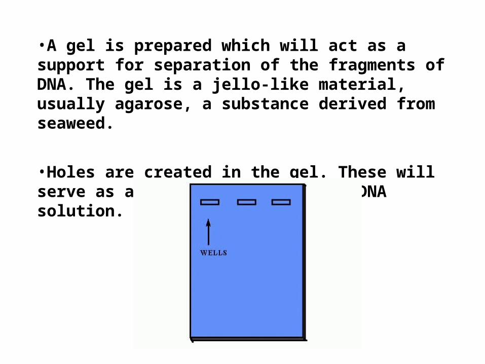
•A gel is prepared which will act as a support for separation of the fragments of DNA. The gel is a jello-like material, usually agarose, a substance derived from seaweed.
•Holes are created in the gel. These will serve as a reservoir to hold the DNA solution.

DNA solutions (mixtures of different sizes of DNA fragments) are loaded in a well in the gel.
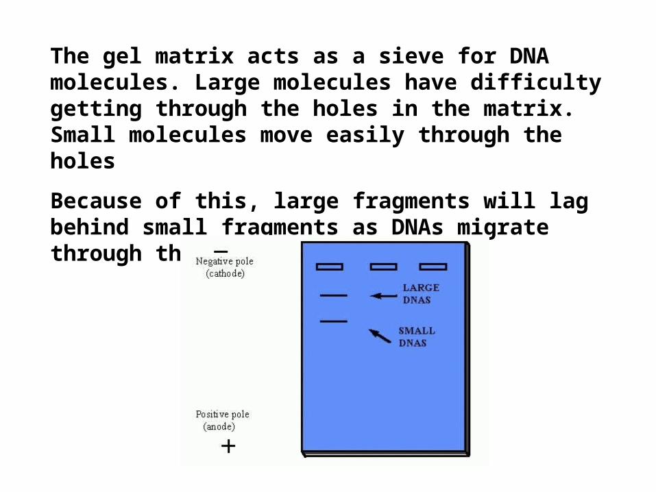
The gel matrix acts as a sieve for DNA molecules. Large molecules have difficulty getting through the holes in the matrix. Small molecules move easily through the holes
Because of this, large fragments will lag behind small fragments as DNAs migrate through the gel.

As the separation process continues, the separation between the larger and smaller fragments increases.

•Molecular weight markers are often electrophoresed with DNAs.
•Molecular weight markers are usually a mixture of DNAs with known molecular weights
•Molecular weight markers are used to estimate the sizes of DNA fragments in your DNA sample
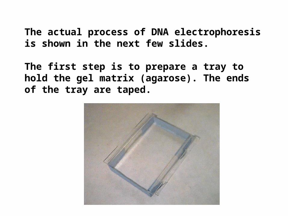
The actual process of DNA electrophoresis is shown in the next few slides.
The first step is to prepare a tray to hold the gel matrix (agarose). The ends of the tray are taped.

A "gel comb" is used to create holes in the gel.

The gel comb is placed in the tray.

Agarose powder is mixed with a buffer solution, usually tris borate EDTA (TBE buffer). The solution is heated until the agarose is dissolved.
The hot agarose solution is poured into the tray and allowed to cool.
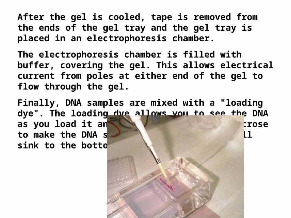
After the gel is cooled, tape is removed from the ends of the gel tray and the gel tray is placed in an electrophoresis chamber.
The electrophoresis chamber is filled with buffer, covering the gel. This allows electrical current from poles at either end of the gel to flow through the gel.
Finally, DNA samples are mixed with a "loading dye". The loading dye allows you to see the DNA as you load it and contains glycerol or sucrose to make the DNA sample heavy so that it will sink to the bottom of the well.

A safety cover is placed over the gel (to keep you from frying yourself) and electrodes are attached to a power supply. Electrical current is applied.
DNA fragments will migrate through the gel at various rates, depending on their size.
When the dye marker indicates that DNA fragments have moved through the gel, the current is turned off and the gel is removed from the tray.

DNAs are visualized by staining the gel with ethidium bromide which binds to DNA and will fluoresce in UV light.
This photograph is of various types of DNA that have been electrophoresed on the same gel. Note that high molecular weight DNAs do not separate well on this gel. This can be corrected by altering gel density.

ENZYMATIC AMPLIFICATION OF DNA
• Polymerase Chain Reactions (PCR)-– a copying machine for DNA molecules
It’s a fast, inexpensive & simple way to make lots of copies of a small sample of DNA

1.Add Target DNA to a test tube which has:
DNA polymerase (TAQ*)
Short oligonucleotide** primer
Copies of the four nucleotides
*TAQ- heat stable DNA polymerase (from hot springs)
**A molecule usually composed of 25 or fewer nucleotides; used as a DNA synthesis primer

Heat to 75ºC to denature DNA to single strands for a minute or so

Cool to 50-65ºc to anneal primers (synthetic sequences of DNA) at target
site
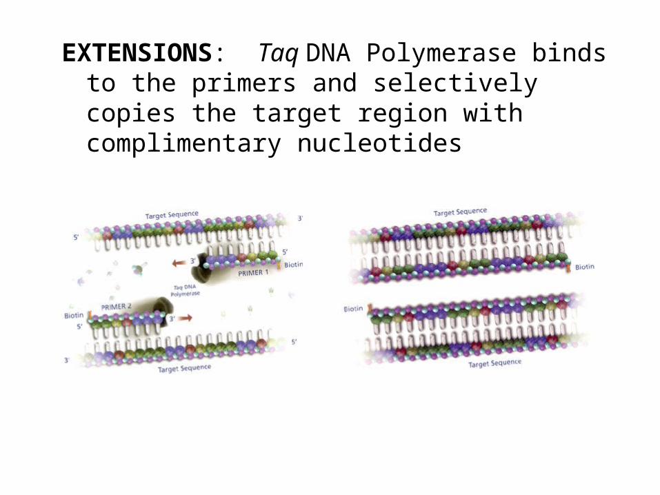
EXTENSIONS: Taq DNA Polymerase binds to the primers and selectively copies the target region with complimentary nucleotides
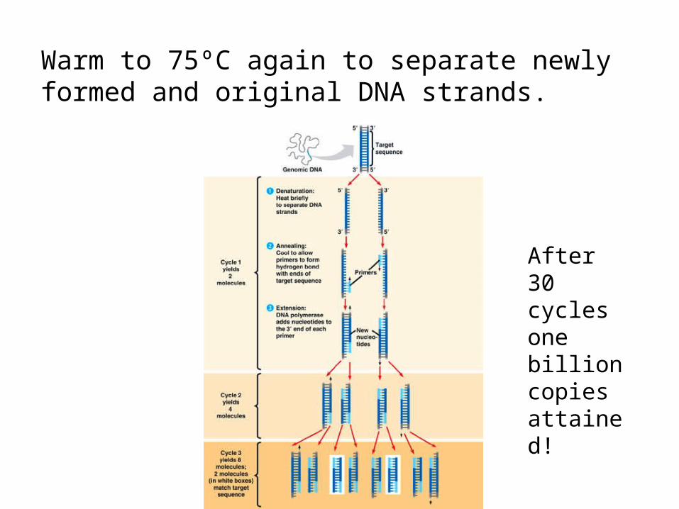
Warm to 75ºC again to separate newly formed and original DNA strands.
After 30 cycles one billion copies attained!

Some Animations
• http://www.sumanasinc.com/webcontent/anisamples/molecularbiology/pcr.html
• http://www.dnalc.org/ddnalc/resources/pcr.html
• http://www.pbs.org/wgbh/nova/sheppard/lab01.html\


