18
Separation of Proteins Part II 180°
| Date post: | 13-Dec-2015 |
| Category: |
Documents |
| Upload: | apolakkiatis |
| View: | 215 times |
| Download: | 0 times |
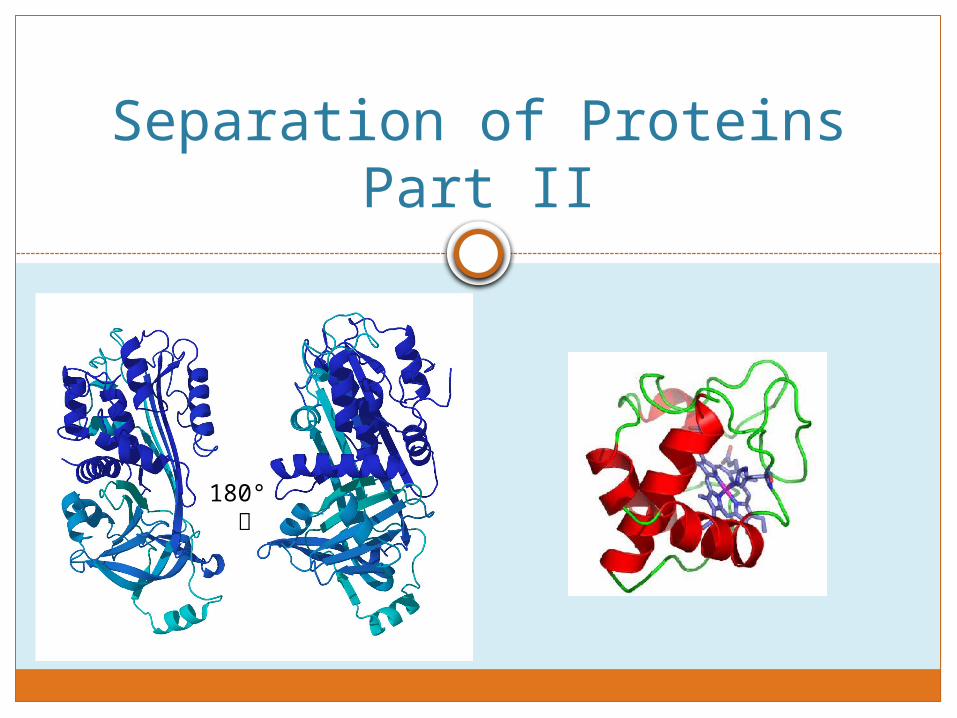
Separation of Proteins Part II
180°
Agenda
Review Separation of Proteins Part I
What is SDS-PAGE and how does it work?
Western Blotting
Today’s procedure
Reminders
Gel loading demo

What did you do two weeks ago?
What two proteins did you separate?
What two types of chromatography did you use to separate the mixture of proteins?

What is SDS-PAGE
Sodium Dodecyl Sulfate-Polyacrylamide Gel Electrophoresis A method of separating proteins based on their
electrophoretic ability (length and mass-to-charge ratio)
What is SDS-PAGE used for? Determination of protein size Protein identification Quantifying proteins Blotting applications- Western blots
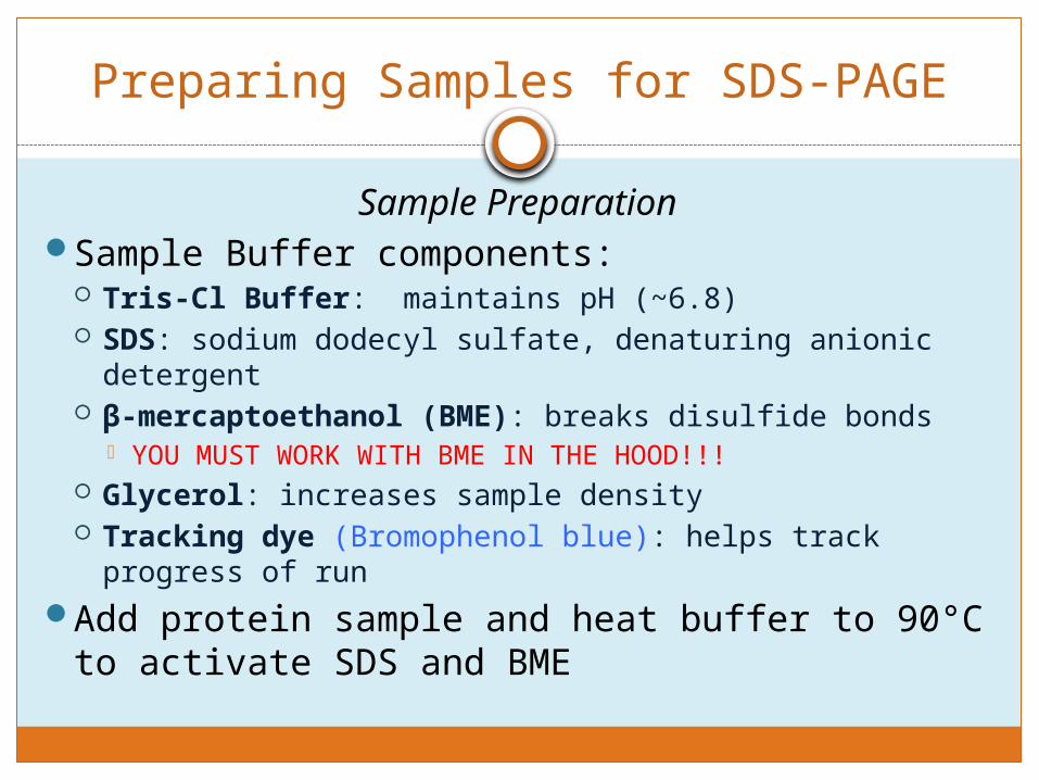
Preparing Samples for SDS-PAGE
Sample PreparationSample Buffer components:
Tris-Cl Buffer: maintains pH (~6.8) SDS: sodium dodecyl sulfate, denaturing anionic
detergent β-mercaptoethanol (BME): breaks disulfide bonds
YOU MUST WORK WITH BME IN THE HOOD!!! Glycerol: increases sample density Tracking dye (Bromophenol blue): helps track
progress of runAdd protein sample and heat buffer to 90°C to
activate SDS and BME
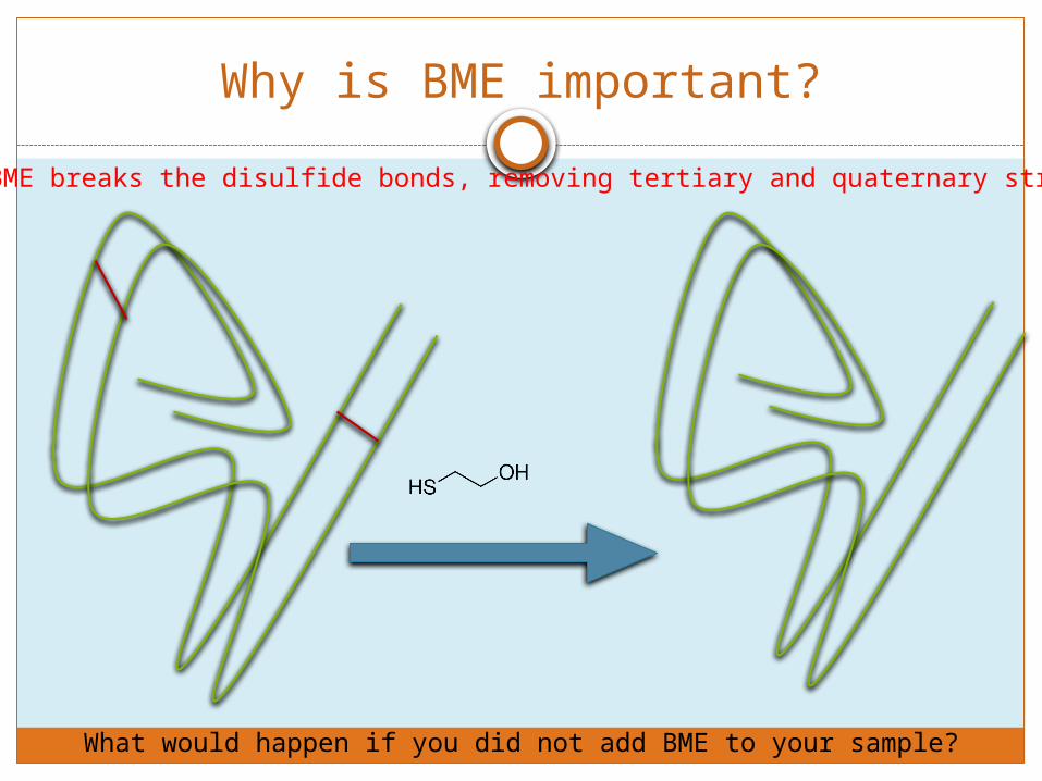
Why is BME important?
BME breaks the disulfide bonds, removing tertiary and quaternary structure
What would happen if you did not add BME to your sample?
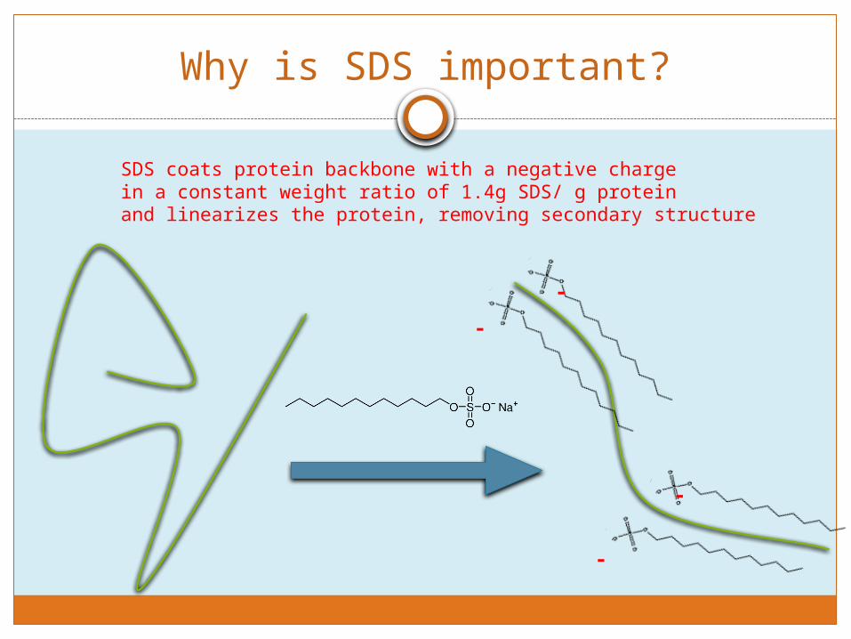
Why is SDS important?
SDS coats protein backbone with a negative charge in a constant weight ratio of 1.4g SDS/ g proteinand linearizes the protein, removing secondary structure
-
-
-
-

SDS-PAGE Gel Set up
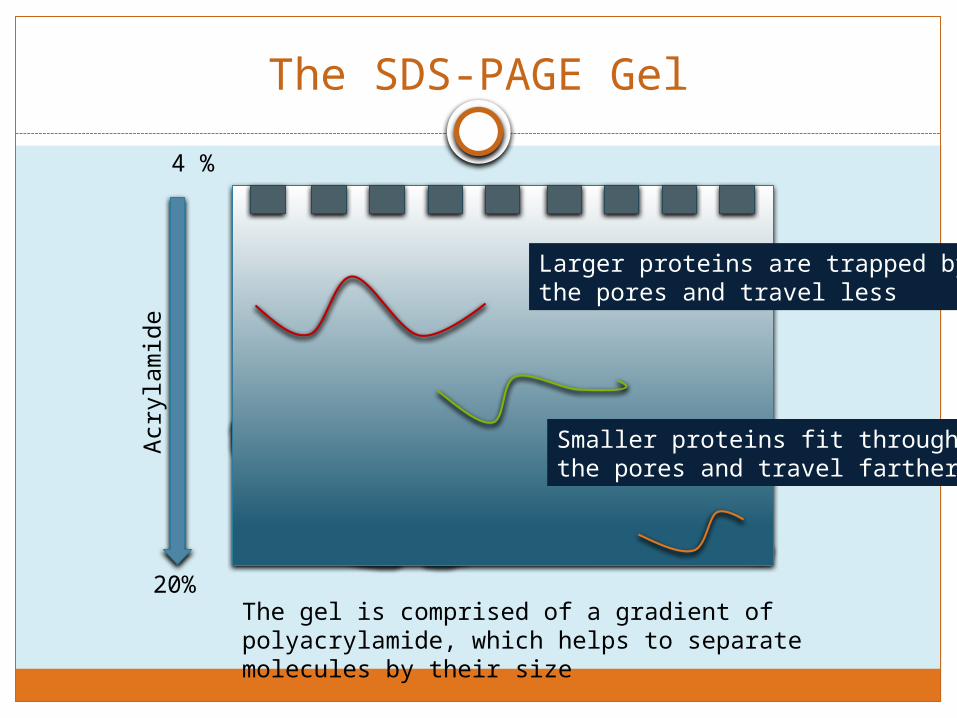
The SDS-PAGE Gel
The gel is comprised of a gradient of polyacrylamide, which helps to separate molecules by their size
4 %
20%
Acr
ylam
ide
Smaller proteins fit through the pores and travel farther
Larger proteins are trapped bythe pores and travel less

How does SDS-PAGE work?
An electric field is applied to the gel, allowing proteins to migrate by charge.
In which direction do the proteins run and why?
If there are multiple bands in the same lane, how would you identify whetherit contains your protein of interest?
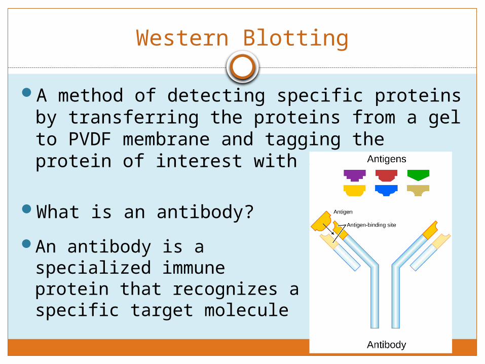
Western Blotting
A method of detecting specific proteins by transferring the proteins from a gel to PVDF membrane and tagging the protein of interest with antibodies
What is an antibody?
An antibody is a specialized immune protein that recognizes a specific target molecule

Today’s Procedure
Add 75μL of Peak
Fraction 1 to 15 μL of
sample buffer
• Repeat for Peak Fraction 2
Heat Sample in heating
block (90°C) for 4
minutes
Centrifuge at max
speed for 30 sec.
• Make sure that tubes all contain the same volume and to properly balance the centrifuge
Load samples
onto gel in the correct
order

Gel Loading Order
Please load 15μL of sample and 10μL of MW marker
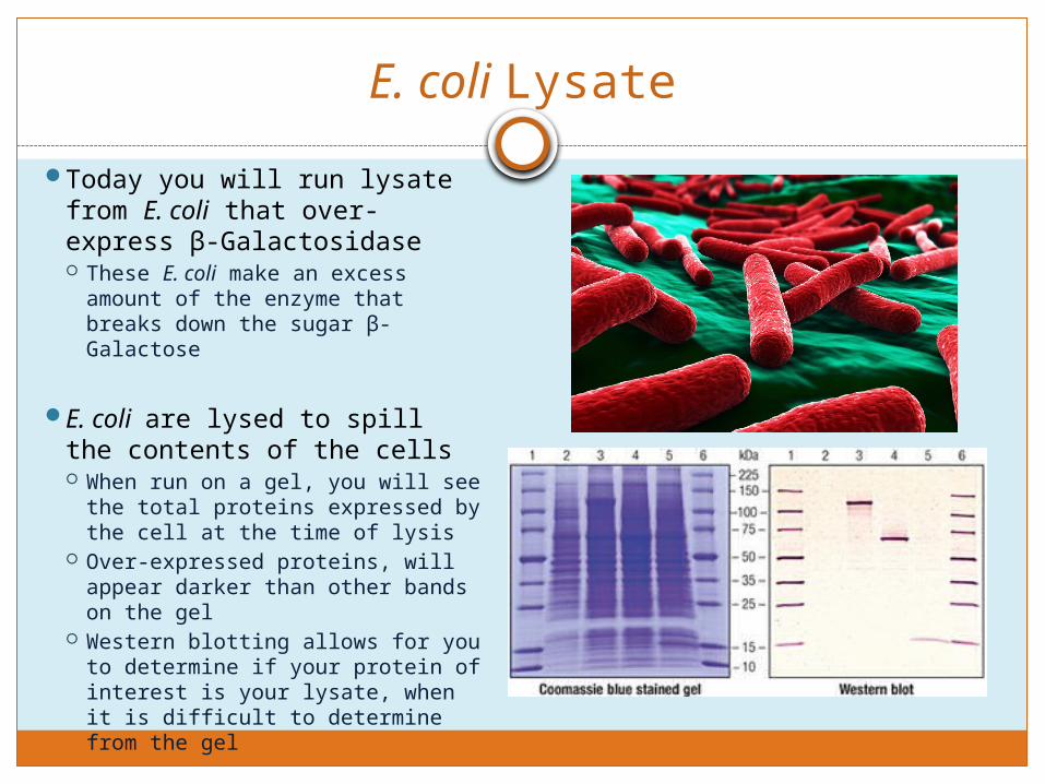
E. coli Lysate
Today you will run lysate from E. coli that over-express β-Galactosidase These E. coli make an excess
amount of the enzyme that breaks down the sugar β-Galactose
E. coli are lysed to spill the contents of the cells When run on a gel, you will see
the total proteins expressed by the cell at the time of lysis
Over-expressed proteins, will appear darker than other bands on the gel
Western blotting allows for you to determine if your protein of interest is your lysate, when it is difficult to determine from the gel

Setting up your Western Blot
Today, you will set up your western blot sandwiches: An electric field will
be run through the blotting apparatus, which will transfer the negatively charged proteins from the gel to the PVDF membrane

Data Analysis
This Week: Determine the protein
concentration using the Beer Lambert Law
Next Week: Calculate Rf values:
Protein migration/dye front migration Measure from the bottom of the well
to the dye front/protein band Create a semi-log plot (use the
correct paper!) MW vs Rf values for MW marker bands Use standard curve to determine the
molecular weight of your peak fractions
Use the 2-cycle semi-log paper
Standard curve of molecular weights of proteins based on Rf
values
(g/mol)
Reminders
Gloves and gogglesBME is toxic
You will find sample buffer in the hood Please place all tips/tubes contaminated with sample buffer in the
appropriately labeled waste containersRemember the order in which you set up your western blotSAVE YOUR FRACTIONS FOR NEXT WEEKLab report for Parts II and III due week of 4/6Problem Set 3 due the week of 3/30
Next week, we will: Analyze the stained gel Continue the western blot Perform the Lowry Protein Assay (Separation Part III)

Reminders
Please remember to properly cite the documents used in your concluding statements/discussions
http://classguides.lib.uconn.edu/content.php? pid=352130&sid=3243107


