JCDA • www.cda-adc.ca/jcda • September 2008, Vol. 74, No. 7 • 617 Point of Care What is the recommended dental management for patients who are receiving oral bisphosphonate therapy? Table 1 Bisphosphonates currently available in Canada for oral administration 1 Generic name Brand name Indications Alendronate Fosamax Osteoporosis, Paget’s disease Fosavance Osteoporosis Clodronate Bonefos, Clasteon Bone metastases of malignant tumours, hypercalcemia of malignancy Etidronate Didrocal Osteoporosis Didronel Paget’s disease Risedronate Actonel Osteoporosis, Paget’s disease of bone Actonel Plus Calcium Osteoporosis Background B y decreasing osteoclastic activity, bisphos- phonate drugs decrease rates of bone re- sorption, resulting in an increase in bone mass when given to patients with osteoporosis. ey also have therapeutic effects for patients with rarer metabolic bone diseases such as Paget’s dis- ease and osteogenesis imperfecta and for cancer patients with metastases to bone. Two forms of bisphosphonate treatment are currently available — oral (Table 1 ) and intravenous (see article on p. 618). Most patients receiving bisphosphonate therapy who are encountered in the general dental setting are receiving oral treatment, usually for osteoporosis. Management Advice It has been hypothesized, though not proven, that oral bisphosphonate therapy may be as- sociated with osteonecrosis of the jaw. e sci- entific data for cases of bisphosphonate-related osteonecrosis of the jaw (BRONJ) are incomplete, and the vast majority of patients receiving oral bisphosphonate therapy do not experience any oral complications. 2 As such, patients should be informed that the health benefits of oral bisphos- phonate therapy far outweigh the minimal risk (if any) of BRONJ. In addition, good oral hygiene, ac- companied by regular dental care, is the best way to minimize this risk, if it exists. Patients receiving bisphosphonate therapy should be advised to con- tact their dentist if any problem develops in the oral cavity. In general, patients who are taking oral bisphosphonates without other risk factors (Box 1 ) can be treated according to normal protocols and procedures, including surgery. For patients receiving oral bisphosphonate therapy, dental treatment recommendations are similar to those for patients not taking the medica- tion, as described in the following sections. Restorative and Prosthetic Dentistry All restorative procedures may be performed. At present, there is no evidence that malocclu- sion or occlusal forces increase the risk of BRONJ. Prosthodontic appliances should be adjusted for fit to avoid mucosal irritation. 2 Periodontal Diseases Treatment protocols are similar to those for the general population (i.e., patients not taking the medication). 2 QUESTION 1 e “Point of Care” section answers everyday clinical questions by providing practical information that aims to be useful at the point of patient care. e responses reflect the opinions of the contributors and do not purport to set forth standards of care or clinical practice guidelines. is month’s contributors are from McGill University’s faculty of dentistry.
Transcript
JCDA•www.cda-adc.ca/jcda • September 2008, Vol. 74, No. 7 • 617
Point of Care
What is the recommended dental management for patients who are receiving oral bisphosphonate therapy?
Table1 Bisphosphonates currently available in Canada for oral administration1
Genericname Brandname Indications
Alendronate Fosamax Osteoporosis, Paget’s disease
Fosavance Osteoporosis
Clodronate Bonefos, Clasteon Bone metastases of malignant tumours, hypercalcemia of malignancy
Etidronate Didrocal Osteoporosis
Didronel Paget’s disease
Risedronate Actonel Osteoporosis, Paget’s disease of bone
Actonel Plus Calcium Osteoporosis
Background
By decreasing osteoclastic activity, bisphos-phonate drugs decrease rates of bone re-sorption, resulting in an increase in bone
mass when given to patients with osteoporosis. They also have therapeutic effects for patients with rarer metabolic bone diseases such as Paget’s dis-ease and osteogenesis imperfecta and for cancer patients with metastases to bone. Two forms of bisphosphonate treatment are currently available — oral (Table 1) and intravenous (see article on p. 618). Most patients receiving bisphosphonate therapy who are encountered in the general dental setting are receiving oral treatment, usually for osteoporosis.
ManagementAdviceIt has been hypothesized, though not proven,
that oral bisphosphonate therapy may be as-sociated with osteonecrosis of the jaw. The sci-entific data for cases of bisphosphonate-related osteonecrosis of the jaw (BRONJ) are incomplete, and the vast majority of patients receiving oral bisphosphonate therapy do not experience any oral complications.2 As such, patients should be informed that the health benefits of oral bisphos-
phonate therapy far outweigh the minimal risk (if any) of BRONJ. In addition, good oral hygiene, ac-companied by regular dental care, is the best way to minimize this risk, if it exists. Patients receiving bisphosphonate therapy should be advised to con-tact their dentist if any problem develops in the oral cavity. In general, patients who are taking oral bisphosphonates without other risk factors (Box 1) can be treated according to normal protocols and procedures, including surgery.
For patients receiving oral bisphosphonate therapy, dental treatment recommendations are similar to those for patients not taking the medica-tion, as described in the following sections.
Restorative and Prosthetic DentistryAll restorative procedures may be performed.
At present, there is no evidence that malocclu-sion or occlusal forces increase the risk of BRONJ. Prosthodontic appliances should be adjusted for fit to avoid mucosal irritation.2
Periodontal DiseasesTreatment protocols are similar to those for
the general population (i.e., patients not taking the medication).2
Q u E s t i o n 1
The “Point of Care” section answers everyday clinical questions by providing practical information that aims to be useful at the point of patient care. The responses reflect the opinions of the contributors and do not purport to set forth standards of care or clinical practice guidelines. This month’s contributors are from McGill University’s faculty of dentistry.
618 JCDA•www.cda-adc.ca/jcda • September 2008, Vol. 74, No. 7 •
––––––– Point of Care –––––
EndodonticsIf the tooth is salvageable, endodontic treat-
ment is preferred to extractions or surgical ma-nipulation. If extractions or surgical manipulations are necessary, such procedures should follow the recommendations discussed in the section “Oral and Maxillofacial Surgery” above. a
THE AUTHORS
Ms. Minyue Dai is a fourth-year dental student at the faculty of dentistry, McGill University, Montreal, Quebec.
Ms. Yang Li is a fourth-year dental student at the faculty of dent-istry, McGill University, Montreal, Quebec.
Dr. Timothy W. Head is an associate professor at the faculty of dentistry, McGill University, Montreal, Quebec.
Dr. Marc D. McKee is a professor at the faculty of dentistry, McGill University, Montreal, Quebec.
Dr. Simon D. Tran is an assistant professor at the faculty of dent-istry, McGill University, Montreal, Quebec. Email: simon.tran @mcgill.ca
References1. Compendium of Pharmaceuticals and Specialties (CPS): the Canadian Drug Reference for Health Professionals. 42nd ed. Ottawa (ON): Canadian Pharmacists Association; 2007.
2. American Dental Association Council on Scientific Affairs. Dental management of patients receiving oral bisphosphonate therapy: expert panel recommendations. J Am Dent Assoc 2006; 137(8):1144–50.
3. Khosla S, Burr D, Cauley J, Dempster Dw, Ebeling PR, Felsenberg D, and others. Bisphosphonate-associated osteonecrosis of the jaw: report of a task force of the American Society for Bone and Mineral Research. J Bone Miner Res 2007; 22(10):1479–90.
What is the recommended dental management for patients who are receiving intravenous bisphosphonate therapy?
Q u E s t i o n 2
Oral and Maxillofacial SurgeryTreatment protocols are similar to those for
the general population (i.e., patients not taking the medication), unless other risk factors are present (Box 1). In such cases, conservative surgical tech-nique, with primary tissue closure, should be con-sidered when extractions or surgery are necessary (including elective dentoalveolar surgical proced-ures such as implant placement, reduction of tori or extraction of asymptomatic teeth).2
Patients may use a chlorhexidine-containing rinse immediately before and after surgical pro-cedures. Systemic antibiotic therapy may be con-sidered for perioperative prophylaxis or if there is evidence of infection.
Background
Since 2003, dentists have been observing osteonecrosis of the jaw as a potential com-plication of intravenously administered bi-
sphosphonate therapy. Bisphosphonate-related osteonecrosis of the jaw (BRONJ) is defined as an area of exposed bone in the maxillofacial region that does not heal within 8 weeks after its iden-tification by a health care provider, in a patient who is receiving or has previously been receiving bisphosphonates and who has not had radiation therapy to the craniofacial region.1
The bisphosphonates currently available on the Canadian market for intravenous (IV) administra-tion are listed in Table 1.2
ManagementAdvice
Dental Management of Patients Receiving IV Bisphosphonate Therapy
The risk of BRONJ appears to range between 1% and 10% in patients receiving IV bisphosphonate treatment.1 Any patient receiving such therapy should be informed of the signs and symptoms of BRONJ. In addition, before the IV bisphosphonate therapy is started, the patient should undergo a dental evaluation by a qualified dental profes-sional, and dental recall examinations should be performed throughout the course of bisphos-phonate therapy. The frequency of such examina-tions will be dictated by the patient’s clinical and dental status.
Box1 Risk factors for bisphosphonate-related osteonecrosis of the jaw in patients receiv-ing oral bisphosphonate therapy2,3
• Concomitant use of estrogen or glucocorticoids
• Comorbid conditions (e.g., malignancy)• Poorly fitting dental appliances• Intraoral trauma• Presence of tori or other bony exostoses• Pre-existing dental or periodontal disease• Older age (> 65 years)• Alcohol and/or tobacco use
JCDA•www.cda-adc.ca/jcda • September 2008, Vol. 74, No. 7 • 619
––––––– Point of Care –––––
It is also important to identify patients with risk factors for BRONJ: dental extraction and/or oral bone surgery; poorly fitting dental appliances; intraoral trauma; presence of tori or other bony exostoses; pre-existing dental or periodontal dis-ease; older age (> 65 years); prolonged exposure to bisphosphonate therapy; concomitant use of estrogen or glucocorticoids; comorbid conditions (e.g., malignancy); alcohol and/or tobacco use.1
Patients with cancer who are receiving IV pamidronate and/or zoledronic acid are at the greatest risk for BRONJ.1
If the patient’s situation permits, invasive dental procedures should be performed before the IV bisphosphonate therapy is started, with follow-up at 14–21 days to ensure complete healing at the surgical site. The following sections outline treatment recommendations for patients who are already receiving IV bisphosphonate therapy.1,3
Restorative and Prosthetic DentistryAll restorative procedures may be performed.
At present, there is no evidence that malocclu-sion or occlusal forces increase the risk of BRONJ. Prosthodontic appliances should be adjusted for fit to avoid mucosal irritation.
Periodontal DiseasesNonsurgical therapy is preferred (such as
scaling and root planing). Periodontal surgery is not recommended. When necessary, surgical
treatment should be aimed primarily at obtaining access to root surfaces.
Oral and Maxillofacial SurgeryWhenever possible, nonsurgical endodontic
or periodontal therapy is preferred to extrac-tion, unless there is a risk of aspiration. Elective dento-alveolar surgical procedures, such as im-plant placement, reduction of tori and extraction of asymptomatic teeth, should be avoided. When an extraction or surgery is necessary, conservative surgical technique, with primary tissue closure, should be considered. The greater incidence of BRONJ in the mandible than the maxilla, espe-cially in the posterior region of the mouth, must be taken into account in the decision to perform surgery.
EndodonticsFor salvageable teeth, endodontic treatment is
preferred to extractions or surgical manipulation. Manipulation beyond the apex should be avoided. Surgical procedures should be guided using the same recommendations mentioned in the section “Oral and Maxillofacial Surgery.”
Considerations for Any Surgical Procedure Patients should use a chlorhexidine-containing
rinse immediately before and after surgical proced-ures. Systemic antibiotic therapy may be considered for perioperative prophylaxis or if there is evidence of infection (and should follow the guidelines of the American Dental Association3) (Table 2).
Table1 Bisphosphonates currently available in Canada for intravenous administration2
Genericname Brandname Indications
Clodronate Bonefos, Clasteon Bone metastases of malignant tumours, hypercalcemia of malignancy
Pamidronate Aredia Bone metastases of malignant tumours, hypercalcemia of malignancy, multiple myeloma, Paget’s disease
Zoledronic acid Aclasta Paget’s diseaseZometa concentrate Bone metastases of malignant tumours, hypercalcemia of
Not allergic to penicillin Amoxicillin 500 mg t.i.d. for 14 days
May be combined with metronidazole 250 mg t.i.d. for 14 days
Allergic to penicillin Clindamycin 300 mg t.i.d. for 14 days
Azithromycin 250 mg t.i.d. for 10 days
620 JCDA•www.cda-adc.ca/jcda • September 2008, Vol. 74, No. 7 •
––––––– Point of Care –––––
Dental Management of Patients with BRONJ If BRONJ is suspected but not yet confirmed
(e.g., duration of unhealed exposed bone less than 8 weeks; Fig. 1), the patient should be followed carefully. Additional common findings include pain, swelling, paresthesia, suppuration, soft-tissue ulceration, intraoral or extraoral sinus tracks, and loosening of teeth. Radiographic findings can vary from changes in bone density to no obvious altera-tion to the bone pattern.
The differential diagnosis for BRONJ includes gingivitis, periodontal diseases (e.g., necrotizing ulcerative periodontitis), osteomyelitis, sinusitis, temporomandibular disorder, trauma, periapical lesions, osteoradionecrosis, bone tumours and me-tastasic lesions.
Standard radiography such as panoramic and periapical radiography may help in the detection of BRONJ in the early stages. Computed tomography may also be considered. No imaging is required for patients with established clinical evidence of BRONJ.
The dental professional should alert the pa-tient’s physician to the diagnosis and should report cases of BRONJ to the appropriate agencies, such as the manufacturer of any agent implicated. There is no published evidence to suggest that discon-tinuation of bisphosphonates will promote resolu-tion of BRONJ.
If pain is present, it should be managed ap-propriately with nonsteroidal anti-inflammatory drugs or narcotic analgesics. The patient should be advised to use chlorhexidine (0.12%) or another similar oral antimicrobial rinse, and systemic
antibiotic therapy may be prescribed if there is evidence of secondary infection. Establishing and maintaining good oral hygiene is essential.
Any patient with established BRONJ needing surgical procedures should be referred to an oral and maxillofacial surgeon, who may consult other qualified specialists as appropriate. Any dentoal-veolar surgical procedure (i.e., extractions, implants or apical surgery) should be avoided since the sur-gical sites will likely result in additional areas of exposed necrotic bone. However, loose teeth should be removed from the exposed bone if there is a danger of aspiration. Similarly, loose segments of bony sequestra should be removed, but without ex-posing uninvolved bone. Sharp bone edges should be removed, to prevent trauma to the adjacent soft tissues. Segmental jaw resection may be required for symptomatic patients with large segments of necrotic bone or pathologic fracture. a
THE AUTHORS
Ms. Li is a fourth-year dental student at the faculty of dentistry, McGill University, Montreal, Quebec..
Ms. Dai is a fourth-year dental student at the faculty of dentistry, McGill University, Montreal, Quebec.
Dr. Head is an associate professor at the faculty of dentistry, McGill University, Montreal, Quebec.
Dr. McKee is a professor at the faculty of dentistry, McGill University, Montreal, Quebec.
Dr. Tran is an assistant professor at the faculty of dentistry, McGill University, Montreal, Quebec. Email: [email protected]
References1. Khosla S, Burr D, Cauley J, Dempster Dw, Ebeling PR, Felsenberg D, and others. Bisphosphonate-associated osteonecrosis of the jaw: report of a task force of the American Society for Bone and Mineral Research. J Bone Miner Res 2007; 22(10):1479–90.
2. Compendium of Pharmaceuticals and Specialties (CPS): the Canadian Drug Reference for Health Professionals. 42nd ed. Ottawa (ON): Canadian Pharmacists Association; 2007.
3. American Dental Association Council on Scientific Affairs. Dental management of patients receiving oral bisphosphonate therapy: expert panel recommendations. J Am Dent Assoc 2006; 137(8):1144–50.
Figure1:A 69-year-old man with necrotic bone and no healing 3 months after extrac-tion of a tooth. The patient had prostate cancer and was receiving zoledronic acid. Photo courtesy of Drs. Emery and Pompura.
Figure1:Interproximal image taken with a storage phosphor sensor.
Figure2:Periapical image taken with a storage phosphor sensor.
Figure3:Interproximal image taken with a direct sensor.
Figure4:Periapical image taken with a direct sensor.
JCDA•www.cda-adc.ca/jcda • September 2008, Vol. 74, No. 7 • 621
––––––– Point of Care –––––
Which digital intraoral sensor is better?
Q u E s t i o n 3
FurtherReadingAdvisory Task Force on Bisphosphonate-Related Osteonecrosis of
the Jaws, American Association of Oral and Maxillofacial Surgeons. American Association of Oral and Maxillofacial Surgeons position paper on bisphosphonate-related osteonecrosis of the jaws. J Oral Maxillofac Surg 2007; 65(3):369–76.
Migliorati CA, Casiglia J, Epstein J, Jacobsen PL, Siegel MA, Woo SB. Managing the care of patients with bisphosphonate- associated osteonecrosis: an American Academy of Oral Medicine position paper. J Am Dent Assoc 2005; 136(12):1658–68.
A patient brochure on bisphosphonates produced by McGill University’s faculty of dentistry is available online at www.cda-adc.ca/jcda/vol-74/issue-7/617.html.
Background
There are 2 types of intraoral sensors: direct sensors and storage phosphor sensors. Direct sensors, whether they use charge-couple-de-
vice or complementary metal oxide semiconductor technology, are equivalent in terms of image quality.1 Image display is instantaneous as these sensors are connected to a computer. The storage phosphor sensor is a plate, with dimensions com-parable to those of conventional film; images are obtained when the plate is inserted into and read by a scanner.
Several experts believe that today’s sensors are reaching their technological limits. Both direct
and storage phosphor sensors are capable of producing diagnostic images for the tasks dentists perform daily, such as diagnosing caries, identifying periapical lesions and evaluating periodontal bone loss (Figs. 1–4).2–7
DigitalSensorCharacteristicsThe characteristics of digital sensors that have
an impact on image quality are contrast resolu-tion, spatial resolution, latitude and sensitivity.
Contrast resolution is the ability to detect dif-ferences between shades of grey. Theoretically, a sensor capable of capturing more shades of grey (greater bit depth) is better. However, because
computer monitors display only 8-bit images, in practice there will be no difference between intraoral sensors that capture 8-bit images (256 levels of grey), 12-bit images (4,096 levels of grey) and 14-bit images (16,384 levels of grey).8,9 In addition, the number of grey shades differentiated by the human eye is between 32 and 60.10
Spatial resolution is the ability to capture details and is measured in line-pairs per millimetre (lp/mm). Film achieves a resolution of up to 20 lp/mm. Newer sensors with a pixel size of 20 µm are able to resolve 25 lp/mm. Storage phosphor systems achieve a lower resolution than direct sensors. Most dentists can perceive 6 lp/mm and up to 10–12 lp/mm with magnification; images magnified above that become pixilated and non-diagnostic. Digital sensors available today have a resolution of 7 lp/mm or more.11
Latitude is the ability of digital re-ceptors to provide diagnostic images
Figure1:Interproximal image taken with a storage phosphor sensor.
Figure2:Periapical image taken with a storage phosphor sensor.
Figure3:Interproximal image taken with a direct sensor.
Figure4:Periapical image taken with a direct sensor.
622 JCDA•www.cda-adc.ca/jcda • September 2008, Vol. 74, No. 7 •
––––––– Point of Care –––––
with a range of exposures. A disadvantage of con-ventional film is that it is easily overexposed or underexposed. Although the latitude of direct sensors is comparable to that of film, storage phos-phor sensors have a greater latitude and, under normal conditions, images are unlikely to be over-exposed or underexposed.10 The downside is the greater dose of radiation that patients will receive if greater exposure is used consistently.12
Sensitivity is the amount of exposure required to produce an image. The more sensitive the re-ceptor, the less exposure is required. One well-known advantage of intraoral digital radiography is the lower dose of radiation to which patients are exposed. The most sensitive intraoral film avail-able is F-speed. Storage phosphor systems can produce images using half the exposure necessary with F-speed film. Direct systems require more exposure than storage phosphor systems, but less than F-speed film.
All imaging software products offer a range of tools for dentists to use to enhance their im-ages. However, the goal is to acquire good-quality diagnostic images that require no enhancement, as modifying images may have deleterious effects.13,14 Clear task-specific indications for the various en-hancement tools have yet to be developed.
ManagementAdvice
Direct SystemsAdvantages• Instantaneousness • Additional images can be obtained without
removing the sensor from the mouth• Spatial resolution superior to storage phosphor
Disadvantages• Sensors are expensive and fragile
• Physical properties of the sensor: thick, rigid, attached cable. Positioning devices are avail-able for all direct sensors to allow the device to be placed parallel to the teeth. However, this technique is not always possible, particularly for patients with a narrow palate. Reverting to the bisecting technique is more frequent than with film. Missed apices are a common problem, particularly for new users of this technology (Fig. 5). The presence of the cable makes obtaining an image of vertical bitewings almost impossible.
• More than one size sensor will be needed. Most companies offer size 1 and 2 sensors whose active areas are smaller than their film counter-parts. Some companies now offer a size 0 sensor for pediatric applications. Size 2 sensors are re-quired for interproximal examinations to view the bone level, but obtaining a distal image of the canines with these large sensors is challen-ging (Fig. 3).
• More exposures are required compared with film because of the smaller active surface area of direct sensors and difficulties in positioning (Fig. 6).
• The learning curve is greater than with storage phosphor sensors.
Storage Phosphor SystemsAdvantages• Latitude superior to direct sensors and film• Sensitivity superior to direct sensors and film• Sensor thickness and flexibility are comparable
to those of film• Plates available in sizes 0 to 4• Plates compatible with standard positioning
devices for obtaining periapical, horizontal and vertical interproximal radiographs
Figure5:This image, taken with a direct sensor, doesn’t show tooth apices.
Figure6:From left: size 2 direct sensor, size 2 plate for a storage phosphor sensor and size 2 film.
Figure7: Storage phosphor sensor plate that is scratched and damaged at the edges.
JCDA•www.cda-adc.ca/jcda • September 2008, Vol. 74, No. 7 • 623
––––––– Point of Care –––––
• Transition from film to storage phosphor is simple
Disadvantages• Spatial resolution inferior to that of direct
sensors• Scanning of exposed plates is required.
Scanning time increases with the size and number of plates and required resolution
• Space for the scanner is required, preferably in a dimmed environment as exposed plates are sensitive to light
• With handling, plates become scratched and damaged at the edges (Fig. 7) and must be re-placed regularly15
Lighting RequirementsThe lighting conditions under which images
are interpreted must be considered. Dental operatories are generally equipped with high ambient light; this must be reduced to create an environment suitable for analysis of digital im-ages.16 Adjusting the contrast and brightness of monitors will also improve image quality.17 Cathode ray tube monitors tend to lose brightness with time.
Transition PeriodRegardless of the system selected, expect a
transition period to adapt to looking at digital images, which appear to have less detail. The evi-dence shows that the information needed to make common diagnoses is there. The medical profes-sion adopted digital radiology to replace conven-tional plain films before the dental profession, possibly because radiologists were used to reading computed tomography and magnetic resonance imaging scans on monitors. However, as stated by Ludlow and Mol, “It is no longer a matter of if but rather when the majority of dental offices will use digital imaging.”11 a
THE AUTHOR
Dr. Marie Dagenais is an oral and maxillofacial radiologist at the faculty of dentistry, McGill University, Montreal, Quebec. Email: [email protected]
References1. Kitagawa H, Scheetz JP, Farman AG. Comparison of comple-mentary metal oxide semiconductor and charge-coupled de-vice intraoral X-ray detectors using subjective image quality. Dentomaxillofac Radiol 2003; 32(6):408–11.
2. Kullendorff B, Nilsson M, Rohlin M. Diagnostic accuracy of direct digital dental radiography for the detection of periapical bone le-
sions: overall comparison between conventional and direct digital radiography. Oral Surg Oral Med Oral Pathol Oral Radiol Endod 1996; 82(3):344–50.
3. Nair MK, Ludlow JB, Tyndall DA, Platin E, Denton G. Periodontitis detection efficacy of film and digital images. Oral Surg Oral Med Oral Pathol Oral Radiol Endod 1998; 85(5):608–12.
4. Abreu M Jr, Mol A, Ludlow JB. Performance of RVGui sensor and Kodak Ektaspeed Plus film for proximal caries detection. Oral Surg Oral Med Oral Pathol Oral Radiol Endod 2001; 91(3):381–5.
5. Khan EA, Tyndall DA, Ludlow JB, Caplan D. Proximal caries detec-tion: Sirona Sidexis versus Kodak Ektaspeed Plus. Gen Dent 2005; 53(1):43–8.
6. Haiter-Neto F, dos Anjos Pontual A, Frydenberg M, Wenzel A. A comparison of older and newer versions of intraoral digital radiog-raphy systems: diagnosing noncavitated proximal carious lesions. J Am Dent Assoc 2007; 138(10):1353–9, quiz 1382–3.
7. Tsesis I, Kamburoglu K, Katz A, Tamse A, Kaffe I, Kfir A. Comparison of digital with conventional radiography in detection of vertical root fractures in endodontically treated maxillary pre-molars: an ex vivo study. Oral Surg Oral Med Oral Pathol Oral Radiol Endod 2008; 106(1):948–52. Epub 2008 Mar 4.
8. Halazonetis DJ. What do 8-bit and 12-bit grayscale mean and which should I use when scanning? Am J Orthod Dentofac Orthop 2005; 127(3):387–8.
9. Wenzel A, Haiter-Neto F, Gotfredsen E. Influence of spatial reso-lution and bit depth on detection of small caries lesions with digital receptors. Oral Surg Oral Med Oral Pathol Oral Radiol Endod 2007; 103(3):418–22. Epub 2006 Aug 2.
10. Sanderink GC. Intra-oral and extra-oral digital imaging: an overview of factors relevant to detector design. In: Nuclear in-struments and methods in physics research. Section A, Accelerators, spectrometers, detectors and associated equipment. 2003; 509(1–3):256–61.
11. Ludlow JB, Mol A. Digital imaging. In: White SC, Pharoah MJ, editors. Oral radiology, principles and interpretation. 5th ed. St Louis: Mosby; 2004. p. 225–44.
12. Berkhout WE, Beuger DA, Sanderink GC, van der Stelt PF. The dynamic range of digital radiographic systems: dose reduction or risk of overexposure? Dentomaxillofac Radiol 2004; 33(1):1–5.
13. Tyndall DA, Ludlow JB, Platin E, Nair M. A comparison of Kodak Ektaspeed Plus film and the Siemens Sidexis digital imaging system for caries detection using receiver operating characteristic analysis. Oral Surg Oral Med Oral Pathol Oral Radiol Endod 1998; 85(1):113–8.
14. Kullendorff B, Nilsson M. Diagnostic accuracy of direct digital dental radiography for the detection of periapical bone lesions. II. Effects on diagnostic accuracy after application of image pro-cessing. Oral Surg Oral Med Oral Pathol Oral Radiol Endod 1996; 82(5):585–9.
15. Bedard A, Davis TD, Angelopoulos C. Storage phosphor plates: how durable are they as a digital dental radiographic system? J Contemp Dent Pract 2004; 5(2):57–69.
16. Kutcher MJ, Kalathingal S, Ludlow JB, Abreu M Jr, Platin E. The effect of lighting conditions on caries interpretation with a laptop computer in a clinical setting. Oral Surg Oral Med Oral Pathol Oral Radiol Endod 2006; 102(4):537–43. Epub 2006 Jun 19.
17. Hellén-Halme K. Quality aspects of digital radiography in gen-eral dental practice. Swed Dent J Suppl 2007; (184):9–60.
624 JCDA•www.cda-adc.ca/jcda • September 2008, Vol. 74, No. 7 •
––––––– Point of Care –––––
plane or the mandibular plane can be chosen as a reference.2 The occlusal plane of the existing or planned prosthesis would also be an adequate ref-erence for measuring the angulation of implants or planning their ultimate position.
It has been suggested that implants should be placed as parallel to the path of insertion of the overdenture and as perpendicular to its occlusal plane as possible. Positioning the implants in ac-cordance with these references facilitates denture insertion and avoids excessive nonaxial loading.
Because of differences in the size and shape of the residual ridge and the maxillomandibular relation, ideal angulation of the implants during placement can be difficult or impossible to achieve. In a preliminary study,3 19% of the implants evalu-ated had an angulation of 90° to the reference plane, 11% had a lingual inclination and 70% were
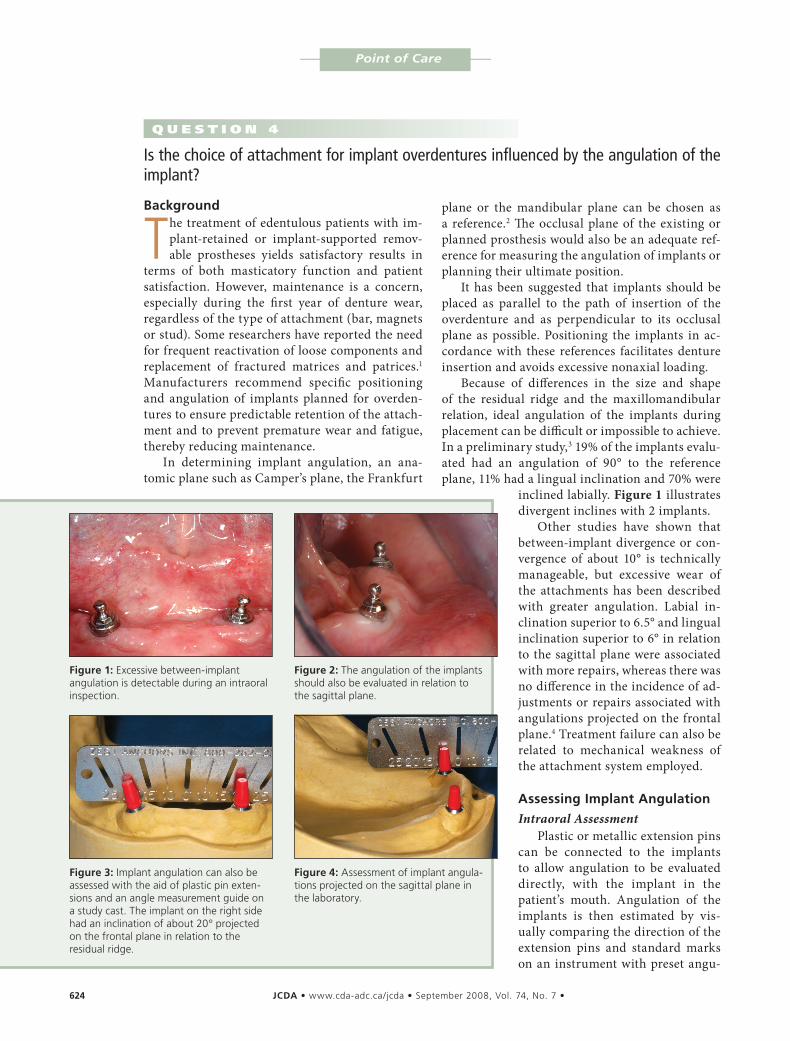
inclined labially. Figure 1 illustrates divergent inclines with 2 implants.
Other studies have shown that between-implant divergence or con-vergence of about 10° is technically manageable, but excessive wear of the attachments has been described with greater angulation. Labial in-clination superior to 6.5° and lingual inclination superior to 6° in relation to the sagittal plane were associated with more repairs, whereas there was no difference in the incidence of ad-justments or repairs associated with angulations projected on the frontal plane.4 Treatment failure can also be related to mechanical weakness of the attachment system employed.
AssessingImplantAngulation
Intraoral AssessmentPlastic or metallic extension pins
can be connected to the implants to allow angulation to be evaluated directly, with the implant in the patient’s mouth. Angulation of the implants is then estimated by vis-ually comparing the direction of the extension pins and standard marks on an instrument with preset angu-
Figure1:Excessive between-implant angulation is detectable during an intraoral inspection.
Figure2:The angulation of the implants should also be evaluated in relation to the sagittal plane.
Figure3: Implant angulation can also be assessed with the aid of plastic pin exten-sions and an angle measurement guide on a study cast. The implant on the right side had an inclination of about 20° projected on the frontal plane in relation to the residual ridge.
Figure4: Assessment of implant angula-tions projected on the sagittal plane in the laboratory.
Background
The treatment of edentulous patients with im-plant-retained or implant-supported remov-able prostheses yields satisfactory results in
terms of both masticatory function and patient satisfaction. However, maintenance is a concern, especially during the first year of denture wear, regardless of the type of attachment (bar, magnets or stud). Some researchers have reported the need for frequent reactivation of loose components and replacement of fractured matrices and patrices.1 Manufacturers recommend specific positioning and angulation of implants planned for overden-tures to ensure predictable retention of the attach-ment and to prevent premature wear and fatigue, thereby reducing maintenance.
In determining implant angulation, an ana-tomic plane such as Camper’s plane, the Frankfurt
Is the choice of attachment for implant overdentures influenced by the angulation of the implant?
Q u E s t i o n 4
Figure5: Moderate inclination of the right-side implant is evident in a frontal panoramic view.
Figure6: Teleradiography of the same patient as depicted in Fig. 5 shows mod-erate discrepancy of the implant positions projected on the sagittal plane.
Figure7:Electron micrograph shows that this ball attachment has become worn after 8 years in function. Note the position and direction of the worn surfaces, about 20° to the long axis of the attachment. (Original magnifica-tion ×30.)
JCDA•www.cda-adc.ca/jcda • September 2008, Vol. 74, No. 7 • 625
––––––– Point of Care –––––
Minor Discrepancies in Between-Implant Angulation
In cases of minor or mild discrepancies in between-implant angulation, the following prin-ciples should be observed: • The matrix parts of ball attachments should be
oriented according to a common path of inser-tion for the individual abutments before the application of acrylic.
• Matrix components with special designs to tol-erate between-implant angulation discrepan-cies are commonly available. Plastic parts with specific levels of resiliency, which are designed to tolerate divergence or convergence between implants of up to 40°, are available for some systems, particularly the cylindrical designs.It should be emphasized that the amount of
wear in cases of between-implant angulation dis-crepancies is directly related to the magnitude of the angle (Fig. 7).
Significant Discrepancies in Between-Implant Angulation
In cases of significant between-implant angula-tion discrepancies:• For some systems, special attachment abut-
ments are manufactured with different angula-tions to compensate for discrepancies. Some of these can be rotated before being tightened in
lations. The device can be positioned on top of the residual ridge, behind the implants, or it can be aligned with the bipupilar plane. This manoeuvre is easy to perform when estimating angulations projected on the frontal plane but is more difficult for angulations on the sagittal plane because the cheeks and lips interfere (Fig. 2).
Laboratory AssessmentThe device described above can also be used
to estimate implant angulations on a dental cast, obtained by pouring an impression of the arch with dental stone (Figs. 3 and 4). Alternatively, a protractor can be used, which will yield a more ac-curate measurement of the angle.
Radiographic AssessmentPanoramic radiography (Fig. 5) and teleradiog-
raphy (Fig. 6) permit evaluation of the direction of the implants relative to a reference plane, such as an anatomic or denture occlusal plane, and meas-urement of between-implant angulation.
ChoiceofAttachmentSystemWhen the location and alignment of the im-
plants are adequate, the choice of attachments should be based on clinical criteria such as the de-gree of retention required, the number of implants and the available prosthetic space.
Figure5: Moderate inclination of the right-side implant is evident in a frontal panoramic view.
Figure6: Teleradiography of the same patient as depicted in Fig. 5 shows mod-erate discrepancy of the implant positions projected on the sagittal plane.
Figure7:Electron micrograph shows that this ball attachment has become worn after 8 years in function. Note the position and direction of the worn surfaces, about 20° to the long axis of the attachment. (Original magnifica-tion ×30.)
626 JCDA•www.cda-adc.ca/jcda • September 2008, Vol. 74, No. 7 •
––––––– Point of Care ––––– Debate& o p i n i o n
place, which adds flexibility in the search for an ideal attachment position.
• Magnet or bar attachments can be used in se-lected cases. For the magnetic type, lack of a mechanical engagement between matrix and patrix prevents problems related to implant angulation, providing a workable solution in even the most severe cases. However, less re-liable retention and maintenance problems, such as wear of components and corrosion of the magnetic alloys, have been frequently re-ported as the main disadvantages of these sys-tems. Bar-clip attachments are another option when angulation of implants is excessive. An adequate path of insertion and adequate reten-tion for overdentures can be easily achieved by splinting the implants with a metallic bar, although this type of attachment usually re-quires more vertical and between-implant space. It also costs more, requires extra labora-tory and chairside time, and is often more dif-ficult for patients to clean than the stud type of attachment.
ConclusionsThe angulation of dental implants can have a
clinically relevant effect on the attachment system for implant overdentures. To prevent excessive wear of the attachment components, loss of re-tention, maintenance problems and unnecessary costs, the most effective system should be chosen after careful assessment of implant angulation. a
THE AUTHORS
Dr. Olivier Fromentin is a professor in the depart-ment of prosthodontics, UFR d’odontologie, University Paris 7 Denis Diderot, and Service d’odontologie, Hôtel-Dieu AP-HP, Paris, France. He is presently visiting professor at the faculty of dent-istry, McGill University, Montreal, Quebec.
Dr. Claire Lassauzay is a professor in the depart-ment of prosthodontics, University d’Auvergne, EA 3847, UFR d’odontologie, and CHU Clermont-Ferrand, Service d’odontologie, Hôtel-Dieu, Clermont-Ferrand. She is presently visiting pro-fessor at the faculty of dentistry, McGill University, Montreal, Quebec.
Dr. Samer Abi Nader is a professor of prosthodontics and director of the division of restorative dentistry, faculty of dentistry, McGill University, Montreal, Quebec. He also maintains a part-time private prac-tice in prosthodontics.
Dr. Rubens F. Albuquerque Jr. is a professor in inte-grated clinics in the faculty of dentistry of Ribeirão Preto, University of São Paulo, Brazil. He is also a visiting professor of prosthodontics at the faculty of dentistry, McGill University, Montreal, Quebec. Email: [email protected]
References1. Trakas T, Michalakis K, Kang K, Hirayama H. Attachment systems for implant retained overdentures: a literature review. Implant Dent 2006 Mar; 15(1):24–34.
2. Krennmair G, Fürhauser R, Krainhöfner M, Weinländer M, Piehslinger E. Clinical outcome and prosthodontic compensation of tilted interforaminal implants for mandibular overdentures. Int J Oral Maxillofac Implants 2005; 20(6):923–9.
3. Mericske-Stern R. Forces on implants supporting overdentures: a preliminary study of morphologic and cephalometric considera-tions. Int J Oral Maxillofac Implants 1993; 8(3):254–63.4. Walton JN, Huizinga SC, Peck CC. Implant angulation: a measure-ment technique, implant overdenture maintenance, and the influ-ence of surgical experience. Int J Prosthodont 2001; 14(6):523–30.