Serial propagation of distinct strains of Aβ prions from Alzheimer’s disease patients Joel C. Watts a,b,1 , Carlo Condello a , Jan Stöhr a,b , Abby Oehler c , Joanne Lee a , Stephen J. DeArmond a,c , Lars Lannfelt d , Martin Ingelsson d , Kurt Giles a,b , and Stanley B. Prusiner a,b,2 a Institute for Neurodegenerative Diseases and Departments of b Neurology and c Pathology, University of California, San Francisco, CA 94143; and d Department of Public Health/Geriatrics, Uppsala University, 751 85 Uppsala, Sweden Contributed by Stanley B. Prusiner, May 13, 2014 (sent for review April 15, 2014) An increasing number of studies argues that self-propagating protein conformations (i.e., prions) feature in the pathogenesis of several common neurodegenerative diseases. Mounting evidence contends that aggregates of the amyloid-β (Aβ) peptide become self-propagating in Alzheimer’s disease (AD) patients. An impor- tant characteristic of prions is their ability to replicate distinct strains, the biological information for which is enciphered within different conformations of protein aggregates. To investigate whether distinct strains of Aβ prions can be discerned in AD patients, we performed transmission studies in susceptible trans- genic mice using brain homogenates from sporadic or heritable (Arctic and Swedish) AD cases. Mice inoculated with the Arctic AD sample exhibited a pathology that could be distinguished from mice inoculated with the Swedish or sporadic AD samples, which was judged by differential accumulation of Aβ isoforms and the morphology of cerebrovascular Aβ deposition. Unlike Swedish AD- or sporadic AD-inoculated animals, Arctic AD-inoculated mice, like Arctic AD patients, displayed a prominent Aβ38-containing cerebral amyloid angiopathy. The divergent transmission behavior of the Arctic AD sample compared with the Swedish and sporadic AD sam- ples was maintained during second passage in mice, showing that Aβ strains are serially transmissible. We conclude that at least two distinct strains of Aβ prions can be discerned in the brains of AD patients and that strain fidelity was preserved on serial passage in mice. Our results provide a potential explanation for the clinical and pathological heterogeneity observed in AD patients. neurodegeneration | bioluminescence imaging | seeding | proteinopathies A lzheimer’s disease (AD) is the most common human neu- rodegenerative disease, and it is characterized by the accu- mulation of extracellular amyloid plaques composed of aggregated amyloid-β (Aβ) peptide as well as intracellular neurofibrillary tangles composed of aggregated and hyperphosphorylated tau protein in the brain. The Aβ peptide is generated by the sequential cleavage of the amyloid precursor protein (APP) by β- and γ-secretase enzymes. The amyloid cascade hypothesis posits that the accumulation and subsequent deposition of Aβ in the brain are the initiating pathological events in AD that lead to the downstream aggregation of tau (1). Although most AD cases are sporadic, a minority results from mutations in the genes encod- ing APP or γ-secretase components. Mounting evidence argues that the progressive nature of AD as well as many other neurodegenerative illnesses may stem from the formation and subsequent spread of self-propagating, β-sheet–rich protein conformations (i.e., prions) in the brain (2– 4). The term prion, derived from the words “protein” and “in- fectious,” was introduced to define a novel pathogen lacking nucleic acids (5). Because the mechanism of prion propaga- tion was shown to involve template-directed conformational change, the prion paradigm was recognized to apply more broadly in biology, including non-Mendelian phenotypic in- heritance in yeast (6) and maintenance of synapse-specific changes in neurons (7). Diverse studies have converged to argue that prions, formed from normal proteins, cause many, if not most, neurodegenerative diseases (8). Some investigators prefer to use other terms for these self-propagating protein aggregates, including prion-like protein aggregates, prionoids, and proteo- pathic seeds (9, 10). A wealth of studies argues that the formation of Aβ prions is involved in the pathogenesis of AD. In AD, cerebral Aβ de- position follows a stereotypical progression, in which the neo- cortex is targeted first, followed by spreading to subcortical regions of the brain (11). Moreover, inoculation of susceptible transgenic (Tg) mice or rats expressing mutant or WT human APP with brain homogenate containing Aβ aggregates, purified Aβ amyloid fibrils, or synthetic Aβ aggregates induced widespread cerebral Aβ deposition, revealing that Aβ aggregates are self- propagating and hence, prions (12–17). An important characteristic of prions is their ability to repli- cate distinct strains, which can be distinguished by their trans- mission behavior as well as their biochemical and pathogenic properties. Prion strain-specific biological information is enci- phered within different conformations of protein aggregates (18– 20). Distinct conformations of Aβ aggregates have been described as formed either spontaneously from synthetic Aβ (21–23) or after seeding of synthetic Aβ by Aβ aggregates from AD brains (24, 25). Furthermore, AD is a clinically heterogeneous disease (26), which could potentially be explained by the existence of multiple Aβ strains. Based on the observation that different mutations in the hu- man prion protein (PrP) encipher distinct strains of PrP prions (19), we hypothesized that heritable AD caused by different APP mutations (particularly mutations that result in the production of Significance The amyloid-β (Aβ) peptide, which plays a central role in Alz- heimer’s disease (AD) pathogenesis, exhibits many properties that are reminiscent of prions (self-propagating proteins that cause neurodegenerative disorders, such as mad cow disease). In the human prion diseases, distinct strains of prions can be distinguished, and therefore, we asked whether different strains of Aβ aggregates might exist in the brains of AD patients. In- oculation of transgenic mice with brain samples from patients with two different heritable forms of AD produced two distinct patterns of cerebral Aβ deposition, and these differences were maintained on serial passage. We conclude that distinct strains of Aβ can be discerned in AD patients, which may help to explain the clinical heterogeneity observed in the disease. Author contributions: J.C.W., C.C., K.G., and S.B.P. designed research; J.C.W., C.C., J.S., A.O., and J.L. performed research; L.L. and M.I. contributed new reagents/analytic tools; J.C.W., C.C., S.J.D., K.G., and S.B.P. analyzed data; and J.C.W., K.G., and S.B.P. wrote the paper. The authors declare no conflict of interest. 1 Present address: Tanz Centre for Research in Neurodegenerative Diseases, University of Toronto, Toronto, ON, Canada M5T 2S8. 2 To whom correspondence should be addressed. E-mail: [email protected]. This article contains supporting information online at www.pnas.org/lookup/suppl/doi:10. 1073/pnas.1408900111/-/DCSupplemental. www.pnas.org/cgi/doi/10.1073/pnas.1408900111 PNAS | July 15, 2014 | vol. 111 | no. 28 | 10323–10328 NEUROSCIENCE
Transcript
Serial propagation of distinct strains of Aβ prionsfrom Alzheimer’s disease patientsJoel C. Wattsa,b,1, Carlo Condelloa, Jan Stöhra,b, Abby Oehlerc, Joanne Leea, Stephen J. DeArmonda,c, Lars Lannfeltd,Martin Ingelssond, Kurt Gilesa,b, and Stanley B. Prusinera,b,2
aInstitute for Neurodegenerative Diseases and Departments of bNeurology and cPathology, University of California, San Francisco, CA 94143;and dDepartment of Public Health/Geriatrics, Uppsala University, 751 85 Uppsala, Sweden
Contributed by Stanley B. Prusiner, May 13, 2014 (sent for review April 15, 2014)
An increasing number of studies argues that self-propagatingprotein conformations (i.e., prions) feature in the pathogenesis ofseveral common neurodegenerative diseases. Mounting evidencecontends that aggregates of the amyloid-β (Aβ) peptide becomeself-propagating in Alzheimer’s disease (AD) patients. An impor-tant characteristic of prions is their ability to replicate distinctstrains, the biological information for which is enciphered withindifferent conformations of protein aggregates. To investigatewhether distinct strains of Aβ prions can be discerned in ADpatients, we performed transmission studies in susceptible trans-genic mice using brain homogenates from sporadic or heritable(Arctic and Swedish) AD cases. Mice inoculated with the ArcticAD sample exhibited a pathology that could be distinguished frommice inoculated with the Swedish or sporadic AD samples, whichwas judged by differential accumulation of Aβ isoforms and themorphology of cerebrovascular Aβ deposition. Unlike Swedish AD-or sporadic AD-inoculated animals, Arctic AD-inoculated mice, likeArctic AD patients, displayed a prominent Aβ38-containing cerebralamyloid angiopathy. The divergent transmission behavior of theArctic AD sample compared with the Swedish and sporadic AD sam-ples was maintained during second passage in mice, showing thatAβ strains are serially transmissible. We conclude that at least twodistinct strains of Aβ prions can be discerned in the brains of ADpatients and that strain fidelity was preserved on serial passage inmice. Our results provide a potential explanation for the clinical andpathological heterogeneity observed in AD patients.
Alzheimer’s disease (AD) is the most common human neu-rodegenerative disease, and it is characterized by the accu-
mulation of extracellular amyloid plaques composed of aggregatedamyloid-β (Aβ) peptide as well as intracellular neurofibrillarytangles composed of aggregated and hyperphosphorylated tauprotein in the brain. The Aβ peptide is generated by the sequentialcleavage of the amyloid precursor protein (APP) by β- andγ-secretase enzymes. The amyloid cascade hypothesis posits thatthe accumulation and subsequent deposition of Aβ in the brainare the initiating pathological events in AD that lead to thedownstream aggregation of tau (1). Although most AD cases aresporadic, a minority results from mutations in the genes encod-ing APP or γ-secretase components.Mounting evidence argues that the progressive nature of AD
as well as many other neurodegenerative illnesses may stem fromthe formation and subsequent spread of self-propagating,β-sheet–rich protein conformations (i.e., prions) in the brain (2–4). The term prion, derived from the words “protein” and “in-fectious,” was introduced to define a novel pathogen lackingnucleic acids (5). Because the mechanism of prion propaga-tion was shown to involve template-directed conformationalchange, the prion paradigm was recognized to apply morebroadly in biology, including non-Mendelian phenotypic in-heritance in yeast (6) and maintenance of synapse-specificchanges in neurons (7). Diverse studies have converged to arguethat prions, formed from normal proteins, cause many, if not
most, neurodegenerative diseases (8). Some investigators preferto use other terms for these self-propagating protein aggregates,including prion-like protein aggregates, prionoids, and proteo-pathic seeds (9, 10).A wealth of studies argues that the formation of Aβ prions is
involved in the pathogenesis of AD. In AD, cerebral Aβ de-position follows a stereotypical progression, in which the neo-cortex is targeted first, followed by spreading to subcorticalregions of the brain (11). Moreover, inoculation of susceptibletransgenic (Tg) mice or rats expressing mutant or WT humanAPP with brain homogenate containing Aβ aggregates, purifiedAβ amyloid fibrils, or synthetic Aβ aggregates induced widespreadcerebral Aβ deposition, revealing that Aβ aggregates are self-propagating and hence, prions (12–17).An important characteristic of prions is their ability to repli-
cate distinct strains, which can be distinguished by their trans-mission behavior as well as their biochemical and pathogenicproperties. Prion strain-specific biological information is enci-phered within different conformations of protein aggregates (18–20). Distinct conformations of Aβ aggregates have been describedas formed either spontaneously from synthetic Aβ (21–23) orafter seeding of synthetic Aβ by Aβ aggregates from AD brains(24, 25). Furthermore, AD is a clinically heterogeneous disease(26), which could potentially be explained by the existence ofmultiple Aβ strains.Based on the observation that different mutations in the hu-
man prion protein (PrP) encipher distinct strains of PrP prions(19), we hypothesized that heritable AD caused by different APPmutations (particularly mutations that result in the production of
Significance
The amyloid-β (Aβ) peptide, which plays a central role in Alz-heimer’s disease (AD) pathogenesis, exhibits many propertiesthat are reminiscent of prions (self-propagating proteins thatcause neurodegenerative disorders, such as mad cow disease).In the human prion diseases, distinct strains of prions can bedistinguished, and therefore, we asked whether different strainsof Aβ aggregates might exist in the brains of AD patients. In-oculation of transgenic mice with brain samples from patientswith two different heritable forms of AD produced two distinctpatterns of cerebral Aβ deposition, and these differences weremaintained on serial passage. We conclude that distinct strains ofAβ can be discerned in AD patients, which may help to explainthe clinical heterogeneity observed in the disease.
Author contributions: J.C.W., C.C., K.G., and S.B.P. designed research; J.C.W., C.C., J.S.,A.O., and J.L. performed research; L.L. and M.I. contributed new reagents/analytic tools; J.C.W.,C.C., S.J.D., K.G., and S.B.P. analyzed data; and J.C.W., K.G., and S.B.P. wrotethe paper.
The authors declare no conflict of interest.1Present address: Tanz Centre for Research in Neurodegenerative Diseases, University ofToronto, Toronto, ON, Canada M5T 2S8.
2To whom correspondence should be addressed. E-mail: [email protected].
This article contains supporting information online at www.pnas.org/lookup/suppl/doi:10.1073/pnas.1408900111/-/DCSupplemental.
mutant Aβ) might result in the formation of distinct Aβ prionstrains. The Arctic mutation in APP (E693G) occurs within thesequence of Aβ (E22G), causes enhanced Aβ protofibril for-mation, produces distinct fibril morphology, and results in a dis-tinct AD pathology (27–30). In contrast, the Swedish mutation(K670M/N671L) occurs outside the Aβ sequence but results inthe overproduction of WT Aβ and typical AD pathology (31–33).Here, we report that brain homogenates from Arctic andSwedish AD patients induced distinct disease phenotypes aftermultiple passages in susceptible Tg mice, suggesting that distinctAβ strains form in the brains of AD patients.
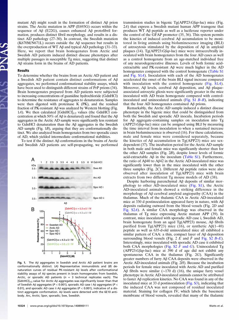
ResultsTo determine whether the brains from an Arctic AD patient anda Swedish AD patient contain distinct conformations of Aβaggregates, we performed conformational stability assays, whichhave been used to distinguish different strains of PrP prions (34).Brain homogenates prepared from AD patients were subjectedto increasing concentrations of guanidine hydrochloride (GdnHCl)to determine the resistance of aggregates to denaturation. Sampleswere then digested with proteinase K (PK), and the residualfraction of PK-resistant Aβ was analyzed by Western blotting (Fig.1A). We then calculated [GdnHCl]1/2 values (the GdnHCl con-centration at which 50% of Aβ is denatured) and found that the Aβaggregates in the Arctic AD sample were significantly less resistantto GdnHCl denaturation than the Aβ aggregates in the SwedishAD sample (Fig. 1B), arguing that they are conformationally dis-tinct. We also analyzed brain homogenates from two sporadic casesof AD, which yielded intermediate conformational stabilities.To test if the distinct Aβ conformations in the brains of Arctic
and Swedish AD patients are self-propagating, we performed
transmission studies in bigenic Tg(APP23:Gfap-luc) mice (Fig.2A) that express a Swedish mutant human APP transgene thatproduces WT Aβ peptide as well as a luciferase reporter underthe control of the GFAP promoter (35, 36). This system permitsthe kinetics of induced cerebral Aβ accumulation to be moni-tored in living animals using bioluminescence imaging (BLI)of astrocytosis stimulated by the deposition of Aβ in amyloidplaques (14). Tg(APP23:Gfap-luc) mice were intracerebrally in-oculated with brain homogenates from the four AD cases as wellas a control homogenate from an age-matched individual freeof any neurodegenerative illnesses. Levels of both formic acid-extractable and PK-resistant Aβ were much higher in the ADhomogenates compared with the control homogenate (Table S1and Fig. S1A). Inoculation with each of the AD homogenatesaccelerated the onset of the brain BLI signal increase comparedwith inoculation with the control homogenate (Fig. S1A).Moreover, Aβ levels, cerebral Aβ deposition, and Aβ plaque-associated astrocytic gliosis were significantly greater in the miceinoculated with AD brain homogenate compared with control-inoculated and uninoculated animals (Fig. S1 B–R), indicatingthat the four AD homogenates contained Aβ prions.Remarkably, the Arctic AD sample induced a distinct disease
phenotype in the bigenic mice that could be distinguished fromboth the Swedish and sporadic AD inocula. Incubation periodsfor Aβ aggregate-containing samples on inoculation into Tg(APP23:Gfap-luc) mice can be assigned using BLI by measuringthe time interval from inoculation to when a sustained increasein brain bioluminescence is observed (16). For these calculations,male and female mice were considered separately, becausethe kinetics of Aβ accumulation in Tg(APP23) mice are sex-dependent (37). The incubation period for the Arctic AD samplein both male and female mice was significantly shorter than forthe other AD samples (Fig. 2B), despite lower levels of formicacid-extractable Aβ in the inoculum (Table S1). Furthermore,the ratio of Aβ40 to Aβ42 in the Arctic AD-inoculated mice wassignificantly lower than in the mice inoculated with the otherthree samples (Fig. 2C). Different Aβ peptide ratios were alsoobserved after inoculation of Tg(APP23) mice with brainextracts from two different Tg mouse models of AD (38).Despite harboring parenchymal Aβ deposits of similar mor-
phology to other AD-inoculated mice (Fig. S1), the ArcticAD-inoculated animals showed a striking difference in themorphology of Aβ cerebral amyloid angiopathy (CAA) in thethalamus. Much of the thalamic CAA in Arctic AD-inoculatedmice at 330 d postinoculation appeared furry in nature, with Aβdeposits radiating outward from the blood vessels (Fig. 2D andFig. S2A). A similar CAA morphology was observed in thethalamus of Tg mice expressing Arctic mutant APP (39). Incontrast, mice inoculated with sporadic AD case i, Swedish AD,brain homogenate from an aged Tg(APP23) mouse, Aβ fibrilspurified from Tg(APP23) mice (16), or synthetic Aβ(1–40)peptide as well as 635-d-old uninoculated mice all exhibited asimilar pattern of CAA: a thin, compact layer of Aβ depositionsurrounding blood vessels (Fig. 2 E and F and Fig. S2 B–E).Interestingly, mice inoculated with sporadic AD case ii exhibitedboth CAA morphologies (Fig. S2 F and G). Uninoculated Tg(APP23:Gfap-luc) mice at 390 d of age did not exhibit anyspontaneous CAA in the thalamus (Fig. 2G). Significantlygreater numbers of furry Aβ CAA deposits were observed in theArctic AD-inoculated animals (Fig. 2H). Because the incubationperiods for female mice inoculated with Arctic AD and purifiedAβ fibrils were similar (∼170 d) (16), the unique furry vesselphenotype in Arctic AD-inoculated animals cannot be attributedto faster Aβ replication kinetics. No CAA was found in any of theinoculated mice at 33 d postinoculation (Fig. S3), indicating thatthe induced CAA was not composed of residual inoculatedmaterial. Staining for collagen IV, which labels the basementmembrane of blood vessels, revealed that many of the thalamic
Fig. 1. The Aβ aggregates in Swedish and Arctic AD patient brains areconformationally distinct. (A) Representative immunoblots and (B) de-naturation curves of residual PK-resistant Aβ levels after conformationalstability assays of Aβ species present in brain homogenates from Swedish,Arctic, or sporadic AD patients (n = 3 technical replicates each). The[GdnHCl]1/2 value for Arctic Aβ aggregates was significantly lower than thatof Swedish Aβ aggregates (P < 0.001), sporadic AD case i Aβ aggregates (P <0.01), and sporadic AD case ii Aβ aggregates (P < 0.001), indicative of a dis-tinct aggregate conformation. Aβ levels were detected with the 6E10 anti-body. Arc, Arctic; Spor, sporadic; Swe, Swedish.
10324 | www.pnas.org/cgi/doi/10.1073/pnas.1408900111 Watts et al.
blood vessels surrounded by the furry Aβ deposits in Arctic AD-inoculated mice, but not Swedish AD-inoculated mice, wereundergoing atrophy (Fig. 2 I and J).A key feature that distinguishes Arctic AD cases from most
sporadic AD cases and cases of heritable AD in which the mu-tation does not occur within the sequence of Aβ is the prefer-ential deposition of Aβ38 peptide, particularly surroundingblood vessels (40). Indeed, PK-resistant Aβ38 was abundant inthe Arctic AD sample and present in sporadic AD case ii butabsent in the Swedish AD and sporadic AD case i samples (Fig.3A). We used mAb BA1-13, which is specific for the Aβ peptideending at residue 38 (Fig. S4A), to assess Aβ38 deposition inAD-inoculated mice. Mice inoculated with either Arctic orSwedish AD samples showed similar levels of formic acid-extractable Aβ38 in their brains (Fig. S4B) and amyloid plaques(Fig. S4 C and D). However, abundant Aβ38-positive CAAwithin the thalamus was observed in mice inoculated with ArcticAβ (Fig. 3B) but not in mice inoculated with Swedish AD orsporadic AD case i. Mice inoculated with sporadic AD case iialso exhibited some thalamic Aβ38-positive CAA (Fig. 3C). Thequantity of Aβ38-positive blood vessels in the thalamus, lep-tomeninges, and the frontal cortex was significantly greater inArctic AD-inoculated mice compared with sporadic AD- andSwedish AD-injected mice (Fig. 3D and Fig. S4 E and F).To confirm these findings, we analyzed a second cohort of
Arctic AD- and Swedish AD-inoculated mice by quantitative
confocal fluorescence microscopy using a different Aβ38-specificantibody (7-14-4) (Fig. S4A). Significantly higher levels of Aβ38staining were observed in thalamic blood vessels in Arctic AD-inoculated mice compared with Swedish AD-inoculated animals(Fig. 3 E–G). Moreover, codeposition of Aβ38 and Aβ40 pep-tides in thalamic blood vessels was observed in mice inoculatedwith Arctic AD or sporadic AD case ii but not in mice inoculatedwith Swedish AD, sporadic AD case i, or other Aβ aggregate-containing preparations (Fig. 3 H and I and Fig. S5 A–G). In-oculation of mice with brain homogenate from aged Tg(APP23)mice, which also contained elevated levels of PK-resistant Aβ38peptide (Fig. S5 H–J), did not result in Aβ38-positive CAAwithin the thalamus (Fig. S5E), suggesting that the conformationof Aβ in the inoculum, not the absolute levels of Aβ38, deter-mines the phenotype in inoculated mice. These observationsargue that the Arctic AD sample induced the preferential de-position of Aβ38 in blood vessels in mice that do not express theArctic mutation. That induced Aβ38 deposition within plaqueswas observed in mice injected with Swedish or Arctic AD (Fig.S4 C and D), despite the absence of PK-resistant Aβ38 in theSwedish AD inoculum (Fig. 3A), may reflect a predispositionof Tg(APP23) mice to depositing Aβ38 in amyloid plaques(Fig. S5I).An important characteristic of prion strains composed of PrP
is that their properties are generally maintained after repeatedpassages in mice. To test if Aβ strains can also be serially
Fig. 2. Arctic AD brain homogenate induces a distinct disease in Tg(APP23:Gfap-luc) mice. (A) Schematic of the inoculation experiment in bigenic Tg(APP23:Gfap-luc) mice, which produce WT Aβ peptide. (B) Incubation periods, determined by BLI, were significantly lower in mice inoculated with Arctic AD than inmice inoculated with Swedish or sporadic AD. Points are from individual mice; mean incubation periods are indicated by the horizontal lines. Female and malemice are shown separately because of sex differences in the kinetics of Aβ deposition in Tg(APP23) mice. (C) The ratio of Aβ40 to Aβ42 was significantly lowerin mice inoculated with Arctic AD than in mice inoculated with Swedish or sporadic AD. (D–G) Aβ CAA in the thalamus of female mice at 330 d postinoculation(dpi) with the indicated brain homogenates. Whereas mice inoculated with Swedish or sporadic AD case i exhibited a thin, compact layer of Aβ depositionsurrounding blood vessels (black arrows), many of the Aβ-positive blood vessels in Arctic AD-inoculated animals appeared furry in nature (red arrows). (G) Thebrain section from an uninoculated mouse at 390 d of age is shown as a control. (Scale bar: D–G, 100 μm.) (H) Significantly greater numbers of furry vesselswere observed in the thalamus of mice inoculated with Arctic AD than in mice inoculated with Swedish or sporadic AD. Quantification was done by visualinspection of stained furry vessels in immunohistochemical slides of inoculated mice (n = 4 each). (I and J) Double labeling of Aβ (white) and collagen IV (red)in the thalamus of Swedish AD- and Arctic AD-injected female mice at 330 dpi. The blood vessels surrounded by furry Aβ deposits in Arctic AD-inoculatedanimals were atrophic (yellow arrows). (Scale bar: I and J, 50 μm.) For D–J, Aβ was labeled using the 4G8 antibody. Arc, Arctic; Spor, sporadic; Swe, Swedish.*P < 0.05; **P < 0.01; ***P < 0.001.
Watts et al. PNAS | July 15, 2014 | vol. 111 | no. 28 | 10325
propagated in mice, we performed a second passage experiment.In these experiments, both the passaged Swedish and Arcticinocula contained identical Aβ sequences, because they bothunderwent initial passage in Tg(APP23:Gfap-luc) mice pro-ducing WT Aβ peptide (Fig. 4A). On second passage, theSwedish and Arctic AD samples as well as sporadic AD case iaccelerated the onset of the brain BLI signal increase and theelevation of cerebral Aβ deposition compared with passaged
control brain homogenate (Fig. S6). Second passage of all ADsamples resulted in incubation periods similar to their respectivefirst passages, with the Arctic AD inoculum showing the shortestincubation periods (Fig. 4B). Similar to first passage, furry AβCAA deposits were observed in the thalamus of mice inoculatedwith the passaged Arctic AD sample but not in mice inoculatedwith the passaged Swedish AD sample (Fig. 4 C–E). Likewise,mice inoculated with the passaged Arctic AD sample, but notmice inoculated with the passaged Swedish or sporadic ADsamples, exhibited prominent CAA deposition of Aβ38 in thethalamus (Fig. 4 F–H) and other areas of the brain (Fig. S7 A andB) as well as codeposition of Aβ38 and Aβ40 in thalamic bloodvessels (Fig. 4 I and J). Levels of formic acid-extractable Aβ38were not significantly different in mice inoculated with thepassaged Arctic or Swedish AD samples (Fig. S7C).
DiscussionIn the studies reported here, we found evidence for distinctstrains of Aβ prions in brain homogenates prepared frompatients with heritable AD. At least certain properties of the Aβprion strains, such as the relative amounts of cerebrovascularAβ38 deposition, were maintained after multiple passages in Tgmice. The existence of distinct Aβ strains and their ability to beserially propagated in mice contend that the Aβ aggregates in thebrains of AD patients show all of the attributes of prions.By melting the conformation of Aβ, we found that the Aβ
prions in the brain of a patient carrying the Arctic mutation andproducing mutant Aβ (E22G) were substantially less resistant toGdnHCl denaturation than those in the brain of a patient withthe Swedish mutation, which does not alter the sequence of WTAβ. We note that Arctic AD patients are heterozygous for themutation, resulting in a mixture of WT and mutant Aβ peptidesin the brain. Although we did not determine the allelic origin ofthe protease-resistant Aβ isoforms, studies of mutant PrPs maybe informative: mutant PrPs in patients harboring the P102L,D178N, or E200K mutations were preferentially converted intothe PrPSc isoform (41–43). Assuming that Arctic Aβ was pref-erentially converted into prions as manifest by protease re-sistance, it seems likely that WT Aβ did not form prions, becausea monophasic melting curve was observed (Fig. 1B). If WT Aβ inArctic AD patients had become a prion and adopted a confor-mation similar to the WT Aβ prions in the brain of the SwedishAD patient, then the melting curve should have been biphasic.Alternatively, WT Aβ may have adopted the same conformationas Arctic mutant Aβ.The analysis of mutant and WT Aβ prions is further compli-
cated by heterogeneous γ-secretase cleavage that generates Aβpeptides varying in length from 37 to 43 aa (44). Each of thesepeptides is likely to possess a different thermodynamic landscapewith respect to conversion into a prion. The difficulties sur-rounding studies of Aβ prion strains are highlighted by a studyusing Tg(APP23) and Tg(APPPS1) mice; the former producesmuch more Aβ40 than Aβ42, whereas the latter produces theopposite (38). In our AD-inoculated mice, the most prominentstrain-specific differences, including the relative amount of Aβ38deposition, were observed in cerebral blood vessels but not pa-renchymal amyloid plaques. However, this difference was notinherent to the Tg(APP23) line, because mice inoculated withdistinct strains of synthetic Aβ prions exhibited strain-specificdifferences in the ratios of Aβ40 and Aβ42 within amyloid pla-ques (45).Our studies suggest that the Arctic AD mutation may encode
a strain of Aβ prions that is distinct from that enciphered by theSwedish mutation, although this result will need to be confirmedusing additional cases. Based on our transmission studies, it seemslikely that the Aβ strains present in the Swedish AD and sporadicAD case i samples are similar and that the sporadic AD caseii sample contains a mixture of the Swedish-like and Arctic-like
Fig. 3. CAA with Aβ38 deposition after inoculation of Tg(APP23:Gfap-luc)mice with Arctic AD but not Swedish AD brain homogenate. (A) Relativelevels of PK-resistant Aβ38 in the various inocula as detected by immuno-blotting using the Aβ38-specific antibody 7-14-4. (B and C) Deposition of Aβ38(BA1-13 antibody) within furry thalamic blood vessels in female Tg(APP23:Gfap-luc) mice at 330 d postinoculation (dpi) with either (B) Arctic AD or (C)sporadic AD case ii brain homogenate. (Scale bar: B and C, 100 μm.) (D)Quantification by visual inspection of immunohistochemical slides of Aβ38-positive (BA1-13 antibody) thalamic blood vessels in mice inoculated with theindicated brain homogenates (n = 4 each). Significantly more Aβ38 depositswere observed in the Arctic AD-inoculated animals. (E and F) Deposition ofAβ38 (7-14-4 antibody) within thalamic blood vessels in a second cohort of Tg(APP23:Gfap-luc) mice at 330 dpi with either (E) Swedish AD or (F) Arctic ADbrain homogenate. (Scale bar: E and F, 50 μm.) (G) Quantification by quan-titative confocal fluorescence microscopy of the relative amount of Aβ38staining (7-14-4 antibody) in thalamic blood vessels in mice inoculated withSwedish AD (n = 51 total blood vessels from three mice) or Arctic AD (n = 31total blood vessels from three mice) brain homogenate. Significantly higherlevels of Aβ38 deposition were observed in the Arctic AD-inoculated animals.(H and I) Double labeling of Aβ38 (7-14-4 antibody; green) and Aβ40 (11A50-B10 antibody; red) in the thalamus of female mice at 330 dpi with either (H)Swedish AD or (I) Arctic AD brain homogenate. (Scale bar: H and I, 15 μm.) (J)Quantification of the ratio of Aβ38 (7-14-4 antibody) to Aβ40 (11A50-B10antibody) staining in thalamic blood vessels in mice inoculated with SwedishAD (n = 51 total blood vessels from three mice) or Arctic AD (n = 31 totalblood vessels from three mice) brain homogenate. Significantly higher ratioswere observed in the Arctic AD-inoculated animals. Arc, Arctic; Con, control;Spor, sporadic; Swe, Swedish. ***P < 0.001.
10326 | www.pnas.org/cgi/doi/10.1073/pnas.1408900111 Watts et al.
strains. Interestingly, the Dutch mutation in Aβ (E22Q) (46),which occurs at the same residue as the Arctic mutation, leadsalmost exclusively to cerebrovascular Aβ deposition and cerebralhemorrhages, implying the existence of additional Aβ strains andsuggesting that residue 22 in Aβ may be critical for determiningthe conformation of Aβ prions. However, mutations within theAβ peptide sequence may not be necessary for the genesis ofdistinct strains, because different conformations of Aβ aggre-gates have been observed in two different cases of sporadic AD(25). Moreover, different subtypes of sporadic AD, such as rapidly
progressive AD (47), may stem from the propagation of uniquestrains of WT Aβ prions in the brain.Because the Tg(APP23:Gfap-luc) mice used herein produce
WT Aβ peptide, we conclude that the conformation of Aβaggregates (not the presence of a mutation per se) enciphers theproperties of Aβ strains. These findings parallel the maintenanceof strain-specified properties observed on transmission of differentinherited forms of human PrP prion disease, each expressing a dif-ferent mutant PrP molecule, to Tg mice expressing chimeric mouse/human PrP (19). The phenotypic differences observed from theArctic and Swedish AD transmissions may be caused by the Arcticmutation favoring the formation of a self-propagating Aβ confor-mation in which Aβ38 is stabilized, leading to its accumulation anddeposition in the brain. However, a similar conformation may in-frequently arise spontaneously in the absence of a mutation in Aβ,which was observed with sporadic AD case ii.Distinct self-propagating strains of protein aggregates have also
recently been described for both the human synucleinopathiesand tauopathies (48–50). Experimental transmission of multiplesystem atrophy to Tg mice revealed a particularly aggressivestrain of WT α-synuclein prions (49). The existence of multipleprion strains causing neurodegenerative diseases poses a chal-lenge for the development of therapeutics. Studies with PrPprions have revealed that therapeutic compounds identified us-ing mouse-passaged strains were efficacious against mouse andchronic wasting disease prions but not against human strains(51). Because the conformation of Aβ aggregates in Tg miceseems distinct from that in AD patients (52), the translationaluse of such mouse models for anti-Aβ therapeutics seemsquestionable. Our ability to propagate distinct AD-associatedAβ strains in the brains of Tg(APP23:Gfap-luc) mice may,therefore, provide a superior tool for assessing drug efficacy.More importantly, heterogeneity in Aβ aggregate conformationssuggests that passive Aβ immunotherapy approaches for treatingAD may only work for certain strains of Aβ that exhibit con-formations similar to those against which the mAb was raised.Several clinical trials involving Aβ mAbs have produced disap-pointing results (53). Although these antibodies may have beenadministered too late in the disease course to produce beneficialeffects, Aβ strain heterogeneity in AD patients may have also con-tributed to their failure.
Materials and MethodsAdditional methods are provided in SI Materials and Methods.
Human Tissue Samples. Frozen brain tissue samples (temporal cortex) wereobtained from neuropathologically confirmed cases of AD. The Swedishmutant sample (Braak stage V–VI with CAA and a minor cerebellar hemor-rhage) was obtained from a 61-y-old female, and the Arctic mutant sample(Braak stage V–VI with CAA and no other copathology) was obtained froma 64-y-old male. Two sporadic AD samples were used: case i (Braak stageV–VI with Lewy body copathology) was obtained from a 62-y-old male, andcase ii (Braak stage V–VI with no other copathology) was obtained from an 85-y-old female. Control brain tissue was obtained from the frontal cortex ofa 79-y-old male who did not exhibit any clinical or pathological signs ofa neurodegenerative disease. Levels of various Aβ peptides in the individualbrain samples are shown in Table S1.
Mice. Tg(APP23) mice, which express human APP (751-aa isoform) containingthe Swedish mutation under the control of the Thy-1.2 promoter (35), weremaintained on a C57BL/6 background. Tg(Gfap-luc) mice, which expressfirefly luciferase under the control of the murine Gfap promoter (36), werea gift from Caliper Life Sciences (Alameda, CA) and maintained on an FVB/Nbackground. To create bigenic Tg(APP23:Gfap-luc) mice, hemizygous Tg(APP23) mice were crossed with homozygous Tg(Gfap-luc) animals. All ani-mal experiments were performed under protocols approved by the In-stitutional Animal Care and Use Committee at the University of California.
Neuropathology. Female mice were euthanized but not perfused beforebrain removal. Brains were then immersion-fixed in 10% (vol/vol) buffered
Fig. 4. Aβ strain properties are maintained on serial transmission in Tg(APP23:Gfap-luc) mice. (A) Schematic of the second passage of AD samples inTg(APP23:Gfap-luc) mice. The passaged Arctic, Swedish, and sporadic ADinocula are composed of WT Aβ peptide. (B) The incubation periods weresignificantly shorter for the passaged Arctic AD sample than for the pas-saged sporadic AD case i and Swedish AD samples. Points are from individualfemale mice; mean incubation periods are indicated by the horizontal lines.(C–E) Aβ CAA in the thalamus of female mice at 330 d postinoculation (dpi)with the indicated passaged brain homogenates. Whereas mice inoculatedwith passaged Swedish AD exhibited a thin, compact layer of Aβ deposition(4G8 antibody) surrounding blood vessels (black arrows), many of the Aβ-positive blood vessels in animals inoculated with passaged Arctic ADappeared furry in nature (red arrows). (F and G) Thalamic CAA with Aβ38deposition (BA1-13 antibody) was observed in two distinct female Tg(APP23:Gfap-luc) mice at 330 dpi with the passaged Arctic AD sample. (Scale bar:C–G, 50 μm.) (H) Quantification of Aβ38-positive (BA1-13 antibody) thalamicblood vessels in mice inoculated with the passaged Swedish (n = 4) or ArcticAD (n = 6) samples. (I and J) Double labeling of Aβ38 (BA1-13 antibody;green) and Aβ40 (11A50-B10 antibody; red) in the thalamus of female miceat 330 dpi with passaged Swedish or Arctic AD samples. Only the mice in-oculated with the passaged Arctic AD sample exhibited codeposition of Aβ38and Aβ40 peptides. (Scale bar: I and J, 100 μm.) Arc, Arctic; Swe, Swedish.*P < 0.05; **P < 0.01; ***P < 0.001.
Watts et al. PNAS | July 15, 2014 | vol. 111 | no. 28 | 10327
formalin followed by embedding in paraffin using standard procedures.Sections were cut at 8 μm, mounted on glass slides, deparaffinized, and thenprocessed for immunohistochemistry. Endogenous tissue peroxidases wereinhibited by incubating the slides in 3% hydrogen peroxide solution for 30min, and sections to be stained with anti-Aβ antibodies were pretreated withformic acid for 5 min. Slides were blocked with 10% (vol/vol) normal goatserum for 1 h and then incubated with primary antibody overnight at 4 °C.The following primary antibodies were used: anti-Aβ mouse mAb 4G8 (1:250dilution; Covance), anti-Aβ40 mouse mAb 11A50-B10 (1:200 dilution; Cova-nce), anti-Aβ38 rabbit mAbs BA1-13 (1:200 dilution; Covance) and 7-14-4(1:250 dilution; Covance), anti-collagen IV rabbit polyclonal antibody ab6586(1:500 dilution; Abcam), and anti-GFAP rabbit polyclonal antibody Z0334(1:500 dilution; Dako). Bound antibody was detected using a Vectastain ABCPeroxidase Kit (Vector Laboratories) and visualized using 3–3′-diaminobenzidine.Slides were counterstained with hematoxylin and then photographed using an
AxioImager.A1 microscope (Carl Zeiss). For fluorescent double-labeling experi-ments, secondary antibodies conjugated to AlexaFluor 488, 568, or 647 (LifeTechnologies) were used. For Aβ/collagen IV double stains, sections were firstpretreated with formic acid and then autoclaved in citrate buffer for epitoperetrieval. All fluorescently labeled samples were visualized using a Leica SP8confocal microscope.
ACKNOWLEDGMENTS. We thank the staff at the Hunter’s Point animalfacility for assistance with the animal experiments, Marta Gavidia formouse genotyping, and Smita Patel for brain homogenization. The Tg(Gfap-luc) mouse line was a gift from Caliper Life Sciences. This work wassupported by National Institutes of Health Grants AG002132, AG010770,AG021601, AG031220, and AG042453 and gifts from the Sherman Fair-child Foundation; the Rainwater Charitable Foundation; and the GlennFoundation for Medical Research. J.C.W. was supported by National Insti-tute on Aging K99 Grant AG042453.
1. Hardy J, Selkoe DJ (2002) The amyloid hypothesis of Alzheimer’s disease: Progress andproblems on the road to therapeutics. Science 297(5580):353–356.
2. Braak H, Braak E (1991) Neuropathological stageing of Alzheimer-related changes.Acta Neuropathol 82(4):239–259.
3. Jucker M, Walker LC (2013) Self-propagation of pathogenic protein aggregates inneurodegenerative diseases. Nature 501(7465):45–51.
4. Guo JL, Lee VM (2014) Cell-to-cell transmission of pathogenic proteins in neurode-generative diseases. Nat Med 20(2):130–138.
6. Wickner RB (1994) [URE3] as an altered URE2 protein: Evidence for a prion analog inSaccharomyces cerevisiae. Science 264(5158):566–569.
7. Si K, Lindquist S, Kandel ER (2003) A neuronal isoform of the aplysia CPEB has prion-like properties. Cell 115(7):879–891.
8. Prusiner SB (2012) Cell biology. A unifying role for prions in neurodegenerative dis-eases. Science 336(6088):1511–1513.
9. Ashe KH, Aguzzi A (2013) Prions, prionoids and pathogenic proteins in Alzheimerdisease. Prion 7(1):55–59.
10. Hardy J, Revesz T (2012) The spread of neurodegenerative disease. N Engl J Med366(22):2126–2128.
11. Thal DR, Rüb U, Orantes M, Braak H (2002) Phases of Aβ-deposition in the humanbrain and its relevance for the development of AD. Neurology 58(12):1791–1800.
12. Meyer-Luehmann M, et al. (2006) Exogenous induction of cerebral beta-amyloido-genesis is governed by agent and host. Science 313(5794):1781–1784.
14. Watts JC, et al. (2011) Bioluminescence imaging of Abeta deposition in bigenic mousemodels of Alzheimer’s disease. Proc Natl Acad Sci USA 108(6):2528–2533.
15. Morales R, Duran-Aniotz C, Castilla J, Estrada LD, Soto C (2012) De novo induction ofamyloid-β deposition in vivo. Mol Psychiatry 17(12):1347–1353.
16. Stöhr J, et al. (2012) Purified and synthetic Alzheimer’s amyloid beta (Aβ) prions. ProcNatl Acad Sci USA 109(27):11025–11030.
17. Rosen RF, et al. (2012) Exogenous seeding of cerebral β-amyloid deposition in βAPP-transgenic rats. J Neurochem 120(5):660–666.
18. Bessen RA, et al. (1995) Non-genetic propagation of strain-specific properties ofscrapie prion protein. Nature 375(6533):698–700.
19. Telling GC, et al. (1996) Evidence for the conformation of the pathologic isoform ofthe prion protein enciphering and propagating prion diversity. Science 274(5295):2079–2082.
20. Tanaka M, Chien P, Naber N, Cooke R, Weissman JS (2004) Conformational variationsin an infectious protein determine prion strain differences. Nature 428(6980):323–328.
21. Petkova AT, et al. (2005) Self-propagating, molecular-level polymorphism in Alzheimer’sbeta-amyloid fibrils. Science 307(5707):262–265.
22. Kodali R, Williams AD, Chemuru S, Wetzel R (2010) Abeta(1-40) forms five distinctamyloid structures whose β-sheet contents and fibril stabilities are correlated. J MolBiol 401(3):503–517.
23. Nilsson KP, et al. (2007) Imaging distinct conformational states of amyloid-beta fibrilsin Alzheimer’s disease using novel luminescent probes. ACS Chem Biol 2(8):553–560.
24. Paravastu AK, Qahwash I, Leapman RD, Meredith SC, Tycko R (2009) Seeded growthof beta-amyloid fibrils from Alzheimer’s brain-derived fibrils produces a distinct fibrilstructure. Proc Natl Acad Sci USA 106(18):7443–7448.
25. Lu JX, et al. (2013) Molecular structure of β-amyloid fibrils in Alzheimer’s disease braintissue. Cell 154(6):1257–1268.
26. Mayeux R, Stern Y, Spanton S (1985) Heterogeneity in dementia of the Alzheimertype: Evidence of subgroups. Neurology 35(4):453–461.
28. Lashuel HA, et al. (2003) Mixtures of wild-type and a pathogenic (E22G) form of Aβ40in vitro accumulate protofibrils, including amyloid pores. J Mol Biol 332(4):795–808.
29. Philipson O, et al. (2012) The Arctic amyloid-β precursor protein (AβPP) mutation re-sults in distinct plaques and accumulation of N- and C-truncated Aβ. Neurobiol Aging33:1010.e1–1010.e13.
30. Kalimo H, et al. (2013) The Arctic AβPP mutation leads to Alzheimer’s disease pa-thology with highly variable topographic deposition of differentially truncated Aβ.Acta Neuropathol Commun 1(1):60.
31. Citron M, et al. (1992) Mutation of the β-amyloid precursor protein in familial Alz-heimer’s disease increases β-protein production. Nature 360(6405):672–674.
32. Mullan M, et al. (1992) A pathogenic mutation for probable Alzheimer’s disease inthe APP gene at the N-terminus of beta-amyloid. Nat Genet 1(5):345–347.
33. Lannfelt L, et al. (1994) Amyloid precursor protein mutation causes Alzheimer’s dis-ease in a Swedish family. Neurosci Lett 168(1-2):254–256.
34. Peretz D, et al. (2001) Strain-specified relative conformational stability of the scrapieprion protein. Protein Sci 10(4):854–863.
35. Sturchler-Pierrat C, et al. (1997) Two amyloid precursor protein transgenic mousemodels with Alzheimer disease-like pathology. Proc Natl Acad Sci USA 94(24):13287–13292.
36. Zhu L, et al. (2004) Non-invasive imaging of GFAP expression after neuronal damagein mice. Neurosci Lett 367(2):210–212.
37. Jucker M (2010) The benefits and limitations of animal models for translational re-search in neurodegenerative diseases. Nat Med 16(11):1210–1214.
38. Heilbronner G, et al. (2013) Seeded strain-like transmission of β-amyloid morphotypesin APP transgenic mice. EMBO Rep 14(11):1017–1022.
39. Rönnbäck A, et al. (2011) Progressive neuropathology and cognitive decline in a sin-gle Arctic APP transgenic mouse model. Neurobiol Aging 32(2):280–292.
40. Moro ML, et al. (2012) APP mutations in the Aβ coding region are associated withabundant cerebral deposition of Aβ38. Acta Neuropathol 124(6):809–821.
41. Gabizon R, et al. (1996) Insoluble wild-type and protease-resistant mutant prionprotein in brains of patients with inherited prion disease. Nat Med 2(1):59–64.
42. Chen SG, et al. (1997) Allelic origin of the abnormal prion protein isoform in familialprion diseases. Nat Med 3(9):1009–1015.
43. Parchi P, et al. (1998) Different patterns of truncated prion protein fragments cor-relate with distinct phenotypes in P102L Gerstmann-Sträussler-Scheinker disease. ProcNatl Acad Sci USA 95(14):8322–8327.
44. Benilova I, Karran E, De Strooper B (2012) The toxic Aβ oligomer and Alzheimer’sdisease: An emperor in need of clothes. Nat Neurosci 15(3):349–357.
45. Stöhr, et al. (2014) Distinct synthetic Aβ prion strains producing different amyloiddeposits in bigenic mice. Proc Natl Acad Sci USA 111:10329–10334.
46. Hendriks L, et al. (1992) Presenile dementia and cerebral haemorrhage linked toa mutation at codon 692 of the β-amyloid precursor protein gene. Nat Genet 1(3):218–221.
48. Guo JL, et al. (2013) Distinct α-synuclein strains differentially promote tau inclusions inneurons. Cell 154(1):103–117.
49. Watts JC, et al. (2013) Transmission of multiple system atrophy prions to transgenicmice. Proc Natl Acad Sci USA 110(48):19555–19560.
50. Clavaguera F, et al. (2013) Brain homogenates from human tauopathies induce tauinclusions in mouse brain. Proc Natl Acad Sci USA 110(23):9535–9540.
51. Berry DB, et al. (2013) Drug resistance confounding prion therapeutics. Proc Natl AcadSci USA 110(44):E4160–E4169.
52. Klunk WE, et al. (2005) Binding of the positron emission tomography tracer Pitts-burgh compound-B reflects the amount of amyloid-β in Alzheimer’s disease brain butnot in transgenic mouse brain. J Neurosci 25(46):10598–10606.
53. Delrieu J, Ousset PJ, Caillaud C, Vellas B (2012) ‘Clinical trials in Alzheimer’s disease’:Immunotherapy approaches. J Neurochem 120(Suppl 1):186–193.
10328 | www.pnas.org/cgi/doi/10.1073/pnas.1408900111 Watts et al.