Biochemistry 1992, 31, 155-162 155 Tsujita, T., & Brockman, H. L. (1987) Biochemistry 26, Tsujita, T., Smaby, J. M., & Brockman, H. L. (1987) Bio- Tsujita, T., Muderhwa, J. M., & Brockman, H. L. (1989) J. Verger, R., & Pieroni, G. (1986) in Lipids and Membranes: Past, Present and Future (Op den Kamp, J. A. F., Roe- lofsen, B., & Wirtz, K. W. A., Eds.) pp 153-170, Elsevier Science Publishers, Amsterdam. Winkler, F. K., D’Arcy, A., & Hunziker, W. (1990) Nature 8423-8429. chemistry 26, 8430-8434. Biol. Chem. 264, 8612-8618. 343, 771-774. Serine Hydroxymethyltransferase: Origin of Substrate Specificity7 Sebastiana Angelaccio,* Stefan0 Pascarella,* Elena Fattori,t Francesco Bossa,* William Strong,§ and Verne Schirch*v§ Department of Biochemistry and Molecular Biophysics, Virginia Commonwealth University, Richmond, Virginia 23298, and Dipartimento di Scienze Biochimiche and Centro di Biologia Molecolare del Consiglio Nazionale delle Ricerche, Universitd La Sapienza, 001 85 Roma, Italy Received July I, 1991; Revised Manuscript Received September 10, 1991 ABSTRACT: All forms of serine hydroxymethyltransferase, for which a primary structure is known, have five threonine residues near the active-site lysyl residue (K229) that forms the internal aldimine with pyridoxal phosphate. For Escherichia coli serine hydroxymethyltransferase each of these threonine residues has been changed to an alanine residue. The resulting five mutant enzymes were purified and characterized with respect to kinetic and spectral properties. The mutant enzymes T224A and T227A showed no significant changes in kinetic and spectral properties compared to the wild-type enzyme. The T225A and T230A enzymes exhibited differences in K,,, and kat values but exhibited the same spectral properties as the wild-type enzyme. The four threonine residues at positions 224,225,227, and 230 do not play a critical role in the mechanism of the enzyme. The T226A enzyme had nearly normal affinity for substrates and coenzymes but had only 3% of the catalytic activity of the wild-type enzyme. The spectrum of the T226A enzyme in the presence of amino acid substrates showed a large absorption maximum at 343 nm with only a small absorption band at 425 nm, unlike the wild-type enzyme whose enzymesubstrate complexes absorb at 425 nm. Rapid reaction studies showed that when amino acid substrates and substrate analogues were added to the T226A enzyme, the internal aldimine absorbing at 422 nm was rapidly converted to a complex absorbing at 343 nm in a second-order process. This was followed by a very slow first-order formation of a complex absorbing at 425 nm. Variation of the initial rapid second-order process as a function of pH suggested that the anionic form of the amino acid forms the first complex with the enzyme. The results are interpreted as being due to the rapid formation of a gem-diamine complex between amino acids and T226A enzyme with a rate- determining formation of the external aldimine. This suggests that Thr-226 plays an important role in converting the gem-diamine complex to the external aldimine complex. Variation of the kinetic constants with amino acid structure suggests that the T226A enzyme distinguishes between substrates and substrate analogues in the formation of the gem-diamine complex. S e r i n e hydroxymethyltransferase (SHMT) catalyzes the conversion of serine and tetrahydrofolate (H,folate) to glycine and 5,10-methyleneH4folate. This reaction is present in a wide variety of cells and is the major source of one-carbon groups required in the biosynthesis of methionine, choline, thymidy- late, and purines (Schirch, 1982). We have previously purified and determined the primary structure of cytosolic and mito- chondrial isoenzymes from rabbit liver (Martini et al., 1987, 1989). SHMT has also been purified and characterizedfrom expression of the Escherichia coli cloned glyA gene (Plamann et al., 1983; Schirch et al., 1985). SHMT activity is dependent on the two coenzymes pyridoxal-P and H4folate. Pyridoxal-P is covalently attached at the active site and serves as a spec- trophotometric probe in determining the structure of en- ‘This work was supported in part by Grant GM 28143 from the National Institutes of Health and a grant from the Istituto Pasteur- Fondazione Cenci Bolognetti. * To whom correspondenceshould be addressed. * Universitd La Sapienza. $ Virginia Commonwealth University. zyme-substrate intermediates in the reaction pathway (Schirch, 1982). As with all pyridoxal-P enzymes, the site of covalent attachment is an internal aldimine between the 4’-aldehyde group on the coenzyme and an e-amino group of a lysyl residue (Davis and Metzler, 1972). Reduction of this external aldimine converts it to a stable secondary amine, which permits isolation of a peptide from proteolytic digests containing the bound pyridoxal-P (Bossa, et al., 1976). These active site peptides have been isolated and sequenced from numerous pyridoxal-P containing enzymes (Vaaler and Snell, 1989; Tanizawa, et al., 1989). The three forms of SHMT which we have studied all contain the nine-residue conserved sequence V-V-T-T-T-T-H-K(Pyr)-T (Martini et al., 1989). The active-site nonapeptide from SHMT is unusual in that five Thr residues have been conserved in the E. coli enzyme and the mammalian isoenzymes in rabbit liver. This suggests that these Thr residues have some functional role in this en- ’ Abbreviations: SHMT, serine hydroxymethyltransferase; H,folate, tetrahydrofolate; pyridoxal-P, pyridoxal 5’-phosphate. 0006-2960/92/043 1-1 55$03.00/0 0 1992 American Chemical Society
Transcript
Biochemistry 1992, 31, 155-162 155
Tsujita, T., & Brockman, H. L. (1987) Biochemistry 26,
Tsujita, T., Smaby, J. M., & Brockman, H. L. (1987) Bio-
Tsujita, T., Muderhwa, J. M., & Brockman, H. L. (1989) J .
Verger, R., & Pieroni, G. (1986) in Lipids and Membranes: Past, Present and Future (Op den Kamp, J. A. F., Roe- lofsen, B., & Wirtz, K. W. A., Eds.) pp 153-170, Elsevier Science Publishers, Amsterdam.
Winkler, F. K., D’Arcy, A., & Hunziker, W. (1990) Nature
8423-8429.
chemistry 26, 8430-8434.
Biol. Chem. 264, 8612-8618. 343, 771-774.
Serine Hydroxymethyltransferase: Origin of Substrate Specificity7
Sebastiana Angelaccio,* Stefan0 Pascarella,* Elena Fattori,t Francesco Bossa,* William Strong,§ and Verne Schirch*v§
Department of Biochemistry and Molecular Biophysics, Virginia Commonwealth University, Richmond, Virginia 23298, and Dipartimento di Scienze Biochimiche and Centro di Biologia Molecolare del Consiglio Nazionale delle Ricerche, Universitd La
Sapienza, 001 85 Roma, Italy Received July I , 1991; Revised Manuscript Received September 10, 1991
ABSTRACT: All forms of serine hydroxymethyltransferase, for which a primary structure is known, have five threonine residues near the active-site lysyl residue (K229) that forms the internal aldimine with pyridoxal phosphate. For Escherichia coli serine hydroxymethyltransferase each of these threonine residues has been changed to an alanine residue. The resulting five mutant enzymes were purified and characterized with respect to kinetic and spectral properties. The mutant enzymes T224A and T227A showed no significant changes in kinetic and spectral properties compared to the wild-type enzyme. The T225A and T230A enzymes exhibited differences in K,,, and kat values but exhibited the same spectral properties as the wild-type enzyme. The four threonine residues at positions 224,225,227, and 230 do not play a critical role in the mechanism of the enzyme. The T226A enzyme had nearly normal affinity for substrates and coenzymes but had only 3% of the catalytic activity of the wild-type enzyme. The spectrum of the T226A enzyme in the presence of amino acid substrates showed a large absorption maximum at 343 nm with only a small absorption band at 425 nm, unlike the wild-type enzyme whose enzymesubstrate complexes absorb at 425 nm. Rapid reaction studies showed that when amino acid substrates and substrate analogues were added to the T226A enzyme, the internal aldimine absorbing at 422 nm was rapidly converted to a complex absorbing at 343 nm in a second-order process. This was followed by a very slow first-order formation of a complex absorbing at 425 nm. Variation of the initial rapid second-order process as a function of pH suggested that the anionic form of the amino acid forms the first complex with the enzyme. The results are interpreted as being due to the rapid formation of a gem-diamine complex between amino acids and T226A enzyme with a rate- determining formation of the external aldimine. This suggests that Thr-226 plays an important role in converting the gem-diamine complex to the external aldimine complex. Variation of the kinetic constants with amino acid structure suggests that the T226A enzyme distinguishes between substrates and substrate analogues in the formation of the gem-diamine complex.
S e r i n e hydroxymethyltransferase (SHMT) catalyzes the conversion of serine and tetrahydrofolate (H,folate) to glycine and 5,10-methyleneH4folate. This reaction is present in a wide variety of cells and is the major source of one-carbon groups required in the biosynthesis of methionine, choline, thymidy- late, and purines (Schirch, 1982). We have previously purified and determined the primary structure of cytosolic and mito- chondrial isoenzymes from rabbit liver (Martini et al., 1987, 1989). SHMT has also been purified and characterized from expression of the Escherichia coli cloned glyA gene (Plamann et al., 1983; Schirch et al., 1985). SHMT activity is dependent on the two coenzymes pyridoxal-P and H4folate. Pyridoxal-P is covalently attached at the active site and serves as a spec- trophotometric probe in determining the structure of en-
‘This work was supported in part by Grant GM 28143 from the National Institutes of Health and a grant from the Istituto Pasteur- Fondazione Cenci Bolognetti.
* To whom correspondence should be addressed. * Universitd La Sapienza. $ Virginia Commonwealth University.
zyme-substrate intermediates in the reaction pathway (Schirch, 1982). As with all pyridoxal-P enzymes, the site of covalent attachment is an internal aldimine between the 4’-aldehyde group on the coenzyme and an e-amino group of a lysyl residue (Davis and Metzler, 1972). Reduction of this external aldimine converts it to a stable secondary amine, which permits isolation of a peptide from proteolytic digests containing the bound pyridoxal-P (Bossa, et al., 1976). These active site peptides have been isolated and sequenced from numerous pyridoxal-P containing enzymes (Vaaler and Snell, 1989; Tanizawa, et al., 1989). The three forms of SHMT which we have studied all contain the nine-residue conserved sequence V-V-T-T-T-T-H-K(Pyr)-T (Martini et al., 1989).
The active-site nonapeptide from SHMT is unusual in that five Thr residues have been conserved in the E. coli enzyme and the mammalian isoenzymes in rabbit liver. This suggests that these Thr residues have some functional role in this en-
0006-2960/92/043 1-1 55$03.00/0 0 1992 American Chemical Society
156
zyme. There are at least two functional roles these residues could have in the mechanism of this reaction. First, they could serve as hydrogen-bond donors or acceptors to either of the two coenzymes or to the substrate amino acids glycine and serine. Serving as hydrogen donors or acceptors, the Thr residues could play a role in determining both reaction and substrate specificity. Second, they could serve as acceptors and donors of protons between various intermediates in the reaction pathway. Including known and proposed mechanistic steps in this reaction, there may be as many as nine different steps in which protons are transferred between some group on the enzyme and either the substrate or both coenzymes.
SHMT utilizes a wide variety of 3-hydroxyamino acid substrates and catalyzes numerous side reactions, which include decarboxylation, transamination, and racemization (Shostak & Schirch, 1988). Previously, we have provided evidence that a conformational change that occurs on substrate binding to SHMT controls reaction specificity (Schirch et al., 1991). What has not been elucidated is at what step in the reaction pathway the enzyme distinguishes between substrate amino acids serine and glycine and nonsubstrate amino acids such as L-alanine and threonine.
The purpose of the study reported here was to determine the function of each of the five Thr residues in E. coli SHMT. Each Thr was changed to an Ala residue by site-directed mutagenesis, and the five resulting mutant proteins were pu- rified and characterized with respect to affinity of coenzymes, substrates, and substrates analogues. Also, kinetic properties in the aldol cleavage of substrates and substrate analogues and ability to catalyze the transamination of D- and L-alanine were studied, and rapid reaction studies to determine the rate of interconversion of enzyme-substrate complexes were per- formed. One of these mutant proteins provides insight into how the enzyme controls which amino acids are accepted as substrates and which amino acids are rejected.
Biochemistry, Vol. 31, No. 1 , 1992 Angelaccio et al.
of the double-stranded expression plasmid in the GS245 cells. Kinetic Studies. Kinetic studies were performed using
several different substrates. When L-serine was used as the changing fmed substrate, H4folate, at concentrations of 10-100 pM, was used as the variable substrate. C,-Tetrahydrofolate synthase was used as the coupling enzyme, and the reaction rate was determined from the increase in absorbance at 340 nm due to the reduction of NADP' (Schirch & Peterson, 1980). K,,, values were determined from double-reciprocal plots. Both allothreonine and threonine were also used as substrates. Each of these reactions was followed by deter- mining the rate of reduction of the product acetaldehyde by alcohol dehydrogenase and NADH at 340 nm (Schirch & Peterson, 1980).
The rates of transamination of both L- and D-alanine were determined from the rate of decrease in absorbance at 425 nm in solutions of enzyme (1 mg/mL), 100 mM D- or L-alanine, and 50 mM potassium phosphate buffer at pH 7.6 at 37 OC (Shostak & Schirch, 1988).
The affinity of each mutant apoenzyme for pyridoxal-P was determined by incubating 0.1 pM solutions of the apoenzyme with concentrations of 0.05-5 pM solutions of pyridoxal-P at room temperature in 20 mM potassium N,N-bis[2-hydroxy- ethyl]-2-aminoethanesulfonate, pH 7.0, for 3 h. Each solution was assayed and the concentration of holoenzyme determined from the fraction of activity of these solutions compared to the activity of the apoenzyme incubated with 50 pM pyri- doxal-P. The concentration of remaining apoenzyme in each solution was the difference between the concentration of the original total apoenzyme and the concentration of holoenzyme determined from the activity measurements. The concentra- tion of unbound pyridoxal-P was determined from the dif- ference in concentrations of pyridoxal-P added to the apo- enzyme solutions and the holoenzyme. Kd values were de- termined from the equation Kd = [apoenzyme] [pyridoxal- P] / [ holoenzyme].
Rapid Reaction Studies. Stopped-flow absorbance mea- surements were performed on a Kinetics Instruments spec- trophotometer. Temperatures were held at either 8,20, or 30 OC by a circulating water bath. Traces were recorded on a MacIntosh IIc using the software provided by Kinetic In- struments. Each study was an average of 4-6 traces. The curves for absorbance versus time were curve-fit by either a single- or double-exponential algorithm. Rate constants and amplitudes for each individual reaction varied less than 10% from the average values for each reaction. The absorbance changes observed in the stopped-flow studies were in agreement with the predicted absorbance changes from equilibrium spectral studies. For studies performed at either 8 or 20 OC, the enzyme concentration in the reaction vessel was 2.0 mg/mL. This gives a subunit concentration of 43 pM. For the reactions done at 30 "C the concentration of enzyme in the reaction vessel was 2.5 mg/mL. The buffer used for both enzyme and substrate solutions was either 50 mM potassium N,N-bis[2-hydroxyethyl] -2-aminoethanesulfonate, pH 7.0, or 50 mM potassium phosphate at the indicated pH. The con- centration of the anionic forms of amino acids were calculated using the Henderson-Hasselbalch equation and pK, values of 9.6,9.2, 10.4, and 9.7 for the amino groups of glycine, L-serine, m-allothreonine, and L-alanine, respectively.
Spectral Studies. Ultraviolet-visible spectra were recorded on a Cary 210 spectrophotometer and circular dichroism spectra were obtained with a Jasco 500 spectropolarimeter.
RESULTS Kinetic Constants. Each of the purified mutant enzymes
containing an alanine in place of a threonine was tested for
EXPERIMENTAL PROCEDURES
Materials. Amino acid substrates, NADP+, NADH, al- cohol dehydrogenase (yeast), and pyridoxal-P were obtained from Sigma. (6RS)-H4folate was purchased from Fluka. C1-Tetrahydrofolate synthase was purified from rabbit liver as previously described ( W a r et al., 1985). Removal of bound pyridoxal-P to form aposerine hydroxymethyltransferase was achieved by incubation of the enzyme with L-cysteine in high salt (Schirch et al., 1973).
Site-Directed Mutagenesis. Threonine residues at amino acid positions 224 and 227 are coded for by ACT and threo- nine residues at the amino acid positions 225, 226, and 230 are coded for by the sequence ACC in the glyA gene (Plamann et al., 1983). These five threonine residues were changed individually to alanine residues by making five oligonucleotides that changed these codons to either GCT or GCC, which code for alanine. Thr-226 was also converted to a Ser residue. The oligonucleotide mutagenesis kit from Amersham was used to obtain each mutant of the glyA gene in a single-stranded M 13mp9 clone. After the sequence of the mutant gene in the M13mp9 insert was verified, the mutant glyA gene was transferred to the plasmid pBR322 and used to transform E . coli strain GS245 as previously described (Hopkins & Schirch, 1986). The mutant form of each enzyme was purified using the same procedure described for the wild-type enzyme (Schirch et al., 1985). For mutant proteins T224A, T225A, T226A, T226S, and T227A, the substitution of either alanine or serine for threonine was verified by amino acid sequence analysis as described by Barra et al. (1991). For mutant protein T230A the mutation was verified by sequence analysis
Serine Hydroxymethyltransferase Biochemistry, Vol. 31, No. I, 1992 157
Table I: Kinetic Constants for Wild TvDe and Five Mutant Forms of E . coli Serine Hvdroxymethvltransferase form of serine hydroxymethyltransferase
substrate kinetic constant wild type T224A T225A T226A T227A T230A threonine Km (mM) 12 10 7 20 8 20
k,, ( m i d ) 2.2 2.9 6 <0.1 4.7 <o. 1 allothreonine Km" (mM) 1.5 0.6 0.4 2.5 0.6 1.2
k,, ( m i d ) 30 28 42 4.7 31 4.1 L-serine Kmb (mM) 0.3 0.5 0.1 0.4 0.4 0.8
Km' (9M) 25 14 7 14 10 330 k,, ( m i d ) 640 870 110 20 720 350
L-alanine k,d ( m i d ) 0.002 0.005 0.04 0.01 0.01 0.003 D-alanine k,d (min-I) 0.005 0.008 0.004 0.004 0.007 0.01
"Based on the concentration of L-allothreonine. bKm value of serine for the enzyme saturated with tetrahydrofolate. K , value of tetrahydrofolate for the enzyme saturated with L-serine. dObserved rate constant for the transamination of the enzyme-bund pyridoxal-P.
catalytic activity using L-threonine, DL-allothreonine, and L-serine as substrates. Each mutant enzyme showed catalytic activity with all three substrates, except for mutant enzymes T226A and T230A, which did not show activity with L- threonine. K, values were determined for substrates and coenzymes for each enzyme form (Table I). There are some differences in K, values for amino acids and H,folate, but except for the T225A and T230A enzymes the differences were less than 3-fold compared to the wild-type enzyme. The T225A enzyme has increased affinity for both amino acid substrates and H4folate compared to the wild-type enzyme. T230A enzyme has a K, for H4folate which is an order of magnitude larger than that of the wild-type enzyme.
The affinity of each enzyme for pyridoxal-P was determined by incubating apoenzyme with increasing concentrations of pyridoxal-P for 3 h and then determining the activity of the reaction. Although Scatchard plots were not linear and showed a considerable amount of error, all five mutant enzymes and the wild-type enzyme showed similar results and have an appreciable affinity for this coenzyme. For each enzyme 50% activity was achieved when both enzyme and pyridoxal-P were 0.1-0.2 pM. Also, none of the holoenzymes lost activity when incubated at 30 O C for 3 h. These results suggest that none of the five threonine residues plays a critical role in binding of pyridoxal-P.
The determination of the catalytic rate constant for cleavage of L-serine showed that only mutant enzymes T225A, T226A, and T230A were significantly less active than the wild-type enzyme (Table I). For the T230A enzyme the decrease in k,, was about 7-fold for the cleavage of allothreonine and 2-fold for L-serine. With T225A SHMT the value of k,, for the cleavage of L-threonine was 2.5-fold larger than the value for the wild-type enzyme, but with L-serine as substrate k,, was 6-fold lower. The decrease in k,,, for T226A SHMT was about 6-fold for allothreonine cleavage but 30-fold for L-serine cleavage (Table I).
One of the unique properties of E. coli SHMT is that it slowly catalyzes the transamination of both D- and L-alanine. We have shown previously that there are different bases on the enzyme which are involved in the removal of the 2s proton from D-alanine and the 2R proton from L-alanine (Shostak & Schirch, 1988). In each case, the removal of the proton results in the formation of an enzyme-quinonoid complex absorbing near 500 nm. The rate of transamination is de- pendent on the formation of this quinonoid complex. The importance of looking at these two reactions with the mutant enzymes is that it allows one to determine how similar the alignment of the amino acid substrate is at the active site with respect to the two bases. If the mutant enzymes do not cat- alyze the transamination of the alanine isomers, then it sug- gests that the external aldimine is not oriented at the active
A
0.05 \ '.-___-------_-
o.osio . . . 350' 390 - - r io WAVaDIGTn Inl)
FIGURE 1: Visible and circular dichroism spectra of T226A SHMT in the absence and presence of L-serine. Curve A circular dichroism spectra of a 2.0 mg/mL solution of enzyme in the absence (solid line) and presence (dashed line) of 40 mM L-serine. Curve B: visible spectra of a 2.0 mg/mL solution of enzyme in the absence (solid line) and presence (dashed line) of 40 mM L-serine.
site in the same way as in the wild-type enzyme. The results of these experiments show, however, that all five mutant en- zymes do catalyze the transamination of both P and L-alanine. For the T225A and T226A enzymes the rates of trans- amination of L-alanine were 20-fold and 5-fold faster than that of the wild-type enzyme, respectively. These two enzymes also catalyze the transamination of L-alanine faster than D-alanine. The rates of transamination of D-alanine are about the same for each mutant enzyme. These transamination reactions inactivate SHMT, which can be reactivated only by binding of free pyridoxal-P. The increased rate of the deleterious transamination of L-alanine by the T225A and T226A en- zymes suggests that the orientation of the external aldimine in these two enzymes, with respect to the base which removes the 2R proton of L-alanine, is different than for the wild-type enzyme.
Spectral Properties. The wild-type enzyme is characterized by an absorption band at 422 nm which is due to a protonated internal aldimine. The addition of saturating concentrations of glycine results in a major absorbance band at 425 nm and smaller bands at 343 and 492 nm. The three absorption bands are due to the formation of the external aldimine, gem-diamine, and quinonoid complexes, respectively. Except for T226A SHMT, the other mutant enzymes exhibit spectra for the
158
enzyme and enzyme-glycine complexes which are very similar to those observed for the wild-type enzyme. As shown in Figure lB, the T226A enzyme exhibits an absorption band at 422 nm indicative of a protonated internal aldimine, but it also has a small peak at 338 nm not present in the wild-type enzyme. This band may represent a hydrated form of the internal aldimine. The addition of glycine to T226A results in almost a complete loss of absorbance at 422 nm with a concomitant increase in an absorption band at 343 nm. A wide variety of amino acid substrates and substrate analogues produce the same spectral shifts with the T226A enzyme. The effect of saturating levels of L-serine are shown in Figure 1B. The T226A enzyme is unstable on storage at temperatures as low as -20 OC. After a few days the enzyme loses most of its catalytic activity and the absorbance at 422 nm decrease with a concomitant increase in absorbance at 335 nm. The enzyme is more stable in phosphate buffer, but the shift in the spectrum of the enzyme and the loss of activity during storage cannot be reversed.
We also made the mutant protein T226S. This mutant enzyme exhibited spectra and kinetic properties similar to those of the wild-type enzyme.
Previous studies with rabbit liver cytosolic SHMT have suggested that the complex absorbing at 343 nm is a gem- diamine in which the 4'-carbon of pyridoxal-P has one bond to the camino group of the active-site lysyl residue and another bond to the amino group of the substrate (Schirch, 1975). The gem-diamines of cytosolic SHMT and E. coli SHMT exhibit positive CD bands. As shown in Figure lA, the absorption band at 343 nm of the T226A serine complex also exhibits a positive band in the CD spectrum. This asymmetry of the complex absorbing at 343 nm is a reflection of the asymmetry of the active site and not the amino acid, since both D- and L-alanine give complexes absorbing at 343 nm which exhibit positive CD bands.
The addition of H,folate to wild-type enzyme, saturated with glycine, results in an order of magnitude increase in the con- centration of the quinonoid complex absorbing near 500 nm. Except for T226A SHMT, this effect of H,folate was also found for each of the mutant enzymes. The T226A enzyme exhibits a small absorption band at 492 nm with saturating levels of glycine, as observed with the wild-type enzyme, but the addition of H4folate did not result in any increase in the concentration of this complex.
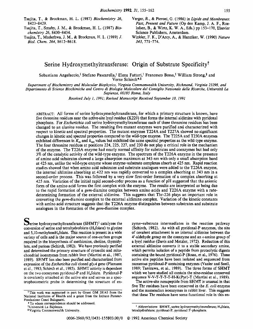
Kinetic Properties of T226A SHMT. The spectral prop- erties of the T226A enzyme and enzyme-amino acid com- plexes suggest that the geminal diamine complex accumulates when the enzyme is saturated with the amino acid substrate. The gem-diamine is an intermediate for all pyridoxal-P en- zymes which form external aldimines with amino acid sub- strates. The spectral results suggest that Thr-226 may play a critical role in the transimination reaction for SHMT. To further investigate the role of Thr-226, we determined the rate of the spectral changes occurring when L-serine was added to the enzyme (Figure 1B). As shown in Figure 2, the decrease in absorption at 422 nm occurs at the same rate as the increase in absorption at 343 nm. Both reactions are first-order pro- cesses and have almost identical amplitude changes.
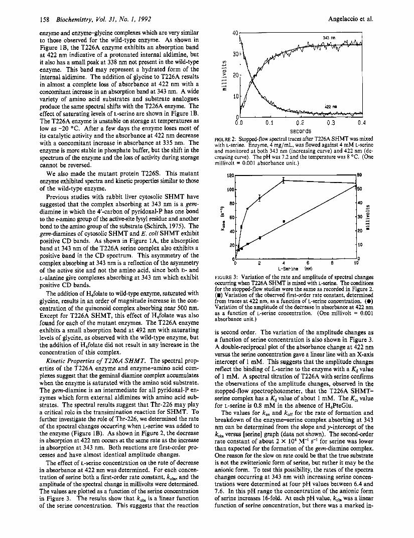
The effect of L-serine concentration on the rate of decrease in absorbance at 422 nm was determined. For each concen- tration of serine both a first-order rate constant, koa, and the amplitude of the spectral change in millivolts were determined. The values are plotted as a function of the serine concentration in Figure 3. The results show that kobs is a linear function of the serine concentration. This suggests that the reaction
Biochemistry, Vol. 31, No. I, 1992 Angelaccio et al.
6.0 0.1 0.2 0.3 0.4 seconds
F i r " 2: Stopped-flow spectral traces after T226A SHMT was mixed with L-serine. Enzyme, 4 mg/mL, was flowed against 4 mM L-serine and monitored at both 343 nm (increasing curve) and 422 nm (de- creasing curve). The pH was 7.2 and the temperature was 8 OC. (One millivolt = 0.001 absorbance unit.)
'0 0 2 4 6 8 10
L-Ser ine InM)
FIGURE 3: Variation of the rate and amplitude of spectral changes occurring when T226A SHMT is mixed with L-serine. The conditions for the stopped-flow studies were the same as recorded in Figure 2. (m) Variation of the observed first-order rate constant, determined from traces at 422 nm, as a function of L-serine concentration. (0) Variation of the amplitude of the decrease in absorbance at 422 nm as a function of L-serine Concentration. (One millivolt = 0.001 absorbance unit.)
is second order. The variation of the amplitude changes as a function of serine concentration is also shown in Figure 3. A double-reciprocal plot of the absorbance change at 422 nm versus the serine concentration gave a linear line with an X-axis intercept of 1 mM. This suggests that the amplitude changes reflect the binding of L-serine to the enzyme with a Kd value of 1 mM. A spectral titration of T226A with serine confirms the observations of the amplitude changes, observed in the stopped-flow spectrophotometer, that the T226A SHMT- serine complex has a Kd value of about 1 mM. The K, value for L-serine is 0.8 mM in the absence of H,PteGlu.
The values for k,, and koff for the rate of formation and breakdown of the enzyme-serine complex absorbing at 343 nm can be determined from the slope and y-intercept of the koa versus [serine] graph (data not shown). The second-order rate constant of about 2 X lo4 M-' s-l for serine was lower than expected for the formation of the gem-diamine complex. One reason for the slow on rate could be that the true substrate is not the zwitterionic form of serine, but rather it may be the anionic form. To test this possibility, the rates of the spectra changes occurring at 343 nm with increasing serine concen- trations were determined at four pH values between 6.4 and 7.6. In this pH range the concentration of the anionic form of serine increases 16-fold. At each pH value, kobs was a linear function of serine concentration, but there was a marked in-
Serine Hydroxymethyltransferase
Table 11: Kinetic Constants for the Rate of Formation of the Complex Absorbing at 343 nm with T226A SHMT and Amino Acid Ligands
kofflkon k,, (M-l s-I k'on' (M-l (M X
ligand pH X lo4) kofl s-I X lo6) 10') M e r 6.47 0.34 + 0.06 33.4 + 2.0 2.0 L-Ser 6.83 1.0 + 0.05 32.6 + 2.4 2.4 M e r 7.19 4.5 + 0.2 24.9 + 2.3 4.5 0.6 L-Ser 7.64 9.4 + 0.35 22.3 + 3.6 3.6 Gly 7.19 1.2 + 0.06 15.1 + 0.6 3.1 1.3 alloThrb 7.19 2.2 + 0.5 43.3 + 2.5 3.5 2.0 L-Ala 7.0 0.39 100 2.0 26
1992 159
'Values corrected for anionic concentration of amino acid ligand. Values based on the concentration of L-allothreonine.
crease in the value of k& with pH (Table 11). When the value of k,, was determined from the slope of the plot of kobs versus the anionic serine concentration, the values only varied from 2 X lo6 to 4.4 X lo6 M-' s-l (Table 11). These pH studies were repeated with glycine at four pH values and essentially the same results were obtained. Between pH values of 6.4 and 7.6, the value of k,, varied less than 2-fold when the anionic form of glycine was used in the calculations.
In addition to serine and glycine, the rates of increase in the formation of the complex absorbing at 343 nm with al- lothreonine and L-alanine were also determined. Both of these amino acids also gave linear plots of kobs versus amino acid concentration. The values of k , , and kOE are recorded in Table 11.
In addition to the rapid spectral changes, observed at 422 and 343 nm, when enzyme was flowed against saturating concentrations of either L-serine or DL-allothreonine, a slow increase in absorption was observed at 422 nm (Figure 4). At these higher concentrations of serine and allothreonine the rapid spectral changes, described by the results in Figure 3, are too fast to measure. The amplitudes of the slow observed increases in absorbance at 425 nm, with both serine and al- lothreonine, were small, which precluded determining either the effect of amino acid concentration on the value of kobs or the shape of the absorption band. However, under saturating conditions of the amino acid substrate, the rates were clearly first order for these slow reactions (Figure 4). For allo- threonine the value of kOb was 3.5 m i d , and for L-serine the value of kobs was 12 m i d . These experiments were done at 30 'C.
DISCUSSION The conservation of five Thr residues at the active site of
rabbit liver cytosolic and mitochondrial SHMT and E. coli SHMT suggests that these residues play some critical role in the structure and function of this enzyme. By changing each of these Thr residues to an Ala in the E. coli enzyme, we hoped to gain some insight into the role of each Thr residue. The first focus of this study was to measure the affinity of both coenzyme and amino acid substrates and to determine k,, values for the five mutant enzymes. In addition to kinetic constants, we also investigated spectral properties of the bound pyridoxal-P and the ability of each enzyme to catalyze the transamination of both D and L-alanine. These studies permit the determination not only of the affinity of substrates and coenzymes for the enzyme but also of the distribution of en- zyme-substrate complexes.
The observation that all five Thr to Ala mutations resulted in enzymes which had both catalytic activity in the aldol cleavage reaction and transaminase activity for both D- and L-alanine, suggests that all five mutant enzymes can form the
Biochemistry, Vol. 31, No. 1,
I
2 3.0 0 > .- 3
1.5 E
0 0 1 ' ' ' ' ' ' ' . I
Y ." 0 10 20 30 40
seconds FIGURE 4: Slow spectral changes occurring at 425 nm after T226A SHMT was mixed with either L-serine or DL-allothreonine. T226A SHMT, 5 mg/mL, was flowed against either 50 mM L-serine or 100 mM m-allothreonine, at pH 7.2 and 30 'C. Panel A shows the spectral trace after enzyme and L-serine were mixed. Panel B shows the trace after enzyme and allothreonine were mixed (one millivolt = 0.001 absorbance unit.)
quinonoid complex absorbing near 500 nm, which is the key catalytic intermediate. Except for T225A SHMT, the relative activities of the various substrates for each mutant enzyme were similar to those for the wild-type enzyme. However, T225A SHMT transaminates L-alanine 20-fold faster and cleaves L-threonine 3-fold faster than the wild-type enzyme (Table I). T225A SHMT cleaves L-serine only one-sixth as fast as the wild-type enzyme. Both the transamination of L-alanine and cleavage of L-threonine are deleterious reactions to the cell, and the role of Thr-225 may be, at least in part, to block these two reactions. This suggests that Thr-225 is important in determining both reaction and substrate specif- icity.
Our results show that for Thr residues 224 and 227 there are no major changes in either the kinetic properties or spectral properties of the mutant enzymes with respect to the wild-type enzyme. Thr residues 226 and 230 play some role in the structure and function of this enzyme since changing them to Ala resulted in an order of magnitude increase in the K, for H4folate for T230A and a 32-fold and 2-fold decrease in k,, for T226A and T230A enzymes, respectively. The mutant T230A enzyme has the same spectral characteristics as the wild-type enzyme, making a kinetic study of its difference with the wild-type enzyme difficult. We have not pursued further where in the reaction sequence this mutation has affected individual rate constants.
The most significant changes in properties occurred when Thr-226 was changed to an Ala. This enzyme had nearly normal K, values for substrates and coenzymes but only 3% of the catalytic activity with serine as the substrate. The large absorption band at 343 nm of the enzyme amino acid com- plexes suggests that the most stable intermediate on the re- action pathway is the gem-diamine rather than the external aldimine, as found with the wild-type enzyme. The gem-di- amine is the first covalent intermediate formed in the con- version of the internal aldimine to the external aldimine. Since this interconversion is a common step in all pyridoxal-P en- zymes metabolizing amino acids, it is of interest to be able to elucidate the role of the enzyme in this step.
In the spectral titration studies, the T226A SHMT-serine complex shows the presence of a band at 425 nm indicative
160 Biochemistry, Vol. 31, No. 1, 1992
of a small amount of external aldimine being present a t sat- urating substrate (Figure 1). When we looked for slower reactions occurring at 343 and 425 nm in the stopped-flow studies, we observed, under saturating amino acid concen- trations at 30 OC, a slow increase in absorbance at 425 nm when either allothreonine or serine was flowed against T226A SHMT. As shown in Figure 4, this increase in absorbance has a small amplitude and is first order. The value of kobs for allothreonine is 3.5 m i d , which is essentially the same as k,, for this substrate (Table I). With L-serine the value of kOk is 12 min-’, which is slightly slower than the observed kat value of 20 m i d . However, unlike with allothreonine, where the values of kobs and k,,, were determined under the same con- ditions of pH and substrate concentration, the value of k,, with serine was determined in the presence of H4folate, which was absent from the determinations of kobs. H,folate could not be used in the stopped-flow studies due to the absorption at 425 nm of oxidation products of this coenzyme. Because of the small amplitude of these slow spectral changes at 425 nm, we were unable to determine either the variation of kobs with amino acid concentration or to accurately determine the spectrum of the complex absorbing near 425 nm. However, the results are consistent with the interpretation that the slow step in the reaction of both allothreonine and serine by T226A SHMT is the conversion of the gem-diamine to the external aldimine shown by structures I11 and IV in Scheme I. Since replacing T226 with a serine residue did not affect catalytic activity, but replacing it with an alanine residue reduced k,,, by 30-fold and caused a substantial change in the distribution of the enzyme-substrate complexes argues that the hydroxyl group of T226 plays an important role in converting the gem-diamine to the external aldimine.
Thr-226 also occupies a position in SHMT which is highly conserved by Ser residues in other pyridoxal-P enzymes. Vaaler and Snell(l989) have listed the enzymes containing a Ser or Thr residue in other pyridoxal-P enzymes occupying an equivalent position with respect to the active-site lysine. These enzymes include histidine, arginine, lysine, glutamate, and tryptophan decarboxylases, cytosolic and mitochondrial aspartate aminotransferases, alanine aminotransaminase, alanine racemase, tryptophanase, D-serine dehydratase, both synthetic and degradative threonine dehydratases, cysta- thionine y-synthase, and methionine y-lyase (Tanizawas et al., 1989). This suggests either a serine or a threonine which is three positions to the amino-terminal side of the active-site lysyl residue plays a critical role in many pyridoxal-P enzymes. Vaaler and Snell (1989) have changed the analogous Ser in histidine decarboxylase to both an alanine and a cysteine. Each of the mutant proteins showed less than 10% of the activity of the wild-type enzyme.
Replacing Thr-226 with a Ser residue produces a fully active enzyme, further suggesting that the hydroxyl group is the critical part of the structure at position 226. Removal of either the Ser or Thr hydroxyl group reduces activity 30-fold. It is possible that the remaining 3% activity is attributable to a solvent water molecule which fills the “hole” left by removal of the Thr hydroxyl. Aspartate aminotransaminase is one of the pyridoxal-P enzymes containing a Ser (S255) at the analogous psition of Thr-226 in SHMT. The crystal structure of aspartate aminotransaminase shows that the serine hydroxyl has polar contacts with the phosphate moiety of pyridoxal-P. In the transformation of the gem-diamine to the external aldimine there is a realignment of pyridoxal-P with respect to the amino acid substrate and active-site pocket (Arnone et al., 1985; Jansonius et al. 1985). It is conceivable that the
Angelaccio et al.
Ser-255 hydroxyl group plays a critical role in this realignment process. The S255A mutation of aspartate aminotransferase has not been reported, so it is not possible at this time to determine if it has altered functions which are similar to those expressed by the T226A mutant form of SHMT.
A second major objective of this study was to use the T226A enzyme to investigate the effect of substrate structure on the mechanism of transimination. In Scheme I is shown a minimal mechanism for transimination of the active-site pyridoxal-P and a sequence of intermediates which are consistent with our experimental results on the properties of E. coli T226A SHMT. For most pyridoxal-P containing enzymes the gem- diamine complexes (structures I1 and I11 in Scheme I) do not accumulate and cannot be directly observed as intermediates. The exception to this is rabbit liver SHMT, where a complex absorbing at 343 nm is observed in both the enzyme-glycine and enzyme-serine complexes (Schirch, 1982). The gem- diamine complex in rabbit SHMT has the same absorption maximum and positive CD spectra observed for the 343-nm complex of T226A SHMT (Figure 1). Rapid reaction studies on the formation of the SHMT-glycine complex absorbing at 343 nm of the rabbit cytosolic enzyme showed that it was formed as the first stable complex (Schirch, 1975). If the 343-nm-absorbing band of the T226A SHMT-amino acid complex is the gem-diamine, this altered protein would rep- resent the first pyridoxal-P enzyme in which this complex is the most stable intermediate. Much of the work in this paper was aimed at determining if the 343-nm band had the char- acteristics of a gem-diamine.
The gem-diamine, shown as structure I1 in Scheme I, should be formed either in a second-order reaction or in a first-order reaction coupled to the rapid second-order formation of a noncovalent complex between serine and SHMT. This model would require that the decrease in absorbance at 422 nm and the increase in absorbance at 343 nm occur at the same rate and be pseudo first order in enzyme. The results shown in Figure 2 confirm this observation with T226A SHMT. A second criterion is that the observed pseudo-first-order rate constant for the appearance of the 343-nm-absorbing complex should be either linear with serine concentration or show saturation kinetics. The results recorded in Figure 3 show that the observed rate constant is a linear function of serine con- centration from 0.1 to 10 mM and that the reaction is first order in L-serine. This shows that the spectral changes ob- served on the binding of serine are the result of the first complex formed on the reaction pathway. It is unlikely that the spectral changes are the result of the formation of a noncovalent complex. The results also suggest that if a non- covalent complex is formed between serine and SHMT prior to the attack of the amino group on the internal aldimine, it has a Kd value which is larger than 10 mM. If a noncovalent complex had been formed, saturation kinetics with respect to the effect of L-serine concentration on kobs would have been observed.
The study of the rate of the spectral changes occurring at 343 nm, when serine and glycine were flowed against SHMT at several pH values, suggests that it is the anionic form of the amino acid which attacks the internal aldimine (structure I in Scheme I). This accounts for the slow rate of formation of the complex. In a study of the rate of formation of the gem-diamine with rabbit SHMT and glycine, a value of k,, of 7 X 10⁴ M-’ s-’ was determined (Schirch, 1975). The value is not significantly different from the values obtained in this study for reaction of E. coli T226A SHMT with amino acid substrates.
Serine Hydroxymethyltransferase
Scheme I ser
E-h-CH-PLP k2 , €I kl . E-#-,-PLP \ +A
B E-N-CH-PLP \
H-N-Ser + I k- 2 :J-ser k- 1
A I 422 nm I1 343 nm 111 343 nm
Biochemistry, Vol. 31, No. I, 1992 161
termined from spectral titrations or steady-state kinetic studies. The values obtained for kOff/kon provide two pieces of infor- mation. First, the results suggest that the affinity of the enzyme for its amino acid substrate is reflected in the inter- actions formed as early as the gem-diamine intermediate. Second, the enzyme distinguishes between amino acids at the gem-diamine complex. The values for k,, for both poor and good amino acids are similar (Table 11). The differences in affinity are reflected in the different values for kOF This would be consistent with the proposal of Federiuk and Shafer that the enzyme distinguishes between amino acids at the step where gem-diamine I1 is converted to gem-diamine I11 (Scheme I). Amino acids such as L-alanine and L-threonine may not be converted to gem-diamine 111, thus accounting for their decreased affinity for the active site.
T226A SHMT also has an absorption band at 338 nm which is not present in the wild-type enzyme. We suggest that this is a hydrated form of the enzyme shown as structure V in Scheme I. However, it could be the result of any nucleophile adding across the double bond of the internal aldimine. Ap- parently, this form of the enzyme is very stable and its slow accumulation leads to inactivation of the enzyme.
Jonsonius, J. N., Eichele, G., Ford, G. C., Picol, D., Thaller, C., & Vincent, M. G. (1985) in Transaminases (Christen, P., & Metzler, D. E., Eds.) pp 110-138, John Wiley & Sons, New York.
Martini, F., Angelaccio, S. , Pascarella, S. , Barra, D., Bossa, F., & Schirch, V. (1987) J . Biol. Chem. 262,5499-5509.
Martini, F., Maras, B., Tanci, P., Angelaccio, S., Pascarella, S., Barra, D., Bassa, F., & Schirch, V. (1989) J. Biol. Chem. 264, 8509-8519.
Plamann, M., Stauffer, L., Urbanowski, M., & Stauffer, G. (1983) Nucleic Acids Res. 11, 2065-2075.
Schirch, L. (1975) J . Biol. Chem. 250, 1939-1945. Schirch, L. (1982) Adv. Enzymol. Relat. Areas Mol. Biol.
Schirch, L., Edmiston, M., Chen, M. S., Barra, D., Bossa, F., Hinds, L., & Fasella, P. (1973) J. Biol. Chem. 248,
Schirch, V., Hopkins, S., Villar, E., & Angelaccio, S. (1985)
Schirch, V., Shostak, K., Zamora, M., & Gautam-Basak, M.
7416-7423.
5 372-5 378.
3363-3369.
53, 83-1 12.
7801-7806.
6456-6461.
J. Bacteriol. 165, 1-7.
(1991) J . Biol. Chem. 266, 759-764.
k4 / k-4 B
E-N-CH-PLP
i V 3 3 8 nm
B il II
E-N: CH-PLP
+N-Ser n
IV 425 nm
Federiuk and Shafer have concluded from their studies of D-serine dehydratase that there must be at least two gem- diamines in the transimination reaction. These two gem-di- maine complexes are the result of a proton transfer from the incoming amino group of the amino acid substrate to the t-amino group of the lysyl residue, which must be expelled in forming the external aldimine (Federiuk and Shafer, 198 1 , 1983). They also point out that a rotation of the C-4 - C-4' bond of pyridoxal-P probably occurs as a means of positioning the leaving amino group of lysine so that it is perpendicular to the plane of the pyridoxal-P ring. They argue that this step is important in determining which amines can form the ex- ternal aldimine and thus will be the step which controls sub- strate specificity.
Our results with T226A and its reaction with amino acids are consistent with the proposal of Federiuk and Shafer con- cerning the origin of substrate specificity. As shown in Figure 3, the amplitude of the decrease in absorbance at 422 nm with increasing serine concentration shows saturation kinetics. This is in contrast to the values of kobs, which are linear with in- creasing serine concentration. This difference in response of amplitude and kob with serine concentration can be explained if a second complex, having the same spectral properties as the first complex, is formed in a rapid second reaction. We suggest in Scheme I that this is the result of a proton transfer from the serine amino group to the lysyl amino group (structure I1 to structure I11 in Scheme I). The change in the amplitude in absorbance at 343 nm, with increasing serine concentration in the stopped-flow studies, is consistent with a & value of about 1 mM for serine, which is similar to the Kd and K,,, values for serine of 0.9 mM determined from steady-state kinetic studies and spectral titrations at 343 nm (Table I). The transfer of a proton in structures I1 and I11 explains the increased stability of the gem-diamine since the incoming serine amino group would no longer be a good leaving group in structure 111.
The data reported in Table I1 suggest that the value for the second-order rate constant, kl, is about 2 X lo4 M-' s-l. However, the ordinate intercepts of 11-50 s-' are probably not the value of k-'. For the reactions shown in Scheme I, where a rapid first-order reaction is preceded by a slower second order reaction, the variation of kob with substrate concentration is given by the following equation (Schirch, 1975). The ordinate intercept value would reflect the ratio of k21k-2 in addition
kobs = kl(Ser) -t k-l/(l + k2/k-,)
to k-1.
One can determine the Kd for each amino acid used in the stopped-flow studies by dividing kOff by kon. As shown in Table 11, these values vary from 0.5 to 20 mM and reflect the affinity of the enzyme for each amino acid. For each amino acid the ratio of these kinetic constants is similar to its Kd values de-
162 Biochemistry 1992, 31, 162-168
Shostak, K., & Schirch, V. (1988) Biochemistry 27, Vaaler, G. L., & Snell, E. E. (1989) Biochemistry 28,
Tanizawa, K., Masu, Y., Asano, S. Tanaka, H., & Soda, K. Villar, E., Schuster, B., Peterson, D., & Schirch, V. (1985) 8007-80 14. 7306-73 13.
Effects of High Pressure on the Catalytic and Regulatory Properties of UDP-Glucuronosyltransferase in Intact Microsomest
Juraj Kavecansky, Andrew J. Dannenberg, and David Zakim* Division of Digestive Diseases, Department of Medicine, Cornel1 University Medical Center, 1300 York Avenue,
New York, New York 10021 Received July 16, 1991; Revised Manuscript Received September 12, 1991
ABSTRACT: The effects of high pressure on the kinetic properties of microsomal UDP-glucuronosyltransferase (assayed with I-naphthol as aglycon) were studied in the range of 0.001-2.2 kbar to clarify further the basis for regulating this enzyme in untreated microsomes. Activity changed in a discontinuous manner as a function of pressure. Activation occurred at pressure as low as 0.1 kbar, reaching one of two maxima at 0.2 kbar. As pressure was increased above 0.2 kbar, activity decreased, reaching a minimum at about 1.4 kbar followed by a second activation. The pathway for activation at pressure > 1.4 kbar was complex. The immediate effect of 2.2 kbar was nearly complete inhibition of activity. The inhibited state relaxed, however, over about 10 min (at 10 “C), to a state that was activated as compared with enzyme at 0.001 kbar or enzyme at pressures between 1.4 and 2.2 kbar, which was the highest pressure we could test. Examination of the detailed kinetic properties of UDP-glucuronosyltransferase indicated that the effects of pressure were due to selective stabilization of unique functional states of the enzyme at 0.2 and 2.2 kbar. Activation at 0.2 kbar was reversible when pressure was released. This was true as well for activation at pressure > 1.4 kbar, but after prolonged treatment at 2.2 kbar, UDP-glucuronosyltransferase became activated irreversibly on release of pressure. The process by which prolonged treatment a t 2.2 kbar led to permanent activation of UDP-glucuronosyltransferase after release of pressure was not reflected, however, by time-dependent changes in the functional state of UDP-glucuronosyltransferase at this pressure. Thus, appearance of the unique functional state of UDP-glucuronosyltransferase at 2.2 kbar occurred within about 10 min after reaching this pressure, and this state of the enzyme persisted for as long as 60 min (longest time studied). By contrast, the distribution of UDP-glucuronosyltransferase between native state and permanently activated state, after release of 2.2 kbar, shifted in favor of the latter with increasing time of treatment at 2.2 kbar. When pressure was released after 60 min at 2.2 kbar, about 80% of UDP-glucuronosyltransferase became permanently activated. We have interpreted this result to mean that treatment a t high pressure perturbs interactions between UDP-glucuronosyltransferase and an undefined regulatory factor in microsomes that is important for maintaining the enzyme in its native conformational state.
E i o r observations of the effect of high pressure on the function of UDP-glucuronosyltransferase indicated that this technique could be useful for studying the regulation of UDP-glucuronosyltransferase in otherwise untreated micro- somes (Dannenberg et al., 1990). Measurements of activity at 2.2 kbar, as a function of temperature (Dannenberg et al., 1990), suggested that high pressure altered the conformation of UDP-glucuronosyltransferase, leading to a functional state different from that at 1 atm, and under some conditions, release of pressure led to irreversible activation of UDP- glucuronosyltransferase. The data for the effects of high pressure on the functional states of UDP-glucuronosyl- transferase were compatible with Scheme I (Dannenberg et al., 1990), in which E is the native state of the enzyme, E* is the activated state produced after release of high pressure, and E + E’ is a reversible change in the state of the enzyme
‘This work was supported by grants from the NSF (DMB 8504014) and the NIH (1K08 DK 1992). A.D. is the recioient of the Dr. Mark Weinstein Liver Scholar Award from the Amerilan Liver Foundation.
* To whom correspondence should be addressed.
at 2.2 kbar, 10 O C . A second intermediate (E”) at high pressure was proposed to explain the observations that E - E’ occurred rapidly at high pressure but complete conversion of E to E*, after release of pressure, required 90 min of treatment at 2.2 kbar. Scheme I
+P -P -P F E’ - E” - E*
In the present work, we have extended observations on the response of UDP-glucuronosyltransferase to high pressure, in otherwise untreated microsomes, by measuring activity as a function of pressure from 1 atm (0.001 kbar) to 2.2 kbar. Thii work shows that UDP-glucuronosyltransferase is sensitive to applied pressures as low as 0.1 kbar and that there are several active states available to UDP-glucuronosyltransferase that are stabilized differentially at pressures above 0.001 kbar. Direct experiments to detect the putative intermediate E” failed to reveal this form of the enzyme at 2.2 kbar. Instead, it appears that high pressure alters a feature of microsomes that determines whether enzyme at high pressure relaxes to
0006-2960/92/043 1-162$03.00/0 0 1992 American Chemical Society