Serine Phosphorylation of GH-Activated SignalTransducer and Activator of Transcription 5a(STAT5a) and STAT5b: Impact on STAT5Transcriptional Activity
SOO-HEE PARK, HIROKO YAMASHITA, HALLGEIR RUI, AND DAVID J. WAXMAN
Division of Cell and Molecular Biology, Department of Biology, Boston University (S.-H.P., D.J.W.),Boston, Massachusetts 02215; and Department of Pathology, Uniformed Services University of theHealth Sciences (H.Y., H.R.), Bethesda, Maryland 20814
Signal transducer and activator of transcription 5b(STAT5b), the major liver-expressed STAT5 form, isphosphorylated on both tyrosine and serine in GH-stimulated cells. Although tyrosine phosphorylationis known to be critical for the dimerization, nucleartranslocation, and activation of STAT5b DNA-bindingand transcriptional activities, the effect of STAT5bserine phosphorylation is uncertain. Presently, weidentify Ser730 as the site of STAT5b serine phos-phorylation in GH-stimulated liver cells. We addition-ally show that the serine kinase inhibitor H7 partiallyblocks the GH-stimulated formation of (Ser,Tyr)-diphosphorylated STAT5b without inhibiting STAT5bnuclear translocation. Evaluation of the functionalconsequences of STAT5b serine phosphorylation bymutational analysis revealed an approximately 50%decrease in GH-stimulated luciferase reporter geneactivity regulated by an isolated STAT5-binding sitewhen STAT5b Ser730 was mutated to alanine andunder conditions where STAT5 DNA-binding activitywas not diminished. No decrease in GH-stimulated
reporter activity was seen with the correspondingSTAT5a-Ser725Ala mutant; however, a decrease inreporter activity occurred when the second estab-lished STAT5a serine phosphorylation site, serine779, was additionally mutated to alanine. Unexpect-edly, STAT5a-Ser725,779Ala and STAT5b-Ser730Aladisplayed approximately 2-fold higher GH- or PRL-stimulated transcriptional activity compared withwild-type STAT5b when assayed using an intact�-casein promoter-luciferase reporter. Finally,STAT5b-stimulated gene transcription was abol-ished in cells treated with H7, but in a mannerunrelated to the inhibitory effects of H7 on STAT5bSer730 phosphorylation. These findings suggestthat the effects of STAT5b and STAT5a serinephosphorylation on STAT-stimulated gene tran-scription can be modulated by promoter context.Moreover, in the case of STAT5a, phosphorylationof serine 779, but not serine 725, may serve toregulate target gene transcriptional activity.(Molecular Endocrinology 15: 2157–2171, 2001)
SIGNAL TRANSDUCER AND activator of transcrip-tion (STAT) factors are signal transducers that
mediate the effects of a broad range of hormones andcytokines on target gene expression (1, 2). STAT pro-tein activation is catalyzed by a cell surface receptor-associated tyrosine kinase of the Janus kinase (JAK)family, which phosphorylates the STAT protein on asingle C-terminal region tyrosine in response to hor-mone or cytokine stimulation. Tyrosine-phosphory-lated STATs undergo rapid homo- or heterodimeriza-tion associated with STAT translocation to the
nucleus, where the dimeric, DNA-binding STAT acti-vates target gene transcription. Dephosphorylationcatalyzed by a phosphotyrosine-specific phosphatase(3) terminates STAT signaling and returns the inacti-vated STAT protein to the cytosol.
In addition to this JAK-catalyzed tyrosine phosphor-ylation reaction, STAT proteins may undergo serinephosphorylation in a manner that can be cell type andstimulus dependent (4–6). In the case of STAT1, -3,and -4, serine phosphorylation is directed at a con-served PMSP motif located about 20–30 amino acidsC-terminal of the conserved tyrosine phosphorylationsite. This phosphorylation can be stimulated by cyto-kine treatment and may be catalyzed by a downstreamkinase, e.g. a cytokine- or growth factor-activatedMAPK. Basal serine phosphorylation of STAT proteinshas also been observed (for a review, see Ref. 7).Mutation of the conserved PMSP serine phosphoryla-tion site (e.g. STAT1-Ser727, STAT3-Ser727, or STAT4-Ser721) decreases cytokine-induced transcriptionalactivity, supporting the hypothesis that serine phos-phorylation is required for maximal transcriptional ac-
Abbreviations: anti-pS730 antibody, Antibody specific forphosphoserine 730 of STAT5b and phosphoserine 725 ofSTAT5a; anti-pY699 antibody, antibody specific for tyrosine699 or 694 of STAT5b and STAT5a, respectively; JAK, Januskinase; PMSP motif, Pro-Met-Ser-Pro; PSP, Pro-Ser-Pro;STAT, signal transducer and activator of transcription; TSTbuffer, 10 mM Tris-HCl (pH 7.6), 5 mM EDTA, 50 mM NaCl, 30mM Na2P2O7 50 mM NaF, 1 mM Na3VO4, 1% Triton X-100,and 1 mM phenylmethylsulfonylfluoride.
Site-specific STAT5 mutants are designated using the sin-gle letter amino acid code; thus, STAT5b-S730A correspondsto STAT5b with Ser730 mutated to alanine.
tivity of these three STATs and may modulate cytokineresponses (4, 8, 9), at least on some promoters (10).Serine phosphorylation can also negatively regulatecytokine-induced tyrosine phosphorylation in the caseof STAT3 (11). STAT nuclear translocation and DNA-binding activity are generally not affected by STATserine phosphorylation, suggesting that serine phos-phorylation modulates STAT’s intrinsic transcriptionalpotential, which is mediated by a C-terminal trans-activation domain just downstream of the serine phos-phorylation site.
Serine phosphorylation of STAT5a and the closelyrelated (�90% identical) STAT5b (12) has been ob-served in cells and tissues stimulated with STAT5-activating ligands such as GH (13–15), PRL (16, 17),and IL-2 (18). In PRL-stimulated cells, both STAT5forms are phosphorylated on a conserved serine res-idue (STAT5a-Ser725 and STAT5b-Ser730) locatedwithin a PSP sequence, which corresponds in locationto the PSMP serine phosphorylation sequence ofSTAT1, -3, and -4 (19). STAT5a is additionally phos-phorylated at a second site, recently identified asserine 779 (20, 21). STAT5 serine phosphorylation mayin part be mediated by the MAPK cascade, as sug-gested by binding interactions between STAT5a andthe MAPKs ERK1 and ERK2 (21) and by the inhibitionof constitutive, but not PRL-inducible, STAT5a serine725 phosphorylation in Nb2 lymphocytes by theMAPK pathway inhibitor PD98059 (19). Functionalstudies of the effects of serine phosphorylation onSTAT5’s transcriptional activity have not provided aconsistent picture. In the case of IL-2-activated STAT5(STAT5a and/or STAT5b), cytokine-stimulated re-porter gene activity is abolished in cells treated withthe serine kinase inhibitor H7, which blocks STAT5serine phosphorylation (18). Similarly, GH-activatedSTAT5a activity is blocked by inhibition of MAPKactivity (22), which may play a role in STAT5a serinephosphorylation (21). In contrast, no difference in PRL-stimulated STAT5 reporter gene activity was seenwhen cells were transfected with serine to alaninemutant forms of STAT5b (Ser730 to Ala) or STAT5a(Ser725 mutated to Ala, Ser779 to Ala, or theSer725,779Ala double mutant) compared with the cor-responding wild-type STAT5 forms (19, 20). Delayedtyrosine dephosphorylation was, however, reportedfor STAT5a-S725A in cells stimulated with PRL (20).These findings raise the possibility that the conse-quences of STAT5 serine phosphorylation may varywith the activating hormone or cytokine, or perhapswith the target gene used to evaluate the impact ofSTAT5 serine phosphorylation. These and relatedissues are investigated in the present study, wherewe evaluate the functional consequences of STAT5serine phosphorylation in GH-stimulated cells usingsite- specific mutants of STAT5a and STAT5b. Ourfindings reveal that GH induces the same pattern ofSTAT5 serine phosphorylation as that previously re-ported for PRL. Moreover, we report that serinephosphorylation can modulate the transcriptional
activity of both STAT5a and STAT5b in a promoter-dependent manner.
RESULTS
Serine Phosphorylation of STAT5b in GH-Stimulated Cells
STAT5a and STAT5b carry out distinct functions inendocrine target tissues. STAT5a is the principal me-diator of mammopoietic and lactogenic signaling stim-ulated by PRL, whereas STAT5b is an important de-terminant of sexual dimorphic liver gene expressioninduced by GH (23). To ascertain whether these dis-tinctive biological roles of STAT5a and STAT5b, mightin part reflect their differential serine phosphorylationin response to GH and PRL, we first investigatedwhether GH induces phosphorylation of STAT5b onthe same serine residue (Ser730) as that reported pre-viously for PRL (19).
Initial experiments were carried out using COS-1cells transfected with GH receptor and either STAT5bor the site-specific mutant STAT5b-S730A and thenstimulated with GH. Western blotting with anti-STAT5b antibody revealed multiple protein bands,which were previously identified as differentially phos-phorylated forms of STAT5b (Fig. 1A). STAT5b band 0(Fig. 1A) migrates as a doublet of bands, neither ofwhich appears to be phosphorylated, as shown pre-viously by phosphatase treatment experiments,whereas STAT5b band 1a corresponds to serine-phosphorylated STAT5b (14). This latter conclusion issupported by the absence of STAT5b band 1a in un-stimulated cells transfected with STAT5b-S730A (Fig.1A, lane 3 vs. lane 1). STAT5b band 2, previouslyidentified as STAT5b phosphorylated on both tyrosineand serine, is the major GH-induced phosphorylatedform of wild-type STAT5b (lane 2). In contrast,STAT5b-S730A was converted to a doublet of proteinsafter GH treatment (Fig. 1A, lane 4 vs. lane 3). Bothbands of the doublet were phosphorylated on ty-rosine, as shown by immunoprecipitation with anti-STAT5b antibody followed by antiphosphotyrosine4G10 Western blotting (data not shown; also see be-low). The lower band corresponds in mobility toSTAT5b phosphorylated on tyrosine alone, i.e.STAT5b band 1, whereas the upper band of the dou-blet remains unidentified (band X). The STAT5b dou-blet dominates in STAT5b-S730A-transfected cellstreated with GH and is not further converted to thedoubly phosphorylated STAT5b band 2, presumablybecause of the block in the secondary serine phos-phorylation on residue 730 (see below). The preciserelationship between STAT5b-S730A bands 1 andband X is uncertain. The two proteins may correspondto the tyrosine-phosphorylated counterparts of thetwo marginally resolved STAT5b protein forms seen inunstimulated cells (both designated band 0; Fig. 1A,lanes 1 and 3).
2158 Mol Endocrinol, December 2001, 15(12):2157–2171 Park et al. • STAT5b Serine Phosphorylation
Western blotting using phospho-STAT5-specificantibodies (anti-pS730-STAT5b and anti-pY699-STAT5b) further supported these STAT5 band identi-fications (Fig. 1A, lanes 5–9). Thus, STAT5b was ba-sally phosphorylated on Ser730 (band 1a; Fig. 1, middlepanel, lane 6), and GH stimulated the formation ofSTAT5b phosphorylated on both Ser730 and Tyr699
(band 2; Fig. 1, lane 7). STAT5b Ser730 phosphoryla-tion was blocked in cells transfected with STAT5b-S730A (Fig. 1, lanes 8 and 9, middle panel), whereasGH stimulated Tyr699 phosphorylation of STAT5b-S730A to form a band that migrated just below band 2(band 1/band X; Fig. 1, upper panel, lane 9). [Of note,STAT5b bands 0, 1 and X are poorly resolved in theright panel of Fig. 1A (c.f. lanes 1 and 4).]
We next investigated whether GH induces phosphory-lation of STAT5b on Ser730, e.g. via a GH-stimulatedserine kinase, and whether phospho-Ser730-STAT5bserves as a substrate for the GH-stimulated tyrosinephosphorylation reaction. These studies were carried outusing the GH-responsive liver cell line CWSV-1, where allthe components required for GH-induced STAT5b ty-rosine and serine phosphorylation are expressed endo-genously (14, 24). CWSV-1 cells were stimulated withGH for varying periods of time, and cell extracts wereprepared and analyzed by immunoprecipitation with an-ti-STAT5b antibody, followed by sequential probing withthe antibodies shown in Fig. 1B. As reported previously(14), STAT5b is basally phosphorylated on serine inCWSV-1 cells (c.f. presence of STAT5b band 1a in un-stimulated cells; Fig. 1B, lane 1, lower panel). At least aportion of this phosphorylation is on Ser730, as indicatedby the reactivity of band 1a with phospho-Ser730-spe-cific anti-STAT5 antibody (anti-pS730; upper panel).Moreover, GH-induced tyrosine phosphorylation ofSer730-phosphorylated STAT5b was readily detectable,as revealed by the appearance of the pS730-reactiveSTAT5b, band 2 (lane 2). The GH-stimulated increase intotal pS730-STAT5b normalized to total STAT5b im-munoreactivity (lane 2 vs. lane 1, lower portion ofFig. 1B) suggests that STAT5b Ser730 can be phos-phorylated by a GH-stimulated serine kinase in additionto the basal Ser730 kinase activity. Both the basal and theGH-stimulated STAT5b Ser730 kinase activity are par-tially inhibited by the serine kinase inhibitor H7 (Fig. 1B,lanes 7 and 8 vs. lanes 1 and 2). This conclusion issupported by densitometric analysis of pS730-STAT5bimmunoreactivity normalized to total STAT5b protein(lower portion of Fig. 1B). Moreover, the specific phos-pho-Ser730 content of STAT5b is seen to decline back tothe basal level from its peak 30 min after GH stimulation.
We cannot determine from the above data whether thediphosphorylated STAT5b (band 2) is preferentiallyformed by GH-stimulated tyrosine phosphorylation ofpreexisting phospho-Ser730-STAT5b (band 1a) or by ty-rosine phosphorylation of STAT5b band 0, followed by asecondary, GH-stimulated Ser730 phosphorylation of aSTAT5b band 1 intermediate. Support for the latterpossibility is provided by Western blot analysis of GH-stimulated CWSV-1 extracts using antibody specific for
STAT5b phosphotyrosine 699. Figure 1C shows that GHstimulates the transient formation of pY699-STAT5b at 5min, followed by conversion to band 2 in an apparentserine phosphorylation reaction (lanes 3 and 4 vs. lane 2).In cells treated with H7, this secondary conversion toSTAT5b band 2 is partially blocked. This finding supportsthe partial inhibition of GH-stimulated S730 phosphory-lation shown in Fig. 1B (note the persistence in Fig. 1C ofthe pY699-STAT5b-immunoreactive doublet, even at40–80 min; lanes 11–13 vs. single band at 20 min in lane15 in the absence of H7). Densitometric analysis of thephospho-Tyr699 signals normalized to total STAT5b pro-tein verified that H7 treatment slows down the decay inSTAT5b signaling, as shown previously (24).
Transcriptional Activity of Site-Specific STAT5Serine Mutants
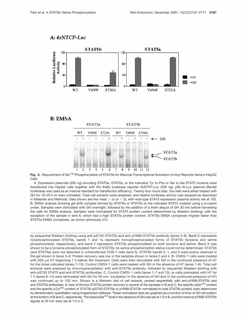
To ascertain the functional significance of GH-stimu-lated STAT5 serine phosphorylation, serine to alaninemutations were introduced at the conserved PSPserine phosphorylation site of both STAT5 forms(STAT5a Ser725 and STAT5b Ser730). The effects ofthese site-specific mutations on GH-stimulated,STAT5-dependent gene transcription were evaluatedin transfection experiments using a luciferase reportergene driven by four copies of a STAT5-binding sitederived from the promoter of the rat ntcp gene (25).These studies were carried out in the liver cell lineHepG2, which has low endogenous GH receptor andSTAT5, but serves as a useful model for STAT reportergene studies and for expression of GH-regulated liverpromoters (25, 26). Wild-type STAT5a and wild-typeSTAT5b trans-activated ntcp promoter activity inthese cells by 32- to 40-fold after GH stimulation (Fig.2A). STAT5a-Y694F and STAT5b-Y699F, which aremutated at the established STAT5 tyrosine phosphor-ylation site, were inactive, consistent with the absoluterequirement of STAT5 tyrosine phosphorylation for ac-tivation of gene transcription. By contrast, mutation ofthe PSP serine phosphorylation site had a more subtleeffect on STAT5-dependent transcriptional activity.No significant change in ntcp promoter activity wasseen with STAT5a-S725A compared with wild-typeSTAT5a, whereas a substantial (�50%) reduction inactivity was seen with the corresponding STAT5b-S730A mutant (Fig. 2A). This effect was observed ineach of four independent HepG2 transfection experi-ments and was confirmed in a second cell model,transfected COS-1 cells (Table 1).
EMSA analysis verified that GH activated the DNA-binding activity of both STAT5b and STAT5b-S730A,as determined using a �-casein promoter STAT5-binding site probe. Similarly, GH activated STAT5aand STAT5a-S725A DNA-binding activity (Fig. 2B). Bycontrast, mutation of the STAT5 tyrosine phosphory-lation site abolished STAT5 DNA-binding activity, asanticipated (Fig. 2B, lanes 4 and 10). We conclude thatphosphorylation of STAT5b at Ser730 is not requiredfor DNA-binding activity, but is required to achieve full
Park et al. • STAT5b Serine Phosphorylation Mol Endocrinol, December 2001, 15(12):2157–2171 2159
Fig. 1. GH-Stimulated Phosphorylation of STAT5b on Ser730 and Tyr699
A, COS-1 cells were transiently transfected with wild-type (wt) STAT5b or STAT5b-S730A as indicated. Thirty-six hours later,cells were either treated with 200 ng/ml GH for 30 min or were untreated. Total cell extracts were analyzed on Western blotsprobed directly with anti-STAT5b antibody (lanes 1–4) or analyzed by immunoprecipitation with anti-STAT5b antibody followed
2160 Mol Endocrinol, December 2001, 15(12):2157–2171 Park et al. • STAT5b Serine Phosphorylation
Fig. 2. Requirement of Ser730 Phosphorylation of STAT5b for Maximal Transcriptional Activation of ntcp Reporter Gene in HepG2Cells
A, Expression plasmids (200 ng) encoding STAT5a, STAT5b, or the indicated Tyr to Phe or Ser to Ala STAT5 mutants weretransfected into HepG2 cells together with the firefly luciferase reporter 4xNTCP-Luc (200 ng). pRL-tk-Luc plasmid (Renillaluciferase) was used as an internal standard for transfection efficiency. Twenty-four hours later, the cells were either treated withGH for 18–20 h or were untreated. Total cell extracts were prepared, and relative luciferase activity was assayed as describedin Materials and Methods. Data shown are the mean � SD (n � 3), with wild-type STAT5 expression plasmid activity set at 100.B, EMSA analysis showing gel-shift complex formed by STAT5a or STAT5b or the indicated STAT5 mutants using a �-caseinprobe. Samples were stimulated with GH overnight, followed by the addition of a fresh aliquot of GH 30 min before harvestingthe cells for EMSA analysis. Samples were normalized for STAT5 protein content (determined by Western blotting), with theexception of the sample in lane 6, which had a high STAT5a protein content. STAT5b EMSA complexes migrate faster thanSTAT5a EMSA complexes, as shown previously (41).
by sequential Western blotting using anti-pS730-STAT5b and anti-pY699-STAT5b antibody (lanes 5–9). Band 0 representsnonphosphorylated STAT5b, bands 1 and 1a represent monophosphorylated forms of STAT5b (tyrosine and serinephosphorylated, respectively), and band 2 represents STAT5b phosphorylated on both tyrosine and serine. Band X wasshown to be a tyrosine-phosphorylated form of STAT5b; its serine phosphorylation status could not be determined. STAT5b(and STAT5a) were not detected in untransfected COS-1 cells (lane 5). STAT5b bands 0, 1, and X were poorly resolved inthe gel shown in lanes 5–9. Protein recovery was low in the samples shown in lanes 2 and 4. B, CWSV-1 cells were treatedwith 200 �M H7 beginning 1 h before GH treatment. Cells were then stimulated with GH in the continued presence of H7for the times indicated (lanes 7–10). Control CWSV-1 cells were treated with GH in the absence of H7 (lanes 1–6). Total cellextracts were analyzed by immunoprecipitation with anti-STAT5b antibody, followed by sequential Western blotting withanti-pS730 STAT5 and anti-STAT5b antibodies. C, Control CWSV-1 cells (lanes 1–7 and 15), or cells pretreated with H7 for1 h (lanes 8–14) were stimulated with GH for 20 min. Incubation in the absence of GH (but in the continued presence of H7)was continued up to 120 min. Shown is a Western blot of cell extracts, probed sequentially with anti-pY699-STAT5b andanti-STAT5b antibodies. In view of the low STAT5b protein recovery in several of the samples in B and C, the specific pSer730 contentand the specific pTyr699 content of STAT5b (pS730-STAT5b or pY699-STAT5b normalized to total STAT5b protein) were determinedby densitometric quantitation using ImageQuant software. These normalized data are graphed as a function of time of GH stimulationat the bottom of B and C, respectively. The basal pSer730 level in the absence of GH was set at 1.0 in B, and the maximal pY699-STAT5bsignals at 20 min were set at 1.0 in C.
Park et al. • STAT5b Serine Phosphorylation Mol Endocrinol, December 2001, 15(12):2157–2171 2161
activation of ntcp promoter activity, as demonstratedin both HepG2 and COS-1 cells. In the case ofSTAT5a, phosphorylation of the corresponding Ser725
is not required for maximal transcriptional activity.
Role of STAT5a Ser779 Phosphorylation
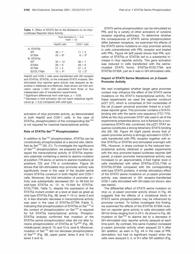
In addition to Ser725 phosphorylation, STAT5a can beconstitutively phosphorylated at a second site, identi-fied as Ser779 (20, 21). To investigate the significanceof Ser779 phosphorylation, we prepared and then as-sayed the transcriptional activity of STAT5a expres-sion plasmids containing a serine to alanine mutationat position 779 alone, or serine to alanine mutations atpositions 725 and 779 in combination. Figure 3Ashows that GH-stimulated ntcp promoter activity wassignificantly lower in the case of the double serinemutant STAT5a construct in both HepG2 and COS-1cells. Moreover, the fold stimulation of promoter ac-tivity was substantially decreased (32- to 38-fold forwild-type STAT5a vs. 12- to 15-fold for STAT5a-S725,779A; Table 1), despite the expression of theSTAT5a mutant protein at a level at least as great aswild-type STAT5a (Fig. 3B, lanes 7–10 vs. lanes 3 and4). A less dramatic decrease in transcriptional activitywas seen in the case of STAT5a-S779A (Table 1),indicating that phosphorylation of STAT5a on Ser779 inthe context of phosphorylation on Ser725 is requiredfor full STAT5a transcriptional activity. Phospho-STAT5a analysis confirmed that mutation of theSTAT5a serine phosphorylation sites did not alter ty-rosine phosphorylation of STAT5a at Tyr699 (Fig. 3B,middle panel, lanes 8, 10, and 12 vs. lane 4). Moreover,mutation of Ser779 did not decrease phosphorylationof Ser725 (Fig. 3B, upper panel, lanes 9 and 10 vs.lanes 3 and 4).
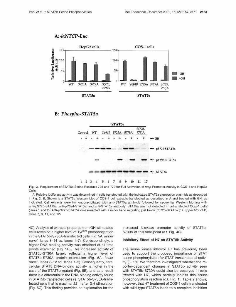
STAT5 serine phosphorylation can be stimulated byPRL and by a variety of other activators of cytokinereceptor signaling pathways. To determine whetherthe consequences of STAT5 serine phosphorylationdiffer between receptors, we examined the effects ofthe STAT5 serine mutations on ntcp promoter activityin cells cotransfected with PRL receptor and treatedwith PRL. Figure 4A (left panel) shows that PRL acti-vation of STAT5a or STAT5b led to a substantial in-crease in ntcp reporter activity. This gene activationwas reduced in cells transfected with the serine-mutated STAT5 forms, STAT5a-S725,779A andSTAT5b-S730A, just as it was in GH-stimulated cells.
Impact of STAT5 Serine Mutations on �-CaseinPromoter Activity
We next investigated whether target gene promotercontext may influence the effect of the STAT5 serinemutations on the STAT’s transcriptional activity. Forthese experiments we used the reporter constructpZZ1 (27), which is comprised of 344 nucleotides ofthe rat �-casein proximal promoter linked to a lucif-erase reporter gene. This promoter contains a STAT5-binding site with the same core sequence (TTC-TTG-GAA) as the ntcp promoter STAT site used in all of theexperiments presented above, but is flanked by a non-consensus STAT5 site; combined, the pZZ1 STAT se-quence constitutes a strong tetrameric STAT5-bindingsite (28, 29). Figure 4A (right panel) shows that �-casein promoter activity is strongly activated in COS-1cells transfected with PRL receptor together with ei-ther STAT5a or STAT5b and stimulated overnight withPRL. However, in sharp contrast to the reduced tran-scriptional activity obtained in parallel experimentswith the ntcp promoter-based luciferase reporter (leftpanel), �-casein promoter transcriptional activity wasincreased to an approximately 2-fold higher level incells transfected with either STAT5a-S725,779A orSTAT5b-S730A compared with the correspondingwild-type STAT5 proteins. A similar stimulatory effectof the STAT5 serine mutations on �-casein promoteractivity was observed in GH receptor-transfectedCOS-1 cells stimulated with GH (data not shown; alsosee below).
The differential effect of STAT5 serine mutation onntcp vs. �-casein promoter activity shown in Fig. 4Asuggests that the effects of a mutational block inSTAT5 serine phosphorylation may be influenced bypromoter context. To further investigate this finding,we examined the effects of the STAT5b S730A muta-tion on reporter gene activity in cells stimulated withGH for times ranging from 3–22 h. As shown in Fig. 4B,mutation of Ser730 to alanine led to a decrease inGH-stimulated ntcp reporter activity assayed at eachtime point. By contrast, this same mutation increased�-casein promoter activity when assayed 22 h afterGH addition, as seen in Fig. 4A in the case of PRLstimulation, but had no significant impact when thecells were assayed 3, 5, or 8 h after GH addition (Fig.
Table 1. Effect of STAT5 Ser to Ala Mutations on 4x-ntcp-Luciferase Reporter Gene Activity
HepG2 and COS-1 cells were transfected with GH receptorand STAT5a, STAT5b, or the indicated STAT5 mutants. GH-stimulated ntcp reporter gene activity was assayed as de-scribed in Materials and Methods. Data shown are fold acti-vation values (�GH/�GH) calculated from three or fourindependent sets of transfection experiments.a Significant differences from wild-type, p � 0.05.b Decrease in fold activation did not reach statistical signifi-cance (p � 0.2) compared with wild-type.
2162 Mol Endocrinol, December 2001, 15(12):2157–2171 Park et al. • STAT5b Serine Phosphorylation
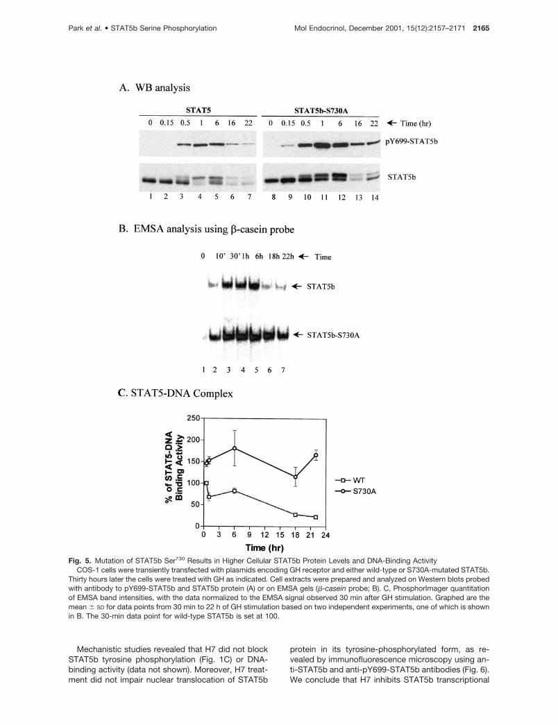
4C). Analysis of extracts prepared from GH-stimulatedcells revealed a higher level of Tyr699 phosphorylationin the STAT5b-S730A-transfected cells (Fig. 5A, upperpanel, lanes 8–14 vs. lanes 1–7). Correspondingly, ahigher DNA-binding activity was obtained at all timepoints examined (Fig. 5B). This increased activity ofSTAT5b-S730A largely reflects a higher level ofSTAT5b-S730A protein expression (Fig. 5A, lowerpanel, lanes 8–12 vs. lanes 1–5). Consequently, totalcellular STAT5 DNA-binding activity is higher in thecase of the STAT5b mutant (Fig. 5B), and as a resultthere is a differential in the DNA-binding activity foundin STAT5b-transfected cells vs. STAT5b-S730A-trans-fected cells that is maximal 22 h after GH stimulation(Fig. 5C). This finding provides an explanation for the
increased �-casein promoter activity of STAT5b-S730A at this time point (c.f. Fig. 4C).
Inhibitory Effect of H7 on STAT5b Activity
The serine kinase inhibitor H7 has previously beenused to support the proposed importance of STATserine phosphorylation for STAT transcriptional activ-ity (8, 18). We therefore investigated whether the re-porter-dependent changes in STAT5b activity seenwith STAT5b-S730A could also be observed in cellstreated with H7, which partially inhibits this serinephosphorylation reaction (c.f. Fig. 1). Table 2 shows,however, that H7 treatment of COS-1 cells transfectedwith wild-type STAT5b leads to a complete inhibition
Fig. 3. Requirement of STAT5a Serine Residues 725 and 779 for Full Activation of ntcp Promoter Activity in COS-1 and HepG2Cells
A, Relative luciferase activity was determined in cells transfected with the indicated STAT5a expression plasmids as describedin Fig. 2. B, Shown is a STAT5a Western blot of COS-1 cell extracts transfected as described in A and treated with GH, asindicated. Cell extracts were immunoprecipitated with anti-STAT5a antibody followed by sequential Western blotting withanti-pS725-STAT5a, anti-pY694-STAT5a, and anti-STAT5a antibody. STAT5a was not detected in untransfected COS-1 cells(lanes 1 and 2). Anti-pS725-STAT5a cross-reacted with a minor band migrating just below pS725-STAT5a (c.f. upper blot of B,lanes 7, 8, 11, and 12).
Park et al. • STAT5b Serine Phosphorylation Mol Endocrinol, December 2001, 15(12):2157–2171 2163
of GH-stimulated, STAT5b-dependent transcription ofthe ntcp-luciferase reporter gene, in contrast to the50% activity decrease seen with the STAT5b-S730Amutant. Moreover, H7 treatment led to a similar inhi-bition of the transcriptional activity of STAT5b-S730A,demonstrating that the inhibitory action of H7 is unre-lated to its effects on STAT5b serine phosphorylation.
Complete inhibition of STAT5b and STAT5b-S730A-stimulated transcription was also observed using thepZZ1 reporter (Table 2). Finally, a similar inhibitoryeffect of H7 on STAT5b-S730A transcriptional activitywas obtained in cells stimulated with GH for a shortertime period (3 h; c.f. 6 h GH stimulation in Fig. 5A; datanot shown).
Fig. 4. Influence of STAT5 Serine Mutations on PRL- or GH-Stimulated STAT5 Activity Assayed with �-casein and ntcp ReportersSTAT5 reporter plasmid, 4xNTCP-Luc or pZZ1-Luc, was cotransfected in COS-1 cells with wild-type STAT5a or STAT5b or the
indicated Ser to Ala mutated STAT5 expression plasmid. Cells were transfected with PRL receptor for PRL-activated reporteractivity (A) or with GH receptor for GH-activated reporter activity (B) and the internal control plasmid pRL-tk-Luc. Twenty-fourhours after transfection, cells were stimulated with 10 nM PRL for 18–20 h (A). For the time-course study (B and C), the cells werestimulated with GH for times ranging from 3–22 h. For all three panels, relative luciferase activities were then determined asdescribed in Fig. 2. Values shown are the mean � SD (n � 3).
2164 Mol Endocrinol, December 2001, 15(12):2157–2171 Park et al. • STAT5b Serine Phosphorylation
Mechanistic studies revealed that H7 did not blockSTAT5b tyrosine phosphorylation (Fig. 1C) or DNA-binding activity (data not shown). Moreover, H7 treat-ment did not impair nuclear translocation of STAT5b
protein in its tyrosine-phosphorylated form, as re-vealed by immunofluorescence microscopy using an-ti-STAT5b and anti-pY699-STAT5b antibodies (Fig. 6).We conclude that H7 inhibits STAT5b transcriptional
Fig. 5. Mutation of STAT5b Ser730 Results in Higher Cellular STAT5b Protein Levels and DNA-Binding ActivityCOS-1 cells were transiently transfected with plasmids encoding GH receptor and either wild-type or S730A-mutated STAT5b.
Thirty hours later the cells were treated with GH as indicated. Cell extracts were prepared and analyzed on Western blots probedwith antibody to pY699-STAT5b and STAT5b protein (A) or on EMSA gels (�-casein probe; B). C, PhosphorImager quantitationof EMSA band intensities, with the data normalized to the EMSA signal observed 30 min after GH stimulation. Graphed are themean � SD for data points from 30 min to 22 h of GH stimulation based on two independent experiments, one of which is shownin B. The 30-min data point for wild-type STAT5b is set at 100.
Park et al. • STAT5b Serine Phosphorylation Mol Endocrinol, December 2001, 15(12):2157–2171 2165
activity at a stage that is downstream of the STAT5bnuclear translocation and DNA-binding steps.
DISCUSSION
STAT5a can be constitutively phosphorylated onserine at two sites, Ser725 and Ser779, whereasSTAT5b, which lacks the COOH-terminal peptide se-quence corresponding to Ser779, can be phosphory-lated at a single serine, Ser730, in a manner that isinducible by either PRL (19) or GH (this report).Whereas mutation of STAT5a Ser725 had no significanteffect on STAT5 transcriptional activity, mutation ofthe corresponding STAT5b Ser730 is presently shownto modulate STAT5b’s transcriptional activity. Thismodulation is manifest as a decrease in transcriptiondriven by a promoter sequence containing four copiesof a STAT5-binding site derived from the ntcp gene,but leads to a time-dependent increase in transcrip-tion from a proximal promoter fragment of the �-casein gene. This latter increase is associated with ahigher cellular level of STAT5 DNA-binding activity inthe case of the Ser730-mutated STAT5b. These find-ings indicate that the functional consequences ofSTAT5b serine phosphorylation can vary from onepromoter to the next, suggesting that STAT5b serinephosphorylation may serve as a mechanism to differ-entially modulate the expression of STAT5 targetgenes. This promoter-dependent effect of STAT5bSer730 phosphorylation appears to reflect a change inSTAT5b’s intrinsic transcriptional activity, insofar asthe decrease in ntcp-luciferase reporter activity ob-served with Ser730-mutated STAT5b occurred in cellswhere there was an increase in STAT5 DNA-bindingactivity. Conceivably, serine phosphorylation maymodulate interactions between STAT5b and othertranscription factors bound to the same promoter, orperhaps may influence the recruitment of STAT-inter-acting coactivator and corepressor proteins (30, 31),which is likely to occur in a promoter-dependent man-ner. Indeed, interferon-�-activated STAT1 interacts
with the nuclear factor minichromosome mainte-nance-5 in a phospho-Ser727-dependent manner (9).Finally, changes in the trans-activation potential ofSTAT5 can also be achieved by mutations elsewherein the C-terminal region (e.g. increased activity ofSTAT5a-T757V) (32).
The increased DNA-binding activity of STAT5b-S730A compared with wild-type STAT5b appears tobe due at least in part to increased expression and/orstability of the Ser730-mutated STAT5 protein. Thisraises the possibility that phosphorylation of Ser730 inthe wild-type protein may enhance the turnover ofSTAT5b. Although the apparent increase in signalingof STAT5b-S730A at longer times of GH stimulationcould additionally involve a decrease in the rate ofSTAT5b tyrosine dephosphorylation, as was sug-gested earlier for STAT5a mutated at serine 725and/or 779 (20), this has not been established. Inde-pendent of the mechanism underlying this effect, theincreased cellular DNA-binding activity of STAT5b-S730A compared with wild-type STAT5b appears toaccount for the enhanced �-casein reporter activityseen at longer times after GH stimulation. Neverthe-less, ntcp reporter activity was decreased under thesesame conditions, highlighting the intrinsic differencesin the effects of Ser730 mutation on transcription of thetwo STAT5-responsive reporter genes, discussedabove.
Of the two STAT5a serine phosphorylation sites,residues 725 and 779, Ser779 appears to be moreimportant for maximal ntcp promoter activity. This issupported by the decrease in ntcp reporter activity incells transfected with STAT5a-S779A, but not in cellstransfected with STAT5a-S725A. This decrease can-not be explained by an effect of the Ser779 mutation onthe phosphorylation of STAT5a on Tyr694 or Ser725
(Fig. 3B). Both STAT5a serine residues are likely to beimportant, however, as suggested by the more sub-stantial decrease in ntcp reporter activity displayed bythe double mutant, STAT5a-S725,779A. Similarly, mu-tation of Ser779, alone or in combination with Ser725,led to an increase in �-casein promoter activity, a
HepG2 cells were transfected with GH receptor and a STAT5 reporter (either 4xNTCP-Luc or pZZ1-Luc, as indicated) togetherwith either wild-type STAT5b or STAT5b-S730A for 24 h. The cells were then pretreated with H7 for 1 h and stimulated with GHin the continued presence of H7, as indicated, for an additional 6 h. Relative luciferase reporter activity was then determined(mean � SD; n � 3). GH-stimulated activity in the absence of GH was set at 100. The reduced transcriptional activity ofSTAT5b-S730A with pZZ1-Luc seen at 6 h in the absence of H7 in this experiment (relative activity, 67% of wild-type STAT5b)is consistent with the 5-h data point shown in Fig. 4C.
2166 Mol Endocrinol, December 2001, 15(12):2157–2171 Park et al. • STAT5b Serine Phosphorylation
response that was also seen with STAT5b-S730A. Incontrast to STAT5b Ser730, whose phosphorylation isinducible, STAT5a Ser725 and Ser779 can both be con-stitutively phosphorylated in a variety of cells and tis-sues, including developing mammary gland in thecase of Ser779 (19, 20). It is unclear, however, whetherboth serine residues can be simultaneously phosphor-ylated in a given STAT5a molecule, leaving open thepossibility that phosphorylation of Ser725 may inhibit,and thereby help regulate, Ser779 phosphorylation andthe resultant phospho-Ser779-dependent transcrip-tional responses.
The stimulatory effects of the STAT5a-Ser779 andSTAT5b-Ser730 mutations on GH-induced �-caseinpromoter activity seen in the present study contrastwith the absence of a clear effect of these mutations inprevious studies in PRL-stimulated cells (19, 20). Thisdiscrepancy does not reflect differences in the stimu-lating hormone, as we were able to duplicate our find-ings in experiments in which STAT5a and STAT5b
were activated via the PRL receptor. Rather, it mayrelate to differences in the time-dependent effects ofthe Ser730 mutation on �-casein promoter activity doc-umented in the present study. Also of note, the twoprevious studies were carried out in COS-7 cells underconditions where only a 2- to 3-fold stimulation of�-casein promoter activity was achieved, which maylimit or mask the modulatory effects of mutating theSTAT5 serine phosphorylation sites. In contrast, thepresent studies were carried out in COS-1 cells underconditions where a 20- to 40-fold activation of the�-casein promoter was routinely achieved. Furtherstudy will be required to clarify this point.
The serine kinase inhibitor H7, which inhibits GH-stimulated STAT5b serine phosphorylation, stronglyinhibited STAT5b-dependent reporter gene activity. Astrong inhibitory action of H7 was also seen withSTAT5b-S730A, independent of whether activity wasassayed with the ntcp or �-casein promoter, indicating
Fig. 6. Immunofluorescence Analysis of GH-Stimulated STAT5b: Effect of H7 on Nuclear Localization of STAT5bShown are confocal microscope images of STAT5b (upper two sets of panels) and anti-pY699-STAT5b indirect immunoflu-
orescence (lower two sets of panels) indicating no effect of H7 treatment (B and D vs. A and C) on STAT5b nuclear translocationafter GH stimulation. Control CWSV-1 cells or CWSV-1 cells pretreated with H7 for 1 h were untreated or were treated with GHfor 30 min in the absence or presence of H7 as indicated. GH-treated cells were fixed and analyzed by immunofluorescencemicroscopy with anti-STAT5b antibody (A and B) or anti-pY699-STAT5b antibody (C and D). Propidium iodide was used to stainnuclei (lower set of images in each panel). STAT5b or pY699-STAT5b green fluorescence and propidium iodide red fluorescenceimages are presented as grayscale images prepared using Adobe Photoshop.
Park et al. • STAT5b Serine Phosphorylation Mol Endocrinol, December 2001, 15(12):2157–2171 2167
that the transcriptional inhibition effected by H7 isunrelated to the resultant changes in STAT5b Ser730
phosphorylation. Mechanistic studies revealed that H7does not interfere with GH-stimulated STAT5b ty-rosine phosphorylation, nuclear translocation, or DNA-binding activity, strongly suggesting that H7 exerts aspecific inhibitory action at the level of STAT5 trans-activation. This effect may thus be distinct from theprolonged signaling by the GH receptor-JAK2 com-plex after H7 treatment that we have previously de-scribed in GH-stimulated liver cells (24). Conceivably,H7 may inhibit STAT5-dependent transcription by al-tering the phosphorylation of a STAT5b-interacting co-activator that is required for the STAT transcriptionalresponse. Alternatively, the inhibition by H7 of STAT5transcriptional activity may be mechanistically linkedto the prolonged signaling by GH receptor-JAK2 toSTAT5b by way of a block in STAT5-stimulated tran-scription of feedback inhibitory regulators of GH re-ceptor-JAK2 signaling, such as SOCS/CIS proteins(33, 34). Further investigation is needed to address thisissue.
The kinase(s) that catalyze the constitutive phos-phorylation of STAT5a on Ser725 and Ser779 and thesignaling pathways that lead to the inducible phos-phorylation of STAT5b on Ser730 in response to GH orPRL stimulation remain to be identified. Inhibitor stud-ies suggest a role for a MAPK-like activity in the con-stitutive phosphorylation of STAT5a on Ser725, but notfor the PRL-inducible phosphorylation of STAT5b onthe corresponding Ser730 (19). Interestingly, in cells inwhich the constitutive phosphorylation of STAT5aSer725 is blocked by the MAPK kinase inhibitorPD98059, PRL can induce phosphorylation at that site(19), demonstrating that Ser725 is intrinsically respon-sive to PRL stimulation, in a manner that is analogousto the inducible phosphorylation of Ser730 in the caseof STAT5b. The additional site of STAT5a phosphor-ylation, at residue Ser779, is within the COOH-terminal20 amino acids of STAT5a, where the two STAT5proteins are highly divergent in sequence. This residueis thus absent from STAT5b. In vitro phosphorylationof STAT5a by MAPK is strongly inhibited by mutationof Ser779, as is the interaction of STAT5a with theMAPK ERK1 and ERK2 (21), suggesting a role forMAPK in this phosphorylation reaction as well. In otherstudies, carried out in a different cell model, phosphor-ylation of STAT5a at Ser779 was not blocked by inhib-itors of MAPK or PI3K (20). Interestingly, Ser779 occurswithin a sequence (RLSPPA) that corresponds to aconsensus motif for phosphorylation by PKA, but notby nine other serine protein kinases, as revealed bycomputer analysis using the web-based Phospho-Base program (35). Accordingly, further investigationof the role of PKA/cAMP-dependent signaling path-ways in the phosphorylation of STAT5a at this COOH-terminal site may be warranted.
STAT5a and STAT5b play distinct physiologicalroles in mediating hormonal responses to PRL(STAT5a) and GH (STAT5b) in the mammary gland and
liver, respectively (36, 37). Although this differentialendocrine function may largely reflect the distinct tis-sue distributions of the two STAT5 forms, there isincreasing evidence that the biological properties ofSTAT5a and STAT5b, although very similar, are dis-tinguishable in several important ways. STAT5a andSTAT5b not only display biochemical differences inapparent DNA binding specificity (38, 39) and propen-sity to bind to DNA as tetramers (STAT5a � STAT5b)(29, 40), but they exhibit potentially important differ-ences in their regulation by serine phosphorylation.Thus, the phosphorylation of STAT5a vs. STAT5b onSer725/730 is not only subject to differential regulation(constitutive phosphorylation of STAT5a vs. induciblephosphorylation of STAT5b), but leads to modulatoryeffects on gene transcription only in the case ofSTAT5b. In the case of STAT5a, such a modulatoryeffect requires phosphorylation on Ser779, a residueunique to this STAT5 form.
MATERIALS AND METHODS
Plasmids and Preparation of STAT5 Mutant Constructs
STAT5 plasmids containing site-specific mutations of serineto alanine (S725A and/or S779A for STAT5a, S730A forSTAT5b) or tyrosine to phenylalanine (Y694F for STAT5a,Y699F for STAT5b) were prepared from double-strandedplasmid DNA using the QuickChange site-directed mutagen-esis kit (Stratagene, La Jolla, CA) and oligonucleotide primersdesigned to introduce each mutation, as described previ-ously (19). Site-specific mutations were verified by DNA se-quence analysis. Expression plasmids for mouse STAT5aand STAT5b (Dr. L. Hennighausen, NIH, Bethesda, MD), ratGH receptor (Dr. N. Billestrup, Hagedon Research Institute,Gentofe, Denmark), and human PRL receptor (Dr. P. Kelly,INSERM, Paris, France) were obtained from the indicatedsources. Luciferase reporter constructs containing either fourcopies of a STAT5 binding site derived from the promoter ofthe rat ntcp gene (4�NTCP-Luc) or the �-casein gene pro-moter (nucleotides �344 to �1; pZZ1-Luc) were respectivelyprovided by Drs. M. Vore (University of Kentucky, Lexington,KY) and B. Groner (Institute for Experimental Cancer Re-search, Freiburg, Germany).
Cell Culture and Transfections
COS-1 and HepG2 cells were maintained in DMEM contain-ing 10% FBS, 50 U/ml penicillin, and 50 �g/ml streptomycin.For transient transfections, cells were seeded in 24-wellplates at a density of 1.3 � 105 HepG2 cells/well or 5 � 104
COS-1 cells/well. Fugene 6 transfection reagent (Roche Mo-lecular Biochemicals, Indianapolis, IN) was used as de-scribed in the manufacturer’s protocol, at a ratio of 1.3:1 ofFugene 6/DNA (vol/wt). Each well received a total of 600 ngDNA, including 150–200 ng luciferase reporter plasmid, 50 ngGH receptor, and 100–200 ng STAT5 expression plasmid.pRL-tk-Luc plasmid (Renilla luciferase; 50 ng DNA) was in-cluded as an internal control for transfection efficiency.Twenty-four hours after transfection, the cells were treatedwith rat GH (200 ng/ml) or rat PRL (10 nM) for an additional18–24 h unless specified otherwise. H7 (200 �M) was in-cluded as indicated. Total cell extracts were prepared using1� lysis buffer (Promega Corp., Madison, WI) for measuringluciferase activities. For Western blot and EMSA analysis,
2168 Mol Endocrinol, December 2001, 15(12):2157–2171 Park et al. • STAT5b Serine Phosphorylation
total cell lysates were centrifuged for 30 min at 15,000 � g.Firefly and Renilla luciferase activities were measured using aDual Reporter Assay System (Promega Corp.) and a Mono-light 2010 luminometer (Analytical Luminescence Laboratory,San Diego, CA). Data shown in the individual figures arerelative values based on normalized luciferase activity (i.e.firefly/Renilla luciferase activities; mean � SD for threereplicates).
Growth and passage of CWSV-1 cells was carried out asdescribed previously (14). For serine phosphorylation stud-ies, CWSV-1 cells were stimulated with GH at 200 ng/ml inthe presence or absence of H7 (200 �M) for varying periods oftime. Total cell extracts were prepared in lysis buffer contain-ing 20 mM HEPES (pH 7.9); 1% Triton X-100; 1 mM each ofEDTA, EGTA, Na3VO4, Na2P2O7, and dithiothreitol; 0.5 mM
phenylmethylsulfonylfluoride; and 1 �g/ml each of pepstatin,antipain, and leupeptin. Total cell extracts were passedthrough a 27-gauge needle seven times, adjusted to 150 mM
NaCl, and centrifuged at 15,000 � g for 30 min at 4 C. Proteinconcentrations were determined using the Dc detergent pro-tein assay kit (Bio-Rad Laboratories, Inc., Hercules, CA).
EMSA Analysis
Total cell extracts (5 �g) were assayed for STAT5 DNA-binding activity using a �-casein STAT5 response elementprobe (14). Gels were exposed to PhosphorImager platesovernight, followed by quantitation of radioactivity intensityusing a Molecular Dynamics, Inc. PhosphorImager and Im-ageQuant software (Sunnyvale, CA).
Western Blotting and Immunoprecipitation
Total cell extracts (20–30 �g) were electrophoresed on 7.5%Laemmli SDS gels, electrotransferred to nitrocellulose mem-branes, and then probed with anti-STAT5b antibodies (cata-logue no. sc-835, Santa Cruz Biotechnology, Inc., SantaCruz, CA). Blocking and probing conditions were describedpreviously (41). Probing with anti-pY699-STAT5b antibody(Cell Signaling Technology, Beverly, MA) was performed witha 1-h incubation of the blot at room temperature in TST buffer[10 mM Tris-HCl (pH 7.6), 0.1% Tween 20, and 100 mM NaCl]containing 5% nonfat dry milk, followed by incubation withanti-pY699 antibody (1:1000 dilution) in 5% BSA-TST bufferovernight at 4 C. Washings were carried out as specified bythe manufacturer. For STAT5 immunoprecipitation, CWSV-1cells grown on 100-mm dishes were solubilized in 1 ml lysisbuffer [10 mM Tris-HCl (pH 7.6), 5 mM EDTA, 50 mM NaCl, 30mM Na2P2O7 50 mM NaF, 1 mM Na3VO4, 1% Triton X-100,and 1 mM phenylmethylsulfonylfluoride] in the presence ofphosphatase inhibitors (14). Clarified total cell extracts wereincubated for 2 h on ice with 2 �l polyclonal rabbit anti-STAT5b antiserum (antibody raised against a peptide corre-sponding to amino acid residues 776–786 of mouse STAT5bwas obtained from Dr. L. Hennighausen, NIH) (42). Immunecomplexes were captured with protein A-Sepharose beads(Pharmacia Biotech, Piscataway, NJ), electrophoresed on7.5% Laemmli-SDS gels, and then transferred onto nitrocel-lulose membranes (Millipore Corp., Bedford, MA). Mem-branes were blocked for 1 h at 37 C with 5% nonfat dry milkin TST buffer. Incubations with site-specific antiphospho-serine STAT5 antibody (anti-pS730) (19) were carried out for16 h at a dilution of 1:5000 in the cold-room. Anti-pS730-STAT5 antibody was raised against the phosphopeptideDQAP[pS]PAVC, corresponding to amino acid residues 726–734 of human STAT5b (19). Antibody binding was visualizedon x-ray film by enhanced chemiluminescence using the ECLkit from Amersham Pharmacia Biotech (Arlington Heights, IL;anti-STAT5b and anti-pS730-STAT5b) or the SuperSignalECL kit from Pierce Chemical Co. (Rockford, IL; anti-pY699-STAT5b).
Immunofluorescence Studies
CWSV-1 cells were seeded at about 60% confluence ontofour-well chamber slides (catalog no. 62409-294, VWR Sci-entific Products, Boston, MA) in RPCD medium (14) contain-ing 3% FBS and allowed to adhere overnight. The mediumwas then replaced with serum-free RPCD medium. The fol-lowing day, the cells were pretreated with H7 (200 �M) for 1 has indicated, then treated with GH (200 ng/ml) and H7 for 30min. Cells were rinsed with ice-cold PBS and fixed with 100%MeOH for 20 min at �20 C. Fixed cells were blocked with 3%charcoal-stripped calf serum in PBS for 1 h at room temper-ature and then incubated with anti-STAT5b antibody (1:500dilution; Santa Cruz Biotechnology, Inc.) in blocking solutionovernight at room temperature. For anti-pY699-STAT5b im-munostaining, fixed cells were blocked with 5.5% charcoal-stripped calf serum in TBST buffer [50 mM Tris-HCl (pH 7.4),150 mM NaCl, and 0.1% Triton X-100] for 1 h at room tem-perature and then incubated for 24 h at 4 C with anti-pY699-STAT5b antibody (1:500 dilution; Cell Signaling Technology,Beverly, MA) in TBS buffer [50 mM Tris-HCl (pH 7.4) and 150mM NaCl] containing 3% BSA. The samples were thenwashed (three times, 5 min/wash) with PBS containing 3%calf serum for anti-STAT5b and with TBST for anti-pY699-STAT5b antibody. Cells were then incubated for 1 h at 37 Cwith fluorescein isothiocyanate-conjugated goat antirabbitIgG antibody (1 �g/ml; Molecular Probes, Inc., Eugene, OR).Cells were counterstained with 50 ng/ml propidium iodide(Sigma) to localize nuclei. For confocal analysis, immunoflu-orescent cells were scanned with an BX-50 confocal laserscanning microscope (Olympus Corp., New Hyde Park, NY)equipped with a �60 objective (Carl Zeiss, New York, NY).
Acknowledgments
The authors thank Drs. L. Hennighausen, N. Billestrup, P.Kelly, M. Vore, B. Groner, and F. Lemaigre for providingplasmid DNAs and antibodies.
Received May 9, 2001. Accepted August 28, 2001.Address requests for reprints to: Dr. David J. Waxman,
Department of Biology, Boston University, Boston, Massa-chusetts 02215. E-mail: [email protected].
This work was supported in part by NIH Grant DK-33765(to D.J.W.).
REFERENCES
1. Darnell JEJ 1997 STATs and gene regulation. Science277:1630–1635
2. Waxman DJ, Frank SJ 2000 Growth hormone action:signaling via a JAK/STAT-coupled receptor. In: Conn PM,Means A, eds. Principles of molecular regulation.Totowa: Humana Press; 55–83
3. Aoki N, Matsuda T 2000 A cytosolic protein-tyrosinephosphatase PTP1B specifically dephosphorylates anddeactivates prolactin-activated STAT5a and STAT5b.J Biol Chem 275:39718–39726
4. Wen Z, Zhong Z, Darnell Jr JE 1995 Maximal activation oftranscription by Stat 1 and Stat3 requires both tyrosineand serine phosphorylation. Cell 82:241–250
5. Boulton TG, Zhong Z, Wen Z, Darnell Jr JE, Stahl N,Yancopoulos GD 1995 STAT3 activation by cytokinesutilizing gp130 and related transducers involves a sec-ondary modification requiring an H7-sensitive kinase.Proc Natl Acad Sci USA 92:6915–6919
6. Ceresa BP, Pessin JE 1996 Insulin stimulates the serinephosphorylation of the signal transducer and activator of
Park et al. • STAT5b Serine Phosphorylation Mol Endocrinol, December 2001, 15(12):2157–2171 2169
8. Lutticken C, Coffer P, Yuan J, Schwartz C, CaldenhovenE, Schindler C, Kruijer W, Heinrich PC, Horn F 1995Interleukin-6-induced serine phosphorylation of tran-scription factor APRF: evidence for a role in interleukin-6target gene induction. FEBS Lett 360:137–143
9. Zhang JJ, Zhao Y, Chait BT, Lathem WW, Ritzi M, Knip-pers R, Darnell JE 1998 Ser727-dependent recruitment ofMCM5 by Stat1� in IFN-�-induced transcriptional acti-vation. EMBO J 17:6963–6971
10. Kim H, Baumann H 1997 The carboxyl-terminal region ofSTAT3 controls gene induction by the mouse haptoglo-bin promoter. J Biol Chem 272:14571–14579
11. Chung J, Uchida E, Grammer TC, Blenis J 1997 STAT3serine phosphorylation by ERK-dependent and -inde-pendent pathways negatively modulates its tyrosinephosphorylation. Mol Cell Biol 17:6508–6516
12. Grimley PM, Dong F, Rui H 1999 Stat5a and Stat5b:fraternal twins of signal transduction and transcriptionalactivation. Cytokine Growth Factor Rev 10:131–157
13. Ram PA, Park SH, Choi HK, Waxman DJ 1996 Growthhormone activation of Stat 1, Stat 3, and Stat 5 in rat liver.Differential kinetics of hormone desensitization andgrowth hormone stimulation of both tyrosine phosphor-ylation and serine/threonine phosphorylation. J BiolChem 271:5929–5940
14. Gebert CA, Park SH, Waxman DJ 1997 Regulation ofSTAT5b activation by the temporal pattern of growthhormone stimulation. Mol Endocrinol 11:400–414
15. Dinerstein-Cali H, Ferrag F, Kayser C, Kelly PA, Postel-Vinay M 2000 Growth hormone (GH) induces the forma-tion of protein complexes involving stat5, erk2, shc andserine phosphorylated proteins. Mol Cell Endocrinol 166:89–99
16. Kirken RA, Malabarba MG, Xu J, Liu X, Farrar WL, Hen-nighausen L, Larner AC, Grimley PM, Rui H 1997 Pro-lactin stimulates serine/tyrosine phosphorylation and for-mation of heterocomplexes of multiple Stat5 isoforms inNb2 lymphocytes. J Biol Chem 272:14098–14103
17. Wartmann M, Cella N, Hofer P, Groner B, Liu X, Hen-nighausen L, Hynes NE 1996 Lactogenic hormone acti-vation of Stat5 and transcription of the �-casein gene inmammary epithelial cells is independent of p42 ERK2mitogen-activated protein kinase activity. J Biol Chem271:31863–31868
18. Beadling C, Ng J, Babbage JW, Cantrell DA 1996 Inter-leukin-2 activation of STAT5 requires the convergent ac-tion of tyrosine kinases and a serine/threonine kinasepathway distinct from the Raf1/ERK2 MAP kinase path-way. EMBO J 15:1902–1913
19. Yamashita H, Xu J, Erwin RA, Farrar WL, Kirken RA, RuiH 1998 Differential control of the phosphorylation state ofproline-juxtaposed serine residues Ser725 of Stat5a andSer730 of Stat5b in prolactin-sensitive cells. J Biol Chem273:30218–30224
20. Beuvink I, Hess D, Flotow H, Hofsteenge J, Groner B,Hynes NE 2000 Stat5a serine phosphorylation. Serine779 is constitutively phosphorylated in the mammarygland, and serine 725 phosphorylation influences prolac-tin-stimulated in vitro DNA binding activity. J Biol Chem275:10247–10255
21. Pircher TJ, Petersen H, Gustafsson JA, Haldosen LA1999 Extracellular signal-regulated kinase (ERK) inter-acts with signal transducer and activator of transcription(STAT) 5a. Mol Endocrinol 13:555–565
22. Pircher TJ, Flores-Morales A, Mui AL, Saltiel AR, Nor-stedt G, Gustafsson JA, Haldosen LA 1997 Mitogen-activated protein kinase kinase inhibition decreasesgrowth hormone stimulated transcription mediated bySTAT5. Mol Cell Endocrinol 133:169–176
23. Davey HW, Wilkins RJ, Waxman DJ 1999 STAT5 signal-ling in sexually dimorphic gene expression and growthpatterns. Am J Hum Genet 65:959–965
24. Gebert CA, Park SH, Waxman DJ 1999 Termination ofgrowth hormone pulse-induced STAT5b signaling. MolEndocrinol 13:38–56
25. Ganguly TC, O’Brien ML, Karpen SJ, Hyde JF, Suchy FJ,Vore M 1997 Regulation of the rat liver sodium-depen-dent bile acid cotransporter gene by prolactin. Mediationof transcriptional activation by Stat5. J Clin Invest 99:2906–2914
26. Delesque-Touchard N, Park SH, Waxman DJ 2000 Syn-ergistic action of hepatocyte nuclear factors 3 and 6 onCYP2C12 gene expression and suppression by growthhormone-activated STAT5b. Proposed model for femalespecific expression of CYP2C12 in adult rat liver. J BiolChem 275:34173–34182
27. Gouilleux F, Wakao H, Mundt M, Groner B 1994 Prolactininduces phosphorylation of Tyr694 of Stat 5 (MGF), aprerequisite for DNA binding and induction of transcrip-tion. EMBO J 13:4361–4369
28. Bergad PL, Shih HM, Towle HC, Schwarzenberg SJ,Berry SA 1995 Growth hormone induction of hepaticserine protease inhibitor 2.1 transcription is mediated bya Stat5-related factor binding synergistically to two �-activated sites. J Biol Chem 270:24903–24910
29. Soldaini E, John S, Moro S, Bollenbacher J, Schindler U,Leonard WJ 2000 DNA binding site selection of dimericand tetrameric Stat5 proteins reveals a large repertoire ofdivergent tetrameric Stat5a binding sites. Mol Cell Biol20:389–401
30. Zhu M, John S, Berg M, Leonard WJ 1999 Functionalassociation of Nmi with Stat5 and Stat1 in IL-2- andIFN�-mediated signaling. Cell 96:121–130
31. Pfitzner E, Jahne R, Wissler M, Stoecklin E, Groner B1998 p300/CREB-binding protein enhances the prolac-tin-mediated transcriptional induction through direct in-teraction with the transactivation domain of Stat5, butdoes not participate in the Stat5-mediated suppressionof the glucocorticoid response. Mol Endocrinol 12:1582–1593
32. Gowri PM, Ganguly TC, Cao J, Devalaraja MN, Groner B,Vore M 2001 Conversion of threonine 757 to valine en-hances stat5a transactivation potential. J Biol Chem 276:10485–10491
33. Ram PA, Waxman DJ 1999 SOCS/CIS protein inhibitionof growth hormone-stimulated STAT5 signaling by mul-tiple mechanisms. J Biol Chem 274:35553–35561
34. Hansen JA, Lindberg K, Hilton DJ, Nielsen JH, BillestrupN 1999 Mechanism of inhibition of growth hormone re-ceptor signaling by suppressor of cytokine signaling pro-teins. Mol Endocrinol 13:1832–1843
35. Kreegipuu A, Blom N, Brunak S 1999 PhosphoBase, adatabase of phosphorylation sites: release 2.0. NucleicAcids Res 27:237–239
36. Liu X, Robinson GW, Wagner KU, Garrett L, Wynshaw-Boris A, Hennighausen L 1997 Stat5a is mandatory foradult mammary gland development and lactogenesis.Genes Dev 11:179–186
37. Udy GB, Towers RP, Snell RG, Wilkins RJ, Park SH, RamPA, Waxman DJ, Davey HW 1997 Requirement ofSTAT5b for sexual dimorphism of body growth rates andliver gene expression. Proc Natl Acad Sci USA 94:7239–7244
38. Boucheron C, Dumon S, Santos SC, Moriggl R, Hen-nighausen L, Gisselbrecht S, Gouilleux F 1998 A singleamino acid in the DNA binding regions of STAT5A andSTAT5B confers distinct DNA binding specificities. J BiolChem 273:33936–33941
39. Ehret GB, Reichenbach P, Schindler U, Horvath CM, FritzS, Nabholz M, Bucher P 2001 DNA binding specificity ofdifferent STAT proteins. Comparison of in vitro specificitywith natural target sites. J Biol Chem 276:6675–6688
2170 Mol Endocrinol, December 2001, 15(12):2157–2171 Park et al. • STAT5b Serine Phosphorylation
40. Verdier F, Rabionet R, Gouilleux F, Beisenherz-Huss C,Varlet P, Muller O, Mayeux P, Lacombe C, GisselbrechtS, Chretien S 1998 A sequence of the CIS gene promoterinteracts preferentially with two associated STAT5Adimers: a distinct biochemical difference betweenSTAT5A and STAT5B. Mol Cell Biol 18:5852–5860
41. Park SH, Liu X, Hennighausen L, Davey HW, WaxmanDJ 1999 Distinctive roles of STAT5a and STAT5b in
sexual dimorphism of hepatic P450 gene expression.Impact of Stat5a gene disruption. J Biol Chem 274:7421–7430
42. Liu X, Robinson GW, Gouilleux F, Groner B, Hen-nighausen L 1995 Cloning and expression of Stat 5 andan additional homologue (Stat 5b) involved in prolactinsignal transduction in mouse mammary tissue. Proc NatlAcad Sci USA 92:8831–8835
2002 Prolactin Gordon Research Conference
The next Gordon Research Conference on Prolactin will be held in Ventura, California, fromJan. 27–Feb. 1, 2002. As has been the case for the past several years, we will also addresssimilar issues for growth hormone. There are two highlighted presentations: The meetingbegins with a debate on Sunday evening entitled, “Do prolactin and growth hormone causecancer?” M. G. Rosenfeld will end the meeting with the Keynote Address on Thursdayevening, speaking on “Genetic control of pituitary development.” Morning and eveningsessions during the week will cover various aspects of PRL and GH secretion, receptors,and actions, including mouse models, microarray analysis and proteomics, PRL and GHphysiology, pituitary development and regulation, agonists, antagonists and assays, effects inperipheral tissues, behavior and lifestyles, and PRL and GH signaling. There will also be eightPlatform Presentations by graduate students, postdoctoral fellows, or junior faculty, who willpresent late-breaking, up-to-the-minute results from their submitted abstracts. The remainingabstracts will be presented in two poster sessions. For more information on the full program orGRC registration, check the Web site at http://www.grc.org.


![REVIEW Open Access Regulation of skeletal muscle growth by ... · IGF1-Akt pathway was not investigated [14]. Activa-tion of IRS types is inhibited by phosphorylation of serine residues](https://static.documents.pub/doc/80x56/6081215dbbc1e17f4e2fb13d/review-open-access-regulation-of-skeletal-muscle-growth-by-igf1-akt-pathway.jpg)
![An assessment of LRRK2 serine 935 phosphorylation in …Aug 29, 2019 · 3 [8], all known LRRK2 kinase inhibitors reduce pS935 in cellular and animal studies [9, 10] indicating its](https://static.documents.pub/doc/80x56/5fb461b5874e44287f652419/an-assessment-of-lrrk2-serine-935-phosphorylation-in-aug-29-2019-3-8-all.jpg)

![RESEARCH ARTICLE Open Access The CRE1 carbon ......sites [10,11]. In addition, phosphorylation of a serine in a conserved short stretch within an acidic domain of T. reesei CRE1 has](https://static.documents.pub/doc/80x56/6104a968f60cbe40f01e8cf1/research-article-open-access-the-cre1-carbon-sites-1011-in-addition.jpg)


