DISEASES OF AQUATIC ORGANISMS Dis, aquat. Org. Published September 9 Serological differences among isolates of viral haemorrhagic septicaemia virus detected by neutralizing monoclonal and polyclonal antibodies N. J. Olesen, N. Lorenzen, P. E. V. Jsrgensen National Veterinary Laboratory, Hangavej 2, DK-8200 Arhus N. Denmark ABSTRACT: The serological variation among 127 isolates of viral haemorrhagic septicaemia virus (VHSV) was examined by plaque neutralization tests (PNT) using a panel of 4 neutralizing monoclonal antibodies (MAbs) and 1 neutralizing rabbit antiserum (PAb). Three distinct neutralization patterns were observed. All isolates in the largest group (I) were neutralized at high titres by the MAbs and the PAb. This group included isolates representing the previously described 'Serotypes' 1 and 2. The iso- lates in Group I1 were efficiently neutralized by 1 of the MAbs and the PAb but not, unless at low titres, by the remaining 3 MAbs. Isolates represented by previously described 'serotype' 3 were in this group. The isolates in Group 111 were not neutralized at all by any of the MAbs and not, or only moderately. neutralized by the PAb. Isolates with this reaction pattern have become the most frequent ones from VHS outbreaks in Denmark in recent years. Pronounced crossreaction between Groups I, I1 & 111 in Western blotting using polyclonal rabbit antibodies combined with the ability of 1 antiserum to neutral- ize 120 out of the 127 isolates indicate that the polyclonal antibodies used define only 1 serotype of VHSV. Groups I to 111 should be regarded as subtypes. The present panel of antibodies did not allow a distinction between pathogenic and nonpathogenic isolates. No relationship was observed between host species and the reaction pattern as all 3 reaction patterns were present among the 5 isolates orig- inating from fish species other than rainbow trout Oncorhynchus mykiss. INTRODUCTION Viral haemorrhagic septicaemia (VHS) is the most serious viral disease affecting the production of rain- bow trout Oncorhynchus mykiss in Europe. The causa- tive agent, viral haemorrhagic septicaemia virus (VHSV) or Egtved virus (Jensen 1965),is a member of the Lyssavirus genus of the family Rhabdoviridae (McAllister & Wagner 1975). The reference strain F1, isolated from rainbow trout in Denmark by Jensen in 1962 was the first isolate of the virus (Jensen 1965). Three 'serotypes' of VHSV have been described: Type 1, represented by Strain F1; Type 2, represented by the Hededam isolate, both isolated from Danish rainbow trout (Jsrgensen 1972, 1980),and Type 3, rep- resented by the French strain 23/75 isolated from brown trout Salmo trutta far10 by Kinkelin & Le Berre (1977). These 'serotypes' were defined by means of dif- 0 Inter-Research 1993 ferent versions of the neutralization test; however, in spite of the existence of serological differences, most polyclonal antisera made against Strain F1 cross- reacted strongly with the other type strains in 50 % plaque neutralization tests (PNT) (Jsrgensen 1974, 1980, this study), immunofluorescence (IF) assays (Jargensen 1972, Meier & Jargensen 1980), enzyme- linked immunosorbent assays (ELISA) (Sanz & Col1 1992) and Western blotting analyses (this study). More recently, isolates of VHSV which are not iden- tifiable by neutralization tests but which react by means of IF have been described (Ahne et al. 1986), and such isolates have appeared with increasing fre- quency in Denmark over the last few years. The present work was performed to determine if an improved picture of the serological variation among isolates of VHSV could be obtained by PNT using a panel of 4 neutralizing monoclonal antibodies (MAbs)
Transcript
DISEASES OF AQUATIC ORGANISMS Dis, aquat. Org.
Published September 9
Serological differences among isolates of viral haemorrhagic septicaemia virus detected by
neutralizing monoclonal and polyclonal antibodies
N. J. Olesen, N. Lorenzen, P. E. V. Jsrgensen
National Veterinary Laboratory, Hangavej 2, DK-8200 Arhus N. Denmark
ABSTRACT: The serological variation among 127 isolates of viral haemorrhagic septicaemia virus (VHSV) was examined by plaque neutralization tests (PNT) using a panel of 4 neutralizing monoclonal antibodies (MAbs) and 1 neutralizing rabbit antiserum (PAb). Three distinct neutralization patterns were observed. All isolates in the largest group (I) were neutralized at high titres by the MAbs and the PAb. This group included isolates representing the previously described 'Serotypes' 1 and 2. The iso- lates in Group I1 were efficiently neutralized by 1 of the MAbs and the PAb but not, unless at low titres, by the remaining 3 MAbs. Isolates represented by previously described 'serotype' 3 were in this group. The isolates in Group 111 were not neutralized at all by any of the MAbs and not, or only moderately. neutralized by the PAb. Isolates with this reaction pattern have become the most frequent ones from VHS outbreaks in Denmark in recent years. Pronounced crossreaction between Groups I, I1 & 111 in Western blotting using polyclonal rabbit antibodies combined with the ability of 1 antiserum to neutral- ize 120 out of the 127 isolates indicate that the polyclonal antibodies used define only 1 serotype of VHSV. Groups I to 111 should be regarded as subtypes. The present panel of antibodies did not allow a distinction between pathogenic and nonpathogenic isolates. No relationship was observed between host species and the reaction pattern as all 3 reaction patterns were present among the 5 isolates orig- inating from fish species other than rainbow trout Oncorhynchus mykiss.
INTRODUCTION
Viral haemorrhagic septicaemia (VHS) is the most serious viral disease affecting the production of rain- bow trout Oncorhynchus mykiss in Europe. The causa- tive agent, viral haemorrhagic septicaemia virus (VHSV) or Egtved virus (Jensen 1965), is a member of the Lyssavirus genus of the family Rhabdoviridae (McAllister & Wagner 1975). The reference strain F1, isolated from rainbow trout in Denmark by Jensen in 1962 was the first isolate of the virus (Jensen 1965).
Three 'serotypes' of VHSV have been described: Type 1, represented by Strain F1; Type 2, represented by the Hededam isolate, both isolated from Danish rainbow trout (Jsrgensen 1972, 1980), and Type 3, rep- resented by the French strain 23/75 isolated from brown trout Salmo trutta far10 by Kinkelin & Le Berre (1977). These 'serotypes' were defined by means of dif-
0 Inter-Research 1993
ferent versions of the neutralization test; however, in spite of the existence of serological differences, most polyclonal antisera made against Strain F1 cross- reacted strongly with the other type strains in 50 % plaque neutralization tests (PNT) (Jsrgensen 1974, 1980, this study), immunofluorescence (IF) assays (Jargensen 1972, Meier & Jargensen 1980), enzyme- linked immunosorbent assays (ELISA) (Sanz & Col1 1992) and Western blotting analyses (this study).
More recently, isolates of VHSV which are not iden- tifiable by neutralization tests but which react by means of IF have been described (Ahne et al. 1986), and such isolates have appeared with increasing fre- quency in Denmark over the last few years.
The present work was performed to determine if an improved picture of the serological variation among isolates of VHSV could be obtained by PNT using a panel of 4 neutralizing monoclonal antibodies (MAbs)
164 Dis, aquat. Org.
and 1 neutralizing polyclonal antiserum (PAb) of rabbit origin. A total of 127 VHSV isolates were examined, in- cludlng isolates of the virus which had not reacted with the antiserum to Strain F1 used in routine neutraliza- tion tests, as well as a number of isolates from fish other than rainbow trout.
MATERIALS AND METHODS
Cells and virus. Epithelioma paprllosum cyprini (EPC) (Fija.n et al. 1983) and bluegill fry (BF-2) (Wolf et al. 1966) cell lines were grown in Eagles MEM supple- mented with 10 % foetal bovine serum, Tris buffer and antibiotics in standard concentrations. Among 127 virus isolates examined, 117 were of Danish origin, isolated during the previous 30 yr at the National Veterinary Laboratory (NVL), with the exception of the isolate from Atlantic cod Gadus morhua (Jensen et al. 1979, Jurgensen. & Olesen 1987) which was received from Dr S. Mellergird, Danish Institute for Fisheries a n d Marine Research, Copenhagen, Denmark. One isolate, DK-87, although isolated at NVL, originated from brown trout received in 1969 from Dr P. Ghittino, Instituto Zooprofilattico, Turin (Jargensen 1980). The isolates were selected to ensure diversity in terms of efficiency of neutralization by a reference rabbit anti- serum to the F1 strain, geographic location, host spe- cies, and year of isolation.
The Danish isolates except for the Atlantic cod iso- late originated from 69 freshwater and 12 seawater farms producing rainbow trout or, in a few cases, brown trout. Isolates from farms on, all Dan.ish streams where VHS had occurred during the last 5 yr (22 streams) and from all new VHS outbreaks in the period from 1 January 1991 to 1 June 1992 (34 isolates) were included among the samples. Among the Danish iso- lates were the type strains F1 (Jensen 1965) and H e (isolate DK-61) (Jerrgensen 1972). One isolate from brown trout, 23/75, representing Type 3, was received from Dr P, d e Kinkelin, lnstitut National d e la Recherche Agronomique, Paris, France. Six isolates were received from Prof W. Ahne, University of Munich, including isolates 49/82 and 67/82 (Ahne et al. 1986) as well as a VHSV isolate from turbot Scophthalmus maximus (Schlotfeld t et al. 1991) Two isolates, one from chinook salmon Oncorhynchus tshawytscha, obtained by K. Hopper (KHV) (Hopper 1989) and one from coho salmon Oncorhynchus ki- sutch, obtained by R. Brunson (RBV) (Brunson et al. 1989), were received from Dr J. W~nton , National Fisheries Research Ccnter, Seattle, USA. One isolate, L59xC from eel Anguilla anguilla (Castric et al. 1992), was received from Dr J Castric, Laboratoire d e Pathologie des Animaux Aquatiques, Brest, France. All
isolates were of salmonid origin except for the 3 iso- lates from cod, eel, and turbot. Apart from certain pas- sage levels of F l , He and the eel virus, the isolates were not cloned before the study. All isolates were passed at least once in both BF-2 and EPC cell lines prior to use. Prior to neutralization, the isolates were plaque-titrated in microplates with EPC cells using overlay medium containing 0.5 % methyl cel.lulose.
Antibodies. Development of mouse hybridoma cell lines producing VHSV-neutralizing MAbs has been described (Lorenzen et al. 1990). Each MAb was quan- tified by means of a sandwich ELISA (Lorenzen et al. 1988) and the IgG concentration adjusted to ap- proximately 5 pg ml-'. Neutralizing rabbit antisera to VHSV Strain F1 were prepared using pelleted virus (K697-2) as described by Jerrgensen (1974) or gradient purified virus (K59) as described by Olesen et al. (1991a). The sera were treated for 30 min at 56 "C be- fore use. A panel of neutralizing reagents containing the polyclonal rabbit antiserum K697-2 and the MAbs 3FlH10, 3F2D4, 3F5A1 & 3F3B4 was assembled. The MAb 3FlH10 was identical to MAb I previously de- scribed by Lorenzen et al. (1990), MAb 3F2D4 be- longed to the IgG2b subclass, and MAbs 3F5A1 & 3F3B4 belonged to the IgG1 subclass.
Rabbit antiserum to the Coleman strain of infectious hematopoietic necrosis virus (IHNV) was prepared as described for VHSV (Olesen et al. 1991a).
50 % plaque neutralization test (PNT). All virus isolates were examined by PNT using the panel of 5 neutralizing reagents. The test was performed as de- scribed by Olesen & J ~ r g e n s e n (1986) except that the complement was omitted. Briefly, 8 successive 2-fold dilutions of antibody, starting at 1:20, were mixed in round bottom, 96-well microplates with equal volumes of virus suspension adjusted to contain approximately 40 plaque-forming units (PFU) per inoculum. Each antibody-virus mixture was incubated for 1 h at 15 "C and then added to 2 replicate wells (10 p1 well- ') con- taining monolayer cultures of EPC cells in 96-well mi- croplates (Nunc, Denmark). After adsorption for 1 h at 15 "C the cell cultures were overlaid with medium con- taining 0.5 "0 methyl cellulose and incubated for 5 d at 15 "C followed by fixation and staining The PNT titre is expressed as the reciprocal value of the highest anti- body dilution causing 50 %, reduction of the average number of plaques when compared to cultures inocu- lated with normal rabbit serum and virus.
Immunofluorescence (IF) assay. Indirect immuno- fluorescence assay on virus infected EPC cell cultures was performed as described by Lorenzen et al. (1988) using monoclonal or polyclonal antibodies.
Double-sandwich enzyme-linked immunosorbent assay (ELISA) The reactiv~ty of selected isolates among the VHSV samples was evaluated by double-
Olesen et al.: Serological ciiflerences among viral isolates
sandwich ELISA using protein-A purified Ig from rab- RESULTS bit antiserum against VHSV Strain F1 (K59) and IHNV Coleman strain (K2702) as catching antibody and Characterization of MAbs horseradish peroxidase (HRP) conjugated Ig from the same antisera as detecting antibody, as described by Immunoblots revealed that all 4 MAbs in the panel Olesen & Jsrgensen (1 991). were directed against the viral glycoprotein (G) and
Competitive ELISA. The relationship among the epi- that they were able to bind only to the non-reduced topes recognized by each of the 4 neutralizing MAbs form of the G protein. By IF, all 4 MAbs stained antigen was examined by a competitive ELISA as described by localized in the presumed Golgi zone of infected cells Lorenzen et al. (1988). Micl-otitre wells were first (not illustrated). The 4 MAbs thus showed the same coated with purified VHSV (Strain F1) at approxima- reaction pattern a s that of MAb 1 (identical with MAb tely 15 ng virus well-' The second layer was either 3FlH10) described by Lorenzen et al. (1990). Against buffer without MAb or MAbs adjusted to 5 pg Ig ml-l, VHSV Strain F1 (8th cell-culture passage) the PNT 50 p1 well-'. After a l h incubation, l 0 v1 of biotinylated titre of each of the 4 MAbs was 2560. MAb (25 pg Ig ml-l), were added to the wells and Competitive binding studies by means of ELISA in- incubation continued for another hour. After washing, dicated that the 4 neutralizing MAbs were all directed the reaction was visualized with HRP conjugated to to the same epitope or to closely related epitopes, a s streptavidin followed by addition of. H202-ortho- the reactivity of each MAb was reciprocally reduced to phenylenediamine substrate. The colour reaction ob- less than 25 O/o by each of the other 3 neutralizing tained with buffer followed by biotinylated MAb in the MAbs. The G-specific non-neutralizing MAb IPlH3, second layer was described as l00 O/o reactivity for however, was directed to a n epitope different from that each of the biotinylated MAbs. The results were ex- recognized by the neutralizing MAbs (Table 1) pressed a s the percent reactivity left after incubation with non-biotinylated MAbs (Lorenzen et al. 1988). The G-specific, non-neutralizing MAb IPlH3 was in- Neutralization patterns cluded to illustrate the reactivity with a non-competing MAb (Lorenzen et al. 1990). Reduction of reactivities to When the 127 isolates of VHSV were reacted with 25 O/o or less of controls was taken as an indication that each of the 5 neutralizing reagents, 3 different neutral- the 2 competing MAbs reacted with the same (or ization patterns were observed. These patterns formed very closely related) epitope whereas reactivities above groups that were based upon the relative efficiency of 75 %indicated that the epitopes were not related. neutralization rather than on the exact titres. The larg-
Immunoblotting. Seven selected VHSV isolates est group (Reaction Pattern I ) , included 55 of the 127 were propagated in BF-2 cells, purified on sucrose gra- isolates tested. These isolates were effectively neutral- dients, and examined by immunoblotting a s described ized by all 4 MAbs and by the rabbit antiserum, with by Lorenzen et al. (1990) using rabbit antiserum K59 median titres of 2560 (Table 2). Strains F1 & He, repre- made against VHSV Strain F1. Total protein staining of senting the previous 'Serotypes' 1 & 2, respectively, blotting membranes was performed with colloidal gold were in this group which also contained the 3 isolates (Moremans et al. 1985). Immunobtots using MAbs known to be non-pathogenic for rainbow trout, i.e. the were performed with and without disruption of disul- 2 North American isolates KHV and RBV (Dr J. Winton phide bonds of VHSV polypeptides as described by pers. comm., and pers obs.) and the isolate from cod Lorenzen et al. (1990). (pers. obs.). Two isolates from brown trout, DK-87 &
DK-566, as well as isolate DK-29, pathogenic Table 1. Competitive bindlng studles of 4 neutralizing MAbs and 1 for brown trout (Jnrgensen 19801, also be- non-neutralizing G-reactive MAb. Results are expressed as percent longed to this group. reactivity remaining after inhibition by competitive MAb compared Reaction Pattern I1 contained isolates which
with reactivity of the MAb with buffer alone were well neutralized by the rabbit antiserum
Competitive Biotinylated MAb MAb 3FlH10 3F2D4 3F5A1 3F3B4 IPIH3
(median titre 5120) and by the MAb 3F5A1 (median titre 640) but not neutralized or neu- tralized a t low titres by the MAbs 3FlH10, 3F2D4 & 3F3B4, median titres being <40 with 320 as the highest titre (Table 2). Twenty-four (19 %) isolates belonged to this group includ- ing the previously described 'Serotype' 3 iso- late, 23/75, from brown trout and L59xC from elvers. Isolates belonging to Group I1 were
Dis. aquat. Org. 16: 163-170, 1993
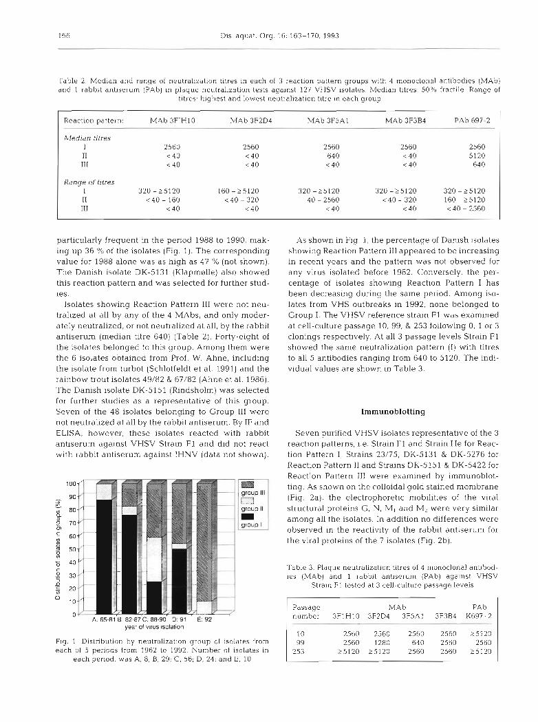
Table 2. Median and range of neutralization titres in each of 3 reaction pattern groups with 4 monoclonal antibodies (MAb) and l rabbit antiserum (PAb) in plaque neutralization tests against 127 VHSV isolates. Median titres: 50% fractile. Range of
titres: highest and lowest neutralization titre in each group
Reaction pattern: MAb 3FIH10 MAb 3F2D4 MAb 3F5A1 MAb 3F3B4 PAb 697-2
particularly frequent in the period 1988 to 1990, mak- ing up 36 % of the isolates (Fig. 1). The corresponding value for 1988 alone was as high as 47 % (not shown). The Danish isolate DK-5131 (Klapmerlle) also showed this reaction pattern and was selected for further stud- ies.
Isolates showing Reaction Pattern 111 were not neu- tralized at all by any of the 4 MAbs, and only moder- ately neutralized, or not neutralized at all, by the rabbit antiserum (median titre 640) (Table 2). Forty-eight of the isolates belonged to this group. Among them were the 6 isolates obtained from Prof. W. Ahne, including the isolate from turbot (Schlotfeldt et al. 1991) and the rainbow trout isolates 49/82 & 67/82 (Ahne et al. 1986). The Danish isolate DK-5151 (Rindsholm) was selected for further studies as a representative of this group. Seven of the 4 8 isolates belonging to Group 111 were not neutralized at all by the rabbit antiserum. By IF and ELISA, however, these isolates reacted with rabbit antiserum against VHSV Strain F1 and did not react with rabbit antiserum against IHNV (data not shown).
m group Ill 1=3 group II m group I
A: 65-81 B: az-sr c: ss-YW U: YT E: 92 year of virus sola at ion
Flg 1 Distribution by neutralization group of isolates from each of 5 periods from 1962 to 1992. Number of isolates in
each period: was A, 8; B, 29; C, 56; D, 24; and E, 10
As shown in Fig. 1, the percentage of Danish isolates showing Reaction Pattern I11 appeared to be increasing in recent years and the pattern was not observed for any virus isolated before 1982. Conversely, the per- centage of isolates showing Reaction Pattern I has been decreasing during the same period. Among iso- lates from VHS outbreaks in 1992, none belonged to Group I. The VHSV reference strain F1 was examined at cell-culture passage 10, 99, & 253 following 0, 1 or 3 clonings respectively. At all 3 passage levels Strain F1 showed the same neutralization pattern ( I ) with titres to all 5 antibodies ranging from 640 to 5120. The indi- vidual values are shown in Table 3.
Irnmunoblotting
Seven purified VHSV isolates representative of the 3 reaction patterns, i.e. Strain F1 and Strain He for Reac- tion Pattern I , Strains 23/75, DK-5131 & DK-5276 for Reaction Pattern I1 and Strains DK-5151 & DK-5422 for Reaction Pattern 111 were examined by immunoblot- ting. As shown on the colloidal gold stained membrane (Fig. 2a), the electrophoretic mobilities of the viral structural proteins G, N, M, and M, were very similar among all the isolates. In addition no differences were observed in the reactivity of the rabbit antiserum for the viral proteins of the 7 isolates (Fig. 2b).
Table 3. Plaque neutralization titres of 4 rnonoclonal antibod- ies (MAb) and 1 rabblt antiserum (PAb) against VHSV
Strain F1 tested at 3 cell-culture passage levels
Passage MAb PAb number 3FlH10 3F2D4 3F5A1 3F3B4 K697-2
Olesen et al.. Serological differences among viral isolates 167
Fig. 2. Immunoblotting using rab- bit antiserum K59 made against VHSV Strain F1 Membrane stained with either (a) colloidal gold or (b) irnrnunostained with K59. Lanes 1: Strain F1 (Group I); Lanes 2: Strain 23/75 (Group 11); Lanes 3: Strain DK-5422 (Group 111); Lanes 4 , Strain DK-5131 (Group 11); Lanes 5, Strain DK- 5151 (Group 111); Lanes 6, Strain He (Group I ) ; Lanes 7, Strain
DK-5276 (Group 11)
Neutralization pattern of isolates from intercon- nected outbreaks of VHS
Some of the 127 isolates used in this study originated from stocks of fish involved in simultaneous outbreaks of VHS a t different trout farms on a single stream and were obtained on the same day or at intervals of a few days. An outbreak in one particular farm was repre- sented by 5 isolates obtained on one day. Table 4 presents the results of the examination of 14 .isolates representing the outbreaks in question, designated A to E. All 5 isolates from Outbreaks A and B showed Re- action Pattern I, isolates from Outbreak C shared Reac-
tion Pattern 11, while isolates from Outbreaks D & E showed Reaction Pattern 111. In no cases were isolates representing different reaction patterns observed in connection with a single outbreak.
DISCUSSION
The use of noncloned virus isolates in this study was motivated by our goal of developing a technique for grouping fresh isolates of VHSV in a n epidemiological context. Cloning represents selection of a certain sub- population from a heterogeneous mixture and thus
Table 4. Plaque neutralization titres of 4 monoclonal antibodies (MAb) and 1 rabbit antiserum (PAb) to 14 virus isolates originating from 5 VHS outbreaks (A to E)
VHS out- lsolate Fish Date of MAb MAb MAb MAb PAb Group break no. farm no." isolation 3FlH10 3F2D4 3F5A1 3F3B4 K697-2
25 May 87 25 May 87 25 May 87
2 Jun 87 2 Jun 87 6 Apr 88 6 Apr 88 9 Dec 90 9 Dec 90 9 Dec 90 9 Dec 90 9 Dec 90
11 Feb 90 11 Feb 90
"Fish farm numbers are used by NVL for registration of fish diseases: first 2 digits give the district, following 2 digits the river system, last 2 digits location on the river
168 Dis. aquat. Org.
would be contraindicated in the present connection. Even using a cloned strain at 2 cell-culture passage levels the individual MAbs gave somewhat varying titres (Table 3). These v a ~ ~ a t i o n s , occasionally corre- sponding to 2 dilution steps, may reflect different ratios between infective and non-infective pa.rticles in the in- dividual virus preparations rather than serological dif- ferences. As a consequence of this, and of the day to day variations observed, only titre differences repre- senting 3 dilution steps or more were considered sig- nificant. The overall reaction patterns, and thus the groupings of the isolates, normally were not influenced by these variations.
From results of the competitive ELISA, it is likely that the 4 neutralizing MAbs bind to the same or very closely related epitopes. No difference in relation to the Ig subclass was observed among the MAbs. MAb 3F5A1, however, was slightly different from the other 3 MAbs in that it inhibited their binding more efficiently than it was inhibited by them. This might be due to higher affinity or to a slightly different epitope specific- ity. Similarly the PNT analyses demonstrated that MAb 3F5A1 differed from the other MAbs by being able to neutralize more isolates. In fact, the same reaction pat- terns with the isolates might have been obtained if the panel had contained only the polyclonal rabbit serum, MAb 3F5A1 and a pool of the remaining 3 MAbs.
It was shown that PNT titres could be increased by lengthening the incubation period of the virus-anti- body mixture from 1 h at 15 "C to overnight at 15 "C prior to inoculation; however, the overall reaction pat- terns of the individual isolates would not be changed as estimated by examination of 6 isolates (unpubl. results).
Three distinct reaction patterns were observed indi- cating the existence of 3 neutralization groups. Although a few Isolates gave titres which were close to those of some isolates belonging to another group, most isolates were clearly separated. In that sense, the present grouping procedure represents a major im- provement compared to the previous ones (Jnrgensen 1972, 1974, Le Berre et al. 1977). However, one should bear in mind that the grouping might be different if other MAbs or more MAbs had been used, perhaps di- rected against different neutralizing epitopes. For that reason, the present grouping is likely to change with time and probably cannot be reproduced in other la- boratories unless the same MAbs are used at the same concentrations. Preliminary results from the present study suggested the existence of 4 neutralization groups (Olesen e t al. 1991b); however, further analysis showed that only 3 groups could be reproducibly dis- tinguished, i.e. 2 of the 4 groups were pooled into 1.
Although clear antigenic differences between the virus isolates were detected by means of MAbs, ~t ap-
peared that at least some neutralizing epitopes must be shared, since the rabbit antiserum against Strain F1 to some extent neutralized 120 out of 127 isolates tested. These results are in agreement with earlier findings (Jmgensen 1972) showing that 72 of 76 isolates were neutralized [log neutralization index (NI) above 4.01 by a rabbit antiserum to Strain F1 with the remaining 4 yielding log NI values between 1.3 and 2.0, close to the borderline value of 1.7 commonly used to differentiate between significant and questionable neutralization. One of the latter strains, termed DK-61 and later He, was considered to represent a second neutralization type, i.e. 'Serotype' 2. In the present study, that strain was neutralized as efficiently as the reference strain F1. This discrepancy may have been due to the differ- ent versions of the neutralizat~on test used in the 2 studies, i.e. determination of NI and PNT respectively. These represent the 'constant serum, varying virus' and the 'constant virus, varying serum' methods, re- spectively (Casals 1967) and are likely to give results which are not directly comparable because the meth- ods deal with different fractions of the virus population under study. PNT thus operates with about 30 to 70 in- fective virus particles per inoculum, whereas determi- nation of NI operates with u p to 106 times more infec- tive particles per inoculum. 'Neutralization variants' would have to occur with a higher frequency in a given virus suspension to be detectable by PNT than is nec- essary for detection by determination of NI. At the same time, PNT titres will be less influenced by the heterogeneity among virus populations. Strain 23/75, the previous 'Serotype' 3 reference strain, was found to be clearly different from strain F1 in the present study, a finding which may reflect the fact that it was origi- nally characterized by means of plaque neutralization tests and not by determination of NI (Kinkelin & Le Berre 1977).
A complete description of the serological relahon- ship between viruses requires cross-neutralization tests using both homologous and heterologous antisera followed by calculation of l /r values (Archetti & Horsfall 1950). Due to the number of isolates in the present study as well as the difficulties in producing neutralizing antisera to VHSV ( J ~ r g e n s e n 1974) such a n approach was impractical. However, on a limited scale, this approach has previously been followed with a few VHSV isolates. Thus cross-neutralization tests using homologous and heterologous antisera have shown close relationship between Strains F1 and He in one study (Jsrgensen 1974), whereas in another (Le Berre et al. 1977) pronounced heterology among Strains F1, He & 23/75 was observed. These differ- ences in results are probably due to differences In the properties of the antisera used, slnce identical neutral- ization techniques were used. This is not unlikely in
Olesen et al.: Serological differences dmong vlral isolates 169
view of the differences in cross-neutralization shown by individual antisera made against isolates of 2 other fish rhabdoviruses, spring viraemia of carp virus and pike fry rhabdovirus (Jsrgensen et al. 1989).
The Group 111 isolates appear to represent a rela- tively new serological variant of VHSV. The first iso- lates in this group were from 2 VHS outbreaks in Germany in 1982, whereas the first Danish isolate was from an outbreak in 1986. Isolates with this reaction pattern have become more common since then and were recovered from fish in 9 out of 10 new VHS out- breaks at facilities on 5 different river systems in 1992. Although Group I1 isolates made up only 24 of the 127 isolates examined in the study, 15 of the 32 isolates from 1988 showed this reaction pattern. This is prob- ably due to the fact that 8 of these 15 isolates origi- nated from 2 water systems with several trout farms on each where fish may have been infected with VHSV from a common source. Apart from the example men- tioned above, the mechanism driving the observed changes in frequency is not understood at present.
It remains to be seen how the non-neutralizable iso- late 02/84 isolated by Dr de Kinke1i.n and recently de- scribed as 'Serotype' 4 of VHSV (Castric et al. 1992) fits into the present grouping system, since this isolate has not yet been made available. However, it appears likely that the isolate may belong to Group 111.
Our Western blotting results using a polyclonal rab- bit antiserum clearly indicated that the representative isolates from the 3 neutralization groups had common determinants on the G, N, M1 and M2 proteins. We did not observe the presence of electropherotype differ- ences among the representative isolates of VHSV as described for IHN virus (Hsu et al. 1986). The very strong crossreactivity of the applied polyclonal rabbit antibodies with the 4 major structural proteins of the representative VHSV isolates in Western blotting in combination with the ability of such antibodies to neu- tralize 120 out of 127 examined isolates in our opinion show that the VHSV isolates are strongly related and make up one single serotype. We recommend that this serotype is referred to as Serotype 1 and that reference strain F1 is regarded as the type strain of this serotype. The 7 non-neutralized isolates represent neutralization variants. Previous distinction between 3 serotypes may have been based on insufficient data, in particular lack of data from Western blotting.
Although thus only 1 serotype was defined by means of polyclonal antibodies it appears that minor differ- ences between isolates at the level of neutralization epitopes can be detected by means of neutralizing MAbs. These differences have enabled us to distin- guish between 3 neutralization groups which have proved useful in epidemiological studies. We recom- mend the designation of these groups as Subtypes I to
111 of Serotype 1 and stress that the subtypes are differ- ent from the previously described 'Serotypes' 1 to 3.
Although the present panel of reagents did not allow distinction between pathogenic and nonpathogenic strains or among isolates from different species of fish (e.g. brown trout isolates were present in neutraliza- tion Group I & 11, the eel isolate was in Group I1 and the turbot isolate was in Group 111) the present grouping system has proved helpful in ep~demiological studies, e .g . in tracing the source of infection of fish stocks (un- publ. results). Such studies in the future might be im- proved considerably if combined with the use of nu- cleic acid probe technology. In this regard, distinction between North American and European isolates of VHSV has been achieved using nucleic acid probes (Dr J. Winton pers. comm.). The typing system would also be improved by producing MAbs against addi- tional VHSV isolates and including these in the panel. Since such reagents are already present in several la- boratories, a cooperative effort towards further devel- opment of the grouping system is recommended.
LITERATURE CITED
Ahne, W., Jsrgensen, P. E. V., Olesen. N. J.. Schafer, W., Stelnhagen, P. (1986). Egtved virus: occurrence of strains not clearly identifiable by means of virus neutralization tests. J . appl. Ichthyol. 2: 187-189
Archetti, I.. Horsfall, F. L. (1950). Persistent antigenic varia- tion of influenza A viruses after incomplete neutralization in vivo with heterologous immune serum. J . exp. Med. 92: 441-462
Brunson, R . , True, K . , Yancey, J . (1989) VHS v ~ r u s isolated at Makah national fish hatchery Am Flsh Soc., Fish l-lealth Sect~on, Newsl. 17: 3-4
Casals, J (1967). Immunological techn~ques for animal vi- ruses In: Mararnorosch, K. , Koprowski, H. (eds.) Methods in v~rology, Vol. 111. Academic Press, New York, p. 201-241
Castric, J., Jeffroy, J., Bearzotti, M., Kinkelin, P. de (1992). Isolation of viral haemorrhagic septicaemia virus (VHSV) from wild elvers Anguilla anguilla. Bull. Eur. Ass. Fish Pathol. 12: 21-23
Fijan, N., Sulimanovic, D., Bearzotti, M., Muzinic, D.. Zwillenberg, L. O., Chilmonczyk, S., Vautherot, J . F., Kinkelin, P. d e (1983). Some properties of the epithelioma papulosum cyprini (EPC) cell line from carp (Cyprinus car- pio). Ann. Virol. (Annls Inst. Pasteur) 134 E. 207-220
Hopper, K . (1989). The isolation of VHSV from chinook sal- mon at Glenwood Springs, Orcas Island, Washington. Am. Fish. Soc., Fish Health Section, Newsl 17. 1-2
Hsu, Y - L , Engelking, H. M. , Leong, J. C. (1986). Occurrence of different types of infectious hematopoietic necrosis virus in fish. Appl. environ. Microbiol. 52: 1353-1361
Jensen, M. H. (1965). Research on the virus of Egtved disease. Ann. N.Y Acad. Sci. 126: 422-426
Jensen, N. J . , Bloch, B., Larsen, J . L. (1979). The ulcus syn- drome in cod Gadus morhua. Nord. Vet.-Med. 31: 436-442
Jsrgensen, P. E. V. (1972). Egtved virus: antigenic variation in 76 virus isolates examined in neutralization tcbts and by
Dis. aquat. Org. 16: 163-170, 1993
means of the fluorescent antibody technique. In: Mawdesley-Thomas, L. E. (ed.) Diseases of fish. Academic Press, London, p. 333-339
Jsrgensen, P. E. V. (1974). A study of viral diseases in Danish rainbow trout. Their diagnosis and control. Ph.D thesis, Royal Veterinary and Agricultural University, Copen- hagen
Jsrgensen, P. E. V. (1980). Egtved virus: the susceptibility of brown trout and rainbow trout to eight virus isolates and the significance of the findings for the VHS control. In: Ahne, W. (ed.) Fish diseases. Springer-Verlag, Berlln, p . 3-7
Jsrgensen, P. E. V., Olesen, N. J. (1987). Cod ulcus syndrome rhabdovirus is indistinguishable from the Egtved (VHS) virus. Bull. Eur. Ass. Flsh Pathol. 7. 73-74
Jsrgensen, P. E. V.. Olesen. N. J.. Ahne, W., Lorenzen. N. (1989). SVCV and PFR viruses: serological examination of 22 isolates indicates close relationship between the two fish rhabdoviruses. In: Ahne. W., Kurstak, E. (eds.) V~ruses of lower vertebrates. Springer-Verlag, Berlin, p. 349-366
Kinkelin, P. de , Le Berre, M. (1977). Isolement d'un rhabdovirus pathogene de la truite fario Salrno trutta L..1766. C . r. Acad. Sci. Paris 284: 101-104
Le Berre, M., Kinkelin, P. de, Metzger, A. (1977). Identification serologique des rhabdovirus des salmonidbs. Bull. Off. int. Epiz. 87: 391-393
Lorenzen. N., Olesen, N. J. , Jsrgensen, P. E. V. (1988). Production and characterization of monoclonal antibodies to four Egtved virus structural proteins. Dis. aquat. Org. 4: 35-42
Lorenzen, N., Olesen, N. J., Jergensen, P. E. V. (1990). Neutralization of Egtved virus pathogenicity to cell cul- tures and in fish by monoclonal antibodies to the viral G- protein. J , gen. Virol. 71. 561-567
McAllister, P. E., Wagner, R. R. (1975). Structural proteins of two salmonid rhabdoviruses. J . Virol. 15: 733-738
Meier, W., Jsrgensen, P. E. V. (1980). Isolation of VHS virus
This article was presented by F: M. Hetrick, College Park, Maryland, USA
from pike fry Esox lucius with haemorrhagic symptoms. In: Ahne, W. (ed.) Fish diseases. Springer Verlag, Berlin, p. 8-17
Moremans, M,, Daniels. G., May, J . de (1985). Sensitive colloidal metal (gold or silver) staining of protein blots on nitrocellulose membranes. Analyt. Biochem. 145: 315-321
Olesen, N. J. , Jarrgensen, P. E. V. (1986). Detection of neutral- izing antibody to Egtved virus in rainbow trout Salrno gairdnen by plaque neutralization test with complement addition. J . appl. Ichthyol. 2: 33-41
Olesen, N. J., Jsrgensen, P. E. V. (1991). Rapid detection of viral haemorrhagic septicaemia virus in fish by ELISA. J . appl. Ichthyol. 7: 183-186
Olesen, N. J. , Lorenzen, N., Jorgensen, P. E. V. (1991a). Detection of rainbow trout antibody to Egtved virus by en- zyme-linked immunosorbent assay (ELISA), immunofluo- rescence (IF), and plaque neutralization tests (50 % PNT). Dis. aquat. Org. 10: 31-38
Olesen, N. J., Lorenzen, N., Jsrgensen, P. E. V. (1991b). Serological differentiation of Egtved virus (VHSV) using neutralizing monoclonal and polyclonal antibodies. Abstract 79. 5th ~ n t . Conf. Eur. Assoc. Fish Pathol. on Diseases of Fish and Shellfish, Budapest
Sanz, F. A., Coll, J . M. (1992). Detection of hemorrhagic septi- cemia virus of salmonid fishes by use of an enzyme-linked immunosorbent assay containing high sodium chloride concentration and two noncompetitive monoclonal anti- bodies against early viral nucleoproteins. Am. J. Vet. Res. 53: 897-903
Schlotfeldt, H. J . , Ahne, W., Jergensen, P. E V., Glende, W. (1991). Occurrence of viral haemorrhagic septicaemia in turbot Scophthalmus maximus - a natural outbreak. Bull. Eur. Ass Fish Pathol. 11: 105-107
Wolf, K., Gravell, M., Malsberger, R. G. (1966) Lymphocystis virus: isolation and propagation in centrarchid fish cell lines. Science 151: 1004-1005
Manuscript first received: December 15, 1992 Revised version accepted: May 24,1993


![Dhf (Dengue Haemorrhagic Fever)[1]](https://static.documents.pub/doc/80x56/577c86e01a28abe054c2ee69/dhf-dengue-haemorrhagic-fever1.jpg)


