presenting a management dilemma. We described the SACQ period and its
patients, and sought biomarkers heralding disease flare in them.
Methods: Patients with a prolonged SACQ period were followed prospectively, with
clinical and laboratory data collected at each visit. Serologically and clinically active
(SACA) and serologically and clinically quiescent (SQCQ) patients served as
positive and negative controls, respectively. Descriptive statistics and logistic
regression analyses were utilized.
Results: SACQ patients were phenotypically unique prior to remission onset, and
did not accrue subclinical organ damage over the quiescent period. Fluctuation in
immunoglobulin isotype did not predict flare. The SACQ interferon and
cytokine/chemokine profiles mirror SQCQ patients.
iii
Conclusions: SACQ patients’ active serology should not guide treatment decisions,
and these patients are best managed conservatively. Alternate biomarkers must be
sought.
iv
Acknowledgements
I feel very fortunate to be surrounded, both personally and academically, by
individuals invested in my successes. It has been in this supportive milieu that I
have been able to devote myself to and learn from this journey.
To my thesis co-supervisors and longstanding mentors, Drs. Dafna Gladman and
Murray Urowitz, I owe a tremendous debt of gratitude, not only for their attentive
oversight of my thesis, but even more importantly for instilling a passion for clinical
research and, specifically, for the study of lupus in me. They have truly led by
example. I am humbled by their confidence in my abilities and enthusiasm for my
ideas, and look so forward to applying their teachings to my career.
To my Program Advisory Committee members, Drs. Earl Silverman and Joan
Wither, I am grateful for their generosity in selflessly sharing their time and expertise
over the years, permitting the exploration of fields outside my comfort zone in my
Master’s research.
I would like to thank Internal and External Appraisers, and Examiner, Drs. Sindhu
Johnson, Hermine Brunner and Brian Feldman, for giving of their time and applying
their unique backgrounds and skill sets to my thesis defence.
Ms Anne MacKinnon was critical in ensuring the seamless scheduling of Program
Advisory Committee Meetings, my final defence, and in providing critical logistic
support all along the way, and was thus truly instrumental in the completion of my
project.
I had the benefit of financial support from numerous sources, which afforded me the
luxury of pursuing this postgraduate degree. Thus the Arthritis Centre of Excellence,
v
Geoff Carr Fellowship/Lupus Ontario, PSI Foundation and UCB/CRA/TAS
Fellowships were central to my success and are thus acknowledged with
appreciation.
Finally, I would be remiss if I did not thank my friends and family, who have seen me
through the triumphs and challenges of this process. To my parents, thank you for
providing me with the wherewithall to remain a student into my 30s, and for your
support all along the way. To my husband and best friend, Nathaniel and our son,
Oscar, I thank you, from the bottom of my heart, for loving me always, cheering for
me often, and providing that extra push as needed. You are my inspiration and
motivation, and make everything in my life worthwhile.
vi
Table of Contents
Chapter 1: Literature Review .................................................................................. 1 Remission in systemic lupus erythematosus (SLE) ................................................ 2 Quantifying remission ............................................................................................. 4 Remission descriptors and correlates .................................................................... 5 The significance of organ damage accrual in SLE over time ............................... 11 The significance of serologic abnormalities in monitoring SLE disease activity ... 13
Serologically active clinically quiescent (SACQ) systemic lupus erythematosus (SLE) .................................................................................................................... 15 Clinically active serologically quiescent (CASQ) SLE .......................................... 25 Interferon alpha in SLE ........................................................................................ 26
Chapter 2: Hypothesis and Research Aims ........................................................ 33 Chapter 3: Prolonged remission in patients with systemic lupus erythematosus ....................................................................................................... 38
No Medication group ........................................................................................ 45 Case-control analysis ....................................................................................... 46 Medication group .............................................................................................. 48 Comparison of No Medication versus Medication groups ................................. 48
Chapter 5: Do differences in anti-dsDNA and anti-chromatin antibody isotype predict flare among patients with serologically active clinically quiescent (SACQ) systemic lupus erythematosus (SLE)? .................................................. 93
Chapter 6: The interferon-Į signature in patients with serologically active clinically quiescent systemic lupus erythematosus ......................................... 114 Abstract ................................................................................................................. 115
Chapter 7: General Discussion, Future Directions and Conclusions ............. 142 Why study remission? ........................................................................................ 143 Describing remission .......................................................................................... 143 Confirming quiescence ....................................................................................... 144 The pursuit of novel biomarkers ......................................................................... 146 Major Contributions ............................................................................................ 147 Limitations .......................................................................................................... 150 Future Directions ................................................................................................ 151
Does SACQ clinical quiescence stem from a lack of autoantigen? ................ 152 Does the SACQ anti-dsDNA and anti-chromatin antibody profile evolve with disease activity within the same patient over time? ........................................ 153 Does the SACQ IFN signature and cytokine/chemokine profile evolve with disease activity within the same patient over time? ........................................ 153 Is TLR tolerance the driver of the SACQ phenotype? ..................................... 154 Could personalized medicine hold the key to averting flare in SLE patients? . 155
Table 3-1: Past remission studies ........................................................................... 57 Table 3-2: SLEDAI-2K clinical characteristics of flare in patients with monophasic course: ..................................................................................................................... 58 Table 3-3: No Medication group matched case-control analysis: ............................ 59 Table 3-4: Clinical characteristics of Medication (MED) compared to No Medication (NO MED) groups at remission start: ....................................................................... 60 Table 3-5: Medication use from clinic entry in Medication (MED) compared to No Medication (NO MED) group: .................................................................................. 61 Supplementary Table 3-1: Patient demographics (p values reflect comparison to No Medication (NO MED) group .............................................................................. 63 Supplementary Table 3-2: Clinical and laboratory characteristics at visit 2 years from clinic entry (if prior to remission; p values reflect comparison to No Medication (NO MED) group) ..................................................................................................... 64 Supplementary Table 3-3: Clinical and laboratory characteristics at visit 5 years from clinic entry (if prior to remission; p values reflect comparison to No Medication (NO MED) group) ..................................................................................................... 65 Supplementary Table 3-4: Clinical and laboratory characteristics at visit 2 years from SLE diagnosis (if prior to remission; p values reflect comparison to No Medication (NO MED) group) .................................................................................. 66 Supplementary Table 3-5: Clinical and laboratory characteristics at visit 5 years from SLE diagnosis (if prior to remission; p values reflect comparison to No Medication (NO MED) group) .................................................................................. 67 Table 4-1: Baseline characteristics of SACQ and control patients .......................... 87 Table 4-2: Serologic abnormalities .......................................................................... 88 Table 4-3: SDI at inception, 3, 5, 7 and 10 years .................................................... 89 Table 4-4: SDI breakdown by corticosteroid effect .................................................. 90 Table 4-5: Renal damage and coronary artery disease (CAD) at five and ten years:................................................................................................................................. 91 Table 5-1: Patient demographics .......................................................................... 108 Table 5-2: Comparison of IgM and IgG anti-chromatin and anti-dsDNA levels in SACQ patients with unselected SLE patients and healthy controls ....................... 109 Table 5-3: Mean anti-chromatin and anti-dsDNA levels during SACQ in patients who flared versus those who remained SACQ (1 sample/pt) ................................ 110 Table 5-4: Mean anti-chromatin and anti-dsDNA levels during SACQ in patients who flared versus those who remained SACQ (all samples) ................................. 111 Supplementary Table 5-1: Clinical characteristics of flare in patients for whom both SACQ and flare samples were available ............................................................... 112 Supplementary Table 5-2: Comparison of autoantibody levels taken in the same patient during SACQ vs during flare ...................................................................... 113 Table 6-1: Patient demographics* ......................................................................... 134 Table 6-2: Associations with SACQ status by logistic regression analysis ............ 135
ix
List of Figures
Figure 3-1: Medication group remission outcomes ................................................. 62 Figure 4-1(a-c): SDI breakdown by corticosteroid (CS) effect ................................ 92 Figure 6-1: Relative gene expression .................................................................. 136 Figure 6-2: IFN score over time ............................................................................ 138 Figure 6-3: Cytokine and chemokine concentrations ............................................ 139
1
Chapter 1: Literature Review
2
Remission in systemic lupus erythematosus (SLE)
In early reports, systemic lupus erythematosus (SLE) was classically described as
an unrelenting disease that would often culminate in death(1). However, the
disease has been increasingly recognized as a chronic, albeit potentially fatal,
relapsing-remitting disease. Given the propensity for severe manifestations and
organ damage over time, remission is a very desirable outcome, and, as such,
efforts have been made to describe it and understand its correlates. In fact, studies
have revealed that the propensity for flare or remission in the initial years of disease
are predictive of long term outcome, with those remitting earlier having a more
favourable disease course(2,3). Substantial variability exists, however, in the nature
and duration of remission, likely attributable to differences in patient cohorts and
inconsistent remission definitions.
Dubois provides one of the first descriptions of remission in a cohort of 163 lupus
patients in 1956(4). He reported that 38% of the patients experienced at least one
“spontaneous remission” prior to treatment with antimalarials or corticosteroids,
including one patient with a 26-year remission, and up to 16% with multiple
remissions. He admitted, however, that most of these patients “did not have the full
picture of systemic lupus erythematosus,” but rather had a rheumatoid arthritis-like
presentation (presumably prominent joint symptomatology in the absence of major
organ manifestations). There was no definition of remission proffered in this historic
paper, but it seemed to be based upon the physician’s global clinical impression. In
3
1964 he and Tuffanelli then corroborated this considerable remission rate, reporting
that 35% of 520 SLE patients experienced “spontaneous remission,” lasting up to 26
years in one case(5). The definition of remission was similarly implicit in this study.
Heller and Schur described serologic and clinical remission in a cohort of 305
patients followed between 1967 and 1981, defined on the basis of change in ANA or
LE cell test from positive to negative in patients who were asymptomatic and without
obvious active organ involvement(6). By contrast with Dubois and Tuffanelli, they
found only 13 (4%) had developed a combined clinical and serologic remission,
ranging in duration from 6 months to 13 years. Their definition of remission included
patients on no therapy, those on hydroxychloroquine, as well as those requiring low
doses of corticosteroids “because they became Addisonian if not receiving therapy.”
Thus, their frequency of remission was much lower than in previous studies, in spite
of having included patients both on and off of immunosuppressive medications. This
disparity is likely attributable to the stringent serologic criteria for remission in this
study, especially as ANA typically persists once present, and is not known to
fluctuate with disease activity(7).
More consistent numerically with Heller and Schur’s findings (but disparate in
definition) was Tozman and colleagues’ determination that the rate of “prolonged
complete remission” in SLE, defined as the absence of clinical manifestations of
disease and off all immunosuppressive therapy, was 4/160 (2.5%)(8). They utilized
both clinical and laboratory variables in their assessment including, for the first time
4
in the setting of remission, the absence of anti-DNA antibodies and C3
hypocomplementemia, both of which are known to run a concordant course with
disease activity in some lupus patients(9-11). These patients had remitted from
previously severe disease, with median remission duration 75 months. Thus,
considerable disparity in duration, definition and frequency of remission existed in
the earlier literature.
In 1996 Drenkard and colleagues published an extensive review encompassing over
2000 person/years of data, and defined remission as “at least one year during which
lack of clinical disease activity permitted withdrawal of all treatment for lupus
proper”(3). They found 23% of their cohort met these criteria, with increasing
proportions of remissions in subsets with longer disease duration. Thus they found
that disease activity waned over time. Unlike Dubois’s observation of frequent
spontaneous remissions, 87% of Drenkard’s cohort required at least some quantity
of prednisone, with or without immunosuppressive agents, to control their disease
prior to remission onset. Survival was highly correlated with remission, with those
achieving at least one remission having a 12.5-fold smaller chance of dying from
SLE than those who did not, regardless of the severity of past SLE manifestations.
Quantifying remission
Since Dubois’s qualitative description of remission, presumably based on his global
assessment of SLE activity, attempts have been made to standardize and quantify
SLE activity through the use of validated disease activity measures(12). The most
5
commonly used among these include the SLE Disease Activity Index (SLEDAI) or its
modification, the SLEDAI-2000 (SLEDAI-2K), Safety of Estrogen in Lupus
Erythematosus National Assessment-SLEDAI (SELENA-SLEDAI), British Isles
Lupus Assessment Group (BILAG), the Systemic Lupus Activity Measure (SLAM)
and European Consensus Lupus Activity Measurement (ECLAM). Each of these
scales provides a quantitative tool for physicians to systematically describe and
compare the extent of lupus disease manifestations in a patient longitudinally(12).
Notably, in 1992 the original SLEDAI was validated, and provided clinicians with a
simple and quantitative method of describing lupus activity(13). However, despite
the availability of these quantitative tools and standardized definitions of flare in
SLE, consensus surrounding the definition of remission has not been met, with the
impact of serologic activity on remission status as well as the use of concomitant
medications remaining the subjects of debate(14). Still, despite these tools, given
the multisystemic nature of SLE, and the importance of distinguishing accurately
between active disease and irreversible disease- and treatment-associated damage,
the facility with which remissions are defined in other medical specialties, such as
Oncology, remains elusive(15).
Remission descriptors and correlates
Researchers began to delve deeper into the nature of remission in SLE, its
correlates and its predictors. Barr and colleagues described patterns of disease in a
cohort of 204 consecutive SLE patients, as defined by the SLEDAI score, modified
to include only clinical (and excluding serologic) components of disease activity,
6
compared to the physician’s global assessment (PGA)(16). They described three
patterns of SLE: relapsing-remitting, chronic active and long quiescent, with chronic
active being the pattern accounting for the largest proportion of patient years among
those studied, and long quiescent being the least frequent. Long quiescent was
defined semi-quantitatively, by the physician’s global assessment score, and
quantitatively, via SLEDAI, as a score of 0 for at least 1 year, irrespective of
serologic features or treatment. It was found that 28% of patients (16% of total
person years) experienced long quiescence, the rarest of the three patterns, by
PGA, and 44% (25% of total person years) by SLEDAI, even though a more
permissive definition of quiescence was tolerated, without heed given to concurrent
treatment. Thus, even within the same study, considerable disparity existed
between measures of disease activity. Furthermore, because a modified SLEDAI
score was used, the long quiescent cohort encompassed those with and without
concurrent serologic activity in patients who, because treatment was permitted, may
have simply been in a state of disease suppression as opposed to true remission.
Similarly, in a cohort comprised of ten European rheumatology centres and 187
patients, it was found that remission, even after ten years of disease, when activity is
thought to wane(2,3), was a rare outcome(17). The SLEDAI and ECLAM were
employed in this study, but not explicitly utilized to define remission. Rather,
remission was defined as “the absence of disease-related signs, without the need
for any treatment.” By these criteria, none of the patients studied were in remission,
as all continued to require some form of treatment, including prednisolone in 72%.
7
The number of patients without any clinical disease activity while taking medications
was not specified, thus an important subset of quiescent patients in whom treatment
was gradually being withdrawn was not described.
As a corollary, Mok and colleagues studied the rate of disease flare in post-
menopausal women, who typically have longer disease duration, as SLE is most
commonly diagnosed during the reproductive years(18). They found that post-
menopausal, compared to premenopausal, flares were significantly less common,
occurring in 44% versus 94%. Their flare definition included new serologic positivity
(in the absence of clinical disease activity), and disease quiescence was not
explicitly defined. Furthermore, while SLEDAI was used to define disease activity,
the definition of flare was based upon the dose of corticosteroid and/or
immunosuppressive agent used, not upon a prospectively determined fluctuation in
SLEDAI score. It is thus difficult to compare the outcomes among these studies, in
spite of the use of a consistent disease activity outcome measure.
As early SLE disease course is predictive of outcome(2,3), Formiga and colleagues
studied remissions among those with high disease activity early in their disease
course(19). They defined remission as disease activity permitting the withdrawal of
all SLE-related treatment over at least one year, and asymptomatic serologic
fluctuations were permissible. Twenty-four percent of their exclusively Caucasian
cohort (of 100 patients) achieved such a remission, at mean 64 months after
diagnosis, and the remissions persisted, on average, over more than 4.5 years.
8
While there were differences in baseline SLEDAI value between those who achieved
remission and those who did not (with those with higher initial SLEDAI scores less
likely to remit), these did not attain statistical significance.
Thus they observed remissions in patients with all disease manifestations, including
major organ involvement. They found a significant correlation between SLEDAI
values and time to remission onset: remission occurred later among those with more
severe baseline disease. It is worthwhile noting that the cohort was comprised
exclusively of Caucasian patients, who have been shown to have consistently more
mild disease – and thus, one would anticipate, more frequent remission - than
Blacks, Hispanics and Asians(20). A comparable study exploring these outcomes in
these ethnicities would be of interest to determine whether remission rates were as
high.
A European study(21) followed an inception cohort for five years, and found 27.5%
of a cohort of 200 patients achieved remission, as defined by Global Physician
Assessment, within the first year of disease. They found no differences, compared
to those with persistently active disease, in age of onset, number of American
College of Rheumatology (ACR) SLE classification criteria fulfilled, or maximum
corticosteroid dose required. They did, however, find that cumulative corticosteroid
dose, maximum SLEDAI score achieved, and organ damage accrual, all over the
first year, were significantly lower in those achieving early remission. While
approximately half of the patients achieving early quiescence maintained their
9
remission through a period of more than a mean four years of follow up, only 25% of
those 145 patients with persistent disease activity within the first year eventually
achieved remission. Those achieving earlier remission evolved to milder disease
than those without, with less active disease and fewer relapses. The frequency of
remission is thus comparable to that described in Formiga’s study(19), but the
comparability of the cohorts is less clear, as a precise remission definition was not
proffered, nor was there mention made of ethnicity (presumably, predominantly
Caucasian).
Wais and colleagues studied laboratory correlates of remission in 57 Caucasian
outpatients with SLE(22). Remission was defined as a BILAG score � 5, and
variables included proinflammatory cytokines, such as IL-6, IL-10 and TNF-Į;
adhesion molecules, such as sICAM and E-selectin; and conventionally concordant
serologic markers, such as complement levels and anti-double stranded DNA
antibodies (anti-dsDNA). They found that, overall, the only difference in lab
indicators between the 39 inactive patients and the 18 active patients was lower
CRP in the former group (p<0.001), concluding that clinically inactive patients
continue to experience some degree of immunoinflammatory activity. The authors
acknowledged, however, that in contrast to others’ studies in patients with SLE, the
level of cytokines was not significantly increased from background levels in many
cases. This finding, of course, may have been indicative of a skew in the cohort
towards relatively quiescent disease, or reflective of differences in definitions of
disease activity.
10
This finding also begets the question of whether organ damage accrues subclinically
in patients with SLE in clinical remission. While that question was beyond the scope
of Wais’s paper, this issue was later addressed and refuted(23), thus supporting the
impression that the “active” cohort was atypically quiescent, and not that the inactive
cohort had increased immunoinflammatory activity. Furthermore, given the
enormous complexity of SLE pathophysiology, one must recognize that Wais’s
findings do not exclude the possibility of significant differences occurring between
groups in other unmeasured immunoinflammatory molecules.
In 2005, Urowitz and colleagues took significant strides toward addressing the
inconsistencies that had plagued the SLE remission literature by quantifying and
describing disease quiescence using incrementally less restrictive criteria(24).
Thus, they defined prolonged remission as at least a five-year period without
disease activity (SLEDAI-2K = 0), while not taking corticosteroids,
immunosuppressives or antimalarials. They found that remission, thus defined, was
a rare event, occurring in only 12 of 703 (1.7%) patients in their cohort. As would be
expected, when progressively less stringent criteria were applied to the remission
definition, encompassing one to five years’ disease quiescence, permitting the
presence of hypocomplementemia and/or anti-dsDNA positivity, and permitting the
use of antimalarials, corticosteroids and immunosuppressive medications, remission
prevalence increased as stringency decreased. When defined as clinical
quiescence (by SLEDAI-2K) for one year, permitting active serology, and requiring
11
the use of medications (the least restrictive definition), remission prevalence was
24.5%. Thus, as elegantly demonstrated by this paper, the question to be asked is
not, “What is the prevalence of remission?” -- a vague and subjective construct -- but
rather, “What type of remission is being quantified?” -- as “remission” is in the eye of
the investigator.
Thus the generalizability of the literature about SLE remission is limited by
differences in definition, with duration, disease activity measure used, the
permissibility of treatment, and serologic activity all being variables that may
significantly affect the result. Furthermore, given the heterogeneity of lupus
presentation, and the impact of ethnicity upon disease manifestations, severity and
prognosis, differences inherent to a cohort, itself, may prove central in the duration
and type of remission achieved. Regardless of how defined, remission remains a
desirable outcome in SLE, but is relatively rarely achieved. A more complete
understanding of SLE pathophysiology may lead to improved management.
The significance of organ damage accrual in SLE over time
Remission, most stringently defined, is a desirable disease state as the patient is
free from both the onslaught of inflammation-associated disease activity and the
untoward effects of treatment. In addition to the immediate manifestations of
disease and treatment, each can impact the patient more permanently, a construct
referred to as “damage.” This outcome is particularly important as mortality has
decreased over the years in the setting of aggressive treatment with corticosteroids
12
and immunosuppressive medications, but the propensity for irreversible organ
damage, attributable to both the disease itself and to its treatment(25), has kept
pace with the increasing survival rate. The Systemic Lupus International
Collaborating Clinics/American College of Rheumatology Damage Index (SDI) is a
reliable and well-validated index which measures damage in individual organ
systems(26,27).
Particularly germane to remission studies, Swaak and colleagues described damage
accrual using the SDI in patients with disease duration of more than 10 years(28), as
disease activity is known to wane over time, as highlighted above. They found the
three most common signs of damage were hypertension (in 40%), osteoporosis and
cardiovascular disease (each in 15%), all of which may be largely attributable to
corticosteroid use. In 2003, Gladman and colleagues tracked the SDI longitudinally
in an inception cohort of SLE patients, followed prospectively for at least 15
years(29). They found that damage accrued linearly over time. In keeping with the
observations of Swaak and colleagues, they found that within the first year of SLE,
42% of damage was disease-attributable, and 58% was possibly or definitely
corticosteroid-attributable. Later in the disease course, only 20% of damage was
disease-attributable, and 80% was possibly or definitely attributable to corticosteroid
use. That much of late damage accrual can be attributed to treatment effects – as
opposed to disease - serves to reinforce the importance of corticosteroid withdrawal,
whenever possible, and the desirability of unmedicated remission.
13
The significance of serologic abnormalities in monitoring SLE disease activity
The ultimate goal of evaluation in the patient with SLE is to monitor for disease
activity, and manage and treat any signs thereof, in hopes of preventing worsening
illness and, ultimately, irreversible organ damage. To diagnose remission, the
clinician must confirm an absence of clinical manifestations of disease, yet the
complex and often non-specific protean manifestations of SLE can be difficult to
interpret. In some patients, serologic activity can provide corroborative evidence of
disease activity, as some biomarkers can fluctuate with disease activity.
Anti-dsDNA
Anti-dsDNA was first described in 1957(30,31), and subsequently implicated in SLE
pathogenesis and organ damage when Koffler and colleagues eluted these immune
complexes from the kidneys of patients with lupus nephritis(32). The presence of
these immune complexes is a highly specific diagnostic marker for SLE, that occurs
in up to 83% of patients(33). Furthermore, studies have correlated fluctuations in
anti-dsDNA titre with clinical disease activity (see below) in all but a unique subset of
patients, termed serologically active clinically quiescent (SACQ), in whom anti-
dsDNA and/or complement levels run a discordant course. Given their utility in
diagnosis and monitoring, commercial kits for measuring the presence of anti-
dsDNA are widely available. These assays, however, are not created equally, and
have varying levels of sensitivity and specificity.
14
The three most commonly employed tests are the Crithidia, ELISA, and Farr assays.
The Crithidia luciliae immunofluorescent test (CLIFT) capitalizes upon this
hemoflagellate’s kinetoplast (that contains circular dsDNA), which is incubated with
patient serum, and an anti-immunoglobulin is applied. The enzyme-linked
immunosorbent assay (ELISA) test is simple, relatively inexpensive and easily
reproducible, utilizing DNA-coated polystyrene plates as substrate for colorimetric
quantitation of serum anti-dsDNA antibodies. Finally, the Farr assay separates
bound and free DNA through ammonium sulphate immunoprecipitation, with bound,
radioactive DNA precipitating with immunoglobulins, and free DNA remaining in the
supernatant(34). Of these, the Farr assay and CLIFT are the most specific, with the
Farr assay best correlating with detection of anti-dsDNA of highest avidity(35,36).
Antibodies of high avidity, in turn, are correlated with the presence of active renal
disease(37).
It has been suggested that not all anti-dsDNA is created equally, with anti-dsDNA of
high avidity, of IgG isotype and complement-fixing IgG sub-class best correlating
with disease activity and renal involvement(38-45). As a corollary, IgM isotype and
non-complement-fixing IgG sub-class best correlate with disease quiescence.
Complement
Measurement of decreasing complement components C3, C4 and CH50 have long
been appreciated to correlate with active lupus, and with the presence of associated
glomerulonephritis(46,47). This is consistent with what is known of their
15
pathophysiologic role, with circulating complement activation products stimulating an
inflammatory cascade, resulting in tissue damage. The measurement of
complement components is most useful when performed serially and thus
comparatively, as in some SLE patients the disease process may not be concordant
with complement values, and in others C4 is chronically low in the context of the C4
null allele, which is not infrequently associated with SLE(46,48,49).
Anti-nucleosome antibodies
There is the general consensus that anti-nucleosome antibodies are both sensitive
and highly specific for SLE(50-53). In a systematic literature review and meta-
analysis, Bizzaro and colleagues determined that the presence of anti-nucleosome
antibodies conferred a 41-fold increase in the risk for SLE (versus 28-fold with anti-
dsDNA)(50). Suleiman et al found that anti-nucleosome antibodies were 98%
specific for the diagnosis of SLE(53). They also found anti-nucleosome antibodies
98% sensitive and 86% specific for detecting active SLE, versus 61% and 84% for
anti-dsDNA sensitivity and specificity, respectively(53). Several studies have
investigated the role of anti-nucleosome antibodies in renal disease. Most found
anti-nucleosome antibodies elevated in the setting of lupus nephritis(51,54,55), with
some finding them of improved sensitivity compared to anti-dsDNA
antibodies(51,56).
Serologically active clinically quiescent (SACQ) systemic lupus
erythematosus (SLE)
16
Recognition of SLE patients with clinical/serologic discordance can first be found in
the literature in 1979, when Gladman and colleagues described 14 patients with
persistently positive lupus erythematosus (LE) preparations and antinuclear
antibodies, hypocomplementemia, and high levels of DNA binding(57). These
patients had displayed typical lupus manifestations in the past, including major
organ manifestations, such as renal or central nervous system involvement, but had
since evolved to a clinically quiescent state. In spite of their clinical quiescence,
these patients had impaired lymphocyte response to concavalin A, suggestive of a
defect in cell-mediated immunity, as was typically seen in patients with active lupus.
These findings were, themselves, discordant from other studies of the time, which
revealed serologic abnormalities were often harbingers of or associated with active
disease(58-60). None of Gladman’s patients, termed “serologically active clinically
quiescent,” or “SACQ,” were taking corticosteroids or immunosuppressive
medications, and had not done so for mean 4.25 years (range 2-11 years). Given
seven years of follow-up revealing no flares among these individuals, Gladman and
colleagues suggested that, in these individuals, close clinical follow-up without
preemptive treatment with corticosteroids or immunosuppressive medications may
be appropriate.
These suggestions contrasted starkly with those made by Swaak and colleagues in
the very same year(61) in their paper describing rise and subsequent precipitous fall
in anti-dsDNA as predictive of flare in SLE patients. In their discussion, Swaak and
17
colleagues advocated for the role for adoption of therapy on the basis of anti-dsDNA
fluctuations. Perhaps even more divergent was a paper published prior to
Gladman’s SACQ observations, describing treatment of SLE patients with up to 100
mg of prednisone daily, guided by immunologic abnormalities, such as depressed
serum complement levels, until these had normalized(62).
Ter Borg and colleagues studied fluctuations in anti-dsDNA and complement levels
prior to SLE flare, and found 27/33 disease exacerbations were accompanied by
elevations in anti-dsDNA antibodies(63). While the test was neither perfectly
sensitive nor specific for disease flare, they concluded that serial assessment of
anti-dsDNA levels, especially by Farr assay, was a reasonable approach to the
monitoring of SLE disease activity. They found anti-dsDNA to be more sensitive for
predicting exacerbations than C3 or C4 levels.
Thus there were two factions emerging among lupus practitioners – those who felt
treatment might be appropriate on the basis of fluctuations in anti-dsDNA and/or
complement levels, and those who argued for close clinical monitoring in lieu of
preemptive treatment.
In 1994 Walz LeBlanc and colleagues conducted a prospective cohort study to
identify SACQ patients and study their clinical predictors of flare(64). SACQ patients
were defined as those in whom “serologic abnormalities – either low C3, C4 or
CH50, or elevated anti-dsDNA antibodies – were present on three consecutive clinic
18
visits in the absence of clinical disease activity as measured by the SLEDAI.” There
were no requirements for medication withdrawal. One hundred-and-six SACQ
periods were analyzed in 74 patients.
The SACQ period was terminated by flare within a year of the 3rd SACQ
assessment in 46 instances: in 31 patients, the SACQ period was observed to occur
between two flares, with both clinical, and serologic activity; in the remaining 15, the
SACQ period followed a serologically and clinically quiescent period and preceded a
clinical flare(64). The remaining 60 SACQ episodes were not associated with
disease flare within the year following the 3rd SACQ assessment. Of these, 35 were
preceded by clinical and serologic activity; 25 were preceded by clinical and
serologic quiescence.
Comparisons were then made between those SACQ patients who flared and those
who did not(64). The only features distinguishing these two groups were increased
use of steroids, and at higher doses, during the SACQ period, a shorter median
duration of the SACQ period, and a slightly greater incidence of vasculitis in those
who flared. There were no differences in demography, or in fluctuations in serology
between groups. Perhaps this suggests that in those who flared SACQ merely
represented a transitional state between flares as opposed to true discordance.
Thus Walz LeBlanc and colleagues’ work emphasized and delineated the difference
between disease quiescence, with true clinical-serologic discordance, versus
disease suppression between flares.
19
In the same year, long-term follow-up was pursued in Gladman’s patients, and
ultimately obtained in 11 of the original 14(64). Seven patients remained clinically
well, off all corticosteroids or immunosuppressive medications. Four patients had
minor clinical flares of SLE requiring intervention that occurred only after a mean of
5.5 years from SACQ onset. This lent further support to the authors’ original
contention that in some patients, serologic abnormalities and clinical disease run
discordant courses, thus the former need not dictate medical intervention. These
findings were situated at a particularly poignant time, given that other authors were
then further experimenting with preemptive dosing of prednisone against SLE flare
in the face of rising titres of anti-dsDNA antibodies.
For instance, in 1995, with a cohort of 156 SLE patients, Bootsma and colleagues
randomly assigned those with an observed increase in anti-dsDNA level to either
conventional treatment, or an addition of 30 mg prednisone to their preexisting
regimen(65). They found the relapse rate was higher in the conventionally-treated
group, with comparable cumulative corticosteroid dosing between groups. Thus the
SACQ cohort appeared to represent a unique subset in whom serology and clinical
features run a discordant course, unlike the patients in the Bootsma study.
A similar study was carried out years later by Tseng and colleagues, in patients who
were serologically active and clinically stable(66). They conducted a prospective,
randomized, double-blind, placebo-controlled trial of prophylactic steroid dosing in
20
154 patients evaluated monthly for up to 18 months. In this study, patients who
were clinically inactive or clinically stable/active, defined as a SELENA-SLEDAI
score of �4 or 5-12, respectively, and who were not receiving more than 15 mg
prednisone per day were eligible. Those with serologic evidence of flare, namely
25% elevation in anti-dsDNA and 50% elevation in C3a, were randomized to receive
either a three-week course of prednisone, with starting dose 30 mg per day, or
placebo. They found that significantly more flares occurred in the placebo group
than in the treatment group (six versus none among 41 patients who experienced
serologic flare, p=0.007). Severe caution must be exercised, however, in
extrapolating these findings to SACQ patients, as Tseng’s patients could have had
active disease requiring up to 15 mg (a moderate dose) of prednisone daily and still
have met inclusion criteria. Since this cohort included patients who continued to
have evidence of active disease despite treatment with corticosteroid, as well as
patients whose clinical manifestations may have been merely suppressed by their
baseline corticosteroid dosing, they were fundamentally different than the SACQ
patients defined by Gladman and colleagues, and this study’s findings cannot be
generalized to them.
Since SACQ patients’ disease activity runs a discordant course from conventional
serology, and since some SACQ patients ultimately do flare, efforts were made to
identify more reliable biomarkers in this unique subset of patients. Ng and
colleagues studied the role for anti-nucleosome antibodies, compared to anti-
dsDNA, in the monitoring of patients with “SACQ” SLE(67). In this study, SACQ was
21
defined as a BILAG score less than 6 for at least six months in the context of anti-
dsDNA positivity by ELISA method. Most patients were taking steroids and/or
immunosuppressive medications. Nine percent (27/290) of the cohort was thus
defined.
The investigators simultaneously measured anti-nucleosome antibodies, and found
that time to first flare after a SACQ period was significantly correlated with their
presence (p=0.0012), with higher titres thereof (p=0.0006), and with the presence of
anti-dsDNA antibodies greater than five times above the upper limit of normal
(p=0.02). They concluded that anti-nucleosome antibodies might be better
predictors of flare in SACQ patients than anti-dsDNA. While numerous past studies
have determined that anti-nucleosome antibodies are reliable indicators of flare in
SLE patients, in general, their reliability in a stringently-defined SACQ group remains
to be determined, as Ng’s SACQ patients were comprised of those whose disease
may have been merely stable, or suppressed by treatment. For instance, a patient
with worsening arthritis and stable, localized discoid skin lesions (BILAG score 5),
on 15 mg of prednisone would have met Ng’s criteria for “SACQ.” However this
patient fundamentally differs from Gladman’s SACQ patient, with persistent
pathogenic active serology, on the one hand, but free of any disease activity, off all
corticosteroids and immunosuppressive medications, on the other.
These stringently-defined SACQ patients thus present a conundrum: How can the
clinician reconcile the presence of potentially pathogenic serologic activity with the
22
clinical picture of complete quiescence? How is this patient best managed? The
more fundamental question to be asked is, ”What are these patients’ outcomes?” as
these dictate management. Two potential SACQ outcomes exist – continued
quiescence or flare. If the former, and SACQ can persist, or evolve to serologic and
clinical quiescence, then it would be prudent for the clinician to spare the patient
from exposure to corticosteroids and immunosuppressive medications and their
associated risks and side effects. Alternately, the SACQ patient who evolves to flare
will ultimately require such treatment. Thus a method to distinguish which SACQ
patients will remain quiescent, versus those who will ultimately flare would be of
clinical utility.
With this goal in mind, I strove to describe the SACQ period and its patients(68).
SACQ was defined as at least two years of persistent serologic activity in the
absence of clinical activity (SLEDAI-2K score of 2 or 4 from anti-dsDNA positivity by
Farr assay and/or hypocomplementemia), with visits �18 months apart, during which
antimalarial medications were permitted, but corticosteroids and
immunosuppressives were not. This restrictive definition was applied in order to
exclude both those patients who had active SLE merely suppressed by ongoing
immunosuppression, and those who were in evolution from or to imminent flare.
Thus this definition was meant to include only those with bona fide clinical/serologic
discordance.
23
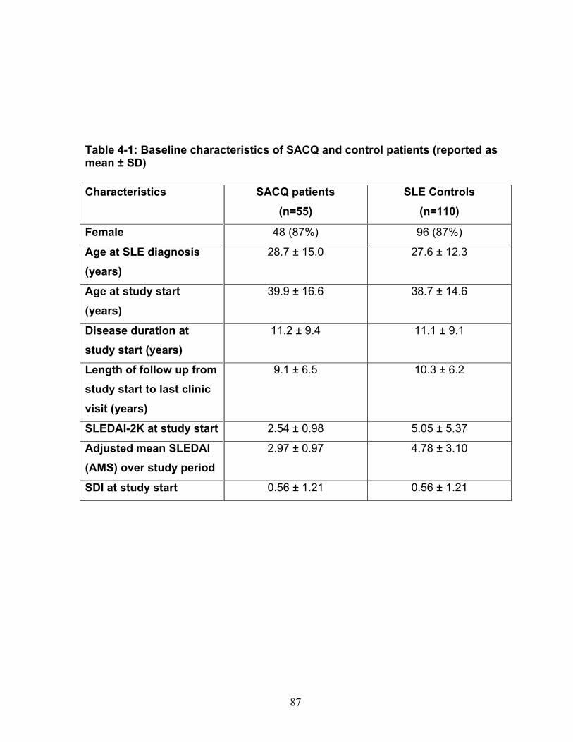
Fifty-six of 924 (6.1%) of the cohort were SACQ, accounting for 70 SACQ periods
among them. Median SACQ duration was 158 weeks (mean 182 weeks), and, on
average, occurred more than a decade after SLE diagnosis. The SACQ period was
characterized by both elevations in anti-dsDNA and hypocomplementemia in the
majority (62.5%) of patients. SACQ patients had milder disease (SLEDAI-2K at
presentation = 7.3 versus 10.1, p=0.01) and required less corticosteroid and
immunosuppressive medications at first clinic visit than did non-SACQ patients.
There was a trend toward fewer deaths in the SACQ group. SACQ patients had
less musculoskeletal, dermatologic and central nervous system involvement over
their disease course.
Of interest, in light of Farr assay positivity in many of the SACQ patients and its
correlation with active renal disease, there was no difference in the prevalence of
renal involvement in patients prior to SACQ onset compared to non-SACQ patients.
Furthermore, that this major organ was comparably involved in SACQ and non-
SACQ patients suggests that SACQ does not merely represent the eventual
outcome of a milder disease course. Anti-dsDNA antibodies were in the moderate
and high range in 20.9% and 9.3% of the SACQ cohort, respectively, thus the
elevations therein were not simply reflective of marginal increases from normal
range.
The first SACQ period was terminated by disease flare in 33 (59%) of patients at
median 155 weeks. Flare most frequently manifested as arthritis, mucous
24
membrane involvement or sterile pyuria. Of the remaining SACQ patients, six
(10.7%) became serologically and clinically quiescent at median 236 weeks, and 17
(30.4%) remained SACQ at their most recent clinic visit. Flares could not be
predicted from fluctuations in anti-dsDNA antibodies and/or complement levels in the
preceding visits. Thus SACQ patients could not be identified a priori. Outcomes
within the thusly-defined SACQ group could not be predicted on the basis of
serologic fluctuations, as was the case in the studies conducted by Swaak, Ter
Borg, Tseng and Bootsma.
In 2012, Conti and colleagues evaluated the frequency of SACQ within their cohort,
using the definition we had proposed in 2010(69). Specifically, SACQ was defined
as at least a two-year period without clinical activity and with persistent serologic
activity, by SLEDAI-2K, during which patients could be taking antimalarials, but
could not be taking corticosteroids or immunosuppressive medications. They found
only 1/45 (2.2%) patient met their SACQ definition, which represents a value slightly
lower than the 6.1% observed in the larger Toronto cohort. The most probable
cause for these discrepant findings lies in Conti and colleagues’ duration of
prospective follow-up, which was only two years, compared to up to 38 years of
follow-up in our longitudinal study. Given that the proportion of time a SLE patient
would spend in a SACQ state is relatively low compared to their overall length of
disease, maximizing the duration of longitudinal follow-up will yield the most
accurate estimate of SACQ prevalence for any patient.
25
Clinically active serologically quiescent (CASQ) SLE
A second discordant cohort has been studied in the clinically active, serologically
quiescent (CASQ) lupus patients. Gladman and colleagues described these
patients, defined as those with three or more consecutive visits with clinical activity
in the absence of serologic activity (hypocomplementemia and elevations in anti-
dsDNA) by SLEDAI-2K score(70). CASQ patients comprised 62/514 (12.1%) of
their cohort, with the CASQ period lasting median eight months, and was associated
with a mean SLEDAI-2K score of 8.9±5.3, indicative of mild to moderate disease
activity. Approximately one-third of CASQ patients ultimately evolved to
serologically and clinically active disease (and thus two-thirds did not). Major organ
involvement, defined as renal, vasculitic or central nervous system manifestations,
occurred in 43 (69%) CASQ patients; of these, 31 (50% of all the CASQ patients)
had renal manifestations during the CASQ period.
Thus, in spite of pathophysiologic plausibility of immune complex deposition leading
to damage in SLE, with anti-dsDNA antibodies having been eluted from the kidneys
of patients(32), this study suggests that (measurable) anti-dsDNA is not the sine qua
non of renal lupus, which had once been dogma. Rather, it lends further support to
the notion that serologic and clinical concordance does not occur in all lupus
patients, and thus other mechanisms, including perhaps as yet unmeasured
antibodies and inflammatory molecules, are the likely drivers of SLE
pathophysiology in some patients.
26
Thus individuals with clinical-serologic discordance represent a clinically significant
minority of SLE patients. The mechanism for this discordance, in spite of the
purported pathogenicity of anti-dsDNA antibodies and/or complement components
remains to be determined. One potential mechanism for discordance may be
pathogenicity of alternate immune complexes that remain unmeasured in the
conventional clinical setting, such as anti-nucleosome antibodies, described above.
A second mechanism, which may be of import in light of its prominence in SLE
pathogenicity, is through the so-called interferon signature, which has been shown
to play an important pro-inflammatory role in SLE pathogenesis(71,72). These two
mechanisms may prove critical in our understanding and clinical follow up of these
discordant patients.
Interferon alpha in SLE
Plasmacytoid dendritic cells (pDCs) are the primary source of interferon-alpha (IFN-
Į), which, in health, is produced in the setting of viral defense. This occurs through
the recognition and subsequent internalization of nucleic acids, such as single-
stranded RNA (ssRNA) or hypomethylated viral or bacterial DNA (73,74). The
nucleic acid is then trafficked, via endosomal compartment, to meet with Toll-Like
Receptors (TLRs) – typically TLRs 7 and 9, which then activate Interferon
Regulatory Factors (IRFs) 5 and 7, whose activation, in turn, results in the
production of IFN-Į. Excess TLRs 7 and 9 signalling, however, leads to a state of
SLE-like autoimmunity, and the IRF 5 polymorphisms that predispose to SLE are
gain-of function mutations, and induce transcription of IFNĮ mRNA. Indeed, there
27
are strong genetic links with IRF 5 variants and the SLE phenotype, in the setting of
autoantibodies, which appear to act as a chronic stimulus for IFN-Į production in this
context(75).
It is thought that self nucleic acids, atypically exposed to the extracellular milieu as a
result of impaired apoptosis thought fundamental to the disease, are the drivers of
the copious IFN production that defines SLE. This was supported by an early study
in which Bave and colleagues induced IFN-Į expression in normal subjects’
peripheral blood mononuclear cells (PBMCs) by exposure to both apoptotic cells
and IgG from SLE patients in vitro(76). The apoptotic cells were sources of nucleic
acid, and the IgG proteins were presumed to be inherent to autoantibodies in the
SLE patients. Neither component alone was sufficient for IFN-Į induction.
IFN-Į then enhances TLRs 7 and 9 signalling, resulting in a positive feedback loop
which, in turn, drives pathogenic inflammation(71,77). Thus, in SLE increased levels
of IFN-Į and IFN-responsive genes are observed, likely in the context of pDC
activation. IFN-Į production has pleotropic effects, including maturation of dendritic
cells; CD8+ T-cell activation, with presentation of self-antigens and resultant loss of
self-tolerance; and differentiation of B cells into long-lived plasma cells that produce
the autoantibodies that are the hallmarks of SLE(71,77). IFN-Į, its associated gene
transcripts, and IFN-associated cytokines and chemokines thus figure prominently
and pathogenically in SLE.
28
Of note, even the earliest studies recognized correlations between levels of IFN-Į
and disease activity. Prior to recognition of nucleic acid apoptotic debris as the
potent IFN-Į-inducing factor, Bengtsson and colleagues found a positive correlation
between SLEDAI, anti-dsDNA and IL-10, and a negative correlation between
complement and leukocyte levels, and serum levels of IFN-Į(78).
Thus there was early evidence for the correlation between levels of IFN-Į and
disease activity. This was further substantiated in 2005, when Dall’Era and
colleagues corroborated a positive correlation between levels of type I IFN (i.e., IFN-
Į) and SLEDAI score, anti-dsDNA, as well as cutaneous disease manifestations.
There was negative correlation with levels of C3, and a trend toward association
with renal disease(79). What remains unanswered in these studies, however, is the
role of IFN-Į in patients with clinical-serologic discordance, who clearly generate the
pathogenic substrate to drive IFN-Į production and the resultant SLE active
phenotype, but in the setting of durable clinical quiescence.
With correlates observed between serum IFN-Į and SLE phenotype, investigators
then studied serologic and clinical factors associated with interferon-inducible genes.
When SLE patients with high and low IFN-inducible gene expression were
compared, those in the former group were notable for increased general and renal
disease activity and increased damage, as well as hematologic involvement and
hypocomplementemia(80). There was an association in the IFN-high group with
anti-Ro antibody positivity, which was thought to be IFN-inducible. It was found that
disease activity permitted withdrawal of all SLE treatment
Yes None 23% (667)
4.6 ± 3.6 years (mean ± SD), 1
– 17.3 years (range)
Barr et al 1999 Arthritis Rheum
Clinical SLEDAI or PGA = 0 for �1 year (one PGA to <1.0
permissible)
Yes N/D
16% of patient-years of follow up
(204)
2.3 ± 1.1 years (mean ± SD),
1.0 – 5.7 years (range)
Formiga et al 1999 Rheumatol
ogy
�1 year during which lack of disease
activity permitted SLE treatment withdrawal
Yes None 24% (100) 55 months (mean)
Swaak et al 1999 Rheumatol
ogy
Absence of disease-related signs with no need for treatment
N/D None 0% (187) N/A
Urowitz et al 2005 J
Rheumatol Clinical SLEDAI = 0
for � 5 years Yes None 1.7% (703)
7.1 ± 5.3 years (mean ± SD), 5
– 17 years (range), 6
years (median)
Nossent et al 2010 Lupus
“By PGA” not otherwise defined,
within 1st year of SLE diagnosis
N/D N/D 27.5% (200)
N/D; 49% achieving remission
maintained over 5 year follow up
Steiman et al 2010 J
Rheumatol Clinical SLEDAI-2K =
0 for � 2 years Yes (all patients) AM 6.1% (924)
182 weeks (mean), 158
weeks (median)
Conti et al 2012 PLoS ONE Clinical SLEDAI-2K = 0 for � 2 years
Yes (all patients) AM 2.2% (45) N/D
AM = antimalarials; CS = corticosteroids; N/A = not applicable; N/D = not described; PGA = Physician Global Assessment; SLEDAI = SLE Disease Activity Index
58
Table 3-2: SLEDAI-2K clinical characteristics of flare in patients with monophasic course:
* Diagnosed clinically and/or radiographically; not a component of SLEDAI-2K **Cumulative corticosteroid dose in patients on corticosteroids at some point ***Cumulative corticosteroid dose all patients. (Assume = 0 in patients never on corticosteroids)
60
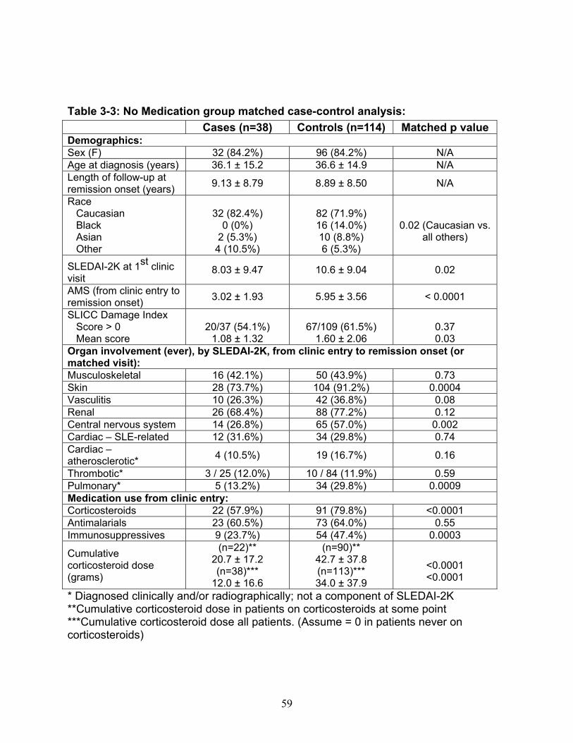
Table 3-4: Clinical characteristics of Medication (MED) compared to No Medication (NO MED) groups at remission start: MED (n=34) NO MED (n=38) Unmatched p
value Sex (F) 33 (97.1%) 32 (84.2%) 0.11 Age at diagnosis (years) 27.9 ± 11.7 36.1 ± 15.2 0.01
**Cumulative corticosteroid dose in patients on corticosteroids at some point ***Cumulative corticosteroid dose all patients. (Assume = 0 in patients never on corticosteroid
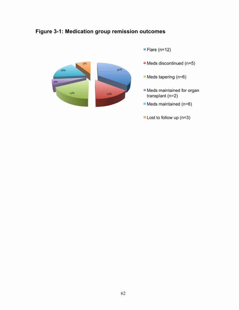
Figu
ure 3-1: MMedicatioon group
62
remissioon outcommes
63
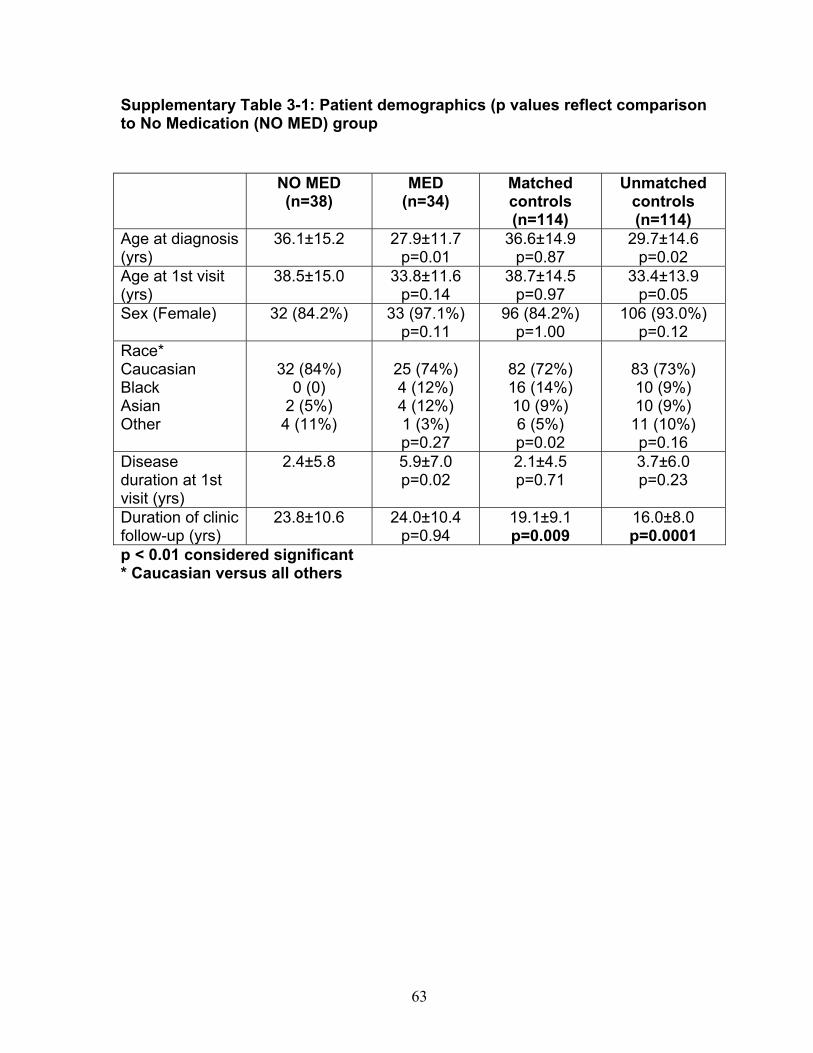
Supplementary Table 3-1: Patient demographics (p values reflect comparison to No Medication (NO MED) group NO MED
(n=38) MED
(n=34) Matched controls (n=114)
Unmatched controls (n=114)
Age at diagnosis (yrs)
36.1±15.2 27.9±11.7 p=0.01
36.6±14.9 p=0.87
29.7±14.6 p=0.02
Age at 1st visit (yrs)
38.5±15.0
33.8±11.6 p=0.14
38.7±14.5 p=0.97
33.4±13.9 p=0.05
Sex (Female) 32 (84.2%) 33 (97.1%) p=0.11
96 (84.2%) p=1.00
106 (93.0%) p=0.12
Race* Caucasian Black Asian Other
32 (84%)
0 (0) 2 (5%)
4 (11%)
25 (74%) 4 (12%) 4 (12%) 1 (3%) p=0.27
82 (72%) 16 (14%) 10 (9%) 6 (5%) p=0.02
83 (73%) 10 (9%) 10 (9%)
11 (10%) p=0.16
Disease duration at 1st visit (yrs)
2.4±5.8 5.9±7.0 p=0.02
2.1±4.5 p=0.71
3.7±6.0 p=0.23
Duration of clinic follow-up (yrs)
23.8±10.6
24.0±10.4 p=0.94
19.1±9.1 p=0.009
16.0±8.0 p=0.0001
p < 0.01 considered significant * Caucasian versus all others
64
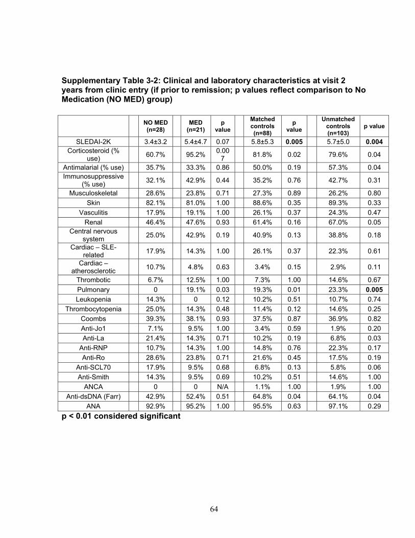
Supplementary Table 3-2: Clinical and laboratory characteristics at visit 2 years from clinic entry (if prior to remission; p values reflect comparison to No Medication (NO MED) group)
ANA 92.9% 95.2% 1.00 95.5% 0.63 97.1% 0.29 p < 0.01 considered significant
65
Supplementary Table 3-3: Clinical and laboratory characteristics at visit 5 years from clinic entry (if prior to remission; p values reflect comparison to No Medication (NO MED) group)
ANA 95.0% 93.3% 1.00 96.6% 1.00 100% 0.16 p < 0.01 considered significant
66
Supplementary Table 3-4: Clinical and laboratory characteristics at visit 2 years from SLE diagnosis (if prior to remission; p values reflect comparison to No Medication (NO MED) group)
ANA 95.7% 85.7% 0.54 89.5% 0.68 93.9% 1.00 p < 0.01 considered significant
67
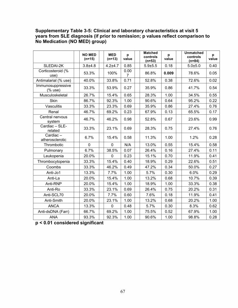
Supplementary Table 3-5: Clinical and laboratory characteristics at visit 5 years from SLE diagnosis (if prior to remission; p values reflect comparison to No Medication (NO MED) group)
Table 5-2: Comparison of IgM and IgG anti-chromatin and anti-dsDNA levels in SACQ patients with unselected SLE patients and healthy controls SACQ (n=23)
SLE control
(n=21) Healthy control (n=49)
p value SACQ vs. healthy controls
Anti-chromatin IgM
0.52 ± 0.67 (0 – 2.2)
0.39 ± 0.53 (0 – 1.76)
0.15 ± 0.02 (0.12 – 0.17)
(n=3)
0.52
Anti-chromatin IgG
0.59 ± 0.57 (0.07 – 2.21)
0.40 ± 0.39 (0.04 – 1.44)
0.08 ± 0.04 (0.01 – 0.20)
< 0.0001
Anti-dsDNA IgM 0.38 ± 0.26 (0 – 0.94)
0.13 ± 0.07 (0 – 0.30)
0.02 ± 0.10 (0.07 – 0.49)
<0.0001
Anti-dsDNA IgG 0.40 ± 0.62 (0.03 – 3.04)
0.22 ± 0.19 (0 – 0.65)
0.10 ± 0.04 (0.04 – 0.28)
0.002
110
Table 5-3: Mean anti-chromatin and anti-dsDNA levels during SACQ in patients who flared versus those who remained SACQ (1 sample/pt) Anti-chromatin Anti-dsDNA
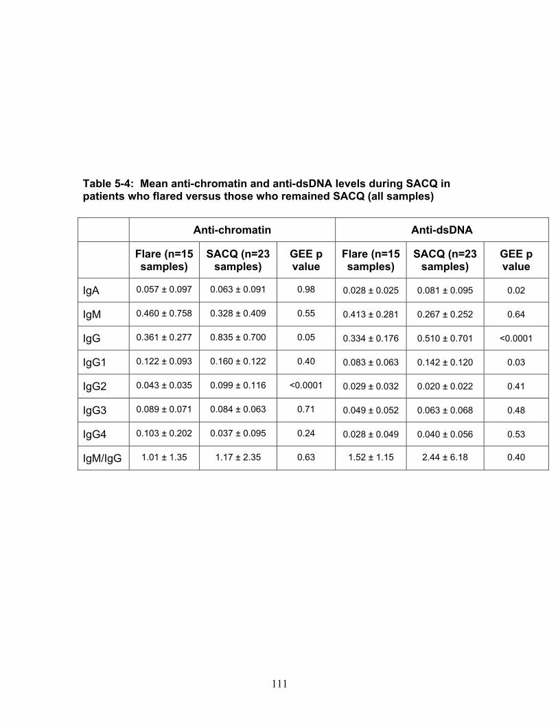
Table 5-4: Mean anti-chromatin and anti-dsDNA levels during SACQ in patients who flared versus those who remained SACQ (all samples) Anti-chromatin Anti-dsDNA
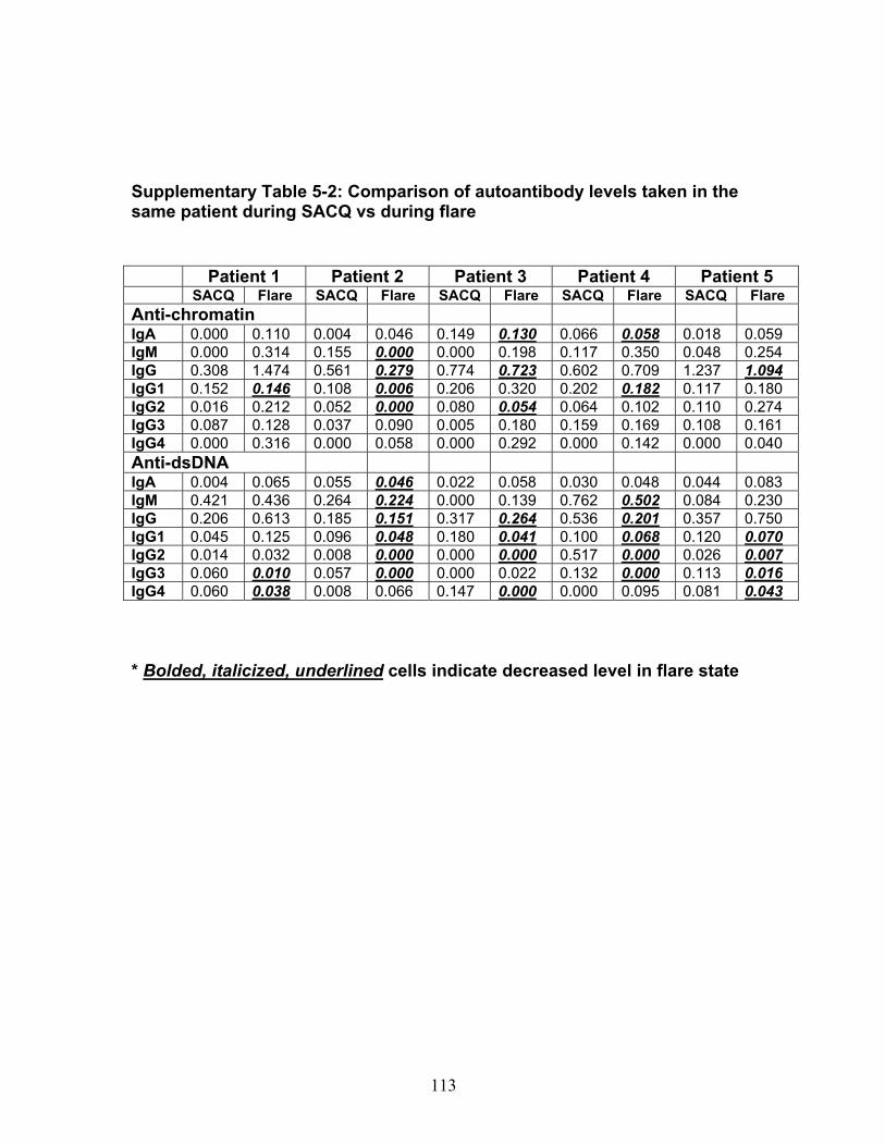
Anti-RNP (% positive) 8 (36.4) 7 (25.9) 29 (74.4) 0.43 0.004 * All durations measured in years
135
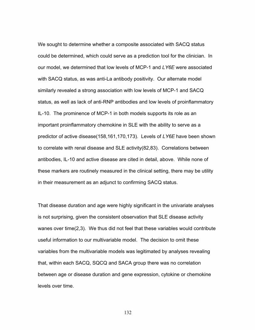
Table 6-2: Associations with SACQ status by logistic regression analysis OR (95% CI) P value Anti-La positivity 168.1 (4.85, >999) 0.005 MCP-1 high 0.87 (0.80, 0.96) 0.003 Ly6e high 0.90 (0.85, 0.97) 0.004
136
Figure 6-1: Relative gene expression
137
138
Figure 6-2: IFN score over time
0 20 40 600
20
40
60
80
100SACASACQSQCQ
Disease Duration (years)
IFN
Gen
e S
core
Per
cent
ile
139
Figure 6-3: Cytokine and chemokine concentrations
GM-CSF
SACQSQCQ
SACA0
200
400
600
800 p = 0.0004
NS
Con
cent
ratio
n (p
g/m
L)
IL-6
SACQSQCQ
SACA0
5
10
15
20
25
NS
p = 0.0018
Con
cent
ratio
n (p
g/m
L)
IL-10
SACQSQCQ
SACA0
10
20
30
NS
p < 0.0001
Con
cent
ratio
n (p
g/m
L)
140
IP-10
SACQSQCQ
SACA0
500
1000
1500
2000
2500
NS
p < 0.0001C
once
ntra
tion
(pg/
mL)
MCP-1
SACQSQCQ
SACA0
500
1000
1500
NS
p < 0.0001
Con
cent
ratio
n (p
g/m
L)
TNF-alpha
SACQSQCQ
SACA0
100
200
300
400
NS
p = 0.0017
Con
cent
ratio
n (p
g/m
L)
141
142
Chapter 7: General Discussion, Future Directions and
Conclusions
143
Why study remission?
Remission, whether spontaneous, or induced by treatment, represents a state of
reprieve from the signs and symptoms of an incurable disease, in this case, SLE.
That remissions occur, rarely, in SLE is widely accepted(3,16,57,68,69). Why
they occur, however, is not understood, but reflects a pathophysiologic shift
which, if harnessed, could represent the “holy grail” of SLE treatment. Of
particular interest are SACQ SLE patients, whose clinical remissions occur
despite the presence of autoantibodies which are known to be pathogenic and/or
hypocomplementemia, which reflects immune activation.
Describing remission
We took a stepwise approach to the study of remission and the patients in whom
it occurs. First, in Chapter 3, we described prolonged remission, precisely and
stringently defining it. We felt this was of paramount importance to ensure
homogeneity of the cohort and thus avert some of the shortcomings of the
studies with broadly defined remission states, described at length in Chapter 1.
It was through carefully defining this group that we could confirm in our case-
control analysis that those patients who had achieved prolonged remission
differed phenotypically from matched SLE controls in terms of their disease
activity in the years preceeding quiescence, and, as a result, the likelihood of
requiring corticosteroids and immunosuppressive medications. Still, these
differences were not sufficiently specific to identify patients who would evolve to
144
a remitted state a priori, and thus did not lend themselves to use in clinical
prediction. We were the first to identify a unique subset of SLE patients, whose
disease to date, with more than two decades of follow-up in some cases, has run
an atypically monophasic course. These patients are of particular interest, as
they have seemingly subverted the mechanism(s) which results in relapse in the
vast majority of SLE patients.
We then subdivided these patients into those with and without pathogenic
serologic activity, as the former group’s clinical-serologic discordance presents a
clinical conundrum, and likely stems from unique pathophysiologic mechanisms
underlying their unique disease state. Finally, we separated those in a clinically
quiescent state under the coverage of medications from those who achieved
quiescence without, which allowed for the identification of patients who were in
true remission versus those whose disease was merely suppressed by ongoing
pharmacotherapy. These two groups are likely unique pathophysiologically, thus
past studies which combined them may have inadvertently obfuscated important
results which could be borne out by their segregation.
Confirming quiescence
Thus in Chapter 3 we carefully defined two related clinical phenotypes: patients
in prolonged remission with or without concomitant pathogenic serology. At the
bedside, patients belonging to each of these groups were identical, but their
serologic profiles differed significantly. How could this serologic activity be so
145
closely linked to – or even be the driver of - disease activity in many SLE patients
and not have any deleterious effect on these seemingly remitted patients? The
next step, then, and the subject of Chapter 4, was to ensure that SACQ patients
were, indeed, in a remitted state, and that their disease course was not
insidiously progressive, falling under the radar of bedside clinical surveillance,
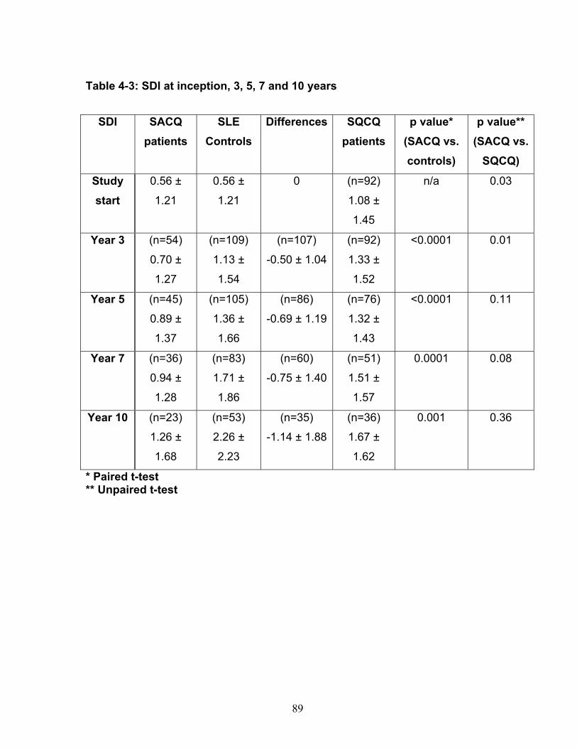
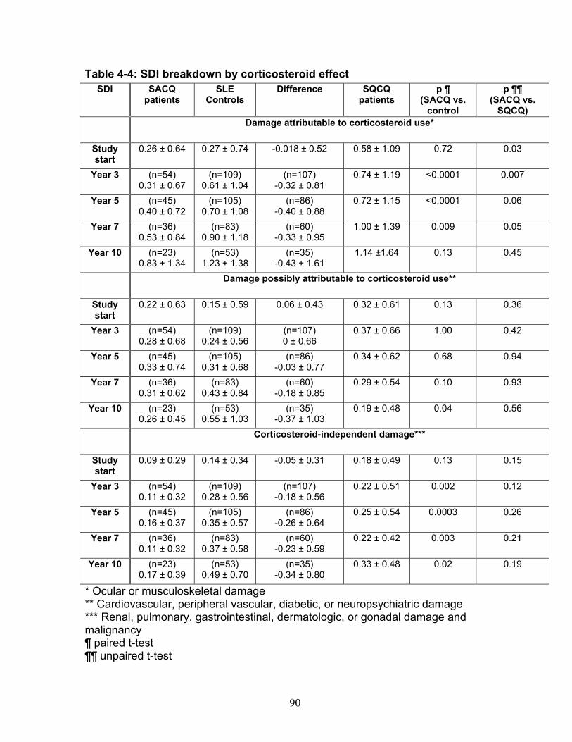
but accruing damage over the long term. We thus compared SACQ patients to
SQCQ and SACA patients and measured damage accrual over a decade. This
study corroborated our initial clinical impression of complete disease quiescence
during a SACQ period, with no evidence of disease-related damage accruing
over time.
From these initial two chapters, we carefully confirmed the existence of a unique
and contradictory cohort, whose discordance, at the patient level, poses a
management dilemma for the physician, who must reconcile a clinically
quiescent state, on the one hand, with serology suggestive of impending flare, on
the other. On an academic level, the existence of these patients and their
discrepant clinical/serologic profiles illuminates an important divergence from the
classic SLE pathophysiologic paradigm of pathogenic immune complex
deposition driving disease activity. Thus, at both a patient and population level,
there are compelling reasons to discover predictors of flare in SACQ patients: at
an individual level, severe relapse could be prevented by early treatment; for
SLE patients, in general, studying SACQ may elucidate unique pathophysiologic
mechanisms which could inform treatment targets.
146
The pursuit of novel biomarkers
Armed with the knowledge that conventional biomarkers were insufficient
predictors of flare in SACQ patients, we sought novel methods to detect
fluctuations in disease activity in this group. In Chapter 5 we explored the
predictive utility of anti-dsDNA and anti-chromatin antibody isotypes. Their
potential relevance as biomarkers in SACQ was founded upon numerous past
studies citing their relative specificity over conventional measures in SLE, in
general, and driven by a dearth of literature exploring their utility in remitted
patients. Studying subsets of anti-dsDNA and related anti-chromatin antibodies
was an intuitive step in confirming that the discordance of anti-dsDNA in SACQ
patients, overall, did not belie an association with disease activity if one were to
delve deeper and subdivide by isotype. While we found no association between
levels of either of these autoantibody isotypes and disease activity in SACQ
patients, and thus did not elucidate a novel biomarker, we felt that this negative
study was critical as it suggested that differences in immunoglobulin expression
did not appear to be the drivers of the SACQ phenotype.
We thus sought an alternate, biologically-plausible mechanism for SACQ clinical-
serologic discordance. That these patients were clinically well, presence of
pathogenic autoantibodies and evidence of immune activation notwithstanding,
suggested that a central player in SLE-associated inflammation was being
circumvented. We postulated that IFN-Į production, driven by TLR stimulation
147
by self nucleic acids, was a candidate. Our hypothesis was bolstered by past
studies revealing blunting of the IFN response in the face of persistent TLR
stimulation, which could account for the discordance of SACQ patients. Our
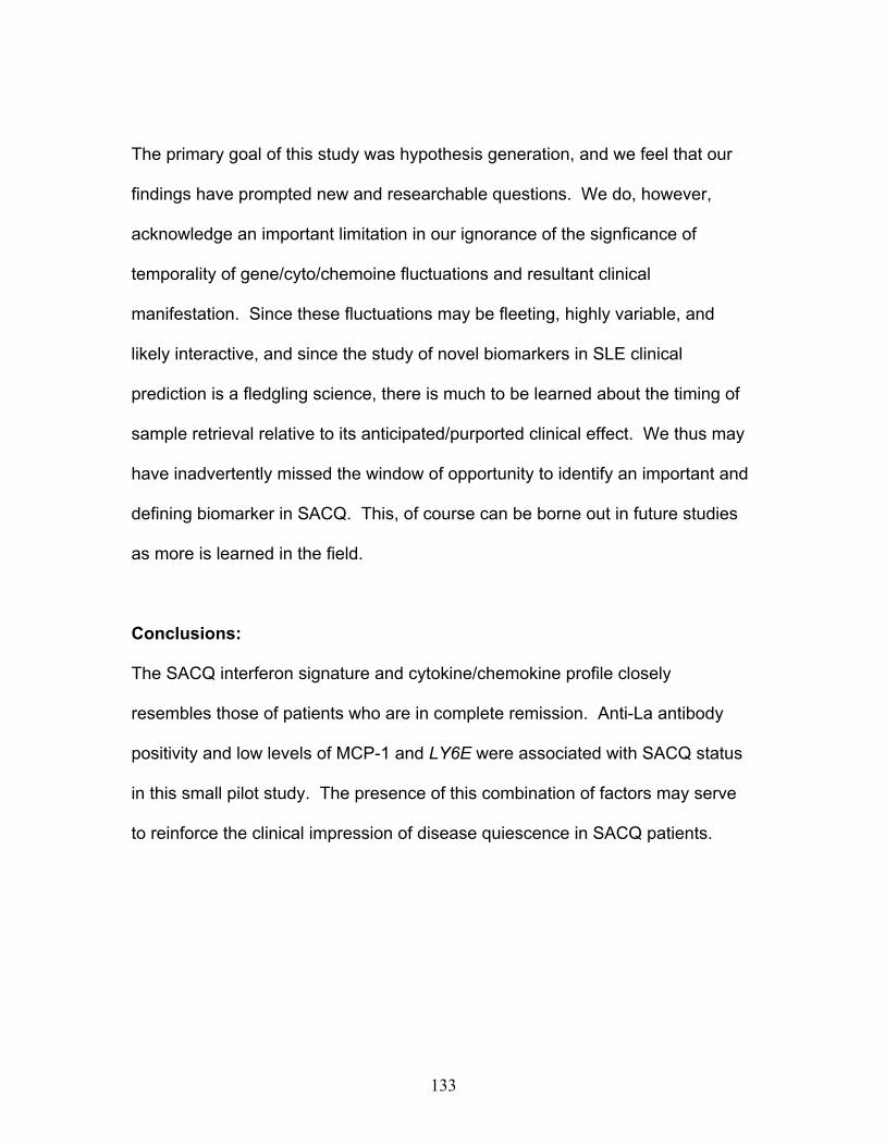
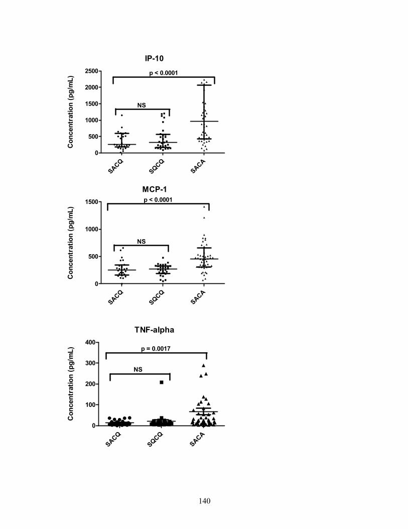
finding of SACQ patients’ IFN signature, cytokines and chemokines mirroring
those of SQCQ patients, and diverging significantly from SACA patients,
supports the theory of IFN blunting in these patients, to be borne out in future
studies. That anti-Ro and anti-La antibodies were significantly more prevalent in
SACQ than SACA patients, and that they have been shown to induce IFN
similarly supports this theory.
Major Contributions
SLE patients with clinical-serologic discordance present a clinical conundrum for
the treating physician – which of these parameters should guide management?
This has resulted in disparity in approach among clinicians, who can be
separated into two factions: those guided by clinical quiescence(67,68), and
those compelled to treat on the basis of serologic activity(65,66). While every
physician acknowledges that the “art” of medicine allows for differences in
approach between clinicians to the same patient, this dichotomy has very
significant ramifications for the SACQ cohort. On the one hand, SLE flare is
associated with significant morbidity, progressive damage accrual, and even
mortality; ongoing, frivolous immunosuppression may be similarly deleterious, on
the other. To our knowledge, this body of work represents the most exhaustive
study of this unique patient subset.
148
A strength of this work is in its meticulous definition and prospective, fastidious
surveillance of patients in remitted states. It thus provides compelling evidence
to suggest that prolonged SACQ periods, spanning years, do occur and, as such,
serologic activity in these patients should not be interpretted as a sign of
impending flare. We have identified a novel subset of SLE patients with a
monophasic clinical course, debunking the dogma that SLE will necessarily
relapse. Furthermore, we have proven that remission in these patients is not
undermined by subclinical, insidious damage accrual, which further reinforces
our position that these patients require close clinical monitoring, without ongoing
coverage with corticosteroids and/or immunosuppressive medications. The onus
thus rests with the treating physician to identify patients exhibiting this
discordance, and to manage them expectantly.
One might argue that the impact of this thesis is limited by the rarity of prolonged,
discordant remission in SLE, and that its findings are thus only applicable to few:
it is true that our most stringently-defined SACQ patients represent less than 2%
of our SLE cohort. However, accepting the published estimated global SLE
prevalence of 52 per 100,000 population(175), our findings, if adopted, could
directly impact the management of at least 62,000 SACQ patients worldwide.
This, in turn, could translate to considerable reduction in needless treatment-
associated morbidity, at the individual level, and have concomitant health
economic effects.
149
Generation of pathogenic autoantibodies is a hallmark of SLE, thought central to
the disease process, and is thus a disease classification criterion(105,176).
However, as outlined in Chapter 5, we found that, in SACQ patients, increased
levels of anti-dsDNA and anti-chromatin immunoglobulin levels (which are
classicaly considered pathogenic) were not predictive of flare, and were actually
numerically (but not statistically significantly) higher in those patients who
remained quiescent. This hypothesis-generating finding suggests that, in these
patients, the effects of these pathogenic antibodies are counteracted, and has
never been reported prior.
The study of differences in cytokine/chemokine and IFN-associated gene
expression between SACQ, SQCQ and SACA patients contributes to a growing
body of literature exploring the utility of novel biomarkers in SLE prognostication,
at large. Most notable is our finding of suppression of the IFN signature in SACQ
patients, despite an abundance of IFN-driving substrate in their robust
autoantibody profile. This finding is surprising, and thus hypothesis-generating,
with the potential for generalizability beyond SACQ patients. Specifically it
implies altered function and/or blockade of the IFN pathway in these patients,
with a resultant clinically quiescent phenotype. It directs future investigation
elucidating the nature of this discrepancy, and may yield important insights
leading to drugable targets.
150
Limitations
Prolonged remission is rare in SLE, and its prospective study is dependent upon
clinical encounters with these quiescent patients which, on average, occur every
six months in our clinic(68). A major issue which arises in studying uncommon
states is that of being underpowered to detect subtle but important differences
between groups. This can be addressed in future studies by lengthening our
recruitment window or by collaboration with other centres.
While it is pragmatic to see patients who are clinically well less frequently, as we
have in the case of SACQ patients, the latency between visits increases the
likelihood that unmeasured/undocumented fluctuations in serologic and/or clinical
status may have evolved. If this were to have occurred, these patients may have
been misclassified as “SACQ,” thus detracting from the homogeneity of the
sample and, potentially, the findings unique to this group. Of course, meticulous
attention was paid to a patient’s intervisit history, which was of utmost
importance in ascertaining continued clinical quiescence.
Given significant interethnic differences in SLE severity and phenotype, coupled
with inconsistent approaches to its management globally, the generalizability of
the findings of a single-centre study to SLE patients, at large, must be
considered. Specifically, the impact of studying a predominantly Caucasian
patient cohort such as ours must be balanced with the knowledge that Black,
Asian and Hispanic patients, phenotypically, have more severe disease, which is
151
likely reflective of both genetic and environmental differences between
groups(20). The generalizability of findings could be confirmed – or improved –
by pursuing a multicentre, international study wherein our previous findings could
be reproduced. This approach would also benefit our sample size and improve
our statistical power.
Future Directions
Despite extensive research to date, SLE genetics and pathophysiology remain
only partially elucidated. This may, in large part, be owing to its protean
manifestations, near-limitless presentations and considerable variability in
severity and phenotype across ethnicities. In fact, the clinical entity referred to
as “SLE” may actually represent the clinical endpoint of several unique
pathophysiologic pathways, unified by a propensity to autoimmunity. It is in this
context that we study the exceptional patients who evolve to a state of prolonged
clinical quiescence: if these patients can be compared to those whose disease
runs a more typical course, the differences discovered between groups may
illuminate important drivers of active disease. Furthermore, SACQ patients,
themselves, present a management dilemma for the treating physician, who
must balance their clinical-serologic discordance in bedside decision-making.
Reliable, objective biomarkers of impending disease activity in this cohort would
be of benefit in averting disease flare and, ultimately, treatment- and disease-
associated organ damage.
152
Thus SACQ patients are of particular interest, as described at length in previous
chapters, because of their surprising clinical-serologic discordance, which refutes
the classic lupus paradigm of loss of self-tolerance resulting in production of
pathogenic autoantibodies and resultant end-organ dysfunction and, ultimately,
damage. Our work to date addresses the clinical correlates of patients who
evolve to SACQ status, their damage accrual over time, and the early exploration
of alternate biomarkers which may assist in clinical decision making. SACQ
patients, however, represent a subset whose unique characteristics deserve
further investigation which may lead to a heightened appreciation of SLE
pathophysiology. There are thus several research questions which arise directly
from our work to date:
Does SACQ clinical quiescence stem from a lack of autoantigen?
SACQ patients unequivocally produce copious, consistent autoantibodies. While
anti-dsDNA antibodies are presumably complexed to nucleic acid autoantigen,
one needs to confirm that their presence is not simply reflective of excess
production of antibody. If this were the case, a state of clinical quiescence would
be unsurprising, as it is immune complex deposition - and not the presence of
excess immunoglobulin - which is thought to drive SLE.
To pursue this, we plan to isolate IgG from SACQ patients. If the IgG is
complexed, the same IFN response should be generated when reassayed, as it
153
is driven by the presence of immune complexes. If antigen exists in SACQ
serum, there should not be an excess of unbound anti-dsDNA antibodies.
Does the SACQ anti-dsDNA and anti-chromatin antibody profile evolve with
disease activity within the same patient over time?
The study outlined in Chapter 5 relied upon the availability of archived serum
samples of SACQ patients, drawn either during a SACQ period or during disease
flare. Serial samples, drawn longitudinally, over both of these disease states for
the same patient were not available. Fluctuations in levels and subtypes of
immunoglobulins within individual SACQ patients may be instructive in
determining patterns in and predictors of flare, both in this patient subset, and
SLE patients, more generally. For example, and as described in Chapter 5, high
levels of IgM may be protective in SLE(44,140). Anti-chromatin antibodies may
be more sensitive and specific in flare prognostication in some SLE
patients(50,53). Thus longitudinal analysis of these autoantibodies may
elucidate useful biomarkers, which may be implemented in daily practice, for
patients in whom fluctuations in anti-dsDNA and/or complement levels are not
concordant with disease activity.
Does the SACQ IFN signature and cytokine/chemokine profile evolve with
disease activity within the same patient over time?
The study outlined in Chapter 6 could be extended to include serial
prospectively-collected samples in SACQ, SQCQ and SACA patients, in order to
154
determine relative stability or, alternately, potentially instructive fluctuations in the
IFN signature and/or cytokine/chemokine profile over time. While, on the one
hand, there is conflicting evidence in the literature pertaining to the stability of the
IFN signature over time(82,83), we found that in SACQ patients, there was a
significant decrease. We speculated that this might stem from progressive TLR
tolerance, which could drive these patients’ phenotype. It would be instructive to
determine whether the IFN signature increases in the context of flare in SACQ
patients.
Similarly, levels of proinflammatory cytokines and chemokines were significantly
lower in clinically quiescent patients than in active patients in our study. We did
not, however, have the opportunity to follow fluctuations therein over time. They
may prove instructive in anticipating flare in SACQ patients, and would thus be of
considerable clinical utility.
Is TLR tolerance the driver of the SACQ phenotype?
The study outlined in Chapter 6 describes the IFN signature, and
proinflammatory cytokine and chemokine levels in SACQ patients compared to
those who are SQCQ and SACA. The mechanism behind the significantly lower
levels of these components in SACQ patients compared to SACA patients,
despite active pathogenic serology in the former group has not yet been
elucidated. A biologically plausible mechanism for SACQ patients’ clinical
phenotype is that of TLR tolerance, as outlined in Pau and colleagues and Kwok
155
and colleagues(87,88), but the theory has not been tested in this patient
population.
A potential method for pursuing this theory would be to reproduce Kwok and
colleagues’ experiment in SACQ patients. First, we would stimulate SACQ
patients’ PBMCs with TLR9 ligands and measure the IFN response. If TLR
tolerance were at the heart of the SACQ phenotype, we would expect a blunted
IFN response. We would then expose healthy control PBMCs to SACQ SLE
patients’ serum to compare IFN response, which we would anticipate would be
robust, given the abundance of TLR ligands in SACQ serum.
A corollary to this experiment would be the measurement of peripheral pDCs in
SACQ patients compared to SLE controls to explore whether a dearth of IFN-
producing cells is culprit for the discordant quiescence in SACQ patients.
Further analyses of pDC function could then be explored.
Could personalized medicine hold the key to averting flare in SLE patients?
The pursuit of novel biomarkers is tempered by both fiscal constraints and by the
limits of our understanding of the significance of derangements in an
exponentially expanding catalogue of cytokines, chemokines and genes. If one
were to hypothetically overcome the former issue, however, there might be
significant knowledge to be derived from prospectively collecting and studying
proinflammatory markers in fluctuating disease states, in all SLE patients
156
throughout their disease course. This pursuit may hold the key to identifying
pathophysiologic drivers of disease activity leading to the clinical phenotype of
SLE. Eventually, this could lead to successful anticipation and early treatment of
flare, or perhaps illuminate drugable targets tailored to the individual patient.
Conclusions
Prolonged clinical remission is a rare and desirable outcome in SLE and, in some
patients, may occur despite ongoing and robust serologic activity. These
remissions are not associated with subclinical damage accrual and can thus be
managed safely with close clinical observation, without prophylactic or
anticipatory treatment with corticosteroids and/or immunosuppressive
medications. The pathophysiologic milieu which permits for serologically
discordant remission is associated with a low IFN signature and proinflammatory
cytokine and chemokine expression, and does not appear to be driven by a
dearth of immunoglobulins/autoantibodies. Future studies exploring pathways
proximal to these biomarkers may elucidate important regulatory steps, which
could hold the key to inducing remissions in SLE patients, and are thus worthy of
pursuit.
157
References:
1. Harvey AM, Shulman LE, Tumulty PA, Conley CL, Schoenrich EH. Systemic lupus erythematosus: review of the literature and clinical analysis of 138 cases. Medicine 1954;33:291-425. 2. Swaak AJ, Nossent JC, Bronsveld W, Van Rooyen A, Nieuwenhuys EJ, Theuns L, et al. Systemic lupus erythematosus. II. Observations on the occurrence of exacerbations in the disease course: Dutch experience with 110 patients studied prospectively. Ann Rheum Dis 1989;48:455-60. 3. Drenkard C, Villa AR, Garcia-Padilla C, Perez-Vazquez ME, Alarcon-Segovia D. Remission of systemic lupus erythematosus. Medicine 1996;75:88-98. 4. Dubois EL. Systemic lupus erythematosus: recent advances in its diagnosis and treatment. Ann Int Med 1956;45:163-84. 5. Dubois EL, Tuffanelli DL. Clinical manifestations of systemic lupus erythematosus. JAMA 1964;190:104-11. 6. Heller CA, Schur PH. Serological and clinical remission in systemic lupus erythematosus. J Rheumatol 1985;12:916-8. 7. Weitzman RJ, Walker SE. Relation of titred peripheral pattern ANA to anti-DNA and disease activity in systemic lupus erythematosus. Ann Rheum Dis 1977;36:44-9. 8. Tozman ECS, Urowitz MB, Gladman DD. Prolonged complete remission in previously severe SLE. Ann Rheum Dis 1982;41:39-40. 9. Isenberg DA, Manson JJ, Ehrenstein MR, Rahman A. Fifty years of anti-ds DNA antibodies: are we approaching journey's end? Rheumatology (Oxford) 2007;46:1052-6. 10. Ho A, Barr SG, Magder LS, Petri M. A decrease in complement is associated with increased renal and hematologic activity in patients with systemic lupus erythematosus. Arthritis Rheum 2001;44:2350-7. 11. Esdaile JM, Joseph L, Abrahamowicz M, Li Y, Danoff D, Clarke AE. Routine immunologic tests in systemic lupus erythematosus: is there a need for more studies? J Rheumatol 1996;23:1891-6. 12. Lam GKW, Petri M. Assessment of systemic lupus erythematosus. Clin Exp Rheumatol 2005;23:S120-S32. 13. Bombardier C, Gladman DD, Urowitz MB, Caron D, Chang CH, SLE CoPSi. Derivation of the SLEDAI. Arthritis Rheum 1992;35:630-40. 14. Mosca M, Bombardieri S. Assessing remission in systemic lupus erythematosus. Clin Exp Rheumatol 2006;24:S-99-104. 15. Schneider M. Response and remission criteria for clinical trials in lupus - what can we learn from other diseases? Lupus 1999;8:627-31. 16. Barr SG, Zonana-Nacach A, Magder LS, Petri M. Patterns of disease activity in systemic lupus erythematosus. Arthritis Rheum 1999;42:2682-8. 17. Swaak AJ, van den Brink H, Smeenk R, Manger K, Kalden JR, Tosi S, et al. Systemic lupus erythematosus: clinical features in patients with a disease duration of over 10 years, first evaluation. Rheumatology 1999;38:953-8.
158
18. Mok CC, Lau CS, Ho CT, Wong RW. Do flares of systemic lupus erythematosus decline after menopause? Scan J Rheumatol 1999;28:357-62. 19. Formiga F, Moga I, Pac M, Mitjavila F, Rivera A, Pujol R. High disease activity at baseline does not prevent a remission in patients with systemic lupus erythematosus. Rheumatology 1999;38:724-7. 20. Pons-Estel GJ, Alarcon GS, Scofield L, Reinlib L, Cooper GS. Understanding the epidemiology and progression of systemic lupus erythematosus. Semin Arthritis Rheum 2010;39:257-68. 21. Nossent J, Kiss E, Rozman B, Pokorny G, Vlachoyiannopoulos P, Olesinska M, et al. Disease activity and damage accrual during the early disease course in a multinational inception cohort of patients with systemic lupus erythematosus. Lupus 2010;19:949-56. 22. Wais T, Fierz W, Stoll T, Villiger PM. Subclinical disease activity in systemic lupus erythematosus: immunoinflammatory markers do not normalize in clinical remission. J Rheumatol 2003;30:2133-9. 23. Steiman AJ, Gladman DD, Ibanez D, Urowitz MB. Outcomes in patients with systemic lupus erythematosus with and without a prolonged serologically active clinically quiescent period. Arthritis Car Res 2012;64:511-8. 24. Urowitz MB, Feletar M, Bruce IN, Ibanez D, Gladman DD. Prolonged remission in systemic lupus erythematosus. J Rheumatol 2005;32:1467-72. 25. Gladman DD, Urowitz MB. Prognostic subsets and mortality in systemic lupus erythematosus. In: Wallace D, Hahn B, eds. Dubois' lupus erythematosus. 6th ed. Baltimore: Williams & Wilkins; 2001:1213-327. 26. Gladman DD, Ginzler E, Goldsmith C, Fortin PR, Liang M, Urowitz M, et al. The development and initial valication of he systemicl upus international collaborating clinics/American College of Rheumatology Damage Index for systemic lupus erythematosus. Arthritis Rheum 1996;39:363-9. 27. Gladman DD, Urowitz M. The SLICC/ACR damage index: progress report and experience in the field. Lupus 1999;8:632-7. 28. Swaak AJG, van den Brink HG, Smeenk RJT, Manger K, Kalden JR, Tosi S, et al. Systemic lupus erythematosus: clinical features in patients with a disease duration of over 10 years, first evaluation. Rheumatology 1999;38:953-8. 29. Gladman DD, Urowitz M, Rahman P, Ibanez D, Tam L-S. Accrual of organ damage over time in patients with systemic lupus erythematosus. J Rheumatol 2003;30:1955-9. 30. Holborow EJ, Weir DM, Johnson GD. A serum factor in lupus erythematosus with affinity for tissue nuclei. BMJ 1957;2:732-4. 31. Ceppelini R, Polli E, Celada F. A DNA-reaction factor in serum of a patient with lupus erythematosus diffuses. Proc Soc Exp Biol Med 1957;96:472-574. 32. Koffler D, Schur PH, Kunkel HG. Immunological studies concerning the nephritis of systemic lupus erythematosus. J Exp Med 1967;126:607-24. 33. Hahn BH. Antibodies to DNA. N Engl J Med 1998;338:1359-68. 34. Isenberg DA, Smeenk R. Clinical laboratory assays for measuring anti-dsDNA antibodies. Where are we now? Lupus 2002;11:797-800. 35. Egner W. The use of laboratory tests in the diagnosis of SLE. J Clin Pathol 2000;53:424-32.
159
36. Smeenk R, Brinkman K, van den Brink H, Swaak T. A comparison of assays used for the detection of antibodies to DNA. Clin Rheumatol 1990;9 (Supplement 1):63-73. 37. Smeenk RJT, Van Rooijen A, Swaak TJG. Dissociation studies of DNA/anti-DNA complexes in relation to anti-DNA avidity. J Immunol Meth 1988;109:27-35. 38. Rothfield N, Stollar B. The relation of immunoglobulin class, pattern of anti-nuclear antibody, and complement-fixing antibodies to DNA in sera from patients with systemic lupus erythematosus. J Clin Invest 1967;46:1785-94. 39. Winfield JB, Faiferman I., Koffler D. Avidity of anti-DNA antibodies in serum and IgG glomerular eluates from patients with systemic lupus erythematosus. J Clin Invest 1977;59:90-6. 40. Puritz EM, Yount WJ, Newell M, Utsinger PD. Immunoglobulin classes and IgG subclasses of human antinuclear antibodies. A correlation of complement fixation and the nephritis of systemic lupus erythematosus. Clin Immunol Immunopathol 1973;2:98-113. 41. Villalta D, Romelli PB, Savina C, Bizzaro N, Tozzoli R, Tonutti E, et al. Anti-dsDNA antibody avidity determination by a simple reliable ELISA method for SLE diagnosis and monitoring. Lupus 2003;12:31-6. 42. Forger F, Matthias T, Oppermann M, Becker H, Helmke K. Clinical significance of anti-dsDNA antibody isotypes: IgG/IgM ratio of anti-dsDNA antibodies as a prognostic marker for lupus nephritis. Lupus 2004;13:36-44. 43. Krippner H, Merle S, Jorgens K, Pirlet K. Antibodies to dsDNA and ssDNA in the immunoglobulin classes IgG and IgM: prognostic value in the course of SLE. Z Rheumatol 1984;43:265-71. 44. Kessel A, Rosner I, Halasz K, Grushko G, Shoenfeld Y, Paran D, et al. Antibody clustering helps refine lupus prognosis. Semin Arthritis Rheum 2009;39:66-70. 45. Bijl M, Dijstelbloem HM, Oost WW, Bootsma H, Derksen RHWM, Aten J, et al. IgG subclass distribution of autoantibodies differs between renal and extra-renal relapses in patients with systemic lupus erythematosus. Rheumatology 2002;41:62-7. 46. Buyon JP, Tamerius J, Belmont HM, Abramson SB. Assessment of disease activity and impending flare in patients with systemic lupus erythematosus. Arthritis Rheum 1992;35:1028-37. 47. Lange K, Wasserman E, Slobody LB. The significance of serum complement levels for the diagnosis and prognosis of acute and subacute glomerulonephritis and lupus erythematosus disseminatus. Ann Int Med 1960;53:636-46. 48. Uko G, Christiansen FT, Dawkins RL. Serum C4 concentration in the monitoring of systemic lupus erythematosus: requirement for C4 allotyping. Rheumatol Int 1986;6:111-4. 49. Sturfelt G, Johnson U, Sjoholm AG. Sequential studies of complement activation in systemic lupus erythematosus. Scan J Rheumatol 1985;14:184-96. 50. Bizzaro N, Villalta D, Giavarina D, Tozzoli R. Are anti-nucleosome antibodies a better diagnostic marker than anti-dsDNA antibodies for systemic
160
lupus erythematosus? A systematic review and a study of metanalysis. Autoimmun Rev 2012;12:97-106. 51. Heidenreich U, Mayer G, Herold M, Klotz W, Stempfl Al-Jazrawi K, Lhotta K. Sensitivity and specificity of autoantibody tests in the differential diagnosis of lupus nephritis. Lupus 2009;18:1276-80. 52. Pradhan VD, Patwardhan MM, Ghosh K. Anti-nucleosome antibodies as a disease marker in systemic lupus erythematosus and its correlation with disease activity and other autoantibodies. Ind J Dermatol Venereol Leprol 2010;76:145-9. 53. Suleiman S, Kamaliah D, Nadeem A, Naing NN, Che Maraina CH. Anti-nucleosome antibodies as a disease activity marker in patients with systemic lupus erythematosus. Int J Rheum Dis 2009;12:100-6. 54. Kiss E, Lakos G, Szegedi G, Poor G, Szodoray P. Anti-nuscleosome antibody, a reliable indicator for lupus nephritis. Autoimmunity 2009;42:393-8. 55. Manson JJ, Ma A, Rogers P, Mason LJ, Berden JH, van der Vlag J, et al. Relationship between anti-dsDNA, anti-nucleosome and anti-alpha-actinin antibodies and markers of renal disease in patients with lupus nephritis: a prospective longitudinal study. Arthritis Res Ther 2009;11:R154. 56. Gutierrez-Adrianzen OA, Koutouzov S, Mota RMS, das Chagas Medeiros MM, Bach J-F, de Holanda Campos H. Diagnostic value of anti-nucleosome antibodies in the assessment of disease activity of systemic lupus erythematosus: a prospective study comparing anti-nucleosome with anti-dsDNA antibodies. J Rheumatol 2006;33:1538-44. 57. Gladman DD, Urowitz MB, Keystone EC. Serologically active clinically quiescent systemic lupus erythematosus: a discordance between clinical and serologic features. Am J Med 1979;66:210-5. 58. Pincus T, Schur PH, Rose JA, Decker JL, Talal N. Measurement of serum DNA-binding activity in systemic lupus erythematosus. N Engl J Med 1969;281:701. 59. Lightfoot RW, Jr., Redecha PB, Levesanos N. Longitudinal studies of anti-DNA antibody levels in SLE. Scan J Rheumatol 1975;Supp 11:52-8. 60. Schur PH, Sandson J. Immunologic factors and clinical activity in systemic lupus erythematosus. N Engl J Med 1968;278:533-8. 61. Swaak A, Aarden L, Statius Van Eps L, Feltkamp T. Anti-dsDNA and complement profiles as prognostic guides in systemic lupus erythematosus. Arthritis Rheum 1979;22:226-35. 62. Lange K, Ores R, Strauss W, Wachstein M. Steroid therapy of systemic lupus erythematosus based on immunologic considerations. Arthritis Rheum 1965;8:244-59. 63. Ter Borg E, Horst G, Hummel E, Limburg PC, Kallenberg CG. Measurement of increases in anti-double-stranded DNA antibody levels as a predictor of disease exacerbation in systemic lupus erythematosus. Arthritis Rheum 1990;33:634-43. 64. Walz LeBlanc BA, Gladman DD, Urowitz MB. Serologically active clinically quiescent systemic lupus erythematosus--predictors of clinical flares. J Rheumatol 1994;21:2239-41.
161
65. Bootsma H, Spronk PE, Derksen R, de Boer G, Wolters-Dicke H, Hermans J, et al. Prevention of relapses in systemic lupus erythematosus. Lancet 1995;345:1595-9. 66. Tseng C-E, Buyon JP, Kim M, Belmont HM, Mackay M, Diamond B, et al. The effect of moderate-dose corticosteroids in preventing severe flares in patients with serologically active, but clinically stable, systemic lupus erythematosus: findings of a prospective, randomized, double-blind, placebo-controlled trial. Arthritis Rheum 2006;54:3623-32. 67. Ng KP, Manson JJ, Rahman A, Isenberg DA. Association of antinucleosome antibodies with disease flare in serologically active clinically quiescent patients with systemic lupus erythematosus. Arthritis Rheum 2006;55:900-4. 68. Steiman AJ, Gladman DD, Ibanez D, Urowitz MB. Prolonged serologically active clinically quiescent systemic lupus erythematosus: frequency and outcome. J Rheumatol 2010;37:1822-7. 69. Conti F, Ceccarelli F, Perricone C, Miranda F, Truglia S, Massaro L, et al. Flare, persistently active disease, and serologically active clinically quiescent disease in systemic lupus erythematosus: a 2-year follow-up study. PLoS ONE 2012;7:e45934. 70. Gladman DD, Hirani N, Ibanez D, Urowitz MB. Clinically active serologically quiescent systemic lupus erythematosus. J Rheumatol 2003;30:1960-2. 71. Niewold TB, Clark DN, Salloum R, Poole BD. Interferon alpha in systemic lupus erythematosus. J Biomed Biotech 2010;2010:948364. 72. Niewold TB. Interferon alpha as a primary pathogenic factor in human lupus. J Interferon Cytokine Res 2011;31:887-92. 73. Bave U, Magnusson M, Eloranta M-L, Perers A, Alm GV, Ronnblom L. Fc gamma RIIa is expressed on natural IFN-alpha-producing cells (plasmacytoid dendritic cells) and is required for the IFN-alpha production induced by apoptotic cells combined with lupus IgG. J Immunol 2003;171:3296-302. 74. Marshak-Rothstein A. Toll-like receptors in systemic autoimmune disease. Nat Rev Immunol 2006;6:823-35. 75. Salloum R, Niewold TB. Interferon regulatory factors in human lupus pathogenesis. Translational Research: J Lab Clin Med 2011;157:326-31. 76. Bave U, Alm GV, Ronnblom L. The combination of apoptotic U937 cells and lupus IgG is a potent IFN-alpha inducer. J Immunol 2000;165:3519-26. 77. Ermann J, Bermas BL. The biology behind the new therapies for SLE. Int J Clin Pract 2007;61:2113-9. 78. Bengtsson AA, Sturfelt G, Truedsson L, Blomberg J, Alm G, Vallin H, et al. Activation of type I interferon system in systemic lupus erythematosus correlates with disease activity but not with antiretroviral antibodies. Lupus 2000;9:664-71. 79. Dall'era MC, Cardarelli PM, Preston BT, Witte A, Davis JC, Jr. Type I interferon correlates with serological and clinical manifestations of SLE. Ann Rheum Dis 2005;64:1692-7. 80. Kirou KA, Lee C, George S, Louca K, Peterson MGE, Crow MK. Activation of the interferon-alpha pathway identifies a subgroup of systemic lupus
162
erythematosus patients with distinct serologic features and active disease. Arthritis Rheum 2005;52:1491-503. 81. Venuturupalli S, Gudsoorkar V, Wallace D. Reconsidering antimalarials in systemic lupus erythematosus: developments of translational clinical interest. J Rheumatol 2012;39:1769-71. 82. Feng X, Wu H, Grossman JM, Hanvivadhanakul P, FitzGerald JD, Park GS, et al. Association of increased interferon-inducible gene expression with disease activity and lupus nephritis in patients with systemic lupus erythematosus. Arthritis Rheum 2006;54:2951-62. 83. Landolt-Marticorena C, Bonventi G, Lubovich A, Ferguson C, Unnithan T, Su J, et al. Lack of association between the interferon-alpha signature and longitudinal changes in disease activity in systemic lupus erythematosus. Ann Rheum Dis 2009;68:1440-6. 84. Morimoto AM, Flesher DT, Yang J, Wolslegel K, Wang X, Brady A, et al. Association of endogenous anti-interferon- autoantibodies with decreased interferon-pathway and disease activity in patients with systemic lupus erythematosus. Arthritis Rheum 2011;63:2407-15. 85. Niewold TB, Kelly JA, Flesch MH, Espinoza LR, Harley JB, Crow MK. Association of the IRF5 risk haplotype with high serum interferon-alpha activity in systemic lupus erythematosus patients. Arthritis Rheum 2008;58:2481-7. 86. Niewold TB, Kelly JA, Kariuki SN, Franek BS, Kumar AA, Kaufman KM, et al. IRF5 haplotypes demonstrate diverse serological associations which predict serum interferon alpha activity and explain the majority of the genetic association with systemic lupus erythematosus. Ann Rheum Dis 2012;71:463-8. 87. Pau E, Cheung Y-H, Loh C, Lajoie G, Wither JE. TLR tolerance reduces IFN-alpha production despite plasmacytoid dendritic cell expansion and anti-nuclear antibodies in NZB bicongenic mice. PLoS ONE 2012;7:e36761. 88. Kwok SK, Lee JY, Park SH, Cho ML, Min SY, Park SH, et al. Dysfunctional interferon-alpha production by peripheral plasmacytoid dendritic cells upon Toll-like receptor-9 stimulation in patients with systemic lupus erythematosus. Arthritis Res Ther 2008;10:R29. 89. Higgs BW, Liu Z, White B, Zhu W, White WI, Morehouse C, et al. Patients with systemic lupus erythematosus, myositis, rheumatoid arthritis and scleroderma share activation of a common type I interferon pathway. Ann Rheum Dis 2011;70:2029-36. 90. Urowitz MB, Gladman DD. Contributions of observational cohort studies in systemic lupus erythematosus: the University of Toronto lupus clinic experience. Rheum Dis Clin North Am 2005;31:211-21. 91. Wold RT YF, Tan EM, Farr RS. Deoxyribonucleic acid antibody: a method to detect its primary interaction with deoxyribonucleic acid. Science 1968;161:806-7. 92. Whicher JT PC, Spencer K. Immunonephelometric and immunoturbidimetric assays for proteins. Crit Rev Clin Lab Sci 1983;18:213-60. 93. Ibanez D, Urowitz MB, Gladman DD. Summarizing disease features over time: I. Adjusted mean SLEDAI derivation and application to an index of disease activity in lupus. J Rheumatol 2003;30:1977-82.
163
94. Aggarwal NR, Szostek JH. 52-year-old man with arthralgias, fever, and fatigue. Mayo Clin Proc 2010;85:568-71. 95. Guillevin L, Lhote F. Polyarteritis nodosa and microscopic polyangiitis. Clin Exp Immunol 1995;101(Suppl 1):22-3. 96. Singh-Grewal D, Schneider R, Bayer N, Feldman BM. Predictors of disease course and remission in systemic juvenile idiopathic arthritis: significance of early clinical and laboratory features. Arthritis Rheum 2006;54:1595-601. 97. Sultan SM, Ioannou Y, Moss K, Isenberg DA. Outcome in patients with idiopathic inflammatory myositis: morbidity and mortality. Rheumatology 2002;41:22-6. 98. Fernandez LS, Sanchez JLA, Ginzler EM. Treatment of lupus nephritis. Rhematol Clin 2008;4:140-51. 99. Ainiala H, Loukkola J, Peltola J, Korpela M, Hietaharju A. The prevalence of neuropsychiatric syndromes in systemic lupus erythematosus. Neurology 2001;57:496-500. 100. Brey RL, Holliday SL, Saklad AR, Navarrete MG, Hermosillo-Romo D, Stallworth CL, et al. Neuropsychiatric syndromes in lupus. Neurology 2002;58:1214-20. 101. Steiman AJ, Urowitz M, Wither J, Ibanez D, Gladman DD. Prolonged serologically active clinically quiescent (SACQ) systemic lupus erythematosus (SLE): novel predictors of flare? Arthritis Rheum 2010;62:S783. 102. Forger F, Matthias T, Oppermann M, Becker H, Helmke K. Clinical significance of anti-dsDNA antibody isotypes: IgG/IgM ratio of anti-dsDNA antibodies as a prognostic marker for lupus nephritis. Lupus 2004;13:36-44. 103. Rahman A, Isenberg DA. Systemic lupus erythematosus. N Engl J Med 2008;358:929-39. 104. Tan EM, Cohen AS, Fries JF, Masi AT, McShane DJ, Rothfield NF. The 1982 reviewed criteria for the classification of systemic lupus erythematosus. Arthritis Rheum 1982;25:1271-7. 105. Hochberg M. Updating the American College of Rheumatology revised criteria for the classification of systemic lupus erythematosus. Arthritis Rheum 1997;40:1725-34. 106. Zonana-Nacach A, Barr SG, Magder LS, Petri M. Damage in systemic lupus erythematosus and its association with corticosteroids. Arthritis Rheum 2000;43:1801-8. 107. Swaak A, Groenwold L, Aarden L, Statius Van Eps L, Feltkamp T. Prognostic value of anti-dsDNA in SLE. Ann Rheum Dis 1982;41:388-95. 108. Kasitanon N, Magder LS, Petri M. Predictors of survival in systemic lupus erythematosus. Medicine 2006;85:147-56. 109. Becker-Merok A, Nossent HC. Damage accumulation in systemic lupus erythematosus and its relation to disease activity and mortality. J Rheumatol 2006;33:1570-7. 110. Sitia S, Atzeni F, Sarzi-Puttini P, Di Bello V, Tomasoni L, Delfino L, et al. Cardiovascular involvement in systemic autoimmune diseases. Autoimmun Rev 2009;8:281-6.
164
111. Sattar N, McCarey DW, Capell H, McInnes IB. Explaining how "high-grade" systemic inflammation accelerates vascular risk in rheumatoid arthritis. Circulation 2003;108:2957-63. 112. Thomas GN, Tam L-S, Tomlinson B, Li EK. Accelerated atherosclerosis in patients with systemic lupus erythematosus: a review of the causes and possible prevention. Hong Kong Med J 2002;8:26-32. 113. Ibanez D, Urowitz MB, Gladman DD. Disease activity over time (Adjusted Mean SLEDAI) predicts mortality and damage in SLE. J Rheumatol 2005;32:824-7. 114. Szekanecz Z, Shoenfeld Y. Lupus and cardiovascular disease: the facts. Lupus 2006;15:3-10. 115. Urowitz MB, Bookman AA, Koehler BE, Gordon DA, Smythe HA, Ogryzlo MA. The bimodal mortality pattern of systemic lupus erythematosus. Am J Med 1976;60:221-5. 116. Lee S-S, Singh S, Link K, Petri M. High-sensitivity c-reactive protein as an associate of clinical subsets and organ damage in systemic lupus erythematosus. Semin Arthritis Rheum 2008;38:41-54. 117. MacGowan JR, Ellis S, Griffiths M, Isenberg DA. Retrospective analysis of outcome in a cohort of patients with lupus nephritis treated between 1977 and 1999. Rheumatology 2002;41:981-7. 118. Bastian HM, Roseman JM, McGwin Jr G, Alarcon GS, Friedman AW, Fessler BJ. Systemic lupus erythematosus in three ethnic groups. XII. Risk factors for lupus nephritis after diagnosis. Lupus 2002;11:152-60. 119. van Bavel CC, van Der Vlag J, Berden JH. Glomerular binding of anti-dsDNA autoantibodies: the dispute resolved? Kidney Int 2007;71:600-1. 120. Swaak AJ, Groenwold J, Bronsveld W. Predictive value of complement profiles and anti-dsDNA in systemic lupus erythematosus. Ann Rheum Dis 1986;45:359-66. 121. Riboldi P, Gerosa M, Moroni G, Radice A, Allegri F, Sinico A, et al. Anti-DNA antibodies: a diagnostic and prognostic tool for systemic lupus erythematosus? Autoimmunity 2005;38:39-45. 122. Lightfoot RW, Jr., Hughes GR. Significance of persisting serologic abnormalities in SLE. Arthritis Rheum 1976;19:837-43. 123. Mikdashi J, Handwerger B. Predictors of neuropsychiatric damage in systemic lupus erythematosus: data from the Maryland lupus cohort. Rheumatology 2004;43:1555-60. 124. Zuniga R, Markowitz GS, Arkachaisri T, Imperatore EA, D'Agati VD, Salmon JE. Identification of IgG subclasses and c-reactive protein in lupus nephritis: the relationship between the composition of immune deposits and FC gamma receptor type IIA alleles. Arthritis Rheum 2003;48:460-70. 125. Ho A, Magder LS, Barr SG, Petri M. Decreases in anti-double-stranded DNA levels are associated with concurrent flares in patients with systemic lupus erythematosus. Arthritis Rheum 2001;44:2342-9. 126. Ibanez D, Gladman DD, Urowitz MB. Adjusted mean Systemic Lupus Erythematosus Disease Activity Index-2K is a predictor of outcome in SLE. J Rheumatol 2005;32:824-7.
165
127. Yee C-S, Hussein H, Skan J, Bowman S, Situnayake D, Gordon C. Association of damage with autoantibody profile, age, race, sex and disease duration in systemic lupus erythematosus. Rheumatology 2003;42:276-9. 128. Prasad R, Ibanez D, Gladman DD, Urowitz M. Anti-dsDNA and anti-Sm antibodies do not predict damage in systemic lupus erythematosus. Lupus 2006;15:285-91. 129. Isenberg D. Anti-dsDNA antibodies: still a useful criterion for patients with systemic lupus erythematosus? Lupus 2004;1:881-5. 130. Mostoslavsky G, Fischel R, Yachimovich N, Yaarkoni Y, Rosenmann E, Monestier M. Lupus anti-DNA autoantibodies cross-react with a glomerular structural protein: a case for tissue injury by molecular mimicry. Eur J Immunol 2001;31:1221-7. 131. Neogi T, Gladman DD, Ibanez D, Urowitz M. Anti-dsDNA antibody testing by Farr and ELISA techniques is not equivalent. J Rheumatol 2006;33:1785-8. 132. Amoura Z, Koutouzov S, Chabre H, Cacoub P, Amoura I, Musset L, et al. Presence of antinucleosome autoantibodies in a restricted set of connective tissue diseases. Arthritis Rheum 2000;43:76-84. 133. Su Y, Jia RL, Han L, Li ZG. Role of anti-nucleosome antibody in the diagnosis of systemic lupus erythematosus. Clin Immunol 2007;122:115-20. 134. Duzgun N, Sahin M, Genc Y, Tutkak H. Antinucleosome antibodies and systemic lupus erythematosus. Ann N Y Acad Sci 2007;1109:421-8. 135. Min DJ, Kim SJ, Park SH, Seo YI, Kang HJ, Kim WU, et al. Anti-nucleosome antibody: significance in lupus patients lacking anti-double-stranded DNA antibody. Clin Exp Rheumatol 2002;20:13-8. 136. Gomez-Puerta JA, Burlingame RW, Cervera R. Anti-chromatin (anti-nucleosome) antibodies: diagnostic and clinical value. Autoimmunity Rev 2008;7:606-11. 137. Ghillani-Dalbin P, Amoura Z, Cacoub P, Charuel JL, Diemert MC, Piette JC, et al. Testing for anti-nucleosome antibodies in daily practice: a monocentric evaluation in 1696 patients. Lupus 2003;12:833-7. 138. Yager TD, McMurray CT, Van Holde KE. Salt-induced release of DNA from nucleosome core particles. Biochemistry 1989;28:2271-81. 139. ter Borg EJ, Horst G, Hummel E, Limburg PC, Kallenberg CG. Rises in anti-double stranded DNA antibody levels prior to exacerbations of systemic lupus erythematosus are not merely due to polyclonal B cell activation. Clin Immunol Immunopathol 1991;59:117-28. 140. Werwitzke S, Trick D, Kamino K, Matthias T, Kniesch K, Schlegelberger B, et al. Inhibition of lupus disease by anti-double-stranded DNA antibodies of the IgM isotype in the (NZB x NZW)F1 mouse. Arthritis Rheum 2005;52:3629-38. 141. Spiegelberg HL. Biological activities of immunoglobulins of different classes and subclasses. Adv Immunol 1974;19:259-94. 142. Flierman R, Daha MR. Pathogenic role of anti-C1q autoantibodies in the development of lupus nephritis--a hypothesis. Molec Immunol \2007;44:133-8. 143. Muller S, Dieker, J., Tincani, A., Meroni, PL. Pathogenic anti-nucleosome antibodies. Lupus 2008;17:431-6.
166
144. Van der Vlag JBJ. Lupus nephritis: role of antinucleosome antibodies. Semin Nephrol 2011;31:376-89. 145. Dieker JWC, van der Vlag J, Berden JHM. Triggers for anti-chromatin autoantibody production in SLE. Lupus 2002;11:856-64. 146. Obermoser G, Pascual V. The interferon-alpha signature of systemic lupus erythematosus. Lupus 2010;19:1012-9. 147. Ronnblom LE, Alm GV, Oberg KE. Possible induction of systemic lupus erythematosus by interferon-alpha treatment in a patient with a malignant carcinoid tumour. J Int Med 1990;227:207-10. 148. Niewold TB, Swedler WI. Systemic lupus erythematosus arising during interferon-alpha therapy for cryoglobulinemic vasculitis associated with hepatitis C. Clini Rheumatol 2005;24:178-81. 149. Ioannou Y, Isenberg DA. Current evidence for the induction of autoimmune rheumatic manifestations by cytokine therapy. Arthritis Rheum 2000;43:1431-42. 150. Paladino P, Cummings DT, Noyce RS, Mossman KL. The IFN-independent response to virus particle entry provides a first line of antiviral defense that is independent of TLRs and retinoic acid-inducible gene I. J Immunol 2006;177:8008-16. 151. Lindau D, Mussard J, Rabsteyn A, Ribon M, Kotter I, Igney A, et al. TLR9 independent interferon alpha production by neutrophils on NETosis in response to circulating chromatin, a key lupus autoantigen. Ann Rheum Dis 2013. 152. Gladman DD, Ibanez D, Urowitz MB. Systemic lupus erythematosus disease activity index 2000. J Rheumatol 2002;29:288-91. 153. Ytterberg SR, Schnitzer TJ. Serum interferon levels in patients with systemic lupus erythematosus. Arthritis Rheum 1982;25:401-6. 154. Niewold TB, Hua J, Lehman TJA, Harley JB, Crow MK. High serum IFN-alpha activity is a heritable risk factor for systemic lupus erythematosus. Genes Immunity 2007;8:492-502. 155. Postal M, Sinicato NA, Pelicari KO, Marini R, Lavras Costallat LT, Appenzeller S. Clinical and serological manifestations associated with interferon- levels in childhood-onset systemic lupus erythematosus. Clinics (Sao Paulo, Brazil) 2012;67:157-62. 156. Sigurdsson S, Nordmark G, Garnier S, Grundberg E, Kwan T, Nilsson O, et al. A risk haplotype of STAT4 for systemic lupus erythematosus is over-expressed, correlates with anti-dsDNA and shows additive effects with two risk alleles of IRF5. Hum Mol Genet 2008;17:2868-76. 157. Weckerle CE, Franek BS, Kelly JA, Kumabe M, Mikolaitis RA, Green SL, et al. Network analysis of associations between serum interferon-alpha activity, autoantibodies, and clinical features in systemic lupus erythematosus. Arthritis Rheum 2011;63:1044-53. 158. Fu Q, Chen X, Cui H, Guo Y, Chen J, Shen N, et al. Association of elevated transcript levels of interferon-inducible chemokines with disease activity and organ damage in systemic lupus erythematosus patients. Arthritis Res Ther 2008;10:R112.
167
159. Tian S, Li J, Wang L, Liu T, Liu H, Cheng G, et al. Urinary levels of RANTES and M-CSF are predictors of lupus nephritis flare. Inflamm Res 2007;56:304-10. 160. Ripley BJ, Goncalves B, Isenberg DA, Latchman DS, Rahman A. Raised levels of interleukin 6 in systemic lupus erythematosus correlate with anaemia. Ann Rheum Dis 2005;64:849-53. 161. Okamoto H, Iikuni N, Kamitsuji S, Yoshio T, Minota S, Kamatani N. IP-10/MCP-1 ratio in CSF is an useful diagnostic marker of neuropsychiatric lupus patients. Rheumatology (Oxford) 2006;45:232-4. 162. Enghard P, Langnickel D, Riemekasten G. T cell cytokine imbalance towards production of IFN-gamma and IL-10 in NZB/W F1 lupus-prone mice is associated with autoantibody levels and nephritis. Scan J Rheumatol 2006;35:209-16. 163. Capper ER, Maskill JK, Gordon C, Blakemore AIF. Interleukin (IL)-10, IL-1ra and IL-12 profiles in active and quiescent systemic lupus erythematosus: could longitudinal studies reveal patient subgroups of differing pathology? Clin Exp Immunol 2004;138:348-56. 164. Bender R, Lange S. Adjusting for multiple testing - when and how? J Clin Epi 2001;54:343-9. 165. Vallin H, Perers A, Alm GV, Ronnblom L. Anti-double-stranded DNA antibodies and immunostimulatory plasmid DNA in combination mimic the endogenous IFN-alpha inducer in systemic lupus erythematosus. J Immunol 1999;163:6306-13. 166. Li QZ, Zhou J, Lian Y, Zhang B, Branch VK, Carr-Johnson F, et al. Interferon signature gene expression is correlated with autoantibody profiles in patients with incomplete lupus syndromes. Clin Exp Immunol 2010;159:281-91. 167. Balboni I, Niewold TB, Morgan G, Limb C, Eloranta ML, Ronnblom L, et al. Detection of anti-Ro, La, Smith and RNP autoantibodies by autoantigen microarray analysis and interferon-alpha induction in juvenile dermatomyositis. Arthritis Rheum 2013. 168. Ching KH, Burbelo PD, Tipton C, Wei C, Petri M, Sanz I, et al. Two major autoantibody clusters in systemic lupus erythematosus. PloS One 2012;7:e32001. 169. Liu X, Mayes MD, Tan FK, Wu M, Reveille JD, Harper BE, et al. Correlation of interferon-inducible chemokine plasma levels with disease severity in systemic sclerosis. Arthritis Rheum 2013;65:226-35. 170. Bauer JW, Petri M, Batliwalla FM, Koeuth T, Wilson J, Slattery C, et al. Interferon-regulated chemokines as biomarkers of systemic lupus erythematosus disease activity: a validation study. Arthritis Rheum 2009;60:3098-107. 171. Ohl K, Tenbrock K. Inflammatory cytokines in systemic lupus erythematosus. J Biomed Biotech 2011;2011:432595. 172. Yao Y, Richman L, Higgs BW, Morehouse CA, de los Reyes M, Brohawn P, et al. Neutralization of interferon-alpha/beta-inducible genes and downstream effect in a phase I trial of an anti-interferon-alpha monoclonal antibody in systemic lupus erythematosus. Arthritis Rheum 2009;60:1785-96.
168
173. Barbado J, Martin D, Vega L, Almansa R, Goncalves L, Nocito M, et al. MCP-1 in urine as biomarker of disease activity in Systemic Lupus Erythematosus. Cytokine 2012;60:583-6. 174. Ghirelli C, Zollinger R, Soumelis V. Systematic cytokine receptor profiling reveals GM-CSF as a novel TLR-independent activator of human plasmacytoid predendritic cells. Blood 2010;115:5037-40. 175. Danchenko N, Satia JA, Anthony MS. Epidemiology of systemic lupus erythematosus: a comparison of worldwide disease burden. Lupus 2006;15:308-18. 176. Petri M, Orbai AM, Alarcon GS. Derivation and validation of the Systemic Lupus International Collaborating Clinics classification criteria for systemic lupus erythematosus. Arthritis Rheum 2012;64:2677-86.