+ All Categories
Home > Documents > Serous Retinal Detachment in Hypertensive Posterior Reversible

Serous Retinal Detachment in Hypertensive Posterior Reversible

Date post: 09-Feb-2022
Category:
Upload: others
View: 2 times
Download: 0 times
Share this document with a friend
3
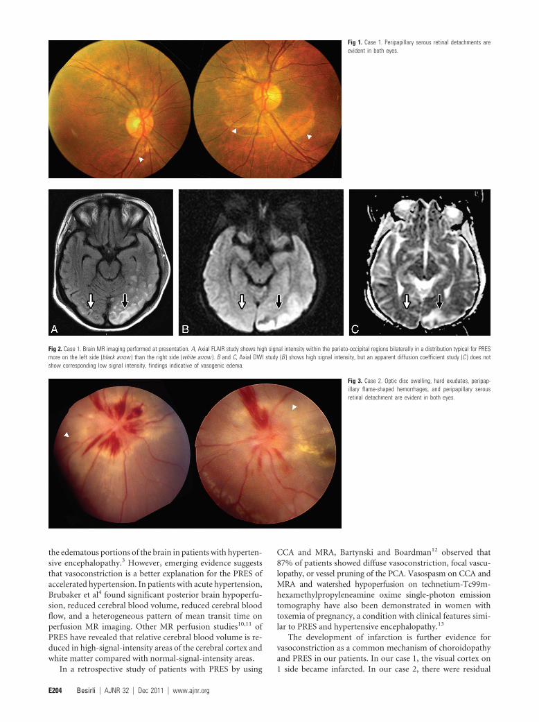
CASE REPORT Serous Retinal Detachment in Hypertensive Posterior Reversible Encephalopathy Syndrome C.G. Besirli P. Sudhakar J. Wesolowski J.D. Trobe SUMMARY: In accelerated hypertension, vasogenic brain edema associated with PRES may repre- sent either autoregulatory breakthrough leading to vasodilation or excessive autoregulation leading to vasoconstriction. We describe 2 patients with PRES in accelerated hypertension who had serous retinal detachments, a vasoconstrictive phenomenon. The concurrence of serous retinal detachment and PRES offers intriguing support for the idea that vasoconstriction rather than vasodilation is the mechanism of vasogenic edema in PRES. ABBREVIATIONS: CCA catheter cerebral angiography; DWI diffusion-weighted imaging; FLAIR fluid-attenuated inversion recovery; MRA MR angiography; PCA posterior cerebral artery; PRES posterior reversible encephalopathy syndrome; RPE retinal pigment epithelium P RES is manifested clinically by headache, seizures, altered mental status, and retrogeniculate visual loss, with scat- tered foci of high T2/FLAIR signal intensity without restricted diffusion, reflecting vasogenic edema. 1 In the setting of ac- celerated hypertension, there are 2 competing hypotheses to explain the vasogenic edema: 1) autoregulatory breakthrough leading to the dilation of cerebral arterial vessels, hyper- perfusion injury to the capillary bed, and secondary cerebral edema 2,3 ; and 2) autoregulatory excess leading to constriction of cerebral arterial vessels, ischemia of vascular endothelial cells, and vascular leakage. 4,5 The choroidopathy of hypertension is attributed to vaso- constriction of choroidal arterioles, 6 which causes RPE necro- sis and overlying serous detachment of the retina. Although commonly reported in acute hypertension, choroidopathy has been described in only 1 case of hypertensive encephalopathy 7 and never in PRES. We describe 2 patients with serous retinal detachment and PRES in the setting of accelerated hypertension. The occur- rence of choroidopathy in PRES offers anecdotal support for vasoconstriction as the mechanism underlying the vasogenic edema of PRES in accelerated hypertension. Case Reports Case 1. A 22-year-old woman with angioimmunoblastic lym- phoma, treated with cyclophosphamide, hydroxydaunorubicin, vincristine, prednisolone, and gemcitabine, developed renal failure, severe hypertension (182/96 mm Hg), generalized seizures, and de- pressed consciousness. Ophthalmoscopy revealed bilateral peripapil- lary serous retinal detachment (Fig 1). After she returned to full consciousness, a complete right homonymous hemianopia was elic- itable. MR imaging demonstrated high T2/FLAIR signal intensity in the posterior parietal and occipital lobes without restricted diffu- sion (Fig 2), a finding compatible with PRES. The serous retinal de- tachments disappeared within weeks, but the right homonymous hemianopia persisted. Repeat brain MR imaging showed disappear- ance of the signal-intensity abnormality in the right occipital lobe and development of restricted diffusion in the left occipital lobe, implying conversion to infarction (not shown). Case 2. A 15-year-old boy presented with a seizure in the setting of severe hypertension (250/140 mm Hg) secondary to crescentic glomerulonephritis. Ophthalmoscopy showed serous retinal de- tachment, optic disc swelling, hard exudates, and peripapillary flame- shaped hemorrhages bilaterally (Fig 3). MR imaging demonstrated occipital T2/FLAIR hyperintensities without restricted diffusion, consistent with PRES (Fig 4). After the patient returned to full con- sciousness, visual acuity was 20/200 in both eyes and Humphrey vi- sual fields showed enlarged blind spots. During the next 6 weeks, visual acuity gradually recovered to 20/20 bilaterally. In view of the recovery of vision, brain MR imaging was not repeated. Six months later, visual acuity remained 20/20 in both eyes, but he had tiny scotomas, corresponding to infarcts of the RPE (not shown). Discussion We have described 2 patients who developed binocular serous retinal detachments in the setting of accelerated hypertension and PRES. Considering that clinical, pathologic, and experi- mental evidence indicates choroidal vasoconstriction as the underlying cause of serous retinal detachments, we believe that these cases fortify the concept that vasoconstriction rather than vasodilation underlies the development of PRES. Ample evidence supports a link between systemic hyper- tension and vasoconstrictive choroidopathy. Histopathologic studies of postmortem specimens drawn from the acute isch- emic phase of hypertension show constriction of choroidal arterioles with obliteration of their lumina. 6 Patchy filling of the choriocapillaris on fluorescein angiography has been doc- umented in patients with hypertension 8 ; and fibrinoid necro- sis of choroidal arteries and arterioles with occlusion of the choriocapillaris has been noted on histopathogic studies of human eyes with accelerated hypertension. 9 Primate models of renovascular hypertension have shown serous retinal de- tachment and RPE lesions associated with delayed or patchy choroidal filling. 6 The vasodilation mechanism of PRES in accelerated hyper- tension is supported by studies showing increased perfusion in Received September 28, 2010; accepted after revision October 14. From the Department of Ophthalmology and Visual Sciences (C.G.B., P.S., J.D.T.), W.K. Kellogg Eye Center, Department of Neurology (J.D.T.), and Department of Radiology (Neuroradiology) (J.W.), University of Michigan, Ann Arbor, Michigan. Paper previously presented at: Annual Meeting of the North American Neuro-Ophthalmology Society, March 6 –11, 2010; Tucson, Arizona. Please address correspondence to Jonathan D. Trobe, MD, Department of Ophthalmology and Visual Sciences, W.K. Kellogg Eye Center, 1000 Wall St, Ann Arbor, MI 48105-0714; e-mail: [email protected] http://dx.doi.org/10.3174/ajnr.A2435 HEAD & NECK CASE REPORT AJNR Am J Neuroradiol 32:E203–E05 Dec 2011 www.ajnr.org E203
Transcript

CASE REPORT

Serous Retinal Detachment in HypertensivePosterior Reversible Encephalopathy Syndrome

C.G. BesirliP. Sudhakar

J. WesolowskiJ.D. Trobe

SUMMARY: In accelerated hypertension, vasogenic brain edema associated with PRES may repre-sent either autoregulatory breakthrough leading to vasodilation or excessive autoregulation leading tovasoconstriction. We describe 2 patients with PRES in accelerated hypertension who had serousretinal detachments, a vasoconstrictive phenomenon. The concurrence of serous retinal detachmentand PRES offers intriguing support for the idea that vasoconstriction rather than vasodilation is themechanism of vasogenic edema in PRES.

ABBREVIATIONS: CCA � catheter cerebral angiography; DWI � diffusion-weighted imaging;FLAIR � fluid-attenuated inversion recovery; MRA � MR angiography; PCA � posterior cerebralartery; PRES � posterior reversible encephalopathy syndrome; RPE � retinal pigment epithelium

PRES is manifested clinically by headache, seizures, alteredmental status, and retrogeniculate visual loss, with scat-

tered foci of high T2/FLAIR signal intensity without restricteddiffusion, reflecting vasogenic edema.1 In the setting of ac-celerated hypertension, there are 2 competing hypotheses toexplain the vasogenic edema: 1) autoregulatory breakthroughleading to the dilation of cerebral arterial vessels, hyper-perfusion injury to the capillary bed, and secondary cerebraledema2,3; and 2) autoregulatory excess leading to constrictionof cerebral arterial vessels, ischemia of vascular endothelialcells, and vascular leakage.4,5

The choroidopathy of hypertension is attributed to vaso-constriction of choroidal arterioles,6 which causes RPE necro-sis and overlying serous detachment of the retina. Althoughcommonly reported in acute hypertension, choroidopathy hasbeen described in only 1 case of hypertensive encephalopathy7

and never in PRES.We describe 2 patients with serous retinal detachment and

PRES in the setting of accelerated hypertension. The occur-rence of choroidopathy in PRES offers anecdotal support forvasoconstriction as the mechanism underlying the vasogenicedema of PRES in accelerated hypertension.

Case ReportsCase 1. A 22-year-old woman with angioimmunoblastic lym-

phoma, treated with cyclophosphamide, hydroxydaunorubicin,

vincristine, prednisolone, and gemcitabine, developed renal failure,

severe hypertension (182/96 mm Hg), generalized seizures, and de-

pressed consciousness. Ophthalmoscopy revealed bilateral peripapil-

lary serous retinal detachment (Fig 1). After she returned to full

consciousness, a complete right homonymous hemianopia was elic-

itable. MR imaging demonstrated high T2/FLAIR signal intensity

in the posterior parietal and occipital lobes without restricted diffu-

sion (Fig 2), a finding compatible with PRES. The serous retinal de-

tachments disappeared within weeks, but the right homonymous

hemianopia persisted. Repeat brain MR imaging showed disappear-

ance of the signal-intensity abnormality in the right occipital lobe

and development of restricted diffusion in the left occipital lobe,

implying conversion to infarction (not shown).

Case 2. A 15-year-old boy presented with a seizure in the setting

of severe hypertension (250/140 mm Hg) secondary to crescentic

glomerulonephritis. Ophthalmoscopy showed serous retinal de-

tachment, optic disc swelling, hard exudates, and peripapillary flame-

shaped hemorrhages bilaterally (Fig 3). MR imaging demonstrated

occipital T2/FLAIR hyperintensities without restricted diffusion,

consistent with PRES (Fig 4). After the patient returned to full con-

sciousness, visual acuity was 20/200 in both eyes and Humphrey vi-

sual fields showed enlarged blind spots. During the next 6 weeks,

visual acuity gradually recovered to 20/20 bilaterally. In view of the

recovery of vision, brain MR imaging was not repeated. Six months

later, visual acuity remained 20/20 in both eyes, but he had tiny

scotomas, corresponding to infarcts of the RPE (not shown).

DiscussionWe have described 2 patients who developed binocular serousretinal detachments in the setting of accelerated hypertensionand PRES. Considering that clinical, pathologic, and experi-mental evidence indicates choroidal vasoconstriction as theunderlying cause of serous retinal detachments, we believethat these cases fortify the concept that vasoconstriction ratherthan vasodilation underlies the development of PRES.

Ample evidence supports a link between systemic hyper-tension and vasoconstrictive choroidopathy. Histopathologicstudies of postmortem specimens drawn from the acute isch-emic phase of hypertension show constriction of choroidalarterioles with obliteration of their lumina.6 Patchy filling ofthe choriocapillaris on fluorescein angiography has been doc-umented in patients with hypertension8; and fibrinoid necro-sis of choroidal arteries and arterioles with occlusion of thechoriocapillaris has been noted on histopathogic studies ofhuman eyes with accelerated hypertension.9 Primate modelsof renovascular hypertension have shown serous retinal de-tachment and RPE lesions associated with delayed or patchychoroidal filling.6

The vasodilation mechanism of PRES in accelerated hyper-tension is supported by studies showing increased perfusion in

Received September 28, 2010; accepted after revision October 14.

From the Department of Ophthalmology and Visual Sciences (C.G.B., P.S., J.D.T.), W.K.Kellogg Eye Center, Department of Neurology (J.D.T.), and Department of Radiology(Neuroradiology) (J.W.), University of Michigan, Ann Arbor, Michigan.

Paper previously presented at: Annual Meeting of the North American Neuro-OphthalmologySociety, March 6 –11, 2010; Tucson, Arizona.

Please address correspondence to Jonathan D. Trobe, MD, Department of Ophthalmologyand Visual Sciences, W.K. Kellogg Eye Center, 1000 Wall St, Ann Arbor, MI 48105-0714;e-mail: [email protected]

http://dx.doi.org/10.3174/ajnr.A2435

HEA

D&

NECK

CASEREPORT

AJNR Am J Neuroradiol 32:E203–E05 � Dec 2011 � www.ajnr.org E203

the edematous portions of the brain in patients with hyperten-sive encephalopathy.3 However, emerging evidence suggeststhat vasoconstriction is a better explanation for the PRES ofaccelerated hypertension. In patients with acute hypertension,Brubaker et al4 found significant posterior brain hypoperfu-sion, reduced cerebral blood volume, reduced cerebral bloodflow, and a heterogeneous pattern of mean transit time onperfusion MR imaging. Other MR perfusion studies10,11 ofPRES have revealed that relative cerebral blood volume is re-duced in high-signal-intensity areas of the cerebral cortex andwhite matter compared with normal-signal-intensity areas.

In a retrospective study of patients with PRES by using

CCA and MRA, Bartynski and Boardman12 observed that87% of patients showed diffuse vasoconstriction, focal vascu-lopathy, or vessel pruning of the PCA. Vasospasm on CCA andMRA and watershed hypoperfusion on technetium-Tc99m-hexamethylpropyleneamine oxime single-photon emissiontomography have also been demonstrated in women withtoxemia of pregnancy, a condition with clinical features simi-lar to PRES and hypertensive encephalopathy.13

The development of infarction is further evidence forvasoconstriction as a common mechanism of choroidopathyand PRES in our patients. In our case 1, the visual cortex on1 side became infarcted. In our case 2, there were residual

Fig 1. Case 1. Peripapillary serous retinal detachments areevident in both eyes.

Fig 2. Case 1. Brain MR imaging performed at presentation. A, Axial FLAIR study shows high signal intensity within the parieto-occipital regions bilaterally in a distribution typical for PRESmore on the left side (black arrow ) than the right side (white arrow ). B and C, Axial DWI study (B ) shows high signal intensity, but an apparent diffusion coefficient study (C ) does notshow corresponding low signal intensity, findings indicative of vasogenic edema.

Fig 3. Case 2. Optic disc swelling, hard exudates, peripap-illary flame-shaped hemorrhages, and peripapillary serousretinal detachment are evident in both eyes.

E204 Besirli � AJNR 32 � Dec 2011 � www.ajnr.org

Elschnig spots in both eyes, reflecting multifocal RPE infarc-tions. Infarction has been previously reported in PRES,4 aphenomenon less likely to occur if vasodilation was themechanism.

If serous retinal detachment and PRES share the samemechanism, why has serous detachment not been reportedbefore in PRES? We speculate that this retinal abnormality hasbeen overlooked because it may present asymptomatically.

AcknowledgmentsThe authors wish to thank Robert Prusak, Department of Photogra-

phy, W.K. Kellogg Eye Center, for his assistance with photographs.

References1. Hinchey J, Chaves C, Appignani B, et al. A reversible posterior leukoencepha-

lopathy syndrome. N Engl J Med 1996;334:494 –5002. Strandgaard S, Olesen J, Skinhoj E, et al. Autoregulation of brain circulation in

severe arterial hypertension. BMJ 1973;1:507–103. Schwartz RB, Jones KM, Kalina P, et al. Hypertensive encephalopathy: findings

on CT, MR imaging, and SPECT imaging in 14 cases. AJR Am J Roentgenol1992;2:379 – 83

4. Brubaker LM, Smith JK, Lee YZ, et al. Hemodynamic and permeability changesin posterior reversible encephalopathy syndrome measured by dynamic sus-

ceptibility perfusion-weighted MR imaging. AJNR Am J Neuroradiol 2005;4:825–30

5. Bartynski WS. Posterior reversible encephalopathy syndrome. Part 2. Contro-versies surrounding pathophysiology of vasogenic edema. AJNR Am J Neuro-radiol 2008;6:1043– 49

6. Kishi S, Tso MO, Hayreh SS. Fundus lesions in malignant hypertension. I. Apathologic study of experimental hypertensive choroidopathy. Arch Ophthal-mol 1985;8:1189 –97

7. Albini TA, Lakhanpal RR, Foroozan R, et al. Retinopathy and choroidopathy asthe initial signs of hypertensive brainstem encephalopathy. Arch Ophthalmol2006;124:1784 – 86

8. MacCumber MW, Flower RW, Langham ME. Ischemic hypertensive choroid-opathy. Arch Ophthalmol 1993;111:705– 06

9. Tso MO, Jampol LM. Pathophysiology of hypertensive retinopathy. Ophthal-mology 1982;10:1132– 45

10. Engelter ST, Petrella JR, Alberts MJ, et al. Assessment of cerebral micro-circulation in a patient with hypertensive encephalopathy using MR perfu-sion imaging. AJR Am J Roentgenol 1999;173:1491–93

11. Sundgren PC, Edvardsson B, Holtås S. Serial investigation of perfusion distur-bances and vasogenic oedema in hypertensive encephalopathy by diffusionand perfusion weighted imaging. Neuroradiology 2002;44:299 –304. Epub 2002Feb 16

12. Bartynski WS, Boardman JF. Catheter angiography, MR angiography, andMR perfusion in posterior reversible encephalopathy syndrome. AJNR AmJ Neuroradiol 2008;29:447–55

13. Ito T, Sakai T, Inagawa S, et al. MR angiography of cerebral vasospasm inpreeclampsia. AJNR Am J Neuroradiol 1995;6:1344 – 46

Fig 4. Case 2. Brain MR imaging performed at presentation. A, Axial FLAIR study shows high signal intensity within the occipital regions bilaterally (white arrows ) in a distribution typicalfor PRES. B and C, Axial DWI (B ) shows moderately high signal intensity in the occipital regions bilaterally (white arrows ), but an apparent diffusion coefficient study (C ) did not showcorresponding low-signal-intensity findings indicative of vasogenic edema.

AJNR Am J Neuroradiol 32:E203–E05 � Dec 2011 � www.ajnr.org E205


Recommended