SERUM IMMUNOGLOBULIN LEVELS AND IMMUNOGLOBULIN HETEROGENEITY IN THE MOUSE CONTROLLING FACTORS. WITH EMPHASIS ON THE INFLUENCE OF THE THYMUS PROEFSCHR!Ff TER VERKRIJGING VAN DE GRAAD VAN DOCTOR IN DE GENEESKUNDE AAN DE ERASMUS UNIVERSITEIT ROTTERDAM OP GEZAG VAN DE RECTOR MAGNIFICUS PROF. DR. J. SPERNA WEILAND EN VOLGENS BESLUIT VAN HET COLLEGE VAN DEKANEN. DE OPENBARE VERDEDIGING ZAL PLAATSVINDEN OP VRIJDAG 30 MEl 1980 DES NAMIDDAGS TE 3.00 UUR DOOR JOHANNES GERARD US MINK GEBOREN TE W AGENINGEN
Transcript
SERUM IMMUNOGLOBULIN LEVELS AND IMMUNOGLOBULIN HETEROGENEITY IN THE MOUSE
CONTROLLING FACTORS. WITH EMPHASIS ON THE INFLUENCE OF THE THYMUS
PROEFSCHR!Ff
TER VERKRIJGING VAN DE GRAAD VAN DOCTOR IN DE GENEESKUNDE
AAN DE ERASMUS UNIVERSITEIT ROTTERDAM OP GEZAG VAN DE RECTOR MAGNIFICUS
PROF. DR. J. SPERNA WEILAND EN VOLGENS BESLUIT VAN HET COLLEGE VAN DEKANEN.
DE OPENBARE VERDEDIGING ZAL PLAATSVINDEN OP VRIJDAG 30 MEl 1980 DES NAMIDDAGS TE 3.00 UUR
DOOR
JOHANNES GERARD US MINK
GEBOREN TE W AGENINGEN
Promotoren
Co-referenten
Prof. Dr. R. Benner Prof. Dr. 0. Vos
Prof. Dr. H. G. van Eijk Prof. Dr. W. Hijmans
Dit proefschrift werd bewerkt binnen de vakgroep Celbiologie en Genetica van de Erasmus Universiteit te Rotterdam.
Het onderzoek werd mede mogelijk gemaakt door financiele steun van de
Stichting Koningin Wilhelmina Fonds.
AAN DORINE, KAREN EN WOUTER
CONTENTS
ABBREVIATIONS
1. INTRODUCTION
1. 1. Genera 1 remarks 1.2. Purpose of the investigation
2. STRUCTURE AND FUNCTION OF MURINE IMMUNOGLOBULINS
2.1. Humoral immunity and antigen elimination 2.2. Structure and classification of immunoglobulins 2.3. Immunoglobulin class distribution of antibody
responses 2.4. Effector functions of immunoglobulins
7
9
9 1 0
12
12 1 3
20 21
3. SERUM IMMUNOGLOBULINS OF THE MOUSE 31
3.1. Compartmentalization of immunoglobulins 31 3.2. Metabolism of circulating immunoglobulins 34 3.3. Antigenic load and the serum immunoglobulin level 36 3.4. Serum immunoglobulin levels during ontogeny and
aging 37 3.5. Serum immunoglobulin levels in different mouse strains 39
4. T CELL REGULATION OF THE HUMORAL IMMUNE RESPONSE 42
4.1. T cell dependence of immunoglobulin production 42 4.2. Generation and function of helper T cells 46 4.3. Generation and function of suppressor T cells 50 4.4. T cell factors 54 4.5. Pathways ofT cell regulation 59
5. HETEROGENEITY OF SERUM IMMUNOGLOBULINS 64
5. 1. Heterogeneity of B cell clones during antibody formation 64
5.2. Paraproteinaemia in lymphoreticular malignancies 65 5.3. lmmunoregulation of neoplastic B lineage cells 70 5.4. Paraproteinaemia in nonmalignant conditions 71 5.5. Influence of the T ce.ll system on the development of
homogeneous immunoglobulin components 72
6. EVALUATION OF THE METHODS USED FOR CHARACTERIZATION OF THE SERUM IMMUNOGLOBULIN SPECTRUM 75
6.1. The rocket electrophoresis method as a quantitative electroimmunoassay 75
6.2. Qualitative analysis of serum immunoglobulins 83
4
7. INTRODUCTION AND DISCUSSION OF THE EXPERIMENTAL WORK 86
8. SUMMARY 93
9. SAMENVATTING 96
10. REFERENCES 99
DANKWOORD
CURRICULUM VITAE
APPENDIX: PUBLICATIONS I-VI
11 7
119
1 21
5
APPENDIX PUBLICATION I 123 Serum and secretory immunoglobulin levels in preleukaemic AKR mice and three other mouse strains J.G. Mink and R. Benner Adv. Exp. Med. Biol. 12i' 605-612, 1979
APPEND I X PUBLICATION I I 133 Serum immunoglobulin levels in mice. Determination of the low lgA level in AKR mice by an irradiation-resistant factor W.B. van Muiswinkel, A.M.M. de Laat, J.G. Mink, A. van Oudenaren and R. Benner Int. Archs. Allergy appl. lmmun. 60, 240-248, 1979
APPENDIX PUBLICATION I I I 145 Serum immunoglobulins in nude mice and their heterozygous l ittermates during aging J.G. Mink, J. Radl, P. van den Berg, J.J. Haaijman, M.J. van Zwieten and R. Benner Immunology, in press
APPENDIX PUBLICATION IV Kinetics of recovery of serum lg levels and of cytoplasmic lg positive cells in various lymphoid organs of nude mice after thymus transplantation J.J. Haaijman, J. Slingerland-Teunissen, A. van Oudenaren, J.G. Mink and R. Benner Immunology, in press
APPENDIX PUBLICATION V Homogeneous immunoglobulins in the serum of irradiated and bone marrow reconstituted mice: the role of thymus and spleen J.G. Mink, J. Radl, P. van den Berg, W.B. van Muiswinkel and R. van Oosterom Immunology 12, 889-894, 1979
APPENDIX PUBLICATION VI Increased frequency of homogeneous immunoglobulins in the sera of nude athymic mice with age J. Radl, J.G. Mink, P. van den Berg, M.J. van Zwieten and R. Benner Cl in. lmmunol. lmmunopathol., in press
6
159
177
185
LIST OF ABBREVIATIONS
A5A
Ars
ATS
ATx
BGG
c CH C-lg cell
CL ConA
CRID
DNP
Fab
Fe
GAT
GF
GMuLV
GT
H-cha in
HGG
H-lg
HRBC
lg
IP
J-chain
K cell
KLH
idiotype of antistreptococcal antibodies
azophenylarsonate
anti thymocyte serum
adult thymectomy
bovine gamma globulin
complement
constant region of the heavy chain
cytoplasmic immunoglobulin-containing cell
constant region of the light chain
concanava l in A
cross reactive idiotype of anti-azophenylarsonate antibodies
dinitrophenyl
antigen binding fragment of immunoglobulin molecule
crystalizable fragment of immunoglobulin molecule
linear copolymer of the L-amino acids L-glutamic acid, L-alanine and L-tyrosine
germ free
gross murine leukemia virus
linear copolymer of the L-amino acids L-glutamic acid and L-tyrosine
heavy chain
human gamma globulin
homogeneous immunoglobulin
horse red blood cells
immunoglobulin
idiopathic paraproteinaemia
joining chain
killer cell
keyhole limpet hemocyanine
7
L-chain
LPS
MHC
MLC
MOPC
NMS
NTx
PC
PEG
sc SCID
SPF
SRBC
SSS I II
STx
T-15
TEPC
(T,G)AL
8
light chain
lipopolysaccharide
major histocompatibility complex
mixed lymphocyte culture
mineral oil-induced plasmacytoma
normal mouse serum
neonatal thymectomy
phosphoryl choline
polyethylene glycol]
secretory component
severe combined immunodeficiency
specific pathogen free
sheep red blood cells
pneumococcal polysaccharide type I I I
sham thymectomy
major idiotype of anti-phosphorylcholine antibodies in BALB/c mice
tetramethylpentadecane-evoked plasmacytoma
branched copolymer of the L-amino acids L-tyrosine, L-glutamine, L-alanine and Llysine
trinitrophenyl
T cell replacing factor
variable region
variable region of the heavy chain
variable region of the light chain
1. INTRODUCTION
1 . 1. General remarks
Vertebrates can mount specific and nonspecific reactions to potentially pathogenic agents such as viruses, bacteria and fungi. Phagocytic cells can move to the site of infiltration to engulf and destroy such foreign invaders in a nonspecific way. In addition to this mechanism, vertebrates have a more specifically operating protective system, the immune system. Immune protection is provided by a dual system consisting of two basic defense mechanisms: the cellular and humoral immune systems. The cellular immune response is particularly involved in reactions against fungi, parasites, intracellular infections and foreign tissue (transplant rejection). The humoral immune response is primarily effective in the extracellular phases of infections with bacteria and viruses. Further, phenomena such as immediate hypersensitivity (e.g., hay fever, asthma) and Arthus reactions are based on this defense mechanism. Humoral immune responses are mediated by antibodies which are released into the blood by plasma cells found within the bone marrow and the lymphoid organs. Cellular immune reactions are directly mediated by cells of the lymphoid system and are transferable only by cells.
The dichotomy of the immune system is based upon two major subpopulations of cells which are morphologically indistinguishable: T and B lymphocytes. T cells mature in the thymus and mediate cellular immune responses. B cells differentiate mostly in the bone marrow. Their progeny produce the antibodies. Both T and B cells can recognize foreign entities of molecules that are not normal constituents of the organism itself. Such entities are called antigens. Antigen-activated T cells can directly eliminate antigenic cells in a cytolytic reaction, enhance engulfment of the antigen by macrophages or help antigen reactive B cells to mature into antibody-secreting plasma cells. These antibodies can combine with the antigen and this facilitates the clearance of the antigen from the body by cytolysis and/ or phagocytosis. The process of antigen clearance via humoral immunity largely depends on the efficiency of the antibody in recognizing the antigen and extent to which other specific or nonspecific defense mechanisms can be recruited (see Chapter 2, section 2.4.).
Besides its specificity, the immune system has another important property which is known as immunological memory. This phenomenon is the ability ofT and B cells to recall a pre-
9
vious antigenic experience. After the first antigenic stimulation, the immune system produces effector cells which are directly involved in the elimination of the antigen. This is called the primary immune re&ponse. However, the progeny of the activated cells include not only effector cells but also so-called memory cells. These memory cells retain the capacity to produce both effector and memory cells upon restimulation by the original antigen. After a renewed antigen contact, the specific immune response is faster and of greater magnitude than the primary response. Hence, the antigen will be removed more rapidly and more efficiently, which reduces the chance of harmful consequences of this contact. Such responses are called secondary or anamnestic immune responses.
A state of humoral immunity to a certain antigen is characterized by the presence of sufficient antibodies in the blood to eliminate the antigen. This antibody activity is mediated by globular proteins in the serum. Since these proteins provide immunity against the antigen, they are called immunoglobulins (I g 's) .
The majority of the lg molecules produced are released into the blood stream. Therefore, it is generally assumed that the lg levels in the serum reflect the overall activity of the humoral immune system. Under normal conditions, the total activity of all 8 cell clones together will yield a heterogeneous spectrum of serum lg molecules. In disease, however, such a heterogeneous serum lg pattern can change. Some disorders of the immune system can lead to imbalanced activity of the 8 cell compartment and this can lead to excessively high (hyperglobulinaemic) or low (hypoglobul inaemic) serum lg levels. Imbalanced 8 cell activity can also lead to a restriction in the heterogeneity of serum lg's and the appearance of homogeneous lg components or paraproteins. The occurrence of lg components in the serum can be temporary or permanent, depending on the degree and nature of the disorder in the lg synthesizing apparatus. Excessive production of such components is often the result of malignant 8 cell transformation.
1.2. Purpose of the investigation
The experiments described in this thesis were performed in attempts to obtain quantitative and qualitative data on the overall activity of the humoral immune system of mice, as reflected by the concentration and heterogeneity of the various lg classes and subclasses in the blood. For most antigens, the
10
humoral immune response is regulated by the thymic dependent limb of the immune system. Therefore, special attention was paid to the influence of the thymus on the concentration and heterogeneity of serum lg's.
11
2. STRUCTURE AND FUNCTION OF MURINE IMMUNOGLOBULINS
2.1. Humora~ irmrunity and antigen elimination
The association between antibody activity and serum globulins was made by Breinl and Haurowitz in 1930 by showing that the bulk of a precipitate formed by horse hemoglobin and specific rabbit antibodies consisted of a protein similar to normal serum globulin (Breinl and Haurowitz, 1930). Subsequent analysis of serum globulins by electrophoresis revealed that antibodies belong to a particular group of globulins. Of the three principle separable globulins of vertebrate sera (a-, s-and Y-globul ins), theY-globulins were observed to be present in increased amounts in the sera of hyperimmunized animals (Tisel ius and Kabat, 1939) _ With the develo-pment of the immunoelectrophoresis technique, it became clear that at least some of the S-globulins can also exhibit antibody activity and are antigenically similar toy-globulins. Hence, it was postulated that all proteins that can behave as antibodies or that have antigenic determinants in common with antibody molecules constitute a single family of proteins, which are now called immunoglobulins (lg's) (Heremans, 1959).
Although most antibody activity can be detected in the serum lg fraction, it was found that serum lg levels are not necessarily a measure for the capacity to produce specific antibodies upon immunization. Thus, mice with comparable serum lg levels can show considerable differences in the capacity to produce specific antibodies after immunization (Amsbaugh et al., 1974). It is also known that, in addition to the formation of specific antibody, immunization can lead to synthesis of lg's lacking the capacity to bind the specific antigen (Urbain-Van Santen, 1970; De Vos-Cloentens et al., 1971; Rosenberg and Chiller, 1979). Since both specific and nonspecific B cell stimulating factors have been described (see Chapter 4, section 4.4.), it is quite possible that bot·h types of factors are released by specifically activated T cells. The release of nonspecific factors stimulating the entire repertoire of B cells can explain why the increase in the serum lg levels after immunization is usually greater than the amount of specific antibodies produced (Moticka, 1974)_ Alternatively, nonspecific lg production might be due to auto-anti-antibodies, since each antibody molecule is itself a potential immunogen (Najjar, 1963). Such auto-anti-antibody formation has been recently observed in several mouse strains reared under low pathogenic conditions (Van Snick and Masson, 1980).
12
Molecular and cellular antigen-antibody complexes have been used in studying biological activity of lg's. Aggregated lg's have also been used for such studies, since such complexes possess many of the properties of antigen-antibody complexes. Antigen-antibody complexes as well as aggregated lg complexes can activate the complement enzymes which are normal constituents of the globulins in the serum. Via a cascade of proteolytic cleavage and protein-binding reactions, activated complement components can cause cell lysis (Mayer, 1973). The in vitro utilization of complement enzymes in antigen elimination is termed complement fixation. lg's can also play an important role in the removal of antigens by phagocytic cells. The ingestion of antigenic particles by phagocytes increases when these particles are coated with antibodies. This process of preparing foreign particles for phagocytic ingestion is called opsonization and the antibodies mediating this process are called opsonins (Unanue, 1972). Besides elimination by complement and/or phagocytes, antigenic cells can be attacked by several antibody-dependent cellular cytotoxic mechanisms. A detailed review of the biological activities of lg's will be given in section 2.4., since the various mechanisms by which antibody molecules eliminate antigens are closely related to their structure.
2.2. Structure and classification of immunoglobulins
Five classes of serum lg's have been recognized on the basis of their physicochemical and antigenic properties: lgG, lgM, lgA, lgD and lgE. In most normal and hyperimmune individuals, the lg fraction in the serum consists mainly of lgG. Accordingly, lgG has been studied most extensively. One of the first approaches to unravel the antibody structure of human lgG was to determine whether the molecules could be separated into subunits or fragments that still had the capacity to bind antigen. Two methods are most frequently used for this purpose, namely, proteolytic cleavage and cleavage by reduction of the interchain disulfide bonds.
Application of proteolytic enzymes such as papain and pepsin achieved a limited cleavage of the lgG molecules into fragments. Papain digestion revealed three fragments (Porter, 1959). Two of these were still capable of binding antigen and they were therefore designated as Fab ("antigen binding fragment"). The third fragment showed no binding capacity. In contrast to intact antibody molecules, this fragment was crystal izable. Hence it was called Fe ("crystal izable fragment"). Of the total digest
13
from papain, about 2/3 is Fab and 1/3 Fe. Comparison of the molecular weights of the respective fragments with the native antibody molecule has led to the conclusion that an intact divalent IgG antibody molecule consists of two univalent Fab fragments joined together by one Fe fragment. Pepsin has been used in a similar manner (Nisonoff et al., 1960). This proteolytic enzyme cleaves the antibody in such a way that the Fe part is broken down into several smaller fragments. The other part can still interact with antigen in a divalent way. Therefore, this fragment is designated as F(ab') 2 .
The second approach to unravel the antibody structure is based upon cleavage of Ig molecules into subunits of polypeptide chains by reduction of the interchain disulfide bonds in a dissociating solvent (Edelman, 1959). This approach revealed that an IgG antibody molecule consists of two different chains of polypeptides, namely, heavy (H; relatively high molecular weight) and light (L; relatively low molecular weight) chains (Edelman and Poul ik, 1961). Correlation of the data of the two approaches has led to the conclusion that IgG is a symmetrical four-chain antibody molecule (Fleischman et al., 1962). It was proposed that the basic unit of each antibody molecule consists of two identical H-chains and two identical L-chains which are linked together by interchain disulfide bonds and noncovalent bonds. The Fab fragment is composed of one L-chain and the amino-terminal-half of the H-chain (the latter is called the Fd fragment). The Fe fragment consists of the carboxy-terminalhalves of the H-chains. The F(ab')2 fragment consists of two Lchains and two Fd fragments (Edelman and Gaily, 1964).
In this model, the antigen-binding sites are localized on the amino-terminal portions of the H- and L-chains. Structural analysis of individual antibody molecules revealed that the aminoterminal half of the L-chains as well as a comparable portion of the amino-terminal part of the H-chains substantially differ in amino acid sequence even when the antibodies belong to the same Ig class or subclass. Therefore, these regions are called variable (V) regions. A comparative amino acid sequence analysis of V regions of both the H-and L-chains (VH and VL, respectively) of different antibodies revealed a further distinction into hypervariable (Wu and Kabat, 1970) and constant (framework) regions (Poljak et al., 1973). The hypervariable regions of the antibody molecules are particularly involved in antigen recognition (Amzel et al., 1974).
The marked heterogeneity of the amino-terminal portions of Hand L-chains contrasts with the relative invariability of the
14
amino acid sequence of the carboxyl-terminal parts of both chains (Cunningham et al., 1971). Consequently, these relatively constant parts of the H-and L-chains have been termed CHand CL, respectively. According to the model of Fleischman and coworkers (1962), the VH region has approximately the same length as the VL region. The CH region of lgG is about 3 times as long as the CL part of the lg molecule.
Data from amino acid sequence analyses have shown that arrangements of intrachain disulfide bonds in homologous regions of both H- and L-chains contribute to certain relatively independent domains in the lg molecule (Edelman and Gall, 1969; Edelman, 1973). L-chains can be folded in two domains, one in the VL region and one in the CL region. H-chains have one domain in their VH part and, depending upon the lg class, 3 or 4 domains in the CH part, namely, CH1, CH2 and CH3 for y and~ chains and an additional domain, CH4, for the~ chain (Nisonoff et al., 1975; Cathou, 1978). A simplified scheme of the composition of an lg molecule is given in Figure 1.
j-' ,.,.o, ot Fob
Of Fob j c '"''"
CH2 Domain
t Crc3 Domo•n
. . . . . . .
.f ~-' ' . . . . .
. ~~ "'"" V7iiii!IZ!!Il L chew>
Carboxy tcrmln•
-s-s- Dl,.ull•d<' bond5
Figure 1. Scheme of the composition of an immunoglobulin molecule. The CH4 domain only occurs in lgM molecules. The.sites of cleavage by the proteolytic enzymes papain and pepsin as well as the corresponding fragments (Fab, Fe and F(ab') 2 , respectively) are indicated (modified from Winkelhake, 1978).
15
For the structural analysis and classification of the various lg's of the mouse, advantage has been taken from the occurrence of lg-producing tumors, since their generally homogeneous products ("myeloma" proteins or paraproteins) can be easily isolated in large quantities from serum or other body fluids (Potter, 1972). The most widely used tumors for this purpose are plasmacytomas, which can be readily induced by intraperitoneal mineral oil injection into BALB/c and NZB mice.
Serological characterization of the paraproteins as well' as their size, electrophoretic mobility, proteolytic peptide maps, occurrence and content of carbohydrates and amino acid sequences have led to a subdivision of lg's. On the basis of structural analysis of Bence Jones proteins, a first division of human L-chains into two groups was made in 1956 by Korngold and Lipari and by Burtin and coworkers. According to the nomenclature of the World Health Organization (1972) L-chains are now divided into two groups: A and K. Characterization of the CL regions of various murine myeloma proteins enabled for mice a subdivision into K L-chains {Gray et al., 1967) and two variants of A L-chains, namely, A1 (Appella, 1971) and A2 (Schulenburg et al., 1971). Serological examinations of normal mouse lg show that 97% contain K-chains (Mcintire and Rouse, 1970). Kchains show an extensive variation in their amino acid sequence (Gray et al., 1967; Hood et al., 1970; McKean et al., 1973). When compared with K and A2 , A1 polypeptides have a conservative amino acid sequence {Weigert et al., 1970).
Based on physicochemical and antigenic properties of the CH part, murine lg's have been divided into the above-mentioned 5 classes: lgM, lgA, lgG, lgD and lgE. In accordance with the nomenclature of the World Health Organization (1972), the Hchain of each lg class is designated by the corresponding Greek letter, i.e., ~, a, y, 8 and E, respectively. These are called H-chain isotypes. Characteristic differences in the Fe fragment are the basis for a further division of murine lgG into the lgG1, lgG2a, lgG2b, lgG3 subclasses {Fahey et al., 1964; Grey et al., 1971; Prouvost-Danon et al., 1972; Melcher et al., 1974; Abney and Parkhouse, 1974).
Native serum lgM molecules are polymers composed of 5 identical subunits which are cyclicly arranged around a central core (Parkhouse et al., 1970). The subunits are held together by disulfide bridges between the Fe regions and a polypeptide called the joining piece or J-chain (Cathou, 1978). Each subunit has a four-chain structure similar to that of lgG.
16
Monomeric lgt1 has been detected as a major constituent of the B lymphocyte membrane (Vitetta et al., 1971; Marchalonis et al., 1972). This lgM consists of two H-and two L-chians (Abney and Parkhouse, 1974; Melcher and Uhr, 1976). Aside from its monomeric form, membrane-associated lgM differs from its serum counterpart by having an extra hydrophobic piece at the carboxyl-terminal end of the molecule (Vitetta and Uhr, 1977).
A distinctive feature of lgA is its occurrence in different molecular (polymeric) forms. As for pentameric lgM, polymeric forms of murine lgA have been demonstrated to contain a J-chain (Rosenstein and Jackson, 1973). In the mouse, most of the serum lgA is dimeric (Vaerman, 1973). In contrast to serum, the external secretions yield a more heterogeneous spectrum of lgA molecules, because, despite a predominance of dimeric lgA, proportionally more lgA occurs in higher polymeric forms (Nash et al., 1970). In addition, another polypeptide chain (unrelated to J-chain) called secretory component (SC) is bound to secretory lgA via noncovalent and disulfide bonds. By binding both a J-chain and a unit of SC, the lgA molecule undergoes characteristic changes in electrophoretic mobility, molecular weight and antigenicity (Vaerman, 1973). \vith respect to the overall structure, the monomeric unit of lgA closely resembles that of lgG. For instance, electron micrographs indicate that monomeric lgG and lgA have the same shape and measurements of Fab and Fe fragments revealed comparable sizes for both isotypes (Munn et al., 1971). Additionally, crystallographic analyses revealed a close similarity between the tertiary and quaternary structures of Fab fragments of murine lgA and human lgG molecules (Segal et al., 1974). No serologically defined subclass specificities have been described for murine lgA. However, some differences in covalent and noncovalent bonds between H- and L-chains have been noted in the basic structure of lgA myeloma proteins from BALB/c and NZB mice, which resemble the two lgA subclasses (lgA1 and lgA2, respectively) in man (Abel and Grey, 1968; Potter, 1972; Heremans, 1974). Recently, more evidence for possible subclasses of murine lgA {lgA1) has been presented (Mushinski et al., 1976; Robinson et al., 1977).
lgG remains exclusively in the monomeric form. Of the four subclasses of lgG {lgG1, lgG2a, lgG2b and lgG3), lgG2a and lgG2b are most closely related serologically (Fahey et al., 1964). Comparison of the primary structure of lgG1, lgG2a and lgG2b myeloma proteins also showed many homologies between lgG2a and lgG2b (De Preval et al., 1970). In this respect, lgG1 was found to be far less similar to each of the two lgG2 subclasses (Svasti and Milstein, 1970). A detailed comparison of the four
17
lgG subclasses, however, has not yet been described. Complete amino acid sequences are available only from the y1 and the y2a chains of myeloma proteins (Beale and Feinstein, 1976; Fougereau et al., 1976; Adetugbo et al., 1977).
Murine lgD was first discovered as a constituent of the B lymphocyte membrane. It was described as an lg molecule consis-ting of two disulfide linked H-and L-chains which can be precipitated from lysates of B lymphocytes with anti-K-chain serum but not with anti-~, anti-y or anti-a sera (Melcher et al., 1974; Abney and Parkhouse, 1974; Melcher and Uhr, 1976). Only recently have minute amounts of lgD been detected in murine serum (Finkelman et al., 1979; Bargelessi et al., 1979). No myelomas of the lgD class are known in the mouse (Goding, 1979), although nomogeneous lg's of the lgD class have been found in sera of aged animals of the C57BL strain (Radl et al, 1980a). From estimations of the molecular weights of the L- and Hchains, a total molecular weight of membrane-associated or serum lgD molecules can be calculated which is intermediate between that of lgM (monomer) and lgG (Melcher and Uhr, 1976; Vitetta and Uhr, 1976; Radl et al., 1980a).
Murine lgE has been immunochemically identified by ProuvostDanon (1972). Since the lgE concentration in the serum is too small (even in heavily immunized animals) to isolate enough material for structural analysis and murine lgE myelomas have not been observed (Potter, 1972), no information is available on the structure of murine lgE. However, a hybridoma cell line producing monoclonal mouse lgE has been recently described (Bottcher, 1978); this will enable a structural analysis of this lg.
The only structural data on rodent lgE presently available are derived from lgE myeloma proteins found in rats (Bazin and Beckers, 1976). Proteolytic cleavage studies suggest that the Fd region of rat lgE contains 3 domains, .whereas the Fe region is composed of two domains. The molecular weight of native lgE is comparable with that of lgG. Therefore, it is likely that lgE in rats is monomeric and that the molecules consist of two H- and two L-chains (Ellerson et al., 1978).
Isotypes, aZZotypes and idiotypes Aside from the structural differences within the CH-and Clparts of lg molecules associated with class, subclass and Lchain type, a further distinction can be made according to serologically defined markers which are specified by structu-
18
ral genes. The genes coding for the constant parts of the Hand L-chains have been shown to occur in various allelic forms. Each allelic alternative is designated as an allotype (Potter and Lieberman, 1967; Herzenberg et al., 1968). For describing the polymorphism of murine allotypes, two nomenclature systems which were proposed by Herzenberg and by Potter and Liebermann, respectively, are used. Only recently have these two systems been incorporated into a generally accepted third proposal which satisfactorily describes the Ig allotypes of mice and the allelic forms of the genes coding for them (Greene, 1979). In the mouse, most allotypic specificities have been associated with the CH2 and CH3 domains, although some allotypes are defined by the CH1 domain (Spring and Nisonoff, 1974; Lieberman, 1978). So far, 3 alleles for the CH genes of the IgM locus, 12 alleles for IgG2a, 6 alleles for IgG2b, 2 alleles for IgG1, 5 alleles for IgA and 2 alleles for IgD have been found (Lieberman, 1978). Up to now, no different allotypes have been described for IgG3 and IgE (Goding, 1979).
As far as the L-chains are concerned, no allotypic determinants have been found on K-chains. On AJ L-chains, one allotypic marker has been identified on the constant part which is common for most, but not for all, inbred mouse strains (Weigert and Potter, 1977).
Isotype and allotype heterogeneity do not contribute to the repertoire of antibody specificities of an individual. In fact, the heterogeneity of antibody specificities is the consequence of the repertoire of V regions that can be expressed. In other words, the unique amino acid sequence of the V region of each antibody molecule contains antigenic determinants which are defined by structural genes coding for VH and VL regions. These antigenic determinants have been called idiotypes (Oudin, 1966). At present, idiotypic markers are used for defining groups of Ig molecules with a related V-region specificity. Comparison of idiotypes of various antigen binding murine myeloma proteins revealed subsets of v-genes whose products are closely related in amino acid sequences (Hood et al., 1976). For instance, the phosphorylcholine (PC) binding properties of various BALB/c myeloma proteins usually correlate with one particular amino acid sequence of the VH region. However, according to some characteristic differences in primary sequences, their VL regions (all of K type) fall into 3 subsets (Potter, 1972; Hood et al., 1976). In this way, several closely related V regions have been described for certain Ig families (i.e., Ig molecules all binding one particular antigen) in the mouse.
19
2.3. Immunoglobulin class distribution of antibody responses
The distribution of antibody activity over the various lg classes and subclasses in adult mice depends upon the distribution of the antigen over the body, i.e., the route of administration determines whether the response will follow a systemic or a localized (mucosal) pattern. Systemic antibody production occurs when the antigen enters the body parenterally (e.g., subcutaneously, intravenously or intraperitoneally). In such a case, the antigen is transported through the body via the blood or lymph. Primary parenteral immunization initially induces the synthesis of lgM, followed later by lgG and eventually lgA antibody production (Uhr and Finkelstein, 1967; Andersson and Dresser, 1972). The secondary or anamnestic response consists primarily of lgG antibodies. These antibodies cause a higher avidity in antigen binding (Sarvas and Makela, 1970).
The situation is different when the immunization occurs via a mucosal surface, particularly via the digestive tract. The antibody appearing in the serum is then largely composed of lgA and to a lesser extent lgM and lgG (Crabbe et al., 1969; Nash et al., 1969; Heremans and Bazin, 1971). After renewed enteric immunization with the same antigen, the kinetics is different from that seen after anamnestic systemic immunization. Primary enteric lgA antibody responses are relatively short lasting and are followed by a long refractory period. Furthermore, the kinetics of the secondary local lgA response do not differ essentially from the primary response (Andre et al., 1973). Ebersole (1979b) has determined the differential response of rats to systemic and local immunization with the antigen dinitrophenyl-bovine gammaglobulin (DNP-BGG). A single injection of this antigen via the hind footpads induced the production of serum antibodies consisting mainly of lgM, lgG and to a small extent lgA, whereas only low levels of lgG antibodies could be detected in the saliva. On the other hand, local immunization with the same antigen in the salivary gland vicinity elicited mainly lgA antibodies in the sal iva.
Besides the route of antigen administration and time after immunization, the relative contribution of each class and subclass to the entire pool of circulating antibody is dependent upon the type and form of the antigen (Nossal et al., 1964; Torrigiani and Roitt, 1965; Kuhara et al., 1978), whether or not an adjuvant is used (White et al., 1963), the type of adjuvant (Torrigiani, 1971), the genetic background of the responder animal (Barth et al., 1965; Minga et al., 1975) and age of the responder (Makinodan and Peterson, 1966; Makinodan et al., 1976).
20
2.4. Effector functions of immunoglobulins
Apart from antigen binding, virtually all biological effector functions of lg's reside in the constant parts of the H-and L-chains. The activation of the complement system and the cellular binding of lg's (lg cytotropism) are reviewed below.
2.4.1. Activation of the complement system
One of the most important effector functions of lg's is the activation of the complement system, since complement represents an effective mechanism to eliminate foreign cells by cytolysis. Antibody plays an important role in this mechanism by identifying the foreign cells and activating and fixing the complement on the surface of the target cell.
The complement system consists of 9 components which cause cytolysis via a cascade reaction of proteolytic cleavages. The reaction must go to completion before lysis occurs. In addition to lysis, some components are also involved in other important activities of the inflammatory response (c.f. figure 2). The split products of the C3 and C5 components (C3a and C5a, respectively) evoke the release of vasoactive amines such as histamine. This results in local blood vessel dilatation and increased permeability of the blood capillaries. C3b is particularly involved in immune adherence and can enhance phagocytosis by promoting opsonization (see 2.4.2.). Furthermore, the fragment C5a and complexes of C5b, C6 and C7 have chemotactic properties for leukocytes (Mayer, 1973; Osler, 1976).
The lytic action of the complement system can occur via two pathways: the classical and the alternative (properdin). The classical pathway includes all 9 components, while only C3 and C5-C9 are involved in the alternative pathway (c.f. figure 2). It has been proposed that the alternative pathway may be used for antigen elimination when sufficient quantities of specific antibody are not available for activation of the classical pathway (Mayer, 1973). Activation of the classical pathway involves binding of the C1 component to antigen-antibody complexes or cross-linked lg's (Augener et al., 1971).
Cl fixation by murine lg's has been reported by Grey et al. (1971) for lgG2. lgGl, lgG3 and lgA were ineffective. In addition, lgM and lgG2a have been found to have the capacity of activating the classical pathway (Spiegelberg, 1974).
Figure 2. Activation pathways and physiological functions of complement components (From Hood et al .• 1978).
Activation of the alternative pathway has been reported for lgG1, lgG2 and lgA (Lambert et al., 1973; Klaus, 1979). The latter report also discusses that autologous complement can not be activated by murine lgM hybridoma antibodies. In contrast, in the presence of heterologous (guinea pig) complement, these hybridoma antibodies do induce lysis of antigencoated erythrocytes (Klaus, 1979). For lgM and lgG2a, the molecular structures responsible for C1 binding have been shown to reside in the CH2 (Kehou and Fougereau, 1969) and the CH4 (Spiegelberg, 1974) domain.
With respect to the humoral immune response, there are indications that the C3 component is required for selective trapping of antigen-antibody complexes in the follicles of the various lymphoid organs (Papamichail et al., 1975; Embling et al., 1978). Follicle-trapped immune complexes can have a prolonged half-life (Tew and Mandel, 1979). The occurrence of these complexes in the follicles is correlated with the generation of an effective secondary humoral immune response (Klaus and Humprey, 1977). It has been proposed that the C3-antigenantibody complexes form an effective antigen specific B cell trap (Ponzio et al., 1977). Their efficiency depends on the lg
22
class of the antibody which has been complexed (Klaus, 1979). It appears that antibodies of the lgG1, lgG2 and lgA classes are active in the process of B cell recruitment, lgG2 being the most effective. lgM immune complexes were shown not to localize the lymphoid follicles (Klaus, 1979).
2.4.2. Immunoglobulin cytotropism 2.4.2.1. Cytophilic antibody binding to phagocytes
lg's can interfere with other elimination systems by virtue of their being recognized by Fe receptors on cells. In this way, lg's can serve as mediators for antigen elimination by phagocytes (macrophages, polymorphonuclear leukocytes).
As far as phagocytes are concerned, two mechanisms of opsonization are possible. One concerns the classical nature of cytophilic lg, in that it is simply mediated through a receptor for the Fe portion of the lg involved. The other mechanism is complement dependent and operates through a receptor for the C3b factor. When soluble lg or immune complexes are bound to phagocytes, this binding stimulates the selective release of lysosomal enzymes (Cardella et al., 1974), the expression of cellmediated cytotoxic effects (Haskill et al., 1976) and the ingestion of particulate materials (Michl et al., 1976; Silverstein et al., 1977).
With respect to the binding of lgG to macrophages or macrophagelike cell lines, there is evidence for the occurrence of more than one type of Fe receptor. These receptors are more or less lg subclass specific (Haeffner-Cavaillon et al., 1979). On mouse macrophages, there are at least two of such Fe receptors: one which can bind lgG2a and a second which can bind lgG2b, and probably also lgG1 (Walker, 1976; Heusser et al., 1977; Unkeless, 1977; Diamond et al., 1978).
Investigation of the binding site of mouse lgG1, lgG2a and lgG2b myeloma proteins to homologous macrophages revealed that the CH3 domain accounts for the cytophylic properties of these lgG subclasses (Dissanayake and Hay, 1975). However, in other studies using lgG2b myeloma proteins with H-chain deletions, it has been shown that the mouse macrophage Fe receptor for lgG2b is located mainly in the CH2 domain of the lgG2b molecule (Diamond et al., 1979), although it could not be excluded that the lgG2b Fe receptor recognizes a region between the CH2 and CH3 domains. There is an analogous discrepancy in the heterologous system. It has been found that human lgG proteins mainly bind via their CH3 domain to Fe receptors
23
of normal murine macrophages (Yasmeen et al., 1976). However, in a transformed murine macrophage cell line, it has been described that human lgG binds via a site which is formed by both the CH2 artd CH3 domains (Haeffner-Cavaillon et al., 1979).
The functions of the two Fe receptors on macrophages are a matter of controversy. A macrophage-dependent cellular cytotoxic reaction against syngeneic murine adenocarcinoma cells has been reported to be mediated by antibody of the lgG2a class (Haskill and Fett, 1976). On the other hand, it has been found that the lgG2a Fe receptors on a macrophage-like cell line can mediate phagocytosis after lgG2a binding, while the lgG2b receptor was found to be responsible for extracellular cytolysis of antibody-coated heterologous erythrocytes (Walker, 1977).
l11acrophage Fe receptors may also play a regulating role in the humoral immune response. In mice, macrophages which specifically bind heterologous erythrocytes sensitized by autologous 7S antibody have been described. Such macrophages caused a feedback inhibition of in vitro antibody formation against these erythrocytes only if the Fe portion of the opsonizing 7S antibodies was present (Abrahams et al., 1973).
No Fe receptors have been found for lgM, lgG3 and lgA (Unkeless and Eissen, 1975; Heusser et al., 1977). However, immune complexes formed by lgM can induce phagocytic activity in the presence of serum complement (Silverstein et al., 1977). The mechanism of attachment is likely to be of the second category, i.e., lgM complexes induce phagocytosis through the C3 receptor of the macrophage. This explanation has been disputed by Walker (1977), who claimed that cytophylic lgM can bind to a trypsin sensitive site on macrophages without complement.
For murine lgE, no data are available on the involvement of cytophylic lgE in phagocytosis. In the rat, it has been reported that lgE antibodies can mediate the immune adherence of parasitic helminths to normal macrophages (Capron et al., 1975).
2.4.2.2. Homocytotropie antibodies
Antibodies that are cytophylic for basophils or mast cells have been called homocytotropie if their binding is species specific. Antigen complexed to homocytotropic antibodies can activate both types of cells when the antibody part is bound to the Fe receptor. When this binding occurs, the cells secrete vasoactive amines and chemotactic factors. These result in increased vascular permeability, bronchial smooth muscle con-
24
traction and eosinophilic influx. All of these events are characteristic for an anaphylactic reaction.
In the mouse, lgE and lgG1 are found to be real homocytotropic antibodies, in the sense that they can sensitize homologous basophils and mast cells (Barth and Fahey, 1965; Prouvost-Danon et al., 1966), while lgG2a is involved only in a heterologous anaphylaxis (heterocytotropic antibody) reaction (Ovary et al., 1965). The mechanism of sensitization seems to be different for lgE and lgG1. Antibodies of the lgE class are firmly fixed to the membrane of mast cells and sensitization with lgE cannot be reversed by washing. In contrast, lgG1 sensitization can be easily abolished by a single washing of mouse mast cells. It appears, therefore, that the anaphylactic reaction produced by lgG is mediated by soluble complexes of lgG1 and antigen which act on the cell membrane (Prouvost-Danon and Binaghi, 1970). The mechanism of lgE sensitization probably involves a simple bridging of membrane bound lgE. It has been demonstrated that chemical cross linking of 2 or 3 rat lgE myeloma molecules on the surface of peritoneal murine mast cells can cause degranulation of these cells (Segal et al., 1977).
2.4.2.3. Cytophylic antibody binding to lymphocytes
The passive binding of exogenous cytophylic antibody to lymphocytes (in contrast to the membrane association of monoclonal lg that has been synthesized in a given cell) has received much attention because there are indications that extracellular lg's play a role in the interactions of immunocompetent cells during an immune response. Lymphocytes appear to bind monomeric lg as well as artificially aggregated lg complexes and antigen-antibody complexes on the lymphocyte membrane (Dickler, 1976). Most evidence indicates that the binding of lg is mediated by sites on the lymphocyte membrane that specifically recognize the Fe portion of the lg molecule. Hence, they are called Fe receptors. The vast majority of B lymphocytes, a substantial minority ofT cells and many weakly defined lymphocyte-like cells appear to have such Fe receptors.
The B cells that bind lg include the precursors of antibodyforming cells (Basten et al., 1972a; Cline et al., 1972; Paraskevas et al., 1972), whereas studies of plasmacytomas have indicated that the plasma cells probably do not bind lg (Basten et al., 1972b; Cline et al., 1972; Ramasamy, 1974). There is general agreement that antibodies of the lgG class bind to B lymphocytes, but the results concerning the relative avidity
25
of binding of the various subclasses are controversial. Most reports indicate that B cells most effectively bind lgG2a and lgG2b (Andersson and Grey, 1974; Soteriades-Vlachos et al., 1974; Gyongyossy et al., 1975). However, equal binding of lgG1, lgG2a and lgG2b has been found by Cline et al. (1972), while Basten et al. (1972) observed that lgG1 was most readily bound to B cells. The reports concerning the cytophilic properties of lgM and lgA are also conflicting. Binding of lgM to murine B ce 11 s has been reported by Basten et a 1. (1972b) and Lamon et al. (1976). Other laboratories have not been able to confirm binding of lgM to the Fe receptor of B cells (Cline et al., 1972; Soteriades-Vlachos et al., 1974; Gyongyossy et al., 1975; Revillard et al., 1975). With respect to lgA binding, no cytophylic lgA has been reported until recently. It is now known that lgA-coated heterologous erythrocytes can specifically bind to a subpopulation of surface lg positive spleen cells (Strober et al., 1978).
Fe receptors have also been described forT cells. Some antigen-activated T cells from spleen, lymph nodes and peritoneal exudate can bind lg, while those of the thoracic duct do not (Basten et al., 1975; Krammer et al., 1975). Evidence is also accumulating that Fe receptors are present on nonactivated T cells in thymus (Andersson and Grey, 1974; Stout and Herzenberg, 1975) and spleen (Soteriades-Vlachos et al., 1974; Stout and Herzenberg, 1975) as well as on T cell lymphomas (Harris et al., 1973; \.Jarner et al., 1975; Krammer et al., 1976). In all studies on the cytophylic activity of the various murine lg H-chain isotypes, it was found that lgG can bind toT cells (Grey et al., 1972; Yoshida and Andersson, 1972; Ramasamy and Munro, 1974; Fridman and Goldstein, 1974). Studies using mouse myeloma proteins suggest that the Fe receptor of murine activated T cells binds mainly (Stout and Herzenberg, 1975) or solely (Krammer et al., 1975) lgG2b molecules. T cell binding activities have also been described for lgG1 and lgG2a, although their relative binding avidities are highly controversial (Dickler, 1976). Some T cells from the spleen were demonstrated to bear Fe receptors specific for lgA (Strober et al., 1978). No Fe receptors binding lgM have been demonstrated on murine T lymphocytes up to now. No studies have been performed in the mouse on possible cytophylic activities of lgG3, lgD and lgE against Tor B cells.
The controversial results concerning the cytophylic properties of the various lg isotypes might be due to different sensitivities of the methods employed (Dickler, 1976). However, some contradictions might be attributed to differences in size of
26
commonly used aggregates of highly purified myeloma proteins in the various bindings assays. Individual myeloma proteins appear to vary in the extent to which they will aggregate and the avidity of lg complex binding to Fe receptors depends on the size of the complex (Andersson and Grey, 1974). In addition, it is important to know to what extent monomeric lg's can interfere with binding of lg aggregates, since they are known to weaken oligomeric binding and increase the exchange rate of bound lg complexes from cells (Segal and Hurwitz, 1977). Therefore, if aggregated lg's are used, a critical evaluation of negative results has to include study of the degree of heterogeneity of the aggregates used, their relative binding constants and the degree·of interference by monomeric lg's.
Data on the domains of the Fe fragments involved in the lg binding to lymphocytes are scant. Based on rosette inhibition of mouse lymph node cells by isologous lgG1 myeloma proteins lacking almost the entire CH1 or C~3 homologous regions, it has been reported that an intact CHj region is essential for lgG1 binding to Fe receptors (Ramasamy et al., 1975). Heterologous binding of human lgG subclasses to the Fe receptor of activated murine T cells required mainly the CH3 domain and, to a lesser extent, that of the CH2 (Klein et al., 1977).
2.4.2.4. The roZe of the Zymphocyte Fe receptcr in the humoraZ immune response
Antigen-antibody complexes are known to be efficient regulators of the immune response (Uhr and Moller, 1968; Fitch, 1975); they can stimulate as well as suppress. At the B cell level, Fe receptors are suggested to play a functional role in feedback inhibition of B cell activity. Although definite proof is lacking, there are some indications suggestive for such activity. For instance, it has been shown that the in vitro proliferative response of B cells to a polyclonal activator such as Escherichia coZi 1 ipopolysaccharide (LPS) is effectively abrogated only if these cells are treated with antimouse lg's with an intact Fe part (Sidman and Unanue, 1976). Sinclair and coworkers (1971; 1976) proposed that cross-linking of antigen and Fe receptors of B cells can effectively block their reactivity. Such a direct blocking of B cell activity has been recently shown in vitro for the induction phase of a primary immune response against heterologous erythrocytes (Oberbarnscheidt and Kelsch, 1978). By adding immune complexes consisting of these erythrocytes and autologous specific antibodies, effective inhibition of the lgM antibody production could be achieved. This suppression was highly effective if the Fe portion of the antibodies
27
in the immune complexes was intact. Complexes with F(ab')z fractions of these antibodies were only partly effective.
In vivo, it has been found that pretreatment ofT cell-deprived mice with covalent hapten-antibody complexes causes suppression of the T-independent response to that hapten and this in vivo depression is also dependent on intact Fe fragments of lg molecules in the immune complexes. Although it is likely that the B cell was the immediate target for this suppression, an influence via macrophages was not excluded (Tite and Taylor, 1979).
On the other hand, single binding of antibody to Fe receptors might provide a stimulating signal forB cell activity. This hypothesis is supported by in vitro experiments which showed that polyclonal stimulation of both the proliferative response and antibody production by murine B cells can be achieved by adding heat-modified Fe fragments of heterologous (human) lgG in either soluble or aggregated form to the cultures. In these experiments, Fab fragments were ineffective (Berman and Weigle, 1977). Analogous experiments were later performed in order to compare the proliferative response of normal spleen cells in the presence of either homologous or heterologous (human) lgG. A comparable stimulation index was found in both situations. Since it was previously shown that most Fe receptorbearing spleen cells are B cells, it was concluded that B cells could be stimulated equally well by homologous lgG, provided that the lg molecules possessed the characteristic conformation of antigen-bound antibody (Berman et al., 1979). Similar conclusions have been reached in studies of the I gH Fe receptor on murine lymphocytes, where it was found that rosette formation could be inhibited by antigen-lgH antibody complexes and pentameric polymers of Fe fragments of human lgM, but not by native murine myeloma lgM or antigen alone (Lamon et al., 1976).
There are also indications for a role of the Fe receptor in the regulation ofT cells during humoral immune responses. Fridman and Goldstein (1974) discovered a suppressor factor for antibody production in spleens of irradiated mice reconstituted with allogeneic thymocytes. This suppressor factor is nonantigenspecific and inhibits the response to both T cell independent and T cell dependent antigens in vitro (Gisler and Fridman, 1975). The T cell factor binds only Fe regions of lgG antigenantibody complexes (Neauport-Sautes et al., 1975) and is expressed on a particular subset of suppressor T cells (Frid-man et al., 1977a). It has been recently reported that a suppressor factor with similar properties can be produced in large amounts by hybri doma T cell lines (Neauport-Sautes et a l.,
28
1979). Some T lymphoma cell lines have also been shown to produce an immunoregulatory factor binding lgG molecules and suppressing in vitro antibody production by mouse spleen cells (Mo 1 enaar et a 1., 1977; Fridman et a 1., 1977b). However, their physiological significance is uncertain, since they originate from a tumor and not from normally functioning T cells. Other, more indirect, evidence suggesting a role of the Fe
-receptor ofT cells in the regulation of humoral immunity is the Fe region dependence of the suppression of the induction phase of specific antibody formation by lgG1 antibody {Gordon and Murgita, 1975). This finding is consistent with others which showed that suppressive antigen-antibody complexes of lgG affect the T-B cooperation via the Fe fragment of the antibody molecules (Kappler et al., 1973; Hoffmann et al., 1974; Hoffmann and Kappler, 1978).
Probably via Fe receptor-binding, antibodies can serve as receptors responsible for specific cell-mediated lytic reactions. A minority of the lymphocytes are efficient antibodydependent killer (K) cells. It is generally supposed that K cells are involved in tumor cell damage, in the immune response to acute and chronic virus infections, in autoimmunity and in transplant rejection. Studies on characterization of the cytolytic cells revealed that the effector cells involved in the antibody-dependent cytotoxicity against heterologous erythrocytes were neither mature T nor mature B cells (Greenberg et al., 1973; Pross et al., 1974). Since they lack both characteristic T cell determinants and endogenously generated surface lg (which is one of the major B cell markers), they belong to the population of "null cells". On the other hand, it has been clearly established that at least some T cells which bind cytophyl ic lgG can exert cytolytic activity against lgGcoated erythrocytes (Kimura et al., 1977). Moreover, antibodydependent cytolytic activity has also been noted for some allogeneically stimulated thymocytes (Rubin and H¢ier-t1adsen, 1977).
Antibody-dependent target cell lysis by lymphocytes requires the presence of the Fe structures on the inducing antibodies (Larsson and Perlmann, 1972; Moller and Shevach, 1972). K cellmediated cytolysis has been observed only if there was a direct and intimate contact between effector cell and target cell (Biberfeld and Perlmann, 1970; Scornik, 1974). The antibodies inducing this type of cytotoxicity belong mainly to the lgG class. With exception of lgM, no antibodies of the other lg classes have thus far proved to be related to K cell activity (Perlmann
29
and Cerottini, 1979). Inhibition studies suggest that Fe receptor affinity of mouse K cells is much stronger for lgG2a than for lgG2b and lgG1 (Greenberg et al., 1975). The K cell activity is greatly dependent on the form of the available lgG antibody. Monomeric lgG binds to K cells with low efficiency; consequently, a relatively high concentration of antibody is neces-sary for lytic K cell activity (Perlmann and Perlmann, 1970; Segal and Hurwitz, 1976; Hurwitz et al., 1977). In contrast, large lgG aggrates (Sul ica et al., 1976) and antigen-antibody complexes (Greenberg and Shen, 1973; Lustig and Bianco, 1976) are effective at low concentrations. K cell-mediated target ce 11 lysis is easily inhibited by small sol ub 1 e immune complexes of lgG, provided they are formed in moderate antigen excess (McLennan, 1972). Therefore, under physiological conditions, the antigen-antibody ratio might be decisive for the actual K cell reactivity.
The recognition site of the different domains of the lgG molecule for the K cell Fe receptor has been well studied only for human lgG. Most evidence is in favour of the concept that both the CH2 and the CH3 domains are necessary for optimal K cell activity (Spiegelberg, 1974; Dickler, 1976).
There are indications that K cell activity can be enhanced by lgM antibodies. Highly purified murine lgM myeloma proteins failed to induce cytotoxicity at any concentration when added alone. However, suboptimal lgG-mediated K cell activity could be enhanced by adding murine lgM myeloma proteins (Perlmann and Cerottini, 1979). This result might suggest that lgM has cytophylic properties forK cells, which would be compatible with the finding of Fe receptors for lgM on both T and non-T lymphocytes in mice (Lamon et al., 1976). K cell activity can also be enhanced by factors of the complement system (C1 and C3) (Peters and Theofilopoulos, 1977; Rouse et al., 1977; Ghebrihwet and /~Oller-Eberhard, 1978). Both the lgl~ and the complement mediated amplification might be based upon increasing the intercellular contact between K cell and target.
30
3. SERUM IMMUNOGLOBULINS OF THE MOUSE
3.1. Compartmentalization of immunoglobulins
lg's of the various classes are present in different concentrations and proportions in different parts of the body. Their physical and cytophylic properties restrict the rate of exchange between the intravascular and extravascular compartments, their localization in external secretions and their transplacental transport (Waldmann and Strober, 1969). The intravascular and extravascular distribution of the various lg classes depends largely on the diffusion coefficient of the protein. Nakamura et al. (1968) reported an inverse relationship between the diffusion coefficient and the ratio of the concentration in serum versus extravascular fluid. This is in agreement with the observation that large molecules such as pentameric lgM are found mainly in the serum (Metzger, 1970), whereas monomeric lgG can usually be found in serum as well as in extravascular compartments (Fahey and Robinson, 1963; Bazin and Malet, 1969; Waldmann and Strober, 1969). Up to now, no conclusive experiments have been performed to assess the diffusion rate of murine serum lgA into extravascular body fluids. However, since murine serum lgA is predominantly dimeric (Nash et al., 1970; Vaerman, 1973), it is expected that only a small amount of murine serum lgA will diffuse from the circulation into extravascular compartments. As compared to serum, thoracic duct lymph contains a high concentration of lgA (Mandel and Asofsky, 1968). Quantitation of lgA in samples of blood and thoracic duct lymph from adult mice revealed that the serum content of lgA was approximately 30% of that in lymph from the thoracic duct (Kaartinen et al., 1978). The explanation for this apparent paradox is that a large proportion of this lgA originates from the gut-associated lymphoid tissues and is transported via lymphatics to the thoracic duct and blood (Vaerman and Heremans, 1970). \Vith regard to lgE, it is generally accepted that a large portion of this isotype is normally present in the extravascular protein pool (Waldman, 1969; Ogawa et al., 1971). This is of physiological importance, since the cells mediating reaginic responses are localized within the blood (basophils) as well as in extravascular compartments (mast cells).
The different pool sizes of the various lg classes are also related to selective transport. In mammals, lgA is the major lg in external secretions (Tomasi and Grey, 1972). There are indications that, in mice, the secretory lgA is produced by an interrelated system consisting of lgA plasma cells which have
31
a specific homing pattern to the various mucous membranes and exocrine glands of the body in order to defend the possible portals of entry for antigen (Cebra et al., 1976; Weisz-Carrington et al., 1979a). Furthermore, there are indications in man that polymeric lgA can be selectively transported across the epithelium to the glandular lumen of the various exocrine glands by means of the SC (Tomasi et al., 1965; Brandtzaeg, 1974; Poger and Lamm, 1974). In this context, it is also of interest to note that rat hepatocytes produce SC which is involved in the selective binding and transport of polymeric serum lgA from liver, via bile, to the intestinal lumen (Orlans et al., 1979; Socken et al., 1979).
Like polymeric lgA, pentameric lgM can also bind to SC and there are indications in man that this lg isotype can also be transported into external secretions in a similar way as lgA (Brown, 1978). However, lgM generally occurs in relatively low concentrations in the external secretions as compared with lgG and lgA (Vaerman, 1973). This might be due to the fact that the binding of SC to lgM is less tight than to lgA polymers (Brandtzaeg, 1975; \veicker and Underdown, 1975). In addition, secretory lg's complexed with SC are better protected against proteolytic enzymes than are noncomplexed lg's (Heremans, 1974). Since SC-IgM complexes dissociate readily in intestinal fluids and only a small amount of lgM in human intestinal fluids is SC-1 inked (Richman and Brown, 1977), it is 1 ikely that transported lgM will be degraded more rapidly in these secretions than will be lgA.
With respect to colostrum and milk, lgM, lgG and lgA have been found in these secretions in most mammals, with a predominance of the latter lg class (Vaerman, 1973). In mice, the predominant lg in colostrum and milk is lgA. The rest consists mainly of lgG (Fahey et al., 1965; Asofsky and Hylton, 1968), while only small amounts of lgM are found (Guyer et al., 1976). Similar results are found in milk of guinea pigs (Vaerman, 1973). In rats, milk and colostrum are reported to contain high levels of only lgA and lgG. lgM was not detectable (Michalek et al., 1975; McGhee et al., 1975). A more detailed quantitative characterization of murine lgG in milk revealed that, of the four lgG subclasses, lgG1 and lgG2a were present in relatively high concentrations. In contrast, the concentration of lgG2b in milk was very low, whereas lgG3 was undetectable (Guyer et al., 1976). An exception to the predominance of lgA in the external secretions in mice has been reported by Osebold et al. (1975), who found that normal mouse lung lavage fluid contains twice as much lgGl and lgG2, respectively, as lgA; lgM could not be detected.
32
Whether the presence of lgG in external secretions is due to local synthesis or to transudation from serum is not fully known. In man and ruminants, ample evidence is available to indicate that lgG in milk originates predominantly from serum (Tomasi, 1976). In disease, the ratios of the various lg subclasses in secretions can change. It has been observed in man that, in secretory fluids of individuals with a selective lgA deficiency, SC complexed lgM can be present in significant amounts (Tomasi and Grey, 1972). This is in accord with experiments with neonatally thymectomized rats. Such animals have severely reduced or even undetectable levels of secretory lgA in saliva. In contrast, they have considerable amounts of secretory lgM after local immunization (Ebersole et al., 1979a). There are also indications for a compensatory role of lgM in secretions in mice if the lgA production is faulty. In the lactating mammary gland of athymic nude mice, the majority of plasma cells contain lgM, while normal mice show a predominance of lgA positive cells (Weisz-Carrington et al., 1979b).
In most mammalian species, including rodents, transfer of passive immunity from mother to young occurs via a selective transport of lgG antibodies (Brambell, 1970). In rats and mice, lgG is the only class of antibodies which can be transported from colostrum or milk into the circulation of young suckling animals, even though these fluids also contain lgA and lgM antibodies (Brown, 1978). By infusing theY-globulin fraction of immune serum into the gastrointestinal tract of young suckling mice and measuring specific antibody activity in the circulation, it was found that lgG is selectively absorbed by the intestinal mucosa (Brambell, 1966). Studies in neonatal rats revealed that monomeric as well as aggregated complexes of homologous lgG can attach to the intestinal epithelial cells of the jejunum via Fe receptors. These receptors bind only lgG, not lgA and lgM (Borthistle et al., 1977). Autoradiography and competitive inhibition experiments have shown that the specific binding of mouse lgG and its Fe fragment to the luminal surfaces of rat enterocytes is a saturable process and that the capacity to absorb lgG is limited mainly to the first 3 weeks of life (Borthistle et al., 1978). Binding could be substantially reduced by treatment of the intestinal loops with trypsin (Borthistle et al., 1976). This finding might explain the limited period in which the rat intestine can absorb lgG, since an abrupt increase in the concentration of pancreatic enzymes in the intestine occurs at about 3 weeks of 1 ife (Mosinger et al., 1959). In maturing mice, the capacity of the intestine to absorp lgG was observed to be limited to the first two weeks of life (Brambell, 1970). Studies on the feeding of neonatal mice with mouse mye-
33
lama proteins indicate that, of the four lgG subclasses, mainly lgG1 and lgGZa bind to the intestinal wall and can enter the circulation. The order of lgG subclass affinity correlates with the subclass levels found in milk of lactating mice. Also in this study lgM and lgA myeloma proteins were shown to be unable to pass the intestinal wall (Guyer et al., 1976).
J. 2. Metabolism of circulating immv.noglobulins
The pool sizes of the different lg's are also related to their pattern of metabolism. With respect to the overall synthetic rates of the major lg's in mice, we are aware of only one conclusive report (Fahey and Sell, 1965). By quantitation of the various serum lg's and determination of their respective catabolic rates, the synthetic rates of lgM, lgA, lgG1 and lgG2 have been estimated. It was found that the rate of synthesis for each lg isotype was of the same order of magnitude (range, 25-50 mg/kg/day). The rate of lg synthesis is largely influenced by the antigenic load of the environment in which the animals are kept. In germ free (GF) and specific pathogen free (SPF) maintained mice, the production rate is low, while conventional and immunized mice show a large amount of lg production (Fahey and Robinson, 1963; Sell and Fahey, 1964). For instance, it was estimated that the synthetic rate of lgG in conventionally kept mice could be 50 times higher than in GF animals.
After release from the lg synthesizing cells, lg's belonging to each of the classes or subclasses have their own characteristic rate of disappearance from the circulation. On determining the total clearance from the body, conventionally kept mice showed a mean survival half-life for lgG of 4.5 days (Humphrey and Fahey, 1961; Fahey and Robinson, 1963; Sell and Fahey, 1964; Tee et al., 1965; Bazin and Malet, 1969). It has been shown that increasing the lgG concentration in serum (via either hyperimmunization, intravenous infusion or development of a myeloma tumor) accelerates the catabolic rate of lgG (Humphrey and Fahey, 1961; Fahey and Robinson, 1963). Studies in which fragments of lgG have been used indicate that, in fact, the Fe fragment was responsible for the regulation of the catabolic rate of lgG (Fahey and Robinson, 1963; Spiegelberg and Weigle, 1965). As indicated in Table I, lgG subclasses have different half-lives; for lgG1, this is in the range of 4 to 9.7 days, for lgG2a 5.1 to 8.3 days, and for lgG2b 2.7 to 3.2 days (Fahey and Sell, 1965; Bazin and Malet, 1969) and for lgG3 4 days (Grey et al., 1971). No subclass specific regulation of the catabolic rates of lgG1, lgG2a and lgG2b isotypes has been
34
reported. In other words, the fractional catabolic rate of each of the lgG subclasses is influenced by the serum levels of the others. Alterations in lgM and lgA serum levels did not affect the catabolism of the lgG subclasses (Fahey and Sell, 1965). By following the clearance from serum after infusion of lgM antibodies or lg~1 myeloma proteins, it was found that lg~1 has a half-life in the range of 0.2 to 0.6 days (Table 1). In similar experiments, lgA myeloma proteins were shown to have a half-1 ife of 0.5 to 1.3 days (Fahey and Sell, 1965; Bazin and Malet, 1969). The regulation of the catabolism of lgM and lgA is different from that of lgG, since the clearance rate of both isotypes from serum is independent of their serum concentrations. Also changes in the various lgG subclass serum levels did not affect the lgM and lgA catabolism (Fahey and Sell, 1965).
TABLE I. HALF-LIVES (IN DAYS) OF MURINE IMMUNOGLOBULINS
N I H-\JS ( 1 ) XVI I (2) BALB/c (3) B6D2F1/HC (4) unknown (5)
lgM 0.2-0.6 0.5
lgG1 4.0 9.7
lgG2a 5. 1 8.3
lgG2b 2.7 3.2
lgG3 4. 0
lgG-tot 4. 1
lgA 1.1 0.5
lgE 0. 4
References: 1. Fahey, J.L. and Sell, S. J. exp. Med. 122, 41, 1965. 2. Bazin, M. and Malet, F. Immunology 17, 345, 1969. 3. Fahey, J.L. and Robinson, A.G. J. exp. Med. 118, 845, 1963. 4. Peeters, S.H. and Carter, B. G. J. lmmunol. 121, 1596, 1978. 5. Grey, H.M., Hirst, J.W. and Cohn, M. J. exp. Med. 133, 289,
1 971 .
35
Recently, passive transfer of homologous lgE antibodies into mice revealed a half-life of lgE in serum of 10.5 hours, as measured by passive cutaneous anaphylaxis in rat skin (Peeters and Carter, 1978). The half-life of circulating rat lgE is independent of its serum level and of the total serum lgG concentration (Tada et al., 1975). In contrast, it has been found in man that the fractional catabolic rate of lgE is inversely related to the serum concentration (Waldmann et al., 1976). This might be related to the unique biological properties of lgE, since this isotype binds to extravascularly localized cells mediating reaginic reactions. Mathematical analysis of the metabolism of lgE revealed that the rate of disappearance of radiolabelled lgE from the serum fits most optimally with a model based upon both intravascular and extravascular catabolism of this lg class (I io et al., 1978).
In Table I I, the serum levels of the various lg heavy chain isotypes are given for some conventionally kept mouse strains. Although there is a large variation among the different strains of mice, the lgG1 and lgG2a serum levels usually predominate over those of lgG2b and lgG3 at adult age. This can also be expected, because the synthetic rate of the various lgG subclasses are within the same range, while lgG1 and lgG2a have a relatively low catabolic rate as compared with lgG2b and lgG3. The same holds true for the serum levels of lgM and lgA: as compared with lgGl and lgG2a, their relatively rapid clearance from the serum roughly correlates with their low serum concentrations (c.f. Tables I and II).
3.3. Antigenic load and the serum immv~oglobulin level
The serum lg level is to a large extent dependent on the antigenic load of the respiratory and digestive tracts. This is apparent from studies in which serum lg levels of mice raised under GF, SPF and conventional conditions were compared. Under SPF conditions, DBA/2 mice have 10 times more serum lgA and 4 to 5 times more serum lgG than GF DBA/2 mice of the same age (Van Snick and Masson, 1980). These differences are even larger if conventional and GF mice are compared. For conventional NIH-\vS mice, they-globulin serum level was 20 times higher than in GF mice of the same strain (Sell and Fahey, 1964). Especially the lgG1, lgG2 and lgA serum levels of GF mice are low. However, the serum lgM levels of such mice are normal or increased (Fahey and Sell, 1965; Asofsky and Hylton, 1968; Nash et al., 1969; Benveniste et al., 1971a; 1971b; Natsuume-Sakai et al., 1977). The influence of the antigenic load on the serum lg level was
36
further demonstrated by inoculating GF animals with pathogenic agents. Such contaminated mice usually show a sharp rise in the serum lgG1 and lgG2 levels, followed some time later by an increase in the lgA level (Benveniste et al., 1971a). However, serum lg level measurement does not fully reflect the lg-synthesizing activity of the immune system. This is due to the different half-lives of the various Jg's and the release of a portion of the synthesized lg's in excretions and extravascular body fluids. Such influences can be avoided by determining the numbers of cytoplasmic lg-containing (C-lg) cells in the various lymphoid organs. By using of this approach, a clear relationship has been showed between the extent of stimulation by external antigens and the total number and class distribution of C-lg cells in C3H mice. Comparison of GF mice reared on a synthetic diet and housed in bedding-free cages, normal GF mice and normal SPF mice revealed an increasing number of C-lg cells depending on the extent of antigenic stimulation. As compared with other lg classes, a relative preponderance of lgM-containing cells was found; this was most pronounced in GF mice reared on a synthetic diet and housed in bedding-free cages (Benner et a 1., 1980). From the above-mentioned data, it is apparent that the conditions of housing as well as the antigenic load of the mice have to be known for comparison of serum lg levels in different mouse strains.
3.4. Servm immunoglobulin levels during ontogeny and aging
Follow-up studies in newborn CBA, C57BL, C3H and BALB/c mice revealed that they can accumulate significant levels of lgM in their sera within one month after birth. The serum levels of lgG and lgA, on the other hand, are low at the end of this period (Fahey and Barth, 1965; Kalpaktsoglou et al., 1973; Haaijman et al., 1977). Within 10-24 hours after birth, no lgA or lgM could be detected in the serum of either strain of mice studied. However, by using the Mancini technique, measurable amounts of both lg isotypes appeared in the serum of C3H and BALB/c mice 3 days after birth (Kalpaktsoglou et al., 1973). In all strains, JgG was present in low but detectable amounts in serum of mice born 10-24 hours previously. It is proposed that this lgG in serum is due partly to prenatal acquisition of maternal lgG via the placenta (Fahey and Barth, 1965; Kalpaktsoglou et al., 1973). During the first 2 weeks of life, suck-] ing mice show a relatively rapid increase and predominance of lgG in their sera, probably due to a selective intestinal uptake of maternal lgG from the milk (see 3.1.). However, serum lgG concentrations mostly show a sharp decrease 1 week after wean-
37
ing. By that time, ingested maternal lgG becomes degraded by proteolytic enzymes in the gut, while the young 1 s own immune system has not yet developed the capacity to compensate for this loss (Fahey and Barth, 1965; Kalpaktsoglou et al., 1973). After one month of age, the serum concentrations of a 11 I g classes steadily increase. Adult levels of most lg classes and subclasses are generally reached at the age of 3 to 6 months (Kalpaktsoglou et al., 1973; Haaijman et al., 1977; NatsuumeSakai et al., 1977). For lgM, the adult range is already reached within 2 months of age, but, for lgA, this range is usually reached in a period ranging from 6 to 12 months of age. No apparent increase or decrease in the lgM and lgA levels occurs after reaching the adult serum levels. For the various lgG subclasses, there are some differences in the rate of appearance of adult serum levels in different mouse strains. C57BL, C3H and BALB/c mice have maximal serum concentrations of lgG1 at 3 to 6 months of age (Kalpaktsoglou et al., 1973; Natsuume-Sakai et al., 1977), in contrast to CBA mice, which still show increasing levels after 30 months (Haaijman et al., 1977). Adult lgG2a levels are reached between 3 and 12 months of age in all four strains. After that time, these levels do not change very much. With respect to lgG2b, again some differences have been noted. CBA and C57BL mice showed a steady increase during the observation periods of 20 and 30 months (Haaijman et al., 1977; Natsuume-Sakai et al., 1977), while C3H and BALB/c mice reached a constant level within 3 months (Kal paktsoglou et al., 1973; Natsuume-Sakai et al., 1977). For lgG3 the only data available are from CBA mice. It was observed that the serum concentration of this lgG subclass reached adult values at 6 months of age and remained at this level up to 30 months of age. In advanced age CBA mice, an increasing variation among individual lg levels occurs (Haaijman et al., 1977). Our own observation with respect to the quantitation of the various lg isotypes in the sera of aging C57BL mice is in accord with this finding (see Chapter 4, section 4.1.).
Investigations in humans have demonstrated that the serum levels of lgG and lgA increase during aging, in contrast to the lgM levels which remain constant after reaching the adult values (Radl 1980). The increase in the lgG level has been reported to be restricted to the subclasses lgG1 and lgG3 (Radl et al., 1975). These authors also reported an age-related increase in variabi 1 ity among individual subjects, notably with regard to lgM and the lgG subclasses, but not for total lgG.
38
3.5. Serum immunoglobulin levels in different mouse strains
Large differences in serum levels of various lg classes and subclasses can be found among different mouse strains, even when factors such as antigenic stimulation and age are the same. For instance, a follow-up study of BALB/c and C3H mice showed that BALB/c mice generally have higher serum lg concentrations than C3H mice. In particular, lgM and lgG1 are clearly ele-vated in BALB/c mice from one month of age onwards (Kalpaktsoglou et al., 1973). In another laboratory, a comparative study was done with BALB/c, C3H and C57BL mice. Also in this study, BALB/c mice were found to have the highest serum lg levels. Furthermore, it was noted that the most prominent serum lg in BALB/c mice is lgG1, while this is lgG2b in C57BL mice (NatsuumeSakai et al., 1977). Recently, Van Snick and Masson (1980) published serum lg levels of various mouse strains (C57BL, 129/SV, DBA/2, C3H and BALB/c). The mice used were more than 20 weeks of age and were raised under low antigenic pressure (SPF). They found that different mouse strains can have clearly different serum lg levels of the various classes and subclasses. For instance, DBA/2 mice were found to have relatively low serum lgM levels, while 129/SV mice showed relatively high lgG and lgA levels in their sera.
Even within a particular mouse strain a large variation can occur in serum lg levels. This has been shown for Swiss mice by selective breeding for high and low responsiveness to various antigens. These mice could be separated into a high and low responder strain which showed not only a difference in height of antibody responses but also in serum lg content. The genetic influence on the serum lg levels was clearly shown for lgM, lgG1, lgG2 and to a lesser extent for lgA. Cross breeding experiments revealed a relatively simple inheritance pattern for lgM, in which the high and low responder genes codominate. However, for the lgG subclasses, a more complicated pattern in which high responder genes seemed to dominate was observed (Lieberman et al., 1972).
Since serum lg levels are influenced by various factors (antigenic load, age and genetic background), a large variation in serum lg levels of mice can be expected in the literature on this subject. This is the more true because no generally accepted absolute standard for mouse lg's is available. Moreover, the reliability of expression of lg concentrations in absolute values depends largely on the quality of the antiserum used, the nature of the reference standard and the nature of the lg's which have to be quantitated (discussed in Chapter 6). To give
39
.... 0
TABLE II, SERUH IH.'1UNOGL0BULIN lEVELS IN BALB/c, CBA, C57BL, C311 AtiO SIJISS HICE KEPT UIIOER COINHITIONI\l CONOITIO!lS
lg (sub)- Seru'l level ,,, Quanti tatlon Reference lg {sub)- Seru'"l level ,,, Quantltation Reference
<:lass r-g/dl rxmths I"'Cthad -class rg/dl r.onths ret hod
2,1LB/J (.','If
~ 15-20 1-12 Man<;:[ni /I at suu-"'-Saka i Tgil 16-30 1-18 Mancini tlatsuu-e-Sakai lgGI 12-195 et al., 1977 lgGI 35-95 et al,, 1977 lgG2a 11-133 1gG2a 15-67 !gG2b 11-59 lgG2b S-20 lgA 5-150 lgA ~-23
lg< 22-65 unkno·,,n Mancini, Kaartinen et al,, lgG1 180-232 1975
1gG2a 87-400 Rocket 1978 lgG2 235-300
lgA 12-57 lgA 76-97
lgK 14 unknm.n Mancini A .. sbaugh et al., lgK 22-26 2 Mancini E!in, 1975
1974 lgG1 265-297 lgG2 349-387 lgA 52-62
-~~ 17-70 1-20 flancini !I at suu-e- Saka 1 lg> 25-40 unknmm cellulose- Blues tore et al., lgGI 5-63 et al., 1977 I gO 372-377 acetate 1973 I gG2a S-83 lgA 13-42 electro-lgG2b 4-112 ir;;-uno lgA 2-69 diffusion
lgK I) 1.5 radlo-iro-unc Briles et al., 1gG1 108 inhibition 1979 lgG2a 373 In this table specifications concerning substraln and origin
lgGZb 185 have been ooltted, For this lnfor>'atlon the reader is referred
lgG3 " to the references.
an indication of this variability and a reference for the strains of mice used in the experiments reported in this thesis, a table of the lg levels of conventionally reared strains in which factors such as age and genetic background (as far as available) are mentioned, has been prepared (Table II). More data on serum lg levels of normal mice can be found in the reports of Sel 1 and Fahey (1964), Fahey and Barth (1965), Nash et al. (1969), Bazin and Doria (1970), Razing et al. (1977) and Van Snick and Masson (1980).
41
4. T CELL REGULATION OF THE HUMORAL IMMUNE RESPONSE
4.1. T cell dependence of immunoglobulin production
The humoral immune system can respond to antigenic stimulation with the production of antibodies belonging to different clas-ses and subclasses~ The actual lg (sub)class distribution of the antibodies depends upon the type of antigen, the antigen dose, the route of immunization, whether or not an adjuvant is used, and the availability ofT cells. Generally, antigens can be subdivided into two classes. The antigens which induce antibody formation without the help ofT cells. They are called Tindependent antigens. If the antibody production requires T cells or is very much helped by T cells, the antigens are referred to as T-dependent antigens. T-independent antigens elicit predominantly antibody responses of the lgM class (Andersson and Blomgren, 1971). T-dependent antigens, as are most conventional antigens, usually induce antibodies of different classes and subclasses. The extent ofT cell help required for antibody production is different for the various lg H-chain isotypes. This is apparent from studies in which the antibody production (i.e., numbers of antibody-forming cells or serum antibody titers) and/or serum lg levels have been measured in T cell-deprived animals.
The effect ofT cell depletion upon the humoral immune response ~n vivo has mostly been studied in the following models: a. Adult thymectomized mice treated with anti-thymocyte serum b. Adult thymectomized, lethally irradiated, bone marrow or fetal
liver reconstituted mice c. Neonatally thymectomized mice d. Congenitally athymic nude mice e. Lethally irradiated mice infused with mixtures of B and T
cells.
a. Adult thymectomized mice treated with anti-thymocyte serum In order to eliminate virtually all T cells from adult thymectomized (ATx) mice, they are used at least 6 weeks after surgery in order to fully deplete short-lived T cells. In addition, the mice are treated with anti-mouse thymocyte (or anti-mouse brain) serum (ATS) shortly before they are used in experiments. This treatment with ATS is necessary to deplete the recirculating T cells. Since these recirculating T cells have a long life span, ATx only hardly affects immune responses to T-dependent antigens like sheep red blood cells (SRBC) (Kappler et al., 1974; Cantor and Simpson, 1975). Recently, it has been calculated that after ATx the maximal lifespan ofT cells capable of mediating delayed
42
type hypersensitivity reactions against SRBC is about 50 months (Vander Kwast and Benner, 1978). After administration of small doses of ATS such long-1 ived T cells are eliminated rapidly (Lance et aL, 1973; Araneo et al., 1975; Cantor and Weissman, 1976). However, treatment with ATS in vivo cannot deplete all long-lived T cells. It has been shown that some T cells from the thoracic duct can not be eliminated by ATS (Miller and Sprent, 1971). Therefore, if ATS has been used forT cell depletion it cannot be completely ruled out that some residual T cells have helped humoral immune responses which were supposed to occur in the absence ofT cells.
b. Adult thymectomized, lethally irradiated, bone marrow or fetal liver reconstituted mice
lnstead of treating ATx mice with ATS, T cells can also be eliminated by ATx followed by lethal irradiation and reconstitution with ATS-treated bone marrow cells or with fetal liver cells as source of stem cells. Studies in nonthymectomized, lethally irradiated and reconstituted mice indicate that the B cell compartment can recover to their normal level within 3-4 weeks after irradiation, while full recovery ·of the T cell population takes 3-6 months (Nossal and Pike, 1973; Van Muiswinkel et al., 1975; Rozing and Benner, 1975). Because of the lack of a thymus in ATx mice, the infused stem cells are not capable of giving rise to the T cell population.
c. NeJaatally thymectomized mice The third way ofT cell deprivation is thymectomy during the first day of life. In order to exclude the possibility that some maternally derived T cells interfere with the experimental observations, the neonatally thymectomized (NTx) mice should be treated with ATS before use. However, a disadvan-tage of NTx is that there is often a low survival of the thymectomized mice. This can be due either to maternal neglect and cannibalism or to the development of wasting disease. The latter occurs more frequently after ATS treatment (Humphrey et al., 1964; Basch, 1966; Avasthi and Anderson, 1971). The point about incomplete elimination of peripheral T cells by ATS, made under a, also holds for the NTx model.
d. Congenitally athymic nude mice Congenitally athymic nude mice do not develop a thymus during pre- and postnatal life as the result of a genetic defect (Pantelouris, 1968). Since they develop a B cell system which has in general similar potentialities as normal mice (Wortis, 1974), these mice have been considered as a very useful animal model to study the T cell (in)dependence of immune respon-
43
ses. However, despite the lack of thymus tissue, some T cell like cells have been demonstrated in adult nude mice (Raff, 1973). Since no conclusive evidence is available which rules out the possibility that such cells participate as helper T cells in the humoral immune response; also for this model some caution seems to be warranted.
e. Lethally irradiated mice infused with mixtures of B and T cells
The requirement for T-B cooperation in lg production can also be assayed by using mice, whose immune system has been destroyed by lethal irradiation. The relevant Band/or T cell populations are mixed together with antigen and then transferred into the irradiated recipients. This procedure is called adoptive transfer. The immune response mediated by the transferred lymphocytes can be assayed by measuring the numbers of antibodyforming cells in the spleen or the antibody levels in the serum.
The above five different animal models have been used for studying the T cell dependence of the production of the various lg's by measuring numbers of antibody-forming cells, serum anti-body titers and serum lg levels. Based upon these systems, evidence has been obtained that B cells committed to lgG antibody production require more T cell help than B cells committed for the production of lgM antibodies (Dresser, 1972; Mitchell et al., 1972; Davie and Paul, 1974; Tingle and Shuster, 1974). Of the four lgG subclasses the production of JgG1 antibodies is most dependent on T cell help (Taylor and \vortis, 1968; Torrigiani, 1972). Furthermore, the lgA (Clough et al., 1971; Benner, et al., 1974; Van Muiswinkel and Van Soest, 1975) and lgE antibody responses (Michael and Bernstein, 1973) are highly dependent on T cell help.
However, the data about the T cell dependency of the antibody production of the various lg classes and subclasses are not always in agreement with the serum lg levels in T cell deprived mice. In NTx mice normal or even raised serum lgA and lgG1 levels have been found (Humphrey et al., 1964; Fahey et al., 1965; Benveniste et al., 1969). ATx mice which have been treated with ATS, generally show similar lgG2a, lgG2b, and lgA serum concentrations as shamoperated animals (Bankhurst et al.,1975). These reports are in agreement with our own determinations of serum lg levels in NTx, ATx, and shamthymectomized (STx) C57BL/ KaLwRij mice at 3-4, 9 and 21 months of age. Except for the initially increased serum lgG1 levels of NTx and ATx mice at 3-4 months of age, no clear differences in lgM, lgG1, lgG2a, lgG2b, lgG3 and lgA serum concentrations among the 3 groups have
Figure 3. Serum lg levels in conventionally kept NTx, STx, and ATx C57BL/KaLwRij mice at 3-4, 9 and 21 months of age. The individual serum levels are expressed in both absolute (mg/dl) and relative values (percentage of 5-month-old barrier maintained SPF C57BL/KalwRij NMS). The horizontal bars represent geometric means of all the individual serum levels from each group of mice.
45
been noticed at the various ages tested (Fig. 3).
In athymic nude mice, usually normal or enhanced serum lgM levels are found, together with decreased lgG and lgA levels (Luzzati and Jacobson, 1972; Manning and Jutila, 1972; Bloemmen and Eyssen, 1973; Crewther and Warner, 1973; Pritchard et al., 1973; Bankhurst et al., 1975). In contrast, normal serum lgA levels have been reported by Gershwin et al. (1975), while the occurrence of clearly decreased as well as excessively high lgG1 serum levels repeatedly have been observed (Brogren et a l., 1977; Okuda ira et al., 1977). So far, the underlying cause for these discrepancies is unclear. Some conflicting data might originate from insufficient elimination ofT cells in some studies. Other from differences in genetic background, age and antigenic load"
4.2. Generation and function of helper T cells
The first direct evidence that T cells are required for optimal B cell function was presented by Claman et al. (1966), who showed that antibody production to SRBC in irradiated mice was greater following transfer of a mixture ofT and B cells than in mice receiving either B or T cells. Two years later, Miller and Mitchel (1968) showed that antibody formation to SRBC by neonatally thymectomized, irradiated and bone marrow reconstituted mice could be restored by T cells from thymus and ductus thoracicus. Also for in vitro antibody formation toT-dependent antigens, cooperation between T and B cells is required. Primary in vitro antibody responses of B cells to SRBC could only be elicited if SRBC-primed T cells were added to the cultures (Hartmann, 1970). Furthermore, in the hapten-carrier system it has been shown that hapten-specific B cells can only mount an effective antibody production in the presence of carrier-specific T cells and the corresponding hapten-carrier conjugate (Mitchison, 1969; Raff, 1970; Mitchison, 1971). Also in antibody formation against haptenated allogeneic cells T cells play an important role (Janeway et al., 1975; Lake and Mitchison, 1976; Vogt and Simpson, 1979). T cells primed to major histocompatibility (MHC) antigens exhibited a clear helper function during anti-hapten antibody responses. These helper T cells were shown to exert their activity only in combination with the alloantigen used for priming.
Recently two different types of helper T cells have been described for the B cell response to the widely used hapten-carrier complex dinitrophenyl-keyhole limpet hemocyanine (DNP-KLH). In in vitro experiments Tada (1978) showed that two subsets of carrier-specific helper T cells can be obtained by passage through
46
a nylon wool column. Both subsets of helper T cells can help DNP-primed B cells. However, if they were added together to the B cells, a synergistic effect upon the anti-DNP antibody formation was noted. Comparable results were obtained by Swierkosz and coworkers (1979). During the first phase of the in vitro anti-DNP response of B cells, helper T cells help the antigenreactive B cells. The genetically restricted helper activity could be replaced by nonspecific concanavalin A (ConA)-induced helper factor. In a later phase the response was helped by T cells which could not be replaced by ConA factor. Both types of helper T cells had the same phenotype as was noticed by Tada, and also in this case both T helper cell populations synergized.
Presently, there are indications that antigen-specific helper T cells can determine the generation of antibodies belonging to a certain lg class, allotype and even idiotype.
Ig class specific T cells Data from in vitro cultures of rabbit lymphocytes suggest that helper T cells for lgE antibody synthesis might be different from those for lgG antibody production since different adjuvants can selectively induce secondary antibody responses of either the lgE or the lgG class (Kishimoto and lshizaka, 1973).
Using an adoptive cell transfer system it has been shown that also in the mouse the way of immunization can cause some dissociation between lgE and lgG antibody responses (Hamaoka et al., 1973; Hamaoka et al., 1974). However, because a number of interrelated and complex variables (e.g., dose of the antigen, nature of the antigen, type of adjuvant, etc.) determine the extent to which lgG and lgE antibodies are produced following immunization, these data have to be considered with caution (Hamaoka et al., 1974). In addition, in the B cell splenic focus assay, it has been demonstrated that an individual antigen(virus)-specific T cell has the capacity to enable primary virus specific antibody responses of more than a single lg H-chain isotype (Pierce et al., 1978).
Some evidence for a separate T cell regulation of lgA in the mouse has been presented by Elson and coworkers (1979). They studied the effect of GenA-activated T cells from different tissues upon the polyclonally induced production of lgM, lgG and lgA by cultured mouse lymphoid cells. Variation in the regulatory T cell activity was only found for lgA. In particular, T cells from Peyer's patches were found to deliver a high level ofT helper activity for lgA synthesis as compared with spleen or peripheral lymph nodes.
47
Allotype-specific T cells Allotype-specific helper T cells are reported to exist in hybrid mice. These T cells specifically help a family of B cell clones bearing common allotypic CH structures. This was shown in adoptive transfer experiments for the secondary lgG2a antibody response to DNP-KLH. Parental helper T cells induced less hapten-reactive hybrid B cells to produce specific antibodies than syngeneic hybrid helper T cells did. In addition, in the presence of parental T cells, hybrid B cells only produced lgG2a antibodies of the allotype corresponding to that of the parent from which the helper T cells were derived (Herzenberg et al., 1976).
Idiotype-specific T cells Specific helper T cells can probably also selectively activate B cells bearing a certain idiotype. By studying T-dependent anti-hapten antibody responses in mice, it was noticed that preferentially certain clones of hapten-specific B cells were activated instead of an activation of all B cells binding that hapten (Woodland and Cantor, 1978; Hetzelberger and Eichmann, 1978b; Eichmann et al., 1978). Eichmann (1974) found that during the anti-streptococcal humoral immune response in A/J mice, a specific enhancement of the antibody production against the A5A idiotype could be achieved by infusion of heterologous (guinea pig) lgG1 antibodies (directed to murine A5A antibodies) before immunization. This phenomenon was dependent on antigen'specific helper T cells, which selectively cooperate with A5A positive B cells (Eichmann and Rajewski, 1975; Black et al., 1976) .
In another idiotypic system it has been found that BALB/c mice respond to PC with antibodies which were virtually all of an idiotype (T15) similar to that of the PC-binding myeloma protein TEPC-15 (Cosenza, 1976). This finding has been correlated with the fact that during the T cell dependent antibody response to PC, the majority of the helper T cells which are involved, bear the T15 idiotype (Cosenza et al., 1977a; 1977b).
After immunization of A/J mice with azophenylarsonate (Ars) coupled to a protein carrier, a large part of the anti-Ars antibodies possess an idiotype which is produced independently of the origin of the carrier. This idiotype, termed cross reactive idiotype (CRID), can be identified by heterologous anti-idiotypic antibodies (Nisonoff et al., 1977). 8 cells producing this idiotype need helper T cells which are also recognized by this antiserumo Selective elimination of these helper cells from the total repertoire of antigen-specific helper T cells before challenge with Ars-KLH did not reduce the total titer
48
of anti-Ars-antibodies in the serum of animals adoptively transferred with KLH-primed helper T cells and Ars-primed B cells. However, in the sera of the recipient mice hardly any anti-Ars antibodies of the CRID idiotype could be found. It was concluded that idiotype-specific helper T cells are necessary for the production of CRID antibodies against Ars (Woodland and Cantor, 1978).
It has been proposed that for the dominance of a particular idiotype in an antibody response in fact two types of helper T cells are required, one recognizing the antigen and the other recognizing the idiotype of the B cell. Both T cell subpopulations can exert their helper function independently of each other. However, in addition to antigen-specific T cell help, idiotypespecific helper T cells may be responsible for the selective production of antibodies of that particular idiotype. It is suggested that both T cell subpopulations are required for an optimal antibody response (Eichmann et al., 1978; Hetzelberger and Eichmann, 1978b; Woodland and Cantor, 1978; Bottomly and Mosier, 1979) .
For various test systems also nonspecific helper T cells have been described by several authors. It has been shown for DNP-KLH that during coculture of irradiated carrier-primed spleen cells and unprimed cells .nonspecific helper T cells are generated which can support the production of lgM antibody to DNP conjugated to several different carriers (e.g., KLH and apoferritin) (Julius and Augustin, 1979). Allogeneically stimulated T cells can help antibody synthesis in vitro to antigens which are irrelevant for the activation of these T cells. This is called the positive allogeneic effect. For instance, it has been found that the lack of primary in vitro anti-SRBC antibody formation by purified B cells could be restored by addition of normal allogeneic spleen cells to the cultures (Hirst et al., 1970; Munro and Hunter, 1970). Limiting dilution analysis revealed that from these allogeneic spleen cells in fact the alloantigenspecific T cells helped the B cell response (Corley et al., 1978) .
There are indications that specific and nonspecific helper T cells are different cell populations. Priming of spleen cells with KLH produced helper T cells which were able to stimulate the response of B cells to both DNP-KLH and (in the presence of KLH) to heterologous erythrocytes. Limiting dilution of the KLH-primed helper T cells revealed that the frequency of helper cells which help the response to TNP-KLH segregated independently from the helper cell which is involved in the
49
response to the heterologous erythrocytes (Marrack and Kappler, 1975). Waldmann and coworkers (1976) confirmed andreinforced this conclusion by using in a similar experimental protocol irradiated antigen-activated helper T cells with the purpose to exclude effects of proliferation.
4.3. Generation and function of suppressor T cells
A large body of evidence has accumulated which revealed that T cells can also suppress antibody production and thereby regulate the humoral immune response (Gershon, 1974). Both specific and nonspecific suppressor T cell activities have been described. As helper T cells, suppressor r cells have been reported to be able to regulate the humoral immune response at the level of antibody class, subclass, allotype and idiotype.
Evidence for suppressor T cells regulating the antigen-specific lgM antibody response has been reported for both T-dependent and T-independent antigens. An example of the latter type is the work of Baker and colleages who studied the regulation of the response to type I I I pneumococcal polysaccharide (SSSI I I) (Baker et al., 1970; 1973; Markham et al., 1977). The lgM response in normal mice after immunization with this antigen was enhanced if these animals were deprived ofT cells by treatment with ATS. ForT-dependent antigens like TNP-KLH it was observed that in vitro induction of antigen-specific suppressor T cells can be achieved by adding high concentrations of KLH to the cultures. These cells were able to abrogate selectively the primary lg/1 response of normal spleen cells to ONP-KLH (Kontianen and Feldman, 1976).
With respect to the lgG antibody response also suppressor T cells have been reported. For instance, antigen-specific suppression of lgG antibody production has been observed in several mouse strains which were immunized with various antigens like deaggregated fowl immunoglobulin (Basten et al., 1974) or haptenated complexes of synthetic copolymers of the amino acids Lglutamic acid, L-alanine and L-tyrosine (GAT) or of L-glutamic acid and L-tyrosine (GT) (Kapp et al., 1975; 1977; Oebre et al., 1976). Mice which become tolerant for such agents generate antigen-specific suppressor T cells which can suppress the total lgG response to these antigens. Furthermore, in vivo and in vitro it has been shown that antigen-specific suppressor T cells regulate the avidity of lgG antibodies during a secondary humoral response against T-dependent ONP-conjugated proteins. Antigenactivated T lymphocytes given shortly before booster immunization
50
selectively suppressed 8 cell clones producing high avidity lgG antibodies during the late phase of the immune response (\.Jarren et al., 1976; \.Jarren and Davie, 1977). Recently an lgG2aspecific suppression has been described. Some congenic mouse strains of C57BL background produce low levels of lgG2a antibodies after SRBC immunization both during the primary and secondary immune response. The authors claim that the suppressor T cells which are responsible for this effect are specific for both the lgG2a subclass and the antigen SRBC (Seman and Zilberfarb, 1979).
Class-restricted suppression has also been reported for lgE. The suppression is mediated by non-antigen-specific suppressor T cells (Watanabe et al., 1976; 1978) and is specific for lgE since concomitant lgG1 antibody production is not affected after adoptive transfer of suppressor T cells. There are indications that the murine lgE suppression can be mediated by two subsets of suppressor T cells; one is normally present in untreated mice, and another arises after hyperimmunization. After adoptive transfer these two types of suppressor T cells act synergistically in suppressing lgE antibody formation (Itaya and Ovary, 1979).
In mice no lgA-specific suppressor T cells have been found so far. However, some patients with a selective lgA deficiency have T cells which can prevent the production of only this lg class. Coculturing of ConA-activated T cells from such patients with normal lymphocytes caused a suppression of the lgA synthesis, whereas the lg/1 and lgG production remained unaffected (Waldmann et al., 1977).
Allotype suppression With regard to the production of antibodies of a particular allotype, it has been reported that in hybrid mice perinatally injected with antibodies specific for one parental allotype, the corresponding allotype is chronically suppressed (Jacobson and Herzenberg, 1972; Herzenberg and Herzenberg, 1974). If this is done for the maternal lgG2a allotype suppressor T cells are generated which, in a hapten-carrier adoptive transfer system can suppress selectively the lgG2a antibody formation of the maternal allotype, whereas the lgG1 production remains unaffected (Herzenberg et al., 1976).
Idiotype suppression In various test systems evidence has been obtained that idiotypespecific suppressor T cells can occur. In the ASA system Eichmann (1976) demonstrated that low doses of the lgG2 fraction
51
of guinea pig anti-ASA idiotype antibodies in mice have the capacity to induce antigen-specific idiotypically related supp~essor T cells, capable of maintaining a chronic suppression of the production of ASA positive antibodies to Streptococcal-A proteins.
The dominant occurrence of the T15 idiotype during the anti-PC response in BALB/c mice can be altered by the administration of anti-T15 antibodies to neonatal or adult mice. This results in suppression of PC-specific clones bearing the T15 idiotype (Cosenza et al.,1977b). In adoptive transfer experiments it was shown by Bottomly et al. (1978) that KLH-primed T cells from adult suppressed mice are unable to cooperate with PC-primed B cells in generating T15 positive antibodies to PC-KLH. In fact this inability was caused by suppressor T cells preventing an effective T-B cooperation. The generation of these suppressor T cells by anti-T15 antibodies required the presence of both the T15 idiotype and the original carrier. Selective removal of these suppressor cells restored the help for T15 positive antibody production to PC in adoptively transferred mice. This indicates that neither anti-T15 treatment by itself, nor the induced suppressor T cell eliminates the helper T cell population or the B cell clones involved in anti-PC T15 positive antibody formation (Bottomly et al., 1978). Also on the basis of in vitro experiments it has been claimed that anti-T15 antibodies, together with specific antigen, generate idiotype-specific suppressor T cells (Kim, 1979). Coculturing of fresh normal BALB/c spleen cells with spleen cells preincubated with anti-T15 antibodies and antigen, almost completely abolished the anti-PC response by the normal spleen cells. The suppressor cell was identified as aT cell. However, the generation of Tl5 idiotypespecific suppressor T cells could not be confirmed by Julius and Heusser. They observed that selective suppression of T15 positive anti-PC antibody production by anti-T15 antibodies could also be induced in BALB/c nude mice. Furthermore, they were unable to transfer suppression of the T15 idiotype by means ofT cells from BALB/c mice which had been injected with monoclonal anti-T15 antibodies shortly after birth (Julius and Heusser, to be published) .
In the Ars-system, adult mice can be maintained in an idiotypically suppressed state by injecting heterologous (rabbit) antibody directed to the CRID idiotype, followed by repeated antigen (KLH-Ars) injection. t1ice which were adoptively transferred with ascitic lymphoid cells from such idiotypically suppressed animals also failed to express the CRIO idiotyoe during the anti-Ars antibody formation (Ju et al., 1977;
52
Nisonoff et al., 1977). T cells from idiotypically suppressed mice revealed an enhanced percentage of cells forming rosettes with autologous erythrocytes coated with F(ab') 2-fragments of the rabbit anti-CRID antiserum as compared with nonsuppressed mice (Owen et al., 1977a). After removal of these rosette-forming T cells, the residual cells did not show suppression of the CRID antibody formation in adoptively transferred animals. Addition of these rosette-forming T cells to anti-Ars-reactive B cells in adoptive transfer experiments caused a severe depression of the CRID expression (Owen et al., 1977b). These results indicate that idiotype-specific suppressor T cells can regulate the CRID antibody formation to the hapten Ars.
Such aT cell dependent suppression of anti-idiotypic antibody formation has also been established under more physiological conditions. It has been claimed that an auto-anti-idiotypic antibody response occurs during the immune response to the Tindependent antigen TNP-ficoll (Goidl et al., 1979; Schrater et al., 1979). This auto-anti-idiotypic response is dependent on T cells since T cell-deprived mice (nude mice as well as thymectomized, irradiated, bone marrow reconstituted mice) did not show anti-idiotypic antibody formation during the anti-TNP response. The anti-TNP response in these animals was more persistent than in normal mice. The production of antibody-forming cells in the T cell-deprived animals could be abrogated by administration of the relevant anti-idiotypic antibodies (Schrater et al., 1979). During the anti-TNP response in BALB/c mice an idiotype is expressed which is related to the TNP-binding myeloma protein MOPC-460. This idiotype was found during the response to several T-independent TNP-conjugates. Removal ofT cells from spleen cell cultures increased the number of antibody-forming cells secreting antibodies of the MOPC-460 idiotype. Addition of T cells from TNP-primed normal BALB/c mice abrogated this idio-type expression. These suppressor T cells were shown to have the capacity to bind specifically the MOPC-460 idiotype. Addition of such T cells to cultures of TNP-primed B cells prevented the production of this idiotypic antibody by the primed B cells (Bona and Paul, 1979).
Apart from the specific suppressor cells also nonspecific suppressor T cells have been described for the humoral immune response. Primed T cells, on culture with (T,G)AL show specific helper activity for this antigen during the first day of culturing. However, if the cells remain in culture for a long time, the ability to mediate specific help to B cells is lost when tested in in vivo assays. Instead, they exert a strong inhibitory effect on lg11 antibody formation against both (T ,G)AL and com-
53
pletely irrelevant antigens like SRBC (Taussig, 1974b). Janeway et al. (1975) described that in an allogeneic mixed lymphocyte culture (MLC) cytotoxic T cells as well as suppressor T cells are generated which can both abrogate the antibody formation in the culture. The activity of the suppressor T cells was not confined to the anti-alloantigen immune response. They were also capable of suppressing secondary antibody responses to antigens like DNP-KLH. This suppressor effect of alloantigen-activated T cells on antibody formation has been called the negative allogeneic effect.
Another way to induce nonspecific suppressor T cells is culturing of normal spleen cells in the presence of mitogenic concentrations of ConA. A marked inhibition of the primary and secondary lgM and lgG antibody production to various antigens (heterologous erythrocytes, TNP-SRBC, TNP-KLH) was noticed when ConA-treated spleen cells were added to cultures of normal spleen cells. Also in this system the induction of suppressor activity by ConA occurs relatively late (Dutton, 1975). These ConA-induced suppressor T cells are not genetically restricted in their activity since they inhibit the secondary response to SRBC by cultured allogeneic spleen cells as effectively as by syngeneic sp 1 een ce 11 s (Dugan et a 1., 1977) .
4.4. T ceZZ factors
Helper factors Experiments have been reported concerning soluble T cell products which can occur in supernatants and lysates of antigenprimed T cells. These factors possess helper T cell replacing activities in the humoral immune response. Such T cell replacing factor(s) (TRF) can be obtained from an allogeneic MLC (Schimpl and \Jecker, 1972), from mitogen-activated normal lymphoid cells (\Jecker et al., 1975), and from antigen-stimulated cultures of primed cells (\Jaldmann, 1977). In view of their molecular weight, the factors are not likely lg's (HUbner et al., 1978). They have been shown to augment lgM and lgG antibody responses to various antigens (Schimpl and Wecker, 1975). In fact these T helper factors have been divided into two types, one which is antigenspecific while the other is nonspecific (Klein et al., 1976).
Using various T-dependent antigens, Feldmann and Basten (1972a) discovered that helper T cells can release a factor capable of triggering primed B cells in vitro to produce antibodies. The factor was restricted in the sense that only syngeneic B cells could be activated. The factor could only exert its activity if it was administered to the cultures together with the relevant antigen. An in vivo active antigen-specific TRF has been des-
54
cribed by Taussig (1974). This factor is produced during in vitro incubation of primed T cells with synthetic antigens like (T,G)AL, and can be found in the supernatant within the first day of culturing. Via adoptive transfer experiments it was shown that the factor can help only those B cell clones which are specific for (T,G)AL. Although the factor bears MHC determinants, it has been shown in vivo as well as in vitro that it is equally effective in inducing T-dependent antibody responses in syngeneic and allogeneic spleen cells (Taussig, 1974a; Taussig et al., 1975; Taussig and Munro, 1976; Howie and Feldmann, 1977). Furthermore, the factor is characterized by having antigen-binding capacities and lg-like determinants. This was concluded from the observation that a heterologous antiserum against mouselgt1 can bind the factor (Howie and Feldmann, 1977). Waltenbaugh (to be published) obtained a helper factor from cyclophosphamidetreated mice primed with GT. This factor specifically enhances the primary immune response to this antigen in vitro as well as in vivo. It was found that also this factor bears MHC determinants (Benacerraf and Germain, 1978).
Another in vivo acting antigen-specific helper factor can be derived from supernatants of allogeneically activated T cells. By injecting nude mice with such supernatants it was shown that the factor has the capacity to replace helper T cells during primary and secondary antibody responses of B cells to histocompatibility antigens, and not to antigens like SRBC (Kindred and Corley, 1977). Such a TRF from supernatants of allogeneically activated T cells was not active in vitro (Waterfield et al., 1979). Allospecific T cells, maintained for 3 years in culture by sequential restimulation, were tested for their ability to induce a positive allogeneic effect, i.e. help of virgin B cells in producing antibodies to SRBC in vitro. It was found that such T lymphocytes could function as helper cells, provided the B cells to be activated had the same t1HC haplotype as the stimulator cells used in the original MLC combination. An equal stimulation index could be achieved by the supernatants of these con- · tinuously activated T cell cultures. However, such supernatants did not exhibit strain specificity. In fact it was already discerned in 1972 that in an allogeneic t1LC T cell factors can be released in the supernatant, which fail to display any degree of antigen specificity, neither in its induction, nor in its action during antibody responses toT-dependent and T-independent antigens (Schimpl and \-iecker, 1972; Feldmann and Basten, 1972b).
At present data are available showing that allogeneic factors can not replace all helper T cell functions. For instance, after administration to DNP-primed B cell cultures, allogeneic helper factor does not help a secondary lgG response
55
to DNP when the hapten is presented in a soluble complex with an irrelevant carrier protein. Only if the antigen is present in a particulate form (e.g., on the surface of macrophages or complexed with antibodies) nonantigen-specific triggering of the lgG antibody response could be achieved by this factor (North et al., 1977; Kemshead and Askonas, 1979). Another observation in this context is that helper factors from allogeneically or ConA-stimulated spleen cells can only enhance the lgM and not the lgG antibody production in nude mice toT-dependent antigens 1 ike heterologous erythrocytes and various hapten carrier complexes (Kindred et al., 1979).
Recently the nonspecific component ofT cell replacing factors has been debated (Bernabe et al., 1979). Helper factor obtained by ConA treatment of cell populations which were enriched for T cells reactive to antigens like SRBC or horse red blood eel ls (HRBC) enables nude spleen cells to mount a considerable primary antibody response in vitro to these antigens. After negative selection ofT cells specific for SRBC or HRBC, the ConA-induced helper factor was selectively incapable of helping nude spleen cells to mount a primary humoral immune response to SRBC and HRBC, respectively. In addition, removal of specific helper activity from ConA supernatants was achieved by absorbing them with antigen-antibody complexes. From these results the authors conclude that nonspecific helper supernatants are in fact a polyclonally induced mixture of various specific helper factors, derived from a whole repertoire of activated T cells (Bernabe et al., 1979).
Stimulation of cultured T cells which were previously primed with the antigen (T,G)AL, revealed an antigen-specific helper factor in the supernatant which could replace the helper function of thymocytes in the production of antibodies to this antigen in irradiated and bone marrow reconstituted mice. The factor could be completely removed by passage through a Sepharose immune absorbent containing anti-idiotypic antibodies to (T,G)AL. This indicates that the helper factor from (T,G)AL-primed T cells bear idiotypic determinants of antibodies specific for this antigen (Mozes and Haimovich, 1979). Also for the A5A system a factor has been described which bear idiotypic determinants. This factor can be found in the supernatants of ConAstimulated T cell cultures in which these T cells were obtained from mice presensitized with lgG1 anti-A5A idiotypic antibodies. This factor carries the A5A idiotype and is most likely involved in the helper activity of A5A-specific helper T cells during the anti-streptococcal antibody response (Eichmann, 1978).
56
Suppressor factors Like helper T cells, suppressor T cells can secrete soluble factors. In most experiments described below, suppressor T cells are proposed to inhibit the humoral immune response by releasing factors which suppress the functioning of helper T cells. Some of these suppressor molecules are genetically restricted in their action and have the capacity to bind the priming antigen. Others are nonspecific and can suppress responses to several antigens, and of B cells from various mouse strains.
With respect to the antigen-specific factors, Tada and cowor-kers (1976) found an antigen (KLH)-specific suppressor factor capable of suppressing selectively the secondary lgG antibody response to DNP-KLH by primed spleen cells in vitro. The suppressor factor was not released from T cells, but could be extracted from sonicated thymocytes and spleen cells previously primed with KLH. The target of this T cell substance was the helper T cell since no effect was seen in the absence of these cells (Taniguchi et al., 1976). Further characterization of this T cell product revealed a heat-labile nature, MHC determinants, no lg determinants and genetic restriction of its activity (Tada et al., 1976). Similar antigen-specific T cell-derived suppressor factors for lgG antibody production have been extracted from thymus and spleen of nonresponder strains of mice which were immunized with GAT or GT (Kapp et al., 1977; Waltenbaugh et al., 1977). However, in contrast to the Tada factor, this T cell product mediates its suppressor activity by stimulating GAT-or GT-specific suppressor T cells. Furthermore, it was found that its activity is not strain specific (Waltenbaugh et al., 1977). With respect to the GAT-specific suppressor factor it was found that it can exert its activity only in association with the specific antigen (Kapp et al., 1977; Germain et al., 1978). Although the factor lacks conventional lg CHand CL determinants, it has been found recently that it has a cross reactive idiotypic determinant in common with GAT-specific antibodies (Germain et al., 1979).
Antigen-specific suppressor T cell factors can also be obtained from hybridoma T cell 1 ines. Such a cell 1 ine was obtained by Kontianen et al. (1978) by hybridization of thymoma cells and suppressor T cells primed with KLH. These authors found a suppressor factor in the supernatant with a specific combining site for the antigen (KLH), with MHC determinants, and with lg-1 ike determinants. A conventional lg molecule structure was excluded on the basis of the relatively low molecular weight. In vitro the factor mediates a selective suppression of lgM and lgG antibody formation to DNP-KLH. No suppression was seen for
57
T-independent antigens. In a similar way Taniguchi and Miller (1978) successfully fused aT lymphoma cell line with human gamma globulin (HGG)-specific suppressor T cells. Sonicates from several of these hybridomas mediated specific as well as nonantigen specific suppression of adoptive secondary response to DNP-HGG. However, only one cell line with specific suppressor properties appeared to be stable during long term culturing. A SR8C-specific suppressor factor produced by a hybridoma T cell 1 ine has been reported by Taussig (1979). This factor inhibits the primary (lgM) antibody response against SR8C in vitro. This T cell product has no lg determinants, binds SR8C and carries determinants of the MHC complex.
lgE-specific suppressor factors have been found in the supernatants of cultures ofT cells which were primed previously with DNP-conjugated mycobacteria. In adoptively transferred mice, the supernatant causes a profound inhibition only of the lgE production in the secondary anti-DNP antibody response. The factor bears MHC determinants and is genetically restricted in its activity. Furthermore, it was shown that it can bind specifically to DNP-primed 8 cells, among them 8 cells positive for lgE (Kishimoto et al., 1978). Mice sensitized with allergic agents (e.g., DNP conjugated to worm extract) can produce immune sera and ascitic fluid which can suppress a secondary lgE anti-DNP response after isologous adoptive transfer (Tung et al., 1978). Also in this experimental set up the suppressive activity was found to be genetically restricted, since only an effective inhibition of the lgE immune response could be achieved if the suppressive serum had been derived from donors which were compatible for the MHC complex with the adoptively transferred cells (Katz and Tung, 1978). Recently it has been discovered that passively infused allogeneic cells or MLC supernatants have a similar capacity to induce such a selective lgE suppression. Also in this case the suppressor substances were only effective in combination with syngeneic ce 11 s (Katz, 1979).
With respect to the suppression of allotypes in mice, it has been reported by Herzenberg and colleagues (1976) that a supressor factor can be found in supernatants of spleen cell cultures of hybrid mice which were suppressed for the maternal allotype of lgG2a. The factor acts on helper T cells which are necessary for the production of this lgG2a allotype.
Nonantigen-specific suppressor T cell products have been reported to occur in the supernatants of cultures of normal spleen cells after stimulation with high concentrations of ConA (Pierce and Kapp, 1976). The suppressive factors in such supernatants
58
have been shown to be potent inhibitors of lgM and lgG antibody responses to both T-dependent and T-independent antigens like SRBC, DNP-ficoll or TNP-LPS. Further characterization of these factors revealed no antigen-binding capacities nor any lg-like determinants (Tadakuma et al., (1976). A nonantigen-specific T cell factor has also been detected in vitro by adding ovalbumin-immune spleen cells to cultures of SRBC-primed spleen cells. The factor could only be produced in the presence of soluble ovalbumin and has the capacity to cause a complete suppression of the anti-SRBC antibody response. It has been proposed that suppressor factors produced under such circumstances participate in the termination of humoral immune responses in vivo (Thomas et al., 1975).
Recently Kontianen reported a nonantigen-specific suppressor T cell factor produced by a stable hybridoma T cell line, which had been produced by fusion of antigen-specific suppressor T cells and thymoma cells (Kontianen et al., 1978). Also this factor was shown to inhibit both lgM and lgG antibody responses. Another well documented nonspecific suppressor T cell factor has been described by Fridman and coworkers. From extracts of spleens of lethally irradiated BALB/c mice injected with C3H thymocytes aT cell factor was isolated which inhibits the in vitro lgM and lgG antibody responses of spleen cells toTdependent as well as T-independent antigens. When such alloantigen-activated T cells were cultured in vitro, the factor was released into the supernatant (Gisler and Fridman, 1975; Neauport-Sautes et al., 1975; Gisler and Fridman, 1976). The factor only binds antigen-antibody complexes containing lgG. This occurs via the Fe part of the lgG. It is proposed that this T cell product is related to a soluble form of the T cell Fe receptor for lgG, which would be expressed on nonantigenspecific suppressor T cells (Fridman et al., 1977a). By the same workers it has been reported that a similar factor can be produced by aT cell hybridoma cell line which was made by fusion ofT lymphoma cells and SRBC-primed spleen cells or alloantigen-activated T cells (Neauport-Sautes et al., 1979).
4.5. Pathways ofT cell regulation
Humoral immune responses are mostly the result of delicately balanced interactions of several cell types. T cells constitute the major cell type involved in immune regulation. In general, three major types ofT cells are distinguished, helper T cells, suppressor T cells and amplifier T cells. The latter subpopulation ofT cells can participate in the regulatory network ofT
59
cells involved in antibody formation by driving antigen-reactive Band/or T cells to extra rounds of proliferation.
With respect to helper T cells and suppressor T cells it has been shown that they belong to discrete subpopulations with their own characteristic phenotype, each incapable of generating cells of the alternative phenotype (Rich and Pierce, 1974; Feldmann et al., 1975; Stout and Herzenberg, 1975). The distinct nature of both cell types has been clearly shown in transfer experiments in which both cell types were allowed to repopulate separately syngeneic, lethally irradiated T cell-deprived mice. As tested with SRBC, such animals display only the immune function associated with the transferred T cell subset (Huber et al., 1976).
It is proposed that both the helper and suppressor T cells are already committed to their effector function before antigen contact. In spleen cell cultures ConA can activate helper as well as suppressor T cells in the absence of antigen. Administration of each of the two activated T cell populations separately to SRBC-stimulated spleen cell cultures caused a stimulation or suppression of the specific antibody formation, depending on the T cell population added. After selective elimination of one of the T cell subpopulations before polyclonal stimulation, the other remains which, after ConA treatment, either stimulates or inhibits the humoral immune response, depending on which cell population had been eliminated (Jandinsky et al., 1976).
One of the pathways in the suppression of B cell act1v1ty is the direct interaction between suppressor T cells and the B cells which mediate the humoral immune response to T-independent antigens 1 ike SSSIII and polyvinyl pyrrol idone (Baker et al., 1970; Kerbel and Eidinger, 1972). Depletion of the T cell population by ATS treatment markely enhanced the antibody production to these antigens. Since noT cell help is required for the antibody formation to such T-independent antigens, it was likely that the immediate target for this normally occurring suppression is the B cell (Baker et al., 1974). However, other experiments indicate that the primary lgM response to SSSI I I is influenced not only by suppressor T cells, but also by amplifier T cells (Baker et al., 1973). The latter cell type was proposed to exist since infusion of peripheral circulating lymphocytes into T cell-deprived mice could cause an additional enhancement of the anti-SSSI I I antibody response. In normal mice it was found that enhancement of the antibody response to SSSI I I by administration of ConA 2 days after immunization depends directly on the function of amplifier T cells, which are suppressed during
60
the early phase of the primary lgM response because of the presence of suppressor T cells. If these suppressor T cells are removed at the time of immunization, the amplifier T cells will stimulate the proliferation of SSSI I !-specific B cells. At present it is supposed that the suppressor T cell is the central cell controlling the magnitude of the humoral immune response to SSSI II; at the one hand by limiting the activity of the amplifier T cells and at the other by suppressing the B cell activity (Markham et al., 1977a; 1977b).
B cells responding toT-dependent antigens can also be directly influenced by amplifier T cells (Muirhead and Cudkowicz, 1978). It was shown in adoptive transfer experiments that initially carrier-primed helper T cells constitute the major cell type which determine the magnitude of a primary lgG anti-TNP antibody response. In a later phase the B cell response could be enhanced by amplifier T cells if the antigen was still present. These cells provided an additional positive signal for the sensitized B cells to proliferate. Both types ofT cells can act synergistically (Muirhead and Cudkowicz, 1978).
The amplifier T cell has also been described to play an important role in the generation of active helper and suppressor T cells (Feldmann et al., 1977; McDougal et al., 1979). Both the helper and the suppressor activity during an in vitro primary lgM response to TNP-KLH requires a T-T interaction with the amplifier T cell for their induction (Feldmann et al., 1977). Especially the interaction of the amplifier T cell with the suppressor T cell is proposed to play an important role in the feedback control of the humoral immune response. During a primary in vitro anti-SRBC response a particular subset of antigen-stimulated helper T cells induces a nonimmune amplifier T cell to participate in specific suppressor activity (Eardly et al., 1978; Cantor et al., 1978). The suppressed B cell activity is at least in part the consequence of inhibition of the T helper activity (Eardly et al., 1978). There are indications that this mechanism of feedback control by T cells is particularly relevant for the duration and intensity of lgEmediated hypersensitivity. This is apparent from the observation that selective elimination of helper and amplifier T cells abolished the suppression of the lgE antibody response in adoptively transferred mice (Watanabe et al., 1977).
The most frequently described interaction among T cells is the influence of suppressor T cells on helper T cell activity. For instance, strains of mice that are low responder for GAT or GT are suppressed in their lgG antibody responses to these anti-
61
gens. This is at least in part due to antigen-specific suppressor T cells which act on helper T cells (Kapp et al., 1974; Kapp et al., 1975; Debre et al., 1975; Debre et al., 1976). The deficiency was not due to an inability to generate helper T cells, since under appropriate conditions nonresponder mice were shown to develop antigen-specific helper T cells (Kapp et al., 1975; Debre et al., 1976; Benacerraf and Germain, 1978). Also in vitro it has been shown that suppressor T cells can interact with helper T cells. Cultures of spleen cells and thymocytes are a rich source of antigen-specific suppressor T cells if during culture a high concentration of the inducing T-dependent carrier (e.g., KLH) is present. These suppressor cells inhibit the lgM response of unprimed B cells to a hapten-carrier complex of the same carrier (e.g., TNP-KLH) via the helper T cells which are necessary for this response (Kontianen and Feldmann, 1976).
Investigations into the mechanism of allotype suppression in hybrid mice revealed that the target for the suppressor T cells involved in this phenomenon is not the B cell itself, but rather an allotype-specific helper T cell, which is required for the production of that allotype (Herzenberg et al., 1976). This was based upon the observation that mice which were suppressed for the maternal allotype of lgG2a lack the specific T helper activity, which is necessary to trigger B cells producing that particular allotype. The suppression must be due to complete elimination of functional helper T cells, since removal of the suppressor T cell population from spleens of suppressed mice did not restore the ability of the B cells to produce antibodies of the suppressed allotype after adoptive transfer. However, recent data suggest that the B cell can also be directly prevented from the production of immunoglobulins of a particular allotype. This can be concluded from the observation that infusion of allotype-specific suppressor T cells in syngeneic hybrid nude mice results in ceasing of the corresponding naturally occurring lgG2a allotype in the serum of these animals (Jacobson, 1978).
Suppressor T ce 11 - he 1 per T ce 11 interactions have a 1 so been reported for the regulation of production of idiotypic antibodies. In the A5A system a selective suppression of A5Aspecific helper T cells by suppressor T cells occurs in mice pretreated with low doses of guinea pig lgG2 anti-A5A idiotypic antibody. Once generated, such specific suppressor T cells prevent effectively the formation of A5A positive antibodies to Streptococcal-A vaccin, since T cell elimination from a suppressed spleen cell population enables the A5A idiotype
62
pos1t1ve B cells to produce antibody if purified helper T cells are added (Hetzelberger and Eichmann, 1978).
As for allotypic suppression, there are indications that idiotypic suppression might be due to a direct influence of suppressor T cells upon the B cells. In the Ars system it has been demonstrated that mice pretreated with anti-CRID generate suppressor T cells which specifically recognize the F(ab') 2 fragments of CRID antibodies (Owen et al.,1977a). It has been shown that such suppressor cells are involved in suppression of CRID antibody formation (Owen et al., 1977b) which might suggest that in T cell-mediated suppression of idiotypic antibody formation the recognition of lg-structures (e.g., F(ab') 2 fragments) is involved. Another indication for such interaction has been described for the regulation of the expression of the MOPC-460 idiotype during anti-TNP responses to various T-independent antigens. Depletion ofT cells from suspensions of spleens cells from immunized mice, before culturing in vitro, significantly increased the production of MOPC-460 positive anti-TNP antibodies. Adsorbtion of these T cells on plastic plates coated with the MOPC-460 myeloma protein revealed a subpopulation ofT cells specifically binding this protein. If such cells were recovered from the plates and added toT cell-depleted spleen cell cultures responding in vitro to TNP coupled to Nocardia mitogen, a marked inhibition was noticed for the production of the antibodies idiotypically related to MOPC-460 (Bona and Paul,,1979).
63
5. HETEROGENEITY OF SERUM IMMUNOGLOBULINS
5.1. Heterog~aeity of B cell clones during antibody formation
Antibody responses are generally characterized by a marked degree of structural and functional heterogeneity. Since individual B lymphocytes produce a homogeneous product (Klinman, 1969), it is assumed that such heterogeneity is the consequence of the stimulation of different clones of B lymphocytes. This heterogeneity in antibody molecules can be expressed both functionally (antigen-binding avidity) as well as physicochemically. As far as the avidity is concerned, it has been shown that the antibodies produced early in the response have a great variability in efficiency of binding the antigen (Eisen and Siskind, 1964). Later, predominantly high avidity antibodies are produced (Miller and Segre, 1972; Hucket and Feldmann, 1973; 11erbl in et al., 1973).
Also with respect to the physicochemical properties, the repertoire of antigen-specific antibodies to most antigens is highly diverse. In the isoelectric focusing assay, a complex spectrum of bands of serum antibodies is found, even when mice are immunized with relatively simple haptenic antigens (Kreth and 11illiamson, 1973). At present, some antibody responses which are restricted in their heterogeneity are known. This is because of the fact that only a limited B cell pool is stimulated. An example of such a restricted B cell response is the antibody production against PC in BALB/c mice. Over 90% of all PC specific antibodies bear the T15 idiotype (Cosenza, 1976; Claflin and Cubberley, 1978). Although this antibody response is primarily restricted to the T15 idiotype, physicochemical heterogeneity can still exist, because clones producing this idiotype can belong to different lg isotypes. This is apparent from the isoelectric spectrum of T15 positive anti-PC antibodies. In the first place, the T15 positive antibodies belong to different lg H-chain isotypes, i.e., lgM, lgG and lgA (Gearhart et al., 1975). In addition, three major subsets of T15 positive lgG antibodies which belong to the subclasses lgG1, lgG2 and lgG3, respectively, have been distinguished. Even in antibodies belonging to one particular subclass, physicochemically different antibody molecules can be produced, since, within each lgG subclass, at least two, but often more, related sets of homogeneous bands are detected in isoelectric focusing (Claflin and Cubberley, 1978).
64
Another well-known example of a restricted antibody response is the immune response to bacterial antigens such as pneumococcal and streptococcal polysaccharides in rabbits and mice (Braun and Jaton, 1974). Animals with high serum titers usually have electrophoretic restrictions of the specific antibodies, with a clonal dominance of one or a fe~ clonotypes. Structural and functional analysis revealed a molecular uniformity and thus a monoclonal origin of these antibodies, i.e., they were shown to belong to a single lg class, L-chain type, allotype and i d i otype.
The heterogeneity of the antibody response is largely dependent on T cells. Thymectomy of mice causes an acceleration of the age-associated loss of lgG and high affinity antibody production toT dependent DNP-conjugated carriers (Weksler et al., 1978). Furthermore, in microcultures of primed B cells responding to TNP-Ovalbumin, only a restricted physicochemical heterogeneity of hapten-specific antibodies has been assessed if limited numbers of helper T cells were available. When large numbers ofT cells were added, the isoelectric spectrum of supernatant antibodies became very heterogeneous and the precise number of clones could no longer be firmly established (Ph ill ips and Waldmann, 1977). Also in vivo it has been found that mice largely depleted ofT cells were incapable of producing a broad antibody spectrum in response to T-dependent antigens. Adult thymectomized mice which had been treated with ATS were capable of developing considerable antibody titers to SRBC after repeated hyperimmunization with this antigen. !soelectric focusing of these sera, however, revealed that the antibodies produced were less heterogeneous than the antibody spectrum found in sera of normal mice (Doenhoff et al., 1979).
5.2. Paraproteinaemia in Lymphoreticular maLi~aancies
The heterogeneity of antibody production is directly reflected in the heterogeneity of serum lg's. Involvement of only a few antigen-reactive B cell clones in an antibody response will be manifested in a restricted heterogeneity of the serum lg spectrum. Dominance of a single B cell clone can lead to excessive production of a particular lg, which will appear in the serum as a homogeneous lg component or paraprotein. In Table II I (according to Radl, 1976), an overview of conditions frequently associated with homogeneous lg components is presented. In this thesis, a homogeneous lg component will be referred to as H-lg if the underlying cause is unknown and its appearance is transient. In situations were the component is persistent
65
and is related to a malignant or a benign disorder of the immune system, it will be referred to as a paraprotein. Consequently, the condition in which the paraprotein occurs is called a paraproteinaemia.
TABLE Ill. OCCURRENCE OF HOMOGENEOUS IMMUNOGLOBULINS IN SERA OF MAN AND SOME OTHER ANIMAL SPECIES
8. lmmunizJtion with hwptcns Jnd polys.:Jcch<Jridcs
From R.:~dl. 1976.
E.g., dextrJn, lcvJn
Mouse (NZB)
Alcuti.:~n mink dis., hwms tc rs with xcnogcne i c tumors?
RJbbits, mice
Frequency: in mJn from 0/.. in the 3rd dccwde up to 19G in the 10th decwde; in CS?BL mouse from o·;; ·1n the 1st month up to 60:t, in the 30th month
GcneticJI\y dctermi r~ed.
The best known group of lymphoreticular malignancies accompanied by paraproteins represent a disorder of the B cell lineage of the immune system. Lymphomatous diseases of B cells include neoplastic transformation at various stages of differentiation. In these cases, the production of a paraprotein depends on the differentiation stage in which the neoplastic event occurs and the capacity for further maturation into an lg secreting cell. In man, paraproteinaemia has been primarily associated with lymphocytoid-plasmacytic or plasmacytic tumors (Waldenstrom macroglobulinemia, multiple myeloma), although neoplastic events in earlier differentiation stages of the B cell lineage
66
(lymphocytic lymphomas, chronic lymphocytic leukemia) can also be associated with paraprotein production (Warner et ai., 1974). \;lith respect to the latter, various lymphoma-] ike tumors 1-1h'ch are accompanied by the production of paraproteins have be,=n described in mice. Rask-Nielsen and coworkers, for instance, described lymphoreticular malignancies with a plasmacytoid morphology (according to their own nomenclature: plasma cell leukemia) which give rise to the appearance of paraproteins in serum. This may be because either the neoplastic cells indirectly induce a dysfunction in the lg-synthesizing apparatus or the neoplastic cells themselves mature into lg-secreting plasma cells (Rask-Nielsen et al., 1960; Clausen et al., 1960; Ebbesen and Rask-Nielsen, 1967; Rask-~Jielsen et al., 1968; Rask-Nielsen and Ebbesen, 1969).
A relatively high frequency of paraproteinaemia occurs in ce~tain mouse strains. Aging NZB and NZB-hybrid mice are extremely susceptible to autoimmune diseases (De Vries and Hijmans, 1966, 1967) and often develop lymphoid malignancies accompanied by lgM paraproteinaemia (East, 1970; Sugai et al., 1973). Paraproteinaemia also frequently occurs in SJL/J mice. During senescence, these mice show a high incidence of spontaneous ret!culum cell tumors (Dunn and Deringer, 1968). Serum electrophoresis revea1ed that, at the early onset~ these malignancies ace often accompanied by restricted hetercgenelty of lg's and/c~ the occurrence of paraproteins ·which are predom~nan·::l·y' of the
· lgG c1ass ('vianebo et al ~ ~, ~956; Mc~ntKre anc! laVo'~ l96?; Hara:~ Ghera et al., 1973) 0 However, a]though these tumors proD·c.b]y oriainate from B cel]s (Carsv..•e]l eta~.,, iS76; leuman eta~~, 197~), it is not likely that there is a direct ccrrelatio~ between the development of a reticulum eel i sarcomc: and p2r2-proteinaemia. This is based on observations ir. C57B!./KaC.wl':ij mice. These mice also show an age-related pathology which is determined to a large extent by the prevalence of developing reticulum cell sarcomas (Biankwater, 1978; Radi et a~.,, 19178) u
Transplantation experiments have shown that, in cases •ACe:-e both a reticulum sarcoma and a paraprote!naemia occurred~ the p2raproteinaemia could not be propagated in young syngeneic recipients (Radl et al., 1979). In the same study, lymphobJ,astic lymphomas were also tested for a direct relationship ',.dtll co~comittant paraproteinaemia .. in one case, seria] tuansp~ar:tZJtion into young recipients revealed that the paraprote"iP-<:emia (lgh, K) could also be propagated. In additcon, it was esta'oi is;1e~ b,, . f ! 1mmuno luorescence that the lgM paraprotein originated fr~m neo-plastic B cells (Radl et al., 1979).
67
The relationship between paraprotein production and tumor development is more evident in the case of the plasmacytic tumors, since there the neoplastic event is usually directly related to the lg-secreting cell population. Although spontaneous neoplastic transformation in plasma cells (plasmacytomas) is quite a rare event in mice (Dunn, 1957), they have been intensively studied because some mouse strains are highly susceptible to artificial induction of these tumors (Potter, 1972). In BALB/c mice, it was discovered that intraperitoneal implantation of solid plastic materials or intraperitoneal injection of adjuvants, mineral oils or pure alkenes (e.g., pristane) induced the formation of granuloma tissue in which plasmacytoma could arise (Merwin and Algire, 1959; Merwin and Redmon, 1963; Anderson and Potter, 1969). When this was done, 60-70% of the mice developed a plasma cell tumor within 5 to 8 months (Potter, 1972; 1973; 1975). A high susceptibility to plasmacytoma induction has also been found in NZB and (NZB x BALB/c) F1 hybrid mice (Goldstein et al., 1966; Warner, 1971; 1975).
Many of these plasmacytomas can be continuously propagated by serial transplantation of neoplastic cells into syngeneic recipients. The take-frequency of stable cell lines is usually near 100%. After transplantation, such cells show continuous proliferation; consequently, their products accumulate in the serum as paraproteins. Very high serum levels of these paraproteins are reached in some cases. In such a state, the levels of some of the other serum proteins (e.g., albumin and lg of other classes and subclasses) are reduced. Mice which receive a transplant of neoplastic plasma cells always have shortened lifespans.
A large proportion of all mouse plasmacytomas produce paraprotein of the lgA class. This is probably due to the fact that these tumors are induced in the vicinity of the digestive tract, an important site of localization of potentially lgA-producing cells. In addition to lgA-producing plasmacytomas, plasmacytomas producing paraproteins of lgM and all subclasses of lgG have also been documented. (Potter, 1972; Warner, 1975). Plasmacytoma-derived paraproteins are thought to represent ordinary lg molecules belonging to the normal repertoire of antibodies. An indication for this is the observation that some of these paraproteins have binding activity for haptenic antigens derived from the environment of the mouse (microbes, food, bedding) (Potter, 1970; 1971; 1977). Furthermore, it has been noted that there can be a great similarity between PC-and dextran-binding paraproteins, on the one hand, and normal antibodies, on the other, with respect to idiotype and cross reactive specificities
68
(Lieberman et al., 1974; Weigert et al., 1974; Hansburg et al., 1976) -
The most common form of plasma cell neoplasia in man is multiple myeloma. This condition is primarily related to infiltration of neoplastic cells into the bone marrow. However, there are indications that B lymphocyte clonal ancestors of the malignant plasma cells are not restricted to the bone marrow, since, in the peripheral blood of patients with multiple myeloma, a proportionally expanded population of cells was found to be idiotypically related to the products of the neoplastic plasma cells in the bone marrow (Mellstedt et al., 1976; Kubagawa et al., 1979). This is not analogous to the artificially induced plasmacytomas in mice, since the localization of the primary tumors is mainly restricted to the peritoneal cavity. Another discrepancy between the plasma cell tumors in mice and man is the fact that murine plasmacytomas predominantly produce lgA paraproteins, while a preponderance of lgG has been noted in multiple myeloma in man (Zawadzki et al., 1967). However, these discrepancies might be due to the method of induction of the plasma cell tumors in mice. A murine equivalent of human myeloma has been recently described in C57BL/KaLwRij mice (Radl et al., 1979). That spontaneously arising tumor produced an lgG2a (K) paraprotein with anti-ONP antibody activity. The bone marrow and spleen of the mouse were found to be heavily infiltrated with neoplastic cells containing cytoplasmic lgG2a. This myeloma cell 1 ine could be continuously propagated without loss of the capacity to produce the original paraprotein.
Salmon and Sel igmann (1974) have advanced a hypothesis which states that the development of myeloma requires two hits. The first hit is due to antigen and leads to monoclonal B cell pro-1 iferation. This may represent a preneoplastic state. Such clones may involute or they may be subsequently transformed into myeloma. The second hit (which may occur months or years later) is the oncogenic or mutagenic stimulus (e.g., oncorna virus) which transforms a susceptible subclone in this population. This subclone then undergoes neoplastic growth.
Once transformed that subclone will initially follow an exponential growth curve. However, with time, this curve will reach a plateau. At this point, only a small percentage of the tumor cells are actively pro] iferating. Salmon and Seligmann proposed that this slowdown of the expansion of tumor cells is the result of some form of feedback inhibition. Evidence supporting such a feedback regulation can be derived from experiments with murine lymphoid tumors.
69
5.3. Immunoregulation of neoplastic B lineage cells
Although malignant lymphoid tumors represent an irreversible imbalance in the immune system, it has been shown that, despite their progressive character, some of these tumors still retain some responsiveness to host immune regulatory influences. Specific suppression of the development of several B cell lymphomas has been observed in mice which were immunized with the paraproteins of these tumors or were infused with the corresponding anti-idiotypic antibodies before inoculation of an otherwise lethal number of tumor cells (Sugai et al., 1974; Haughton et al., 1978). Inhibition of the growth of a transplanted plasmacytoma in mice can occur if the animal mounts a humoral immune response to the paraprotein of that plasmacytoma (Meinke et al., 1974; Eisen et al., 1975). Recently, a direct humoral influence on the paraprotein production of plasmacytoma cells has been observed in vitro. Culturing the anti-DNP antibody (lgA) producing plasmacytoma MOPC-315 with homologous or heterologous GNPconjugated y-globulin complexes specifically inhibited lgA antibody synthesis and secretion by this plasma cell tumor (Abbas and Klaus, 1978).
T cells can also modulate the paraprotein production of plasmacytomas. Allotype specific suppressor T cells which can regulate the lgG2a allotypic antibody levels can also suppress the growth of an lgG2a producing plasmacytoma cell line (Bosma and Bosma, 1977). Furthermore, allogeneically stimulated, MHC specific T cells can abrogate the antibody production of cultured lg-secreting tumors such as ~10PC-104E (lgM) and MOPC-315 (lgA) (Abbas, 1979). Rohrer and Lynch (1978) have shown that the progressive differentiation of MOPC-315 from nonsecreting lymphocytoid cells into lg-secreting plasmacytes in intraperitoneal-ly implanted diffusion chambers can be modulated by specific helper and suppressor factors ofT cell origin. In a later study, it was reported that the paraprotein production by this tumor is susceptible to feedback regulation, since isologous immunization with the paraprotein induces idiotype-specific T cells which, after adoptive transfer, suppress lgA secretion by MOPC-315 cells (Rohrer et al., 1979). When MOPC-315 cells were subcutaneously or intravenously inoculated into mice which contained antigen-specific helper T cells or suppressor T cells, the progressive character of the tumor cells was initially promoted and antagonized, respectively. With time, only inhibition of the tumor growth occurred in both groups of mice (Rohrer and Lynch, 1979).
In conclusion, several lymphoid tumors can be manipulated by
70
immunoregulatory mechanisms. However, the physiological significance of these effects is still unclear.
5.4. Paraproteinaemia in nonmalilfaant conditions
H-lg or paraproteins have also been observed in individuals without a B cell malignancy. In man as well as in mice, an increasing incidence of serum lg's of restricted heterogeneity accompanied by H-lg and/or paraproteins has been observed during aging. Evaluation of clinical data revealed that, in cases of permanent paraproteinaemias, the benign forms (i.e., without clinical signs of malignancy) are about 100 times more frequent than those caused by B cell neoplasia. Their frequency increased from 0% in the third decade of life to 19% in the tenth decade (Radl et al., 1975). Such persistent forms of paraproteinaemia in apparently healthy individuals have been given many different names, but here the term idiopathic paraproteinaemia (IP) (Radl and Masopust, 1964) will be used. In contrast to malignant paraproteinaemia, IP does not influence the life span of the affected individuals. With respect to the class distribution of the idiopathic paraproteins, the majority belongs to the lgG class (Zawadzki and Edwards, 1972; vlaldenstri'im, 1973; Radl et al., 1975; Axelsson, 1977).
Comparative investigations of various mouse strains revealed a great variability with respect to the onset and incidence of H-lg in the sera of aging mice. The highest frequency was found in C57BL/KaLwRij mice, where H-lg components have been detected as early as 3 months of age. In contrast, CBA and BALB/c mice have a very low incidence of H-lg components, which are not seen before 21 months of age. In a fo 11 ow-up study, it was es tablished that the components were persistent and not associated with malignancy in the majority of the animals. Although no exact estimation was made, it was suggested that the ratio between benign and malignant paraproteinaemias in C57BL mice might be much higher than 100 : 1, a figure which has been reported for man. The class distribution of these permanent H-lg components showed a predominance of lgG2a followed by lgG1, lgG2b, lgM, lgG3 and lgA in order of decreasing incidence (Radl et al., 1978). Because of the very high frequency of these persistent benign H-lg components in the C57BL/KaLwRij mice, this strain has been chosen for studying to what extent these components can be considered as the murine counterpart of the human IP. Comparison of the characteristics of human IP and the persistent components in C57BL/KaLwRij mice revealed no essential differences. Briefly, the criteria for murine IP are:
71
1. Presence of a paraproteinaemia which persists for at least 6 months;
2. The level of the paraprotein does not exceed a concentration of 400 mg/dl in serum;
3. The serum level of other lg classes and subclasses is not or only slightly affected by the paraprotein;
4. The condition of IP is not accompanied by signs of malig-nancy or Bence Jones proteinuria.
As in malignant proteinaemias, the B cell clones responsible for IP can be further propagated in young healthy syngeneic recipients by bone marrow or spleen cell transfer. However, in contrast to malignant clones, the "take"-frequency of transplanted IP clones decreases with each subsequent transplantation and is limited to about 4 generations (Radl et al., 1978; 1979).
During the studies on the incidence of H-lg components in aging C57BL/KalwRij mice, it was found that a minority of these components (10%) was transient and usually disappeared within 1 to 4 months (Radl et al., 1978). Transient H-lg are a usual finding during the reconstitution process after a successful] bone marrow transplantation. Children with severe combined immunodeficiency (SCID) disease developed multiple transient H-lg after receiving a suitable bone marrow graft (Radl and Van den Berg, 1973). The same phenomenon was observed in rhesus monkeys which w.ere lethally irradiated and reconstituted with syngeneic or autologous bone marrow cells. For a number of these transient H-lg, antibody activity towards antigenic stimulation could be established (Radl et al., 1974; Van den Berget al., 1976). In lethally irradiated and bone marrow reconstituted mice, similar changes in lg heterogeneity have been observed during the observation period of 1.5 to 3.5 months after reconstitution (Van Muiswinkel et al., 1976). In clinical as well as animal studies, these H-lg were most frequently found during the first months of the reconstitution period. They gradually changed into a heterogeneous lg spectrum as the reconstitution proceeded.
5.5. Influence of the T cell system on the development of homogeneous immu~~globulin components
There are several indications that restrictions of the lg heterogeneity can be the consequence of an impairment of the T cell system. H-lg are frequently found in patients with immunodeficiencies such as Nezelof syndrome, Di-George syndrome and Wiskott-Aldrich syndrome and bone marrow grafted patients recovering from SCID (Hitzig and Jako, 1971; Radl et al., 1973; 1976; 1979). In general, conditions under which these H-lg
72
arise do not represent a defect in B cell functioning, but an impairment in the T cell system. In both the reconstitution process of bone marrow grafted SCID patients and lethally irradiated and reconstituted mice, it has been found that there are differences in the rate of recovery of the T and B cell compartments. In reconstituted mice, B cells reach their normal numbers within 4-6 weeks after bone marrow transplantation, while the T cells are still below their normal level at 30 weeks after reconstitution (Nossal and Pike, 1973; Razing and Benner, 1 976) .
A direct relationship of the influence ofT cells on the occurrence of H-lg has been demonstrated in lethally irradiated mice which received a syngeneic T cell depleted bone marrow graft. In such animals, different grades ofT cell impairment were induced by either thymectomy before irradiation and reconstitution or infusion ofT cells in STx animals during the first week of recovery. As controls, mice which were only shamthymectomized before irradiation were used. During the reconstitution period, the highest H-lg incidence occurred in the group of ATx mice and the lowest in the group ofT cell-infused mice (Van Muiswinkel et al., 1976).
Thymectomy of neonatal and young adult mice also substantially increased the occurrence of lg's of restricted heterogeneity, transient H-lg and IP during aging (Radl et al., 1979; 1980b). This effect of thymectomy was observed in mice which are prone to develop H-lg and/or paraproteins (C57BL/KaLwRij) as well as in mice which normally show a low incidence of these abnormalities (CBA/BrARij).
On the basis of above mentioned observations, a three-stage hypothesis has been worked out for the development of IP during aging (Radl, 1979b):
Stage 1. Age-related immunodeficiency During aging, involution of the thymus and a genetically determined selective gradual decay of certain T cell subpopulations leads to alterations in the T cell functions. The progress and extent of these changes are under the influence of some extrinsic factors such as environment, chronic antigenic stimulation and virus infection. Stage 2. Restricted heterogeneity of Ig, transient paraproteins As consequences of the above-mentioned changes cooperation with and control of B cells by the T cells become impaired. The resulting imbalance in the immune system network leads to a restriction of heterogeneity of the immune response and excessive
73
clonal proliferations with an overshoot production of H-lg antibodies. Stage 3. Persistent paraproteinaemia The repeated monoclonal and oligoclonal expansions result in a greater probability for either a spontaneous or a virus-induced mutation of the regulatory genes within a given B cell clone. This clone, originally turned on by an antigenic stimulus, will continue to proliferate and secrete its lg product even after that stimulation has disappeared. This intrinsic defect in cell regulation is different from that in B cell malignancies. IP can be considered to represent a benign tumor of a B cell line.
With respect to this hypothesis, it can be mentioned that, apart from the absence of clinical signs of malignancy, the benign character of the JP has been further substantiated by the recent finding that, while increasing the frequency of IP, thymectomy does not substantially influence the incidence of lymphoreticular neoplasms or malignant paraproteinaemias in aging C57BL and CBA mice (Radl et al., 1980b). Therefore, it is likely that there are different pathways for the development of the two types of B cell tumors.
In conclusion, the finding of Jg's with restricted heterogeneity and of transient H-lg and idiopathic paraproteins in high frequencies in individuals with aT immune system impairment supports the concept that T cells play an important role in the regulation of the heterogeneity in the synthesis of Jg's. The development of such benign disorders of the lg-synthesizing apparatus is also dependent on the genetic background.
74
6. EVALUATION OF THE METHODS USED FOR CHARACTERIZATION OF THE SERUM IMMUNOGLOBULIN SPECTRUM
6.1. The rocket electrophoresis method as a quantitative electroimmunoassay
6.1.1. Formation of precipitates
Principle Rocket electrophoresis can be used as a quantitative method which involves electrophoresis of antigen in an antibody containing gel (in our case, agarose). During application of an electric field, the antigen moves into the gel and forms antigenantibody complexes which aggregate to form visible stationary precipitates if an equivalent ratio has been achieved between antigen and antibodies. At first, precipitating complexes occur at the sidelines of the pathway in which the antigen travels, since antigen excess at the front prevents precipitation of the antigen-antibody complexes. The successive consumption of antigen during formation of the sidelines finally results in a precipitation area which has the appearance of a rocket. Its length is related to the antigen concentration (Laurel], 1966; Merrill et al., 1967).
App Zication In general, rocket electrophoresis is applicable as quantitation method only if the antigen has an electric charge which is other than that of the antibody molecules in the gel. lg levels in a test sample cannot be estimated quantitatively as easily as most other proteins, since their mobility is very similar to that of the antibodies in the gel. However, carbamylation of the amino acid groups of the lg's under investigation by potassium cyanate before electrophoresis appears to be a satisfactory method for resolving this problem. Partial carbamylation (in our case, 30 minutes at 37°C in potassium cyanate excess) increases the charge of the lg molecule, while only a slight decrease in antibody affinity occurs (Weeke, 1968). Under high pH conditions (pH 8-9), the antibodies in the gel remain essentially stationary during electrophoresis, in contrast to carbamylated lg's, which move to the anode.
Appearance of rocket shaped precipitates Prolongation of the electrophoresis time after the final size of the rocket has been reached does not usually change the height of the rocket. However, immune complexes of relatively low molecular weight might still slowly diffuse through the gel without changing the shape of the rocket. Only the sharpness of the side] ines of the rocket will decrease. Comparison of peak heights
75
and area measurement of rockets of serially diluted samples revealed, in our hands, a greater accuracy for the latter method. Therefore, the area of the rocket has been used as the quantitation criterion. The accuracy of measurement of the surface of the rocket diminishes when the sidelines become faint. This diffusion of the precipitate can be reduced by adding 1-3% polyethyleneglycol (PEG, MW 6000-7500) to the agarose before casting the gel. This enhances complex formation and immobilizes large particles, since the free water content and the space in the gel available for migration is reduced (Zeppezauer and Brishammar, 1965; Harrington et al., 1971; Adams and Jerry, 1974).
The shape of the rocket largely depends on experimental conditions such as quality of the gel, buffer, electric field, etc. In the context of a general description of the rocket electrophoresis method, the extensive evaluation of these factors by Laurell (1972) should be reviewed. Here, only some general aspects of the antigen and the corresponding antiserum will be mentioned in relation to the most frequently occurring rocket forms. In principle, the shape of a rocket is mainly determined by (a) the molecular weight of the antigen and (b) the ratio betweer. the concentrations of antigen and antibody.
As described in Chapter 2, there is a great diversity in the molecular form of the different Ig's. IgA can occur in different molecular forms (monomeric or polymeric, with or without SC and/or J-chain), while native IgM molecules are pentameric. IgG, on the other hand, occurs only in the monomeric form. The more polymers present in an Ig sample, the more the normal and clear-cut type of precipitate (narrow rocket-like precipitates with sharp sidelines) will change into one which is more difficult to interpret. For instance, the occurrence of highly polymeric molecules in secretory IgA can lead to rockets which have a smudged inner demarcation. In extreme cases, precipitation zones instead of lines can also be obtained. In such cases, erroneous measurement of the area of the rocket can result, since not all precipitates contribute to the outer sidelines. This precipitate trailing of the antigens is due to blocking of the gel pores by large immune complexes. By diminishing voltage (thus decreasing the reaction speed) and prolongation of the electrophoresis time, together with high dilution of both antigen and antibodies, most of this trailing phenomenon can be avoided.
The ratio between antigen and antibody can also influence the shape of the rocket. For most antigens, there is an optimum range for maximum sharpness and minimum precipitate trailing.
76
In general, rockets between 1 and 4 em in height are considered to represent as closely as possible the amount of antigen applied. Rockets less than 1 em high can have distinct and sharp sidelines, but they are too small for accurate area measurement. On the other hand, rockets higher than 4 em often develop blunt peaks and oval precipitate lines because of the fact that not all antigen molecules have been optimally bound during the usual electrophoresis time of 18-20 hours. Consequently, these types of rockets are also inappropriate for quantitation.
As stated above, the strength of the antiserum can also contribute to the type of the rocket. A relatively weak antiserum can still cause erroneous rockets within the appropriate range of 1 to 4 em in height. In this case, the affinity or concentration of effective antibodies for the antigen is so low that visible precipitates can be achieved only if a relatively high concentration of antiserum is applied. However, a high protein level of antibodies might interfere with the space that is available for migration in the gel, which, in turn, can cause precipitate trailing and hairy rocket borders. Increasing immune complex formation via PEG is a possibility to reduce the required antiserum concentration; however, the effect is relative, since PEG tends to decrease the pore size of the gel. Another advantage of adding PEG to the antibody containing gel is that otherwise nonprecipitating antibodies can be involved in the precipitation of antigens (Adams and Jerry, 1974).
In summary, it is clear that reliable estimations of the lg content in test samples can be made only if sufficient knowledge is available on physical characteristics such as diffusion constant, reaction speed and strength of the antigen-antibody interaction. In this context, the molecular composition of the lg's under investigation and the ratio between the amounts of antibody and antigen are important factors in determining the form of the rocket. However, the reliability of a quantitation of the lg concentration in a test sample depends not only on the development of well-shaped rockets but also on the antigenic heterogeneity of the lg's and the specificity of the available antisera. These aspects of lg quantitation by rocket electrophoresis will be discussed below.
6.1.2. Quantitative determinations of immunoglobulins
Essential oonoepts Reliable quantitation of lg levels and expression in absolute values is extremely difficult. This is apparent from the discrepancies among the results of different laboratories which tes-
77
ted one particular serum sample of human origin for its content of various lg classes and subclasses by using different techniques (Rowe et al., 1972). Similar discrepancies were found when ustng one particu1ar quantitation method for various human serum samples (Ferguson et a1., 1974). Evaluation of these discrepancies revealed three important variables which have to be properly considered before an lg level can be expressed in absolute values. These variab]es are: (a) the antigenic specificity and antigen binding capacity of
the applied antiserum; (b) the nature of the antigen, which includes the antigenic
and molecular heterogeneity of the ig's under investigation; (c) the nature of the reference standard which is used for ex
press~ng the resu1ts in absolute values.
For accurate determination of ~g levels~ these three variables must be closely related to each other. This means that, in principle, a useful specific antiserum must cover the antigenic spectrum of the corresponding lg class or subclass in both the test sample and the reference standard. In addition, the molecular and antigenic heterogeneity of the lg class or subclass in the reference standard should be related as closely as possible to that of the test sample.
Antiserum properties and immunoglobulin heterogeneity The reliability of most immunoassays for characterization of lg's depends primarily on the quality of the antiserum used. ln the studies reported in this thesis, the murine lg classes and subclasses were tested in immunoprecipitation techniques such as ~mmunoe1ectrophoresjs, :mmunof~xation, Ouchterlony and rocket electrophores1s. Therefore, the first requirement for a suitable antiserum was its !g precipitating capacity. Other requirements which had to be fuJf~~1ed were: high antibody titer and a high grade of purity and specificity. For mice, antisera against l.gM, lgG1, lgG2a, igG2b, igG3 and lgA are commercially avai1ab1e. Hovvever, the quality can vary from one supp 1 i er to another and also between different batches from the same supplier. Therefore, it is imperative to test an antiserum thoroughly in different immunoassays before it is applied in the experiments for which it is ultimately destined. in our case, antisera for determinations of lg classes or subclasses were tested against normal mouse serum (NMS) in immunoelectrophoresis for isotype specificity and precipitating capacities. Furthermore, the antiserum was tested by rocket electrophoresis in different antigen (NMS)-antibody ratios in order to determine whether only one precipitate would develop. After these tests, the optimal conditions for electrophoresis (antigen-antibody
78
ratio, application of PEG, current conditions, etc.) were adapted to the nature of the lg's in the test samples and the reference standard in order to obtain measurable rockets.
Most heterologous antisera of commercial origin are raised against artificially induced murine myeloma proteins, since these are easy to isolate in large quantities and in a pure state. However, since serum lg molecules are physicochemically heterogeneous, even within a certain lg class or subclass, such antisera might be inappropriate for quantitation. This is because there is the possibility that such antisera are restricted in their capacity to recognize the full spectrum of antigenic determinants of the lg class or subclass under investigation. If various specific antisera are available, each raised against a different myeloma protein of the same lg (sub)class, it is desirable to check this possibility by comparing results after separate testing of the same heterogeneous sample with all of these different antisera. Alternatively, the chance of obtaining erroneous data can be reduced by pooling such antisera before use in quantitative analyses. This problem was avoided by using NMS as a reference stanard. In this standard, the absolute lg levels of the various classes and subclasses were determined via calibration against myeloma preparations of known protein content (c.f. Table IV).
Characteristics and calibration of the reference standard As stated above, the accuracy of quantitation of lg levels is dependent on the reference standard from which the absolute values are derived. In man, serum lg levels are usually measu-red by using an appropriate internal standard. In this internal standard, the lg levels are calibrated against an international standard from the Horld Health Organization (Rowe et al., 1972). However, no internationally accepted standard of comparable nature is presently available for mouse lg's. Although preparations consisting of one or more myeloma proteins of various lg classes and subclasses are commercially available, their use for directly expressing experimental data in absolute values is not recommended. The reason for this is twofold. Firstly, their antigenic spectrum is not as heterogeneous as that of normal serum lg's. Secondly, standard preparations of mixtures of purified myelomas are very expensive. The usual procedure for expressing serum lg levels in absolute values includes an intermediate step in which a pool of NMS is used. Such an intermediate reference (secondary) standard consists of material comparable to that in the test samples. By calibrating this internal standard to different myeloma preparations, it is possible to make rough estimations of the absolute values for concentrations of different lg classes and subclasses in test samples.
79
TABLE IV. SPECIFICATION OF ANTISERA AND PRIMARY STANDARDS USED FOR DETERMINATION OF THE IMMUNOGLOBULIN CONCENTRATIONS IN THE CBA AND C57BL NMS SECONDARY STANDARDS.
ANT I SERUM BATCH IMMUNOGEN PRIMARY STANDARD CBA NMSx C57BL NMS OUCHTERLONY TEST (mg/dl) (mg/dl) FOR ANTI-IDIOTYPE
RAM/ I gA TEPC-15 TEPC-15 211.1 NO po> (from Dr. J. Rad 1) (Meloy)
X The CBA reference serum was a pool of sera of conventionally kept mice of approxi~ mately 1 year old. The C57BL reference serum was a pool of sera of SPF barrier maintained 5-month-old mice.
XX Meloy reference standard J401, Springfield, Ohio, USA.
XXX Neg refers to negative, pos to positive, and NO to not determined.
xxxx The lgG3 level in the CBA and C57BL reference serum was measured against a secondary standard of normal C57BL serum with known lgG3 content. The lgG3 level of this standard had been determined previously by Dr. J. Radl by comparison with an isolated lgG3 paraprotein (FLOPC-21).
Factors leading to erroneous quantitation of murine Ig's. In introducing a secondary standard, the main error in absolute quantitation resides in the accuracy of calibration of this standard from primary reference preparations. \-le used a preparation from Meloy Laboratories (Springfield, Virginia, USA) consisting of a mixture of myeloma proteins and containing
80
one myeloma protein from each lg class and subclass (c.f. Table IV). However, if antisera which have been raised against a single myeloma protein are used, one has to consider the possibility that the determination of lg levels in the secondary NMS standard are based on erroneous calibration data, especially if that same myeloma protein is present as a reference in the primary standard. This is because of the fact that, in addition to antibodies specific for H-chain isotype determinants, such an antiserum usually also contains antibodies against idiotypic determinants of the myeloma protein. Consequently, this can lead to erroneously high values in determinations for the corresponding lg class or subclass in the NMS standard. An example of such an anti-idiotype activity is presented in Table IV for the quantitation of lgA in the secondary CBA standard. In a combination of GAM/IgA (TEPC-15) and a primary standard of TEPC-15, the serum lgA content in the CBA standard was calculated to be 211.1 mg/dl, in contrast to a value of 178.8 mg/dl when a different combination was used. Therefore, we used only antisera raised against different myeloma proteins which were the same as those found in the Meloy preparation. In addition, all antisera used for calibration of the CBA and C57BL NMS standards were tested for anti-idiotype activity. A small amount of each antiserum was absorbed with NMS and tested for the absence of these antibodies by Ouchterlony immunodiffusion, using the same myeloma proteins as present in the Meloy standard in separate combinations. No anti-idiotype activity was found in any of the antisera used (c. f. Table IV).
Concerning the quanti tat ion of secretory I gA in m i 1 k, it is possible that some underestimations have been made because the molecular composition of lgA in our standard (NMS) differs in two important respects from the lgA molecules in milk: (a) Due to the binding of a secretory piece to secretory lgA
molecules, there are some antigenic differences between secretory lgA and serum lgA. l-Ie used an antiserum which was raised to the lgA myeloma protein MPC-110. Since myeloma proteins lack the characteristic configuration of secretory lgA molecules, it is 1 ikely that the antibody repertoire of the antiserum will not optimally recognize secretory lgA.
(b) Proportionally more lgA molecules occur in highly polymeric forms in secretions than in serum. By precipitate trailing, such polymers can cause rockets which do not completely reflect the lgA concentration actually present in the test sample.
For the other lg's in milk (mainly lgG), such quantitation problems would not be expected with NMS as a reference, since lgG most likely diffuses from the blood into the milk and lgG in milk is monomeric.
81
Another factor which can affect the outcome of lg quantitation is the possible prevalence of heterologous antisera to precipitate lg's of a particular allotype. Most commercially available antisera to murine lgG subclasses are raised against BALB/c myeloma proteins. It is generally assumed that lgG subclassspecific hetero-antisera recognize species specific antigenic determinants common to all mice. This assumption is not always justified, since antisera raised against BALB/c myeloma proteins have been shown to bind more lgG from mouse strains which have the same allotype as BALB/c mice than lgG of other mouse strains in some instances. Such an allotypically restricted capacity of antisera to precipitate lgG was primarily found for lgG2a. Especially the efficiency of binding of C57BL lgG2a molecules was extremely low (Epstein and Gottlieb, 1977).
No insufficient binding of lgG1, lgG2b or lgG3 proteins has been noted in the various strains of mice studied with our antisera. However, for lgG2a specific antisera (obtained from Meloy laboratories), also a relative deficiency in precipitating C57BL lg's as compared with BALB/c and (allotypically similar) CBA lg's was found. Therefore we employed an antiserum specific for the C57BL lgG2a allotype in experiments in which lgG2a levels had to be determined in mice with a C57BL background (see Chapter 4, section 4.1. and Appendix publication lV). In addition, a pool of normal C57BL NMS instead of a CBA standard was used as the secondary standard in these cases.
Finally, the occurrence of a paraprotein in the serum might influence the quantitation of the lg class in which such a component occurs. This might be because the antigenic and molecular heterogeneity of such a sample is different from the NMS reference standard. This possibility was tested for lg~\ by adding known amounts of a ~10PC-1 04E pa rap rote in to serum samples conta ining a normal heterogeneous lgM. For the rocket electrophoresis, it was found that the- lgM concentration was directly related to the amount of paraprotein added, even if half of the lgM in the sample consisted of MOPC-104E molecules. Thus, at least with the antiserum we used for quantitation of lgM, no clear effect of the presence of a paraprotein on the quantitation of that lg class was found.
In conclusion, a number of factors have to be considered for an adequate determination of lg levels. Some difficulties (such as anti-idiotype activity) can be avoided by expressing the lg levels as a percentage of a secondary standard consisting of NMS. However, in order to enable comparison with other data in the literature, the levels are also given in absolute values (mg/ d 1) •
82
6.2. Qualitative «aalysis of serum immunoglobulins
Assessment of homogeneous immunoglobulins Two criteria have to be fulfilled in the demonstration of a H-lg or paraprotein. These are: (a) Homogeneity of the constant part of the molecules of the
suspected lg component. Thus, the component must belong to only one H-chain isotype, allotype and L-chain type.
(b) Homogeneity of the variable part of the lg molecules. Thus, it must possess an unique idiotype and, if antibody activity can be demonstrated, it must bind to only one antigen.
In the isoelectric focusing assay, a H-lg or paraprotein will give rise to only a few closely related bands. This microheterogeneity in bands is due to postsynthetic changes in the biosynthetically homogeneous product such as deamination of some amino acids in the amino-terminal part of the molecules (Williamson et al., 1973). However, the different physicochemical and serological techniques for establishing the homogeneous nature of a paraprotein are not always possible to perform and a selection is usually made in order to detect and to characterize a paraproteinaemia. For the studies reported in this thesis, we have used agar electrophoresis according to the method of Wieme (1959), immunoelectrophoresis (Scheidegger, 1955) and the technique of immunofixation (Cejka and Kithier, 1976).
As can be seen in Figure 4, a serum sample separates into several fractions after electrophoresis: the albumin-, a 1-, a-2 , s,-, Sz- andy-globulin fractions. Initially, the sera are screened for paraproteins by Wieme- and immunoelectrophoresis. The criterion for designation of a paraprotein in a serum sample after Wieme electrophoresis is the occurrence of a narrow homogeneous extra band in the S-y region. \1ith this sensitive technique, paraproteins in concentrations of 50 mg/dl or more can be detected (Radl, 1979). A disadvantage of this technique is that it may be difficult to assess some paraproteins of the lgM and lgA classes and fast moving components of the lgG class which migrate into the az-S regions during electrophoresis. In this region, paraproteins can be masked by overlapping non-lg serum proteins (c.f. Fig. 4).
By using specific antisera to lg classes, subclasses and Lchain types in the immunoelectrophoresis technique, detection of paraproteins (indicated by agar electrophoresis) can be confirmed and in some instances extended. The criterion for a paraprotein in this technique is a symmetric deviation of a precipitin line in the same region as had been detected by agar
83
Albumin
1¢' (__. i i !
Albumin
photom<>trle
CUfV<.'
i',, lmmuoo-
<'i<.'etrophor .. sls
'"
Figure 4. Simplified scheme of a photometric and immunoelectrophoretic pattern of human serum (modified from Weir, 1973).
electrophoresis. Paraproteins can be most easily detected in immunoelectrophoresis, if agar with a high electroendosmosis is used. In that case, all lg's will drift towards the cathode to form long asymmetric (heterogeneous) precipitin lines. Moreover, this has the advantage that the precipitin lines are not distorted by t.he starting well. For optimal detection of paraproteins, uncontrolled variations in electrophoretic conditions (e.g., deviations in current) between different "runs" can be excluded by performing the complete classification analysis of one serum sample on one glass slide. Another advantage of this arrangement is the possibility to include NMS as a reference point to which each test sample can be compared. Furthermore, the detection of minor abnormalities (which are often seen for lgM) is easier if the lg's under investigation are precipitated in duplicate from two troughs which are parallel to the line of electrophoretic migration of the test sample.
Paraproteinaemia can not always be established with certainty in agar electrophoresis and/or immunoelectrophoresis. This may be because either the lg pattern does not show a clear deviation characteristic for a homogeneous component or the serum H-lg level is too low. This latter may be the case, e.g., for lgG2b and lgA in barrier maintained (SPF) animals (c.f. Appendix publication VI). In such cases, immunofixation on the agar electrophoretic plates was also performed. After overlaying the gel with strips of acetate cellulose containing antisera with anti-lg
84
or anti-Fab activity, distinct precipitin bands develop in case of paraproteins. Heterogeneous lg's, on the other hand, form only a diffuse precipitate pattern. In an optimal antigen(lg)antibody ratio, this method can detect paraproteins at concentrations which are too low to be detected by agar electrophoresis. Using monospecific antisera, this technique offers the possibility to identify class, subclass and L-chain type of the paraproteinaemia. As in immunoelectrophoresis, the detection of paraproteins is not hampered by overlapping non-lg proteins in the sample. A disadvantage of immunofixation is that it is an expensive assay, since relatively large amounts of antisera with high antibody titers are necessary for optimal detection.
A restriction in the heterogeneity of serum lg's will be manifested in these techniques as a general reduction in the normally broad and heterogeneous lg spectrum. Then, some lg populations are reduced and others are increased in a rather homogeneous way. This is detected in immunoelectrophoresis as a reduction in both length and asymmetric form of an lg precipitin line. In immunofixation, small mono- or oligoclonal lg components can often be detected. Such an imbalance can be accompanied by an unusual ratio between the amount of K and A L-chains. In that case, immunoelectrophoretic analyses may reveal heavier precipitin lines of lg's of the A type than found for normal, heterogeneous serum lg spectra. Normally, A L-chains constitute about 5% of the total lg population (see Chapter 2). Therefore, antisera specific for A L-chains hardly form visible precipitates with normal mouse serum.
85
7. INTRODUCTION AND DISCUSSION OF THE EXPERIMENTAL WORK
Characterization of the serum lg spectrum can be valuable in clinical diagnosis, since the level and the heterogeneity of serum lg's may change in disease. Particularly, disorders of the immune system can be accompanied by changes in serum lg levels (e.g., selective lgA deficiency, severe combined immunodeficiency (SCID) disease) and serum lg heterogeneity (as in Nezeloff, Di George and >liskott-Aldrich syndromes) (Ammann and Hong, 1973; Radl, 1976; 'laldman et al., 1977). Deviations in the serum lg spectrum can be due to abnormalities in theTas well as the 8 cell lineage, since: (a) The progeny of 8 cells actually represent the lg-synthesi
zing apparatus; and (b) T cells regulate the synthesis of most lg classes and sub
classes.
Immune system disorders can also affect the serum lg spectrum in experimental animals. The purpose of the experimental work presented in this thesis was to obtain quantitative and quali~ tative data on murine serum lg's and the cellular background of deviations from the normal spectrum.
The serum lg levels of normal untreated mice reported by different authors are much varied (c. f. Table I I). This might be due partly to differences in age and antigenic experience of the animals (data of which are not always mentioned) or due to limitations of techniques used for lg quantitation. As outlined in Chapter 6, many different factors may affect the outcome of the quantitation of lg's (e.g., specificity spectra of the antisera used, allotype specificity, nature of the reference). Because we have realized these problems, a special attention was paid to the technical aspects of the qualitative and quantitative studies performed for this thesis.
Appendix publication I deals with serum and secretory lg levels (lgl-1, lgGl, lgG2 and lgA, respectively) in AKR, C3H, CBA and C57BL mice. This was of interest because only one report on the concentrations of lgM, lgA and all four lgG subclasses in milk of mice is presently found in the literature (Guyer et al ., 1976). However, this paper gives no information on the mouse strain used and the extent to which the concentrations of the various lg H-chain isotypes can vary in milk among different strains of mice. We were especially interested in AKR mice, because these mice have been reported to have very low serum lgA levels (Potter and Lieberman, 1967) and are known to be in-
86
fected with Gross murine leukemia virus (GMuLV) throughout life. Because of this infection, they develop a malignant lymphoma from which they die between 6 and 14 months of age (Hays, 1973; Klein, 1975). There are several indications in man for a relationship between selective lgA deficiency and congenital virus infections (Soothill et al., 1966; Lawton et al., 1972) and between lgA deficiency and malignancy (Ammann and Hong, 1973). Comparison of the results of the 4 mouse strains used confirmed the observation of Potter and Lieberman that AKR mice have proportionally low lgA levels in the serum. The same was found for milk of AKR mice. The levels of lgM, lgG1 and lgG2 in serum and milk of AKR mice were quite comparable with those of the other strains. However, the low lgA level in AKR mice is probably not a major predisposing factor in the development of lymphoma. This view is based upon studies on the relationship between the serum lgA level and the susceptibility to lethal GMuLV infection, which are still in progress. Using 20 congenic lines derived from the F2 generation of a cross breeding of AKR and C57L mice, we could not find a positive correlation between the serum lgA level and the incidence of the GMuLV-induced lymphoma. Instead, we found a positive correlation between the low serum lgA level and the lg-1d allotype (Taylor, Cherry, Mink and Benner, to be published).
In Appendix publication II, the cellular background of the low lgA serum level of AKR mice was investigated by transplantation of AKR bone marrow cells into C3H mice and vice versa. C3H mice were chosen for this purpose for two reasons. Firstly, the AKR and C3H strains differ greatly in serum lgA content; this enables characterization of the lgA levels in the sera of the chimeric mice as "AKR-type" or "C3H-type" (Potter and Lieberman, 1967; Appendix publication 1). Secondly, AKR and C3H mice are compatible for the MHC and this favors a successful grafting of allogeneic bone marrow cells. By quantitation of the recovery of the serum lgA level after allogeneic bone marrow transplantation, it was established that AKR hemopoietic stem cells can give rise to a C3H-l ike lgA level. This lgA was of the AKR allotype. On the other hand, after transplantation of C3H bone marrow cells into lethally irradiated AKR mice, only a low serum lgA level was found in these chimeric mice. Apparently, in this experimental set up, the serum lgA level is primarily determined by the genetic background of the host and not by the genetic background of the B cells. Since a normal catabolic rate of lgA was found and no autoimmunity to lgA could be demonstrated in AKR mice, we conclude from the above studies that the low serum lgA level in AKR mice can be determined by two, possibly independent, factors. One of these is related to the lg-1d allotype and the other is an irradiation-resistant factor capable of preventing
87
the transplanted bone marrow cells from producing high serum lgA 1 eve 1 s.
The influence ofT cells on the serum lg spectrum was studied in mice which were devoid of a thymus. Two models were used: congenitally athymic nude mice and adult thymectomized mice.
A~pendix publication I I I describes the influence of the absence o the thymus on the serum concentrations of lgM, lgGl, lgG2a, lgG2b, lgG3 and lgA as well as on their heterogeneity in 6, 40 and 110-week-old nude mice. \Vith respect to the serum lg levels, the T cell-deficient state of athymic nude mice is known to have a profound effect on the serum concentrations of most lg classes and subclasses. Conventionally kept nude mice aged between one and six months generally show reduced serum levels of lgG and lgA (Pantelouris, 1978).
No long-term observations on serum lg levels in untreated nude mice have been described in the literature. This is probably due to the fact that it is impossible to keep these animals alive and healthy for a long period if they are raised under conventional conditions. However, such investigations are made possible by maintaining nude mice under low pathogenic conditions. Appendix publication I I I deals with barrier maintained SPF nude mice kept for up to 110 weeks of age. With the exception of lgG1, the data from the 1 iterature were confirmed, in that serum levels of lgG2a, lgG2b and lgA were lower in athymic nude mice than in their euthymic (heterozygous) 1 ittermates. For the lgG1 and lgG2 serum levels in nude mice, there is a discrepancy between their production after intentional immunization and their serum concentrations. Nude mice generally produce primarily antibodies of the lgr1 class (Pritchard et al., 1973; Wort is, 1974). Therefore, it was not expected to find in the 40-week-old group that a considerable number of the nude mice tested had serum levels of lgG1 and (to a smaller extent) lgG2a and lgG2b which were equal to or even increased as compared with the heterozygous littermates. This discrepancy might be related to nonspecific lg synthesis which generally accompanies specific antibody production (Avrameas et al., 1976; Rosenberg and Chiller, 1979). Another possible cause might be the hepatitis virus infection which was found in virtually all 40-week-old nude mice. It is known that such an infection in nude mice can induce considerable lgG antibody production (Tamura et al., 1978). Although this second possibility seems to be attractive, the possibility that the high serum levels of lgG1 of nude mice are independent of infection has to be considered, since similar observations have been made in germ free nude mice (Okudaira et al., 1977).
88
In Appendix publication IV, the deficiency in the lg-synthesizing apparatus of nude mice was further analysed by comparing the levels of various serum lg's and their corresponding numbers of cytoplasmic lg-containing cells (C-Ig cells) in the lymphoid organs. This was done in untreated and thymus transplanted nude mice as well as in untreated heterozygous littermates. At 4, 8, 12 and 32 weeks after thymus. transplantation, the influence of the recovery of the T cell system on the number of C-lg cells was determined. This experimental set-up was of interest because the long-term effect of thymus transplantation in nude mice on the number of C-lg cells in various lymphoid organs and the lg class distribution of the C-lg cells are unknown. In addition, there are no available data in the literature on the kinetics of the recovery of the lg-synthesizing apparatus in nude mice after thymus transplantation, although it is known that transplantation enables them to develop normal serum levels of all lg classes (Wortis, 1974). In this study, it was shown that the mean serum lg levels of the various classes and subclasses in untreated athymic nude mice roughly correlated with the mean number of the corresponding C-lg cells, if corrected for pool size and metabolic rate. lgM clearly predominated in the nude mice. In recently thymusgrafted nude mice, a sharp increase in serum lg levels as well as in C-lgG1, C-lgG2 and C-lgA cells was observed. Two months after thymus transplantation, the values for thymus transplanted nude mice even surpassed those of the heterozygous 1 ittermates. Especially for lgG1 and lgG2 was a clear simultaneous increase found in the number of C-lg cells in spleen and bone marrow and in the serum lg concentration. Since the C-lg cell number in the other lymphoid organs were hardly increased in the same period, these results are compatible with the view that the spleen and bone marrow are the major sources of serum lgG1 and lgG2 in young adult mice.
Besides the effect of the athymic state on the quantities of serum lg's, we investigated the influence of this condition on the heterogeneity of the serum lg's. Disorders of the immune system are accompanied by the production of H-lg components and/or paraproteins. The paraproteins can be of a benign or a malignant character. Benign paraproteinemias are far more frequent than the malignant ones. The T cell system plays an important role in the regulation of the heterogeneity of serum lg's. In the serum of individuals with aT system impairment, restrictions in the lg heterogeneity and the occurrence of H-lg components are frequent findings (Radl, 1979a). A direct correlation between the severity of the T system impairment and the frequency of H-lg components was demonstrated in adult thymectomized, lethally irradiated, reconstituted mice with different
89
grades ofT cell deficiency (Van Muiswinkel et al., 1976). In this investigation, T cell depleted mice showed a high H-lg incidence during the observation period of 3 months, in contrast to animals which received infusions ofT cells after irradiation and reconstitution.
In Appendix publication V this observation was extended by comparing the influence of. previous thymectomy, splenectomy and the combination of thymectomy and splenectomy on the occurrence of H-lg in lethally irradiated, bone marrow reconstituted mice. The splenectomy was incorporated into the experimental design because it has been claimed that the absence of the spleen can affect the cooperation between T and B cells in adoptive antibody formation (Bucsi et al., 1972; Wargon et al., 1975). Both the total incidence and the lg class distribution of H-lg were determined in the various experimental groups. During the follow-up period of 9.5 months, most H-lg components appeared to be transient; this is in accord with clinical and experimental studies of individuals who received a successful bone marrow graft (Radl and van den Berg, 1973; Radl et al., 1974). It was further found that thymectomy alone can increase the H-lg incidence. The peak incidence occurred between 1.5 and 3.5 months after bone marrow transplantation. In this period, the H-lg components belong primarily to the lgG1 and lgG2 subclasses. Later the H-lg incidence of the lgM class also increased. H-lg of the lgA class were found only sporadically. The predominance of the components of the lgG class in operated as well as shamoperated mice might be for two closely related reasons: (a) the lgG antibody production is relatively T cell dependent
as compared with lg~1 (Dresser, 1972; Mitchell et al., 1972; Davie and Paul, 1974; Tingle and Shuster, 1974); and
(b) an imbalance between T and B cells can cause a dysregulat-ion of the T cell dependent lg production.
The latter is based on the observation that, after lethal irradiation and bone marrow reconstitution, the B cell population recovers at a faster rate than does the T cell population (Nossal and Pike, 1973; Rozing and Benner, 1976). The reason for the low incidence of H-lg components of the lgA class in the various groups of mice is unclear. However, technical limitations might contribute to this result, since H-lg in the lgA class might be easily masked under the electrophoresis conditions applied (see Chapter 6, section 6.2.).
The influence of the T cell system on the heterogeneity of the lg production was also investigated in aging athymic nude mice. In view of the marked increase in the incidence of H-lg in thymectomized, irradiated, bone marrow reconstituted mice (Ap-
90
pendix publication V) and the increase in the incidence of H-lg and/or nonmalignant paraproteins during aging in both humans and mice (Radl, 1980), relatively frequent disturbances in lg heterogeneity were expected to occur in nude mice during senescence.
Indeed, the results of such studies presented in Appendix publication I I I confirm this expectation. In that study, an increased incidence of H-lg components was found in the sera of 40- and 110-week-old nude mice as compared with their euthymic 1 ittermates. Since the H-lg were predominantly found in the lgG1 subclass, we questioned whether these components could account for the frequent occurrence of unexpectedly high lgG1 serum levels in nude mice. However, statistical analysis of such a potential influence revealed no significant correlation. This finding is in accord with observations of Radl et al. (1975), who also could not detect a marked influence of IP on serum lg levels in advanced age man.
In Appendix publication VI, the onset and lg class distribution of H-lg was further investigated in a follow-up study of individual nude mice, their heterozygous littermates and mice of the background strains into which the nude gene was introduced (BALB/c and CBA). It was found that the incidence in the heterozygous mice was intermediate between that of the nude mice and the corresponding background strain. This result suggests that the thymus of mice heterozygous for the nude gene is functionally inferior to that of the homozygous background strain. In 13-month-old nude mice, 46% of the H-lg components persisted for more than 6 months. Such a persistent occurrence of the same component is one of the criteria for the diagnosis of IP (see Chapter 5, section 5.4.). Therefore, it is likely that athymic nude mice are highly susceptible to developing IP. In agreement with results of Appendix publications I I I and V, determination of the class distribution of the H-lg and/or paraproteins of nude mice revealed a predominance of the lgG1 subclass, although such disturbancies could also be found in the lgM, lgG2a, lgG2b, lgG3 and lgA H-chain isotypes. The incidence of H-lg and nonmalignant paraproteins is not only enhanced in case of homozygosity in the nude gene. The genetic background can also affect their incidence at other levels. This has been found in man (Meyers et al., 1972) as well as in various mouse strains (Radl et al., 1978). The latter authors reported that a high incidence of H-lg and IP occurs among C57BL mice, in contrast to the BALB/c and CBA strains, which show a low incidence and only at advanced age.
91
In conclusion, the studies reported in this thesis and the reviewed 1 iterature show that both the serum lg level and the serum lg heterogeneity are highly dependent on the genetic background and T cell function of an individual. lg's of different classes and subclasses are unequally affected by defi-cient T cell function. When T cell impairment occurs, the serum levels of lgM and lgG3 are hardly affected, but the levels of the other lg classes and subclasses are generally altered. In nude mice, this is usually reflected in an increased lgG1 level and decreased levels of lgG2 and lgA. Deviations from the normally heterogeneous serum lg spectrum due toT cell impairment can become manifest as restricted heterogeneity, transient H-lg components and idiopathic paraproteins. Especially the lgG1 subclass is predisposed to undergo such changes in heterogeneity.
92
8. SUMMARY
The vertebrate immune system depends on two major subpopulations of lymphoid cells: T and B lymphocytes. T cells develop in the thymus and mediate cellular immune responses. B cells differentiate to a large extent in the bone marrow. After antigenic stimulation, the B cell's progeny of plasma cells produce antibodies which mediate humoral immune responses. An optimal humoral immune response to most antigens requires the interaction of BandT cells. These latter can regulate B cell activity positively as well as negatively. The majority of the antibodies produced .are released into the blood stream. Their activity can usually be found in certain serum globulin fractions. These globulins mediating immunity are called immunoglobulins (lg' s). lg molecules in the serum of the adult mouse belong to the lgM, lgA, lgG1, lgG2a, lgG2b, lgG3, lgE and lgD classes. Their concentration in serum depends largely on age, antigenic stimulation genetic background and the development of the thymus dependent limb of the immune system. The experimental work on the serum lg spectrum of mice which is presented in the publications appended to this thesis deals mainly with the latter two aspects.
Appendix publication I concerns an investigation on to what extent AKR mice are deficient for lgA in serum and secretions. This was of interest because, in man as well as in animals, lgA deficiency frequently is related to autoimmunity and malignancy and AKR mice are predisposed to develop lymphoma between 6 and 14 months of age. Quantitation of the lgM, lgG1, lgG2 and lgA levels in serum and milk of 5-month-old AKR, C3H, CBA and C57BL mice showed that, of these mouse strains, AKR have the lowest serum and secretory lgA levels. However, these levels were not far below those of the CBA mouse, which is a long-lived strain and exhibits no specific immune pathology. Therefore, it is unlikely that the lymphoma development in AKR mice is closely related to the low lgA content.
In Appendix publication I I the genetic influence on the serum lgA level was further investigated by determining the capacity of AKR bone marrow cells to give rise to serum lgA in lethally irradiated allogeneic hosts. For this purpose, the AKR bone marrow cells were transplanted into C3H mice and vice versa. C3H mice were chosen because they have a relatively high serum lgA level (a difference of a factor 4 at 5 months of age; c.f. Appendix publication I) and bone marrow transplantation in this combination is usually successful because of MHC compatibility. It was found that AKR mice reconstituted with C3H bone marrow
93
cells developed lgA serum levels within the normal (relatively low) ,AKR range, In C3H mice reconstituted with AKR bone marrow cells, however, high levels of serum lgA showing the AKR allotype were found, Therefore, it is concluded that the low serum lgA concentration of AKR mice is not a reflection of a genetically determined incapacity of the B cell line to produce lgA, but rather a manifestation of a genetically determined capacity to prevent lgA synthesis,
The second aspect studied was the influence of the thymus on the lg production. T immune system defects are known to influence the serum lg spectrum by affecting the serum levels as well as the heterogeneity of lg's.
Appendix publication II I describes investigations on the influence of the thymusless state in aging nude mice kept under low pathogenic (SPF) conditions. At advanced ages (40- and 110-week-old, respectively), the nude mice showed characteristic differences in their serum lg spectra as compared with their heterozygous littermates. Generally, they showed decreased levels of lgG2a, lgG2b and lgA. In contrast, the lgG1 serum levels were often significantly increased, Furthermore, 40- and 110-week-old nude mice showed a high incidence of restricted lg heterogeneity and H-lg components which were predominantly of the lgG1 subclass. Investigation of the possible relationship between the occurrence of H-lg components of lgG1 and the unexpectedly high serum levels of this lg subclass did not reveal a positive correlation.
In Appendix publication IV the deficiency in the lg-synthesizing act1v1ty in nude mice was further studied by transplanting a neonatal thymus lobe under the kidney capsule of 8-week-old nude mice. In these mice, the recovery was monitored with respect to the number of cells positive for cytoplasmic lgt1, lgG1, lgG2 and lgA and the serum levels of the corresponding lg's. It was found that the initial predominance of cells containing cytoplasmic lgM in untreated nude mice changed into an almost normal percentage distribution of cells containing the various lg Hchain isotypes. An overshoot of both the number of cells producing lgG1 and lgG2 and their respective serum levels was noted at 8 weeks posttransplantation. Thus, by that time, thymusreconstituted nude mice have built up serum levels of all lg classes and subclasses to an extent that is comparable to mice which normally have the disposal of aT cell system.
The heterogeneity of serum lg's after irradiation and reconstitution and in athymic nude mice was investigated in Appendix
94
publications V and IV, respectively. In Appendix publication V, the occurrence of H-lg of various lg classes and subclasses (lgM, lgG1, lgG2 and lgA) in the serum was determined during the recovery of lethally irradiated mice, reconstituted with syngeneic bone marrow cells. These mice had previously been thymectomized and/or splenectomized. Only thymectomy was found to cause an increase in the frequency of H-lg components, with a peak incidence between 1.5 and 3.5 months posttransplantation. During the entire observation period of 9.5 months, the components mainly occurred in the lgG class. Most H-lg were transient and disappeared within some months.
To further investigate the role of the thymus in the occurrence of H-lg components, the serum lg spectra of aging nude mice were analysed for the presence of H-lg. This study is reported in Appendix publication VI. The results reveal an earlier onset and an increased incidence of H-lg in nude mice as compared with euthymic controls. Determination of the class distribution of the various components showed similar results as were obtained in Appendix publications I I I and V, namely, a relatively high H-lg incidence of lgG1. However, H-lg of the lgM, lgG2a, lgG2b and lgG3 H-chain isotypes were also a frequent finding. A striking observation was that, in 13-month-old nude mice, 46% of the paraproteins persisted for at least 6 months. This persistent occurrence suggests that nude mice are highly susceptible to the development of the benign 8 cell disorder idiopathic paraproteinaemia.
95
9. SAMENVATTING
Het immuunsysteem van gewerve1de dieren is afhanke1ijk van twee subpopu1aties lymfocyten, n. 1. Ten 8 cel1en. T ce11en worden in de thymus gevormd en zijn verantwoordelijk voor de ce11u1aire immuunrespons. 8 ce11en differentieren voorname1ijk in het beenmerg. Na antigene stimulatie ontstaan uit B ce11en p1asmace11en die de anti1ichamen produceren die verantwoorde1ijk zijn voor een humorale immuunrespons. Voor een optima1e humora1e immuunrespons tegen de meeste antigenen zijn T ce11en nodig, die zowe1 in positief a1s in negatief opzicht de 8 eel activiteit kunnen regu1eren. Aangezien de meeste anti1ichamen die geproduceerd worden in de b1oedbaan terecht komen, is anti1ichaam activiteit over het a1gemeen terug te vinden in bepaa1de fracties van serumg1obu1inen. Deze anti1ichaam activiteit bevattende g1obu-1 inen worden immunog1obu1inen (lg's) genoemd. De lg's in het serum van een vo1wassen muis behoren tot the lgM, lgA, lgGl, lgG2a, lgG2b, lgG3, lgE en lgD klassen. De serum concentratie van deze verschi1lende- lg's is sterk afhanke1 ijk van de 1eeftijd, de aard en intensiteit van de antigene stimu1atie vanuit de omgeving, de genetische achtergrond en de volledige ontwikkeling van het T eel systeem. Het experimentele werk dat in dit proefschrift beschreven wordt, is met name gericht op de twee laatstgenoemde aspekten van de serum lg produktie in de muis.
De serum concentraties van de diverse lg klassen en subklassen kunnen aanzienl ijk varieren in verschi1lende muizenstammen. In Appendix publikatie I is onderzocht in hoeverre AKR muizen deficient zijn voor lgA in serum en secreten. Deze vraagstel-1 ing was gebaseerd op gegevens uit de l iteratuur dat er bij mens en dier een relatie kan bestaan tussen lgA deficientie enerzijds, en autoimmuniteit en maligniteit anderzijds, en het feit dat AKR muizen op een leeftijd varierend van 6 tot 14 maanden een lymfoom ontwikkelen. Kwantitatieve bepal ingen van lgf1, lgGl, lgG2 en lgA concentraties in serum en melk van 5 maanden oude AKR, C3H, CBA en C578L muizen toonden aan dat van de geteste stammen AKR muizen het laagste gehalte aan lgA hadden in serum en secreten. Omdat deze waarden niet erg afweken van de concentraties in C8A muizen (muizen met een lange levensduur, zonder duidel ijke pathologische afwijkingen van het immuunsysteem), is het niet waarschijnlijk dat de lymfoomontwikkeling in AKR muizen direct gerelateerd is aan hun, in het algemeen lage, gehalte aan lgA.
In Appendix publ ikatie I I is de genetische invloed op de serum lgA concentratie verder onderzocht door te bepalen in hoeverre
96
transplantatie van AKR beenmergcellen naar letaal bestraalde allogene recipienten kan zorgen voor lgA produktie in deze recipienten. Voor deze vraagstell ing zijn AKR beenmergcellen getransplanteerd naar C3H muizen en vice versa, omdat C3H muizen op een leeftijd van 5 maanden een veel hogere (ongeveer 4 keer zo veel) serum lgA concentratie hebben dan AKR muizen (zie Appendix publikatie I) en omdat beenmergtransplantatie in deze com- · binatie een redel ijke kans van slagen heeft (be ide stammen zijn 11HC compatibel). AKR muizen, die gereconstitueerd werden met C3H beenmergcellen, vertoonden na enige maanden nog steeds serum lgA concentraties die karakteristiek zijn voor normale onbehandelde AKR muizen. C3H muizen gereconstitueerd met AKR beenmergcellen daarentegen, ontwikkelden relatief hoge serum lgA concentraties. Dit lgA had het AKR allotype. De conclusie uit deze waarnemingen was dat de ]age serum lgA concentratie in AKR muizen niet zozeer het gevolg is van een genetisch bepaald onvermogen van AKR B cellen om lgA te produceren, maar eerder van het vermogen om lgA synthese te voorkomen.
Het tweede aspect dat ten aanzien van de serum lg produktie bestudeerd is, betreft de invloed van de thymus op het serum lg spectrum. Het is bekend dat defecten in het T eel systeem zowel de serum concentraties van de verschillende lg's kunnen beinvloeden als hun heterogeniteit.
In Appendix publikatie I I I is de invloed van het ontbreken van een thymus op het serum lg spectrum onderzocht in nude muizen van 6, 4D en 110 weken oud die onder laag pathogene (SPF) omstandigheden gehouden werden" Thymusloze nude muizen van 40 en 110 weken oud vertoonden karakteristieke verschillen vergeleken met de heterozygote nestgenoten die wel een thymus hadden. Over het algemeen waren in nude muizen de serum spiegels van lgG2a, lgG2b en lgA verlaagd in tegenstelling tot lgG1, dat vaak in sterk verhoogde concentratie aanwezig was. Bovendien bleken 40 en 110 weken oude nude muizen slechts een beperkt vermogen te hebben om een electrophoretisch heterogeen lg spectrum te ontwikkelen in de diverse lg klassen en subklassen" Dit onvermogen werd voor een deel weerspiegeld in de sterk verhoogde incidentie aan homogene lg (H-Ig) componenten in het serum. Deze componenten waren voornamelijk van de lgG1 subklasse" Er is nagegaan of er een relatie bestaat tussen de aanwezigheid van H-lg in deze subklasse en de hoge lgG1 serum concentraties in nude muizen. Er kon daarbij geen statistisch significant verband aangetoond worden.
In Appendix publikatie IV is het defect in de lg synthese van nude muizen verder onderzocht door bij jong volwassen nude muizen een neonatale thymuslob onder het nierkapsel te transplanteren.
97
In dergel ijke muizen werd het verloop van het reconstitutieproces gevo 1 gd a an de hand van zowe 1 het aanta 1 ce 11 en dat I gM, I gG1 , lgG2 en lgA in het cytop1asma vertoonden als de serum concentraties van deze lg's. De aanvankelijk overheersende aanwezigheid van lgM positieve eel len (in onbehandelde nude muizen) veranderde in een vrijwel normale procentuele distributie van de verschillende lg klassen en subklassen over de lg producerende eel len. Acht weken na thymus transplantatie werden ten aanzien van zowel het aantal lg producerende eel len als de serum concentraties van de diverse lg's hogere waarden gevonden dan in de heterozygote nestgenoten. Op dat moment bezitten de thymus gereconstitueerde nude muizen blijkbaar het vermogen om voor aile lg klassen en subklassen serum concentraties te ontwikkelen die vergelijkbaar zijn met dieren die normaal over een T eel systeem kunnen beschikken.
In Appendix publikaties V en VI is de heterogeniteit van serum lg's verder onderzocht in letaal bestraalde, beenmerg gereconstitueerde muizen en in thymusloze nude muizen. In Appendix pub] ikatie V werd nagegaan in hoeverre H-lg in serum lgM, lgG1, lgG2 en lgA voorkomen tijdens het herstel van muizen waarin na letale bestral ing syngeen beenmerg getransplanteerd werd. Deze muizen waren voor de bestraling gethymectomeerd en/of gesplenectomeerd. Aileen als de thymus ontbrak bleek de H-lg frequentie verhoogd te zijn met een maximum tussen 1,5 en 3,5 maand na transplantatie. Tijdens de gehele observatieperiode van 9,5 maand werden de meeste componenten in de lgG klasse gevonden. De H-lg componenten die in deze muizen gevonden werden, bleken van tijdel ijke aard te zijn, aangezien ze gewoonl ijk een paar maanden na detectie weer verdwenen waren.
De rol van de thymus in het ontwikkelen van H-lg is verder onderzocht door serum lg spectra te analyseren van verouderende nude muizen. De resultaten van deze analyses zijn vermeld in Appendix publ ikatie VI. Nude muizen ontwikkelen op relatief jonge leeftijd en in verhoogde mate H-lg, wanneer ze vergeleken worden met heterozygote nestgenoten. De bepaling van de lg klasse distributie van de diverse componenten gaf dezelfde resultaten als al in Appendix pub] ikaties Ill en V vermeld zijn, n.l. een relatief hoge incidentie aan H-lg in de lgG1 subklasse. Alhoewel in mindere mate, werden ook regelmatig H-lg componenten aangetroffen in de lgM klasse en in de subklassen lgG2a, lgG2b en lgG3. Slechts incidenteel werd een H-lg gevonden in de lgA klasse. Een opmerkelijke observatie was dat in 13 maanden oude nude muizen 46% van de aangetroffen componenten reeds tenminste 6 maanden aanwezig was. Dit suggereert dat nude muizen gepredisponeerd zijn voor de ontwikkel ing van de goedaardige B eel tumor idiopathische paraproteinemie.
98
REFERENCES
Abbas A.K. (1979) T lymphocyte-mediated suppression of myeloma function in Ditro. I. Suppression by allogeneically activated T lymphocytes. J. lmmunol. 123, 2011.
Abbas A.K . .Jnd Kl<Jus G.G.B. (1978) Antigen-<:mtibody complexes suppress antibody production by mouse plasmacytom.J cells in Ditr'o. Eur. J. lmmunol. 8, 217.
Abel C.A. and Grey H.M. (1968) Studies on the structure of mouseY A Myelom-J proteins. Biochemistry 7, 2682.
Abney E.R. and P.Jrkhouse R.M.E. (1974) Candid-Jte for immunoglobulin D present on murine B lymphocytes. N<Jture 252, 600.
Abney E.R., Hunter I.R. and Parkhouse R.M.E. (1976) Pre'paration .Jnd ch<:~racterisation of .Jn <:Intiserum to the mouse c.Jndid<:~te for immunoglobulin 0. N.Jture 259, 404.
Abrahams S., Phillips R.A. and Miller R.G. (1973) Inhibition of the immune response by 7S antibody. Mechwnism and site of .Jction. J. Exp. Med. 137, 870.
Adams L.S. and Jerry L.M. (1974) Detection of human lgA subc).Jsses <1nd allotypes by polymer-induced precipit<ltion in agarose gel. J. lmmunol. Meth. 6, 157.
Adetugbo K., Milstein C. and Secher D.S. (1977) Molecular analysis of spontaneous somatic mutants. Nature 265, 299.
Ammann A.J. and Hong R. (1973) Selective lgA deficiency. In: Immunologic disorders in infants and children. p. 199. Eds. E.R. Stiehm .Jnd W.A. Fulginitti, W.B. Saunders Company, Phil.Jdelphia.
Amsb'"ugh D.F., H<1nsen C.T., Prescott B., St<lshak P.W., Asofsky R. and Baker P.J. (1974) Genetic control of the antibody response to type II I pneumococcwl polysaccharide in mice. I I. Relationship between lgM immunoglobulin levels wnd the <1bility to give .Jn lgM <lntibody response. J. Exp. Med. 139, 1499.
Amzel L.M., Poljak R.J., S.Jul F., Varga J.M. and Richards F.F. (1974) The three dimensional structure of a combining region-ligand complex of immunoglobulin NEW at 3.5-E: resolution. Proc. Natl. Acad. Sci. USA 71, 1427.
Anderson C.L. <Jnd Grey H.M. (1974) Receptors for aggregated 1gG on mouse lymphocytes. Their presence on thymocytes, thymus-derived, and bone marrow-derived 1 ymphocytes. J. Exp. Med. 139, 1175.
Anderson D.W. and Barrett J.T. (1979) Ultrasound: a new immunosuppressant. Clin. lmmunol. Jrnmunopathol. 14, 18.
Anderson H.R. and Dresser O.W. (1972) The long-term distribution of antibody-forming cells. Eur. J. lmmunol. 2, 410.
Anderson P.N. and Potter M. (1969) Induction of plasma cell tumors in BALB/c mice 1vith 2, 6, 10, 14-tetramcthylpentadccane (prist21ne). Nature 222, 994.
Andersson B. <lnd B 1 omg ren H. ( 1971) Evidence for thymus independent humor a 1 anti body production in mice <1gainst polyvinylpyrrolidone and E-coli lipopolysaccharide. Cell. lmmunol. 2, 411.
Andr.:; C .• Bazin H. ,:'ind Heremans J.F. (1973) Influence of repeated administration of antigen by the oral route on specific .Jntibody-producing cells in the mouse spleen. Digestion 9, 166.
Appella E. (1971) Amino acid sequences of two mouse immunoglobulin 1-Jmbda chains. Proc. Nat]. Acad. Sci. USA 68, 590.
Araneo B.A., Marrack (Hunter) P.C. and Kappler J.W. (1975) Functional heterogeneity among the Tderived lymphocytes of the mouse. J I. Sensitivity of subpopulations to anti-thymocyte serum. J. lmmunol. 114, 747.
Asofsky R. and Hylton M.B. (1968) Secretory lgA synthesis in germfree and conventionally reared mice. Fed. Proc. 27, 617.
Augener W., Grey H.M., Cooper N.R. and M"Uller-Eberhard H.J. (1971) The reaction of monomeric and <1ggregated immunoglobulins with C1. Immunochemistry 8, 1D11.
Avasthi P. and Anderson R.E. (1971) Effects of antilymphocyte serum in neonatally thymectomized mice. Amer. J. Path. 65. 357.
Avrameas S .• Antoine J.C., Ternynck T. and Petit C. {1976) Development of immunoglobu1 in and antibody-forming cells in different stages of the immune response. Ann. lmmunol. 127C, 551.
Axelsson U. (1977) An eleven-ye<Jr follow-up on 64 subjects with' M-components. Acta Med. Scand. 2D1 ' 173.
Baker P.J., Barth R.F., Stash.Jk P.W. and Amsbaugh D.F. {1970) Enhancement of the antibody response to type Ill pneumococc.Jl polysaccharide in mice treated with .Jntilymphocyte serum. J. lmmunol. 104, 1313.
Baker P.J., Reed N.D., Stashak P.W., Amsb-Jugh O.F. and Prescott B. (1973) Regulation of the antibody response to type Ill pneumococcal polysaccharide. I. Nature of regul.Jtory cells. J. Exp. Med. 137, 1431.
Baker P.J., Stash.Jk, P.W., Amsbaugh D.F. and Prescott B. (1974) Regulation of the antibody response to type I II pneumococca 1 pol ysaccha ride. I I. Mode of action of thymic derived suppressor cells. J. lmmunol. 112, 404.
Bankhurst A.D., Lambert P.H. -Jnd Micscher P.A. (1975) Studies on the thymic dependence of the immunoglobulin classes of the mouse. Proc. Soc. Exp. Biol. Med. 148, 501.
B.:trgellesi A., Corte G., Casulich E. and Ferr.:trini M. (1979) Presence of serum !gO and !gO-containing plasma cells in the mouse. Eur. J. lrnmunol. 9, 490.
99
Barth W.F. and Fahey J.L. (1965) Heterologous and homologous skinsensitizing activity of mouse 7Sy1- and 7Sy2-globul ins. Nature 206, 730.
Barth W.F., McLaughlin C.L. and F<:~hey J.L. (1965) The immunoglobulins of mice. VI. Response to immunization. J. lmmunol. 95, 781.
Basch R.S. (1966) Immunologic competence after thymectomy. Int. Arch. Allergy 30, 105. Basten A., Miller J.F.A.P., Sprcnt J. and Cheers C. (1974) Cell-to-cell interaction in the immune
response. X. T cell-dependent suppression in tolerant mice. J. Exp. Med. 140, 199. Basten A., Miller J.F.A.P., Sprent J. and Pye J. (1972a) A receptor for antibody on 8 lymphocytes.
J. Exp. Med. 135, 610. Basten A., Miller J.F.A.P., W<Jrner N.L., Abraham R., Chia E. and Gamble J. (1975) A subpopulation
ofT cells be<:~ring Fe receptors. J. lmmunol. 115, 1159. Basten A., Warner N.L. and Mandel (1972b) A receptor for antibody on B lymphocytes. 11. lmmunochemi
cal and electron microscopy characteristics. J. Exp. Med. 135, 627. Bazin H. and Beckers A. (1976) lgE myelom.Js in rats. In: Molecular .:~nd biologic<:~] aspects of the
acute allergic reaction. p. 125. Eds. S.G.O. Johansson, K. Strandberg .Jnd B. Uvn.:~s. Plenum Press, New York.
Bazin H. and Dori.1 G. (1970) The met.:~bolism of different immunoglobulin classes in irradiated mice. III. Effects of supra-lethal doses of X-rays. Int. J. R.:ldi<lt. Biol. 17, 359.
Bazin H. and Malet F. (1969) The met.:~bolism of different immunoglobulin classes in irradiated mice. I. Catabolism. Immunology 17, 345.
B.:~zin H., Levi G. and Heremans J.F. (1971) The met.:~bolism of different immunoglobulin classes in irr.:.diated mice. IV. Fate of circulating lgA of turrour or trans origin. Immunology 20, 563.
Beale D. and Feinstein A. (1976) Structure and function of the const<lnt regions of immunoglobulins. Rev. Biophys. 9, 135.
Benacerraf B . .1nd Germain R.N. {1978) The immune response genes of the major histocomp<ltibility complex. Immunologic.:~! Rev. 38, 70.
Benner R., Mcima F., VanderMeulen G.M. and V.1n Ewijk \.J. (1974) Antibody formation in mouse bone marrow. I II, Effects of route of priming and antigen dose. Immunology 27, 747.
Benner R., V<:~n Oudcnaren A., Ha<lijman J.J., Slingerland-Teunissen J., Schuit, H.R.E., Westman B.S. and Hijmans W. (1980) Regulation of the spontaneous (b.:~ckground) immunoglobulin synthesis. A study of germfree CBA nude mice .1nd of C3H mice r<lised on a synthetic diet. Submitted for public.:~tion.
Benveniste J., Lespinats G., Adam C. and Salomon J.C. (1971a) Irrrnunoglobulins in intact, immunized and cont<:~minated axenic mice: study of serum lgA. J. lrm1unol. 107, 1647.
Benveniste J .• Lespinats G. <:~nd Salomon J.C. (1969) Study of immunoglobulins in axenic mice thym'ectomized at birth. Proc. Soc. Exp. Bioi. 130, 936.
Benveniste J., Lespinats G. and Salomon J.C. (1971b) Serum .1nd secretory lgA in <lxcnic and holoxenic mice. J. lmmunol. 103, 1656.
Berman M.A. and Weigle W.O. (1977) B lymphocyte .JctiV<ltion by the Fe region of lgG. J. Exp. Med. 146, 241.
Berman M.A., Spiegelberg H.L. and Weigle W.O. (1979) Lymphocyte stimulation with Fe fragments. t. Class, subclass and domains of .:~ctive fragments. J. lmmunol. 122, 89.
Bern.:~b.S R.R., Martinez-Alonso C. and Coutinho A. (1979) The specificity of nonspecific concanavalin A-induced helper f.Jctors. Eur. J. trrrnunol. 9, 546.
Beverley P.C.L., Woody J., Dunkley M . .1nd Feldmann M. (1976) Separation of suppressor and killer T cells by surf<:~ce phenotype. Nature 262, 495.
Biberfeld P. and Perlmann P. (1970) Morphological observ.:~tions on the cytotoxicity of human blood lymphocytes for .Jntibody coated chicken erythrocytes. Exp. Cell Res. 62, 433.
Black S.J., H5mmerling G.J., Berek C., R<ljewsky K. and Eichmann K. (1976) ldiotypic an<:~lysis of l'ymphocytes in tJ'ity•o. I. Specificity and heterogeneity of BandT lymphocytes reactive with antiidiotypic .:~ntibody. J. Exp. Med. 143, 846.
Blankwater M.J. (1978) Ageing and the humoral immune response in mice. Thesis, University of Utrecht. Bloemmen J. and Eyssen H. (1973) Immunoglobulin levels of ser.1 of genetic<llly thymusless (nude)
mice. Eur. J. lmmunol. 3, 117. Bluestone R., Goedberg L.S., Kalmanson G.M. and Guze L.B. (1973) Systemic <lnd urin<lry immune response
in experiment.:~! E.coli pyelonephritis of mice. Int. Arch. Allergy 45, 571. Bona C. and P.:~ul W.E. (1979) Cellular basis of regul<~tion of expression of idiotype. I. T suppressor
cells specific for MOPC 460 idiotype regulate the expression of cells secreting anti-TNP antibodies bearing 460 idiotype. J. Exp. Med. 149, 592.
Borthistlc B.K., Grey H.M., Kubo R.T. and Brown W.R. (1976) Immunoglobulin receptors on rat intestinal epitheli.:~l cells. Gastroenterol. 70, 967.
Borthistle B.K., Kubo R.T., Brown W.R. and Grey H.M. (1977) Studies on receptors for \gG on epithelial cells of the r.:~t intestine. J. lmmunol. 119, 471.
Borthistle B.K., Jsobe K. and Brown W.R. (1978) Studies on translocation of immunoglobulins across intestinal epithelium. 111. Failure of cholera enterotoxin to stimulate lgA secretion by rilbbit intestine. Am. J. Oig. Dis. 23 (2), 134.
Bosma M.J. i.lnd Bosm.1 G.C. (1977) Prevention of lgG2a production .:IS .1 result of allotypespecific interaction between T and B cells. J. Exp. Med. 145, 743.
100
BOttcher I. and H::immerl ing G. (1978) Continuous production of monoclonal mouse lgE antibodies with known <Jllcrgenic specificity by a hybrid cell line. N<Jture 275. 761.
Bottomly K. <Jnd Mosier O.E. (1979) Mice whose 6 cells cannot produce the T15 idiotype also lz.ck <Jn antigen-specific helper T cell required for T15 expression. J. Exp. Med. 150, 1399.
Bottomly K .• M21thieson B.J. and Mosier D.E. {1978) Anti-idiotype induced regul<ltion of helper cell function for the response to phosphorylcholine in adult BALB/c mice. J. Exp. Med. 148, 1216.
Br21mbell F.W.R. (1966) The transmission of immunity from mother to young and the c<Jtabolism of immunoglobulins. Lancet 2, 1087.
Brambell F.W.R. (1970) The transmission of passive immunity from mother to young. In~ Frontiers of Biology. vol. 18, eds. A. Neuberger and E.L. Tatum, North-Holland Publ. Company Amsterdam, London.
Brandtzaeg P. (1974) Mucosal and glandular distribution of immunoglobulin components: differential localiz<Jtion of free <lnd bound SC in secretory epithelial cells. J, lmmunol. 112, 1553.
Br<Jndtz<leg P. {1975) Human secretory immunoglobulin M: <Jn immunochemical <lnd immunohistochemical study. lmmunol. 29, 559.
Braun D.G. and Jaton J.C. (1974) Homogeneous antibodies: induction <lnd value as probe for the antibody problem. In: Current topics in microbiology and immunology. Vol. 66, p. 29. Springer-Verlag, Berlin, Heidelberg, New York.
Breinl F. and Haurowitz F. (1930) Chemische Untersuchung des Pr::izipitates <Jus Hdmoglobin und AntiHJmoglobin-Scrum und Bemerkungen Uber die Natur der AntikOrper. Ztschr. Physiol. Chern. 192, 45.
Briles D. E., Perlmutter R.M., Hansburg D .• Russel Little L. <:~nd Davie J.M. (1979) lrrmune response deficiency to BSVS mice. I I. General i zed deficiency of thymusdependcnt anti gens. Eur. J, I mmuno 1 . 9. 255.
Brogren C.H., Warren H.S .• Nielsen E. and Ryg<J<Jrd J. (1977) Quantitative immunoelectrophoretic an.Jlysis of serum proteins and immunoglobulins in the serum and produced by spleen cell cultures of individual nude mice. In: Proceedings of the second international workshop on nude mice. p. 157. Eds. T. Nomura. N. Ohsawa. N. Tam<Joki and K. Fujiwara. University of Tokyo Press, Tokyo/Gustav Fischer Verl<Jg, Stuttg.Jrd.
Brown W.R. (1978) Relationships between immunoglobulins and the intestinal epithelium. Gastroenterol. 75. 129.
Bucsi R.A., Borek F. and Battiste J.R. (1972) Splenic replenishment of synergistic ability to bone m.Jrrow <Jnd thymic cells of neon<Jt<Jlly splenectomized CBA mice. J. Exp. Med. 136, 761.
Burtin P., Hartmann L.. Fauvert R. and Graber P. {1956) Clinic.Jl study of technics of identification of the Bence-Janes protein <lrtd their di<Jgnostic value. Rev. Franc. Etudes Clin. Bioi. 1, 17.
C.Jntor H. <Jnd Simpson E. (1975) Regul<Jtion of the immune response by subcl<lsses ofT lymphocytes. I. Interactions between pre-killer T cells <Jnd regulatory T cells obtained from peripheral lymphoid tissues of mice. Eur. J. lmmunol. 5. 330.
Cantor H. and Weisman I. (1976) Development and function of subpopulations of thymocytes and T lymphocytes. Prog. Allergy 20, 1.
Cantor H., McVa)l' Boudreau L •• Hugenbergcr J., Naidorf K., Shen F.'w'. and Gershon R.K. {1978) lmmunoregul<Jtory circuits among T cell sets. II. Physiologic role of feedback inhibition in. vivo: absence in NZB mice. J. Exp. Med. 147, 1116.
C.Jpron. A., Dess<Jint J.P. and C<lpron M. (1975) Specific lgE antibodies in immune adherence of normal macrophages to Schistosoma mansoni schistosomules. Nature 253, 474.
Carde1l<J C.J., Davies P. and Allison A.C. (1974) Immune complexes induce selective release of lysosomal hydrol.Jses from macrophages. Nature 247, 46.
Carswell E.A., Lerman S.P . .:.nd Thorbecke G.J. (1976) Properties of reticulum cell sarcom.Js in SJL/J mice. II. Fate of labeled tumor cells in norm<Jl <Jnd irrodi.:.tcd syngeneic mice. Ccll.lmmunol. 23,39.
Cathou R.E. (1978) Solution conformation and segment<Jl flexibility of immunoglobulins. In: Comprehensive immunology. vol. 5. p. 37. Eds. G.W. Litman and R.A. Good, Plenum Press, New York.
Cebr<J J.J •• Ge.Jrhart P.J., Kamat R .• Robertson S.M . .:.nd Tseng J. (1976) Origin and differentiation of 1 ymphocytes i nvo 1 vcd in the secretory I gA response. In: Co 1 d Spring Harbor Symp. Quant. Bi o 1 . , Vol. XLI. p. 201.
Cejka J. <Jnd Kithier K. (1976) A simple method for the classific.Jtion and typing of monoclonal immunoglobu1 ins. Immunochemistry 13, 629.
Cl<Jflin J.L. and Cubberley M. (1978) Clonal nature of the immune response to phosphocholine. VI. Molecul<Jr uniformity of a single idiotype among BALB/c mice. J. lmmunol. 121, 1410.
ClamCJn H.N., Ch.:~pcron E.A. and Faser Triplett R. (1966) Thymus-marrow cell combinations. Synergism in antibody production. Proc. Soc. Exp. Med. (N.Y.) 122, 1167.
Clauo;,en J., Rask~Nielsen R., Christensen H.E., Lontie R. and Herem<Jns J.F. (1960) Macroglobul inemia in a transplantable mouse leukemia. Proc. Soc. Exp. Biol. Med. 103, 802.
Cline M.J. and Warner N.L.(1972)1mmunoglobulin receptors on a mouse mast cell tumor. J.lmmunol.108,339. Cline M.J., Sprent J., Warner N.L. and H21rris A.W. (1972) Receptors for immunoglobulin on B lympho
cytes and cells of a cultured plasma cell tumor. J. lm:nunol. 108, 1126. Clough J.D., Mims L.H. and Strober w. (1971) Deficient lgA antibody responses to arsanilic acid
bovine serum albumin {BSA) in neonatally thymectomized rabbits. J. llllTiunol. 106, 1624. Cohn D.A. and Hamilton J.B. (1976) Sensitivity to androgen and the immune response: immunoglobulin
levels in two strains of mice, one with high and one with low target organ responses to androgen. J. Reticuloend. Soc. 20. 1.
101
Corley R.B., Kindred B. and Lefkovits I. (1978) Positive and neg<Hive allogeneic effects mediated by MLR~primed lymphocytes: quantitation by limiting dilution dn<:~lysis. J. lmmunol. 121, 1082.
Cosenza H. (1976) Detection of .::.nti-idiotype re.;ctivc cells in the response to phosphorylcholine. Eur. J. lmmunol. 6, 114.
Cosenz<J H., Augustin A. and Julius M.H. (1977-J) Induction and ch<Jracterization of "autologous" antiidiotypic .Jntibodics. Eur. J. Jmmunol. 7, 273.
Cosenza H., Julius M.H. wnd Augustin A.A. (1977b) ldiotypes <Js vari<Jble region markers: an.Jlogies between receptors on phosphorylchol inc-specific T <Jnd B lymphocytes. lmmunol. Rev. 34, 3.
Cr.1bbC P.A., Bazin H., Eyssen H. and Herem<:~ns J.F. (1968) The normal microbial flor;;~ .JS .:1 major stimulus for proliferation of plasm.J cells synthesizing lgA in the gut. Int. Arch. Allergy 34, 362.
Crabbe P.A., Nash D.R., Bazin, H., Eyssen, H. and Heremans J.F. (1969) Antibody of the lgA type in intestinal plasm.J cells or germfree mice <:~fter or<:~l or parenter<:~l immuniz<:~tion with ferritin. J. Exp. Med. 130, 723.
Crewther P. and Warner N.L. (1972) Serum immunoglobulins .:1nd antibodies in congenitally athymic (nude) mice. Aust. J. Exp. Biol. Med. Sci. 50, 625.
Cunningham B.A., Gottlieb P.O., Pflumm M.N. and Edelman G.M. (1971) Immunoglobulin structure: diversity gene duplication, arid domains. In: Progress in Immunology, vel. I. p. 3. Ed. B. Amos, Ac.Jdemic Press, New York, London.
Davie J.M. and Paul W.E. (1974) Role ofT lymphocytes in the humoral immune response. I. Pro] iferation of B lymphocytes in thymus-deprived mice. J. lmmunol. 113, 1438.
Debn~ P., Kapp J.A., Dorf M.E. and Benacerraf B. (1975) Genetic control of specific immune suppression. I I. ~52-1 inked dominant genetic control of immune suppression by the random copolymer L-glutamic acid -L-tyrosine5°(GT). J. Exp. Med. 142, 1447.
Debre P., Waltenbaugh C., Dorf M.E. and Ben<:~cerr<Jf B. (1976) Genetic control of specific immune suppression. Ill. M.:.pping of H-2 complex complementing genes controlling '1mmune suppression by the random copolymer L-glutamic acid5°-L-tyrosinc50(GT). J. Exp. Med. 144, 272.
DePrev.:.l C., Pint J.R.L . .;md Milstein C. (1970) Variilbility of interchilin binding of immunoglobulins. Nature 228. 930.
De Vos-Cloetens C., Mins.Jrt-Baleriaux V . .:lnd Urbain-Vansanten G. (1971) Possible relationships between antibodies and non-specific immunoglobulins simultaneously induced after antigenic stimul.Jt-ion. Immunology 20, 955.
De Vries M.J. and Hijmans W. (1966) A deficient development of the thymic epithelium <Jnd autoimmune disease "1n NZB mice. J. P.:~th. Bact. 91, 487.
De Vries M.J. and Hijm.:~ns W. (1967) Pathological ch<:~nges of thymic epithelial cells <lnd <Jute-immune disease in NZB, NZW and (NZB x NZW)F1 mice. lmunol. 12, 179.
Diamond B., Birshtein B.K. and Scharff M.D. (1979) Site of binding of mouse lgG2b to the Fe receptor on mouse macrophages. J. Exp. Med. 150, 721.
Diamond B., Bloom B.R. and Scharff M.D. (1978) The Fe receptors of primary and cultured phagocytic cells studied with homogeneous .Jntibodies. J. lmmunol. 121, 1329.
Dickler H.B. (1976) Lymphocyte receptors for immunoglobulin. Adv. lmmunol. 24, 167. Dissanayake S. and Hay F.C. (1975) Investigation of the binding s·1te of mouse lgG subcl0sses to
homologous peritone<Jl macrophages. Immunology 29, 1111. Doenhoff M.J., Musallam R., Keeler K.D. and Dresser D.W. (1979) Restricted heterogeneity of .Jntibody
synthesized by T cell deprived mice. Immunology 38, 57. Dresser D.W. (1972) The role ofT cells .:.nd adjuvant in the immune response of mice to foreign ery
throcytes. Eur. J, lmmunol. 2, 50. Dugan E., Niederhuber J.E., Frelinger J.A., Mayo L. and
sor cells: suppression with I region incomp<Jtibility. Dunn T.B. (1957) Pl<.~sm.:~ cell neoplasms beginning in the
C.:.ncer lnst. 19, 371.
Shreffler D.C. (1977) Con A induced suppresCell. lmmunol. 29. 322.
ileocec.:~l area in strain C3H mice. J. Natl.
Dunn T.B. and Deringer M.K. (1968) Reticulum cell neoplasm, type B, or the "Hodgkin's-like-lesion" of the mouse. J. Natl. C.:~ncer lnst. 40, 771.
Dutton R.W. (1975) Suppressor T cells. Tr;:~nsplant. Rev. 26, 39. Eardley D.O., Hugenberger J., McVay-Baudreau L., Shen F. I~ .• Gershon R.K . .:lnd C<:~ntor H. (1978) lmmu
noregulatory circuits .Jmong T cell sets. I. T helper cells induce other T cell sets to exert feedback inhibition. J. Exp. Med. 147, 1106.
East H. (1970) Immunopathology and neoplasms in New Ze<:~land Black (NZB) .Jnd SJL/J mice. Progr. Exp. Tumor Res. 13, 84.
Ebbesen P. and Rask-Nielsen R. (1967) On amyloidosis and paraproteinemia in seven transplantation sublines of a murine plasma cell leukemia. J. Natl. Cancer lnst. 38, 723.
Ebersole J.L., Taubman M.A • .Jnd Smith O.J. (1979a) The neonatally thymectomized rat: Cl model for compensatory lgM antibody formation in exocrine secretions. Immunology 38, 33.
Ebersole J.L., Taubman M.A. and Smith D.J. (1979b) Thymic control of secretory antibody responses in the rat. J. lmmunol. 123, 19.
Edelman G.M. (1959) Dissociation of y-globul in. J. Amer. Chern. Soc. 81, 3155.
102
Edelm<Jn G.M. {1973) Antibody structure <md moleculwr immunology. Science 180, 830. Edelman G.M. and Gall W.E. {1969) The antibody problem. Ann. Rev. Biochcm. 38, 415. Edelman G.M. and Gally J.A. {1964) A model for the 7S antibody molecule. Proc. Nat]. Acad. Sci.
51, 846. Edelm<ln G.M . .:~nd Paulik M.D. {1961) Studies on structural units of they-globulins. J. Exp. Med.
113, 861. Eichm.:~nn K. {1974) ldiotype suppression. I. Influence of the dose <lnd of the effector functions of anti~idiotypic antibody on the production of an idiotype. Eur. J. lmmunol. 4, 296.
Eichmann K. {1975) ldiotype suppression. IJ. Amplification of <l suppressor T cell with anti-idiotypic activity. Eur. J. lmmunol. 5, 511.
Eichmann K. (1978) Expression and function of idiotypes on lymphocytes. Adv. lmmunol. 26, 195. Eichmann K. 21nd Rajewsky K. (1975) Induction ofT and 8 cell immunity by <lnti-idiotypic antibody.
Eur. J. lmmunol. 5, 661. Eichmann K., Falk I, and Rajewsky K. (1978) Recognition of idiotypes in lymphocytes interactions.
I I. Antigen-independent cooperation between T and 8 lymphocytes that possess similar and complement<lry idiotypes. Eur. J. lnmunol. 8, 853.
Eisen H.N. and Siskind G.W. (1964) Variations in <:~ffin'1t'1cS of antibodies during the immune response, Biochemistry 3, 996.
Eisen H.N., Sakata S. and Hall S.J. (1975) Myelomaproteins as tumor-specific antigens. Transplant. Proc. 7. 209.
El in R.J. (1975) The effect of magnesium deficiency in mice on serum immunoglobulin concentrations and <lntibody plwqueforming cells. Proc. Soc. Exp. Biol. 11ed. 148, 620.
Ellerson J.R .• Karlsson T. and Bennich H. (1978) Rat lgE. Differences between free and cell~bound tgE in susceptibility to proteolytic clc.:~v.1ge. In: Prot ides of biological fluids, Vol. 25. p. 739. Ed. H. Peeters. Perg<:~mon Press, Oxford.
Elson C.O., Heck J.A. and Strober W. (1979) T cell regulation of murine lgA synthesis J. Exp. Med. 149. 6)2.
Embling P.H., Evans H., Guttierez C., Holborow E.J., Johns P., Johnson P.M., Papamichail M. <lnd Stanworth D.R. (1978) The structural requirements for immunoglobulin aggregates to localize in germinal centres. Immunology 34, 781.
Epstein S.L. .1nd Gottlieb P.O. {1977) Quantitative measurement of mouse lgG subclasses with the use of hctcroantiser.1: the importance of allotype considerations. J. lmmunol. 118, 935,
Fahey J.L. and Barth W.F. (1965) The immunoglobulins of mice. IV. Serum immunoglobulin changes following birth. Proc. Soc. Exp. Biol. Med. 118, 596.
F0hey J.L. and Robinson A.G. (1963) F<lctors controlling serum y-globulin concentr<Jtion. J. Exp. Med. 118, 845.
Fcihey J.L . .Jnd SellS. (1965) The immunoglobulins of mice. V. The metabolic (c.:~tabolic) properties of five immunoglobulin classes. J. Exp. Med. 122, 41.
Fahey J.L., Barth W.F. and Law L.W, (1965) Normal immunoglobulins and <lntibody response in neonatally thymectomized mice. J. Nat. Cancer lnst. 35, 663.
Fahey J.L., Wunderlich J. <lnd Mishcll R. (1964) The immunoglobulins of mice. I. Four major classes of immunoglobulins: 7S y2-, 7Sy1-, y1A(I32A)- and 18S ylm-globul ins. J. Exp. Med. 120, 223.
Feldmann M. ;:.nd B<Jstcn A. {1972.J) Cell interactions in the immune response in vitr•o. Ill. Specific coll<:~bor<Jtion <Jcross .J cell impermeable membrane. J. Exp. Med. 136, 49.
Feldmann M. ;:.nd 8<Jstcn A. (1972b) Cell inter.Jctions in the immune response in vitr·o. IV. Comparison of the effects of antigen-specific and allogeneic thymus-derived cell factors. J. Exp. Med. 136, 722.
Feldmann M., Beverley P.C.L., Woody J., <Jnd McKenzie I.F.C. (1977) T-T interactions in the induction of suppressor and helper T cells: <Jnalysis of membrane phenotype of precursor and amplifier cells. J. Exp. Med. 145, 793.
Fenton M.R. and Hav21s H.F. (1975) The effect of plasm.:~cytomas on serum immunoglobulin levels of BALB/c mice. J. lmmunol. 114, 793.
Ferguson A., Dick H.M., Fallon R.J., Logan R.W. and Mendell C.M. (1974) Interlaboratory discrepancies in serum immunoglobulin estimations. Scot. Med. J. 19, 113.
Finkelman F.D., Woods V.L., Berning A . .Jnd Scher I. (1979) Demonstration of mouse serum JgD. J. lmmunol. 123, 1253.
1, 174. Fougcreau M., Bourgers A., De Preval C., Rocca-Serra J. and Schiff C. (1976) The complete sequence
of the murine monoclon<:~l immunoglobulin MOPC 173 (lgG2.:~): genetic implications. Ann. lmmunol. 127, 6D7.
Fridman W.H. and Goldstein P. (1974) lmmunoglobul in-binding factor present on <Jnd produced by thymus-precessed 1 ymphocytcs (T cc 11 s) . Ce 11. I rm1uno 1. 11, 442.
Fridman W.H., Fradelizi D., Guimezanes A., Plater C. and Leclerc J.C. (1977a) The role of the Fe receptor (FeR) of thymus-derived lymphocytes. II. Presence of FeR on suppressor cells and direct involvement in suppression. Eur. J. lmrnunol. 8, 549.
103
Fridm<Jn W.H., Guinez<Jnes A. <.lnd Gisler R.H. (1977b) Ch<Jr<Jcteriz<Jtion of suppressive immunoglobulin binding f<Jctor (!SF). I. Production of IBF by <l 9-positive lymphom<J (L-5178-Y). J, lmmunol. 119, 1266.
Gearhart P.J., Sig<Jl N.H. ;:.nd Klinman N.R. (1975) Product.ion of <Jntibodies of identical idiotype but diverse immunoglobulin cl<Jsses by cells derived from <l single stimulated B cell. Proc. Nat. Acad. Sci. 72, 1707.
Germain R.N .. Ju. S.T., Kipps T.J., Ben21cerraf B. <Jnd Dorf M.E. (1979) Sh.Jred idiotypic determinants on agoibodies and T cell-deriyfid suppressor factor specific for the r<Jndom terpolymer L-glut<Jmic acid -L-alanine3°-L-tyrosine . J, Exp. Med. 149, 613.
Germ<Jin R.N., Th~ze J., K<lpp J.A. <.lnd Bgn<Jcerraf B. 61978) Antigen-specific T cell-medi<Jted suppression. l. Induction of L-glut<Jmic 21cid 0-L-<Jlanine3 -L-tyrosine 1D specific suppressor T cells in ,;iti'O requires both <Jntigens-specific T cell-supressor factor and .Jntigen. J. Exp. Med. 147, 123.
Gershon R.K. (1974) T cell control of <Jntibody production. Contemp. Top. lmmunobiol. 3, 1. Gershwin M.E., Merch<Jnt B., Gclf<Jnd M.C., Vickers J., Steinberg A.D. wnd Hansen C.T. (1975) The na
tural history and immunopathology of outbred athymic (nude) mice. Clin. lmmunol. Immunopathol. 4, 324,
Ghebrehiwet B. <Jnd MUller-Eberh<Jrd H.J. (1978) Lysis of C1q-coated chicken erythrocytes by hum<:~n lymphoblastoid cell lines. J. lmmunol. 120, 27.
Gisler R.H. <Jnd Fridman W.H. (1975) Suppression of -in oitr•o <Jntibody synthesis by immunoglobulinbinding factor. J. Exp. Med. 142, 507.
Gisler R.H. <.lnd Fridman W.H. (1976) Inhibition of their~ rJitr•o 19S and 7S antibody response by immunoglobulin-binding f<Jctor (IBF) from <Jllo<Jntigen-activated T cells. Cell. Immunol. 23, 99.
Goding J. W. ( 1978) A 11 otypes of I gM <Jnd I gO receptors in the mouse: a probe for 1 ymphocyte d i fferentiation. Contemp. Top. lmmunobiol. 8, 203.
Goidl E.A., Schrater A. F., Siskind G.W. and Thorbecke G.J. (1979) Production of <Juto-anti-idiotypic <lntibody during the norm<:~l immune response to TNP-ficoll. II. H<Jpten-reversible inhibition of anti-TNP plaque-forming cells by immune serum .JS <Jn assay for .Juto-<Jnti-idiotypic <Jntibody. J. ~xp. Mcd. 150, 154,
Goldstein G., Warner N.L. and Holmes M.C. (1966) Plasmacell tumor induction in (NZB x BALB/c)Fl hybrid mice. J. Natl. C<Jncer lnst. 37. 135.
Gordon J. and Murgita R.A. (1975) Suppression and augment.Jtion of the primary -ire ~·-i.t;r•o immune response by different classes of .Jntibodies. Cell. lmmunol. 15, 392.
Gr<Jy W.R., Dreyer W.J. and Hood L. (1967) Mech<Jnism of antibody synthesis: size differences between mouse kappa chains. Science 155, 465.
Green M.C. (1979) Genetic nomenclature for the immunoglobulin loci of the mouse. 1mmunogenetics 8, 89,
Greenberg A. H. and Shen l. (1973) A class of specific cytotoxic cells demonstr<Jted i1c 1'itr·o by arming with <:~ntigen-antibody complexes. Nature New Biol. 245, 282.
Greenberg A.H., Shen l., \-lalker L., Arn<Jiz-Villena A. and Roit I.M. (1975) Ch<:~r.:lcteristics of the effector cells mediating cytotoxicity ag.Jinst antibody-co.Jted target cells. It. The mouse nonadherent K cell. Eur. J. lmmunol. 5. 474.
Grey H.M., Hirst J.W. and Cohn M. (1971) A new Grey H.M., Kubo R.T. and Cerottini I.C. (1972)
a receptor site for lgG and absence of l<Jrge Exp. Med. 136, 1323.
mouse immunoglobulin: lgG3. J. Exp. t1ed. Thymus-derived (T) cell immunoglobulins. amounts of "buried" lg determinants on T
133, 289. Presence of cells. J.
Guyer R.l., Koshl<Jnd M.E. <Jnd Knopf P.M. (1976) lmmunoglobul in binding by mouse intestinal epitheli.:ll cell receptors. J. lmmunol. 117. 587.
GyOngyOssy M.I.C., Arn<Jiz-Villena A., Soteri<Jdes-Vlachos C . .:lnd PL<Jyfair J.H.L. (1975) Rosette formation by mouse lymphocytes. IV. Fe <Jnd C3 receptors occuring together and sep.Jr<Jtely on T cells and other leukocytes. Cl in. Exp. Immunol. 19, 485.
Haaijman J.J., V.:ln den Berg P. <Jnd Brinkhof J. (1977) Immunoglobulin class <Jnd subcl21ss levels in the serum of CBA mice throughout life. Immunology 32, 923.
Haeffner-C<Jvaillon N., Darrington K.J. <Jnd Klein M. (1979<J) Studies of the Fey receptor of the murine macrophage-like cell line P388DT. II. Binding of hum.Jn IgG subclass proteins and their proteolytic fragments. J. lmmunol. 123, 1914.
Haeffner-C.:lvaillon N., Klein M. and Derrington K.J. (1979b) Studies on the Fey receptor of the murine macroph<:~ge-like cell line P388D1. I. The binding of homologous and heterologous immunoglobulin G. J. lmmunol. 123, 1905.
Hamaoka T., Katz D.H. and Ben<Jcerr.:lf B. (1973) Hapten-specific lgE .Jntibody responses in mice. I1. Cooperative interactions between <Jdoptively transferred T and B lymphocytes in the development of 1gE response. J. Exp. Med. 138, 538.
Ham<Jok<J T., Newburger P.E., K<ltz D.H. and Benacerraf B. (1974) Hapten-specific tgE antibody responses in mice. 1!!. Est.Jblishment of p<.lr<:~meters for gener.Jtion of helper T cell function regulating the primary and second<Jry responses of I gE and I gG B 1 ymphocytes. J. 1 mmuno 1. 113, 958.
Hansburg D., Briles D.E. and D<Jvie J.M. (1976) An.Jlysis of the diversity of murine <:~ntibodies to dextran B 1355. I. Generation of a large p<Juci-clonal response by 21 b<Jcteri<Jl v<Jccine. J. lmmunol. 117. 569.
104
H~r~n-Gher~ N., Ben-Yoakov M., Peled A. and Bentwich Z. (1973) Immune status of SJL/J mice in relation to age and spontaneous tumor development. J. Nat]. Cwncer lnst. 50, 1227.
Hwrris A.W., Bankhurst A.D., Mason S. and Wwrner N.L. (1973) Differentiated functions expressed by cultured mouse lymphoma cells. II. e antigen, surface immunoglobulin and a receptor for antibody on cells of a thymoma cell line. J. Jmmunol. 110, 431.
H<lrtmann K.U. (1970) Induction of a hemolysin response in vitr•o. !nterwction of eel Is of bone marrow origin and thymic origin. J. Exp. Med. 132, 1267.
Haski 11 J. S. and Fett J .W. ( 1976) Poss i b 1 e evi de nee for anti body-dependent macrophage-mediated cytotoxicity directed against murine adcnocarcinom<l cells -in oivo. J. lmmunol. 117, 1992.
Hwughton G., Lanier L.L., B<~bcock G.F. <lnd Lynes M.A. {1978) Antigen-induced murine B cell lymphomas. II. Exploit21tion of the surface idiotypes as tumorspecific antigens. J. lmmunol. 121,2358.
Hays E.F. {1973) Immune responses in AKR mice: their relationship to the development of spontaneous leukemia in this strain. In: Virus tumorigenesis and immunogenesis. p. 321. Eds. IJ.S. Ceglowski and H. Friedman. Academic Press, New York.
Herem<~ns J.F. {1959) Jmmunochemical studies on protein p<lthology. The irrmunoglobulin concept. Clin. Chim. Acta 4, 639.
Heremans J.F. (1974) Immunoglobulin A. In: The antigens, vol. II. p. 365. Ed. M. Sela. Academic Press, New York.
Heremans J.F. and B<lzin H. (1971) Antibodies induced by loc.1l antigenic stimulation of mucosal surfaces. Ann. N.Y. Ac<ld. Sci. 190, 268.
Herzenberg L.A. and Herzenberg L.A. (1974) Short-term .:.nd chronic allotype suppression in mice. Contemp. Top. lmmunobiol. 3, 41.
Hcrzenberg L.A., McDevit H.O. ~nd Herzenberg L.A. (1968) Genetics of .1ntibodies. Ann. Rev. Genet. 2, 209.
Herzenberg L.A., Okumura K., C<:lntor H., Sato V.L., Shen F.W., Boyse E.A . .1nd Herzenberg L.A. (1976) T cell regulation of ~ntibody responses: Demonstr.Jtion of allotype-specific helper T cells .and their specific remov<ll by suppressor T cells. J. Exp. Med. 144, 330.
Hetzel berger D. <tnd Eichm.:.nn K. (1978a) ldiotype suppression. II I. Induction of unresponsiveness to scnsitizCJtion with <tnti-idiotypic antibody: identific<~tion of the cell types tolerized in high zone and in low zone, suppressor cell-medi21ted, idiotype suppression. Eur. J. lmmunol. 8, 839.
Hetzelberger D . .Jnd Eichm.Jnn K. {1978b) Recognition of idiotypes in lymphocyte interactions. 1. !dietypic selectivity in the cooper.1tion between T and B lymphocytes. Eur. J. IITUTiunol. 8, 846.
Heusser C.H., Anderson C.L. Zlnd Grey H.M. (1977) Receptors for lgG: subcl<~ss specificity of receptors in different mouse cell types and the definition of two distinct receptors on 21 macroph.:.ge cell line. J. Exp. Med. 145, 1316.
Hirst J.A. and Dutton R.W. (1970) Cell components in the immune response. Ill. Neonatal thymectomy: restoration in culture. Cell. Immune!. 1, 190.
Hitzig W.H. <Jnd J<'ik6 J. (1971) Monoclonal immunoglobulins in children. In: Protides of biological fluids. vol. 18. p. 139. Ed. H. Peeters, Pergamon ?ress, Oxford.
Hoffmann M.K. and K.Jppler J.W. (1978) Two distinct mech.:~nisms of immune suppression by .Jntibody. N<Jture 272, 64.
Hoffman M.K., K<lppler J.W., Hirst J.A. <lnd Oettgen H.F. (1974) Regul.1tion of the immune response. V. Antibody-mediated inhibition ofT .1nd B cell cooperation in the in oitro response to red cell ant'1gens. Eur. J. lmmunol. 4, 282.
Hood L., Loh E., Hubert J., B<Jrsti.ld P., Eaton B., EZ~rly P., Fuhrman J., Johnson N., Kronenberg H. and Schilling J. (1976) The structure and genetics of mouse immunoglobulins: An <Jn.Jlysis of NZB myelom.J proteins .Jnd sets of 8ALB/c myelom<l proteins binding par::icul.Jr haptens. Cold Spring Harbor Symp. Qu.:~nt. Biol. XLI, 817.
Hood L.E., Potter M. and McKean D.J. (1970) Jmmunoglobul in structure:· Zlmino terminal sequences of k.Jppa ch.Jins from genetically similu mice (BALB/c). Science 170, 1207.
Howie S. and Feldmann M. {1977) In o1:tro studies on H-2-1 inked unresponsiveness to synthetic polypeptides. Ill. Production of .:~n .:.ntigen-specific T helper cell factor to (T,G)-A-L. Eur. J. lmmunol. 7, 417.
Huber B., Cantor H., Shen F.W . .Jnd Boyse E.A. (1976) Independent differentidtive pdthways of Ly and Ly 23 subcl<JSSes ofT cells. Experimental production of mice deprived of selected T cell subcl<Jsses. J. Exp. Med. 144, 1128.
Htlbner L., MUller G., Schimpl A., and Wecker E. (1978) ?.Jrtial ch.:~racteriz.Jtion and purific.Jtion of murine T cell-replacing factor, TRF-11. Biochemical ch.:~racteristics. Immunochemistry 15, 33-
Hucket R. and Feldmann M. (1973) Studies on the Zlntibody affinity in mice. Eur. J. lmmunol. 3, 49. Hudson L. and HZly F.C. {1976) ?r.1ctical Immunology. Bl.Jckwcll scientific publications. Oxford,
London, Edingburgh, Melbourne. Humphrey J.H. {1964) Studies on globulin and .Jntibody production in mice thymectomized dt birth.
Immunology 7, 419.
105
Humphrey J.H. and Fahey J.L. (1961) The metabolism of normal pl<:~sma proteins <Jnd g<Jmm.J myeloma protein in mice bearing pl.Jsr:Kl cell tumors. J. Clin. Invest. 40, 1696.
Hurwitz E., Ziltz M.M. wnd Scg<Jl D.M. (1977) The biologic.Jl ;:.ctivity of .:lffinity cross-linked oligomers of rwbb it i mmunog 1 obu 1 in G ws tcs ted by -Jnt i body-dependent ce 11-med i a ted cyto 1 ys is {ADCC) . J. lmmunol. 118, 1348.
I io A., Wwldm.Jnn T.A. wnd Strober W. (1978) Metwbol ic study of human lgE: evidence for an extravascular C<lt<lbol ic p<:~thway. J. lmmunol. 120, 1696.
Irwin M.R. (1975) Enhanced resistilncc to Coryneb<Jctcrium pseudotuberculosis infections associ<Jtcd with reduced serum immunoglobulin levels in lev<Jmisolc-treated mice. Infection and Immunity 12, 1098.
ltay.J T. <md Ovary Z. (1979) Suppression of lgE antibody production in SJL mice. IV. Interaction of primed and unprimed T cells. J. Exp. Med. 150, 507.
Jacobson E.B. (1978) Adoptive trdnsfer of allotype-specific suppressor cells inhibits thymus-independent immunoglobulin production in syngeneic .Jthymic mice. J. Exp. Med. 148, 607.
Jacobson E. B. and Herzc:nberg L.A. (1972) Active suppression of immunoglobulin .:~llotype synthesis. I. Chronic suppression <Jfter pren.:~tal exposure to maternal antibody to p.Jternal allotype in (SJL x BALB/c)F1 mice. J. Exp. Med. 135, 1151.
Jandinski J., C<Jntor H., T.Jd<Jkuma T., Pe<Jvy D.L. and Pierce C.W. (1976) Separation of helper T cells from suppressor T cells expressing differentLy components. I. Polyclon.Jl activation: suppressor and helper activities are inherent properties of distinct T cell subclasses. J. Exp. Med. 143, 1382.
J.Jneway C.A .• Sh<Jrrm~ S.D . .Jnd Simpson E. (1975) T cell popul.:~tions with different functions. Nature 253. 544.
Ju S.T.. Owen F.L. and Nisonoff A. (1976) Structure and immunosuppression of a crossreactive idiotype associ<Jted 1~ith <Jnti-p-azophcnylarsonate antibodies of str.:~in A mice. Cold Spring H.:~rbor Symp. Quant. Biol. XLI, 699.
Julius M.H. <Jnd Augustin A.A. (1979) Helper activity ofT cells stimulated in long-term culture. Ei.Jr. J. lmmunol. 9. 671.
Kaartinen M., lmir T .• Klockars M., Sandholm M . .Jnd M:o:ikela 0. (1978) lgA in blood and thoracic duct lymph: concentr<Jtion and degree of polymeriz<Jtion. Scand. J. lr.-munol. 7. 229.
K.:~lp<Jktsoglou P.K . .Jnd Good R.A. (1973) Mortality .:~nd five classes of mouse immunoglobulins in early splenectomy. Int. Arch. Allergy 44, 697.
K.:~lp<Jktsoglou P.K .. Hong R. and Good R.A. (1973) The five classes of immunoglobulins in norm.:~] C3H and BALB/c mice. Immunology 24, 303.
Kapp J.A., Pierce C.W. <Jnd Benacerraf B. (1974) Genetic COT)trol of imm1,1ne responses ·/u :Jitr•o. Ill. To! erogenic properties of the terpolymer L-glut.Jmic <Jcidb 0-L-.:ll<Jninej 0-L-tyrosine 10 (GAT) for spleen cells from nonresponder (H-2s and H-2q) mice. J. Exp. Med. 140, 172.
Kapp J.A .• Pierce C.W • .Jnd Benacerraf B. (1975) Genetic control of immune responses in ;J·itM. VI. Experimental cot;Jditions for the development of helper T cell activity specific for the terpolymer L-glutamic .:~cidbO_L-alanine30_L-tyrosine 1 D (GAT) in nonresponder mice. J. Exp. /~ed. 142, 50.
Kapp J.A,, Pierce C.W. and Benacerr<Jf B. (1977) lmmunosuppres56ve f.Jctor(~) extracted from lymphoid cells of nonresponder mice primed with L-glutamic acid -L-alanine)D_L-tyrosine 10 (GAT). II. Cellul.:~r source and effect on responder .1nd nonresponder mice. J. Exp. Med. 145, 828.
Kappler J.\J., Hunter P.C., Jacobs D. and Lord E. (1974) Functional heterogeneity among the T-derived lymphocytes of the mouse. I. Analysis by adult thymectomy. J. lmmunol. 113, 27.
Kappler J.W., Vander Hoven A., Dharmar.:~jan U. and Hoffman M. (1973) Regulation of the immune response. IV. Antibody-medi<Jted suppression of the immune response to haptens and heterologous erythrocyte .Jn t i gens £,. :·i tl•o. J. I mmuno 1. 111 • 1228.
Katz D.H. (1979) Regulation of lgE .Jntibody production by serum molecules. Ill. Induction of suppressive activity by allogeneic lymphoid cell interactions and suppression of lgE synthesis by the allogeneic effect. J. Exp. Mcd. 149, 539.
Katz D.H. and Tung A.S. (1978) Regul.nion of lgE antibody production by serum molecules. II. Str.:~inspecificity of the suppressive .Jctivity of serum from complete Freund's .Jdjuv.Jnt-immune low responder mouse donors. J. Immune!. 120, 2060.
Kehoe J.M. and Fougere.Ju M. (1969) lmmunoglobul in peptide with complement fixing .:~ctivity. Nature 224, 1212.
Kemshe<:~d J.T. and Askonas B.A. (1979) Thymus dependence of the lgG response: the role ofT cells is restricted to non-specific r.:~ther than antigen-specific factors. Immunology 37, 603.
Kerbel R.S. and Davies A.J.S. (1974) The possible biological significance of Fe receptors on m<Jmma-1 ian lymphocytes and tumor cells. Cell 3, 105.
Kerbel R.S. and Eidinger D. (1972) Enhanced immune responsiveness to a thymus-independent antigen early .1fter adult thymectomy: evidence for short-lived inhibitory thymus-derived cells. Eur. J. lmmunol. 2, 114.
Kim B.S. (1979) Mechanisms of idiotype suppression. I. I·: )·i~ro gencr<Jtion of idiotypc-specific suppressor T cells by anti-idiotype antibodies and specific .Jntigen. J. Exp. Med. 149, 1371.
Kimura A.K., Rubin B. and Anderson L.C. (1977) Evidence of 'K'-ccll killing by allo-activ<Jted, Fe receptor-be<Jring cytotoxic T lymphocytes. Sc<:~nd. J. Immune]. 6, 787.
106
Kindred B. and Corley R.B. (1977) AT cell-repl.:.cing f.Jctor specific for histocomp.Jtibility antigens in mice. Nature 268, 531.
Kindred B., B15sing-Schneider R. 21nd Corley R.B. (1979) In vivo activity of a nonspecific T cellreplacing factor. J. lmmunol. 122, 350.
Kishimoto T. 21nd lshizak.J K. (1973) Regulation of antibody response -in. Ditr-o. VI. C<:~rrier-specific helper cells for lgG and !gE antibody response. J. lll111unoJ. 111, 720.
Kishimoto T., Hir<Ji Y., Suemura M., Nak.Jnishi K • .Jnd Yamamura Y. (1978) Regulation of antibody response in different immunoglobulin classes. IV. Properties and functions of "!gE-class specific" suppressor factor(s) rele.:~scd from DNP-mycobacterium-primed T cells. J. lmmunol. 121,2106.
Klaus G.G.B. (1979) Gener<Jtion of memory cells. II!. Antibody class requirements for the generation of B memory cells by <Jntigen-antibody complexes. Immunology 37. 345.
Kl.Jus G.G.B. <Jnd Humphrey J.H. (1977) The gener<Jtion of memory cells. I. The role of C3 in the gcne-r.:~tion of B memory cells. Immunology 33, 31.
Klein J. (1975) Genetic control of virus susceptibility. In: Biology of the mouse histocomp.Jtibility -2 complex. p. 389. Springer Vcrlag,Berl in.
Klein J. (1976) Comp.:.rison of B cell CJCtiv.:.tion factors. Cold Spring Harbor Symp. Quant. Biol. XLI, 625.
Klein M., Ne.Juport-S.:.utes C., Ellerson J.R . .Jnd Fridm<Jn W.H. (1977) Binding site of human lgG subclasses <Jnd their dom<Jins for Fe receptors of activ<Jted murine T cells. J. !mmunol. 119, 1077.
Kl inm<Jn N.R. (1969) Antibody with homogeneous .Jntigen binding produced by splenic foci in organ cul-ture. Immunochemistry 6, 757.
Kent ian en S. and Fe 1 dm.:~nn M. {1976) Suppressor ce 11 induction in tr:.t:r•o. I . Kinetics of induction of .:~ntigen-specific suppressor cells. Eur. J. Immune]. 6, 296.
Kontianen S., Simpson E., Bohrer E., Beverley P.C.L., Herzenberg L.A., Fitzp.:~trick W.C., Vogt P., Torano A., McKenzie I.F.C . .Jnd Feldmann M. (1978) T cell Jines producing antigen-specific suppressor factor. Nature 274, 477.
Korngold L . .Jnd Lip.Jri R. (1956) Multiple-myelom.J proteins. Ill. The antigenic rel.:~tionship of Bence Jones proteins to norm.:~ I gamm.:~globul in <~nd multiple-myeloma serum proteins. Cancer 9, 273.
Krammer P.H., Citronbaum R., Read S.E., Forni L. and Lang R. {1976) Murine thymic lymphom.:~s as model tumors for T cell studies. T cell markers, immunoglobulins .Jnd Fc-receptors on AKR thymom.JS. Cel 1. Immune]. 21, 97.
Kr.Jmmer P.H., Hudson L. <Jnd Sprent J. (1975) Fc-receptors, 1.:~-.:mtigens and immunoglobulin on normal and <Jctiv.Jted mouse T lymphoc'ytes. J. Exp. Med. 142, 1403.
Kreth H.W . .Jnd \.Jilliamson A.R. {1973) The extent of diversity of .Jnti-hapten antibodies in inbred mice: <Jnti-NIP (4-hydroxy-5-idio-3-nitro-phenyl.:~cetyl) .Jntibodies in CBA/H mice. Eur. J. lmmunol. 3. 141.
Kub.:.g<Jwa H., Vvgler L.B., Capra J.D., Conr.Jd M.E., L.Jwton A.R. <Jnd Cooper M.D. (1979) Studies on the clon<Jl origin of multiple myeloma. Use of individually specific (idiotype) .Jntibodies to tr.Jce the oncongenic event to its e.:~rliest point of expression in B cell differenti.:~tion. J. Exp. Med. 150, 792.
Kuh<Jra T., Fujiwar.J M. and K<Jw.Jmura A. (1978) Antibody response of .Jthymic nude mice to h.Jpten coup 1 ed to thymus-dependent C.J rri er: the effect of hapten density. Ce 11. I rrrnuno 1. 41, 397.
L21ke P . .:~nd Mitchison rLA. (1976) Regul.Jtory mechanisms in the immune response to cell-surf<Jce .:Intigens. Cold Spring H<Jrbor Symp. Quant. Biol. 41, 589.
L.Jmbert P.H., Perrin L. 21nd Auderset M.J. (1973) Qu.Jntit.Jtive studies of the conversion of C3PA(GBG) to C3A(GGG). Fed. Proc. Amer. Soc. 32, 960.
Lamon E.W., Andersson B., Whitten H.D., Hurst M.M. Jnd Ghanta V. (1976) lgM complex receptors on subpopul<Jtions of murine lymphocytes. J. lmmunol. 116, 1199.
L.Jnce E.M., Med.1w.1r P.B • .:~nd Taub R.N. (1973) Antilymphocyte serum. Adv. lrnnunol. 17, 1. L.:~rsson A. and Perlm.:~nn P. (1972) Study of Fab and F(.:~b') 2 from r.:~bbit lgG for capCJcity to induce
lymphocyte-medi<Jted t.:~rgct cell destruction in vitro. Int. Arch. Allergy 43, 80. Laurel] C.B. (1967) Qu.Jntitative estim.:~tion of proteins by electropho-resis in CJgarose gel cont.:::aining
antibodies. An<Jl. Biochem. 15, 45. L<Jurell C.B. (1972) Electroimmunoass.Jy. Scand. J. Clin. Lab. Invest. 29, suppl, 124, 21. L.Jwton A.R., Royal S.A., Self K.S . .Jnd Cooper M.D. (1972) JgA determin.Jnts on B-lymphocytes in p.J
tients with deficiency of circul.Jting lgA. J. L<Jb. Clin. Med. 80, 26. Lerm.Jn S.P., Ponzio N.M., Carswell E.A. and Thorbecke G.J. (1976) Properties of transplantable reti
culum cell s<.Jrcomas in SJL/J mice. Adv. Exp. Med. Biol. 66, 691. Lieberm21n R. (1978) Genetics of lgCH (allotype) locus in the mouse~ Springer Semin.:~rs lmmunopathol.
1, 7. Lieberm.:~n R., Potter M., Mushinski E.B., Humphrey W • .Jnd Rudikoff S. (1974) Genetics of a new lgVH
(T15 idiotype) marker in the mouse regulating natur.Jl <Inti body to phosphorylchol inc. J. Exp. Med. 139. 983.
Lieberm<Jn R., Stiffel C., Asofsky R., Morton D., Biozzi G. and Ben.:~cerr<:~f B. (1972) Genetic f<:~ctors controlling anti-sheep erythrocyte .Jntibody response and immunoglobulin synthesis in b.Jcl<;cross and F2 progeny of mice genetically selected for "high" or "low" antibody synthesis. J. Exp. Med. 136, 790.
107
Lustig H.J. and Bianco C. (1976) Antibody-mediated cell cytotoxicity in a defined system: regulation by antigen, antibody and complement. J. Immune]. 116, 253.
Luzzati A.L. and J<Jcob5on E.B. {1972) Serum immunoglobulin levels in nude mice. Eur. J. lmmunol. 2, 47).
Makinodan T., Albright J.W., Good P.l., Peter C.P. and Heidrick M.L. (1976) Reduced humoral immune <Jctivity in long-1 ived old mice: An <Jppro<Jch to elucidating its mechanisms. lmmunol. 31, 903.
Makinodan T. and Peterson W.J. (1966) Further studies on the secondi'lry antibody-forming potentii'll of juvenile, young adult, ddult and aged mice. Develop. Biol. 14, 112.
Mandel M.A. and Asofsky R. (1968) Studies of thoracic duct lymphocytes of mice. 1. lmmunoglobul in synthesis in vitr•o. J. tmmunol. 100, 363.
Manning D.D. <:~nd Jutila J.W. {1972) Immunosuppression in mice injected with heterologous anti-immunoglobulin antisera. J. lmmunol. 108, 282.
March<:~lonis J.J., Cone R.E. and Atwell J.L. {1972) Isolation and partial characterization of lymphocyte surface immunoglobulins. J. Exp. Med. 135, 956.
Markham R.B., Reed N.D., Stash.:Jk P.W., Prescott B., Amsbaugh D.F. and Baker P.J. {1977<:~) Effect of concan.:~va 1 in A on 1 ymphocyte interactions i nvo 1 ved in the anti body response to type I I I pneumococcal polysacch.:~ride. II. Ability of suppressor T cells to <Jet on both B cells and amplifier T cells to limit the magnitude of the antibody response. J. lmmunol. 119, 1163.
Markham R.B., St.:~shak P.W., Prescott B., Amsbaugh D.F. and B;:~kcr P.J. (T977b) Sensitivity of amplifier T cells involved in the antibody response to type Ill pneumonocc<:~l polysaccharide to .:~ntilymphocyte serum. J. lmmunol. 119, 1159.
Marr.:~ck (Hunter) P.C • .:~nd K<:~ppler J.W. (1975) Antigen-specific i'lnd nonspecific mediators ofT cell/ B cell cooperation. I. Evidence for their production by different T cells. J. lmmunol. 114, 1116.
Mayer M.M. (1973) The complement system. A foreign cell in the body is identified by <:~ntibody, but the cell is destroyed by other .;gents. Among them is "complement" • .:ln intricately linked set of enzymes. Scient. Americ<Jn 229. 54.
McDoug<:~l J.S., Shen F.W. and Elster P. (1979) Generation ofT helper cells in /Jitr·o. V. Antigenspecific L.y 1+ T cells mediate the helper effect and induce fcedbJck suppression. J. lmmunol. 122, 437.
McGhee J.R., Michalek S.M . .:~nd Gh.Jnt~ V.K. (1975) Rat immunoglobulins in serum and secretions: purification of r.:~t lgM, lgA and lgG and their quantitation in serum, colostrum, milk and saliva. Immunochemistry 12, 817.
Mcintire K.R. and L.:~w L.\.1. (1967) Abnormal serum immunoglobulins occuring with reticul<:~r neoplasms in an inbred str~in of mouse. J. Nwtl. C~ncer Res. 39, 1197.
Mcintire K.R. and Rouse A.M. (1970) Mouse lmmunoglobul in 1 ight chains: Alter.;tion of K:A ratio. Fed. Proc. Fed. Amer. Soc. Exp. Bioi. 29, 704.
McKean D., Potter M. and Hood L. {1973) Mouse immunoglobulin chains. Pwttern of sequence vari~tion among K chains with 1 imited sequence differences. Biochemistry 12, 761.
Mclennan !.C.M. (1972) Antibody in the induction and inhibition of lymphocyte cytxicitiy. Transpl.;nt. Rev. 13, 67.
Meinke G.C., McConahey P.J. and Spiegelberg H.L. {1974) Suppression of plasmacytom~ growth in mice by immunization with myelom<J proteins. Fed. Proc. 33, 792.
Melcher U. and Uhr J.W. (1976) Cell surface immunoglobulin. XVI. Polypeptide ch;:~in structure of mouse lgM <Jnd lgD-like molecules. J. lmmunol. 116. 409.
Melcher U., Vitetta E.S., 11c\Jilliams M., Lamm M.E., Phillips-Quagli.Jt<J J.M. and Uhr J.\.1. (1974) Cell surface immunoglobulin. X. Identification of an lgD-1 ike molecule on the surface of murine splenocytes. J. Exp. Med. 140, 1427.
Mellstedt H., Pettersson D. and Holm G. (1976) Monoclonwl B lymphocytes in peripheral blood of pwtients with plasmacell myeloma. Relation to activity of the dise.Jse. Scwnd. J. Haem. 16, 112
Merrill D., Hartley T.F. and Claman H.N. (1967) Elcctroimmunodiffusion (EID): A simple rapid method for quantitation of immunoglobulins in dilute biological fluids. J. Lab. Clin. Med. 69, 151.
Merwin R.M . .:~nd Algire G.H. (1959) Induction of pl.:~sma cell neoplasms and fibrosilrcomas in BALB/c mice carrying diffusion chambers. Proc. Soc. Exp. Siol. Med. 101, 437.
Merwin R.M. and Redmon L.\.1. (1963) Induction of plasmacell tumors and sarcomas in mice by diffusion chambers placed in the peritoneal cavity. J. Natl. Cancer lnst. 31, 997.
Metzger H. (1970) Structure ilnd function of y M macroglobul ins. Adv. lmmunol. 12, 57. Meyers K.A.E., de Leeuw B. and Voormolen-Kalova M. (1972) The multiple occurrence of myeloma and
.;symptom.nic par.Jproteinaemi.J within one f<:~mily. Clin. Exp. lmmunol. 12, 185. Michael J.G. and Bernstein I.L. (1973) Thymus dependence of reaginic antibody form.:~tion in mice.
J. !mmunol. 111, 1600, Michalek S.M., R<:~hman A.F.R. and McGhee J.R. {1975) R.Jt immunoglobulins in serum and secretions:
comparison of lgM, lgA and lgG in serum, colostrum, milk and saliva of protein malnourished ~nd norm.Jl rats. Proc. Soc. Exp. Biol. Med. 148, 1114.
Michl J., Ohlbaum D.J. and Silverstein S.C. (1976) 2-deoxyglucose selectively inhibics Fe and complement receptor-mediated phagocytosis in mouse peritone.Jl m~croph.Jges. I. Description of the inhibitory effect. J. Exp. Med. 144, 1465.
108
Miller G.W. 21nd Segre D. (1972) Determinwtion of relative <Jffinity <Jnd heterogeneity of mouse antiDNP antibodies by a plaque-inhibition technique. J. lmmunol. 109, 74.
Miller J.F.A.P. and Mitchell G.F. (1968) Cell to cell interaction in the immune response. I. Hemolysin-forming cells in neonatally thymectomized mice reconstituted with thymus or thoracic duct lymphocytes. J. Exp. Med. 128, 801.
Miller J.F.A.P. and Sprent J, (1971) Thymus-derived cells in mouse thor<Jcic duct lymph. Nature New Biol. 230, 267.
Minga U.M., Segre M. and Segre D. (1975) Numbers and avidity of anti-DNP antibody plaques in diffe-rent inbred mouse strains. Immunogenetics 2, 369.
Mitchell G.F., Grumet F.C. and McDevitt H.O. (1972) Genetic control of the immune response. The effect of thymectomy on the primary and secondary antibody response of mice to poly-L(Tyr,Glu)poly-D, L-Ala--poly-L-lys. J. Exp. Med. 135, 126.
Mitch'1son N.A. (1969) Celpopulations involved in the immune response. In: Immunologic.:~] tolerance. p. 149. Eds. M. Landy and W. Braun. Ac.Jdemic Press, New York.
Mitchison N.A. (1971) The carrier effect in the secondary response to hapten-protein conjugates. I J. Cellular cooperation. Eur. J. lmmunol. l, 18.
Molen.:~.:~r J.L., Van G<Jlcn M., Hannema A.J., Zeylemaker 1~. and Pondman K.W. (1977) Spontaneous release of Fe receptor-like material from human lymphoblastoid cell Jines. Eur. J. lmmunol. 7, 230.
MOller G. and Svenag S.E. (1972} Specificity of lymphocyte-mediated cytotoxicity induced by in vitr·o antibody-coated target cells. Cell. Jmmunol. 4, 1.
Mosinger B. and Placer Z. (1959) Passage of insulin through the w.:~ll of the gastro-intestinal tract of the infant r<Jt. Nature 184, 1Z45.
Moticka E.J. (1974) The non-specific stimulation of immunoglobulin secretion following specific stimulation of the immune system. Immunology 27, 401.
Mozes, E. and H21imovich J. (1979) Antigen specific T cell helper factor cross reacts idiotypically with antibodies of the same specificity. Nature 278, 56.
Muirhe.Jd D.Y. and Cudkowicz G. (1978) Subpopulat·lons of splenic T cells regulating 21n anti-h.Jpten antibody response. II. Distinct functions of, and sequential requirements for, helper and amplifier cells. J. lmmunol. 121, 130.
Munn E.A .• Feinstein A. and Munro A.J. (1971) Electron microscope examination of free lgA molecules and of their complexes with antigen. N.Jture 231, 527.
Munro A. and Hunter P. (1970) In vitr•o reconstitution of the immune response of thymus-deprived mice to sheep red blood cells. Nature 225, 277.
Mushinski J.F., and Dungan M.M. (1976) lgA hwlf molecules. I I. Genetic variants of lgA detected in normal mouse intestinal contents. J. lmmunol. 117, 1668.
~jjar V.A. (1963) Some .:~spects of antibody-antigen re.:~ctions and theoretical considerations of the immunologic response. Physiol. Rev. 43, 243.
Nak.Jmura R.M., Spiegelberg H.L., LeeS. and Weigle W.D. (1968) Relationship between molecular size and intra- and extravascular distribution of protein antigens. J. lmmunol. 100, 376.
Nash D.R .• Crabbt: P.A .• B21zin H., Eyssen H. and Heremans J.F. (1969) Specific .Jnd non-specific immunoglobulin synthesis in germfree mice immunized w'1th ferritin by different routes. Experient'1a 25/10, 1094.
Nash D.R .• Vaerman J.P., Bazin H. and Heremans J.F. (1970) Comp21rative molecular size of mouse lgA in different body fluids. Int. Arch. Allergy 37. 167.
Natsuume-S<:~kai S .• Motonishi K. and Mig ita S. (1977) Qu.Jntitative estimations of five classes of immunoglobulin in inbred mouse stra·lns. Immunology 32, 861.
Neuuport-Suutes C, Dupuis D . .Jnd FridmJn \J.H. {1975) Specificity of Fe receptors of activated r cells. Relation with released immunoglobulin binding factors. Eur. J. lmmunol. 5. 849.
Ne<:~uport-Sautcs C., Rabourdin-Combc C. and Fridman W.H. (1979) T cell hybrids bear Fey receptors and secrete suppressor immunoglobulin binding factor. Nature 277, 656.
Nisonoff A .• Hopper J.E. and Spring S.B. (1975) The antibody molecule. Eds. F.J. Dixon 21nd N.G. Kunke 1. Academic Press. New York.
Nisonoff A., Ju, S.T. and Owen F.L. (1977) Studies of structure and immunosuppression of a cross-reactive idiotype in strain A mice. lmmunologic<:~l Rev. 34, 89.
Nisonoff A., Wissler F. C., Lipman L.N. and Woernley D.L. (1960) Separation of univ.:~1ent fragments from the bivalent rabbit antibody molecule by reduction of disulfide bonds. Arch. Biochem. Biophys. 89. 230.
North J.R., Kemshead J.T. and Askonas B.A. {1977) Non-specific factor replaces T cells in an lgG response to soluble antigens. Immunology 33, 21.
Nossal G.J.V. and Pike B.L. (1973) Differentiation of B lymphocytes from stem cell precursors. Adv. Exp. Med. Biol. 29, 11.
Nossal G.J.V., Ad21 G.L. and Austin C.M. (1964) Antigens in immunity. I I. Immunogenic properties of flagella, polymerized flagell in 21nd flagell in in the primary response. Aust. J. Exp. Bio1. Med. Sci. 42, 283.
Oberbarnscheidt J. and KOlsch E. (1978) Direct blockade of .Jntigcn-reactive B lymphocyteS by immune comp 1 exes. An ''off'' signa 1 for precursors of I gM producing ce 11 s provi dcd by the 1 i nkage of antigen- and Fe- receptors. Immunology 35, 151.
109
Ogawa M., Mcintyre O.R., lshizaka K., lshizaka T., Terry D.T. and Waldmann T.A. (1971) Biologic properties of lgE myeloma proteins. Amcr. J. Med. 51, 193.
Okud.:lira H., Kom.Jg<:Jt<J Y., Ghodw A . .Jnd lshiz<:~k<J K. (1977) Thymus-independent .Jnd -dependent <:~spects of immunoglobulin synthesis and specific antibody formation in nude mice. In: Proceedings of the second intern.Jtional workshop on nude mice. p. 167. Eds. T. Nomura, N. Ohsawa, N. Tamaoki and K. Fuj iw<:~rw. University of Tokyo Press, Tokyo, Gustwv Fischer Verlag, Stutgart.
Orl.Jns E., Peppard J., Fry J.F., Minton R.H. and Mullock B.M. (1979) Secretory component .JS the receptor for polymeric lgA on r.Jt hcp.Jtocytes. J. Exp. Med. 150, 1577.
Osebold J.W., Nedin N.l. and Zee Y.C. (1975) lmmunochemicwl wnalysis of serum-rel.Jted proteins in the respiratory tract secretions of normal mice. Infection <Jnd lrrrnunity 12, 1141.
Oster A.G. (1976) Complement. Mechanisms and functions. Prentice-Hall, Inc., Englewood Cliffs, New Jersey.
Oudin J. (1966) The genetic control of immunoglobulin synthesis. Proc. Roy. Soc. {B) 166, 207. Ovary Z., Barth W.F. and Fahey J.L. (1965) The immunoglobulins of mice. Ill. Skin sensitizing
activity of mouse immunoglobulins. J. lmmunol. 94, 410. Owen F.L., Ju S.T. and Nisonoff A. (1977<:~) Binding to idiotypic determin<Jnts of large proportions
of thymus-derived lymphocytes in idiotypically suppressed mice. Proc. Natl. Acad. Sci. 74, 2084. Owen F.L., Ju S.T. <1nd Nisonoff A. (1977b) Presence on idiotype-specific suppressor T cells of recep
tors that interact with molecules be.Jring the idiotype. J. Exp. Med. 145, 1559. Pcmtelouris E.M. (1968) Genetics. Absence of thymus in <1 mouse mutant. Nature 217, 370. Pantelouris E.M. (1978) Involution of the thymus and <:~ging. In: The nude mouse in experimental and
clinical research. p. 51. Eds. J. Fogh and B.C. Giovanell.:~, Academic Press, New York. Papamich<:~il M., Gutierrez C., Embling P., Johnson P., Holborow E.J. and Pepys M.B. (1975) Complement
dependence of localisation of aggreg<Jted lgG in germinal centres. Sc<Jnd. J. lmmunol. 4, 343. Pwrkhouse R.M.E., Askonas B.A. and Dourmashkin R.R. (1970) Electron microscopic studies of mouse
immunoglobulin M; structure and reconstitution following reduction. Immunology 18, 575. Par.:~skev<Js F., Lee S.T., Orr K.B. and lsraels L.G. (1972) A receptor for Fe on mouse B lymphocytes.
J. lmmunol. 108, 1319. Peeters, S.H. and Carter B.G. (1978) Regulation of the lgE antibody response in mice. !. Long-term
production of lgE <lnti-ovalbumin antibody in irr.:~diated recipients. J. lmmunol. 121, 1596. Perlm<lnn P. and Cerottini J.C. (1979) Cytotoxic lymphocytes. In: The <Jntigens. Vol. V, p. 173. Ed.
M. Sela. Ac<Jdemic Press, New York. Perlm<Jnn P. <Jnd Perlmann H. (1970) Cont.:~ctu<Jl lysis of antibody-coated chicken erythrocytes by
leukemia vir.:~l <Jntigens: studies with human lymphoblastoid cell lines and human peripheral lymphocytes as effector cells comparing rabbit, go.Jt and mouse antisera. J. lmmunol. 119, 1089.
Pierce S.K., C<:~ncro M.P. and Kl inman N.R. (1978) Individual antigen-specific T lymphocytes: helper function in enabling the expression of multiple .:~ntibody isotypes. J. Exp. Med. 148, 759.
Pierce C.W. and Kapp J.A. (1976) Regulation of immune responses by suppressor T cells. Contemp. Top. lmmunobiol. 5. 91.
Phillips J.M. and Waldmann H. (1977) Monogwmous T helper cell. Nature 268, 641. Poger M.E. and Lamm E.L. (1974) Localization of free and bound secretory component in human intes
tinal epithelial cells. A model for the assembly of secretory lgA. J. Exp. Med. 139. 629. Poljak R..J., Amzel, L.H., Avey H.P., Chen B.L .. Phizackerley R.P . .:~nd S.:~ul F. {1973) Three-dimensio
nal structure of the Fab' frag.nent of a hum<Jn immunoglobulin at 2.8-5!. resolution. Proc. Nat!. Acad. Sci. 70, 3305.
Ponzio N.M., Chapman-Alexander J.M. and Thorbecke G.J. (1977) Transfer of memory cells into antigenpretrewtcd hosts. I. Functional detection of migration sites for antigen-specific B cells. Cell. Immune!. 34, 79.
Porter R.R. (1959) The hydrolysis of rabbit y-globulin and <Jntibod.ies with crystalline p.:~pain. Biochem. J. 73. 119.
Potter M. (1970) Mouse lgA myelom<:~ proteins that bind polysaccharide antigens of enterobacterial origin. Fed. Proc. 29, 85.
Potter M. (1971) Antigen-binding myelom.:~ proteins in mice. Ann. N.Y. Acad. Sci. 190, 306. Potter M. (1972) Immunoglobulin-producing tumors and myeloma proteins of mice. Phys. Rev. 52, 631. Potter M. (1973) The developmental history of the neopl.:~stic plasmacell in mice: a brief review of
recent developments. Scm. Hematol. 10, 19. Potter M. (1975) Pathogenesis of plasmacytomas in mice. In: Cancer <1 comprehensive trewtise. vel. 1.
p. 161. Ed. F.F. Becker. Plenum Press, New York. Potter M. (1977) Antigen-binding myeloma proteins of mice. Adv. lmmunol. 25, 141. Potter M. and Liebermwn R. (1967) Genetics of immunoglobulins in the mouse. Adv. lrrrnunol. 7. 91. Pritchard H., Ridd<Jw<Jy J. ilnd Micklem H.S. (1973) Immune responses in congenitally thymus-less mice.
I I. Quantitative studies of serum immunoglobulins, the antibody response to sheep erythrocytes and the effect of thymus allografting. C1in. exp. lmmunol. 13, 125.
Pross H. F., Tr.:~cey D.E., Wigzell H. and Schirrmacher V. (1974) Antibody-dependent cell-mediilted cytotoxocity in the mouse. I. Surface characteristics of .:m effector cell. Sc<Jnd. J. lmmun. 3, 769.
110
Prouvost-D~non A. and Binaghi R. {1970) In vitFo sensitization of mouse peritone~l ~st cells with reagenic antibody. Nature 228, 66.
Prouvost-Danon A., Binaghi R., Roch.:ls S • .:lnd Boussac-Aron Y. (1972) lnmunological identification of mouse JgE. Immunology 23, 481.
Prouvost-Danon A., Lima M.S. and Javierre M.Q. (1966) Active an.Jphylactic re~ction in mouse peritoneal m~st cells in vitr-o. Life Sci. 5, 289.
Radl J. (1976) Immune system disorders in man and experimental models accompanied by the production of homogeneous immunoglobulins-p.Jraproteins. In: Protides of Biologic.Jl Fluids. vel. 23. p. 405. Ed. H. Peeters, Pergamon Press, Oxford.
Radl J. (1979a) The influence of the T immune system on the appe.Jr.Jnce of homogeneous immunoglobulins in man and experiment.Jl animals. In: Humoral immunity in neurol-ogical diseases. p. 517. Eds. D. Karcher, A. Lowenthal and A.P. Strosberg, Plenum Press, New York, London.
Radl J (1979b) ldiop~thic paraprotein.Jemi.:. -A consequence of age- related deficiency in the T immune system. Three-stage development- A hypothesis. Clin. lmmunol. Jnmunop.Jth. 14, 251.
Radl J. (1980) Immunoglobulin levels and abnorm~lities in aging humans ~nd mice. In: Immunology of aging, vel. V. in press. Eds. W.H. Adler .Jnd A.A. Nordin, CRC Press, W. Palm Beach, Florida.
Radl J. and MasopUst J. (1964) ldiopathische paraprotein::imie. Schweiz. Med. Wschr. 94, 961. R.Jdl J. and van den Berg P. (1973) Transitory appearance of homogeneous immunoglobulins -"Parapro
teins"- in children with severe combined immunodeficiency before and after transpl<lntation treatment. In: Protides of biological fluids. Vol. 20. p. 263. Eds-. H. Peeters, Perg.'lmon Press Oxford.
Radl J., Dooren L.J:, Vossen J.M.J.J., Morell A., Skv~ril F. <lnd Uittenbogaart C.H. (1976) lmmunoglobul ins and transient paraproteins in sera of patients with the Wiskott-Aldrich syndrome: a follow-up study. Clin. Exp. lmmunol. 25, 256.
Radl J., De Glopper E., Schuit H.R.E. and ZUrcher c. (1979) Idiopathic p~raproteinemia. II. Tr<lnsplantation of the paraprotein-producing clone from old to yo~ng C57BL/KaLwRij mice. J. lmmunol. 122, 609.
Radl J., van den Berg P. and Jol-van der Zijde C.M. {1980a) lgD idiopathic paraproteinemia in the <lg'1ng C57BL/KaLwRij mouse. J. lmmunol. in press.
R<ldl J., De Glopper E., vwn den Berg P. and Van Zwieten M.J. (1980b) Idiopathic p<lr<lproteinemia. Ill. Increased frequency of paraproteinemia in thymectomized aging C57BL/KaLwRij and CBA/BrARij mice. J. lmmunol. in preSs.
Radl J., Hollander C.F., Van den Berg P. and De Glopper E. (1978) Idiopathic par<lproteinemi<l. I. Studies in an animal model - the ageing CS7BL/KalwRij mouse. Cl in. Exp. lmmunol. 33, 395.
Radl J., Sepers J.M. Skvaril F .• Morell A. and Hijmans W. (1975) Immunoglobulin patterns in humans over 95 years of age. Cl in. Exp. lmmunol. 22, 84.
Radl J., Van den Berg P., Voormolen M., Hendriks W.D.H. and Schaefer U.W. (1974) Homogeneous immunoglobulins in sera of rhesus monkeys .Jfter lethal irradiation and bone marrow transplantation. Clin. Exp. lmmunol. 16, 259.
Raff M.C. (1970) Role of thymus-derived lymphocytes in the secondary humoral immune response in mice. N.Jture 226, 1257.
Raff M.C. {1973) 8-be.'lring lymphocytes in nude mice. Nature 246, 350. R.Jmas.Jmy R. and Munro A.J. (1974) Surf.Jce T and B cell markers on murine lymphomas <lnd plasm.'lcytom.Js.
Immunology 26, 563. Ramasamy R., Munro A . .Jnd Milstein C. (1974) Possible role for the Fe receptor on B lymphocytes.
Nature 249, 573. Ramas.'lmy R., Secher D.S. and Adetugbo K. {1975) CH3 domain of lgG as binding site to Fe receptor on
mouse lymphocytes. Nature 253, 656. Rask-Nielsen R. and Ebbesen P. (1969) Spont.Jneous reticular neopl<lsms in (CBA x DBA/2)Fl mice, with
special emphasis on the occurrence of plasma cell neoplasms. J. Nat!. Cancer Jnst. 43, 553. Rask-Nielsen R., Christensen H.E. and Clausen J. (1960) Electrophoretic and morphologic studies of
a transplantable reticulum-cell neoplasm in mice during .Jmyloidosis. J. Nat!. Cancer lnst. 25, 315. Rask-Nielscn R., Mcintire R . .:~nd Ebbesen P. (1968) Pl<lsma eel 1 leukemia in BALB/c mice inoculated
with subcellular materi<ll. II. Serological chwnges. J. Nat]. Cancer lnst. 41, 495. Revillard J.P., Samarut C., Cordier G. and Brochier J. (1975) Char.'lcterization of human lymphocytes
bearing Fe receptors with special reference to cytotoxic (K) cells. In: Membrane receptors of lymphocytes. p. 171. Eds. M. Seligmwnn, J.L. Preud'homme, F.M. Kourilsky, North-Holland Publ. Comp., Amsterdam.
Rich R.R. and Pierce C.W. (1974) Biologic.Jl expressions of lymphocyte activation. J II. Suppression of plaque-forming cell responses in vitr'o by supernat<lnt fluids from concanavalin A-.Jctivated spleen cell cultures. J. lmmunol. 112, 1360.
Richman L.K. and Brown W.R. {1977) Ch<lracterization of !gM in human intestinal fluids. Clin. Res. 25, 112A.
Robinson E.A. and Appell.:~ E. (1977) Amino acid sequence of the first 217 residues of a mouse heavy chain (MOPC-47A) with a domain deletion. Proc. Natl. Acad. Sci. 74, 2465.
Rohrer J.W. and Lynch R.G. (1978) Antigen-specific regulation of myeloma cell differentiation in vivo by carrier-specific T cell factors and macrophages. J. lmmunol. 121, 1066.
111
Rohrer J.W. and Lynch R.G. (1979) lmmunoregul.ation of loQI ized <::~nd dissemin.ated murine myeloma: .antigen-specific regul.ation of MOPC-315 stem cell prolifer.ation .:~nd secretory cell differentiation. J. lmmunol. 123, 1083.
Rohrer J.W., Odermatt B • .and Lynch R.G. (1979) lmnunoregul.ation of murine myeloma: isologous immunization with M315 induces idiotype-specific T cells that suppress lgA secretion by MO?C~315 cells t.n v1-00. J. lmmunol. 122, 2011.
Rosenberg Y.J. and Chiller J.M. (1979) Ability of antigen~specific helper cells to effect a cl<::~ss restricted increase in tot.Jl Jg-secreting cells in spleens after immunization with the antigen. J. Exp. Med. 150, 517.
Rosenstein R.W. and Jackson ?. (1973) The binding of a naphth.:llene dye .:~ssociated with J-chain attachment in an immunoglobulin A mouse immunoglobulin. Biochemistry 12, 1659.
Rowe D.S., Gr.ab B . .:~nd Anderson S.G. (1972) An intern.ational reference preparation for human serum immunoglobulins G, A wnd M: content of immunoglobulins by weight. Bull. Wid. Hlth. Org. 46, 67.
Razing J • .J:ld Benner R. (1976) The recovery of the B cell comp.:~rtment in leth<::~l ly i rradi.ated and reconstituted mice. Adv. Exp. Med. Bioi. 66, 203.
Razing J., Brons N.H. C. and Benner R. (1977) B Lymphocyte differentiation in leth.Jily irradiated and reconstituted mice. Ill. The influence of splenectomy on the recovery of the B cell popul.ation. Cell. lmnunol. 31, 340.
Rubin B . .Jnd H0ier-M.adson M. (1977) Antibody-dependent mocyte populations. Nature of the effector cell (s).
Ryan J.L. and Henk.art P.A. (1976) Fe receptor-medi.ated J. Exp. Med. 144, 768.
cell-mediated cytotoxicity by <lCtivated thyAct.:~ P.ath. Microbial. Sc.Jnd. C. 85, 267. inhibition of murine B lymphocyte .activation.
Salmon S.E . .:~nd Seligmann M. (1974) B cell neoplasia in m.:m. Lancet 23, 1230. Sarvas H . .and Mdkelcl 0. (1970) H.aptenated b.:~cteriophage in the assay of .:~ntibody qu.:~ntity and affi
nity: Matur<::~tion of .:~n immune response. Immunochemistry 7, 933. Scheidegger J.J. (1955) Une micro-m~thode de l'immuno~lectrophorl!se. Int. Arch. Allergy 7, 103. Schimpl A. and Wecker E. (1972) Replacement of T cell function by .:1 T cell product. N.ature New
Bioi. 237. 15. Schimpl A. and Wecker E. (1975) A third signal in B cell activ.:~tion given by TRF. Transplant. Rev.
23. 176. Schrater A.F., Goidl E.A., Thorbecke G.J. and Siskind G.W. (1979a) Production of .:lUto-anti-idiotypic
antibody during the normal immune response to TNP-Ficoll. I. Occurrence in AKR/J and BALB/c mice of h.Jpten-augmentab 1 e .anti-TNP p 1 <lque-formi ng ce 11 s <::~nd their .Jccel er<::~ted .:~ppe.a ranee in recipients of immune spleen cells. J. Exp. Med. 150, 138.
Schr.ater A.F., Goidl E.A., Thorbecke G.J • .Jnd Siskind G.W. (1979b) Production of <::~uto-.:mti-idiotypic .antibody during the normal immun::! response to TNP-ficoll. I I I. Absence in nu/nu mice: Evidence forT cell dependence of the anti-idiotypic-<lntibody response. J. Exp. Med. 150, 808.
Schulenburg E.?., Simms E.S., Lynch R.G., Br.Jdshaw R.A . .Jnd Eisen H.N. (1971) Amino acid sequence of the light ch.Jin from .a mouse myclom.J protein with anti-h.Jptcn <lCtivity: Evidence for .a third type of light ch.Jin. Proc. N<::~tl. Acad. Sci. 68, 2623.
Scornik J.C. (1974) Antibody-dependent cell-mediated cytotoxicity. II. E.Jrly interactions between effector and target cells. J. lmmunol. 113, 1519.
Segal O.M. and Hurwitz E. (1977) Binding of affinity cross-linked oligomers of lgG to cells be<::~ring Fe receptors. J. ! mmuno 1. 118, 1338.
Seg.:~l D.M., Padl.an E.A., Cohen G.H., Rudikoff S., ?otter M . .Jnd Davi~s D.R. (1974) The three-dimensional structure of a phosphorylchol inc-binding mouse immunoglobulin Fab and the nature of the antigen binding site. ?roc. Natl. Acad. Sci. 71, 4298.
Seg<ll D.!" .. , Taurog J.D. and Metzger H. (1977) Dimeric immunoglobulin E serves as a unit signal for mast cell degranul.Jtion. ?roc. N.:~tl. Acad. Sci. 74, 2993.
Seman M. and Zilberfarb V. (1979) Genetic control of the lgG2a response to sheep erythrocytes in mice: isotype- .:~nd antigen-specific T cell-mediated suppression in low responders. J. lmmunol. 122, 2534.
Sci 1 S. <::~nd F.:~ hey J.L. (1964) Relationship between y-globul in met.:~bol ism and low serum y-globul in in germfree mice. J. lmmunol. 93, 81.
Sidm<ln C.L. and Un<lnue E.R. (1976) Control of B lymphocyte function. I. Inactivation of mitogenesis by interactions with surf<lce immunoglobulin and Fc-receptor molecules. J. Exp. Med. 144, 882.
Sincl<lir N.R. and Chan P.L. (1971) Regul;:~tion of the immune response. IV. The role of the Fc-fragment in fecdb.Jck inhibition by <lntibody. Adv. Exp. Med. Bioi. 12, 609.
Sincl.air N.R., Lees R.K . .Jnd Chan P.L. (1976) Interference with antibody-feedback by irr<::~diation, thymus cells, the .allogenetic effect and serum factors. Adv. Exp. Med. Bioi. 66, 623.
Silverstein S.C., Steinman R.M. <lnd Cohn Z.A. (1977) Endocytosis. Ann. Rev. Biochem. 46, 669. Socken D.J., Khursheed N., Jeejeebhoy N., B<lzin H. <lnd Underdown B.J. (1979) ldentific.ation of
secretory component <lS .:~n lgA receptor on rat hepatocytes. J. Exp. Med. 150, 1538. Soothill J.F., Hayes K. <lnd Dudgeon J.A. (1966) The immunoglobulins in congenital rubella. Lancet
1-2, 1384.
112
Sotcri.Jdes-Vlachos C., GyOngyOssy M.I.C . .Jnd Playfair J.H.L. (1974) Rosette form.Jtion by mouse lymphocytes. Ill. Receptors for immunoglobulin on normal Jnd wctivated T cells. Clin. Exp. lmmunol. 18, 187.
Spiegelberg H.L. (1974) Biologic.:~] <Jctivities of immunoglobulins of different cl.Jsses and subcl<:~sses. Adv. lrmlUnol. 19, 259.
Spiegelberg H.l. and Weigle W.O. (1965) The catabolism of homologous and heterologous 75 gammaglobu-1 in fr.Jgments. J. Exp. Mcd. 121, 323.
Spring S.B • .;'Jnd Ni'sonoff A. {1974) Allotypic markers on Fab fr<Jgmcnts of mouse immunoglobulins. J. lmmunol. 113, 470.
Steclle E.J . .Jnd Cunningham A.J. (1978) High proportion of lg-producing cells making auto<:lntibody in normo:~l mice. N.Jture 274, 483.
Stout R.D. <:~nd Herzenberg L.A. (1975a). The Fe receptor on thymus-derived lymphocytes. I. Detection of .J subpopulation of murine T lymphocytes bearing the Fe receptor. J. Exp. Med. 142, 611.
Stout R.D. o:~nd Herzenberg L.A. {1975b). The Fe receptor on thymus-derived lymphocytes. II. Mitogen responsiveness ofT lymphocytes be.:~ring the Fe receptor. J. Exp. Med. 142, 1041.
Strober W., H.:.gue N.E., Lum l.G. <Jnd Henkart P.A. {1978) lgA-Fc receptors on mouse lymphoid cells. J. lmmunol. 121, 2440.
Sug<:~i S., Palmer W.W., Tala] N. and Witz J.P. (1974) Protective and ccllul<Jr immune responses to idiotypic detcrmin<Jnts on cells from spontaneous lympholll<l of NZB/NZW F1 mice. J. Exp. Med. 140, 1547.
Sugai S., Pill.Jrisetty R. and Talal N. (1973) Monoclonal m<Jcroglobulinemia in NZB/NZW F1 mice. J. Exp. Med. 138, 989.
Sulic.J A., L.Jky M., Ghcrman M., Ghetie V. and SjOquist J. (1976) Arming of lymphoid cells by lgG .Jntibodies treated with protein A from staphylococcus aureus. Scc:md. J. Immune]. 4, 1191.
Svasti J. and Milstein C. (1970) lnterchain bridges of mouse lgGl. Nature 228, 932. Swierkosz J.E., M.Jrrack P. and Kappler J.IJ. (1979) Function.:~] analysis ofT cells expressing Ia
antigens. I. Demonstration of helper T cell heterogeneity. J. Exp. Med. 150, 1293. Tada T., Okumura K., Platteau B., Beckers, A. and B<Jzin H. {1975) H<Jlf-lives of two types of rat
homocytotropic .:mtibodies in circulation and in the skin. Int. Arch. Allergy 48, 116. Tado:~ T., T.:tkemori T., Okumur<J K., Monilka M., ;Jnd Tokuhis<J T. (1978) Two distinct types of helper T
cells involved in the second;Jry antibody response: independent .Jnd synergistic effects of Ia- and ],"l+ helper T cells. J. Exp. Med. 147, 466.
T.:tda T., T.:~niguchi M. and D.:tvid C.S. (1976) Properties of the antigen-specific suppressive T cell factor in the regulation of antibody response of the mouse. IV. Speci<Jl subregion assignment of the genc(s) that codes for the suppressive T cell factor in the H-2 histocompatibility complex. J. Exp. Med. 144, 713.
T.Jd.Jkuma T., KUhner A.l., Rich R.R., David J.R. and Pierce C.V. (1976) Biological expressions of lymphocyte activ,J.tion. V. Characterization of ,J. soluble immune response suppressor (SIRS) produced by concanavalin A-activated spleen cells. J. lmmunol. 117, 323.
To:~mura T., Machii K., Ueda K. and Fujiw<Jra K. (1978) Modific<Jtion of immune response in nude mice infected with mouse hep<Jtitis v"1rus. Microb"1ol. Immune]. 22, 557.
T.:~niguchi M. and Miller J.F.A.P. (1978) Specific suppressive f<Jctors produced by hybridomas derived from the fusion of enriched suppressor T cells .:md aT lymphoma cell line. J. Exp. Med. 148, 373.
Taniguchi M., Hayakawa K. and T.:~d<J T. (1976) Properties of the <Jntigen-specific suppressive T cell f.:~ctor in the regulation of antibody response of the mouse. II. In vitr•o activity and evidence for the I region gene product. J. Immune]. 116, 542.
Tcwssig M.J. (1974.:~) T cell factor which can repl<Jce T cells in IJioo. Nature 248, 234. Taussig M.J. (l974b) Demonstration of suppressor T cells in a popul<:~tion of 'educ.Jted' T cells.
Nature 248, 236. Taussig M.J. and Munro A.J. {1974) Remov<Jl of specific cooperative T cell factor by anti-H-Z but not
by anti-lg ser.J. N<:~ture 251, 63. Taussig M.J. and Munro A.J. (1976) Antigen-specific T cell factor in cell cooperation and genetic
control of the immune response. Fed. Proc. 35, 2061. Taussig M.J., Corvalan J.R.F., Binns R.M. and Holliman A. {1979). Production of an H-2-related sup
pressor factor by a hybrid T cell line. N<Jture 277, 305. Taussig M.J., Munro A.J., Campbell R., David C.S . .Jnd Staines N.A. (1975) Antigen-specific T cell
factor in cell cooper.Jtion. Mapping within the I region of the H-2 complex and ability to cooper.:~te .Jcross allogeneic barriers. J. Exp. Med. 142, 694.
Taylor R.B. i!nd Wort is H.H. (1968) Thymus dependence of antibody response: vari.Jtion with dose of antigen i!nd class of antibody. Nature 220. 927.
Tee D.E.H., Watkins J. <Jnd Wang M. (1965) Breakdown r<Jtc of serum ?Sy-globulins in mice. Nature 208, 251.
Tew J.G. <.~nd Mandel T.E. {1979) Prolonged antigen h.Jlf-life in the lymphoid follicles of specific.Jlly immunized mice. Immunology 37, 69.
Thomas D.W., Roberts W.K. and Talm<Jge D.W. {1975) Regul<Jtion of the immune response: production of a soluble suppressor by immune spleen cells in vitro. J. lmmunol. 114, 1616.
113
Tingle A.J. and Shuster J. (1974) The role ofT lymphocytes in the prim.:~ry humor.:~] antibody response to brucellin. J. lmmunol. 112, 716.
Tiscl ius A . .Jnd K.Jb.Jt E.A. (1939) An electrophoretic study of immune ser.:~ and purified .:~ntibody prep<Jr.o~tions. J. Exp. Med. 69, 119.
Tite J.P. and Taylor R.B. (1979) lmmunoregulation by covalent antigen-omtibody complexes. II. Suppression of aT cell independent anti-hapten response. Immunology 38, 325.
Tomasi T.B. (1976) The immune system of secretions. Eds. A.G. Osler .:tnd L. Weiss,Prentice Hall Inc., London.
Tomasi T.B. and Grey H.M. (1972) Structure .:~nd function of immunoglobulin A. Progr. Allergy 16, 81. Tomasi T.B., Tan E.M. Solomon A. and Prendergast R.A. (1965) Characteristics of an immune system
common to certain external secretions. J. Exp. Med. 121. 101. Torrigiwni G. (1971) Qu<Jntitative estimation of antibodies in the immunoglobulin classes of the mouse.
I. Effect of adjuvants on the antibody response to human serum .:~lbumin and keyhole limpet haemocyanin. Clin. Exp. lmmunol. 11, 125.
Torrigiani G. (1972) Quantit.:~tive estimation of antibody in the immunoglobulin classes of the mouse. 11. Thymic dependence of the different classes. J. lmmunol. 108, 161
Torrigiani G. and Roitt I.M. (1965) The enhancement of 19S antibody production by particulate antigen. J. Exp. Med. 122, 181.
Tung A.S., Chiorazzi N. and Katz D.H. (1978) Regulation of lgE antibody production by serum molecules. I. Serum from complete Freund's adjuvant-immune donors suppresses irradiation-enhancement !gE production in low responder mouse strains. J. lmmunol. 120, 2050.
Uhr J.W. and Finkelstein M.S. (1967) The kinetics of antibody formation. Progr. Allergy 10, 37. Uhr J.W. and MOller G. (1968) Regulatory effect of antibody on the immune response. Adv. lmmunol.
8, 81. Un.:~nue E.R. (1972) The regulatory role of macrophages in antigenic stimulation. Adv. lmmunol. 15, 95. Unkeless J.C. (1977) The presence of two Fe receptors on mouse macrophages: evidence from a variant
cell line and differentiwl trypsin sensitivity. J. Exp. Med. 145, 931. Unkeless J.C. and Eisen H.N. (1975) Binding of monomeric immunoglob•,.ll ins to Fe receptors of mouse
macrophages. J. Exp. Med. 142, 1520. Urbain-V.:~nsanten G. (1970) Concomitant synthesis, in separate cells. of non-reactive immunoglobulins
and specific antibodies after immunization with tobacco mosaic virus. Immunology 19, 783. Vaerman J.P. (1973) Comperative immunochemistry of lgA. ln: Research in immunochemistry and immune
biology, vol. 3. p. 91. Ed. E.D. Day. University Park Press, Baltimore. Vaerman J.P. and Heremans J.F. (1970) Origin and molecular size of immunoglobulin A in the mesenteric
lymph of the dog. Immunology 18, 27. Van den Berg P., Radl J., LOwenberg B. and Swart A.C.W. (1976) Homogeneous antibodies in lethally
irradiated and autologous bone marrow reconstituted Rhesus monkeys. Clin. Exp. lmmunol. 23, 355. Van der Kwwst T.H. and Benner R. (1978) Tl .:~nd T2 lymphocytes in primary and secondary delayed type
hypersensitivity of mice. I. Contribution in the response to sheep red blood cells and to allogeneic spleen cells. Cell. lmmunol. 39, 194.
Van Muiswinkel W.B. and Van Soest P.L. (1975) Thymus dependence of the lgA response to sheep erythrocytes. Immunology 28, 287.
Van Muiswinkel W.B., Radl J. and Van der Wal D.J. (1976) The regulatory influence of the thymusdependent immune system on the heterogeneity of immunoglobulins in irrildiated and reconstituted mice. Adv. Exp. Med. Bioi. 66, 617.
Van Muiswinkel W.B., Van Seek J.J. and Van Soest P.L. (1975) The recovery of the B cell population on adult thymectomized, lethally irradiated and bone marrow reconstituted mice. Immunology 29, 327.
Van Snick J.L. and Masson P.L. (1980) Incidence and specificities of lgA and lgM anti-lgG autoantibodies in v.arious mouse strwins and colonies. J. Exp. Med. 151, 45.
Vitetta E.S. and Uhr J.\~. (1976) Cell surface immunoglobulin. XIX. Susceptibility of lgD and lgM on murine splenocytes to cleavage by papain. J. 1mmunol. 117, 1579.
Vitetta E.S. and Uhr J.I.J. (1977) lgD and B cell differentiiltion. Immunological Rev. 37. 50. Vitetta E.S., Baur S. and Uhr J.W. (1971) Cell surface immunoglobulin. 1!. Isolation and chilractcri
z.ation of immunogl::.bulin from mouse splenic lymphocytes. J. Exp. Med. 134, 242. Vogt P. and Simpson E. (1979) In !)itr-o evidence from .anti-hapten antibody responses forT helper and
suppressor cells directed against major histocompatibility antigens in the mouse. Participation of I region determinants in the induction ofT helper cells. Eur. J. lmmunol. 9, 561.
Wi.!ldenstrOm J.G. (1973) Benign monoclonal gammapathics. In: Multiple myeloma and related disorders, vol. 1. p. 247. Eds. A. Azar and M. Potter. H.arper and Row, Hagerstown, M.Jryl.and.
Waldman, H. (1977) Conditions determining the generation and expression ofT helper cells. lmmuno-logic.al Rev. 35, 121.
Waldm.ann T.A. (1969) Disorders of immunoglobulin metabolism. New Eng. J. Med. 281, 1170. Waldmann T.A. and Strober W. (1969) Metabolism of immunoglobulins. Progr. Allergy 13, 1. Waldmann T.A., Broder S., Durm M., Goldman C. and Muul L. (1977) Defects in immunoregulatory cell
interactions in patients with immune dysfunction. Progr. lmmunol. 111, 635. Waldmann T.A., I io A., Ogawa M., Mcintyre O.R. and Strober W. (1976) Studies in normal individuals
and in a patient with lgE myelomz. J. lmmunol. 117, 1139.
114
Waldmann H., Lefkovits I. and Feinstein A. (1976) Restriction in the function of single helper T cells. Immunology 31, 353.
W.:~lker W.S. (1976) Sepwrate Fc-receptors for ·Immunoglobulins JgG2a and lgG2b on an estdblished cell line of mouse m.Jcrophwges. J. lmmunol. 116, 911.
Walker W.S. (1977) Mediation of macrophage cytolytic and phagocytic activities by antibodies of different classes and class-specific Fc-receptors. J. lmmunol. 119, 367.
Waltenbauch C., Debre, P., Thi::ze J .• and Benacerraf B. (1977a) Immunosuppressive factor(s) specific for l-glutamic acid5D-l-tyrosine50 (GT). J. Production, characterization and lack of H-2 restriction for activity in recipient strain. J. lmmunol. 118, 2073.
Waltcnbauch C., Thi::ze J., Kapp J.A. <lnd Benacerr<lf B. (1977b) Immunosuppressive factor(s) specific for l-glutamic acid 50-L-tyrosineSO (GT). Ill. Generation of suppressor T cells by a suppressive extract derived from GT-primed lymphoid cells. J. Exp. Med. 146, 970.
Wanebo H.J., G.:~llmeier W.M., Boyse, E. A, and Old C.J. (1966) P<lraproteinemia and reticulum cell Silrcoma in an inbred mouse strain. Science 154, 901.
Wargon l.B., lozzio B.S. and Wust C.J. (1975) Alteration of bone marrow-thymus cell synergism in hereditary asplenic and adult splenectomized mice. Proc. Soc. Exp. Biol. Med. 148, 925.
W.:~rner N.l. (1971) Auto-immunity and origin of plasma cell tumors. J. lmmunol. 107, 936. W.:~rner N.L. (1975) Autoimmunity .1nd the p.:~thogenesis of pl<lsma cell tumor induction in NZB inbred and
hybrid mice. Immunogenetics 2, 1. Warner N.l., Harris A.W. and Gutman G.A. (1975) Membrane immunoglobulin and Fe receptors of murine T
and B cell lymphomas. In: Membrane receptors of lymphocytes. p. 203. Eds. M. Seligmann, J.l. Preud 1 -
homme and F.M. Kourilsky. North-Holland Publishing Company, Amsterdam. Warner N.L., Potter M. and Metcalf D. (1974) Multiple myeloma and related immunoglobulin-producing
neoplasms. U. 1 .C.C. Technical Report Series Genevw 13, 37. Warren R.W. and Davie J.M. (1977) late .Jcting T cell depression of high avidity lgG antibody secret
ion in the secondary response. J. lmmunol. 119, 1306. Warren R.W., MurphyS. and Davie J.M. (1976) Role ofT lymphocytes in the humoral immune response.
II. T cell-mediated regulation of antibody avidity. J. Jmmunol. 116, 1385. Watanabe N. and Ovary Z. (1978) Enhancement of lgE antibody production in AKR mice. Int. Arch.
Allergy 57, 554. Watanabe N., Kojima S. and Ovary Z. (1976) Suppression of lgE antibody production in SJl mice. I.
Nonspecif"1c suppressor T cells. J. Exp. Med. 143, 833. Watanabe N., Kojima S., Shen F.\..1. and Ovary Z. (1977) Suppression of lgE antibody production in SJl
mice. II. Expression of ly-1 antigen on helper and nonspecific suppressor T cells. J. lrnmunol. 118, 485.
Waterfield J.D., Dennert G., Swain S.L. and Dutton R.W. (1979) Continuously proliferating allospecific T cells. I. Specificity of cooperation with allogeneic 8 cells in the humor<:~] antibody response to sheep erythrocytes. J. Exp. Med. 149, 808.
Wecker E., Schimpl A., HUnig T. and KUhn l. (1975) AT cell-produced mediator subst.Jnce active in the humoral immune response. Ann. N.Y. Acad. Sci. 249, 258.
Weeke B. (1968) Carb.amylated hum<:~n immunoglobulins tested by electrophoresis in agarose and antibody containing agarose. Scand. J. Clin. lab. Invest. 23, 351.
Weicker J. and Underdown B.J. (1975) A study of the .associ.ation of human secretory component with lgA and lgM proteins. J. Jmmunol. 114, 1337.
Weigert M. and Potter M. (1977) Antibody variable-region genetics. Immunogenetics 4, 401. Weigert M.G., Cesari I.M., Yonkov"1ch S.J. and Cohn M. (1970) Variability in the lambda light chain
sequences of mouse antibody. N<.Jture 228, 1045. Weigert M., Raschke W.C., Carson 0. and Cohn M. (1974) lmmunochemical analysis of the idiotypes of
mouse myeloma proteins with specificity foT levan or dextran. J. Exp. Med. 139. 137. Weir D.M. (1973) Handbook of experimental immunology. 2nd ed. Blackwell Scient. Publ., Oxford,
p. 19-25. Weisz-Carrington P., Raux M.E., McWilliams M., Philips-Quagliat.J J.M. and Lamm M.E. (1979.:~) Organ
and isotype distribution of plasma cells producing specific antibody after oral irnmuniz.:~tion:
Evidence for .J generalized secretory immune system. J. lrnmunol. 123, 1705. Weisz-Carrington P., Schrater A. F., lamm M.E. and Thorbecke G.J. (1979b) Immunoglobulin isotypes
in plasma cells of norm.:~] and athymic mice. Cell. lmmunol. 44, 343. Weksler M.E., Innes J.B • .and Goldstein G. (1978) Immunological studies of .aging. IV. The contribution
of thymic involution to the immune deficiencies of aging mice and reversal with thymopoietin. J. Exp. Med. 148, 996.
Werblin T.P., Kim Y.T., Quagliata F. and S"1skind G.W. (1973) Studies on the control of antibody synthesis. Ill. Changes in heterogeneity of antibody affinity during the course of the immune response. Immunology 24, 477.
White R.G., Jenkins G.C. and Wilkinson P.C. (1963) The production of .skin-sensitizing antibody in the guinea-pig. Int. Arch. Allerg. 22, 156.
W.H.O. (1972) Recommmendations for the nomenclature of human immunoglobulins. J. lmmunol. 108, 1733. Wieme R.J. (1959) Studies on ag.:~rgelelectrophoresis. Arscia, Bruxelles. Williamson A.R., Salaman M.R. and Kreth H.W. (1973) Microheterogeneity and allomorphism of proteins.
Ann. N.Y. Acad. Sci. 209, 210.
115
Winkelh.ake J.L. (1978) lmmunoglobul in structure and effector functions. Immunochemistry 15, 695. Woodland R. and Cantor H. (1978) ldiotype-specific T helper cells are required to induce idiotype
positive B memory cells to secrete ~ntibody. Eur. J. lmmunol. 8, 600. Wortis H. H. (1974) lmmunologic.al studies of nude mice. Contemp. Top. lmmunobiol. 3, 243. Wu T.T. and Kabat E.A. (1970) An analysis of the sequences of the variable regions of Bence Jones
proteins and myelom.a light chains and their implic~tions for antibody complement~rity. J. Exp. Med. 132, 211.
Yasmeen D., Ellerson J.R., Derrington K.J. and Painter R.H. (1976) The structure and function of immunoglobulin domains. IV. The distribution of some effector functions among the Cy2 and Cy3 homology regions of human immunoglobulin Gl. J. lmmunol. 116,518.
Yoshida T.O. and Andersson 8. (1972) Evidence for a receptor recognizing antigen complexed immunoglobulin on the surface of activated mouse thymus lymphocytes. Scand. J. lmmunol. 1, 401.
Zawadzki Z.A. and Edwards G.A. (1967) M-components in immunoprol iferative disorders. Electrophoretic and immunologic analysis of 200 cases. Am. J. Cl in. Path. 48, 418.
Zawadzki Z.A. and Edwards G.A. (1972) Nonmyelomatous monoclonal immunoglobul inemia. In: Progress in clinical immunology, vol. 1. p. 105. Eds. R.S. Schartz. Grune and Stratton, New York.
Zeppezauer M. and Brishammar S. (1965) Protein precipitation by uncharged watersoluble polymers. Biochem. Biophys. Acta 94. 581.
11.6
DANKWOORD
Op deze plaats wil ik iedereen bedanken die op enigerlei WIJZe aan het tot stand komen van dit proefschrift een bijdrage geleverd heeft. In de eerste plaats dank ik mijn promotoren Prof.Dr. R. Benner en Prof.Dr. 0. Vos voor de mogelijkheden en hulp, die zij mij geboden hebben om het in dit proefschrift beschreven onderzoek te kunnen uitvoeren. Prof.Dr. R. Benner wil ik in het bijzonder danken voor de stimulerende wijze waarop hij mij de afgelopen drie jaren heeft begeleid. Zijn enthousiasme en steun vormden een belangrijke bijdrage aan het slagen van het onderzoek en het tot stand komen van dit proefschrift.
Dr. J. Radl dank ik voor zijn stimulerende en constructieve medewerking. Zijn vele suggesties ten aanzien van het uitvoeren van experimenten en zijn kritische evaluatie van grote delen van de tekst van dit proefschrift waren erg waardevol.
De beide coreferenten, Prof.Dr. H.G. van Eijk en Prof.Dr. W. Hijmans, ben ik zeer erkentelijk voor het snel en zorgvuldig doornemen van de tekst van dit proefschrift.
Dr. W.B. van Muiswinkel dank ik voor de wijze waarop hij m1J kennis heeft laten maken met de immunologie. Zijn belangstelling en enthousiaste medewerking aan sommige experimenten heb ik zeer gewaardeerd.
Ook dank ik Joost Haaijman, Theo van Zwieten, Pleunie van de Berg, Adri van Oudenaren, Joke Sl ingerland-Teunissen, Ad de Laat en Rob van Oosterom voor hun bijdragen aan het experimentele dee] van dit proefschrift. Dr. A.C. Ford dank ik voor het corrigeren van het Engels van een groot deel van de inleiding van het proefschrift.
Van vele medewerkers van de afdel ing Celbiologie en Genetica heb ik de afgelopen jaren steun gekregen. Cary t1eijerink-Clerkx dank ik voor de snelle en nauwkeurige wijze waarop zij het vele typewerk verrichtte. Tar van Os ben ik zeer erkentelijk voor het snelle en zorgvuldige fotograferen van de figuren die steeds zo uitstekend door Pim Visser en Piet Hartwijk getekend zijn. Ewoud Noordegraaf, t1ieke Erkens-Versluis, Lidia Odijk en Ellen de Vreede dank ik voor hun hulp bij het persklaar maken van het manuscript voor dit proefschrift. Verder dank ik Rein Smid voor het bestellen van de benodigde materialen; Joop Bos en Cornelie de Kreeke-Francke voor de verzorging van de proefdieren en Mevr. A.M. Godijn en Jopie Belman voor de verzorging van het glaswerk en het zetten van de verse koffie.
117
Mijn ouders ben ik dankbaar dat zij mij de mogelijkheid geboden hebben om te studeren en dat ik altijd op ze kan rekenen als er hulp nodig is.
Dorine, jouw aandeel is het belangrijkst geweest. Jouw begrip en hulp hebben mij erg gesterkt, vooral de laatste maanden.
118
CURRICULUM VITAE
Na het behalen van het diploma HBS-8 aan het Heldring College te Zetten in 1970 begon ik de studie Biologie aan de Landbouw Hogeschoo l te \vag en i ngen. Het kand i daatsexamen heb i k afge l egd in september 1974. In de doctoraalfase van mijn studie heb ik gedurende de praktijkperiode onder leiding van Dr. 0.8. Zaalberg op het Medisch-Biologisch Laboratorium in Rijswijk (ZH) onderzoek gedaan naar de optimale kweekomstandigheden van miltcellen. Het diploma Landbouwkundig lngenieur werd verkregen op 24 januari 1977. Van september 1976 tot begin januari 1977 was ik part time leraar biologie aan de Christelijke Koningin Wilhelmina ~1AVO te Wageningen. Per 1 februari 1977 trad ik in dienst als wetenschappelijk assistent bij de afdeling Celbiologie en Genetica van de Erasmus Universiteit te Rotterdam. Binnen deze afdeling werd, onder leiding van Prof.Dr. R. Benner en Prof.Dr. 0. Vos, het in dit proefschrift beschreven onderzoek verricht.
119
APPENDIX PUBLICATION I
SERUM AND SECRETORY IMMUNOGLOBULIN LEVELS IN PRELEUKAEMIC AKR MICE
AND THREE OTHER MOUSE STRAINS
J.G. Mink and R. Benner
Dept. of Cell Biology and Genetics
Erasmus University, Rotterdam, The Netherlands
SUMMARY
Levels of IgM, IgG 1, IgG2 and IgA were determined in serum and milk of AKR mice, which spontaneously develop lymphoma at 6-14 months of age. As a reference C3H, CBA and C57BL mice were studied. Of the four mouse strains studied AKR had the lowest serum and secretory IgA levels. The values of the other immunoglobulins in AKR mice were comparable to those of CBA mice. C3H and C57BL mice had significantly higher immunoglobulin levels. Serum of lactating mice showed fairly decreased IgG 1 and IgGz levels as compared with non-lactating mice, probably due to transudation into the milk. The serum IgM and IgA levels were not consistently affected by lactation.
INTRODUCTION
There are several indications that in animals and man IgA deficiency is related to autoimmunity and malignancy (1). Furthermore, people who have suffered from a congenital infection with rubella virus (2,3) have a relatively high incidence of selective IgA deficiency. The relationship between IgA deficiency, autoimmunity and malignancy is unclear at present.
AKR mice have a naturally occurring infection with an endogenous virus, the Gross murine leukaemia virus, which is present from birth (4,5). Infection with this virus causes lymphoma in all AKR mice between 6 and 14 months of age, but not necessarily in other mouse strains (4,5). In addition, AKR mice have been reported to have
123
very low serum IgA levels (6). Concerning immunoglobulin levels 1n secretions of mice, there is a lack of data in the literature. Serum and secretory IgA are .reported to be produced by largely separate populations of immunoglobulin-synthesizing cells (7,8). Therefore, we decided to investigate whether the IgA deficiency of AKR mice only holds for serum, or is general. This paper presents quantitative data on levels of IgA and the other immunoglobulin classes in serum and milk of AKR mice. For comparison similar determinations were done in C3H, CBA and C57BL mice.
MATERIALS AND METHODS
Mice. Five months old female AKR/FuRdA, C3H/fA, CBA/Rij and C57BL/Rij mice were used. The AKR (H-2k) mice were bred at our own department, the C3H (H-zk) mice were purchased from the Laboratory Animals Centre of the Erasmus University, Rotterdam, and the CBA (H-2q) and C57BL (H-2b) mice were purchased from the Medical Biological Laboratory TNO, Rijswijk, The Netherlands.
Collection of serum and milk. Blood was taken with a capillary pipette from the orbital plexus. After isolation of the serum, the samples were stored at -70°C prior to use. Milk was collected (0.3-0.5 ml during 15 min) 5 to 9 days after parturition, by asparation using a modification of the milking device described by McBurney et al (9). The offspring was separated from the mothers 18 hr before milking. Lactating mice were intraperitoneally injected with 0.4 u.i. Oxytosin (Piton-s\R); Organon, Oss, The Netherlands) per kg body weight 30 min before milking, in order to stimulate milk-flow. After collection the milk was centrifuged twice for 30 min at 27,000 gin order to collect clear milk-serum.
Quantitation of immunoglobulins. The levels of IgM, IgG 1, IgGz and IgA in serum and milk were determined by using the rocket electrophoresis method according to Laurell (10). The agarose solution was supplemented with 1% po1yethy1eneg1yco1 (MW 7000). Before electrophoresis the immunoglobulins were carbamylated according to Weeke (11). For quantitations peak surfaces instead of peak heights were calculated. A large pool of normal mouse serum obtained from approximately one-year-old CBA mice was used as a reference serum. The concentration of IgM, IgG 1 , IgGz and IgA in the reference seru~ was determined by comparing with a mouse immunoglobulin reference .. standard containing myeloma proteins (Meloy Laboratories, Inc., Springfield, USA). The reference serum contained 38.5 mg IgM, 230.2 mg IgG 1 , 432.8 mg IgG2 and 178.8 mg IgA per 100 m1. The antisera to mouse IgM, IgG 1 and IgG2 were raised in rabbits by Nordic Immunological Laboratories, Tilburg, The Netherlands. The antiserum to mouse IgA was raised in a goat by Meloy Laboratories. All antisera were found to be specific for the respective mouse immunoglobulins as tested by immunoelectrophoresis and rocket electrophoresis.
124
TAB. 1: Immunoglobulin concentrations in serum of 5-month-old AKR, C3H, CBA and C57BL mice.
a) Total im.r.nunoglobulin concentration was calculated by summing up the figures for the various classes and subclasses.
b) Numbers of mice tested in parentheses.
c) Figures represent the arithmetic mean + 1 SEM in mg per 100 ml serum. Total immunoglobulin levels of AKR and CBA mice were significantly lower than in C3H and C57BL mice (p < 0.005).
Quantitation of protein in milk. milk was determined by the Lowry assay, Weinstein (12).
RESULTS
Protein concentration in as modified by Bensadoun and
Serum IgM, IgG 1 ~ IgG2 and IgA levels were determined in S~onthold AKR mice. For comparison these Ig levels were also measured in H-2 compatible C3H mice and in CBA and C57BL mice of the same age. AKR serum contained 18.1 2:. 1.7 mg IgM, 133.9 2:. 20.4 mg IgGl, 220.9 2:. 19.8 mg IgG2 and 39.5 2:. 1.5 mg IgA per 100 m1 serum (Tab.1). These figures are within the range of the immunoglobulin levels found in CBA mice, except for IgA. No consistent pattern in the relative amount of the different immunoglobulin (sub) classes was found in the four mouse strains tested. C57BL mice were found to have a relatively high level of IgG2 , while C3H mice were proportionally high for IgG 1 and IgA. Total immunoglobulin concentration was the lowest in AKR mice, while the highest immunoglobulin levels were found in C3H and C57BL mice.
In order to investigate whether the relatively low IgA level in AKR mice also holds for secretio~s, immunoglobulin levels were compared in the milk of the aforementioned four strains of mice. The
125
~
~
TAB. 2: Immunoglobulin concentrations in serum and milk of 5-month-old lactating AKR, C3H, CBA anct CS7BL mice.
House Samplea) IgH IgGI IgG2 IgA Total Igb) strain
a) Serum and milk were collected 5 to 9 days after parturition.
b) Total immunoglobulin concentration was calculated by summing up the figures for the various classes and subclasses,
c) Number of mice tested in parentheses,
d) Figures represent the arithmetic mean + 1 SEH in mg per 100 ml serum or milk.
effect of lactation upon the serum immunoglobulin levels was also determined in these mice. It was found that the IgA level in milk was the lowest for AKR mice (Tab.2). However, this value was nearly as high as found in CBA mice. Also the IgG 1 and IgG2 concentration in milk of AKR and CBA mice were of about the same magnitude. The levels of IgGl, IgG2 and IgA in milk of C57BL mice were about twice as high as in AKR and CBA mice. In the milk of neither strain of miCe IgM was clearly present. Comparison of the concentrations of various immunoglobulin classes and subclasses in milk and serum showed that the ratio between secretory and serum immunoglobulin level was the highest for IgA.
Lactation did not affect the serum IgM and IgA levels, as can be deduced from comparison of Tables 1 and 2. Only in C3H mice the serum IgA level was reduced during lactation. A relatively large decrease of the serum IgGl and IgGz levels during lactation was a consistent finding. This lowered the total serum immunoglobulin level by at least 50 per cent.
Differences in secretory immunoglobulin levels between various mouse strains might be related to the protein concentration in these secretions. Therefore, the protein concentration was determined in the various milk samples. Milk of AKR, CBA and C57BL mice appeared to contain about equal amounts of protein, but the protein concentration in milk of C3H mice was clearly higher (Tab.3). Consequently, relative immunoglobulin levels (calculated as milligram immunoglobulin per gram protein) in milk of C3H mice (Tab.3) were less high than the absolute immunoglobulin levels (Tab.2). These relative immunoglobulin levels of C3H mice were the lowest of the four mouse strains tested. C57BL mice on the other hand, appeared to have the lighest relative immunoglobulin levels.
DISCUSSION
AKR, NZB and SJL/J mice have a high incidence of spontaneously occurring tumors. Since there is a relationship between immunocompetence and malignancy, these strains have been investigated for a possible immunodeficiency at the humoral and cellular level (cf. ref. 13 and 14). While most authors agree that leukaemic AKR mice have a decreased immunocompetence, preleukaemic AKR mice have been reported to have normal or somewhat decreased humoral and cellular immune reactivity (cf. ref. 4 and 13). Potter and Liebermann (6) reported that AKR mice are deficient for serum IgA. In view of these data we considered it worthwhile to quantitate the serum and secretory IgA levels of AKR mice, and to compare that with those of other, low tumor incidence, mouse strains. Of the four mouse strains tested, our AKR/FuRdA mice (which have a medium life span of 32 weeks) were found to be the lowest for IgA in serum (Tab.l) and milk (Tab.2).
127
TAB. 3: Protein concentration and relative immunoglobulin levels in milk of 5-mouth-old AKR, C3H, CBA and C57BL mice.
Mouse Protein IgM IgG1 IgG2 IgA Total Ig strain (g/lOOml)
b) Relative immunoglobulin levels were calculated as milligram immunoglobulin per gram protein. For these calculations the immunoglobulin concentrations from Tab.2 were used.
However, these levels in AKR mice were not far below those of CBA mice, which is a long-lived strain without specific immune pathology (15). Therefore, the low IgA level of AKR mice is probably not a major permitting factor in the development of lymphoma.
As most other mammalian species, mouse serum IgA consists predominantly of molecules sedimenting in the 9S range (16), indicating that it has the dimer form, just like the majority of secretory IgA. The origin of mouse serum IgA was thought to be predominantly the gut associated lymphoid tissue (17). However, studies of Haaijman et al. (18) have shown that the mouse bone marrmv also contains large numbers of IgA containing cells, indicating that this organ might substantially contribute to the serum IgA level.
There is a lack of data in the literature concerning immunoglobulin levels in mouse colostrum and milk. It has been shown that mouse colostrum contains IgA, IgG 1 and IgGz, but no IgM (19,20). Our results show that the same holds for milk, when collected 5-9 days after parturition (Tab.2). Quantitatively, IgA was predominant. Similar results have been reported for milk of rats (21,22) and many other mammals (8). In the neonatal mouse the IgG 1 ,IgG2 and IgA from milk are probably resorbed into the blood. This supposition is supported by the observation that in newborn mice the serum IgG 1 , IgGz and IgA levels increase fastly after birth, and decrease after weaning (20,23).
There is a large body of evidence that the majority of IgA in secretions is synthetized locally. In vitro experiments have shown
128
that mouse mammary tissue can synthesize IgA (19). In how far transudation of IgA from serum contributes to the secretory IgA in mouse milk is unclear. If there is any contribution, this is not consistently reflected in a decrease of the serum IgA level. On the other hand, serum IgG1 and IgG2 level decrease by about 50% during lactation (cf. Tables land 2). This might be due to transudation into the milk. Indeed, there is ·ample evidence that IgG in milk is predominantly derived from serum (8). This even holds for the large amounts of IgG in bovine colostrum and milk (24).
REFERENCES
l. Ammann, A.J., Hong, R., In: Immunologic disorders in infants and children (E.R. Stiehm, W.A. Fulginiti, Eds.) pp. 199-214, Saunders, Philadelphia (1973).
24. Newby, T.J., Bourne, J., J. Immunol. 118 (1977) 461.
ACKNOWLEDGEMENTS
The authors thank Prof. 0. Vos for his continuous support and interest, and Dr. J. Radl for critical reading of the manuscript.
130
APPENDIX PUBLICATION II
International Archives of Allergy and Applied Immunology Editors-in-Chief: R. R. A. Coombs, Cambridge; P. Kall6s, Helsingborg; F. Milgrom, Buffalo, N.Y. Z. Trnka, Basel; G. B. West, Epsom
Publishers: S. Karger, Basel Reprint (Printed in Switzerland)
Serum Immunoglobulin Levels in Mice Determination of the Low IgA Level in AKR Mice by an Irradiation-Resistant Factor
W. B. van Muiswinkel, A.M. M. de Laat, J. G. Mink, A. van Oudenaren and R. Benner
Department of Experimental Animal Morphology and Cell Biology, Agricultural University, Wageningen and Department of Cell Biology and Genetics, Erasmus University, Rotterdam
Abstract. A comparison was made between the serum immunoglobulin (Ig) levels in H-2 compatible AKR and C3H mice. The IgG1 and especially the IgA level in preleukemic AKR mice was much lower than in age-matched C3H mice, while the IgM concentration was hardly different for AKR and C3H. Lethally irradiated AKR and C3H mice reconstituted with syngeneic bone marrow (BM) cells showed a return to serum Ig levels which are normal for these strains. In AKR mice reconstituted with C3H BM cells low IgA levels were observed. On the other hand, in C3H mice reconstituted with AKR BM cells high quantities of IgA appeared, showing the AKR allotype. It is concluded that the low serum IgA concentration in AKR mice is not a reflection of a genetically determined inability of the B cell line to produce IgA, but rather a manifestation of a genetically detemtined capability to prevent IgA synthesis.
Introduction
Serum innnunoglobulin (Ig) concentrations in mammals vary widely according to species, genetic background, age and immune status [10, 21, 31]. During ontogeny [12, 21], after lethal irradiation and reconstitution of mice [17, 30], and after bone marrow (BM) transplantation in children with a severe combined immunodeficiency [27, 33] the Ig's of the various classes appear in the sequence IgM-IgG-IgA. Synthesis of these various Ig's require T cell help to a various extent [2, 19, 26]. Thus, genetically athymic nude mice have normal or enhanced levels of IgM, while levels of
IgG1, IgG2 and IgA are severely reduced in most animals [2, 26]. However, in nude mice on a Balb/c background a hyperglobulinemic state for IgG 1 can occur [22, own observations]. In thymus-bearing animals enhanced T suppressor cell activity can cause severely decreased serum levels of a single Ig class, e.g. IgA [3] and IgE [5].
AKR and C3H are H-2 compatible strains of mice with largely different levels of the various Ig classes [15, 25]. In 5-month-old mice the IgM level is almost comparable in both strains, whereas the IgG1 and IgG2 level in serum of AKR is about half of that in C3H. The serum IgA level of AKR mice is even less than 25'/o of
133
Serum lgA in Mice
that in C3H mice [15, 25]. Generally, the concentration of an Ig of a particular class depends on the rate of synthesis and the rate of catabolism, which both might be affected by the genetic background of an individual. In this paper we describe BM transplantation experiments in which low IgA AKR mice were reconstituted with BM cells from the high IgA producer C3H and vice versa. These experiments were done to investigate the influence of the genetic background of AKR and C3H mice upon their respective serum Ig levels. The experiments show that infusion of lethally irradiated AKR mice with C3H BM cells is not able to enhance the serum IgA level. This result suggests, that the low IgA level of AKR mice is mainly caused by an irradiation-resistant factor, and not by a genetically determined deficiency in AKR B cells.
Materials and Methods
Animals. AKR/FuRdA (H~2k), C3H/fA (H~2k) and (C57BL/Rij X CBNRij)F 1 (H~2l.!/q) female mice were used. The animals were bred at the Department of Cell Biology and Genetics and the Laboratory Animals Centre of the Erasmus Uni~ versity at Rotterdam. Blood was obtained by cardiac puncture under Nembutal (Abbott SA, SaintRemy-sur-Avre, France) anesthesia (70 mg/kg body weight) or from the orbital plexus under ether anesthesia.
X-Irradiation and Reconstitution. For lethal irradiation 8-week-old mice were exposed to 850 (C3H) or 1,000 rad (AKR) X-irradiation delivered at a dose rate of 32 radlmin by a Philips Mueller MG 300 X-ray machine operating at 250 kV and 11 rnA with a Cu filter. The distance to target was 53 em. Further details are described by Vos [32]. The animals were injected intravenously with 2 X 10⁶ femoral BM cells or 5 X 10⁶ fetal liver cells within 3 h after irradiation. Fetal liver cells were derived from embryos at 14-15 days gesta~ tion.
134
Quantification of Ig's. The serum levels of Ig's were measured by rocket electrophoresis according to Laurell [11] using 10fo agarose (No.4, Nordic, Tilburg, The Netherlands) in high resolution buffer pH 8.8 (Gelman, Ann Harbor, Mich.). Samples (8 ,ul) of serum and standard were ap~ plied in duplicate and subjected to electrophoresis at a constant voltage of 50 V for 16 h at room temperature. Before electrophoresis the samples were carbamylated overnight according to Weeke [35]. A pool of normal mouse serum from (DBA/ 2 X C57BL!Rij) F 1 was used as a secondary stand~ ard. It is well known that expression of the results in absolute values gives much less accurate information than relative quantification [4, 29], because there arc no proper reference standards of heterogeneous Ig's of individual classes and subclasses. Only reference standards consisting of myeloma proteins are available for calibration of a secondary standard. Our (DBA/2 X C57BURij) F 1 standard was related to a reference standard which contained the following myeloma proteins: 104 mg/dl lgM, 514 mg/dl lgA and 674 mg/dl IgG1 (Meloy Laboratories Inc., USA). Because of the known problems giving calibration with one particular myeloma protein, only rough estimations can be made about the content of different Ig classes and subclasses in a secondary standard. Therefore, we prefer to give our Ig concentrations in relative units. One unit approximately represents 1 mg.
Specific Antisera. A rabbit antiserum to mouse lgM was obtained from the Central Laboratory of the Netherlands Red Cross Blood Transfusion Service (Amsterdam). Goat antisera to mouse IgG1 and lgA were purchased from Meloy Laboratories Inc.
The antiserum to the allotype of C3H~IgA was raised in A mice according to the method described by Mage et al. {14]. As immunogen the purified myeloma protein TEPC-15 (Bionetics, Kensington, USA) was used. Serum antibody activity and specificity against TEPC~15 and the allotype of C3H-lgA was examined by Ouchterlony immunodiffusion and by immunoelectrophoresis [24]. The unabsorbed antiserum developed clearly visible precipitin lines with TEPC-15 and normal C3H serum in the immunodiffusion test but no precipitate with a serum from nonleukemic AKR mice.
The anti-Thy-1.2 antiserum was obtained from serum of AKR mice immunized with C3H thymocytes according to Reij and Allen [28]. The anti-Thy-1.1 antiserum was raised as ascitic fluid in C3H mice after immunization with AKR thymocytes according to the method of Munoz [20]. The specific cytotoxic potency of both antisera for corticosteroid-resistant thymocytes was determined in the trypan blue excl~sion assay as described previously [18].
lgA Catabolism. A slight modification of the method of Crewther and Warner [6] was used. In short, mouse lgA myeloma protein (TEPC-15) was labelled with Natu;J (IMS 30, The Radiochemical Centre, Amersham, England) according to the chloramine-T method of Hunter [8]. AKR and (C57BL X CBA)F 1 mice were injected intravenously with 3.25 ftg of t2SI-TEPC-15 (specific activity 5.8 X 10⁷ cpm/ pg). 50 ttl blood samples were collected and counted for radioactivity at various intervals after injection.
DeterminatiOil of the Chimeric Nature of Mice Reconstituted with Allogeneic Cells. Two methods were applied to determine the chimeric nature of allogeneically reconstituted AKR and C3H mice. By means of Ouchterlony analysis the allotype of serum IgA of individual mice was tested using an alloantiserum against the C3H IgA-allotype. Pre-
60 UtgMIIOO m1 ~1 U1gG1/100 ml
50 250
" 200
30 w
20 '00
10 50
0 6W 24W 6W 24W
Fig.l. The level of IgM, lgG1 and lgA in serum of normal C3H (open columns) and AKR (hatched columns) mice. Samples from 6-7 female mice were examined at an age of 6 weeks (6 W)
van Muiswinkel!de Laat/Mink/van Oudenaren/Benner
CIPitation was performed in 10/o agarose supplemented with 30/o polyethyleneglycol (MW 6,000-7,500).
The trypan blue exclusion test was performed to determine the Thy-1 isotype on thymocytes of chimeric mice, which were reconstituted with bone marrow or fetal liver cells. In a two-step procedure thymocytes (101/ml) were incubated with either anti-Thy-1.1 or anti-Thy-1.2 antiserum in melting ice for 30 min. As control thymocytes were mixed with normal mouse serum obtained from AKR mice. After washing in a balanced salt solution the cells were incubated at 37 °C for 15 min with guinea pig complement (Flow Laboratories, Rockville, Md.) which had been absorbed previously with mouse spleen cells. The percentage of dye excluding cells was determined after addition of 0.20fo trypan blue to the cell suspension.
Results
Ig Levels in Normal AKR and CJH Mice The serum Ig levels in H-2 compatible
AKR and C3H mice are shown in figure 1. From 6 till 24 weeks of age an increase in
600 UtgA/100 ml
500
400
300
200
100
and 24 weeks (24 W). The concentration of each Ig class is given in U/100 ml serum. The results are expressed as arithmetic mean ± 1 SE.
135
Serum IgA in Mice
the amount of Ig was observed in the IgM and IgA classes. These data are in agreement with the observations of NatsuumeSakai et al. [21]. The amount of IgG1 showed only a slight increase in C3H mice and was the same in AKR mice aged 6 and 24 weeks. Due to the frequent occurrence of spontaneous lymphoma in AKR older than 24 weeks no determinations were performed in older mice. Interesting are the relative amounts of the different Ig classes in AKR and C3H mice. The IgM levels did not differ significantly at an age of 6 and 24 weeks. However, the amount of IgG1 and IgA was significantly lower in AKR than in C3H mice. This difference between AKR and C3H mice was most apparent for IgA (AKR: 15 and 31 U/100 ml; C3H: 103 and 320 U/100 ml at an age of 6 and 24 weeks, respectively). The difference in IgG1 levels was less striking (AKR: 93 and 92 U/ 100 ml; C3H: 194 and 251 U/100 ml at an age of 6 and 24 weeks, respectively).
I gA Catabolism Data about the IgA catabolism were ob
tained by following the decrease in radioac~ tivity in blood samples obtained at various times after intravenous injection of 3.25 1-'g '"l-Iabeiied IgA myeloma protein (total activity 189 X 10' cpm) into the low IgA producer AKR and the relatively high IgA producer (C57BL X CBA)F, mice. Within 4 h after injection, the radioactivity in the blood had decreased ti11 2.5 X 10'-3 X 10' cpm/50 f-'1, i.e. 10% of the original value in the case of 3 m1 blood. This fast early disappearance has also been reported by others [6, 23] and is probably due to secretion into bile [23]. A logarithmic decrease of radioactivity in blood of both AKR and (C57BL X CBA)F, mice was
136
0 10 20 30 Hours
Fig. 2. Catabolism of t2::iJ~Iabelled lgA myeloma protein (TEPC-15) in (C57BL X CBA)F1 and AKR mice. The mean radioactivity count (cpm ± 1 SE) in 50 ,ul blood samples is plotted against the time (hours) after injection of a con~ stant amount in 5 mice.
observed (fig. 2). The rate of decline in both mouse strains was nearly the same. The time to clear half of the level of "'I-IgA was 20.1 h for AKR and 21 h for F, mice. It is concluded that there is only a minimal difference between the IgA catabolism in AKR and (C57BL X CBA)F, mice.
Ig Levels in Irradio.ted and Reconstituted Mice Lethal irradiation and reconstitution
with syngeneic of allogeneic BM cells induced a sharp decrease of all Ig classes during the first 2 weeks after treatment (fig. 3). In the period between 4 and 9 weeks after irradiation the AKR and C3H mice reconstituted with syngeneic BM cells showed a return to Ig levels normal or slightly subnormal for AKR and C3H mice of the same age. In the same period C3H mice reconsti-
Fig. 3. The level of lgM, IgG 1 and lgA in seM rum of C3H and AKR mice after irradiation and bone marrow (BM) transplantation. &.week-old animals were bled before irradiation (01 C3H; CD AKR). Irradiation and reconstitution with BM cells ( t) was performed at an age of 8 weeks. BM transplantation was either syngeneic (D
Fig. 4. Results of an Ouchterlony immunodiffusion test to determine the chimeric nature of irradiated AKR and C3H mice at 9 weeks after reconstitution with allogeneic bone marrow (BM) cells. The peripheral holes in the agarose gel were filled with sera from the following reconstitutions: C3H + C3H BM (!), C3H + AKR BM (2),
C3H + C3H BM, e AKR + AKR BM) or allogeneic (B C3H + AKR BM; 0 AKR + C3H BM). The concentration of each Ig class is given in units per 100 ml serum. The results are expressed as arithmetic mean ± 1 SE. Each group consisted of at least 6 mice.
AKR + AKR BM (3), AKR + C3H BM (4), and from untreated nonleukemic AKR mice, 11 months old (5) and untreated C3H mice, 9 months old (6). The central holes were filled with a mouse anti-allotype serum specific for C3H-IgA (7) or a goat antiserum against mouse IgA (8).
137
Serwn IgA in Mice
tuted with AKR BM cells showed quantities of Ig more or less comparable to normal C3H mice. This observation provides evidence that a genetic defect in AKR hemopoietic stem cells is probably not the cause of the low IgA level in AKR mice. In AKR mice reconstituted with C3H BM cells the serum Ig pattern was the same as in AKR mice reconstituted with syngeneic BM cells.
Chimeric Nature of Mice Reconstituted with Allogeneic Cells To reveal the origin of the B cell popula
tion in chimeric mice an aBo-antiserum
Table I. Determination of the Thy-I isotype on thymocytes of AKR and C3H mice reconstituted with allogeneic hemopoietic stem cells
1 The trypan blue exclusion assay was used to determine the Thy-1 isotype on thymocytes of chimeric AKR and C3H mice 25 weeks after transplantation. 2 Mice were lethally irradiated and reconstituted with either 2 x 10& bone marrow (BM) or 5 x 1()6 fetal liver (FL) cells. Number of mice in parentheses. 3 Figures represent the arithmetic mean ± 1 SEM. The percentage for chimeric mice sometimes reached values higher than 100%, since the number surviving thymocytes treated with anti-Thy-1 sera was compared with cells treated with normal mouse serum (NMS) derived from AKR mice.
138
against the C3H IgA allotype was used. It is shown in figure 4 that serum IgA in C3H mice reconstituted with AKR BM cells lacks the C3H allotype. However, in C3H mice reconstituted with C3H BM cells the C3H allotype was clearly demonstrated. The low IgA level in AKR mice reconstituted with either allogeneic or syngeneic BM cells did not allow a visible precipitation reaction in the Ouchterlony immunodiffusion technique.
To reveal the origin of the T cell population, antisera against the Thy-1 isotype were used in a cytotoxicity assay. The data presented in table I provide evidence for the statement that the T cell population in chimeric mice is also from donor origin. In AKR mice reconstituted with C3H hemopoietic stem cells almost all thymocytes were killed by treatment with an antiThy-1.2 antiserum. The same antiserum had no effect on thymocytes from C3H reconstituted with AKR fetal liver cells. The opposite results were obtained when thymocytes of the same animals were treated with antiThy-1.1 serum.
Discussion
The experiments reported in this paper show that AKR mice have a relatively low TgA level, in contrast to H-2 compatible C3H mice (fig. 1). In lethally irradiated C3H mice the progeny of AKR BM cells produced quantities of IgA comparable to those produced by cells derived from C3H BM (fig. 3). On the other hand, after lethal irradiation of AKR mice, neither AKR nor C3H BM cells were able to improve the serum IgA level of the recipients. Apparently the height of the serum IgA level in AKR
van MuiswinkeVde Laat!Mink/van Oudenareo!Benner
and C3H mice is determined by the genetic background of the environment in which the progeny of the transplanted hemopoietic stem cells synthesize the Ig's. The recovery of the serum IgG 1 level in allogeneically reconstituted AKR and C3H mice was not only determined by the host, but also by the type of hemopoietic stem cells transplanted. These mice attained IgG 1 levels intermediate between the high level of syngeneically reconstituted C3H mice, and the low level of syngeneically reconstituted AKR mice (fig. 3). Obviously, these effects are not related to the major histocompatibility complex, since both AKR and C3H are H-2k. For chickens, evidence has been obtained for a genetic relationship between IgA deficiency and alleles at the major histocompatibility locus [13].
In man, low IgA levels are frequently due to the production of anti-IgA autoantibodies [1 J. This is probably not the cause of the low serum IgA level in AKR, since TEPC-15 IgA myeloma protein injected into AKR was found to have the same halflife as after injection into relatively high IgA-producer (C57BL X CBA)F, mice (fig. 2). Furthermore, the presence of antiIgA antibodies in AKR serum could not be demonstrated in Ouchterlony immunodiffusion test, even not when tested against samples containing substantial amounts of AKR IgA or IgA from other mouse strains [data not shown]. Therefore, the low serum IgA level of AKR and the high level of C3H probably reflects a low and high IgA synthetic activity in AKR and C3H mice, respectively.
Most serum IgA is produced by the progeny of B lymphocytes already committed to IgA synthesis. This has been shown in cell transfer experiments using the fluores-
cent-activated cell sorter to separate B cells with different Ig isotypes [9]. The difference between IgA synthetic activity in AKR and C3H mice might be due to a different rate of development of this B cell subpopnlation during ontogeny and after lethal irradiation and BM transplantation. Alternatively, a different IgA synthetic activity might be caused by different proportions of T helper and T suppressor cells in AKR and C3H mice [16]. In chickens it has been shown that enhanced suppressor cell activity indeed can cause IgA deficiency [3].
AKR mice have also been reported to be low IgE producers [5, 34]. This low IgE production is due to nonantigen specific suppressor T cell activity [ 5]. Just like the low IgA level, the low reagin level of AKR is not linked to the H-2 complex, since the H-2 compatible C3H and CBA mice are capable of persistent IgE antibody production [34]. Several laboratories have shown that elimination of suppressor (T) cells could convert low IgE responders into high IgE responders [5, 7, 34]. Watanabe and Ovary [34] were able to do that for AKR mice by means of X -irradiation. However, this procedure was effective only at an early stage of immunization, suggesting that the mature suppressor cells are irradiation resistant. When similar suppressor cells cause the low IgA level of AKR mice, these cells are probably also responsible for the low IgA level in lethally irradiated AKR mice reconstituted with C3H BM cells (fig. 3). Interestingly, in C3H recipients AKR lymphoid cells would not give rise to such suppressor cell activity.
In conclusion, the results presented in this paper suggest that the low serum IgA concentration of AKR mice is not a reflection of a genetically determined inability to
139
Serum IgA in Mice
produce IgA, but rather a manifestation of a genetically determined capability to prevent IgA synthesis.
Acknowledgements
The authors thank Professor Dr. 0. Vos and Dr. J. Radl for their continuous suppon and interest. Furthermore, they thank Dr. F. Klein for practical advice, Mr. P. L. van Soest for excellent technical assistance, Mr. W. V alen for the illustrations, Mr. T. van Os for expert photography and Mrs. C. Meijerink-Clerkx for typing the manuscript.
References
1 Amman, A. K. and Hong, R.: Selective IgA deficiency; in Stiehm and Fulginiti, Immunologic disorders in infants and children, pp. 199-214 (Saunders, Philadelphia 1973).
2 Bankhurst, A. D.; Lambert, P. H., and Miescher, P. A.: Stu-dies on the thymic dependence of the immunoglobulin classes of the mouse. Proc. Soc. exp. Bioi. Med. 148: 501-504 (1975).
3 Blaese, R. M.: Personal communication. 4 Bruin, A. M. de and Klein, F.: The validity of
the radial immunodiffusion method for the quantitative determination of human IgM. Evaluation of a modified method. J. immunol. Methods ll: 311-320 (1976).
5 Chiorazzi, N.; Fox, D. A., and Katz, D. H.: Hapten-specific IgE antibody responses in mice. VII. Conversion of IgE 'nonresponder' strains to IgE 'responders' by elimination of suppressor T cell activity. J. Immun. 118: 48-54 (1977).
6 Crewther, P. and Warner, N. L.: Serum immunoglobulins and antibodies in congenitally athymic (nude) mice. Aust. J. exp. Bioi. med. Sci. 50: 625-635 (1972).
7 Gollapudi, S. V. S. and Kind, L. S.: Inhibition of IgE production in mice by non-specific suppressor T cells. Int. Archs Allergy appl. Immun. 53: 395-401 (1977).
140
8 Hunter, W. M.: Radioimmunoassay; in Weir, Handbook of experimentaf immunology, vol. 1, pp.17.1-17.36 (Blackwell, Oxford 1973).
9 Jones, P. P.; Craig, S. W.; Cebra,. J. J., and Herzenberg, L. A.: Restriction of gene expression in B lymphocytes and their progeny. II. Commitment to immunoglobulin heavy chain isotype. J. exp. Med. 140: 452-469 (1974).
10 Kalpaktsoglou, P. K.; Hong, R., and Good, R. A.: The five classes of immunoglobulins in normal C3H and Balb/c mice. Immunology 24: 303-314 (1973).
11 Laurell, C. B.: Elcctroimmuno assay. Scand. J. clin. Lab. Invest. 29: suppl.124, 21-37 (1972).
12 Lawton, A. R.; Self, K. S.; Royal, S. A., and Cooper, M. D.: Ontogeny of B-lymphocytes in the human fetus. Clin. Immunol. Immunopathol. 1: 84--93 (1972).
13 Luster, M. I.; Leslie, G. A., and Cole, R. K.: Selective lgA deficiency in chickens with spontaneous autoimmune thyroiditis. Nature, Lond. 263: 331 (1976).
14 Mage, R.; Liebermann, R.; Potter, M., and Terry, W. D.: Immunoglobulin allotypes; in Sela, The antigens, pp. 299-376 (Academic Press, New York.f973).
15 Mink, J. G. and Benner, R.: Serum and secre~ tory immunoglobulin levels in preleukaemic AKR mice and three other mouse strains. Adv. Exp. Med. Biol., vol. 114 (in press, 1979).
16 Moretta, L.·; Mingari, M. C.; Webb, S. R.; Pearl, E. R.; Lydyard, P. M.; Grossi, C. E.; Lawton, A. R., and Cooper, M. D.: Imbalances in T cell subpopulations associated with immunodeficiency and autoimmune syndromes. Eur. J. Immunol. 7: 696-700 (1977).
17 Muiswinkel, W. B. van; Beek, J. J. van, and Soest, P. L. van: The recovery of the B-cell population in adult thymectomized, lethally irradiated and bone marrow reconstituted mice. Immunology 29: 327-336 (1975).
18 Muiswinkel, W. B. van and Soest, P. L. van: The T cell dependent period of the immune response to sheep erythrocytes. Immunology 31: 111-118 (1976).
19 Muiswinkel, W. B. van and Soest, P. L. van: Thymus dependence of the IgA response to sheep erythrocytes. Immunology 28: 287-291 (1975).
20 Munoz, J.: Production in mice of large vol-
umes of ascites fluid containing antibodies. Proc. Soc. exp. Bioi. Med. 95: 757-759 (1957).
21 Natsuume-Sakai, S.; Motonishi, K., and Migi~ ta, S.: Quantitative estimations of five classes of immunoglobulin in inbred mouse strains. Immunology 32: 861..S66 (1977).
22 Okudaira, H.; Komagata, Y.; Ghoda, A., and Ishiza.ka, K.: Thymus~independent and -de~
pendent aspects of immunoglobulin synthesis and specific antibody formation in nude mice: in Nomura, Oksawa, Tamaoki and Fujiwara, Proceedings of the Second International Work~ shop on Nude Mice, pp. 167-115 (University of Tokyo Press, Tokyo/Fischer, Stuttgart 1977).
23 Orlans, E.; Peppard, J .; Reynolds, J ., and Hall, J.: Rapid active transport of immunoglobulin A from blood into bile. J. exp. Med. 147: 58S-592 (1978).
24 Ouchterlony, 0. and Nilsson, L. A.: Immuno~ diffusion and immunoelectrophoresis; in Weir, Handbook of experimental immunology, vol. 1, pp. 19.1-19.39 (Blackwell, Oxford 1973).
25 Potter, M. and Liebermann, R.: Genetics of immunoglobulins in the mouse. Adv. Immunol. 7: 91-145 (1967).
26 Pritchard, H.; Riddaway, J., and Micklem, H. S.: Immune responses in congenitally thymus~ less mice. II. Quantitative studies of serum immunoglobulins, the antibody response to sheep erythrocytes, and the effect of thymus allograft~ ing. Clin. exp. Irnmunol. 13: 125-138 (1973).
27 Radl, J.; Dooren, L. J.; Eijsvoogel, V. P.; Went, J. J. van, and Hijmans, W.: An immunological study during post-transplantation fol~ low-up of a case of severe combined immuno~ deficiency. Clin. exp. Immunol. 10: 367-382 (1971).
28 Reif, A. E. and Allen, J. M.: Mouse thymic iso-antigens. Nature, Lond. 209: 521-523 (1966).
29 Rowe, D. S.; Grab, B., and Anderson, S. G.: An international reference preparation for hu-
van MuiswinkeVde Laat'Mink/van Oudenaren!Benner
man serum immunoglobulins G, A and M: content of immunoglobulins by weight. Bull. Wid H!th Org. 46: 67-79 (1972).
30 Rozing, J.; Brons, N. H. C., and Benner, R.: B lymphocyte differentiation in lethally irradiated and reconstituted mice. II. Recovery of humoral immune responsiveness. Cell. Immunol. 29: 37-53 (1977).
31 Vaerman, J. P.: Comparative immunochemistry of lgA; in Kwapinski, Research in immunochemistry and immunobiology, vol. 3, pp. 91-183 (University Park Press, Baltimore 1973).
32 Vos, 0.: Radiation sensitivity and post-irradiation repair of mouse lymphatic cells in vivo and in vitro. A study with the parent to F 1 hybrid graft-versus-host reaction. Int. J. Radiat. Bioi. 13: 317-333 (1967).
33 Vossen, J. M.; Koning, J. de; Bekkum, D. W. van; Dicke, K. A.; Eijsvoogel, V. P.; Hijmans, W.; Loghem, E. van; Radl, J.; Rood, J. J. van; Waaij, D. van der, and Dooren, L. J.: Successful treatment of an infant with severe combined immunodeficiency by transplantation of bone marrow cells from an uncle. Clin. exp. lmmunol. 13: 9-20 (1973).
34 Watanabe, N. and Ovary, Z.: Enhancement of IgE antibody production in AKR mice. Int. Arcbs Allergy appl. Immun. 57: 554-559 (1978).
35 Weeke, B.: Carbamylated human immunogloJ>. ulins tested by electrophoresis in agarose and antibody containing agarose. Scand. J .. clin. Lab. Invest. 21: 351-354 (1968).
Received: November 14, 1978
Correspondence to: Dr. W. B. van Muiswinkel, Department of Experimental Animal Morphology and Cell Biology, Agricultural University, Zodiac, Marijkeweg 40, NL-6709 PG Wageningen (The Netherlands)
141
APPENDIX PUBLICATION III
SERUM IMMUNOGLOBULINS IN NUDE MICE AND THEIR HETEROZYGOUS LITTERMATES DURING AGING
J.G. Mink, J. Radlx, Pleuntje van den Bergx, J.J. Haaijmanx, M.J. van Zwietenx and R. Benner
Department of Cell Biology and Genetics, Erasmus University, Rotterdam and Institute for Experimental Gerontology TNO, Rijswijk (ZH), The Netherlands
SUMMARY
Serum immunoglobulin (I g) levels were investigated in 6, 40 and 110-week-old congenitally athymic (nude) mice and their heterozygous littermates. Concentrations of lgM, lgG1, lgG2a, lgG2b, lgG3 and lgA were determined by rocket electrophoresis. At 6 weeks of age, lgM was the most prominent serum lg in both nude and heterozygous mice. Except for lgM and lgG3, some nude mice displayed unquantifiable levels of some of the other lg classes or subclasses. At this age, the average levels of the various lg classes and/or subclasses did not differ significantly between the two groups of mice. At the ages of 40 and 110 weeks, most nude mice showed serum lg spectra in which all classes and subclasses were present. Young (6 wk) and middle-aged (40 wk) nude mice generally showed a wider variation in lg levels than did their heterozygous littermates. The most striking differences between aged nude mice and aged heterozygous mice were: (a) the generally decreased levels of lgG2a, lgG2b, and lgA; (b) the frequent occurrence of increased serum levels of lgG1;
and, (c) the increased incidence of homogeneous lg components ("para
proteins") in the sera of nude mice.
INTRODUCTION
The serum levels of immunoglobulins (I g) in nude mice have been the subject of several investigations, because of the possibi-1 ity to study the influence of a T immune system deficiency on lg production. In all available reports, conventionally kept nude mice aged between one and six months have been used. In general, these nude mice have comparable serum lgt1 levels to those of heterozygous littermates, while lgG and lgA levels are depressed (Pantelouris, 1978). However, increased serum levels of lgM in nude mice have been found by Bankhurst, Lambert and Miescher (1975), while a particular stock of nude mice of Gershwin,
145
Herchant, Gelfand, Vickers, Steinberg and Hansen (1975) showed approximately normal serum lgA levels. No long-term observations on serum lg levels in untreated nude mice were reported. Such investigations may be possible in specific pathogen free (SPF), barrier maintained nude mice, which have a much longer 1 ifespan than conventionally kept ones (Holland, t1itchell, Gipson and Whitaker, 1978).
Also no information is available on the heterogeneity of the serum lg's in nude mice. In the sera of mice with a T immune system defect, a restriction in the lg heterogeneity and an increased frequency of homogeneous lg components (H-Ig) were observed (Mink, Radl, Van den Berg, Van Muiswinkel and Van Oosterom, 1979). Similar phenomena have also been reported to occur during aging in both humans and mice (reviewed by Radl, 1979). Disturbances in the lg heterogeneity in the aging nude mice may, therefore, be expected. The following aspects were the subjects of our study in 6, 40 and 110-week-old nude mice and their heterozygous littermates: (a) the serum levels of lgM, lgG1, lgG2a, lgG2b, lgG3 and lgA
isotypes; and (b) the frequency of H-lg in the sera of the various groups.
MATERIALS AND METHODS
Mice. Six, 40 and 110-week-old nude mice and their heterozygous littermates (7 to 24 per age group) were purchased from the Radiobiological Institute TNO, Rijswijk (ZH), The Netherlands. The 6 and 40-week-old mice were the second and third generations after cross-breeding of male nude mice on a CBA background and female BALB/c mice. Nude and heterozygous mice of 110 weeks of age had a CBA background. All animals were from a pathogen-free and barrier-maintained colony. They received sterilized pelleted food (Hope Farms, Woerden, The Netherlands) and sterilized water ad libitum. The mice of the 40-week-age group were removed from the barrier colony 2 weeks prior to bleeding and were kept in a conventional animal room. All mice from the 6 and 110 week age groups remained in the barrier-maintained facility until the day of bleeding.
Antisera. Rabbit antisera to mouse lgM (batch nr. 10-476) and lgG1 (batch nr. 6-576) and a goat antiserum to lgG3 (batch nr. 31-977) were obtained from Nordic Immunological Laboratories, Tilburg, The Netherlands. Goat antisera directed against mouse lgG2a (batch
146
nr. 8104-88570), lgG2b (batch nr. 8105-77028) and lgA (batch nr. 8106-64392) were purchased from Meloy Laboratories, Springfield, Ohio, USA. All antisera were found to be specific for the respective mouse lg's as tested by immunoelectrophoresis and rocket electrophoresis.
Serum a:na l-ysis. Anaesthetized mice, 7-24 per age group, were bled from the retroorbital sinus. Serum levels of lgM, lgG1, lgG2a, lgG2b, lgG3 and lgA were determined by rocket electrophoresis (Laurell, 1972) as described previously (Mink and Benner, 1979). As a reference standard, a large serum pool from normal CBA mice was used. The absolute concentrations of the various lg classes and/or subclasses in the CBA serum pool were determined by comparison with a mouse lg standard (Meloy Laboratories Inc., Springfield, Ohio, USA). This latter standard was a mixture of the following myeloma proteins: MOPC 104E (lgM, (:\), 117 mg/dl), TEPC 15 (lgA, (K), 507 mg/dl), MOPC 31C (lgG1 (K), 638 mg/dl), RPC 5 (lgG2a (K), 756 mg/dl), and MOPC 195 (lgG2b (K), 513 mg/dl). The lgG3 level in the CBA standard was measured against a secondary standard of normal C57BL serum, which contained 70 mg/dl lgG3 as determined by comparison with an isolated lgG3 (K) myeloma protein FLOPC 21. The optimal electrophoretic conditions for our experiments allowed quantifications down to 2. 0 mg/dl for lgM, 2.1 mg/dl for lgA, 3.1 mg/dl for lgG1, 4.4 mg/dl for lgG2a, 3.6 mg/dl for lgG2b and 2.8 mg/dl for lgG3. To avoid calibration errors due to anti-idiotype activity, a small amount of each antiserum was absorbed with normal mouse serum and tested for the absence of these antibodies by Ouchterlony immunodiffusion, using in separate combinations the same myeloma proteins as present in the Meloy standard. t<o anti-idiotype activity was found.
All serum samples were tested for the presence of homogeneous immunoglobulins (H-Ig) by agar electrophoresis according to Wieme (1959) and by immunoelectrophoresis. In the sera in which the presence of H-lg could not unambigeously be demonstrated by agar electrophoresis and immunoelectrophoresis, the technique of immunofixation (Cejka and Kithier, 1976) was applied.
Necropsies. A number of nude mice in this study were killed with C02 after the collection of blood. The mice were necropsied in order to asses their general health, as well as to verify their thymusless condition. Tissues from 15 nude mice of the 40-week-age group were fixed in 10 per cent buffered formal in, embedded in paraffin and sectioned for histological examination.
147
Statistical analysis. Differences in the concentration of serum lg between nude mice and heterozygous controls was investigated by application of the Wilcoxon test for two independent samples. The difference in dispersion of serum lg levels between the two groups of mice was investigated with the Siegel-Tukey test (Lehmann, 1975). Confidence intervals for geometric means of serum lg levels with and without a H-lg component in that particular subclass were obtained by assuming a log-normal distribution. Significant differences in geometric means were verified with the Studentt-test.
RESULTS
No structures clearly resembling thymic remnants were present in any of the nude mice examined grossly" In 5 of the 15 cases examined histologically, small cysts were observed in the anterior mediastinum. These cysts were lined by a single layer of flattened or low cuboidal epithelium, and were sometimes associated with several acini of ectopic salivary gland tissue. No lymphocytes were consistently associated with these structures.
Fourteen of the 15 nude mice of the 40-week-age group had histological lesions compatible with mouse hepatitis virus (MHV) infection. These consisted in most cases of mild to severe multifocal hepatic necrosis and syncytial giant cell formation in mucosal epithelium of the colon and cecum. In 6 cases such syncytial giant cells were found in the absence of liver lesions. There was no histological evidence for the presence of any other infectious diseases in these mice.
lg's of all classes and subclasses were present in the sera of 6-week-old mice heterozygous for the nu-gene, lgM being the most prominent. Comparable concentrations of lgM and lgG3 were found in nude mice of the same age. However, in most nude mice, the lgG1, lgG2a, lgG2b and lgA levels were too low to be quantitated (Fig. 1). Two nude mice showed higher serum levels for all lgG subclasses than did the heterozygous l ittermates. The occurrence of extremely low lgG and lgA levels in most 6-week-old nude mice made it difficult to properly analyse the potential statistical differences between serum lgG and lgA levels of nude and heterozygous mice. Therefore, the statistical analysis of such differences as indicated in Fig. 1 has to be considered with caution. Analysis of the dispersion of the lg levels in both groups of mice showed significantly larger variations for lgG2a, lgG2b, lgG3 and lgA in nude mice.
Figure 1. Serum levels in nude mice and their heterozygou: littermates at the age of 6 weeks. The individual serum levels are expressed 1n both abso~ut~ . (mg/dl) and relative values (percentage of our CBA reference serum). SJgnJflcances in differences of the geometric average values of serum levels between the two groups of mice were calculated by the Wilcoxon test. No H-lg components were found in 6-week-old mice. NS means not significant.
At 40 weeks of age, all nude mice had developed clearly detectable serum levels of all lg classes and/or subclasses (Fig. 2). At this age, nude mice showed normal levels of lgM, while the lgG2b, lgG3 and lgA levels were lower than in the euthymic littermates. A considerable number of nude mice had increased serum levels of lgG1 and lgG2a. Statistical evaluation revealed that this increase was significant for lgG1 (p (0.01) but not for lgG2a. Analysis of the dispersion of the lg levels in nude and heterozygous mice showed a significantly greater variation for lgG2a in nude mice. For the other lg classes and subclasses, no significant differences in dispersion were found between nude and heterozyggous mice.
Nude mice aged 110 weeks showed lgM, lgG1 and lgG3 serum levels similar to those of the heterozygous littermates, while their lgG2a, lgG2b and lgA levels were significantly lower (Fig. 3). No lgG1 or lgG2a could be detected in the sera of a few nude mice. As far as the dispersion at the age of 110 weeks is concerned, only the variation of lgG1 levels was found to be significantly greater in nude mice than in the heterozygous littermates (Fig. 3).
Investigations of the heterogeneity of individual lg classes failed to demonstrate a H-lg component in sera of 6-week-old animals. The occurrence of H-lg in the other groups is indicated in Figs. 2 and 3. The incidence of H-lg in the various groups is summarized in Table I.
149
<00
120 $0 '" 0
100 ~ 0 750 "' 1.0 ~ i J 0 .!:.
60 - s £00 JO ~ e .t/:J e
GO .. 200 osoW- ~
, 0 000
'" 20 10 '"
1g l~v•l N5
d<>po•~lo~ N5
0 . . : :.
p<O.Ot
"
I~ C02"
,, ~00 -o
00 o!l' JOO 0
0
•oo
'" '" 00
'" ~ : 120
'"
.. 7~ 60 ot105 .co
:J,- GO e ~ 100
•o 48o"z • eo Go:. 70 -v-• 40 aoa-...-
cozoo:;-<»so •o- ~ dlr op GO 0
1 • " ~ JO tOO 25 ~
" 0
JS 20 zo 40 ~ 20 i
00 00 0
nul~~
""'+ nu1nu nul+
p< 0 02 p<O 01 p<O 01 p<O 01
~0 W((~5 Or AGC
Figure 2. Serum lg levels in 40-week-old mice; the same conditions as in Figure 1. !n addition, an assessment of the heterogeneity of the lg levels in individual animals is given by using the following symbols: e !g of normal heterogeneity in all classes; 0 normal heterogeneous lg pattern of a given class or subclass, but one or more H-!g components in another class or subclass; D lg class or subclass containing H-lg components.
At 40 weeks of age, 12 of 24 nude mice showed one or more H-lg components, in contrast to an incidence of 2/10 in the heterozygous littermates. At the age of 110 weeks, the nude mice displayed a H-lg incidence of 5/12. Heterozygous control mice of this age also frequently showed H-lg components (3/7). Two or three simultaneously occurring H-lg components of different classes or subclasses were found in some nude mice. Moreover, in 3 of the 24 nude mice tested at 40 weeks of age, the occurrence of two different H-lg components was noted within a single lg subclass, namely lgGl. No multiple H-lg were found in the serum of a single mouse in the phenotypically normal control group.
The class and/or subclass distribution of the H-lg was as follows: of 24 nude mice at 40 weeks of age, 9 animals displayed H-lg of the lgG1 subclass, 3 of lgG2a, 3 of lgG2b and 2 of the lgG3 subclass. Of 10 heterozygous controls, one animal developed a H-lg of lgt1 and another of the lgG1 subclass. At the age of 110 weeks, of 12 nude mice, one animal developed H-lg of lgM, 4 of lgG1, and 1 of lgG2a, while of the 7 control mice, 1 H-lg was of the lgG1 and 2 of the lgG2a subclass. No H-lg of the lgA class was found in either group of mice.
The potential influence of the occurrence of H-lg components on serum lg levels was investigated for lgG1 in 40-week-old nude mice. This was done by comparing lgG1 serum levels in mice which contained a H-lg component within that subclass and lgG1 levels
150
120 50 120 450
12~0 c
90 4
0 ;, i. 450 1000 . ,00
" ~ " ~
" 0
" , 0
""'"~nul+
I~ I9Y<'i >I$
dl~p•r~lon N$
~00 7$0 .. GO 225
,, >OO
0
I 1$0 •
"' 250.p i 75 •
0 •
o o - :J;" o"'-o'---'.._-""1"",.~1· ""1
""nul+
p<0.05 p<O.Ot
"'
>00 0
75 ao . 120 7$
90 GO
~ 0
0 •
.. '0
'O'
, . -45 7 • 40 0 -dL
-Go 0 °20 ~
40 • g " .
20 ir 0
""'"" nul+
p<O.O\
"
~ 0 t 0
no wee~:; OP .o.cc
Figure 3- Serum lg levels in 110-week-old mice; the same conditions as in Figure 2.
in mice which showed no H-lg component in their sera. The geometric mean of the lgGl level of the mice which were positive for H-lg of lgG1 was 510 mg/dl with a 95% confidence interval of 334-779 mg/dl. In mice with a normal heterogeneous lg spectrum, the average lgG1 serum level was 427 mg/dl (286-639 mg/dl). Statistical evaluation via a student-t-test did not reveal significant differences between the two groups. For the other lg classes and/or subclasses, this analysis was impossible, due to the relatively small numbers of mice with a H-lg component in these classes and subclasses (Table 1).
DISCUSSION
Most investigations performed with conventional athymic nude mice under 6 months of age demonstrated low serum levels of lgG and lgA, but normal concentrations of lgM (Pantelouris, 1978). This selective deficiency was attributed to the T immune system defect in the nude mice. Production of antibodies of the lgG and lgA classses is known to be dependent to a large extent on T cell help. lgM production, on the other hand, is less dependent on the help ofT cells (Pritchard, Riddaway and Micklem, 1973). The same may be valid for the lgG3 subclass as indicated from work of Bankhurst (1975). Our results, obtained with SPF mice, were comparable with those reported in the literature for 6-week-old nude mice. At this age, lgG1, lgG2a and lgG2b levels are very low, but the lgG3 level is normal (Fig. 1).
At the age of 40 weeks, the nude mice showed clearly detectable serum levels for all lg classes and/or subclasses. In 110-week-
151
~
~
TABLE I. INCIDENCE AND CLASS DISTRIBUTION OF HOMOGENEOUS SERUM IMMUNOGLOBULINS IN AGED NUDE MICE AND THEIR HETEROZYGOUS CONTROLS
40 weeks 11 0 weeks
y1 y2a y2b Y3 total incidence y1 y2a y2b y3 total incidence
)J a of H-lg )J a of H-1 g
nu/nu ox 9 3 3 2 0 12/24xx 1 4 1 0 0 0 5/12
nu/+ 1 1 0 0 0 0 2/10 0 1 2 0 0 0 3/7
X Figures represent the number of mice l'lhich display one or more homogeneous immunoglobulin (H-Ig) components in their serum immunoglobulin spectra.
xx The total incidence of H-lg (numerator) found in the number of mice tested in a particular group (denumerator). In both groups of nude mice, H-lg of more than one class or subclass were found in a single mouse. For this reason, the total incidence is lmoJer than can be deduced from summing up fractional incidences of H-lg.
old mice (nude mice as well as euthymic 1 ittermates), the average levels of most lg classes and/or subclasses had decreased as compared with those of 40-week-old mice.
An extraordinary finding in all three age groups of nude mice was the occurrence of excessive lgG1 production, a phenomenon that has been described previously for nude mice (Brogren et al., 1977), conventional as well as germfree (Okudaira et al., 1977). This observation contrasts with the generally held view that the T cell dependency of lgG1 production is reflected in reduced serum lgG1 levels in nude mice (IVortis, 1974). However, the presence ofT cells is not an absolute requirement for induction and secretion of lgG. Nude mice infected with hepatitis virus have been shown to have the capacity to generate an indirect PFC response after immunization with SRBC (Tamura, Machii, Ueda and Fujiwara, 1978). In our study the majority of the 40-week-old nude mice were infected with this virus. Therefore this infection may (together with a possible genetic predisposition (Okudaira et al., 1977)) account for the significantly raised lgG1 serum levels in the nude mice of this age group as compared with their heterozygous controls. Recently it has also been shown that spironucleosis infection in nude mice increased serum levels of lgG1, lgG2a and lgG2b (Kunstyr, Meijer and Ammerpohl, 1977). Furthermore an appreciable lgG anti-TNP immune response has been detected in nude mice after immunization with TNP-LPS (Humbert, Motta and Truffa-Bach, 1979). Other relevant indications are observations in several other mouse mutants which are immunodeficient in the T cell system (e.g. "lethargic" and "dwarf" mutants). These animals can also display elevated serum lgG1 levels as compared to their normal littermates of the same age (Dung, Lawson and Stevens, 1977; Duquesnoy, Christensen, Pedersen and Kemp, 1975).
Analysis of the dispersion of serum lg levels showed a clear difference between nude mice and their heterozygous littermates at 6 weeks of age. At this age, nude mice have a wider dispersion for all lg classes and/or subclasses except for lgM and lgG1. Such a difference might be related to the T immune system defect in nude mice, since 6-week-old euthymic mice have a normal functioning T system. No significant differences in variability between the two groups of mice were present at the ages of 40 and 110 weeks. As compared to younger mice, 110-week-old heterozygous littermates showed a general increase in dispersion of serum lg levels. This confirms earlier observations of Radl et al. (1975) and Haaijman, van den Berg and Brinkhof (1977) concerning increased variability of serum immunoglobulins in aging man and CBA mice, respectively. A defect in the T immune system is accom-
153
panied by an increased incidence of restricted heterogeneity of serum lg's and the occurrence of H-lg (Radl, 1979). Nude mice at 40 weeks of age also showed an increased incidence of H-lg as compared to their heterozygous littermates. This difference was no longer found at the age of 110 weeks; this may be explained by an increased incidence of H-lg components in the heterozygous mice as a consequence of aging (Radl, Hollander, Van den Berg and De G I opper, 1978).
The analysis of the class distribution of H-lg components revealed a predominance of H-lg in the lgG class, particularly in the lgG1 subclass (Table 1). We have previously obtained similar results in thymectomized, lethally irradiated mice after reconstitution with syngeneic bone marrow cells (Mink et al., 1979).
The high incidence of H-lg in the lgG1 subclass in 40-week-old nude mice allowed a statistical analysis of the potential difference between serum lgG1 levels in mice which were positive for H-lg in that subclass and in mice which showed no H-lg component. It was found that the presence of H-lg components within the lgG1 subclass did not alter significantly the serum level of that subclass. This finding is in accord with observations of Radl, Sepers, Skvaril, Morell and Hijmans (1975), who noted that the appearance of idiopathic paraproteinaemia only slightly influenced serum lg levels in aged man. The observation that the occurrence of H-lg in a particular class or subclass does not affect the corresponding serum lg level indicates that the excess production of H-lg is accompanied by a reduced production of the other heterogeneous lg's of the same class or subclass.
In conclusion, the results presented in this paper show that nude mice are able to produce lg's of all classes and subclasses. However, these lg's are often of restricted heterogeneity or homogeneous. The underlying cause of this H-lg development in nude mice is presently under investigation.
ACKNOWLEDGMENTS
We thank Prof.Dr. 0. Vos and Prof.Dr. 1<. Hijmans for their continuous support and interest, Drs. H.E. Schouten for statistical advice, Mr. A. van Oudenaren for skilful! technical assistance and Mrs. C.J.M. Meijerink-Clerkx for typing the manuscript.
154
REFERENCES
Adler, W.H., Jones, K.H. and Nariuchi, H. (1977) Ageing and Immune Function. p. 77. Recent Advances in Clinical Immunology (ed. by R.A. Thompson). Churchill Livingstone, Edinburgh, London and New York.
Bankhurst A.D., Lambert P.H. and Miescher P.A. (1975) Studies on the thymic dependence of the immunoglobulin classes of the mouse. Proc. Soc. Exp. Biol. Med. (NY) 148, 501.
Brogren C. H., Warren H.S., Nielsen E. and Rygaard J. (1977) Quantitative immunoelectrophoretic analysis of serum proteins and immunoglobulins in the serum and produced by spleen cell cultures of individual nude mice. p. 157. Proceedings of the Second International Workshop on Nude Mice (ed. by T. Nomura, N. Ohsawa, N. Tamaoki and K. Fujiwara). University of Tokyo Press, Tokyo. Gustav Fischer Verlag, Stuttgart.
Cejka J. and Kithier K. (1976) A simple method for the classification and typing of monoclonal immunoglobulins. Immunochemistry 13, 629.
Dung H.C., Lawson R.L. and Stevens r1. (1977) A study of the increased level of lgG1 in "lethargic" mice combined with a depressed thymus-dependent lymphoid system. J. Immunogenetics 4, 287.
Duquesnoy R.J., Christensen K., Pedersen G.M. and Kemp R.G. (1975) Development of immunodeficiency of pituitary dwarf mice. Amer. Zool. 15, 107.
Gershwin M.E., Merchant B., Gelfand M.C., Vickers J., Steinberg A.D. and Hansen C.T. (1975) The natural history and immunopathology of outbred athymic (nude) mice. Clin. lmmunol. lmmunopath. 4, 324.
Haaijman J.J., van den Berg P. and Brinkhof J. (1977) Immunoglobulin class and subclass levels in the serum of CBA mice throughout life. Immunology 32, 923.
Holland J.r1., Mitchell T.J., Gipson L.C. and Whitaker M.S. (1978) Survival and cause of death in aging germfree athymic nude and normal inbred C3Hf/He mice. J. Natl. Cancer lnst. 61, 1357.
Humbert J., Motta I. and Truffa-Bach P. (1979) TNP-LPS induces an lgG anti-TNP immune response in mice. Cell.lmmunol. 47, 211.
Kunstyr 1., Meijer B. and Ammerpohl E. (1977) Spironucleosis in nude mice: an animal model for immune-parasitologic studies. p. 17. Proceedings of the Second International \vorkshop on Nude Mice (ed. by T. Nomura, N. Ohsawa, N. Tamaoki and K. Fujiwara). University of Tokyo Press, Tokyo, Gustav Fischer Verlag, Stuttgart.
Lehmann E.L. (1975) Nonparametrics; Statistical methods based on ranks. p.33, Holden-Day Inc., San Francisco, McGraw-Hill internat. book comp., New York.
Mink J.G., Rad.l J., Van den Berg P., Van Muiswinkel W.B. and Van Oosterom R. (1979) Homogeneous immunoglobulin in the serum of irradiated and bone marrow reconstituted mice: the role of thymus and spleen. Immunology 37, 889.
Mink J.G. and Benner R. (1979) Serum and secretory immunoglobulin levels in preleukaemic AKR mice and three other mouse strains. Adv. Exp. Med. Biol. 114, 605.
Okudaira H., Komagata Y., Ghoda A. and lshizaka K. (1977) Thymusindependent and -dependent aspects of immunoglobulin synthesis and specific antibody formation in nude mice. p. 167. Proceedings of the Second International 14orkshop on Nude Mice (ed. by T. Nomura, N. Ohsawa, N. Tamaoki and K. Fujiwara). University of Tokyo Press Tokyo. Gustav Fischer Verlag, Stuttgart.
Pantelouris E.M. (1978) Involution of the thymus and aging. p. 51. The Nude Mouse in Experimental and Clinical Research (ed. by J. Fogh and B.C. Giovanella). Academic Press, New York, San Francisco, London.
Pritchard H., Riddaway J. and Micklem H.S. (1973) Immune responses in congenitally thymus-less mice. II. Quantitative studies of serum immunoglobulins, the antibody response to sheep erythrocytes and the effect of thymus allografting. Clin. Exp. I mmuno 1. 1 3, 125.
Radl J., Hollander C.F., van den Berg P. and de Glopper E. (1978) Idiopathic paraproteinaemia. I. Studies in an animal model in the ageing C57BL/KaLwRij mouse. Cl in. exp. lmmunol. 33, 395.
Radl J., Sepers J.M., Skvaril F., Morell A. and Hijmans 14. (1975) lmmunoglobul in patterns in humans over 95 years of age. Clin. exp. lmmunol. 22, 84.
Radl J. (1979) The influence of the T immune system on the appearance of homogeneous immunoglobulins in man and experimental animals. p. 517. Humoral Immunity in Neurological Diseases (ed. by D. Karcher, A. Lowenthal and A.D. Strosberg). Plenum Press, New York and London.
Tamura T., Machi i K., Ueda K. and Fujiwara K. (1978) Modification of immune response in nude mice infected with mouse hepatitis virus. Microbiol. lmmunol. 22, 557.
ltieme R.J. (1959) Studies on Agar gel electrophoresis. Arscia, Bruxelles.
KINETICS OF RECOVERY OF SERUM Ig LEVELS AND OF CYTOPLASMIC Ig POSITIVE CELLS IN VARIOUS LYMPHOID ORGIJNS OF NUDE MICE AFTER THYMUS TRANSPLANTATION
X J.J. Haaijman, Joke Slingerland-Teunissen, A. van Oudenaren , J.G. MinkX and R. Benner
Institute for Experimental Gerontology TNO, Rijswijk (ZH), and xDepartment of Cell Biology and Genetics, Erasmus University, Rotterdam, The Netherlands
SUMMARY
The long-term effects of thymus-transplantation in nude mice was studied with regard to the number of (cytoplasmic immunoglobulin positive) plasmablasts and plasma cells (C-Ig cells) in various lymphoid organs and their immunoglobulin (lg) class distribution profile. These data were correlated with the serum lg levels of the same mice. Already four weeks after thymus transplantation the number of C-lg cells in the spleen of nude mice had increased to the 2-3 fold of that found in normal nude mice and normal heterozygous littermates of the same age. This overshoot already subsided at eight weeks after thymus transplantation. The increase of ~he C-lg cell number in the other lymphoid organs tested (bone marrow, mesenteric lymph nodes and Peyer's patches) started later than in spleen, and did not show a clear overshoot. Almost complete recovery of the C-lg cell pattern to that of normal littermates was found 32 weeks posttransplantation.
Analysis of the lg class distribution of the C-lg cells showed that the increase of the C-lg cell numbers after thymus transplantation in nude mice was almost exclusively confined to lgG1, lgG2 and lgA. The increase of C-lgG1 and C-lgG2 cells in spleen and bone marrow correlated with a simultaneous increase of the serum lgG1 and lgG2 level, suggesting that these organs are the major source of serum lgG in young adult mice.
INTRODUCTION
Hairless mice homozygous for the nu (nude) gene lack a functiona 1 thymus (Pante 1 our is, 1978) . These mice fa i 1 to respond normally to thymus-dependent antigens like sheep red blood cells, but react as good or even better than normal mice to thymusindependent antigens 1 ike bacterial 1 ipopolysaccharide (Man-
159
ning, Reed and Jutila, 1972) or polymeric flagell in (Feldmann, \vagner, Basten and Holmes, 1972). The antibodies produced in response to these antigens are mainly of the lgM class. Consequently, nude mice have normal or increased serum levels of lgM, and reduced lgG and lgA levels (Pantelouris, 1978). Recently we found that this difference is less clear in aged nude mice (Mink, Radl, van den Berg, Haaijman and Benner, 1980).
Previously we have shown that the total number of plasmablasts and plasma cells as revealed by cytoplasmic fluorescence (C-Ig cells) do not differ greatly between nu/nu and nu/+ mice. In nu/nu mice most of these C-lg cells produce lgM (Haaijman, Slingerland-Teunissen, Benner and van Oudenaren, 1979). Both in nu/nu and in nu/+ mice there is a shift of the majority of lgsecreting cells from spleen to bone marrow as the animals get older. The nude mice seemed to be retarded, however, in this respect.
In mice receiving no intentional antigenic stimulation we observed a more or less transient appearance of C-lg cells in the gut-associated lymphoid organs, i.e., the mesenteric lymph nodes and Peyer's patches (Haaijman and Hijmans, 1978). The number of C-lg cells in these organs increases sharply around 5-weeks of age, is constant up to 6 months of age, and than declines to almost zero.
In this paper we describe the long-term effects of thymus transplantation in nu/nu mice on the number of C-lg cells in various lymphoid organs and on the lg class distribution of the C-lg cells. These results were correlated with the effect of thymus transplantation upon the serum lg levels of the same mice.
MATERIALS AND METHODS
Animals. Nude mice on a B10.LP background and their heterozygous littermates were purchased from the Central Institute for the Breeding of Laboratory Animals, TNO, Zeist, The Netherlands. During the experiments the animals were kept in laminar flow hoods. Each experimental group consisted of 5-11 animals.
Thymus transplantation. Single thymic lobes of neonatal B10.LP mice were transplanted under the kidney capsule of 8-week-old B10.LP nu/nu mice. The thymus donors were irradiated with 300 rad (Phi 1 ips-Muller MG-300 X-ray machine) as described previously (Benner and van
160
Oudenaren, 1975). Control groups (nu/nu and nu/+ mice) received a sham-surgery.
Enumeration of C-Ig cells. Cell suspensions of spleen, femoral bone marrow, mesenteric lymph nodes and Peyer's patches and cytocentrifuge slides were prepared as described previously (Haaijman et al., 1979). The total number of C-lg cells was determined by immunofluorescence microscopy using a fluorescein labelled goat antiserum directed against mouse immunoglobulins (GAM/FITC, lot no. 2-873, Nordic Immunological Laboratories, Tilburg, The Netherlands). From the number of positive cells per slide, the number of cells spun down onto the slide and the total cell yield of a given organ follows the total number of C-lg cells per organ. For bone marrow it was assumed that 2 femurs contain 12.8% of the total bone marrow in mice (Benner and van Oudenaren, 1975). The immunoglobulin class distribution was determined according to Hijmans, Schuit and Klein (1969), using combinations of fluorescein and rhodamine labelled antisera specific for the heavy chains of lgM, lgGl, lgG2 and lgA. The fluorescent conjugates were generously supplied by Dr. J. Radl and t1iss P. van den Berg of the Institute for Experimental Gerontology, TNO. The antisera met all the specificity criteria described earlier (Haaijman et al., 1979). The specificity of the reagents was corroborated by the fact that preparations of quite a number of nu/+ animals did not show any C-lg cell containing lg of two different (sub)classes, notably in Peyer's patches and mesenteric lymph nodes. The fluorescence microscope equipment has also been described in a previous paper (Haaijman et al., 1979).
Estimation of Ig levels in serum Rocket electrophoresis according to Laurell (1972) was used to determine the absolute and relative amount of various lg (sub) classes in sera. Details of the antisera used, the absolute standard for and sensitivity of the method have been described extensively in a previous paper (Mink et al., 1980). The levels of lgM, lgG1, lgG2b and lgA were related to a serum pool from normal CBA mice. In order to avoid possible influences of allotype specificities of the anti-lgG2a antiserum (Epstein and Gottlieb, 1977), the levels of lgG2a were determined relative to a serum pool from C57BL/KaLwRij mice. The lgG2a level in the C57BL/ KaLwRij standard was determined by comparison with a purified C57BL lgG2a (K) paraprotein (5T2), which was kindly supplied by Dr. J. Radl.
161
~
Rl TABLE I. THE NUMBER OF C-lg CELLS x 10-3 PER ORGAN IN nu/nu, nu/nu + TH AND nu/+ MICE
OF VARIOUS AGES
Mice
nu/nu nu/+
Age(wks)
8 8
nu/nu 2) 12 nu/nu + TH 12 nu/+ 12
nu/nu 16 nu/nu + TH 16 nu/+ 16
nu/nu 20 nu/nu + TH 20 nu/+ 20
nu/nu 40 nu/nu + TH 40 nu/+ 40
Spleen
122(69) 1) 130(28)
380(81) 818(83) 257 (39)
464 (84) 466 (59) 189(37)
290 (83) 326(55) 172(25)
140(33) 277(24) 159(13)
Bone marrmv
48(27) 292(62)
63(14) 139(14) 376(56)
74(13) 250(32) 308(60)
51 (15) 221 (38) 489(71)
288 (67) 805(69)
1023 (85)
Mesenteric lymph nodes
4 (2) 29 (6)
21 ( 5) 22(2) 22 (3)
12 (2) 62 (8) 7 ( 1)
7 (2) 24 ( 4) 19 ( 3)
2 (< 1) 80 (7) 19 (2)
Peyer's patches
2 ( 1) 18(4)
3 (< 1) 1 ( <1)
12 (2)
1 (< 1) 6 ( 1) 9 (2)
1 ( <1) 17 (3) 12 (2)
1 ( <1 ) 9 ( 1) 6 (<1)
Total
176 469
447 980 667
551 784 513
349 588 692
431 11 71 1207
1) Values in brackets indicate the percentage contributed by the different organs to the total number of C-lg cells.
2) Thymus-lobes from 300 rad irradiated neonatal mice \vere transplanted under the kidney capsule of 8-week-old nude mice. Such mice are indicated as nu/nu + TH.
RESULTS
C-Ig cells in different lymphoid org«as The distribution of C-lg cells over spleen, bone marrow, mesenteric lymph nodes and Peyer's patches is shown in Table I for nude mice with (nu/nu + TH group) and without (nu/nu group) thymus transplant, and their heterozygous littermates (nu/+ group) at various ages. The per cent contribution of the four lymphoid organs to the total number of C-lg cells is indicated within brackets. The data confirm our earlier finding that at young age the majority of the C-lg cells is localized in the spleen, while the bone marrow becomes the major site of C-lg cells in adult animals (Haaijman and Hijmans, 1978; Haaijman et al., 1979). As shown previously (Haaijman et al., 1979), nude mice were retarded in the development of the full complement of C-lg cells. This was evident especially for the bone marrow, which has in nude mice significantly less C-lg cells up to an age of 40 weeks. At 12 and 16 weeks of age, the spleen of nude mice contained more C-lg cells than the spleen of the normal littermates. The gut-associated lymphoid organs of nude mice were highly deficient in C-lg cells. The mesenteric lymph nodes of nude mice did contain some C-lg cells. Their number varied as a function of age in much the same way as was seen in the euthymic littermates.
Four weeks after transplantation of a thymic lobe under the kidney capsule the number of C-lg cells in the spleen of nude mice had increased 2-3 fold as compared to normal nude mice and normal heterozygous littermates of the same age. By that time the C-lg cell number in the bone marrow was also slightly increased. Eight weeks after transplantation the overshoot of splenic C-lg cells already subsided, while the bone marrow C-lg cell number further increased. An almost complete recovery of the C-lg cell pattern as compared with euthymic 1 ittermates was observed 32 weeks posttransplantation.
Ig class distribution of the C-Ig cells The lg class distribution of C-lg cells in spleen and bone marrow of nu/nu, nu/nu + TH and nu/+ animals at various ages is given in Tables II and Ill, respectively. The majority of splenic C-lg cells in nude mice was of the lgM class. High numbers of C-lg cells positive for both lgM and lgG were observed especially in young nude mice. Wherever the analysis was extended to the lgG subclasses 1 and 2, it appeared that the number of cells containing both lgM and lgG1 and the number of the cells containing both lgM and lgG2 were roughly equal in spleen but often markedly different in bone marrow. The number of doubly produ-
163
~
~
TABLE II. lg CLASS DISTRIBUTION OF C-lg CELLS IN THE SPLEEN OF nulnu, nulnu + TH AND nul+ MICE
1 ) In the combinations M stands for lgM, G for lgG and A for lgA. 2) Average percentage + standard error of the mean. 3) nd = not determined~ 4) TH stands for thymus transplantation in nude mice at 8 v1eeks of age.
cing cells was somewhat higher in the nu/nu and nu/nu + TH groups than in the heterozygous littermates. An exception was the 40-week-old group in which the nude mice showed hardly any cells producing more than one lg heavy chain.
The percentages of C-lgG and C-lgA cells were consistently low in nude mice of the ages studied. In the spleen of euthymic mice the lg class distribution of the C-lg cells was more or less constant from 8 to 40 weeks of age. The bone marrow contained a relatively high percentage of C-lgA cells next to C-lgG cells. Percentagewise the number of C-lgM cells was smaller than in the spleen.
Transplantation of a thymus in nude mice restored the Jg class distribution of the spleen and bone marrow in 8-12 weeks to a pattern characteristic for heterozygous littermates. The C-lgM cells gradually disappeared and were replaced by C-JgG and C-JgA ce 11 s.
'" Total
-•-h--'h"'P'i"'~ 0 ., o4e1ZEW ~ o,anEm ~
'"
I r-il
+-rl'"'F'l'. "'1"'~ 0 4 8121620 40
'"
Figure 1. Cytoplasmic immunoglobulin containing cells (C-Ig cells) in the spleen (left) and bone marrow (right) of nu/nu (0), nu/nu + TH (8) and nu/+ (())mice as a function of age. Calculation of C-lg cell numbers in the total bone marrow was done on the assumption that two femurs contain 12.8% of the total marrow. One neonatal thymus lobe was transplanted under the kidney capsule of nu/nu mice at 8 weeks of age. Each group consisted of 5-11 animals.
Jg class distributions have been shown to be more or less independent of the total number of C-Jg cells (Haaijman and Hijmans, 1978). Therefore, the absolute number of C-lgM, C-lgG and C-lgA cells have been plotted as a function of time in Figs. 1 and 2 for spleen and bone marrow, and for Peyer's patches, respectively. The cells containing more than one Jg class have not been taken into account in the figures. For the sake of clarity the standard errors were omitted. Fig. 1 (left) shows that thy-
165
~
m m
TABLE I II. lg CLASS DISTRIBUTION OF C-lg CELLS IN THE BONE MARROW OF nu/nu, nu/nu + TH AND nu/+ MICE
mus transplantation did not appreciably affect the number of C-lgM cells in the spleen. The number of C-lgG cells had already dramatically increased four weeks after transplantation. Their number returned to the nu/+ level by 12 weeks posttransplantation. Four weeks after receiving a thymus graft the number of C-lgA cells in the nu/nu + TH group was already comparable to that of nu/+ mice. No overshoot was observed for C-lgA cells in the spleen. Also in the bone marrow the C-lgM cell number was hardly influenced by the thymus transplantation (Fig. 1, right). However, the C-lgG and C-lgA cell number in the marrow of nude mice with a thymus transplant showed a clear age-related increase, just as in nu/+ mice.
Figure 2. Cytoplasmic immunoglobulin containing cells (C-lg cells) in the Peyer's patches of nu/nu (0), nu/nu + TH (e) and nu/+ (0) mice as a function of age. Note the difference in the ordinate scale (lo-3 in stead of lo-5) between this figure and figure 1. See for further details the legend to figure 1.
Thymus transplantation did tremendously increase the C-lg cell number in the Peyer' s patches. Peak numbers were observed 12 weeks posttransplantation (Fig. 2). However, it should be stressed that these absolute numbers were small as compared to spleen and bone marrow. The increase was almost equally distributed over the lgM, lgG and lgA C-lg cell compartments. The C-lgG cell compartment was the first showing an increase around 8 weeks after transplantation. By forty weeks of age the C-lg cell numbers in Peyer's patches of nude mice with a thymus transplant were no longer significantly different from those in the heterozygous littermates.
Serum Ig levels after thymus transplantation The serum levels of lgM, lgG1, lgG2a, lgG2b and lgA in the nu/nu mice and the relevant control groups were measured 4 and 8 weeks after thymus transplantation. These sera were from the same mice
167
as used for C-lg cell determinations (Figs. 1 and 2). In 12-weekold nude mice lg of most (sub) classes were clearly present in all animals tested (Fig. 3). Only lgG1 was at the border] ine of detectability in tbe sera of most nude mice. One animal showed an extremely high lgG1 level, evidenced by the large standard error of the mean in that group. In that particular mouse the C-lgG1 cell numbers in the various lymphoid organs tested were also increased, but by far not to the same extent. Serum levels of lgG1, lgG2a, lgG2b and lgA were lower in nude mice than in the heterozygous littermates (p<0.01). Thymus transplantation at 8 weeks of age raised the serum level of lgG1 in nude mice significantly (p< 0.05) within 4 weeks. The increase was not significant for lgG2a and lgG2b, while for lgM and lgA even no indication for an increase of the serum level was obtained by that time. At 8 weeks after thymus transplantation the lgG1, lgG2a and lgG2b levels had further increased, while the lgA level of nude mice with a thymus transplant was now also higher than in the controls. The serum levels of the three subclasses of lgG tested had significantly surpassed the levels in nu/+ mice (p< 0.02). Due to the large variation between the individual lg levels, the overshoot of the lgM and lgA levels in nude mice with a thymus transplant was not significant as compared to the heterozygous littermates.
Figure 3- Serum lg levels in nu/nu, nu/nu + TH and nu/+ mice at 12 (open columns) and 16 weeks (arced columns), respectively. The nu/nu + TH mice received a thymus graft at 8 weeks of age. The levels are expressed in both absolute (mg/dl) and relative values (percentage of a CBA (lgM, lgGl, JgG2b and lgA) or C57BL (lgG2a) reference serum). The bars indicate the 95 per cent confidence limits of the geometric mean.
DISCUSSION
Nude mice are deficient in the formation of antibodies against certain antigens which have been called henceforward T dependent antigens. This deficiency can be alleviated by thymus transplantation or infusion of thymus cell suspensions (Kindred, 1978). The restoration of the humoral immune responses against injected antigens have been monitored by enumerating plaqueforming cells (PFC) in spleen and by measuring serum antibody levels. \-/hen young adult nude mice are compared with their heterozgygous littermates the C-lg cell pattern of nude mice is characterized by a normal or increased incidence of C-lgM cells, a deficiency of C-lgG and C-lgA cells, and an increased incidence of C-lg cells producing more than one lg heavy chain (Haaijman et al., 1979). In view of these data it was of interest to follow the course of restoration of the C-lg cell pattern in the various lymphoid organs of nude mice after thymus transplantation. The results presented in this paper clearly show that nude mice have a full B cell potential able to generate in the course of 8-12 weeks a normal C-lg cell complement, provided T cell help is available. This holds for the spleen as well as the bone marrow and gut-associated lymphoid organs. In the spleen the reaction to the thymus transplantation is most swift for lgG: already 4 weeks after transplantation there is a 6-fold increase of the number of C-lgG cells (Fig. 1), with a predominance of C-lgG2 cells (Table 2). Expansion of the C-lgA cell population in the spleen also occurs already within 4 weeks after thymus transplantation. The influence of the thymus transplant on the C-lgM cells is difficult to assess because the decline with time is superimposed on the normal decline seen in nude mice during ageing.
The bone marrow becomes the major site of C-lg cells around half a year of age (Haaijman, Schuit and Hijmans, 1977). This is especially true for lgA, suggesting that the marrow is the major source of serum lgA. There is no evidence so far that bone marrow-derived lgA contributes to lgA in secretions.
The large number of C-lg cells in the bone marrow of advancedaged mice is in concord with earlier data (Benner and Van Oudenaren, 1975), showing that the spleen is the major source of PFC and serum antibodies during primary responses, but the bone marrow for secondary responses. VIe have already shown (Haaijman et al., 1979) that the bone marrow in nude mice acquires C-lg cells in comparable numbers to normal mice, but only in old age (around 2 years of age), and almost exclusively of the lgM class. The results presented here indicate that the
169
retarded appearance of marrow C-lg cells is not due to a genetic defect of the bone marrow microenvironment but to lack of function a 1 T ce 11 s.
\vhile the lag time before the increase of the C-lg cell numbers was already longer for bone marrow than for spleen, Peyer's patches reacted even more slowly upon thymus transplantation (Figs. 1 and 2). Between 4 and 12 weeks after thymus transplantation the number of C-lgt1, C-lgG and C-lgA cells reach and even surpass the level in the heterozygous littermates (Fig. 2). Subsequently, the Peyer's patches of thymus-grafted nude mice follow the age-related decline characteristic for normal, thymus bearing, mice.
From the difference in timing of the C-lg cell increase between Peyer's patches and spleen it is suggestive that most Peyer's patch B cells do not differentiate into mature immunoglobulin secreting cells in situ. This is in harmony with observations of Cooper, Halliday and Thonard (1967) who inoculated Salmonella and Shigella antigens into intestinal blind loops of rats, and found that PFC appeared first in the spleen, and subsequently in Peyer's patches. Initially this was interpreted as evidence against a significant share of the gut in the immune response against antigens of enteric origin. Recently, Kagnoff (1977) showed that the number of antigen specific B cells in the Peyer's patches decrease upon antigen feeding. Taken together, these results suggest that antigen-activated B cells from Peyer's patches are likely to migrate to extra-intestinal lymphoid tissues, proliferate there and differentiate into antibody-secreting cells. These cells eventually accumulate in the lamina propria, Peyer's patches and exocrine tissue, where they differentiate into mature plasma cells (Cebra, Gearhart, Kamat, Robertson and Tseng, 1977; Weisz-Carrington, Roux, t1dlilliams, Phillips-Quagliata and Lamm, 1979).
An alternative explanation for the late appearance of C-lg cells in the Peyer's patches would be that Peyer's patch B cells have special requirements forT cells which either take long to develop or long to arrive in the gut environment. lgA specific T helper cells have been postulated by Elson, Heck and Strober (1979). \ve think this explanation to be less likely in view of the rapid lgA development in spleen (Fig. 1).
Up to 27% in spleen (nu/nu, 20-week-old) and 18% in bone marrow (nu/nu, 12-week-old) of the C-lg cells contain lg of more than one heavy chain (sub)class. The most frequently observed combination was lg/1 + lgG, and in cases in which a distinction was
170
made between lgG1 and lgG2, the combination lgM + lgG2. With exception of the 40-week-old group, the highest number of doubly producing cells was found in nude mice, thus implying a role of functional T cells in the heavy chain isotype switching process. The number of cells containing more than one heavy chain isotype was markedly lower in the gut-associated lymphoid organs (data not shown) than in spleen and bone marrow. This finding might be due to the antigenic load of the gut (Gearhart and Cebra, 1979). Experiments suggesting that the incidence of doubly producing cells is also dependent upon the antigenic load of the animal will be presented elsewhere.
The data on the serum lg levels of the mice tested and the effect of thymus transplantation (Fig. 3) confirm the data of previous studies (Pantelouris, 1978; Mink et al., 1980). In general, nude mice have similar or increased lgM levels when compared with heterzygous littermates, whereas lgG and lgA levels are depressed. These differences are alleviated by thymus transplantation. The present study enables comparison of C-lg cell numbers in the various lymphoid organs and serum lg levels of the same mice. No correlation was found between the serum lgM levels and number of C-lgM cells, which may be explained by the short half life of lgM in serum. Such a correlation was found for the levels of lgG1, lgG2a and lgG2b and the numbers of C-lgG1 and C-lgG2 cells in spleen and bone marrow, indicating that these organs are the major source of serum lgG at the ages tested. The overshoot of C-lgG cells in spleen and bone marrow of nude mice with a thymus transplant 4 and 8 weeks postransplantation causes a higher serum level of the various lgG subclasses than present in the thymus-bearing littermates. The elevated levels of serum lgG in transplanted nude mice between 4 and 8 weeks posttransplantation in spite of the decrease of the total C-lgG cell number might be due to the relatively long half life of lgG (Spiegelberg, 1974).
As for lgM, no correlation could be found between the serum lgA levels and the C-lgA cell numbers in the various· lymphoid organs tested. This might be due to a quantitatively important synthesis of lgA at other sites such as the lamina propria. Alternatively, one might argue that the serum lgA level is rather independent of the actual amount of lgA which is synthesized, since, at least in rats, a large proportion of serum lgA is excreted via the bile (Orlans, Peppard, Reynolds and Hall, 1978).
171
ACKNOWLEDGMENTS
We thank Dr. J. Radl and Miss P. van den Berg sincerely for making the fluorescent conjugates available to us. Furthermore we thank Prof.Dr. \v. Hijmans and Prof.Dr. 0. Vas for continuous support and discussion and Mrs. C.J.M. Meijerink-Clerkx for typing the manuscript.
REFERENCES
Benner, R. and van Oudenaren, A. (1975) Antibody formation in mouse bone marrow. IV. The influence of splenectomy on the bone marrow plaque-forming cell response to sheep red blood cells. Cell. lmmunol. 19, 167.
Cebra, J.J., Gearhart, P.T:", Kamat, R., Robertson, S.M., and Tseng, J. (1977) Origin and differentiation of lymphocytes involved in the secretory lgA response. In: Cold Spring Harbor Symp. Quant. Bioi., XLI, part 1, pp.201.
Cooper, G.M., Halliday, \v.J. and Thonard, J.C. (1967) Immunological reactivity associated with antigens in the intestinal tract of rats. J. Pathol. Bacterial. 93, 223.
Elson, C.C., Heck, J.A. and Strober, W. (1979T T cell regulation of murine lgA synthesis. J. exp. Med. 149, 632.
Epstein, S.L. and Gottlieb, P.D. (1977) Quantitative measurement of mouse lgG subclasses with the use of heteroantisera: the importance of allotype considerations. J. lmmunol. 118, 935.
Feldmann, M., \vagner, H., Basten,· A. and Holmes, t1. (1972) Humoral and cell mediated responses in vitro of spleen cells from mice with thymic aplasia (nude mice). Austr. J. Exp. Bioi. Med. Sci. 50, 651.
Gearhart, P.J-.-and Cebra, J.J. (1979) Differentiated B lymphocytes: potential to express particular antibody variable and constant regions depends on the site of lymphoid tissue and antigen load. J. exp. Med. 149, 216.
Haaijman, J.J., Schult, H.R.E.and Hijmans, W. (1977) Immunoglobulin-containing cells in different lymphoid organs of the CBA mouse during its lifespan. Immunology 32, 427.
Haaijman, J.J. and Hijmans, W. (1978) The inflUence of age on the immunological activity and capacity of the CBA mouse. Mech. Ageing Develop. 7, 375.
Haaijman, J.J., Slingerland-Teunissen, J., Benner, R. and van Oudenaren, A. (1979) The distribution of cytoplasmic immunoglobulin containing cells over various lymphoid organs of congenitally athymic (nude) mice as a function of age. Immunology~, 271.
172
Hijmans, W., Schuit, H.R.E. and Klein, F. (1969) An immunofluorescence procedure for the detection of intracellular immunoglobulin. Cl in. exp. lmmunol. 4, 457.
Kagnoff, M.F. (1977) Functional characterization of Peyer' s patch lymphoid cells. IV. Effect of antigen feeding on the frequency of antigen specific B-cells. J. lmmunol. ~. 992.
Kindred, B. (1978) The nude mouse in studying T cell differentiation. In: The Nude Mouse in Experimental and Clinical Research \J. Fogh and B.C. Giovanella, Eds.) pp. 111-134, Academic Press, New York.
Laurell, C. B. (1972) Electro-immune assay. Scand. J. Cl in. Lab. Invest. 29, suppl. 124, 21.
Manning, J.K:-, Reed, N.D. and Jutila, J.W. (1972) Antibody response to Escherichia coli lipopolysaccharide and type I I I pneumococcal polysaccharide by congenitally thymus-less (nude) mice. J. lmmunol. 108, 1470.
Mink, J.G., Radl, J., van denBerg, P., Haaijman, J.J. and Benner, R. (1980) Serum immunoglobulins in nude mice and their heterozygous littermates during ageing. Immunology, in press.
Orlans, E., Peppard, J., Reynolds, J. and Hall, J. (1978) Rapid active transport of immunoglobulin A from blood to bile. J. exp. Med. 147, 588.
Pantelouris, E.M-:--(1978) Involution of the thymus and ageing. In: The Nude Mouse in Experimental and Clinical Research \J. Fogh and B.C. Giovanella, Eds.) pp. 51-73, Academic Press, New York.
Spiegelberg, H.L. (1974) Biological activities of immunoglobulins of different classes and subclasses. Advan. lmmunol. Q, 259.
Weisz-Carrington, P., Roux, M.E., McWilliams, M., Phillips-Quagl iata, J.M. and Lamm, M.E. (1979) Organ and isotype distribution of plasma cells producing specific antibody after oral immunization: evidence for a generalized secretory immune system. J. lmmunol. 123, 1705.
173
APPENDIX PUBLICATION V
Immunology 1979 3i 889
Homogeneous immunoglobulins in the serum of irradiated and bone marrow reconstituted mice: the role of thymus and spleen
J. G. MINK. J. RADL.' PLEUNTJE VAN DEN BERG.' W. B. VAN MUISWINKELt & R. VAN OOSTEROMt Department of Cell Biology and Genetics, Erasmus University, Rotterdam, "'Institute for Experimental Gerontology TNO. Rijswijk, and tDepartment of Experimental Animal Morphology and Cell Biology,
Agricultural University, Wageningen, The Netherlands
Accepted for publication I March 1979
Summary. The influence of thymectomy and splenectomy on the frequency and class distribution of homogeneous immunoglobulins (H-Ig) in serum was studied in lethally irradiated (DBA/2 x C57BI/Rij)F1
mice reconstituted with syngeneic bone marrow. During four follow-up periods in the first 9 months after transplantation. the sham-operated controls and splenectomized animals developed transient H-lg in an average frequency of 14·2 and 15·7% respectively. There were no marked differences in the incidence of H-lg within these two groups. In contrast. thymectomized mice and mice both thymectomized and splenectomized showed H-lg in much higher frequencies (average percentages 31·6 and 36·5, respectively). The highest frequency of H-lg was observed between 1·5 and 3·5 months after transplantation. H-Ig of the IgG l and IgG2 subclasses were most frequent in aU groups during the first 3·5 months. Later, H-Ig belonging to the IgM class also appeared in somewhat higher numbers. H-Ig of the IgA class was a very rare finding at any time. These results indicate that the presence of the thymus. but not necessarily of the spleen. is an important factor in the regulation of the immuno-
Correspondence: Dr J. G. Mink. Dept of Cell Biology and Genetics. Erasmus University. P.O. Box. 1738. Rotterdam, The Netherlands.
globulin heterogeneity during the reconstitution of the immune system in lethally irradiated and bone marrow reconstituted mice.
INTRODUCTION
Transient homogeneous immunoglobulins (H-Ig) have frequently been observed during the reconstitution process in the sera of recipients of bone marrow grafts in both clinical and experimental transplantations (reviewed by Radl. 1979). Children with severe combined immunodeficiency and patients with aplastic anaemia or leukaemia. who received pre-treatment with an immunosuppressive regimen. developed immunoglobulins of restricted heterogeneity and transient H-Ig (para proteins) in their sera. In lethally irradiated and bone marrow reconsti'tuted monkeys and mice. similar changes in immunoglobulin heterogeneity were observed. It was shown. that these H-Ig were specific antibodies which gradually became heterogeneous as the reconstitution proceeded (Van den Berg. Radl. LOwenberg & Swart, 1976).
As an explanation for this phenomenon. the pre~ sence of insufficient numbers of newly developing B-cell clones capable of responding to an antigenic stimulus was first suggested (Radl, Dooren, Eijsvoogel. Van Went & Hijmans, 1972). Later, imbalanced
177
J. G. Mink et al.
T -B cell interactions due toT -cell deficiency as a result of a slower maturation of the T cells. were considered to be responsible for the restrictions in the antibody heterogeneity {Van Muiswinkel. Radl & VanderWal. 1976). In the present study. the effect of thymectomy. splenectomy and both operations on the incidence and class distribution ofH~Ig in sera of lethally irradiated and reconstituted mice was investigated. The presence of the thymus. but not the spleen, was shown to be an important factor in the regulation of the immunoglobulin heterogeneity during reconstitution of the immune system after lethal irradiation and bone marrow transplantation.
MATERIALS AND METHODS
Animals (DBA/2 x C57Bl/Rij)F 1 male and female mice were used. They were obtained from the Medical Biological Laboratory TNO. Rijswijk. The Netherlands.
Surgery Splenectomy (Sx), sham splenectomy (SSx). thymectomy (Tx) and sham thymectomy (STx) were performed when the mice were 6-7 weeks of age. The mice were anaesthetized by intraperitoneal injection of 0·5 ml of a 10% solution of Evipan (Bayer A. G .. Leverkusen. G.F.R.) in saline containing 0·03% atropin sulphate (Bayer A.G.). For splenectomy, the splenic blood vessels were tied with a single suture, then cut and the spleen was removed. Thymectomy was performed as described by Miller (1960). When splenectomy and thymectomy were carried out in the same animal. both operations were performed within I h. Sham surgery was performed similarly. except that the spleen and thymus were not removed.
Irradiation The recipient mice received 850 rad whole body irradiation, generated in a Philips Miiller MG 300 machine. Animals were irradiated in well aerated circular perspex cages. Physical constants of the irradiation were: 250 or 300 kV (10 rnA; l·O mmCufiltration; dose rate: 32 or 75 rad/min). Maximal back scatter was achieved by placing the cage on a layer of II em hardboard. During irradiation. the dose was measured with a Baldwin Ionex dosimeter. Irradiation was carried out 2-4 weeks after surgery.
178
Cell suspensions . After killing the mice with ether, lymphoid organs and/or femurs were isolated in a balanced salt solution (BSS) according to Tyrode (1910) or Mishell & Dutton (1967). supplemented with 5-SO/o newborn calf serum (NCS). Bone marrow was obtained by flushing the femurs with BSS solution containing NCS. Single cell suspensions of spleens, mesenteric lymph nodes and bone marrow were counted for viable nucleated cells in a haemocytometer. using 0·2% trypan blue in BSS solution as a diluent.
Bone marrow transplantation Irradiated mice were reconstituted by intravenous (i.v.) injection of 3 x 10⁶ viable bone marrow cells in 0·5 ml BSS within 4 h after irradiation. In one experiment, the bone marrow cells were treated with antiThy-1.2 antiserum and guinea-pig complement in vitro before i.v. injection. The anti~Thy-1.2 antiserum was raised in AKR mice and used for selective elimination ofT cells as described previously (Van Muiswinkel & Van Soest. 1976).
Serum analysis/or H-Ig At various times after irradiation and reconstitution, the anaesthetized mice were bled from the retroorbital sinus or by heart puncture. The sera were
Figure L Wieme's agar electrophoresis of pooled normal (DBA/2 x C57Bl/Rij)FJ mouse serum (NMS) and sera from irradiated and bone marrow reconstituted mice (1 and 2). Note the distinct homogeneous immunoglobulin component in serum I and the faint but still distinct band in serum 2, both indicated by arrows. The anode is to the left.
Immunoglobulin heterogeneity
Figure 2. Immunoelectrophoresis of pooled normal mouse serum (NMS) and sera from irradiated and bone marrowreconstituted mice (1 and 2). Rabbit antiserum to mouse immunoglobulins (A) and to mouse IgG2 (B) were placed in the troughs. Note the homogeneous component of different Ig subclasses in sera I and 2; they are indicated by arrows. The anode is to the left.
examined for the presence of H-Ig by agar electrophoresis according to Wieme (1959) and by immunoelectrophoresis according to Scheidegger (1955) using polyvalent and monospecific antisera to mouse immunoglobulin classes and subclasses (Nordic, Tilburg, The Netherlands and Meloy Laboratories Inc., Springfield, Virginia, U.S.A.). The criterion for the designation of H-Ig in a serum sample was the occurrence of a narrow, homogeneous extra band in the P -y region when tested by electrophoresis according to Wieme (Fig. I), a symmetric deviation of an immunoglobulin precipitin line when examined by immunoelectrophoresis (Fig. 2). In some sera in which the presence of H-Ig could not be established with
certainty. a technique of immunofixation according to Cejka & Kithier (1976) was applied.
RESULTS
The incidence ofH-Ig
H-Ig were detected in a number of sera of splenectomized (Sx). thymectomized (Tx) and sham-operated mice during the entire observation period. The percentage of mice with H-lg at various times after irradiation and reconstitution is given for each experimental group in Table 1. The sham-operated and Sx mice developed H-Ig in their sera in comparable average frequencies. i.e. 14·2 and 15·7<'/o. respectively. There was no clear-cut indication for a peak incidence in any of the four follow-up periods in either of the two groups. In contrast. Tx and animals undergoing both Tx and Sx showed H-Ig in much higher frequencies (average percentages .of 31·6 and 36·5. respectively). Here. the highest percentage of H-Ig was observed in the second period of observation. i.e. between 1·5 and 3·5 months after transplantation. During the follow-up of individual mice. it was noted that most of the H-Ig were transient in their appearance. In some animals, however. they persisted for a period of2 or 3 months.
Class distribution of H-Ig
The class distribution of the H-Ig in the four experimental groups is shown in Table 2. In some animals. two components of H-Ig of different Ig classes were found at the same time. For that reason. the sum of the
Table 1. Percentage of mice with homogeneous immunoglobulins after irradiation and reconstitution with bone marrow cells
Surgery'" Months after reconstitution Mean percentage before of the
Exp. irradiation 0·5--1·0 1·5-3·5 4·0--5·5 6·0-9·5 total period
'" Sx. splenectomy; Tx. thymectomy: Control. sham Sx and/or Tx. All mice were lethally irradiated and reconstituted with 3 x 10⁶ syngeneic bone marrow cells.
t Number of mice tested in parentheses. + In this particular experiment reconstitution was done with bone marrow cells,
treated with Thy-1.2 antiserum and complement in vitro before transfer. § n.t .• not tested.
179
J. G. Mink eta/.
Table 2. Class distribution of homogeneous immunoglobulins in irradiated and reconstituted mice*
Surgery Months after reconstitution before
Exp. irradiation 0·~1·0 1·~3·5 4·0-5·5 6·0-9·5
" . y, nt " . y, Y2 " • y, 1'2 " • y, Y2
I Sx 0 I 6 12 I 0 3 5 14 0 4 6 9 0 4 I I! Tx 0 2 19 16 5 I 18 18 n.t. 10 2 7 7 III Sx.+Tx. 0 0 19 14 0 0 24 24 5 0 14 29 0 0 5 21 Control 0 0 5 14 0 0 7 7 "I 0 4 0 5 I 5 9
* Data were obtained from the same mice used to determine the incidence of H-Ig at various times after irradiation and reconstitution (Table 1). In some groups H-Ig of more than one class or subclass were found in a single mouse. That is why the sum of the percentages ofH-Ig in some groups of Table 2 is larger than in the groups of Table I. For abbreviations. see Table 1.
t Percentage of mice with H-Ig in this particular Ig class or subclass.
percentages ofH-Ig in some groups ofT able 2 is higher than in the corresponding group of Table I. During the two first observation periods (0·5-3·5 months), H-Igofthe IgG I and lgG2 subclasses predominated in all groups of mice. H-Igofthe lgM class were absent in control and both Tx and Sx animals or low in number in Tx and Sx mice. After 4·0-9·5 months. H-lg of the lgM class also appeared in higher numbers, while the incidence of H-Ig of the IgG class decreased or remained approximately the same. H-Ig belonging to the lgA class were a very rare finding in any of the experimental groups.
DISCUSSION
Immunodeficient individuals who were successfully treated by bone marrow transplantation, frequently developed transient H-Ig in their sera during the reconstitution period (Radl, 1978). The mechanisms, responsible for the appearance ofH-Igare not yet fully understood. Some of the H-Ig appearing early in the reconstitution process, might reflect the immune responsiveness of single B-cell clones, which are available to respond to an antigenic stimulation in a similar way as observed in limiting dilution experiments (Kreth & Williamson, 1973) or by mechanisms, described under the term clonal dominance (Williamson, Zitron & McMichael, 1976). Oth~r observations (Radl, 1978: Van Muiswinkel et al., 1976). including those reported here, indicate that it is mainly a defect of the T immune system and not of the B immune system which leads to the restriction in the hetero-
180
geneity of immunoglobulins and to the appearance of H-Ig. During reconstitution. this kind of deficiency may occur as a result of a gradual and unequal development, where the T system matures more slowly than the B system. It has been shown, that B cells in mice reach their normal values 4-6 weeks after irradiation and reconstitution (Nossal & Pike, 1973; Rozing & Benner. 1976). whereas the T-cell system is still below its normal level at 30 weeks after reconstitution (Rozing & Benner, 1976). In accordance with this view are experiments which showed that repeated infusions of additional T cells to the reconstituted mice remarkably reduced the phenomena of restricted heterogeneity of the immunoglobulins during the recovery period (Van Muiswinkel eta! .. 1976). The question of the role ofT helper or suppressor cells or both in the regulation of the heterogeneity of the immune response, remains open and should be studied in similar experiments using separated T -cell subpopulations.
The influence of splenectomy on the heterogeneity of serum immunoglobulins during the reconstitution process was investigated, because of the known major role, which this organ plays in the immune response. Congenital absence of the spleen as well as neonatal and adult splenectomy affects the humoral immune response (reviewed by Auerbach, 1978). BandT lymphocytes generated by hereditarily asplenic mice and neonatally splenectomized mice have been reported to be deficient in their capacity to cooperate in adoptive antibody formation; later studies, however, did not confirm this observation. Our experiments were designed in order to answer the question, whether the absence of the spleen would be detrimental to the
Immunoglobulin heterogeneity
function and retard the maturation of both T and B cells (as reflected in the lg heterogeneity) during the reconstitution period. The results demonstrated no differences in the frequency of H-Ig between splenectomized and sham-operated controls. Splenectomy performed in addition to thymectomy also showed no clear-cut additive effect as far as the numbers of paraproteins were concerned. These results indicate. that other sites of the immune system can readily compensate the removal of the spleen as was also observed in normal splenectomized mice (Rozing. Brons & Benner. 1978).
The follow-up study showed that the serum pattern might be used as a sensitive indicator of the reconstitution process. Considering a heterogeneous immunoglobulin spectrum in the serum of all mice as a criterion for the completion of the reconstitution. none of the experimental groups achieved that during the observation period of 9·5 months. This indicates that normalization of the immune system after a lethal irradiation and bone marrow transplantation is a long-lasting process. In irradiated and reconstituted monkeys. a normal heterogeneous Ig spectrum was achieved only after 1-2 years following transplantation (Van den Berget a!., 1976).
Under normal conditions. the (DBA/2 x C57Bl/Rij)F 1 mice used in this study show no spontaneous H-Ig in their sera during the first 12 months oflife: they develop H-Ig spontaneously in increasing frequencies. however, later during ageing (Radl, Hollander. Van den Berg & De Glopper. 1978). These H-Ig in aged animals are not transient and they reflect a different condition. the so-called idiopathic paraproteinaemia. Because of the fact that the H-Ig in mice in the present experiments were transient. it is unlikely that our figures were distorted by. e.g. premature ageing of the irradiated animals. Additional influence of the irradiation. thymectomy and splenectomy on the appearance of idiopathic paraproteinaef!lias in these animals during ageing was not studied.
The analysis of the class distribution of H-Ig showed a clear-cut predominance of the IgG class in all four experimental groups and during the entire observation period. H-Ig of the IgM class were absent or in very low numbers during the first three months after transplantation. Only later they appeared in somewhat higher numbers. H-Ig of the IgA class were found only sporadically in any of the four groups and during the whole experiment. This frequency distribution of H-Ig can perhaps be explained by the high thymus dependency of the B cells committed to the
production of antibodies of the IgG class (Pritchard, Riddaway & Micklem. 1973~ Van Muiswinkel & Van Soest, 1975). The high representation of H-Ig of this class. especially in the Tx and both Tx and Sx groups. may support this idea. Only investigations using specific T dependent and T independent antigen-antibody systel!ls could clarify this point.
ACKNOWLEDGMENTS
We are very grateful to Mr F. C. W. Luiten, Mr P. L. van Soest and Mr J. G. M. N. van der Stap for technical advise and assistance. We also express our thanks to the Medical Biological Laboratory TNO at Rijswijk for housing the animals during one of the experiments. Dr R. Benner (Erasmus University, Rotterdam) is gratefully acknowledged for helpful advice and criticisms during the preparation of the manuscript and Mrs C. J. M. Meijerink-Clerkx for typing the manuscript.
REFERENCES
AUERBACH R. (1978) Ontogeny of immune responsiveness in the absence of the spleen: a review. Dev. camp. Jmmunol. 2.219.
BENNER R. & VAN 0UDENAREN A. (1975) Antibody formation in mouse bone marrow. IV. The influence of splenectomy on the bone marrow plaque-forming cell response to sheep red blood cells. Ceii.Jmmunol. 19, 167.
CEJKA J. & KJTHJER K. (1976) A simple method for the classification and typing of monoclonal immunoglobulins. Immunochemistry. 13,629.
KRETH H.W. & WILLIAMSON A.R. ( 1973) The extent of diversity of anti-hapten antibodies in inbred mice: anti-NIP (4-hydroxy~S-iodo-3-nitro-phenacetyl) antibodies in CBA/H mice. Europ. J.lmmunol. 3, 141.
MILLER. J.F .A.P. { 1960) Studies on mouse leukemia. The role of the thymus in leukaemogenesis by cell-free leukaemic filtrates. Br. J. Cancer. 14, 93.
MISHELL R.I. & DUTTON R.W. (1967) Immunization of dissociated spleen cell cultures from normal mice. J. exp. Med. 126, 423.
NOSSAL G.J.V. & PlK.E B.L. (1973) Differentiation of 8 lymphocytes from stem cell precursors. Microenvironmental aspects of immunity. Adv. exp. Med. Bioi. 29. II.
PRITCHARD H.. RIDDAWAY J. & MICKLEM H.S. {1973) Immune responses in congenitally thymus-less mice. Il. Quantitative studies of serum immunoglobulins, the antibody response to sheep erythrocytes and the effect of thymus a\lografting. Clin. exp.lmmunol. 13, 125.
RADL J. (1979) The influence of the T immune system on the appearance of homogeneous immunoglobulins in man and experimental animals {a mini review). In: The Pro-
181
J. G. Mink eta/.
ceedings of the Advanced Study InstiLUte on Humoral Immunity in Neurological Diseases (Ed. by A. Lowenthal). Plenum Press. New York. (In press.}
R.AoL J .• OOOREN L.J .. EUSVOOGEL V.P .. VAN WENT J.J. & HIJMANS W, (1972) An immunological study during posttransplantation follow-up of a case of severe Combined immunodeficiency. Clin. exp.lmmunol. 10,367.
RAoLJ .. HOLLANDER C.F .. VAN DEN BERG P. &DEGLOPPER E. (1978} Idiopathic paraproteinaemia. I. Studies on an animal model-the ageing C57Bl/KaLw Rij mouse. Clin. exp. Immunol. 33,395.
ROZJNG J. & BENNER R. (1976) The recovery of the B cell compartment in lethally irradiated and reconstituted mice. Adv, exp. Med. Bioi. 66,203.
ROZING H .• BRONS N.H.C. & BENNER R. (1978) Effect of splenectomy on the humoral immune system. A study in neonatally and adult splenectomized mice. Immunology. 34,909.
ScHl!IDEGGER J.J. (1955) Une micro-methode de l'inununoClectropborCse. Int. Arch. Allergy, 1, 103.
TYR.ODE M.V. (1910) The mode of action of some purgative salts. Arch. Int. Pharmacodyn. 20, 205.
182
VAN DEN BERG, P., RADL J .. UIWENBERG B. & SWART A.C. W, (1976) Homogeneous antibodies in lethally irradiated and autologous bone marrow reconstituted Rhesus monkeys. Clin. exp. lmmunol, 23, 355.
VAN MutsWINKEL W.B., RADL J. VANDERWAL OJ. (1976} The regulatory influence of the thymus-dependent immune system on the heterogeneity of immunoglobulins in irradiated and reconstituted mice. Adv. exp. Med. BWL 66,617.
VAN MU1SWINKEL W.B. & VAN SoEST P.L. (1975) Thymus dependence of the lgA response to sheep erythrocytes. Immunology. 28,287.
VAN MUISWINKEL W.B. & VAN SoEST P.L. (1976) The T -cell dependent period of the immune response to sheep erythrocytes. Immunology, 31, 111.
WIEME R.J. (1959) Studies on Agar Gel Electrophoresis. Ars-cia. Bruxelles.
WILUAMSON A.R .. ZITRON I.M. & MCMICHAEL A.J. (1976) Clones of B lymphocytes: their natural selection and expansion. Fed. Proc. 35, 2195.
APPENDIX PUBLICATION VI
INCREASED FREQUENCY OF HOMOGENEOUS IMMUNOGLOBULINS IN THE SERA OF NUDE ATHYMIC MICE WITH AGE
J. Radl, J.G. Minkx, P. van den Berg, M.J. van Zwieten and R. Benner
- Institute for Experimental Gerontology TNO, Rijswijk and XDepartment of Cell Biology and Genetics, Erasmus University, Rotterdam, The Netherlands.
SUMMARY
Sera from aging nude athymic mice, their heterozygous littermates and normal mice of the BALB/c and CBA background strains were investigated for the presence of homogeneous immunoglobulins (H-Ig) by agar electrophoresis, immunoelectrophoresis and immunofixation. While both the normal CBA and BALB/c mice showed only a very low incidence and a late onset of H-lg, the barrier maintained and even more so the conventionalized nude mice developed H-lg in very high frequencies with age. The incidence curve of H-lg in the heterozygous mice occupied an intermediate position between that for the nude mice and the mice of their background strains. Follow-up studies of 50 barrier maintained nude mice demonstrated persisting H-lg in 46% and transient H-lg in 20% of the cases. The percentage distribution of individual lg isotypes among 320 H-lg components from the sera of nude mice was 1, 17, 43, 19, 8 and 12 for lgA, lgM, lgG1, lgG2a, lgG2b and lgG3, respectively. Of these H-lg, 15% contained A and 85% K 1 ight chains. These findings stress the importance of the T immune system in the regulation of the heterogeneity of lg and are compatible with the hypothesis on the crucial role of an impairment in the T system in the development of age-related lg abnormalities, including idiopathic paraproteinemia.
INTRODUCTION
Changes occurring during aging in the spectrum of serum immunoglobulins (lg) are characterized by the appearance of lg of restricted heterogeneity, transient homogeneous immunoglobulins (H-Ig) and persistent H-lg, the so called idiopathic paraproteinemia (IP). On the basis of results of investigations in humans and experimental laboratory animals (reviewed in 1) a hypothetical explanation of these changes has been offered (2), suggesting that all of the phenomena appear as
185
a consequence of an age-related deficiency of the T immune system. The resulting imbalance in the T-B immune system network 1 eads to restricted (as far as the numbers of c 1 ones are concerned) but overshooting responses to antigenic stimulation (i.e., antibodies of restricted heterogeneity and transient H-lg). Eventually a defect in cell regulation within one B cell clone can occur which then continues at its high rate of proliferation and H-lg production even after the antigenic stimulus has disappeared (i.e, IP). The crucial role of a defect in the T immune system in the development of these agerelated changes is indicated by previous studies in thymectomized C57BL and CBA mice (3) and further supported by the present investigation in congenitally athymic nude mice.
MATERIALS AND METHODS
Mice Nude athymic (Nu) mice and their heterozygous littermates, normal BALB/c and CBA/BrARij mice were bred and maintained in the mouse colonies of the REP institutes TNO in Rijswijk. Nu mice and their heterozygous littermates were the second and third generations after cross breeding of male Nu mice on a CBA background and a female BALB/c mice (mice up to 2 years of age) or they were of a CBA background only (mice over 2 years of age). At the beginning of the experiments, all Nu mice and their heterozygous 1 ittermates were pathogen free and barrier maintained. A number of these mice was transferred to conventional facilities at the ages of 3, 10 and 12 months; they are referred to in the text as conventionalized. The other tju and heterozygous mice were barrier maintained throughout the experiments. The normal BALB/c and CBA mice were bred and kept under conventional conditions. All barrier maintained mice received sterilized pelleted food (Hope Farms, Woerden, The Netherlands) and sterilized water. Conventionally kept mice received similar, but unsterilized food and acidified water (pH 3-4). Food and water were available ad libitum. Small blood samples were taken at intervals varying from 1 to 3 months and the serum was investigated within 24 hours or stored frozen at -20°C for later use. In addition to crosssectional studies on Nu mice of different ages, two groups of barrier maintained Nu mice were followed-up monthly for periods of more than 6 months in order to determine the frequency of transient and per~istent H-lg in their sera. Prolonged follow-up of the conventionalized Nu mice was not possible because of their short survival time (2 to 3 months) under conventional conditions. Complete necropsies and histo-
186
logical examinations were performed on animals which were killed and on those dying spontaneously if not severely autolyzed. The necropsies were done according to a standard protocol.
Detection of homogeneous immunoglobulins All sera were investigated for the presence of H-lg by agar electrophoresis according to \•lieme (4) and by immunoelectrophoresis using specific antisera to individual mouse lg isotypes. In doubtful cases and in the follow-up studies, the technique of immunofixation (5) performed on Wieme's agar plates (4) was used. Preparation and specificity testing of antisera and methods of evaluation of the lg spectra have been reported in detail elsewhere (1,6,7).
RESULTS
Histopathology Histological examination was carried out on 40 barrier maintained Nu mice killed at approximately 15 months of age, and on 30 conventionalized Nu mice dying between 12 and 15 months of age. In addition, 14 barrier maintained heterozygous mice killed at approximately 20 months of age were examined histologically. Careful examination of the anterior mediastinal region revealed the presence of thin-walled epithelial-lined multiloculated cysts in the majprity of the Nu mice, and partially atrophic thymic lobes in the heterozygous mice.
Lesions consistent with mouse hepatitis virus infection were found in 28 of the 30 conventionalized Nu mice. These lesions included mild to severe multifocal hepatic necrosis, syncytial giant cell formation in mucosal epithelium of cecum and colon, in endothelium of meningeal blood vessels, and in lymph nodes, spleen and bone marrow. In addition, a number of these mice showed multifocal interstitial pneumonia associated with syncytial giant cells in bronchial mucosa and free in alveolar lumens.
Lymphoreticular neoplasms were found in 5 of the 40 barrier maintained Nu mice. Three of these were lymphoblastic malignant lymphomas, and 2 were compatible with reticulum cell sarcoma type B. None of the mice with malignant lymphomas had persistent H-lg. An intestinal malignant lymphoma was found in one of the 30 conventionalized Nu mice; it was not accompanied by H-lg. No lymphoreticular tumors were identified in the barrier maintained heterozygous mice. The reason for the higher incidence of lymphoreticular tumors in barrier maintained Nu
187
mice as compared with mice from the other two groups is not clear at present.
Other neoplasms found in the barrier maintained Nu mice included pulmonary alveologenic carcinoma (1 case), adrenal cortical adenoma (2 cases), ureter transitional cell carcinoma (1 case), and Harderian gland adenoma (1 case). The only non-lymphoreticular neoplasm found in the group of conventionalized Nu mice was a pulmonary alveologenic carcinoma. Seven of the 14 barrier maintained heterozygous littermates had alveologenic carcinomas. Other tumors in this group included one adrenal cortical adenoma, and one Harderian gland adenoma.
IEP
Ia }sG2a.
lb jlgA+IgG3
la
lb .jlgGl
/lsG2b la
l.b /IsM
/>-la
Figure 1. Demonstration of lg abnormalities in the sera of five conventionalized nude athymic mice by means of immunoelectrophoresis. !ndiv·idual serum samples are indicated by numbers (left side), the antisera used (specific for individual lg classes and subclasses) are indicated next to each corresponding trough (right side). Note the symmetric deviations of the precipitin lines (arr:ows) indicating the presence of H-lg of the lgG2a (la). lgG3 (3- two paraproteins, both of the A light chain type- and 5), and lgGl (la and lbthe same mouse at age of 10 and 12 months, respectively, paraprotein of A light chain type- and samples 2 (A), 4 and 5) subclass. Note also the restrictions in the 1 gG2a (S), I gM (S) and the 1 gG2b (2, 4) 1 i nes and the absence of the 1 gA (S), 1gG3 (4) and the lgG2b (1a) precipitin 1 ines.
AnaLysis of serum Ig The serum lg of the Nu mice generally showed a distinctly abnormal pattern. A decreased level or deficiency in lg of some of the classes or subclasses (usually of the lgA and lgG2b isotypes) was often accompanied by the appearance of lg of restricted heterogeneity and/or of H-lg components of the other isotypes (Fig. 1). These H-lg were usually rather low in concentration (below 2mg.ml-1) in the barrier maintained Nu mice; therefore, they were usually detectable only by immunoelectrophoresis and immunofixation but not by agar electrophoresis. In
188
contrast, the level of H-lg in the conventionalized Nu mice was often higher; consequently, these components could also be seen on the agar electrophoresis (Fig. 2). With increasing age, multiple H-lg components were often found in individual Nu mice of both groups. Such abnormalities in the serum lg were far less pronounced in the heterozygous mice. Even though some of the aging individuals in this group also showed H-lg in their sera, multiple forms were never found in a single mouse.
Agar EP +
1
2 ~- 1 - ' 3
4
5
6 ,.._
7 _.
Figure 2. Comparison of the agar electrophoresis pattern of seven different sera from conventionalized 1 year old nude mice. Note the homogeneous components (arrows) in samples 1) 3) 4, 5 ·and 7, the 11empty 11 (2,6) or restricted pattern (3,4) of the y-globul in region.
The increase in frequency of H-lg with age in the sera of both barrier maintained and conventionalized nude mice and in sera of heterozygous mice is demonstrated in Fig. 3. For comparison, data on the incidence of H-lg in the sera of the background strains are also given. While both the normal aging CBA and BALB/c mice showed only a very low incidence and a late onset of H-lg, the barrier ma-intained Nu mice, and even more so the conventionalized Nu mice, developed H-lg in their sera with age in very high frequencies. The incidence curve for H-lg in the heterozygous mice occupied an intermediate position between that for the Nu mice and mice of their background strains.
Follow-up studies of 50 barrier maintained Nu mice for periods of more than 6 months demonstrated persisting H-lg in 46% of the mice and a transient appearance of H-lg in 20%. However,
189
about 50% of the mice with the persistent paraproteinemia also showed additional transient H-lg in their sera during the observation period. An example of such a follow-up examination by immunofixation is shown in Fig. 4. Histopathological analysis performed in 40 of these 50 mice excluded the possibility that the persistent H-lg were products of a lymphoreticular malignancy.
Figure 3. Frequency of homogeneous lg in the sera of nude athymic mice, their heterozygous littermates and control background CBA and BALB/c mouse strains during aging. (e--e) conventionalized nude mice (n = 169); (4---:A.) barrier maintained nude mice- the last two values from 7 mice only (n = 190); (11-111) barrier maintained heterozygous mice (n =59); (~)conventional control BALB/c mice (n = 327); (~) conventional control CBA mice (n = 359).
The percentage distribution of individual lg classes and subclasses among 320 H-lg components from the sera of Nu mice was 1, 17, 43, 19, 8 and 12 for lgA, lgt1, lgG1, lgG2a, lgG2b and lgG3 respectively. Typing of the light chains of the H-lg revealed that 15% of these belonged to the A and 85% to the K type. Too few of H-lg components were available for a comparable classification in the heterozygous and control mice.
DISCUSSION
The results of this study are in agreement with those of our previous investigations (3,8,9,10), showing that mice with a severe impairment in the T immune system are able to produce immunoglobulins. However, these are often of restricted hetero-
190
geneity or are homogeneous, reflecting deficiencies in some and excess proliferation in other B cell clones. With aging, these abnormali-ties increase, are more pronounced and appear earlier in animals with a higher antigenic load (i.e., in the conventionalized Nu mice). The majority of the dead Nu mice from the conventionalized groups examined histologically were shown to suffer from a hepatitis virus infection. Considering the reports of Tamura et al. (11) and Okudaira et al. (12) on the stimulating influence of the mouse hepatitis virus on the immune system in Nu mice, it would be interesting to know to what extent this specific infection was responsible for the increased frequency of H-lg in our Nu mice.
I FIX +
15
F
13
F
11.5
F
10 I
F
9
F
Figure 4. Example of the follow-up investigation of H-lg in the (1 :4 diluted) serum of one barrier maintained nude mouse by immunofixation. Symbols: age in months- left side; antiser~ used for the fixation of the serum lg (I ~rabbit antiserum to all lg classes and subclasses, F =rabbit antiserum to the Fab fragment of heterogeneous lgG) - right side. H-lg are indicated by arrows.
Compared with the Nu mice, the heterozygous littermates distinctly showed fewer abnormalities in their serum lg spectrum with age. However, in comparison with the mice of the background strains, the frequency of H-lg was much higher and the onset markedly earlier in the heterozygous mice. This may indicate that the T immune system in mice heterozygous for the
191
Nu gene is not entirely normal. If proved correct, such a conclusion would have consequences for the results of experiments on the immune functions where abnormal Nu mice are compared with their ''normal" littermates. As especially indicated by reconstitution experiments (reviewed in 13), it seems that the appearance of lg with restricted heterogeneity and of transient H-lg in serum may be considered as very sensitive indicators of an impairment in the T immune system and a consequent imbalance of the T-B immune system network.
The question of how many of the H-lg in the sera of Nu mice represent permanent idiopathic paraproteinemia is difficult to answer with certainty at present. According to our previous studies in aging C57BL mice (7), the criteria for IP can be briefly summarized as follows: a long lasting paraproteinemia (at least 6 months) with a steady level of the paraprotein not exceeding a concentration of 4 mg.ml-1; levels of the other, normal lg within normal range or only slightly decreased;, absence of Bence Jones protein in the urine; absence of any clinical signs of malignancy in the animals. A more recent investigation (14) demonstrated that an IP-producing clone can be propagated in young healthy recipients of the same strain by a bone marrow or spleen cell transfer; however, this could be maintained for only three to four generations and with a decreasing "take" frequency. Because of the technical problems involved in the handling and follow-up of larger numbers of barrier maintained Nu mice, only some of the parameters could be checked. Nearly one half of the Nu mice from the follow-up study showed a paraproteinemia which persisted at about the same concentration (below 4 mg.ml-1). for the entire observation period of more than 6 months. There were no clinical signs of a malignancy in any of the mice and also no histopathological findings of a lymphoreticular malignancy in the Nu mice with persistent H-lg in this group. These data may be sufficient to make the assumption that the majority of H-lg in the aging Nu mice were not products of a malignant B cell clone. It is 1 ikely that many of the H-lg reflected an impairment in B cell regulation due to the inherited defect in the T immune sytem and that many of the B cell clones which proliferated in excess also became targets for an - up to now unknown - event leading to the development of IP, most probably a benign neoplasm of a B cell line (2). Further studies, e.g., on the transplantability of these H-lg producing clones, are warranted in order to prove whether or not this assumption is correct.
The percentage distribution of individual lg classes and subclasses among the H-lg components in the sera of Nu mice showed
192
the most prevalent to be the lgG1 and the least to be the lgA and the lgG2b isotypes. In fact, this distribution of H-lg in the sera of Nu mice from this study roughly correlates with that of the individual lg isotypes according to their average serum concentration as determined in Nu mice in our previous investigation (10). However, this percentage distribution of the lg isotypes among the paraproteins in the Nu mice differs from that found in both normal and thymectomized C57BL mice (the most prevalent being the lgG2a isotype) as well as in thymectomized CBA mice (the H-lg occurring most frequently were of the lgM isotype) (3). These differences may reflect genetic influences on the repertoire of specific antibodies within individual isotypes; however, they may also indicate that B cells committed to the production of a certain isotype might have a genetically determined increased susceptibility to develop an intrinsic cellular defect in the control of cell proliferation and H-lg production.
In conclusion, the findings of immunoglobulins with restricted heterogeneity and of transient and persisting homgeneous lg components in high frequencies in the sera of the aging nude athymic mice add new evidence for the notion on the important role of the T immune system in the regulation of the heterogeneity of immunoglobulins, i.e., antibodies. They are also compatible with the hypothesis (2) on the crucial role of an impairment in the T immune system for the development of agerelated lg abnormalities, includi.ng the benign proliferative B cell disorder designated as idiopathic paraproteinemia or benign monoclonal gammapathy.
ACKNOWLEDGMENT
The authors wish to thank Ms. R.J. Brondijk and Mr. A.L. Koopman for their skillful assistance and Dr. C. Zurcher for helpful discussion concerning this manuscript.
REFERENCES
1. Radl~ J., In "Immunology of Aging", vol. 5. of the Uniscience series Methods in Aging Research, Edited by \v.H. Adler and A.A. Nordin, CRC Press, W. Palm Beach, Fla. (in press), 1980.
3. Radl, J., De Glopper, E., van den Berg, P., and van Zwieten, M.J., Idiopathic paraproteinemia. I I I. Increased frequency of paraproteinemia in thymectomized aging C57BL/ KaLwRij and CBA/BrARij mice, submitted.
4. Hieme, R.J., "Studies on agar electrophoresis, Techniques -Applications". Arscia, Brussels, 1959.
5. Cejka, J., and Kithier, K., Immunochemistry 13, 629, 1976. 6. Bloemmen, F.J., Radl, J., Haaijman, J.J., van den Berg, P.,
Schult, H.R.E., Hijmans, H., J. lmmunol. Methods 10, 337, 1976.
7. Radl, J., Hollander, C.F., van den Berg, P., and de Glopper, E., Cl in. Exp. lmmunol. 33, 395, 1978.
8. Van Muiswinkel, H.B., Radl, J., and van der Hal, D.J., Adv. Exp. Med. Biol. 66, 617, 1976.
9. Mink, J.G., Radl, J., van den Berg, P., van Muiswinkel, \v.B., and van Oosterom, R., Immunology 3), 889, 1979.
10. Mink, J.G., Radl, J., van den Berg, P., Haaijman, J.J., van Zwieten, M.J., and Benner, R., Immunology, in press.
11. Tamura, T., Machii, K., Ueda, K., and Fujiwara, K., Microbioi. lmmunol. 22, 557, 1978.
12. Okuda ira, H., Komagata, Y., Ghoda, A., lshizaka, K., "Thymus independent and dependent aspects of immunoglobulin synthesis and specific antibody formation in nude mice", Proceedings of the Second International Horkshop on Nude Mice (ed. by Nomu, T., Ohsawa, N., Tamaoki, N., and Fujiwara, K.), University of Tokyo Press Tokyo, Gustav Fischer Verlag Stuttgart, 1977.
13. Radl, J., In "Humoral Immunity in Neurological Diseases" (ed. by Karcher, D., Lowenthal, A., and Strosberg, A.D.), Plenum Press, New York and London, p. 517, 1979.
14. Radl, J., de Glopper, E., Schult, H.R.E. and Zurcher, C. J. lmmunol. 122, 609, 1979.


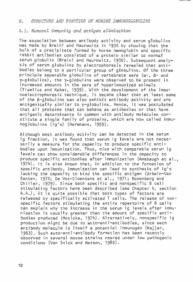


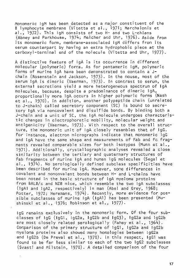


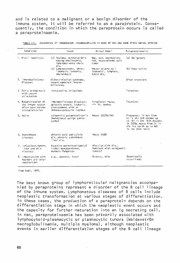


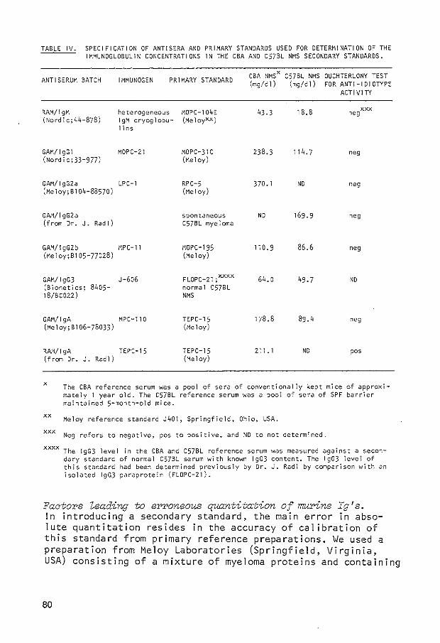


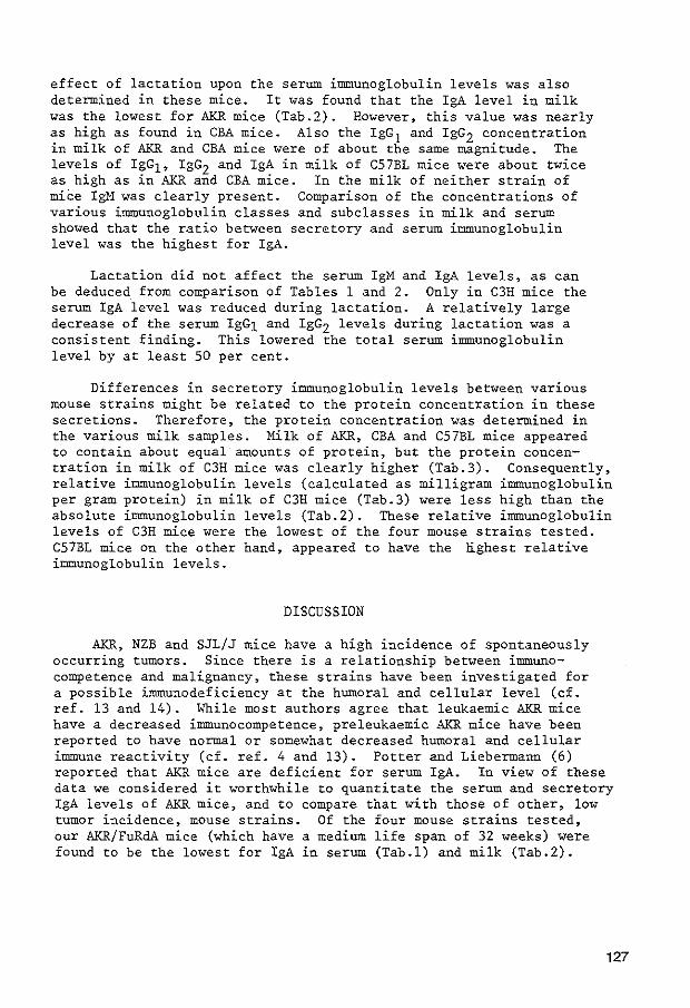
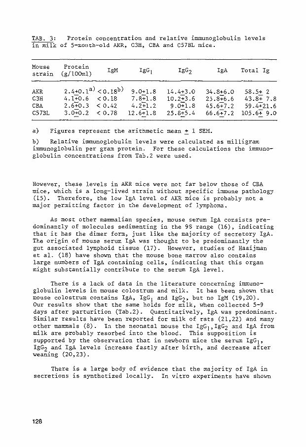


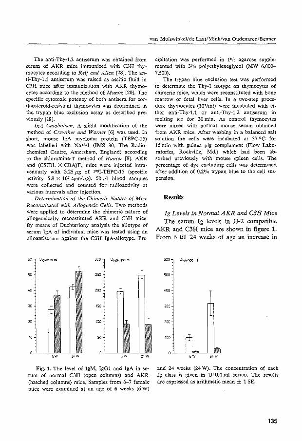
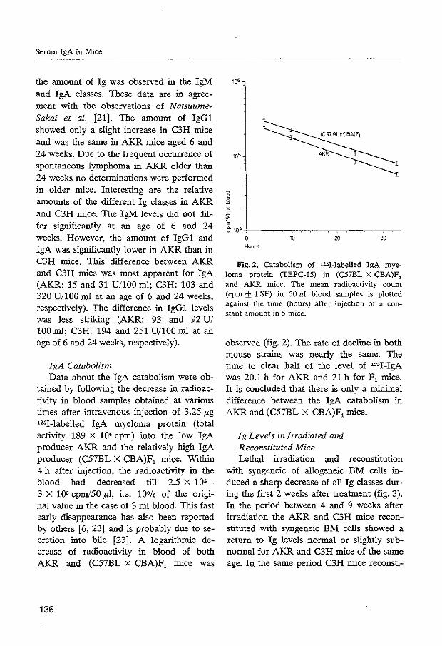

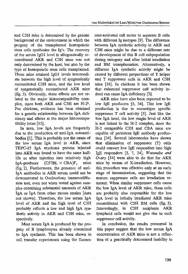


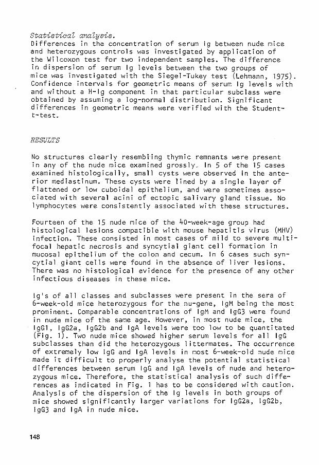
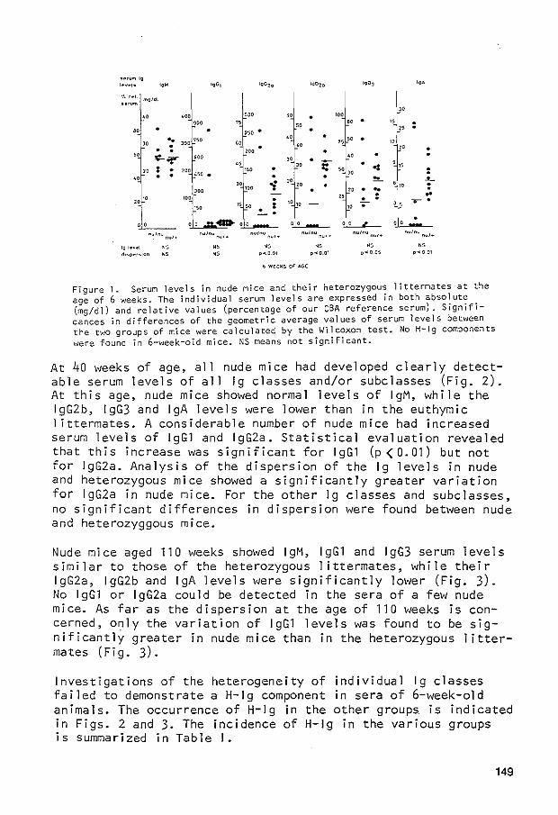

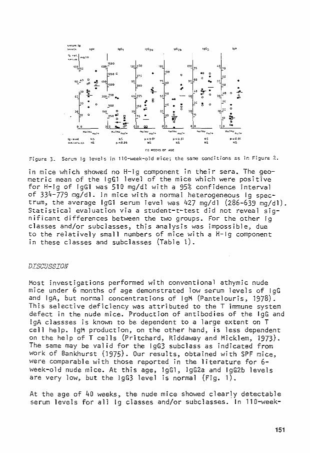
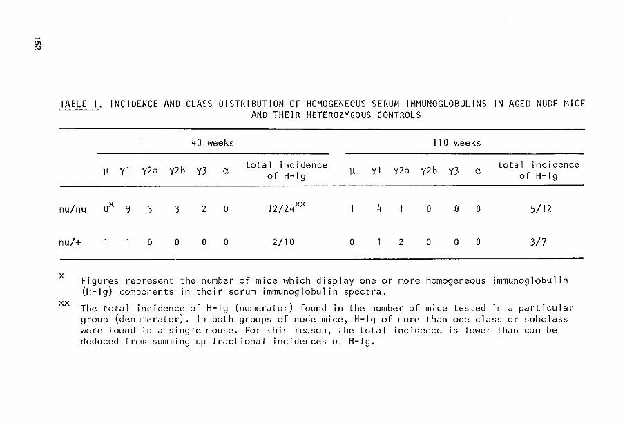


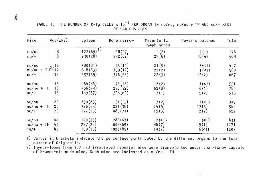


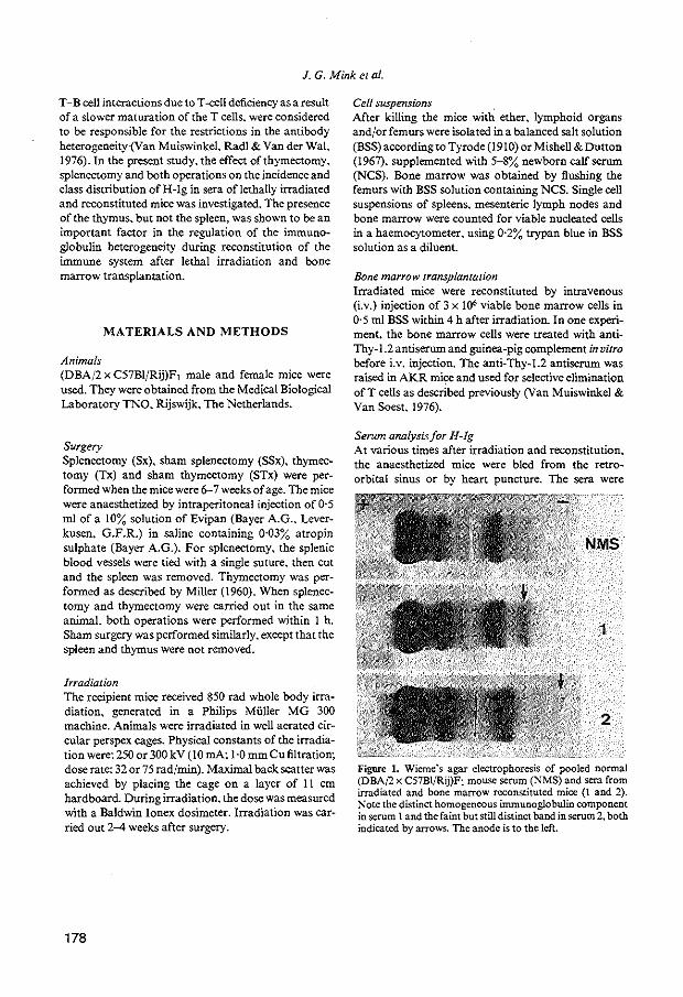


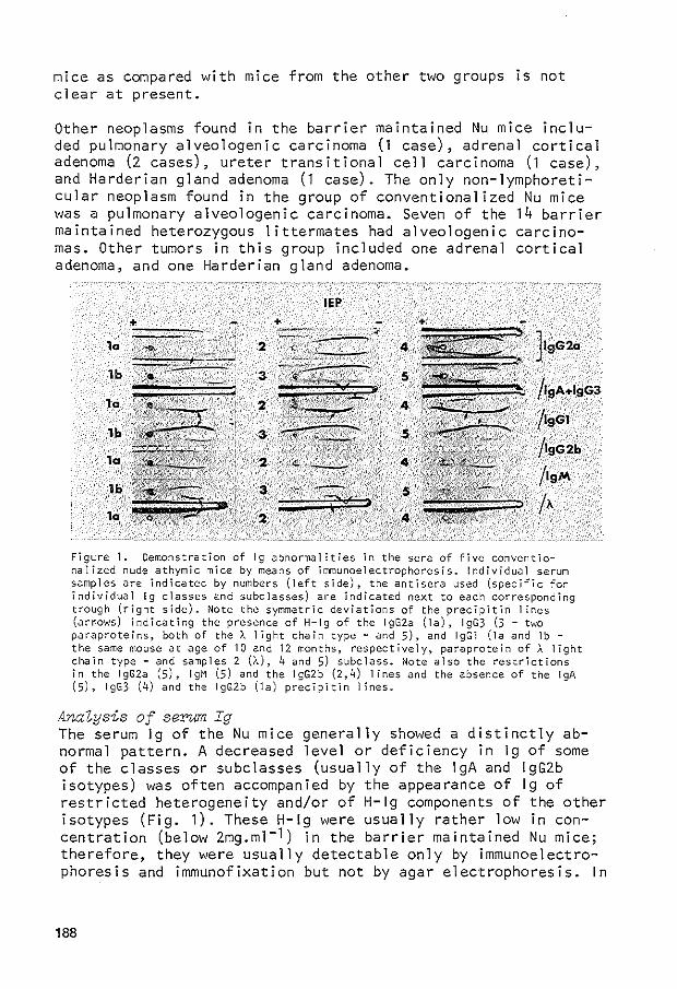

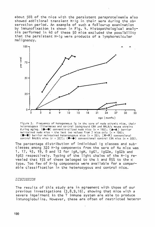
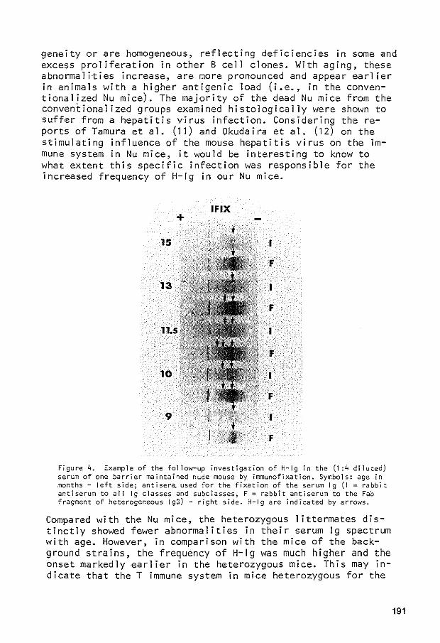


![The serum immunoglobulin G glycosylation signature of ......dysplasia, and finally adenocarcinoma [6]. Helicobacter pylori is a bacterium that infects the gastric epithelium of approximately](https://static.documents.pub/doc/80x56/5f4637eb1f92b745a0349ef3/the-serum-immunoglobulin-g-glycosylation-signature-of-dysplasia-and-inally.jpg)


