Received 8 February 2013; Accepted 30 September 2013
Abstract: Background: Approximately half of patients with chronic urticaria (CU) have functional autoantibodies against FceRIα or against IgE that induce histamine release from basophils and cutaneous mast cells. In addition to intracutaneous autologous serum skin test (ASST), more recently upregulation of CD63 and CD203c molecules on basophils has been proposed to detect the presence of autoantibodies in sera of CU patients. Objective: Our aim was to assess the effect of serum from CU patients on basophil CD63 and CD203c expression and to correlate serum-induced basophil activation with ASST. Methods: Sera were obtained from 128 patients with CU and 30 healthy controls. Patients or controls serum was incubated with atopic donor whole blood and activated basophils were identified by flow cytometry on basis of presence of CD63 or CD203c on high-expressing IgE positive cells. ASST was performed in all CU patients and 10 healthy controls. Results: ASST was positive in 33.6% CU patients. Serum from 36.7% patients induced upregulation of CD63 while serum from 45.3% patients upregulated the CD203c molecule. There was a positive correlation between ASST and upregulation of CD63, but no correlation was observed between ASST and serum-induced CD203c. The sensitivity and specificity of the ASST in vitro assays were 55.8% and 72.9% for CD63; 55.8% and 60.0% for CD203 respectively. Conclusions: Serum from CU patients upregulated both CD63 and CD203c molecules on blood basophils. A positive correlation between CD63 assay and ASST indicates the potential usefulness of this test for the diagnosis of the autoimmune form of CU.
1 Vilnius University, Faculty of Medicine, Clinic of Infectious, Chest Diseases, Dermatovenerology and Allergology, M.K. Ciurlionio 21, 03101 Vilnius, Lithuania
2 Vilnius University, Faculty of Medicine, Clinic of Cardiovascular Diseases, M.K. Ciurlionio 21, 03101 Vilnius, Lithuania
3 Vilnius University Hospital Santariskiu Klinikos, Santariskiu 2, 08661 Vilnius, Lithuania
4 - Medical University of Lodz, Department of Immunology, Rheumatology and Allergy, ul. Pomorska 251, 92-213 Lodz, Poland
Anzelika Chomiciene*1,3, Laimute Jurgauskiene2,3,Marek L. Kowalski4, Audra Blaziene1,3
Research Article
1. IntroductionChronic urticaria (CU) is a common skin disorder char-acterized by recurrent appearance of wheals and/or an-gioedema for more than 6 weeks. CU may affect up to 1% of the population and is associated with significantly impaired quality of life [1]. The etiology of symptoms cannot be determined in majority of the patients. Recently, it has become clear that 30 to 50% of pa-tients with CU have functional autoantibodies directed against the α-chain of the high-affinity immunoglobu-
lin E (IgE) receptor (FcεRI) or less commonly against IgE [2]. The term chronic “autoimmune urticaria” (CAU) has been increasingly used for this group of patients reflecting advances in our knowledge about functional autoantibodies that activate mast cells and basophils through cross-linking the FcεRI to secrete histamine [3]. Patients with CAU generally have more severe and dif-ficult to control symptoms than those with CU without autoantibodies [4,5]. The autologous serum skin test (ASST) is the only in vivo method to test for functional autoantibodies in CU patients [6]. A positive test is sug-
gestive, but not diagnostic of autoimmune basis for the patient’s urticaria symptoms. ASST may either prove the presence of autoantibodies or show histamine-releasing properties of the tested serum. The sensitivity and spec-ificity of this test have been reported to be about 70 and 80% respectively [7]. Techniques to detect the autoantibody to FcεRI and IgE in vitro include binding assays, Western blot and ELISA [8], however, they fail to identify antibodies with histamine releasing properties. The basophil histamine release assay (HRA) currently remains the “gold stan-dard” for confirmation of functional antibodies in the se-rum of patients with CU [9]. The HRA used by some au-thors is less sensitive than ASST in detection of patients with histamine releasing factors in their blood [10]. Fur-thermore, HRA is time consuming and cumbersome to perform. There is still no simple and reproducible clinical test for functional antibodies and improved screening tests are being sought. Recently, the ability of CU pa-tient’s serum to evoke expression of CD63 and CD203c on donor human basophils assessed by flow cytometry has been tested. There are a few published studies as-sessing serum-induced CD63 expression measurement as a diagnostic tool in CAU [11-16] and only 2 studies done by measured CD203c expression [16,17]. The primary objective of the current study was to as-sess the effect of serum from CU patients on basophil CD63 and CD203c expression. The secondary objec-tive was to correlate CD63 and CD203c expression with ASST and other autoreactivity markers in patients with chronic urticaria.
2. Materials and methods2.1 Patients and controls128 patients (26 males and 102 females; mean age 43±13, range 20-78 years) with CU with presenting al-most daily symptoms of urticaria and with continuous disease mean duration of 38±70 month (range 2 month - 40 years) were included in the study. Thirty-two (25%) of CU patients had elevated thyroid peroxidase antibod-ies (TPO), 7 (8.4%) had elevated antinuclear antibod-ies (ANA) and 12 (9.4%) reported hypersensitivity to non-steroidal anti-inflammatory drugs (NSAID). Serum was obtained from patients at the time of the ASST and was stored at -80°C. Antihistamines were withheld for at least 5 days before skin testing. Serum from 30 healthy adult individuals were used as normal controls. The study was approved by Lithuanian Bioethics Committee. The basophil donor was atopic and had serum IgE level of 1350 U/ml. The basophil donor provided in-formed consent and was bled less than once per week.
Disease activity was evaluated by using urticaria ac-tivity score (UAS).
3. ASSTThe test was performed by injecting 0.05 ml of the pa-tient‘s own serum intradermally into the volar aspect of the forearm. Sterile saline was used as negative control. A skin prick test with histamine 10 mg/ml was carried out as positive control to exclude any residual effect of anti-histamine drugs. Wheal and flare reactions were measured after 30 minutes. A mean wheal diameter of at least 1.5 mm greater than negative control with saline was considered to be a positive ASST.
3.1 Measurement of CD203c surface expression CD203c surface expression was measured using hepa-rinized blood from the donor within 3 to 4 hours after collection. Aliquots of the donor‘s heparinized whole blood (200 µl) were incubated for 20 minutes at 37ºC with 40 µl of serum from a patient with CU or from nor-mal controls and 40 µl basophil stimulation buffer (BSB) (Becton Dickinson (BD), USA). Phosphate buffer saline (PBS) (40 µl) was added to the donor blood and used as a negative control, in addition, 40 µl-µM N-Formil-Met-Leu-Phe chemotactic peptide (fMLP) (Sigma, USA) and 40 µl of a 1:50 dilution of anti-FcεRI receptor antibody (Upstade, USA) in Ca++ Mg++ free PBS were used as a positive control. The reactions were stopped by plac-ing the tubes on ice for 5 min. Cells (8-10 × 105 cells per test) were stained with 40 µl phycoerithrin (PE)-con-jugated antihuman CD45 (BD, USA), peridinin-chloro-phyll-protein (PerCP)-antihuman CD203c (Immunotech, USA), and fluorescein isothiocyanate (FITC)-conjugat-ed antihuman IgE (Invitrogen, USA) antibody cocktail at room temperature in the dark for 30 minutes. Red cells were lised with 2 ml FACS Lysing Solution (BD, USA). The cells were washed once with 2 ml PBS and fixed in 0.5% paraformaldehyde. Samples were analyzed on us-ing FACSCalibur flow cytometer (BD). Data on at least 1000 basophils were acquired and the percentage of CD203c-expressing basophils was calculated.
3.2 Measurement of CD63 surface expressionCD63 surface expression was measured using heparin-ized blood within 3 to 4 hours after collection from the donor with standard BD FastImmune set. Aliquots of the donor‘s heparinized whole blood (100 µl) were incubated for 20 minutes at 37ºC with 100 µl serum from patients with CU or normal controls and 20 µl BSB. For the con-trol, 100 µl PBS was added to the donor blood and used as negative control, and 100 µl 1-µM fMLP was used as
340
A. Chomiciene et al.
a positive control. The reactions were stopped by plac-ing the tubes on ice for 5 min. Cells were stained with 20 µl CD63FITC/ CD123PE/ Anti-HLA DR PerCP antibody cocktail at room temperature in the dark for 30 minutes. Red cells were lised with 2 ml FACS Lysing Solution. The cells were washed once with 2 ml PBS and fi xed in 0.5% paraformaldehyde. The cells were analyzed on a FACSCalibur fl ow cytometer (BD). Data on at least 1000 basophils were acquired, and the percentage of CD63-expressing basophils was calculated.
3.3 Statistical analysisStatistical signifi cance was determined by using the Wil-coxon matched pairs test, the Man-Whitney test, or χ2
test, where appropriate. A ROC curve analysis was used to estimate the cut-off of CD63 and CD203c serum-induced assay able to discriminate ASST positive CU from ASST negative CU patients, regarding optimal val-ues of sensitivity and specifi city. A p value of less than 0.05 was considered as signifi cant. Statistical analysis of results was performed with SSPS 15 program.
4. Results4.1 Serum-induced basophil activation Figure 1 and Figure 2 shows typical examples of the FACScan fl ow cytometric analysis of donor basophils. On average serum from CU patients, but not from healthy controls, signifi cantly upregulated CD63 and CD203c expression on atopic donor basophils. CD63 and CD203c expression following incubation with CU patient serum were 10.8±10.9% and 11.8±12.4%, respectively, as compared to 3.10±1.5% and 1.8±0.5%, respectively, with control serum (p<0.001 for both CD63 and CD203c ex-pression tests). The data are presented in Figure 3 and 4. A ROC curve analysis was used to establish the abil-ity of the serum-induced CD63 and CD203c expression assay to discriminate CU patients from controls, when the whole blood from atopic donor was used. The AUC for CD63 assay was 0.691 (p<0.001) and for CD203c assay – 0.552 (p=0.339). By analyzing the ROC curve, the cut-off value with the highest sensitivity and specifi c-ity (56% and 73% respectively for CD63 assay; 56% and 60% for CD203c assay) was found to be 10% (Figure 5). This threshold was used for both CD63 and CD203c in further investigations.
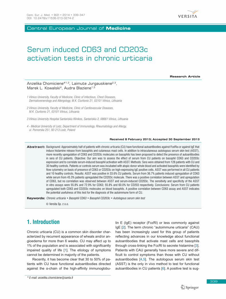
Figure 1. Basophil identification and expression of CD63. Basophils were gated with R1 and R2 (boxed area). Gate R1 (A) isolates the low side scatter (SSC), CD123+ basophils population. Gate R2 (B) detects a well defined population of cells highly positive for CD123, but negative for HLA-DR. Histogrames of CD63 expression after addition of buffer (PBS) for negative control (C), fMPL for positive control (D) and patient sera (E).
341
Basophil activation tests in Chronic Urticaria
Figure 2. Basophil identification and expression of CD203c. Basophils located between the monocytes and lymphocytes were gated with R1 and R2 (boxed area). Gate R1 (A) isolates the side scatter and CD45+ basophils population. Gate R2 (B) detects a population of cells highly positive for IgE and CD203c expression. Histograme of CD203c expression after addition of buffer (shaded area) and after addition of serum of patient with CU and positive ASST.
Symbol used in diagram: Box – Q1-Q3 quartiles
Line inside box – median
– mean
–maximum
– minimum
– extrieme values
– outliers
Figure 3. CD 3 expression (%) in patients (n=128) vs healthy controls (n=30), positive (fMLP) and negative (buffer) control results on donor basophils
0
10
0
0
0
0
0
0
0
p 0 001
Figure 3. CD63 expression (%) in patients (n=128) vs healthy controls (n=30), positive (fMLP) and negative (buffer) control results on donor basophils.
Symbol used in diagram: Box – Q1-Q3 quartiles
Line inside box – median
– mean
–maximum
– minimum
– extrieme values
– outliers
Figure 4. CD203c expression (%) in patients (n=128) vs healthy controls (n=30), positive (fMLP) and negative (buffer) control results on donor basophils
10
0
10
0
0
0
0
0
0
0
0
%
p 0 001
Figure 4. CD203c expression (%) in patients (n=128) vs healthy controls (n=30), positive (fMLP) and negative (buffer) control results on donor basophils.
342
A. Chomiciene et al.
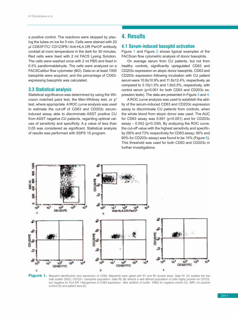
Considering the estimated cut-off, we found that serum from 36.7% patients induced upregulation of CD63 and se-rum from 45.3% of patients upregulated CD203c molecule. As the CD203c area under ROC curve was about 0.5 and p>0.05, we suggesting that the CD203c did not have sufficient diagnostic value, and we used it for ex-perimental purpose in further investigations.
4.2 Correlation of the basophil activation tests with ASST and other autoreactivity markersASST was positive in 43 (33.6%) of the 128 CU patients. In healthy control group (n=10) ASST was negative in all subjects. Upregulated (>10%) CD63 expression was ob-served in 24 (55.8%) patients with positive ASST and in 23 (27.1%) patients with negative ASST. Upregulated CD203c expression was measured in 24 (55.8%) pa-tients with positive ASST and 34 (40%) with negative ASST. A significant correlation was found between CD63 expression upregulation and ASST (r=0.282, p=0.001), however there was no correlation between CD203c upregulation and ASST (r=0.150, p=0.091). The mean CD63 expression was statistically higher in the positive than in the negative ASST group (p<0.001), and there were no differences in CD203c expression between the positive and negative ASST groups (p=0.339). No sig-nificant correlation was found between basophil activa-tion tests, ASST and other autoreactivity markers (ele-vated TPO, positive ANA) and hypersensitivity to NSAID (Figure 6,7 and Table 1,2).
There was no association between duration of CU, disease activity, incidence of angioedema and posi-tivity of ASST with CD63 or CD203c expression (p>0.05). The prevalence of positive ASST was higher in the elderly.
Figure 5. CD63 and CD203c ROC curves
Figure 5. CD63 and CD203c ROC curves.
CD203c CD63Negative Positive Negative Positive
N (%) N (%) N (%) N (%)
CU patients 43 (33.6%) 85 (66.4%) 43 (33.6%) 85 (66.4%)Control group 30 (100.0%) 0 (0.0%) 30 (100.0%) 0 (0.0%)
Table 1. CD203c, CD63 and other autoreactivity markers.
ASST
Negative PositiveN (%) N (%)
CU patients 85 (66.4%) 43 (33.6%)Control group 10 (100.0%) 0 (0.0%)
Elevated TPO 20 (62.5%) 12 (37.5%)p=0.589
ANA positive 6 (75.0%) 2 (25.0%)p=0.092
Hypersensitivity to NSAID‘s 6 (50.0%) 6 (50.0%)p=0.206
Table 2. ASST positivity and other autoreactivity markers.
343
Basophil activation tests in Chronic Urticaria
0102030405060708090
100
Tota
l CU
ASST
+ C
U
ASST
-CU
Cont
rols
Hype
rsen
sitiv
ity t
o NS
AID‘
s
Elev
ated
TPO
Posi
tive
ANA
CD 20
3c E
xpre
ssio
n, %
n=128 n=40 n=85 n=30 n=12 n=32 n=8
Figure 7. Serum-induced CD203c expression in the whole group of CU patients and in various patient’s subpopulations (ASST+ and ASST- CU patients, CU patients with other autoreactivity markers and controls). No significant differences in CD203c expression between different groups of CU patients were found.
Figure 7. Serum-induced CD203c expression in the whole group of CU patients and in various patient’s subpopulations (ASST+ and ASST- CU patients, CU patients with other autoreactivity markers and controls). No significant differences in CD203c expression between different groups of CU patients were found.
0
10
20
30
40
50
60
70
80
90
100
Tota
l CU
ASST
+ C
U
ASST
-CU
Cont
rols
Hype
rsen
sitiv
ity t
o NS
AID‘
s
Elev
ated
TPO
Posi
tive
ANA
CD63
Exp
ress
ion,
%
n=128 n=40 n=85 n=30 n=12 n=32 n=8
p=0.01
Figure 6. Serum-induced CD63 expression in the whole group of CU patients and in various patient’s subpopulations (ASST+ and ASST- CU patients, CU patients with other autoreactivity markers and controls). A significant difference in CD63 expression was found only between ASST+ and ASST- patients, but not between other groups of patients with CU.
Figure 6. Serum-induced CD63 expression in the whole group of CU patients and in various patient’s subpopulations (ASST+ and ASST- CU patients, CU patients with other autoreactivity markers and controls). A significant difference in CD63 expression was found only between ASST+ and ASST- patients, but not between other groups of patients with CU.
344
A. Chomiciene et al.
5. DiscussionCurrently, there is no reliable laboratory diagnostic test to confirm autoimmune mechanisms in chronic urticaria. Recently upregulation of CD63 and CD203c molecules on basophils has been used to detect the presence of autoantibodies in serum of patients with CU. Our study is one of the first to compare CD63 and CD203c ex-pression in a group of CU patients. Based on experi-ence of other authors, who demonstrated that atopic donor basophils showed marked upregulation of CD63 and CD203c molecules after stimulation with serum from CU patients in this study we used highly atopic donor basophils [12,14,15]. Our study confirmed that serum from patients with CU, but not control sera, on average significantly upregulated atopic donor basophil CD63 and CD203c expression. The estimated cut-off value for CD63 and CD203c expression to distinguished CU patients was 10%. In an earlier study by Frezzo-lini A. et al. [12], the cut-off with the highest sensitivity and specificity (95% and 91%, respectively) was found to be 15%, - a difference that can be easily explained by basophil donor sensitivity and/or differences in various flowcytometry techniques. Significantly more patients with positive ASST (55.8%), than with negative ASST (27.1%), had elevat-ed CD63 expression. On the contrary, the percentage of elevated CD203c expression was found to be similar in patients with positive (55.8%) and with negative ASST (40%). The presence of positive basophil activation tests (BAT) in a significant proportion of ASST negative pa-tients could be explained by the fact that BAT are more sensitive to detect autoantibodies. Another explanation is that there are other undefined serum factors present in CU sera that can upregulate CD63 and CD203c on basophils. Whereas negative CD63 and CD203c ex-pression was found in 19 (44.2%) ASST positive pa-tients, it can be explained by the existence of a mast cell-specific histamine releasing factor, causing positive ASST. Also, false-positive ASST results could be due to bradykinin or C5a generated in serum during clotting. We found significant correlation between the CD63 expression test and ASST, but there was no correlation between CD203c and ASST. After ROC curve analysis, we concluded that CD203c did not have sufficient diag-nostic power. Other authors, who tested CD63 [11-15] or CD203c expression [17] in CU patients, found signifi-cant correlation between ASST and both basophil acti-vation tests positivity. The discrepancy between CD63 and CD203c correlations with ASST in our study can reflect either different mechanism of CD63 and CD203c molecules upregulation or differences in methodology of tests. Recently, Gentinetta et al. [16] conducted the
study of basophil activation tests in CU using differ-ent basophil donors and made a conclusion that CD63 activation test using IL-3 priming could be used as al-ternative to ASST. In authors opinion CD203c as baso-phil activation marker is less suitable for this purpose as they reported variable activation patterns, rendering the discrimination between patient and healthy control serum impossible in certain basophil donors. A. Tede-schi et al. [18] in their paper commented the results of the Genginetta study and wrote, that BAT and ASST do not overlap and reflect, at least in part, different phe-nomena. In author’s opinion further efforts are needed to identify the factors causing basophil and mast cell ac-tivation in CU. We agree with authors, that BAT can be complementary test but not substitute of ASST. Recent a study by Confino-Cohen et al. found strong association between CU and major autoimmune diseases [19]. We found quite frequent (25%) occur-rence of elevated TPO in our CU patients. We looked for association of CAU tests (ASST, basophil activation tests) and other autoreactivity markers (elevated TPO, positive ANA), however we did not find a significant correlation. Several studies reported that autoreactiv-ity is highly prevalent in patients with multiple NSAIDs intolerance [20,21]. In our patients with hypersensitiv-ity to NSAIDs reported from history, the prevalence of a positive ASST or basophil activation tests was not sig-nificantly higher than in other patients without reactions to NSAIDs. We also could not find any relationship be-tween basophil activation tests, ASST and disease char-acteristics (duration of CU, disease activity, incidence of angioedema). The prevalence of positive ASST was higher in the elderly. In our hands, measurement of serum-induced CD63 expression turned out to be more sensitive and specific than CD203 to detect CU. Accordingly, a difference be-tween sensitivity of both test has been previously re-ported in the diagnosis of allergy. Authors comparing ba-sophil activation tests, using either CD63 and CD203c in diagnosis of latex allergy, insect venom allergy, drug allergy and found that sensitivity of CD203c was consid-erably higher than CD63 for the diagnosis of latex aller-gy [22] and insect venom allergy [23], whereas in diag-nosis of allergy to muscle relaxants Sudheer et al. [24] found better sensitivity of CD63 assay (79%) in compari-son with CD203c assay (36%). In summary, our results indicate that although serum from patients with CU can upregulate both CD63 and CD203c molecules on basophils, the results of basophil activation tests support an autoimmune origin of dis-ease. Correlation between CD63 assay and ASST, indi-cates the potential usefulness of this test for the diagno-sis of the autoimmune form of CU. CD63 expression test
345
Basophil activation tests in Chronic Urticaria
could be used as a complementary test to ASST. Further research of basophil activation tests is needed in a large sample size to establish the validity and superiority of basophil activation tests in general population.
AcknowledgmentsWe wish to extend our heartfelt gratitude the Lithuanian State Studies Foundation for the support of the research project “Autoreactivity and innate immunity markers in patients with chronic urticaria”(T-91/08).
References
[1] Powell R.J., Du Toit G.L., Siddique N., Leech S.C., Dixon T.A., Clark A.T., et al., BSACI guidelines for the management of chronic urticaria and angio-oedema, Clin Exp Allergy, 2007, 37(5), 631-650
[2] Sheikh J., Autoantibodies to the high-affinity IgE receptor in chronic urticaria: how important are they, Curr Opin Allergy Clin Immunol., 2005, 5(5), 403-407
[3] Grattan C. E., Autoimmune urticaria, Immunol Allergy Clin North Am., 2004, 24(2), 163-181
[4] Irinyi B., Szeles G., Gyimesi E., Tumpek J., Heredi E., Dimitrios G., et al., Clinical and laboratory examinations in the subgroups of chronic urticaria, Int Arch Allergy Immunol., 2007, 144(3), 217-225
[5] Sabroe R.A., Seed P.T., Francis D.M., Barr R.M., Black A.K., Greaves M.W., Chronic idiopathic urticaria: comparison of the clinical features of patients with and without anti-FcepsilonRI or anti-IgE autoantibodies, J Am Acad Dermatol., 1999, 40(3), 443-450
[6] Zuberbier T., Bindslev-Jensen C., Canonica W., Grattan C.E., Greaves M.W., Henz B.M. et al., EAACI/GA2LEN/EDF guideline: definition, classification and diagnosis of urticaria, Allergy, 2006, 61(3), 316-320
[7] Sabroe R.A., Grattan C.E., Francis D.M., Barr R.M., Kobza Black A., Greaves M.W., The autologous serum skin test: a screening test for autoantibodies in chronic idiopathic urticaria, Br J Dermatol., 1999, 140(3), 446-452
[8] Fiebiger E., Maurer D., Holub H., Reininger B., Hartmann G., Woisetschlager M., et al., Serum IgG autoantibodies directed against the alpha chain of Fc epsilon RI: a selective marker and pathogenetic factor for a distinct subset of chronic urticaria patients, J Clin Invest., 1995, 96(6), 2606-2612
[10] Asero R., Lorini M., Chong S.U., Zuberbier T., Tedeschi A., Assessment of histamine-releasing activity of sera from patients with chronic urticaria showing positive autologous skin test on human basophils and mast cells, Clin Exp Allergy, 2004,34(7), 1111-1114
[11] De Swerdt A., Van Den Keybus C., Kasran A., Cadot P., Neyens K., Coorevits L., et al., Detection of basophil-activating IgG autoantibodies in chronic idiopathic urticaria by induction of CD 63, J Allergy Clin Immunol., 2005,116(3), 662-667
[12] Frezzolini A., Provini A., Teofoli P., Pomponi D., De Pita O., Serum-induced basophil CD63 expression by means of a tricolour flow cytometric method for the in vitro diagnosis of chronic urticaria, Allergy, 2006 Sep, 61(9), 1071-1077
[13] Gyimesi E., Sipka S., Danko K., Kiss E., Hidvegi B., Gal M., et al., Basophil CD63 expression assay on highly sensitized atopic donor leucocytes-a useful method in chronic autoimmune urticaria, Br J Dermatol, 2004 Aug;151(2), 388-396
[14] Szegedi A., Irinyi B., Gal M., Hunyadi J., Danko K., Kiss E., et al. Significant correlation between the CD63 assay and the histamine release assay in chronic urticaria, Br J Dermatol, 2006, 155(1), 67-75
[15] Wedi B., Novacovic V., Koerner M., Kapp A., Chronic urticaria serum induces histamine release, leukotriene production, and basophil CD63 surface expression--inhibitory effects ofanti-inflammatory drugs, J Allergy Clin Immunol, 2000, 105(3), 552-560
[16] Gentinetta T., Pecaric-Petkovic T., Wan D., Falcone F. H. , Dahinden C. A., Pichler W. J., et al., Individual IL-3 priming is crucial for consistent in vitro activation of donor basophils in patients with chronic urticaria, J Allergy Clin Immunol, 2011, 128, 1227-1234
[17] Yasnowsky K.M., Dreskin S.C., Efaw B., Schoen D., Vedanthan P.K., Alam R. et al., Chronic urticaria sera increase basophil CD203c expression, J Allergy Clin Immunol, 2006, 117(6), 1430-1434
[18] Tedeschi A., Asero R., Cugno M., Basophil activation test and autologous serum skin test: Not overlapping tests in chronic urticaria, J Allergy Clin Immunol, 2012, 280-281
[19] Confino-Cohen R., Chodick G., Shalev V., Leshno M., Kimhi O., Goldberg A., Chronic urticaria and autoimmunity: Association found in a large population study, J Allergy Clin Immunol, 2012, 129, 1307-1313
346
A. Chomiciene et al.
[20] Asero R., Tedeschi A., Lorini M., Autoreactivity is highly prevalent in patients with multiple intolerances to NSAIDs, Ann Allergy Asthma Immunol, 2002, 88 (5), 468-472
[21] Erbagci Z. Multiple NSAID intolerance in chronic idiopathic urticaria is correlated with delayed, pronounced and prolonged autoreactivity. J Dermatol, 2004, 31(5), 376-382
[22] Boumiza R., Monneret G., Forissier M.F., Savoye J., Gutowski M.C., Powell W.S. et al., Marked improvement of the basophil activation test
by detecting CD203c instead of CD63, Clin Exp Allergy, 2003, 33(2), 259-265
[23] Eberlein-Konig B., Varga R., Mempel M., Darsow U., Behrendt H., Ring J., Comparison of basophil activation tests using CD63 or CD203c expression in patients with insect venom allergy, Allergy, 2006, 61(9), 1084-1085
[24] Sudheer P.S., Hall J.E., Read G.F., Rowbottom A.W., Williams P.E., Flow cytometric investigation of peri-anaesthetic anaphylaxis using CD63 and CD203c, Anaesthesia, 2005, 60(3), 251-256


![Urticaria e infeccion[1][1]](https://static.documents.pub/doc/80x56/5466656daf79596f338b50a1/urticaria-e-infeccion11.jpg)


