48
Serum vs Plasma Which specimen should you use ? American Society for Clinical Laboratory Science Michigan Annual Meeting Sol Green PhD FACB March 31 2016

Serum vs Plasma
Which specimen should you use ?
American Society for Clinical Laboratory Science
Michigan Annual Meeting
Sol Green PhD FACB
March 31 2016
Introduction • This presentation is intended to provide laboratorians with an in-
depth discussion/review of serum and heparin plasma.
• Heparin plasma is the specimen of choice in clinical chemistry for an
increasing number of laboratories.
• Heparin plasma represents a more complex specimen than serum.
• We will cover:
TAT Factors Influencing Specimen Quality
Analyte Stability Instrument/Assay Considerations
Summary

Serum vs Plasma
Chemistry
Lithium Heparin
Mix and spin
Centrifuge
1,100 - 1,300g
10 min
Plasma
Clot activator
30 min clot time
Centrifuge
1,100 - 1,300g
10 min
Serum
Serum Tubes
• Non gel blood collection tube
– Serum tube 30 – 60 min clot
– Need to be aliquot to avoid cell contamination
• Gel blood collection tube
– Serum tube with activator gel separates cells from serum
– 30 minute clot for routine chemistry
• Thrombin blood collection tubes (RST)
– 5 minutes for STAT serum determination in chemistry
• Inversion at blood collection: 8-10
• Centrifugation:
– 1100-1300 g for 10 minutes at room temperature
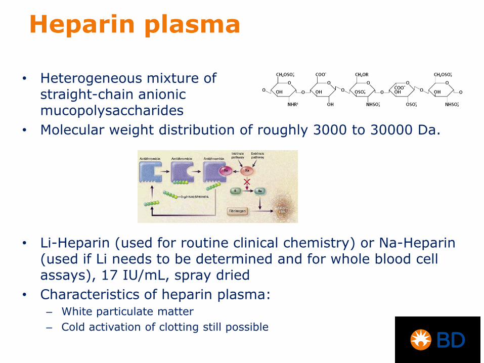
Heparin plasma
• Heterogeneous mixture of straight-chain anionic mucopolysaccharides
• Molecular weight distribution of roughly 3000 to 30000 Da.
• Li-Heparin (used for routine clinical chemistry) or Na-Heparin (used if Li needs to be determined and for whole blood cell assays), 17 IU/mL, spray dried
• Characteristics of heparin plasma: – White particulate matter
– Cold activation of clotting still possible

Polling Question
A. Only serum
B. Only plasma
C. Mainly serum with some plasma
D. Mainly plasma with some serum
E. Almost even split
F. Don’t know
Which sample does your laboratory use ?

Jones BA, et al. PhysicianPhysician satisfactionsatisfaction with clinical laboratory services: a College of American Pathologists Q-probes study of 138 institutions. Arch Pathol Lab Med. 2009;133:38-43.
TAT represents top 3/5 categories listed by physicians as the most important
TAT = 36,5%
How Important is TAT?

Jones BA, et al. PhysicianPhysician satisfactionsatisfaction with clinical laboratory services: a College of American Pathologists Q-probesstudy of 138 institutions. Arch Pathol Lab Med. 2009;133:38-43.
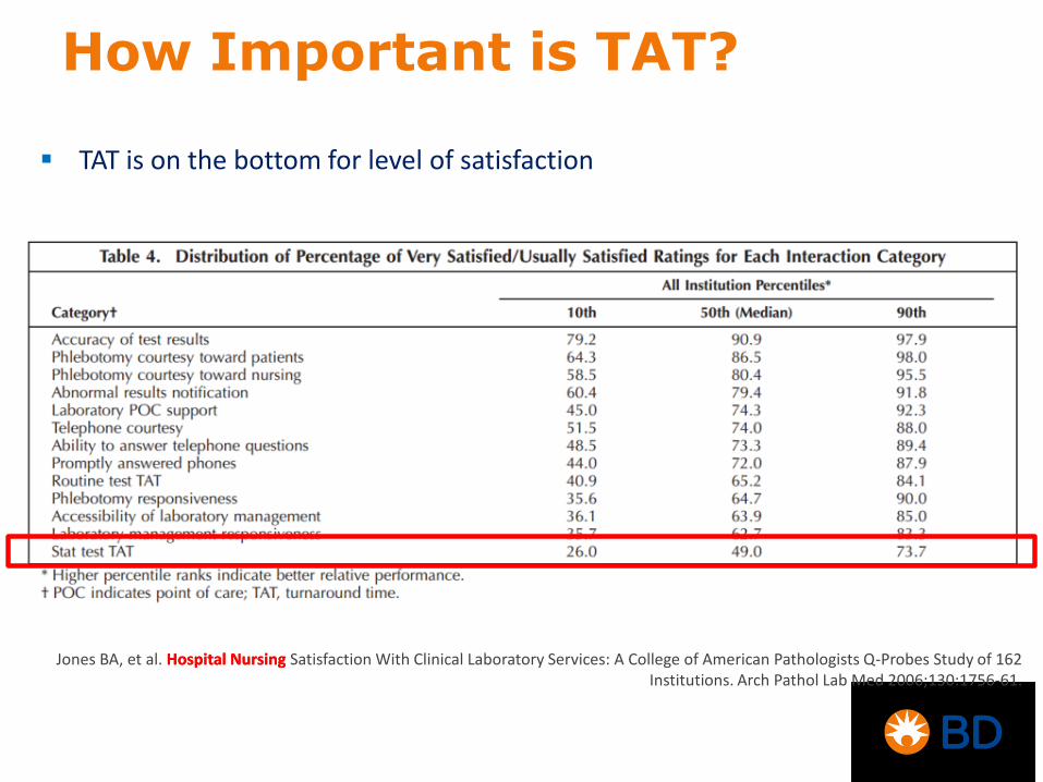
TAT is on the bottom for level of satisfaction
How Important is TAT?

Jones BA, et al. Hospital Nursing Hospital Nursing Satisfaction With Clinical Laboratory Services: A College of American Pathologists Q-Probes Study of 162 Institutions. Arch Pathol Lab Med 2006;130:1756-61.
Stat TAT represents most important category listed by nurses
How Important is TAT?
Jones BA, et al. Hospital Nursing Hospital Nursing Satisfaction With Clinical Laboratory Services: A College of American Pathologists Q-Probes Study of 162 Institutions. Arch Pathol Lab Med 2006;130:1756-61.
TAT is on the bottom for level of satisfaction
How Important is TAT?

How To Reduce TAT ?
Fast Analytical Phase
Reduce Preanalytical Handling & Processing
Speed Up Sample Transport
Use Plasma

Turnaround Time
Recommended clotting times for serum blood
collection tubes generally range from 30-60
minutes.
Use of plasma allows laboratories to process
and test specimens upon receipt, while avoiding
latent fibrin formation due to incomplete clotting.

Serum Specimen Quality
Specimen quality has been another factor
prompting some laboratories to switch to plasma.
Serum specimens are subject to latent fibrin
formation when clotting is inadequate.
insufficient clotting time
patients receiving anticoagulant or
thrombolytic therapy
Fibrin can range from thin strands to large cloud-
like masses.
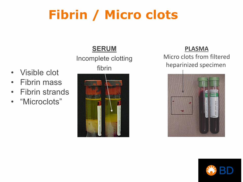
Fibrin / Micro clots
• Visible clot
• Fibrin mass
• Fibrin strands
• “Microclots”
SERUM
Incomplete clotting
fibrin
PLASMA Micro clots from filtered heparinized specimen

Fibrin – Tube Mixing
• Mixing immediately after collection facilitates dispersion
of additive into the blood.
• Incomplete mixing may lead to incomplete or delayed
clotting (serum) or incomplete anticoagulation (plasma)
• Typical manufacturer recommendations:
Serum tubes: 5-6 inversions
Plasma (heparin) tubes: 8-10 inversions
= 1 inversion
Fibrin
• Can cause
significant
disruption to
instrument
operation and
process workflow.

Physical obstruction of sampling probe
Insufficient sampled volume
Gradual deposition of fibrin in reaction chambers
or pathways
Interference with measurement systems or
reagents
Potential consequences: instrument downtime,
failure to provide test results, or erroneous test
results.
Issues Due To Fibrin

Fibrin – Instrument Operation
Gradual deposition of fibrin
in reaction pathway; "plaque"
Build up leads to obstruction
Even with no obstruction,
potential interference from
light scattering or reagent
interference
Erroneous results
Physical, chemical
or immunological
Aspiration of "micro
clots" not sufficiently
large to obstruct
probe
Latent fibrin
formation inside
instrument
Reaction
Pathway or
Reaction
Chamber
Sampling problem,
insufficient quantity aspirated
Erroneous results
Physical
Aspiration of fibrin
causing probe
obstruction
Sampling
Probe
Potential Result Type of
Interference Event Location
Addressing Fibrin Issues
Approaches to minimize the impact of fibrin in
serum specimens often require user intervention,
increase TAT, and may not be recommended.
To help reduce these issues, some laboratories
have switched to plasma.
However, plasma specimens also have unique
characteristics concerning specimen quality and
integrity.
Plasma Trends
World wide generally increasing use of plasma
Increasing use of plasma in some European countries
US also increasing number of labs are moving to plasma due to TAT

Plasma Trends

Gel Movement
The presence of a solid clot in serum gel tubes also
leads to a difference in the movement of gel during
centrifugation.
Serum: Gel must move up and around the clot,
against the tube wall.
Plasma: Gel moves up in pieces similar to a ‘lava
lamp’.

serum tube
plasma tube
Simulated Gel Movement

unmixed mixed
What type of sample is this?
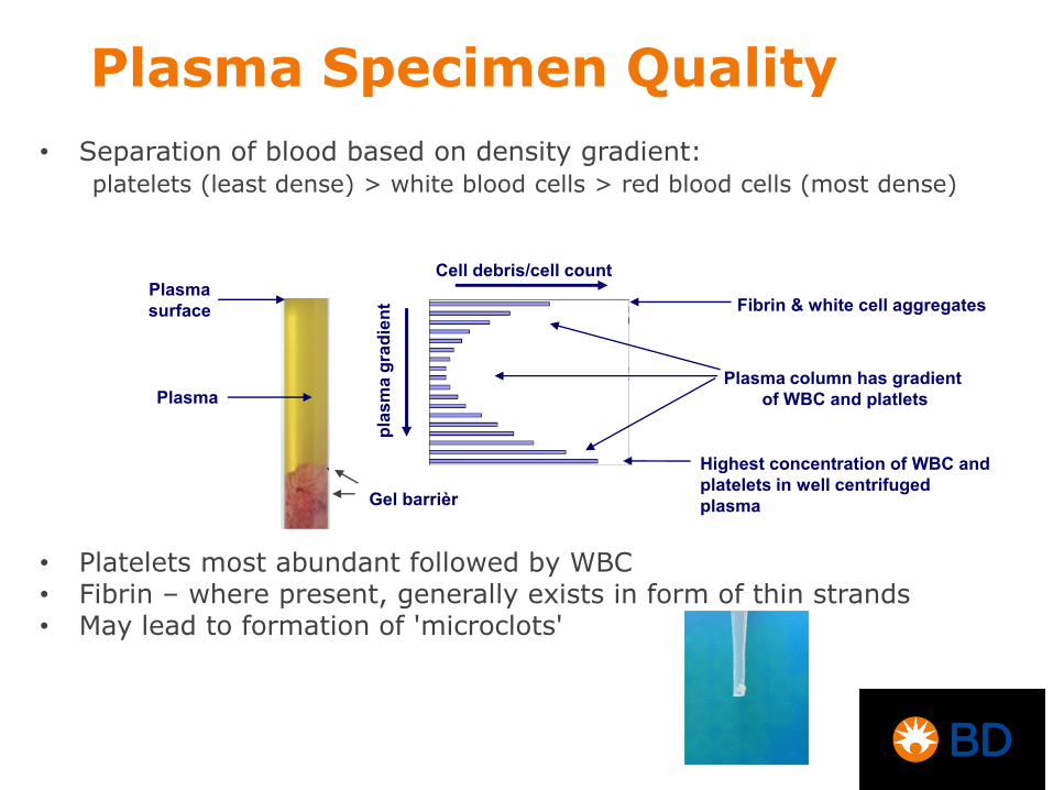
• Separation of blood based on density gradient: platelets (least dense) > white blood cells > red blood cells (most dense)
• Platelets most abundant followed by WBC • Fibrin – where present, generally exists in form of thin strands • May lead to formation of 'microclots'
Plasma
Gel barrièr
Cell debris/cell count
pla
sm
a g
rad
ien
t
Plasma
surface Fibrin & white cell aggregates
Plasma column has gradient
of WBC and platlets
Highest concentration of WBC and
platelets in well centrifuged
plasma
Plasma Specimen Quality

Plasma Specimen Quality
• As a result of the potential for variable amounts of cells, platelets, fibrin, and WPM, heparin plasma is generally a more complicated matrix to manage than serum.
• A proper understanding of the factors that influence plasma specimen quality is needed.

Ideal Plasma Specimen
• Ideal plasma specimen would be one which is cell/platelet free and in which the anticoagulant functions to inhibit clotting and fibrin formation for extended periods of time
• Often not attained with heparin plasma specimens
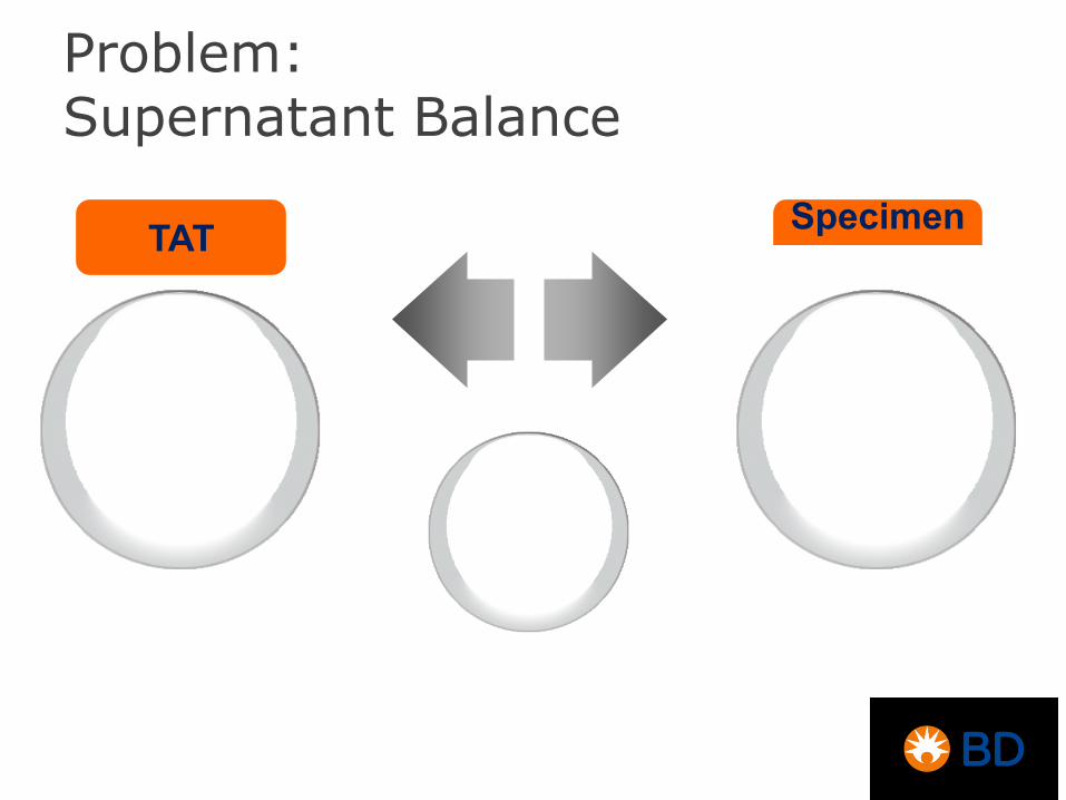
Problem: Supernatant Balance
Specimen
Quality TAT

Plasma Test Results
In general, most assays in clinical chemistry are
compatible with both serum and heparin plasma,
and test results are sufficiently equivalent that the
same reference ranges can be used.
However for certain assays or test methods,
plasma specimens may be unacceptable, or
differences in results may be sufficient to warrant a
change in reference range.
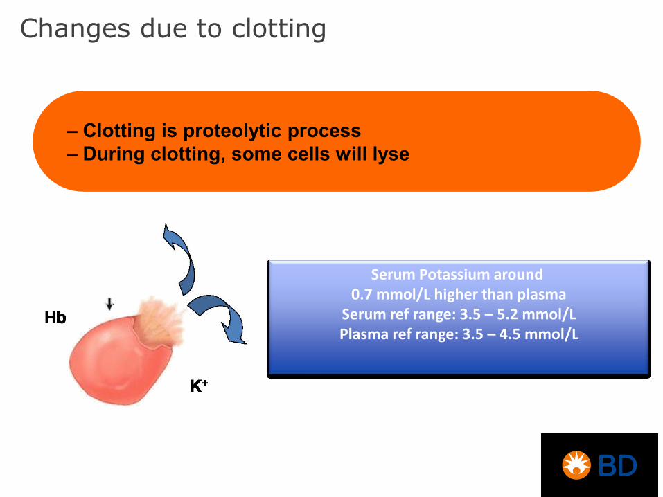
Changes due to clotting
– Clotting is proteolytic process
– During clotting, some cells will lyse
Serum Potassium around 0.7 mmol/L higher than plasma
Serum ref range: 3.5 – 5.2 mmol/L Plasma ref range: 3.5 – 4.5 mmol/L

Potassium
Potassium and phosphorus increased in serum
due to release from cells/ platelets during
clotting.
A linear correlation has been shown between
platelet count and the increase in serum
potassium.
World Health Organization. Use of anticoagulants in diagnostic laboratory investigations.
WHO/DIL/LAB/99.1 Rev.2, 2002.
Harr R, Bond L, Trumbull D. A comparison of results for serum versus heparinized plasma for 30 common
analytes. Laboratory Medicine 1987 Jul;18(7):449-55.
Ciuti R, Rinaldi G. Serum and plasma compared for use in 19 common chemical tests performed in the
Hitachi 737 analyzer. Clin Chem. 1989 Jul;35(7):1562-3.
Guder WG, Narayanan S, Wisser H, Zawta B. Samples: from the patient to the laboratory. 3rd ed.
Darmstadt, Germany: Wiley-VCH; 2003, pp. 32-3.
Miles RR, Roberts RF, Putnam AR, Roberts WL. Comparison of serum and heparinized plasma samples
for measurement of chemistry analytes. Clin Chem. 2004;50:1704-5.
Burtis CA, Ashwood ER, eds. Tietz fundamentals of clinical chemistry. 4th ed. Philadelphia, PA: W.B.
Saunders Company; 1996:499.

Total Protein
Slightly increased in plasma due to presence of
fibrinogen.
World Health Organization. Use of anticoagulants in diagnostic laboratory investigations.
WHO/DIL/LAB/99.1 Rev.2, 2002.
Harr R, Bond L, Trumbull D. A comparison of results for serum versus heparinized plasma
for 30 common analytes. Laboratory Medicine 1987 Jul;18(7):449-55.
Ciuti R, Rinaldi G. Serum and plasma compared for use in 19 common chemical tests
performed in the Hitachi 737 analyzer. Clin Chem. 1989 Jul;35(7):1562-3.
Guder WG, Narayanan S, Wisser H, Zawta B. Samples: from the patient to the laboratory.
3rd ed. Darmstadt, Germany: Wiley-VCH; 2003, pp. 32-3.

Other Tests
Differences in certain enzymes (e.g., LD, ALKP, AST)
may be seen.
Lithium/sodium increased with use of lithium or
sodium heparin.
Interference from fibrinogen may also make plasma
an unsuitable specimen for certain protein analysis
methods (e.g., SPEP - protein electrophoresis).
Heparin may interfere with certain immunoassays.
Lee DC, Klachko MN. Falsely elevated lithium levels in plasma samples obtained in lithium
containing tubes. J Toxicol Clin Toxicol. 1996;34(4):467-9.
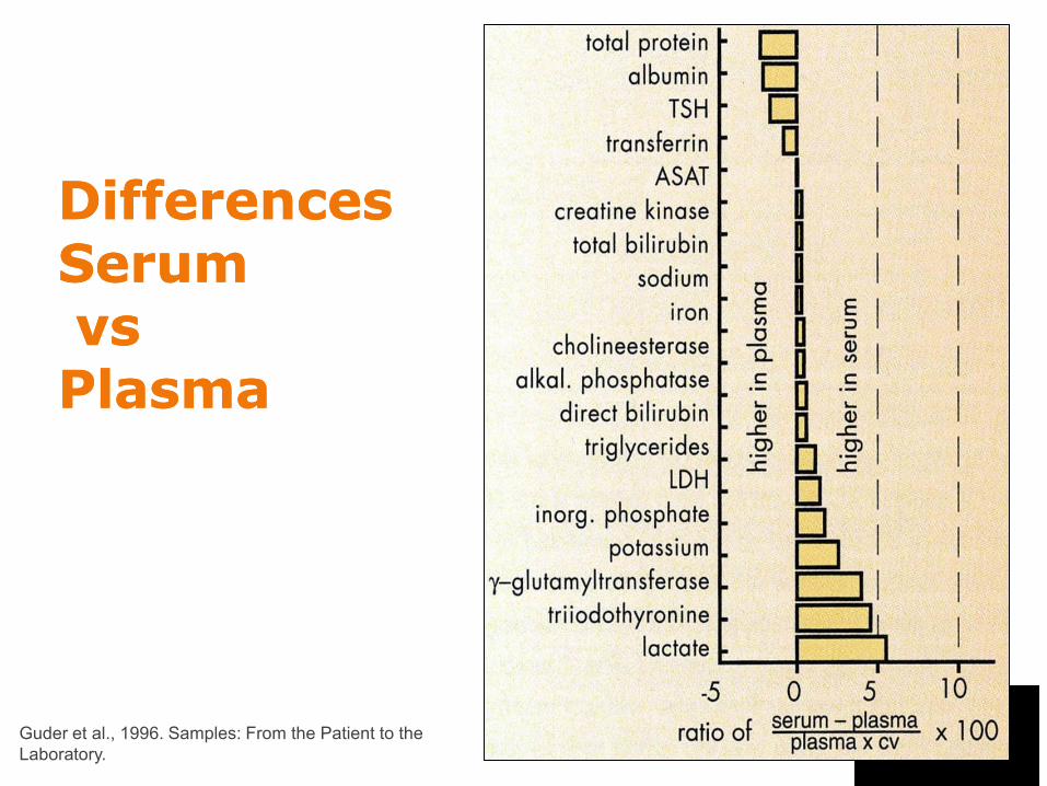
DifferencesDifferences SerumSerum vsvs PlasmaPlasma
Guder et al., 1996. Samples: From the Patient to the
Laboratory.
Effects over Time
Reduced stability in plasma of certain common
analytes that are involved in cell/platelet-
mediated metabolic processes and/or are
present in higher concentrations in cells or
platelets.
Serum-plasma differences may be evident with
these analytes depending on plasma cell/platelet
content and time between centrifugation and
testing.

• Heparin plasma specimens with increased cell/platelet concentrations
exhibit reduced stability of certain common analytes.
• Analytes affected are involved in cell/platelet-mediated metabolic
processes and/or are present in higher concentration in cells or
platelets.
Routine Analyte Stability in Plasma
Dependence on Handling and Test Methodology
The occurrence and magnitude of serum-plasma
differences can depend on specimen handling
and processing procedures and/or the specific
instrument/assay methodology used.
Plasma specimens may also exhibit an
increased frequency of duplicate errors with
certain instrument/test combinations, due to
platelets, cell aggregates, or microclots.
Bakker AJ, Mirchi B, Dijkstra JT, Reitsma F, Syperda H, Zijlstra A. IFCC method for lactate
dehydrogenase measurement in heparin plasma is unreliable. Clin Chem. 2003;49(4):662-4.
Dimeski G, Badrick T, Flatman R, Ormiston B. Roche IFCC methods for lactate
dehydrogenase tested for duplicate errors with Greiner and Becton-Dickinson lithium-
heparin and Greiner serum samples. Clin Chem. 2004 Dec;50(12):2391-2.

Fibrin – Test Interference
• Erroneous FSH results caused by insufficient clotting of
serum specimens and fibrin formation within analyzer
reaction vessel.1
• Falsely elevated Troponin-I due to fibrin in serum samples.2
• Duplicate errors in LD due to micro clots or cell aggregates
in plasma samples.3
1. Zweig MH, Glickman J, Csako G. Analytical interference caused by incompletely clotted serum specimens. Clin Chem.
1994;40:2325-6.
2. Nosanchuk JS, Combs B, Abbott G. False increases of troponin I attributable to incomplete separation of serum. Clin
Chem. 1999;45:714.
3. Dimeski G, Badrick T, Flatman R, Ormiston B. Roche IFCC methods for lactate dehydrogenase tested for duplicate
errors with Greiner and Becton-Dickinson lithium-heparin and Greiner serum samples. Clin Chem. 2004;50:2391-2.
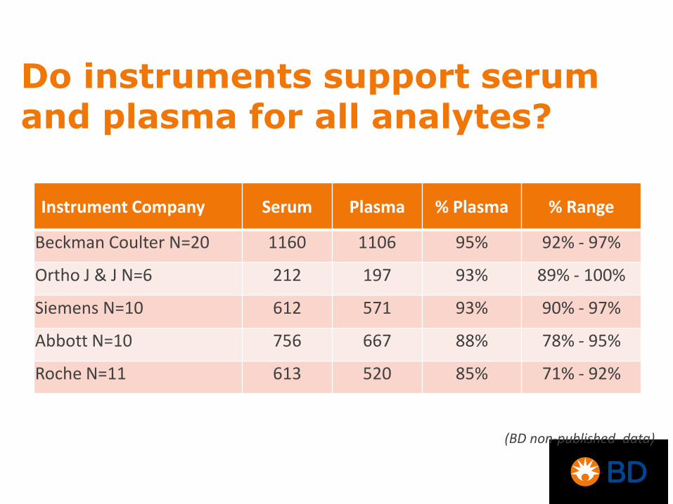
Do instruments support serum and plasma for all analytes?
Instrument Company Serum Plasma % Plasma % Range
Beckman Coulter N=20 1160 1106 95% 92% - 97%
Ortho J & J N=6 212 197 93% 89% - 100%
Siemens N=10 612 571 93% 90% - 97%
Abbott N=10 756 667 88% 78% - 95%
Roche N=11 613 520 85% 71% - 92%
(BD non-published data)

Specimen Yield
The formation of a physical
clot in serum blood
collection tubes also leads
to other differences between
serum and plasma
specimens.
% Yield of serum is slightly
lower due to serum trapped
between the clot and the
tube wall.

Serum vs. Plasma
– nearly cell-free
– good storage stability for most analytes
– wide range of assays available
– shorter TAT: can be centrifuged immediately
– faster gel movement in gel tubes
– more reproducible gel barrier formation
– increase supernatant yield 15-20% > serum

Serum vs. Plasma
– longer TAT
– instrument or test interference from fibrin,
esp. with anticoagulation therapy
– may cause pseudohyperkalemia
– analytical variation due release from
cells/platelets during clotting
– Higher cell counts
– reduced storage stability for certain analytes
– fibrin formation during storage
– interference from anticoagulant
– interference from fibrinogen
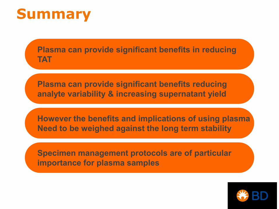
Summary
Plasma can provide significant benefits in reducing
TAT
However the benefits and implications of using plasma
Need to be weighed against the long term stability
Plasma can provide significant benefits reducing
analyte variability & increasing supernatant yield
Specimen management protocols are of particular
importance for plasma samples
Proper Sample Selection
• The selection of serum vs. heparin plasma may be
dependent on the specific setting/population.
• Example: plasma for stat testing or patients on
heparin therapy.
• Example: serum to preserve sample quality over
extended periods of time or transportation
• Standardizing on one sample type may be desirable
but not always practical

1. Select appropriate sample type (serum vs. heparin plasma)
based on pre-centrifugation time and patient population (and
assay compatibility).
2. Ensure correct collection technique to minimize hemolysis.
3. Fill evacuated blood collection tubes to the stated draw volume.
This will ensure the proper blood-to-additive ratio.
4. Ensure correct number of complete tube inversions immediately
after collection to ensure blood and additive are mixed
thoroughly.
5. Ensure correct (minimum or longer) clotting time for serum
tubes.
TOP 10 LIST Preparing High Quality Specimens
6. Ensure centrifuge g-force (RCF) and spin time are sufficient to
obtain adequate sedimentation of cells, platelets and other
debris.
7. Carefully aliquot samples from non-gel tubes after
centrifugation.
8. Avoid mixing/agitation of plasma gel tubes between
centrifugation and testing.
9. To help reduce fibrin formation over time, keep heparin
plasma at room temperature.
10.Recentrifugation of sample aliquots can help “clean up”
samples but, do not re-spin gel tubes.
TOP 10 LIST Preparing High Quality Specimens

Thank you


